Note: Descriptions are shown in the official language in which they were submitted.
INTRAOCULAR PRESSURE REDUCTION WITH
INTRACAMERAL BIMATOPROST IMPLANTS
[1]
BACKGROUND OF THE INVENTION
1. Field of the Invention.
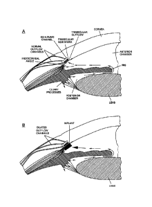
[2] The present invention relates to a method of treating an ocular
condition, comprising the
step of placing a biodegradable intraocular implant in an eye of the patient,
the implant
comprising a prostamide and a biodegradable polymer matrix that releases drug
at a rate
effective to sustain release of an amount of the prostamide from the implant
to provide an
amount of the prostamide effective to prevent or reduce a symptom of the
ocular condition,
wherein said ocular condition is elevated 1OP.
2. Summary of the Related Art
[3] The anterior and posterior chambers of the eye are filled with
aqueous humor, a fluid
predominantly secreted by the ciliary body with an ionic composition similar
to the blood. The
function of the aqueous humor is two-fold: to 1) supply nutrients to the
avaseular structures of
the eye, such as the lens and cornea and 2) maintain intraocular pressure
(lOP) within its
physiological range. Maintenance of IOP and supply of nutrients to the
anterior segment arc
factors that are critical for maintaining normal visual acuity. Aqueous humor
is predominantly
secreted to the posterior chamber of the eye by the ciliary processes of the
ciliary body and a
minor mechanism of aqueous humor production is through ultrafiltration from
arterial blood.
Aqueous humor then reaches the anterior chamber by crossing the pupil and
there are convection
currents where the aqueous, adjacent to the iris, flows upwards, and the
aqueous, adjacent to
the cornea, flows downwards. There are two different pathways of aqueous humor
outflow, both
located in the iridocorneal angle of the eye. The uveoscleral or
nonconventional pathway refers
to the aqueous humor leaving the anterior chamber by diffusion through
intercellular spaces
among ciliary muscle fibers. Although this seems to be a minority outflow
pathway in humans,
the uveoscleral or nonconventional pathway is the target of specific anti-
hypertensive drugs such
as the hypotensive lipids, e.g. bimatoprost, that increase the functionality
of this route through
1
CA 2796443 2017-10-25
CA 02796443 2012-10-15
WO 2011/130462 PCT/US2011/032393
remodeling of the extracellular matrix. In addition, bimatoprost may improve
aqueous outflow
through the trabecular meshwork ("TM") mediated through a prostamide receptor.
In the
human eye, the main outflow route is the trabecular or conventional outflow
pathway. This
tissue contains three differentiated layers. From the inner to the outermost
part, the layer of
tissue closest to the anterior chamber is the uvcal meshwork, formed by
prolongations of
connective tissue arising from the iris and ciliary body stromas and covered
by endothelial cells.
This layer does not offer much resistance to aqueous humor outflow because
intercellular spaces
are large. The next layer, known as the corneoscleral meshwork, is
characterized by the
presence of lamellae covered by endothelium¨like cells on a basal membrane.
The lamellae are
formed by glycoproteins, collagen, hyaluronic acid, and elastic fibers. The
higher organization of
the corneoscleral meshwork in relation to the uveal meshwork as well as their
narrower
intercellular spaces are responsible for the increase in flow resistance. The
third layer, which is
in direct contact with the inner wall of endothelial cells from Schlemm's
canal, is the
juxtacanalicular meshwork. It is formed by cells embedded in a dense
extracellular matrix, and
the majority of the tissue resistance to aqueous flow is postulated to be in
this layer, due to its
narrow intercellular spaces. The layer of endothelial cells from Schlemm's
canal has expandable
pores that transfer the aqueous into the canal and accounts for approximately
10% of the total
resistance. It has been postulated that aqueous humor crosses the inner wall
endothelium of
Schlemm's canal by two different mechanisms: a paracellular route through the
junctions formed
between the endothelial cells and a transcellular pathway through
intracellular expandable pores
of the same cells. Once there is entry into Schlemm's canal, the aqueous
drains directly into the
collector ducts and aqueous veins that anastomose with the episcleral and
conjunctival plexi of
vessels. Aqueous humor outflow via the trabecular pathway is TOP dependent,
usually measured
as outflow facility, and expressed in microliters per minute per millimeter of
mercury. The
episcleral venous pressure controls outflow through the collector channels and
is one factor that
contributes to the intraocular pressure. Increases in the episcleral venous
pressure such as seen
with carotid-cavernous sinus fistulas, orbital variccs, and Sturgc-Weber
Syndrome, can lead to
difficult to manage glaucoma. Reducing episcleral venous pressure in disease
states, such as
treating carotid-cavernous sinus fistulas, can normalize the episcleral venous
pressure and reduce
the intraocular pressure. The mechanism of action of modern ocular hypotensive
agents for
treating ocular hypertension and open angle glaucoma are as follows: 1- reduce
aqueous humor
production, 2- improve uveoscleral outflow, 3- improve outflow through the TM
with miotic
agents by providing tension as the scleral spur with stimulation of the
ciliary body muscle, 4-
combination of any of the above.
2
CA 02796443 2012-10-15
WO 2011/130462 PCT/US2011/032393
Brief Summary of the Invention
[4] Unexpectedly, when sustained-release implants releasing bimatoprost
were placed in an
intracameral location, the outflow channels emanating from Schlemm's Canal
were visibly
dilated (See Figure 4). This results in a profound reduction in the
intraocular pressure, i.e. -60%
10P reduction from baseline. (See Figure 5), This reduction is significantly
more than what is
typically observed with topical bimatoprost, i.e. -35% 10P reduction)
The redirection of
aqueous flow towards the TM is illustrated in Figure 1, lower image. The usual
mechanism of
prostamides is to remodel both the anterior ciliary body near the ciliary band
and the TM. The
intracameral implants, which are located adjacent to the TM , as shown in
Figure 3, provide a
high drug concentration into the outflow channels and dilate the vessels in
the episcleral and
conjunctival venous plexus, thereby resulting in a novel mechanism of 1OP
reduction. The
dilation appears 360 degrees around the eye since drug released from an
implant positioned at
the 6:00 O'clock position is well-mixed throughout the anterior segment
through the convection
currents.
[5] This incremental reduction in the 1OP with the intracameral bimatoprost
implants is
advantageous for patients with ocular hypertension and open angle glaucoma
that require
sustained reduction in TOP to prevent progressive optic neuropathy. Patients
can avoid the need
for combination eye drops and/or surgery (including incisional surgery such as
trabeculectomy,
laser procedures such as ALT and SLT, and aqueous humor bypass stents), if
they are able to
achieve profound reductions in 1OP with the intracameral implant described
herein.
Brief Description of the Drawings
[6] Figure 1 (upper image) shows aqueous humor is predominantly secreted to
the posterior
chamber of the eye by the ciliary processes of the ciliary body.
[7] Figure 1 (lower image) shows an intracameral sustained-release
bimatoprost implant
releasing drug directly into Schlemm's canal resulting in visible dilation of
the outflow channels.
[8] Figure 2 shows that aqueous humor reaches the anterior chamber by
crossing the pupil
and there are convection currents where the flow of aqueous adjacent to the
iris is upwards, and
the flow of aqueous adjacent to the cornea is downwards.
[9] Figure 3 is a slit lamp photograph through a gonioscopy lens showing an
intracameral
bimatoprost implant placed adjacent to the trabecular meshwork in the dog eye.
[10] Figure 4 is a photograph showing the outflow vessels that are dilated
as a result of
treatment of a dog with the high-release bimatoprost intracameral implant of
Example 1.
3
CA 02796443 2012-10-15
WO 2011/130462 PCT/US2011/032393
[11] Figure 5 shows the TOP of a dog treated with the high-release
bimatoprost intracameral
implant described in Example 1 was reduced to approximately -60% from baseline
and such
reduction was sustained for at least 5 months.
[12] Figure 6 is a photograph showing the outflow vessels that are dilated
as a result of
treatment of a dog with the low-release bimatoprost intracameral implant of
Example 2.
[13] Figure 7 shows the 10P of a dog treated with the low-release
bimatoprost intracameral
implant described in Example 2 was reduced to approximately -40% from baseline
and such
reduction was sustained for at least 42 days.
[14] Figure 8 shows the in vitro release rate of the Implant formulation
used in Example 1
(arrow).
[15] Figure 9 shows the in vitro release rate of the Implant formulation
used in Example 2
(arrow).
[16] Figure 10 shows the TOP is lowered in a dog treated with a single
bimatoprost implant
according to Example 3.
[17] Figure 11 shows the TOP is lowered in a dog treated with two
bimatoprost implants
according to Example 3.
Detailed Description of the Invention
[18] As disclosed herein, controlled and sustained administration of a
therapeutic agent
through the use of one or more intraocular implants may improve treatment of
undesirable ocular
conditions, in particular elevated IOP. The implants comprise a
pharmaceutically acceptable
polymeric composition and are formulated to release one or more
pharmaceutically active
agents, such as a prostamide, over an extended period of time. The implants
are effective to
provide a therapeutically effective dosage of the agent or agents directly to
a region of the eye to
treat or prevent one or more undesirable ocular conditions. Thus, with a
single administration,
therapeutic agents will be made available at the site where they are needed
and will be
maintained for an extended period of time, rather than subjecting the patient
to repeated
injections or repeated administration of topical drops.
[19] The above implants are utilized in a method of treating an ocular
condition, comprising
the step of placing a biodegradable intraocular implant in an eye of the
patient, the implant
comprising a prostamide and a biodegradable polymer matrix that releases
prostamide at a rate
effective to sustain an amount of prostamide effective to prevent or reduce a
symptom of the
ocular condition, wherein said ocular condition is elevated IOP and said
implant is placed in an
4
CA 02796443 2012-10-15
WO 2011/130462 PCT/US2011/032393
intracameral location to dilate the outflow channels of the eye emanating from
Schlemm's
Canal.
[20] An intraocular implant in accordance with the disclosure herein
comprises a therapeutic
component. The therapeutic component comprises, consists essentially of, or
consists of, a
prostamide. A drug release sustaining component may be associated with the
therapeutic
component to sustain release of an effective amount of the prostamide into an
eye in which the
implant is placed. The amount of the prostamide is released into the eye for a
period of time
greater than about one week after the implant is placed in the eye, and is
effective in treating or
reducing a symptom of an ocular condition.
[21] The implant is made of polymeric materials to provide maximal
approximation of the
implant to the iridocorneal angle. In addition, the size of the implant, which
ranges from a
diameter of approximately 0.1 to 1 mm, and lengths from 0.1 to 6 mm, enables
the implant to be
inserted into the anterior chamber using an applicator with a small gauge
needle ranging from 22
to 30G.
DEFINITIONS
[22] For the purposes of this description, we use the following terms as
defined in this section,
unless the context of the word indicates a different meaning.
[23] As used herein, an "intraocular implant" refers to a device or element
that is structured,
sized, or otherwise configured to be placed in an eye. Intraocular implants
are generally
biocompatible with physiological conditions of an eye and do not cause adverse
side effects.
Intraocular implants may be placed in an eye without disrupting vision of the
eye.
[24] As used herein, a "therapeutic component" refers to a portion of an
intraocular implant
comprising one or more therapeutic agents or substances used to treat a
medical condition of the
eye. The therapeutic component may be a discrete region of an intraocular
implant, or it may be
homogenously distributed throughout the implant. The therapeutic agents of the
therapeutic
component are typically ophthalmically acceptable, and are provided in a form
that does not
cause adverse reactions when the implant is placed in an eye.
[25] As used herein, a "drug release sustaining component" refers to a
portion of the
intraocular implant that is effective to provide a sustained release of the
therapeutic agents of the
implant. A drug release sustaining component may be a biodegradable polymer
matrix, or it
may be a coating covering a core region of the implant that comprises a
therapeutic component.
[26] As used herein, "associated with" means mixed with, dispersed within,
coupled to,
covering, or sun-ounding.
CA 02796443 2012-10-15
WO 2011/130462 PCT/US2011/032393
[27] As used herein, an "ocular region" or "ocular site" refers generally
to any area of the
eyeball, including the anterior and posterior segment of the eye, and which
generally includes,
but is not limited to, any functional (e.g., for vision) or structural tissues
found in the eyeball, or
tissues or cellular layers that partly or completely line the interior or
exterior of the eyeball.
Specific examples of areas of the eyeball in an ocular region include the
anterior chamber, the
posterior chamber, the vitreous cavity, the choroid, the suprachoroidal space,
the conjunctiva,
the subconjunctival space, the episcleral space, the intracorneal space, the
epicorneal space, the
sclera, the pars plana, surgically induced avascular regions, the macula, and
the retina.
[28] As used herein, an "ocular condition" is a disease, ailment or
condition which affects or
involves the eye or one of the parts or regions of the eye. Broadly speaking
the eye includes the
eyeball and the tissues and fluids which constitute the eyeball, the
periocular muscles (such as
the oblique and rectus muscles) and the portion of the optic nerve which is
within or adjacent to
the eyeball.
[29] An anterior ocular condition is a disease, ailment or condition which
affects or which
involves an anterior (i.e. front of the eye) ocular region or site, such as a
periocular muscle, an
eye lid or an eye ball tissue or fluid which is located anterior to the
posterior wall of the lens
capsule or ciliary muscles. Thus, an anterior ocular condition primarily
affects or involves the
conjunctiva, the cornea, the anterior chamber, the iris, the posterior chamber
(behind the retina
but in front of the posterior wall of the lens capsule), the lens or the lens
capsule and blood
vessels and nerve which vascularize or innervate an anterior ocular region or
site.
[30] Thus, an anterior ocular condition can include a disease, ailment or
condition, such as for
example, aphakia; pseudophakia; astigmatism; blepharospasm; cataract;
conjunctival diseases;
conjunctivitis; corneal diseases; corneal ulcer; dry eye syndromes; eyelid
diseases; lacrimal
apparatus diseases; lacrimal duct obstruction; myopia; presbyopia; pupil
disorders; refractive
disorders and strabismus. Glaucoma can also be considered to be an anterior
ocular condition
because a clinical goal of glaucoma treatment can be to reduce a hypertension
of aqueous fluid
in the anterior chamber of the eye (i.e. reduce intraocular pressure).
[31] A posterior ocular condition is a disease, ailment or condition which
primarily affects or
involves a posterior ocular region or site such as choroid or sclera (in a
position posterior to a
plane through the posterior wall of the lens capsule), vitreous, vitreous
chamber, retina, optic
nerve (i.e. the optic disc), and blood vessels and nerves which vascularize or
innervate a
posterior ocular region or site.
[32] Thus, a posterior ocular condition can include a disease, ailment or
condition, such as for
example, acute macular neuroretinopathy; Behcet's disease; choroidal
neovascularization;
6
CA 02796443 2012-10-15
WO 2011/130462 PCT/US2011/032393
diabetic uveitis; histoplasmosis; infections, such as fungal or viral-caused
infections; macular
degeneration, such as acute macular degeneration, non-exudative age related
macular
degeneration and exudative age related macular degeneration; edema, such as
macular edema,
cystoid macular edema and diabetic macular edema; multifocal choroiditis;
ocular trauma which
affects a posterior ocular site or location; ocular tumors; retinal disorders,
such as central retinal
vein occlusion, diabetic retinopathy (including proliferative diabetic
retinopathy), proliferative
vitreoretinopathy (PVR), retinal arterial occlusive disease, retinal
detachment, uveitic retinal
disease; sympathetic opthalmia; Vogt Koyanagi-Harada (VKH) syndrome; uveal
diffusion; a
posterior ocular condition caused by or influenced by an ocular laser
treatment; posterior ocular
conditions caused by or influenced by a photodynamic therapy,
photocoagulation, radiation
retinopathy, epiretinal membrane disorders, branch retinal vein occlusion,
anterior ischemic
optic neuropathy, non-retinopathy diabetic retinal dysfunction, retinitis
pigmentosa, and
glaucoma. Glaucoma can be considered a posterior ocular condition because the
therapeutic goal
is to prevent the loss of or reduce the occurrence of loss of vision due to
damage to or loss of
retinal cells or optic nerve cells (i.e. neuroprotection).
[33] The term "biodegradable polymer" refers to a polymer or polymers which
degrade in
vivo, and wherein erosion of the polymer or polymers over time occurs
concurrent with or
subsequent to release of the therapeutic agent. Specifically, hydrogels such
as methylcellulose
which act to release drug through polymer swelling are specifically excluded
from the term
"biodegradable polymer". The terms "biodegradable" and "bioerodible" are
equivalent and are
used interchangeably herein. A biodegradable polymer may be a homopolymer, a
copolymer, or
a polymer comprising more than two different polymeric units.
[34] The term "treat", "treating", or "treatment" as used herein, refers to
reduction or
resolution or prevention of an ocular condition, ocular injury or damage, or
to promote healing
of injured or damaged ocular tissue. A treatment is usually effective to
reduce at least one
symptom of an ocular condition, ocular injury or damage.
[35] The term "effective" as used herein, refers to the level or amount of
agent needed to treat
an ocular condition, or reduce or prevent ocular injury or damage without
causing significant
negative or adverse side effects to the eye or a region of the eye. In view of
the above, a
therapeutically effective amount of a therapeutic agent, such as a prostamide,
is an amount that
is effective in reducing at least one symptom of an ocular condition.
[36] Intraocular implants have been developed which can release drug loads
over various time
periods. These implants, which when inserted into an eye, such as the vitreous
of an eye,
provide therapeutic levels of a prostamide for extended periods of time (e.g.,
for about 1 week or
7
CA 02796443 2012-10-15
WO 2011/130462 PCT/US2011/032393
more). The disclosed implants are effective in treating ocular conditions,
such as ocular
conditions associated with elevated intraocular pressure, and more
specifically in reducing at
least one symptom of glaucoma.
[37] In one embodiment of the present invention, an intraocular implant
comprises a
biodegradable polymer matrix. The biodegradable polymer matrix is one type of
a drug release-
sustaining component.
The biodegradable polymer matrix is effective in forming a
biodegradable intraocular implant. The biodegradable intraocular implant
comprises a
prostamide associated with the biodegradable polymer matrix. The matrix
degrades at a rate
effective to sustain release of an amount of the prostamide for a time greater
than about one
week from the time in which the implant is placed in ocular region or ocular
site, such as the
vitreous of an eye.
[38] The prostamide component of the implant includes one or more types
of prostamides. In
certain implants, the prostamide component comprises a compound having the
formula (I) .
R1
A-B .=' X
R2
wherein the dashed bonds represent a single or double bond which can be in the
cis or trans
configuration, A is an alkylene or alkenylene radical having from two to six
carbon atoms, which
radical may be interrupted by one or more oxide radicals and substituted with
one or more
hydroxy, oxo, alkyloxy or akylcarboxy groups wherein said alkyl radical
comprises from one to
six carbon atoms; B is a cycloalkyl radical having from three to seven carbon
atoms, or an aryl
radical, selected from the group consisting of hydrocarbyl aryl and heteroaryl
radicals having from
four to ten carbon atoms wherein the heteroatom is selected from the group
consisting of nitrogen,
oxygen and sulfur atoms; X is --N(R4).2 wherein R.4 is independently selected
from the group
consisting of hydrogen and lower alkyl radicals having from one to six carbon
atoms,
[39]
Z is =0; one of Ri and R2 is =0, -OH or a -0(CO)R6 group, and the other one is
-OH or
-0(CO)R6, or Ri is =0 and R2 is H; wherein R6 is a saturated or unsaturated
acyclic
hydrocarbon group having from 1 to about 20 carbon atoms, or -(CH2)mR7 wherein
m is 0-10,
and R7 is cycloalkyl radical, having from three to seven carbon atoms, or a
hydrocarbyl aryl or
heteroaryl, as defined above; or a pharmaceutically-acceptable salt thereof or
a
pharmaceutically-acceptable salt thereof.
8
[40] Pharmaceutically acceptable acid addition salts of the compounds of
the invention are
those formed from acids which form non-toxic addition salts containing
pharmaceutically
acceptable anions, such as the hydrochloride, hydrobromide, hydroiodide,
sulfate, or bisulfate,
phosphate or acid phosphate, acetate, maleate, fumarate, oxalate, lactate,
tartrate, citrate,
gluconate, saccharate and p-toluene sulphonate salts.
Preferably, the prostamide has the following formula (II)
-1
-
X
,S,-(Y)n
(CH2)y(0)x
R2 R3
wherein y is 0 or 1, x is 0 or 1 and x+y are not both 1, Y is a radical
selected from the group
consisting of alkyl, halo, nitro, amino, thiol, hydroxy, alkytoxy,
alkylcarboxy and halo
substituted alkyl, wherein said alkyl radical comprises from one to six carbon
atoms, n is 0 or an
integer of from 1 to 3 and R1 is .=0, --OH or --0(CO)R6 and hatched lines
indicate the .alpha.
configuration and solid triangles indicate the .beta. configuration.
[41] In at least one type of intraocular implant, the prostamide comprises
a compound wherein
RI, R2 and R3 arc OH, y is 1, x is 0, n is 0 and X is N(H)(C2H5) ,c.g..
cyclopentane N-ethyl
heptenamide-5-cis-2-(3a.-hydroxy-5-pheny1-1-trans-penteny1)-3, 5-di hydroxy,
[1õ.
[42] The compound, cyclopentane N-ethyl heptenamide-5-cis-2-(3(1.-hydroxy-5-
pheny1-1-
trans-penteny1)-3, 5-dihydroxy, [1 a.,2[3.,3õ,5õ ] , is also known as
bimatoprost and is publicly
available in a topical ophthalmic solution under the tradename, Lumigan.RTIVI.
(Allergan, Inc.,
CA).
[43] Alternatively, the prostamide may be any of the prostamides disclosed
in US Patent
No. 6,395,787 .
[44] Thus, the implant may comprise a therapeutic component which
comprises, consists
essentially of, or consists of bimatoprost, a salt thereof, or mixtures
thereof.
[45] The prostamide may be in a particulate or powder form and it may be
entrapped by the
biodegradable polymer matrix. Usually, prostamide particles will have an
effective average size
less than about 3000 nanometers. In certain implants, the particles may have
an effective
average particle size about an order of magnitude smaller than 3000
nanometers. For example,
the particles may have an effective average particle size of less than about
500 nanometers. In
9
CA 2796443 2017-10-25
CA 02796443 2012-10-15
WO 2011/130462 PCT/US2011/032393
additional implants, the particles may have an effective average particle size
of less than about
400 nanometers, and in still further embodiments, a size less than about 200
nanometers.
[46] The prostamide of the implant is preferably from about 10% to 90% by
weight of the
implant. More preferably, the prostamide is from about 20% to about 80% by
weight of the
implant. In a preferred embodiment, the prostamide comprises about 20% by
weight of the
implant (e.g., 15%-25%). In another embodiment, the prostamide comprises about
50% by
weight of the implant.
[47] Suitable polymeric materials or compositions for use in the implant
include those
materials which are compatible, that is biocompatible, with the eye so as to
cause no substantial
interference with the functioning or physiology of the eye. Such materials
preferably are at least
partially and more preferably substantially completely biodegradable or
bioerodible.
[48] Examples of useful polymeric materials include, without limitation,
such materials
derived from and/or including organic esters and organic ethers, which when
degraded result in
physiologically acceptable degradation products, including the monomers. Also,
polymeric
materials derived from and/or including, anhydrides, amides, orthoesters and
the like, by
themselves or in combination with other monomers, may also find use. The
polymeric materials
may be addition or condensation polymers, advantageously condensation
polymers. The
polymeric materials may be cross-linked or non-cross-linked, for example not
more than lightly
cross-linked, such as less than about 5%, or less than about 1% of the
polymeric material being
cross-linked. For the most part, besides carbon and hydrogen, the polymers
will include at least
one of oxygen and nitrogen, advantageously oxygen. The oxygen may be present
as oxy, e.g.
hydroxy or ether, carbonyl, e.g. non-oxo-carbonyl, such as carboxylic acid
ester, and the like.
The nitrogen may be present as amide, cyano and amino. The polymers set forth
in Heller,
Biodegradable Polymers in Controlled Drug Delivery, In: CRC Critical Reviews
in Therapeutic
Drug Carrier Systems, Vol. 1, CRC Press, Boca Raton, Fla. 1987, pp 39-90,
which describes
encapsulation for controlled drug delivery, may find use in the present
implants.
[49] Of additional interest are polymers of hydroxyaliphatic carboxylic
acids, either
homopolymers or copolymers, and polysaccharides. Polyesters of interest
include polymers of
D-lactic acid, L-lactic acid, racemic lactic acid, glycolic acid,
polycaprolactone, and
combinations thereof. Generally, by employing the L-lactate or D-lactate, a
slowly eroding
polymer or polymeric material is achieved, while erosion is substantially
enhanced with the
lactate racemate.
CA 02796443 2012-10-15
WO 2011/130462 PCT/US2011/032393
[50] Among the useful polysaccharides are, without limitation, calcium
alginate, and
functionalized celluloses, particularly carboxymethylcellulose esters
characterized by being
water insoluble, a molecular weight of about 5 kD to 500 kD, for example.
[51] Other polymers of interest include, without limitation, polyvinyl
alcohol, polyesters,
polyethers and combinations thereof which arc biocompatible and may be
biodegradable and/or
bioerodible.
[52] Some preferred characteristics of the polymers or polymeric materials
for use in the
present invention may include biocompatibility, compatibility with the
therapeutic component,
ease of use of the polymer in making the drug delivery systems of the present
invention, a half-
life in the physiological environment of at least about 6 hours, preferably
greater than about one
day, not significantly increasing the viscosity of the vitreous, and water
insolubility.
[53] The biodegradable polymeric materials which are included to form the
matrix are
desirably subject to enzymatic or hydrolytic instability. Water-soluble
polymers may be cross-
linked with hydrolytic or biodegradable unstable cross-links to provide useful
water insoluble
polymers. The degree of stability can be varied widely, depending upon the
choice of monomer,
whether a homopolymer or copolymer is employed, employing mixtures of
polymers, and
whether the polymer includes terminal acid groups.
[54] Equally important to controlling the biodegradation of the polymer and
hence the
extended release profile of the implant is the relative average molecular
weight of the polymeric
composition employed in the implant. Different molecular weights of the same
or different
polymeric compositions may be included in the implant to modulate the release
profile. In
certain implants, the relative average molecular weight of the polymer will
range from about 9 to
about 64 kD, usually from about 10 to about 54 kD, and more usually from about
12 to about 45
kD.
[55] In some implants, copolymers of glycolic acid and lactic acid are
used, where the rate of
biodegradation is controlled by the ratio of glycolic acid to lactic acid. The
most rapidly
degraded copolymer has roughly equal amounts of glycolic acid and lactic acid.
Homopolymers, or copolymers having ratios other than equal, are more resistant
to degradation.
The ratio of glycolic acid to lactic acid will also affect the brittleness of
the implant, where a
more flexible implant is desirable for larger geometries. The % of polylactic
acid in the
polylactic acid polyglycolic acid (PLGA) copolymer can be 0-100%, preferably
about 15-85%,
more preferably about 35-65%. In some implants, a 50/50 PLGA copolymer is
used.
[56] The biodegradable polymer matrix of the intraocular implant may
comprise a mixture of
two or more biodegradable polymers. For example, the implant may comprise a
mixture of a
11
CA 02796443 2012-10-15
WO 2011/130462 PCT/US2011/032393
first biodegradable polymer and a different second biodegradable polymer. One
or more of the
biodegradable polymers may have terminal acid groups.
[57] Release of a drug from an erodible polymer is the consequence of
several mechanisms or
combinations of mechanisms. Some of these mechanisms include desorption from
the implant's
surface, dissolution, diffusion through porous channels of the hydrated
polymer and erosion.
Erosion can be bulk or surface or a combination of both. As discussed herein,
the matrix of the
intraocular implant may release drug at a rate effective to sustain release of
an amount of the
prostamide component for more than one week after implantation into an eye. In
certain
implants, therapeutic amounts of the prostamide component are released for no
more than about
30-35 days after implantation. For example, an implant may comprise
bimatoprost, and the
matrix of the implant degrades at a rate effective to sustain release of a
therapeutically effective
amount of bimatoprost for about one month after being placed in an eye. As
another example,
the implant may comprise bimatoprost, and the matrix releases drug at a rate
effective to sustain
release of a therapeutically effective amount of bimatoprost for more than
forty days, such as for
about six months.
[58] One example of the biodegradable intraocular implant comprises a
prostamide associated
with a biodegradable polymer matrix, which comprises a mixture of different
biodegradable
polymers. At least one of the biodegradable polymers is a polylactide having a
molecular weight
of about 63.3 kD. A second biodegradable polymer is a polylactide having a
molecular weight
of about 14 kD. Such a mixture is effective in sustaining release of a
therapeutically effective
amount of the prostamide for a time period greater than about one month from
the time the
implant is placed in an eye.
[59] Another example of a biodegradable intraocular implant comprises an
prostamide
associated with a biodegradable polymer matrix, which comprises a mixture of
different
biodegradable polymers, each biodegradable polymer having an inherent
viscosity from about
0.16 dl/g to about 1.0 dl/g. For example, one of the biodegradable polymers
may have an
inherent viscosity of about 0.3 dl/g. A second biodegradable polymer may have
an inherent
viscosity of about 1.0 dl/g. Additional implants may comprise biodegradable
polymers that have
an inherent viscosity between about 0.2 dl/g and 0.5 dUg. The inherent
viscosities identified
above may be determined in 0.1% chloroform at 25° C.
[60] One particular implant comprises bimatoprost associated with a
combination of two
different polylactide polymers. The bimatoprost is present in about 20% by
weight of the
implant. One polylactide polymer has a molecular weight of about 14 kD and an
inherent
viscosity of about 0.3 dl/g, and the other polylactide polymer has a molecular
weight of about
12
CA 02796443 2012-10-15
WO 2011/130462 PCT/US2011/032393
63.3 kD and an inherent viscosity of about 1.0 dl/g. The two polylactide
polymers are present in
the implant in a 1:1 ratio. Such an implant may be effective in releasing the
bimatoprost for
more than two months. The implant is provided in the form of a rod or a
filament produced by
an extrusion process.
[61] A preferred implant formulation for the invention is API 30%, R203S
45%, R202H 20%,
PEG 3350 5% or API 20%, R203S 45%, R202H 10%, RG752S 20%, PEG 3350 5%, wherein
the API is bimatoprost. The range of concentrations of the constituents that
can be used in the
preferred implant formulation are API 5 to 40%, R203S 10 to 60%, R202H 5 to
20%, R0752S 5
to 40%, PEG 3350 0 to 15%. The PLA/PLGA polymers are from the Resomer product
line
available from Boehringer Ingelheim in Ingelheim, Germany and include the
following:
Resomer Monomer ratio i.v. dL/g
RG502, 50:50 poly (D, L-lactide-co-glycolide) 0.2
RG502H, 50:50 poly (D, L-lactide-co-glycolide) 0.2
RG503, 50:50 poly (D, L-lactide-co-glycolide) 0.4
RG504, 0.5
RG505, 0.7
RG506, 0.8
RG752, 75:25 poly (D,L lactide-co-glycolide) 0.2
RG755, 75:25 poly(D,L lactide-co-glycolide) 0.6 (40000)
RG756, 0.8
RG858, 85:15 poly (D,L-lactide-co-glycolide) 1.4
R202H, poly (D,L-lactide) 0.3
R203 poly (D,L-lactide) 1.0 (40000)
R206. poly (D,L-lactide) ; acid end 0.2
R104 poly (D,L-lactide) (3500)
[62] The release of the prostamide from the intraocular implant
comprising a biodegradable
polymer matrix may include an initial burst of release followed by a gradual
increase in the
amount of the prostamide released, or the release may include an initial delay
in release of the
prostamide component followed by an increase in release. When the implant is
substantially
completely degraded, the percent of the prostamide that has been released is
about one hundred.
Compared to existing implants, the implants disclosed herein do not completely
release, or
release about 100% of the prostamide, until after about one week of being
placed in an eye.
[63] It may be desirable to provide a relatively constant rate of release
of the prostamide from
the implant over the life of the implant. For example, it may be desirable for
the prostamide to
be released in amounts from about 0.01 µg to about 2 µg per day for the
life of the
implant. However, the release rate may change to either increase or decrease
depending on the
formulation of the biodegradable polymer matrix. In addition, the release
profile of the
13
CA 02796443 2012-10-15
WO 2011/130462 PCT/US2011/032393
prostamide may include one or more linear portions and/or one or more non-
linear portions.
Preferably, the release rate is greater than zero once the implant has begun
to degrade or erode.
[64] The implants may be monolithic, i.e. having the active agent or agents
homogenously
distributed through the polymeric matrix, or encapsulated, where a reservoir
of active agent is
encapsulated by the polymeric matrix. Due to ease of manufacture, monolithic
implants arc
usually preferred over encapsulated forms. However, the greater control
afforded by the
encapsulated, reservoir-type implant may be of benefit in some circumstances,
where the
therapeutic level of the drug falls within a narrow window. In addition, the
therapeutic
component, including the prostamide, may be distributed in a non-homogenous
pattern in the
matrix. For example, the implant may include a portion that has a greater
concentration of the
prostamide relative to a second portion of the implant.
[65] The intraocular implants disclosed herein may have a size of between
about 5 µm and
about 10 mm, or between about 10 µm and about 1 mm for administration with
a needle,
greater than 1 mm, or greater than 2 mm, such as 3 mm or up to 10 mm, for
administration by
surgical implantation. For needle-injected implants, the implants may have any
appropriate
length so long as the diameter of the implant permits the implant to move
through a needle. For
example, implants having a length of about 6 mm to about 7 mm have been
injected into an eye.
The implants administered by way of a needle should have a diameter that is
less than the inner
diameter of the needle. In certain implants, the diameter is less than about
500 µm. The
vitreous chamber in humans is able to accommodate relatively large implants of
varying
geometries, having lengths of, for example, 1 to 10 mm. The implant may be a
cylindrical pellet
(e. g., rod) with dimensions of about 2 mm×0.75 mm diameter. Or the
implant may be a
cylindrical pellet with a length of about 7 mm to about 10 mm, and a diameter
of about 0.75 mm
to about 1.5 mm.
[66] The implants may also be at least somewhat flexible so as to
facilitate both insertion of
the implant in the eye, such as in the vitreous, and accommodation of the
implant. The total
weight of the implant is usually about 250-5000 µg, more preferably about
500-1000 µg.
For example, an implant may be about 500 µg, or about 1000 µg. For non-
human
individuals, the dimensions and total weight of the implant(s) may be larger
or smaller,
depending on the type of individual. For example, humans have a vitreous
volume of
approximately 3.8 ml, compared with approximately 30 ml for horses, and
approximately 60-
100 ml for elephants. An implant sized for use in a human may be scaled up or
down
accordingly for other animals, for example, about 8 times larger for an
implant for a horse, or
about, for example, 26 times larger for an implant for an elephant.
14
CA 02796443 2012-10-15
WO 2011/130462 PCT/US2011/032393
[67] Thus, implants can be prepared where the center may be of one material
and the surface
may have one or more layers of the same or a different composition, where the
layers may be
cross-linked, or of a different molecular weight, different density or
porosity, or the like. For
example, where it is desirable to quickly release an initial bolus of drug,
the center may be a
polylactate coated with a polylactate-polyglycolate copolymer, so as to
enhance the rate of initial
degradation. Alternatively, the center may be polyvinyl alcohol coated with
polylactate, so that
upon degradation of the polylactate exterior the center would dissolve and be,
rapidly washed
out of the eye.
[68] The implants may be of any geometry including fibers, sheets, films,
microspheres,
spheres, circular discs, plaques and the like. The upper limit for the implant
size will be
determined by factors such as toleration for the implant, size limitations on
insertion, ease of
handling, etc. Where sheets or films are employed, the sheets or films will be
in the range of at
least about 0.5 mm×0.5 mm, usually about 3-10 mm×5-10 mm with a
thickness of
about 0.1-1.0 mm for ease of handling. Where fibers are employed, the fiber
diameter will
generally be in the range of about 0.05 to 3 mm and the fiber length will
generally be in the
range of about 0.5-10 mm. Spheres may be in the range of about 0.5 µm to 4
mm in
diameter, with comparable volumes for other shaped particles.
[69] The size and form of the implant can also be used to control the rate
of release, period of
treatment, and drug concentration at the site of implantation. Larger implants
will deliver a
proportionately larger dose, but depending on the surface to mass ratio, may
have a slower
release rate. The particular size and geometry of the implant are chosen to
suit the site of
implantation.
[70] Preferably the implant is sized to fit the anatomy of the iridocorneal
angle of the eye.
[71] The proportions of the prostamide, polymer, and any other modifiers
may be empirically
determined by formulating several implants with varying proportions. A USP
approved method
for dissolution or release test can be used to measure the rate of release
(USP 23; NF 18 (1995)
pp. 1790-1798). For example, using the infinite sink method, a weighed sample
of the implant is
added to a measured volume of a solution containing 0.9% NaC1 in water, where
the solution
volume will be such that the drug concentration is after release is less than
5% of saturation.
The mixture is maintained at 37° C. and stirred slowly to maintain the
implants in
suspension. The appearance of the dissolved drug as a function of time may be
followed by
various methods known in the art, such as spectrophotometrically, HPLC, mass
spectroscopy,
etc. until the absorbance becomes constant or until greater than 90% of the
drug has been
released.
[72] In addition to the prostamide included in the intraocular implants
disclosed herein, the
intraocular implants may also include one or more additional ophthalmically
acceptable
therapeutic agents as described in US Patent Application 10/837,260.
[73] For example, one implant may comprise a combination of bimatoprost and
a beta-
adrenergic receptor antagonist. More specifically, the implant may comprise a
combination of
bimatoprost and Timolol® Or, an implant may comprise a combination of
bimatoprost and
a carbonic anyhdrase inhibitor. For example, the implant may comprise a
combination of
bimatoprost and dorzolamide (Trusopt®).
[74] One implant may comprise a combination of bimatoprost and latanoprost.
Another
implant may comprise a combination of bimatoprost and travoprost.
[75] In addition to the therapeutic component, as described in US Patent
Application
10/837,260, the intraocular implants disclosed herein may include effective
amounts of buffering
agents, preservatives and the like.
[76] In at least one of the present implants, a benzylalkonium chloride
preservative is
provided in the implant, such as when the prostamide consists essentially of
bimatoprost.
[77] Additionally, release modulators such as those described in U.S. Pat.
No. 5,869,079 may
be included in the implants. The amount of release modulator employed will be
dependent on
the desired release profile, the activity of the modulator, and on the release
profile of the
prostamide in the absence of modulator. Electrolytes such as sodium chloride
and potassium
chloride may also be included in the implant. Where the buffering agent or
enhancer is
hydrophilic, it may also act as a release accelerator. Hydrophilic additives
act to increase the
release rates through faster dissolution of the material surrounding the drug
particles, which
increases the surface area of the drug exposed, thereby increasing the rate of
drug bioerosion.
Similarly, a hydrophobic buffering agent or enhancer dissolve more slowly,
slowing the
exposure of drug particles, and thereby slowing the rate of drug bioerosion.
In certain implants, an implant comprising bimatoprost and a biodegradable
polymer matrix is
able to release or deliver an amount of bimatoprost between about 0.1 mg to
about 0.5 mg for
about 3-6 months after implantation into the eye. The implant may be
configured as a rod or a
wafer. A rod-shaped implant may be derived from filaments extruded from a 720
µm nozzle
and cut into 1 mg size. A wafer-shaped implant may be a circular disc having a
diameter of
about 2.5 mm, a thickness of about 0.127 mm, and a weight of about 1 mg.
[78] Various techniques may be employed to produce the implants described
herein, as
described in US Patent Application 10/837,260 .
16
CA 2796443 2017-10-25
CA 02796443 2012-10-15
WO 2011/130462 PCT/US2011/032393
[79] The present implants are configured to release an amount of prostamide
effective to treat
an ocular condition, such as by reducing at least one symptom of the ocular
condition. More
specifically, the implants may be used in a method to treat glaucoma, such as
open angle
glaucoma, ocular hypertension, chronic angle-closure glaucoma, with patent
iridotomy,
psuedoexfoliative glaucoma, and pigmentary glaucoma. By implanting the
prostamide-
containing implants into the vitreous of an eye, it is believed that the
prostamide is effective to
enhance aqueous humour flow thereby reducing intraocular pressure.
[80] The implants disclosed herein may also be configured to release the
prostamide or
additional therapeutic agents, as described above, which to prevent or treat
diseases or
conditions, such as described in US Patent Application 10/837,260.
[81] In one embodiment, an implant, such as the implants disclosed herein,
is administered to
a posterior segment of an eye of a human or animal patient, and preferably, a
living human or
animal. In at least one embodiment, an implant is administered without
accessing the subretinal
space of the eye. For example, a method of treating a patient may include
placing the implant
directly into the posterior chamber of the eye. In other embodiments, a method
of treating a
patient may comprise administering an implant to the patient by at least one
of intravitreal
injection, subconjuctival injection, sub-tenon injections, retrobulbar
injection, and
suprachoroidal injection.
[82] In at least one embodiment, a method of reducing intraocular pressure
in an eye of a
patient comprises administering one or more implants containing a prostamide,
as disclosed
herein, to a patient by at least one of intravitreal injection, subconjuctival
injection, sub-tenon
injection, retrobulbar injection, and suprachoroidal injection. A syringe
apparatus including an
appropriately sized needle, for example, a 22-30 gauge needle, such as a 22
gauge needle, a 27
gauge needle, a 28 gauge needle, or a 30 gauge needle, can be effectively used
to inject the
composition with the posterior segment of an eye of a human or animal. Repeat
injections are
often not necessary due to the extended release of the prostamide from the
implants.
[83] In addition, for dual therapy approaches to treating an ocular
condition, the method may
include one or more additional steps of administering additional therapeutic
agents to the eye,
such as by topically administering compositions containing timolol,
dorzolamide, and iatoprost,
among others.
[84] In certain implants, the implant comprises a therapeutic component
which consists
essentially of bimatoprost, salts thereof, and mixtures thereof, and a
biodegradable polymer
matrix. The biodegradable polymer matrix may consist essentially of PLA, PLGA,
or a
combination thereof. When placed in the eye, the implant releases about 40% to
about 60% of
17
CA 02796443 2012-10-15
WO 2011/130462 PCT/US2011/032393
the bimatoprost to provide a loading dose of the bimatoprost within about one
day after
placement in the eye. Subsequently, the implant releases about 1% to about 2%
of the
bimatoprost per day to provide a sustained therapeutic effect. Such implants
may be effective in
reducing and maintaining a reduced intraocular pressure, such as below about
15 mm Hg for
several months, and potentially for one or two years.
[85] Other implants disclosed herein may be configured such that the amount
of the
prostamide that is released from the implant within two days of being placed
in the eye is less
than about 95`)/0 of the total amount of the prostamide in the implant. In
certain implants, 95% of
the prostamide is not released until after about one week of being placed in
an eye. In certain
implants, about 50% of the prostamide is released within about one day of
placement in the eye,
and about 2% is released for about 1 month after being placed in the eye. In
other implants,
about 50% of the prostamide is released within about one day of placement in
the eye, and about
1% is released for about 2 months after being placed in the eye.
[86] The following examples are intended to illustrate the present
invention.
[87] Example 1 Intracameral Bimatoprost Implant with High Initial
Release Rate
[88] A bimatoprost implant comprising Bimatoprost 30%, R203S 45%, R202H
20%, PEG
3350 5% was manufactured with a total implant weight of 900 mg (drug load 270
ug). The in
vitro release rates of this implant are shown in Figure 8. This implant
releases ¨70% over first
30 days. An implant with a 270 ug drug load would release 189ug over first 30
days or 6.3ug
per day. The remainder of the implant (8 lug) is released over the next 4
months (i.e. 675ng per
day).
[89] A normal beagle dog was given general anesthesia and a 3 mm wide
keratome knife was
used to enter the anterior chamber of the right eye. The intracameral
bimatoprost implant was
placed in the anterior chamber and it settled out in the inferior angle within
24 hours. As shown
in Figure 5, the TOP was reduced to approximately -60% from baseline and this
was sustained
for at least 5 months (See Figure 5). As shown in Figure 4,the episcleral
vessels are dilated.
[90] Example 2 Intracameral Bimatoprost Implant with Slow Initial
Release Rate
[91] A bimatoprost implant comprising Bimatoprost 20%, R2035 45%, R202H
10%, RG7525
20%, PEG 3350 5% was manufactured with a total implant weight of 300ug or
600ug (drug
loads of 60 or 120 ug, respectively). The in vitro release rates of this
implant are shown in
Figure 9. The implant releases ¨15% of the drug load over the first month. An
implant with a
6Oug drug load would release 9 ug over first 30 days or 300 ng per day,
thereafter, it releases
¨50 ug over 60 days or ¨700ng/day. Like Example 1, it was found that the
episcleral vessels
were dilated.
18
[92] Example 3.
[93] The following experiment was carried out by inserting the implants
described below in
six Beagle dogs:
[94] Implant Formulations:
[95] 2mm Bimatoprost implant in applicator ( 20% Bimatoprost, 45% R203s,
20% R0752s,
10% R202H, 5% PEG-3350)
[96] 2mm, Placebo implant in applicator (56.25% R203s, 25% RG752s, 12.25%
R202H,
6.25% PEG-3350)
[97] Dog 1,2,3: API implant intracameral OD (one 2 mm implant), OS placebo
implant
[98] Dog 4,5,6: API implant intracameral OD (two 2 mm implants), OS placebo
implant
Dog ID Implant WeightDrug Dose
(mg) (20% load, ug)
CYJ AUS 0.317 63.4
CYJ AYE 0.326 65.2
CYJ AUR 0.315 63.0
CYJ AUG 0.302 126.6
0.331
CYJ BAV 0.298 125.4
0.329
CYJ BBY 0.306 126.6
0.327
[99] Surgical Procedure: Implants were loaded in a customized applicator
with a 25G UTW
needle. Under general anesthesia, normal beagle dogs had the implant inserted
in the anterior
chamber through clear cornea and the wound was self-sealing. The applicator is
described in
Published United States Patent Application 20080033351 .
[100] The experimental results are reported in Figures 10 and 11. There was
a reduction of
10p up to 40% in dogs treated with intracameral bimatoprost implants with a
greater mean
reduction at most time points in animals with 2 implants. As shown in Figure
6, the dilation of
the episcleral outflow vessels was observed in the animals with the active
implants in this
Example 3, but said vessels were less dilated compared with the test animal
treated with the
faster drug releasing implant used in Example 1.
[101] Example 4
19
CA 2796443 2017-10-25
CA 02796443 2012-10-15
WO 2011/130462 PCT/US2011/032393
[102] Pre-filled applicators were used to administer the implant to 4 dogs
per dose. (It was
noted that the Bimato IC DDS, which is disclosed in Published US Patent
Application
20080033351, releases only the amide. In Figures 12 and 13, PK data with
different doses of the
implant is shown. It is noted that there is a dose response, and the
predominant species,
especially in the 1CB, is the amide.)
[103] The present invention is not to be limited in scope by the
exemplified embodiments,
which are only intended as illustrations of specific aspects of the invention.
Various
modifications of the invention, in addition to those disclosed herein, will be
apparent to those
skilled in the art by a careful reading of the specification, including the
claims, as originally
filed. In particular, while the present invention, as disclosed above
discloses a prostamide as the
active pharmaceutical ingredient or API, one may utilize a prostaglandin (or a
drug that is
effective to lower the elevated TOP of a patient) or a prodrug thereof as the
API. The
prostaglandin or prodrug thereof of the implant may include one or more types
of prostaglandin
or prodrug thereofs. In these implants, the prostaglandin or prodrug thereof
comprises a
compound having the formula (I) .
R1
A¨B X
wherein the dashed bonds represent a single or double bond which can be in the
cis or trans
configuration, A is an alkylene or alkenylene radical having from two to six
carbon atoms, which
radical may be interrupted by one or more oxide radicals and substituted with
one or more
hydroxy, oxo, alkyloxy or akylcarboxy groups wherein said alkyl radical
comprises from one to
six carbon atoms; B is a cycloalkyl radical having from three to seven carbon
atoms, or an aryl
radical, selected from the group consisting of hydrocarbyl aryl and heteroaryl
radicals having from
four to ten carbon atoms wherein the heteroatom is selected from the group
consisting of nitrogen,
oxygen and sulfur atoms; X is ¨(0R4). wherein R.4 is independently selected
from the group
consisting of hydrogen and a lower alkyl radical having from one to six carbon
atoms,
Z is =0; one of R ] and R2 is =0, -OH or a -0(CO)R6 group, and the other one
is -OH or -
0(CO)R6, or R] is =0 and R2 is H; wherein R6 is a saturated or unsaturated
acyclic hydrocarbon
group having from 1 to about 20 carbon atoms, or -(CH2)mR7 wherein m is 0-10,
and R7 is
cycloalkyl radical, having from three to seven carbon atoms, or a hydrocarbyl
aryl or heteroaryl,
as defined above.
CA 02796443 2012-10-15
WO 2011/130462 PCT/US2011/032393
[104] Preferably, the prostaglandin or prodrug thereof has the following
formula (II)
X
õ-
(CH2)y(0)x
R2 R3 ¨)
wherein y is 0 or 1, x is 0 or 1 and x+y are not both 1, Y is a radical
selected from the group
consisting of alkyl, halo, nitro, amino, thiol, hydroxy, alkyloxy,
alkylcarboxy and halo
substituted alkyl, wherein said alkyl radical comprises from one to six carbon
atoms, n is 0 or an
integer of from 1 to 3 and R3 is .=0, --OH or --0(CO)R6 and hatched lines
indicate the .alpha.
configuration and solid triangles indicate the .beta. configuration.
[105] In at least one type of intraocular implant, the prostaglandin
prodrug comprises a
compound wherein Ri, R2 and R3 are OH, y is 1, x is 0, n is 0 and X is (0C3H7)
,e.g..
cyclopentane hepten-5-oic acid-cis-2-(3a.-hydroxy-5-phenylpenty1)-3, 5-
dihydroxy, isopropyl
ester [1õ,213.,3,,5a. ], i.e. latanoprost.
[106] In at least other one type of intraocular implant, the prostaglandin
prodrug comprises a
compound wherein Ri, R2 and R3 are OH, y is 0, x is 1, n is 1, Y is CF3 and X
is (0C3H7) ,e.g..
cyclopentane hepten-5-oic acid-cis-2-(3a.-hydroxy-5-phenylpenty1)-3, 5-
dihydroxy, isopropyl
ester [10,.,213.,3a,5a. ], i.e. travoprost.
[107] Alternatively, the prostaglandin may be unuprostone. Thus, the
implant may comprise a
therapeutic component which comprises, consists essentially of, or consists of
latanoprost, or
travoprost or unoprostone.
[108] It is intended that all such modifications will fall within the scope
of the appended
claims.
21