Note: Descriptions are shown in the official language in which they were submitted.
CA 02796865 2012-10-18
WO 2011/139589 PCT/US2011/033491
CATHETER APPARATUSES, SYSTEMS, AND METHODS FOR
RENAL NEUROMODULATION
REFERENCE TO RELATED APPLICATION(S)
[0001] The present application claims the benefit of U.S. Provisional Patent
Applications No. 61/328,105, filed April 26, 2010, and No. 61/405,472, filed
October
21, 2010, and U.S. Patent Applications No. 12/790,639, filed May 28, 2010, and
12/871,457, filed August 30, 2010, each of which are incorporated herein by
reference in their entireties.
TECHNICAL FIELD
[0002] The technologies disclosed in the present application generally relate
to
catheter apparatuses, systems and methods for intravascular neuromodulation.
More particularly, the technologies disclosed herein relate to catheter
apparatuses,
systems, and methods for achieving intravascular renal neuromodulation via
application of thermal and/or electrical energy.
BACKGROUND
[0003] Hypertension, heart failure, chronic kidney disease, insulin
resistance,
diabetes and metabolic syndrome represent a significant and growing global
health
issue. Current therapies for these conditions include non-pharmacological,
pharmacological and device-based approaches. Despite this variety of treatment
options, the rates of control of blood pressure and the therapeutic efforts to
prevent
progression of these disease states and their sequelae remain unsatisfactory.
Although the reasons for this situation are manifold and include issues of non-
compliance with prescribed therapy, heterogeneity in responses both in terms
of
efficacy and adverse event profile, and others, it is evident that alternative
options
are required to supplement the current therapeutic treatment regimes for these
conditions.
[0004] Reduction of sympathetic renal nerve activity (e.g., via denervation),
can
reverse these processes. Ardian, Inc., of Palo Alto, CA, has discovered that
an
CA 02796865 2012-10-18
WO 2011/139589 PCT/US2011/033491
energy field, including and comprising an electric field, can initiate renal
neuromodulation via denervation caused by irreversible electroporation,
electrofusion, apoptosis, necrosis, ablation, thermal alteration, alteration
of gene
expression or another suitable modality.
[0005] Catheter-based intervention is widely used for medical treatments where
access to a location in the body is obtained, for example, through a vessel of
the
cardiovascular system. Ardian, Inc. has shown that an energy field can be
applied to
the sympathetic renal nerves from within a renal artery. The renal artery has
features
unique from other vessels or parts of the body and thus applying an energy
field to
the sympathetic renal nerves from within the renal artery is not trivial.
Accordingly, a
need exists for a catheter capable of effectively delivering energy to the
renal
sympathetic nerves from within a renal artery, where the catheter is better
configured
to i) navigate through a renal artery with reduced risk of applying traumatic
force to
the artery wall; ii) precisely place an energy delivery element at a desired
location on
the vessel wall; and iii) maintain stable contact between the energy delivery
element
and the location on the vessel wall during blood flow pulsatility and
respiratory
motion of the renal artery.
SUMMARY
[0006] The following summary is provided for the benefit of the reader only,
and
is not intended to limit the disclosure in any way. The present application
provides
catheter apparatuses, systems and methods for achieving electrically- and/or
thermally-induced renal neuromodulation by intravascular access.
[0007] One aspect of the present application provides apparatuses, systems,
and methods that incorporate a catheter treatment device comprising an
elongated
shaft. The elongated shaft is sized and configured to deliver at least one
energy
delivery element to a renal artery via an intravascular path that includes a
femoral
artery, an iliac artery and the aorta. Different sections of the elongated
shaft serve
different mechanical functions when in use. The sections are differentiated in
terms
of their size, configuration, and mechanical properties for (i) percutaneous
introduction into a femoral or brachial artery through a small-diameter access
site;
(ii) atraumatic passage through the tortuous intravascular path through an
iliac
artery, into the aorta, and into a respective left/right renal artery,
including
-2-
CA 02796865 2012-10-18
WO 2011/139589 PCT/US2011/033491
(iii) accommodating significant flexure at the junction of the left/right
renal arteries
and aorta to gain entry into the respective left or right renal artery;
(iv) accommodating controlled translation, deflection, and/or rotation within
the
respective renal artery to attain proximity to and a desired alignment with an
interior
wall of the respective renal artery; (v) allowing the placement of at least
one energy
delivery element into contact with tissue on the interior wall in an
orientation that
optimizes the active surface area of the energy delivery element; and (vi)
allowing
substantially stable contact force between the at least one energy delivery
element
and the interior wall during motion of the renal artery with respect to the
aorta due to
respiration and/or blood flow pulsatility.
BRIEF DESCRIPTION OF THE DRAWINGS
[0008] Figure 1 is a conceptual illustration of the sympathetic nervous system
(SNS) and how the brain communicates with the body via the SNS.
[0009] Figure 2 is an enlarged anatomic view of nerves innervating a left
kidney
to form the renal plexus surrounding the left renal artery.
[0010] Figures 3A and 3B provide anatomic and conceptual views of a human
body, respectively, depicting neural efferent and afferent communication
between
the brain and kidneys
[0011] Figures 4A and 4B are, respectively, anatomic views of the arterial and
venous vasculatures of a human.
[0012] Figure 5 is a perspective view of a system for achieving intravascular,
thermally-induced renal neuromodulation, comprising a treatment device and a
generator.
[0013] Figures 6A to 6D are anatomic views of the intravascular delivery,
deflection and placement of various embodiments of the treatment device shown
in
Figure 5 through the femoral artery and into a renal artery.
[0014] Figures 7A to 7D are a series of views of the elongated shaft of the
treatment device shown in Figure 5, showing the different mechanical and
functional
regions that the elongated shaft incorporates.
[0015] Figure 7E shows an anatomic view of the placement of the treatment
device shown in Figure 5 within the dimensions of the renal artery.
-3-
CA 02796865 2012-10-18
WO 2011/139589 PCT/US2011/033491
[0016] Figures 8A to 8C show the placement of a thermal heating element,
which is carried at the distal end of the elongated shaft of the treatment
device
shown in Fig, 5, into contact with tissue along a renal artery.
[0017] Figures 9A and 9B show placement of the thermal heating element
shown in Figures 8A to 8C into contact with tissue along a renal artery and
delivery
of thermal treatment to the renal plexus.
[0018] Figures 10A and 10B show a representative embodiment of the force
transmitting section of the elongated shaft of the treatment device shown in
Figure 5.
[0019] Figures 11A to 11C show a representative embodiment of the proximal
flexure zone of the elongated shaft of the treatment device shown in Figure 5.
[0020] Figures 12A to 12D show a representative embodiment of the
intermediate flexure zone of the elongated shaft of the treatment device shown
in
Figure 5.
[0021] Figures 13A to 13C show alternative embodiments of the intermediate
flexure zone of the elongated shaft of the treatment device shown in Figure 5.
[0022] Figures 14A to 14C show alternative embodiments of the intermediate
flexure zone of the elongated shaft of the treatment device shown in Figure 5.
[0023] Figures 15A to 15C show a representative embodiment of the distal
flexure zone of the elongated shaft of the treatment device shown in Figure 5.
[0024] Figures 15D to 15F show multiple planar views of the bending
capability of the distal flexure zone corresponding to the elongated shaft of
the
treatment device shown in Figure 5.
[0025] Figures 15G and 15H show alternative embodiments of the distal
flexure zone corresponding to the elongated shaft of the treatment device
shown in
Figure 5.
[0026] Figures 16A and 16B show a representative embodiment of a rotational
control mechanism coupled to the handle assembly of the treatment device shown
in
Figure 5.
[0027] Figures 17A and 17B show an alternative representative embodiment
of an elongated shaft for a treatment device like that shown in Figure 5,
showing
-4-
CA 02796865 2012-10-18
WO 2011/139589 PCT/US2011/033491
examples of the different structural, mechanical and functional regions that
the
elongated shaft can incorporate.
[0028] Figures 18A to 18C show additional alternative representative
embodiments of an elongated shaft for a treatment device like that shown in
Figure
5, showing examples of the different structural, mechanical and functional
regions
that the elongated shaft can incorporate.
[0029] Figures 19A to 19C show additional alternative representative
embodiments of an elongated shaft for a treatment device like that shown in
Figure
5, showing examples of the different structural, mechanical and functional
regions
that the elongated shaft can incorporate.
[0030] Figures 20A and 20B show additional alternative representative
embodiments of an elongated shaft for a treatment device like that shown in
Figure
5, showing examples of the different structural, mechanical and functional
regions
that the elongated shaft can incorporate.
[0031] Figures 21A to 21C show a representative embodiment of the third
flexure zone of the elongated shaft of the treatment device shown in Figure 5.
[0032] Figure 21 D shows an anatomic view of the placement of the treatment
device shown in Figure 5 within the dimensions of the renal artery.
[0033] Figures 21E to 21G show an anatomic view of the placement of the
treatment device shown in Figure 21A to 21C within the dimensions of the renal
artery.
[0034] Figures 21 H to 21 L show examples of configurations of force
redirecting
elements.
[0035] Figures 21 M and 21 N show alternative embodiments of the force
dampening section corresponding to the elongated shaft of the treatment device
shown in Figure 21A.
[0036] Figures. 22A to 22G show additional alternative representative
embodiments of an elongated shaft for a treatment device, showing second
flexure
zones comprising a pre-shaped bend.
-5-
CA 02796865 2012-10-18
WO 2011/139589 PCT/US2011/033491
[0037] Figures 22H to 22K show additional alternative representative
embodiments of an elongated shaft for a treatment device, showing second
flexure
zones longitudinally offset from a pre-shaped bend.
[0038] Figures 23A to 23G show additional alternative representative
embodiments of an elongated shaft for a treatment device, showing examples of
the
different structural, mechanical and functional regions that the elongated
shaft can
incorporate.
[0039] Figures 24A to 24D show additional alternative representative
embodiments of an elongated shaft for a treatment device, showing examples of
the
different structural, mechanical and functional regions that the elongated
shaft can
incorporate.
[0040] Figures. 25A to 25C show an alternative representative embodiment of
the second flexure zone of the elongated shaft of the treatment device shown
in
Figure. 5 configured for deflection in multiple directions.
[0041] Figures. 25D to 25M show additional alternative representative
embodiments of an elongated shaft for a treatment device like that shown in
Figure.
25A, showing examples of the different structural, mechanical and functional
regions
that the elongated shaft can incorporate, wherein the second flexure zone
comprises
a centrally positioned spine.
[0042] Figures. 25N to 25W show additional alternative representative
embodiments of an elongated shaft for a treatment device like that shown in
Figure.
25A, showing examples of the different structural, mechanical and functional
regions
that the elongated shaft can incorporate.
[0043] Figures. 26A to 26L show additional alternative representative
embodiments of an elongated shaft for a treatment device showing examples of
shafts deforming in to helical shapes.
[0044] Figures. 27A to 27F show additional alternative representative
embodiments of an elongated shaft for a treatment device showing examples of
shafts deforming in to a complex bend.
-6-
CA 02796865 2012-10-18
WO 2011/139589 PCT/US2011/033491
[0045] Figures 28A and 28B show additional alternative representative
embodiments of an elongated shaft for a treatment device having an
electrically
activated deflectable section.
[0046] Figures 29A and 29E show additional alternative representative
embodiments of an elongated shaft for a treatment device having hinge joint.
[0047] Figure. 30A is a perspective view of an additional embodiment of the
system of Fig. 5 configured for active cooling of the treatment device.
[0048] Figure 30B shows an open circuit system for actively cooling the
thermal
heating element and/or the contacted tissue and its surroundings.
[0049] Figures 30C and 30D are side-sectional and cross-sectional views,
respectively, of a closed circuit system for actively cooling the thermal
heating
element and/or the contacted tissue and its surroundings.
[0050] Figure 31A is a cross-sectional view of the renal artery at the
treatment
site, demonstrating an impact of active cooling.
[0051] Figure 31B is a graph plotting temperature against tissue depth in the
presence and in the absence of active cooling with other parameters kept
constant.
[0052] Figure 32A is a cross-sectional view of the renal artery at the
treatment
site, demonstrating an alternative impact of active cooling.
[0053] Figure 32B is a graph of temperature against tissue depth in the
presence and in the absence of active cooling in combination with increased
energy
delivery during active cooling.
[0054] Figures 33A and 33B are, respectively, graphs of temperature vs. time
at
a target tissue depth in the presence and in the absence of active cooling
showing i)
a faster rate of increase of temperature, and ii) a greater magnitude of
temperature,
resulting in decreased treatment duration during active cooling.
[0055] Figures 13A-13L show additional representative embodiments of an
open circuit system for actively cooling the thermal heating element and/or
the
contacted tissue and its surroundings.
[0056] Figure 35 is a graph plotting power and temperature against time at the
tissue surface and at lesion depth in the presence of active cooling.
-7-
CA 02796865 2012-10-18
WO 2011/139589 PCT/US2011/033491
[0057] Fig. 36 is a graph plotting power and temperature against time at the
tissue surface and at lesion depth in the presence of active cooling when
utilizing an
algorithm incorporating intermittent power delivery and cooling.
[0058] Figure 37 shows an additional representative embodiment of a closed
circuit system for actively cooling the thermal heating element and/or the
contacted
tissue and its surroundings.
[0059] Figures 38 to 41 show additional representative embodiments of an open
circuit system for actively cooling the thermal heating element and/or the
contacted
tissue and its surroundings.
[0060] Figure 42 shows an additional representative embodiment of an open
circuit system for actively cooling the thermal heating element and/or the
contacted
tissue and its surroundings
[0061] Figures 43A to 43H show the intravascular delivery, placement,
deflection, rotation, retraction, repositioning and use of a treatment device,
like that
shown in Fig. 5, to achieve thermally-induced renal neuromodulation from
within a
renal artery.
[0062] Figures 431 to 43K show the circumferential treatment effect resulting
from intravascular use of a treatment device, like that shown in Fig. 5.
[0063] Fig. 43L shows an alternative intravascular treatment approach using a
treatment device, like that shown in Fig. 5.
[0064] Fig. 44 shows an energy delivery algorithm corresponding to the
energy generator of a system, like that shown in Fig. 5.
[0065] Fig. 45 shows several components of a system and treatment device
packaged within a single kit.
[0066] Figs. 46A to 46C show fluoroscopic images of a treatment device, like
that shown in Fig. 5, in multiple treatment positions within a renal artery of
an animal.
[0067] Figs. 46D and 46E show fluoroscopic images of a treatment device,
like that shown in Fig. 5, in multiple treatment positions within a renal
artery during a
human study.
-8-
CA 02796865 2012-10-18
WO 2011/139589 PCT/US2011/033491
DETAILED DESCRIPTION
[0068] Although the disclosure hereof is detailed and exact to enable those
skilled in the art to practice the disclosed technologies, the physical
embodiments
herein disclosed merely exemplify the various aspects of the invention, which
may
be embodied in other specific structures. While the preferred embodiment has
been
described, the details may be changed without departing from the invention,
which is
defined by the claims.
1. Pertinent Anatomy and Physiology
A. The Sympathetic Nervous System
[0069] The Sympathetic Nervous System (SNS) is a branch of the autonomic
nervous system along with the enteric nervous system and parasympathetic
nervous
system. It is always active at a basal level (called sympathetic tone) and
becomes
more active during times of stress. Like other parts of the nervous system,
the
sympathetic nervous system operates through a series of interconnected
neurons.
Sympathetic neurons are frequently considered part of the peripheral nervous
system (PNS), although many lie within the central nervous system (CNS).
Sympathetic neurons of the spinal cord (which is part of the CNS) communicate
with
peripheral sympathetic neurons via a series of sympathetic ganglia. Within the
ganglia, spinal cord sympathetic neurons join peripheral sympathetic neurons
through synapses. Spinal cord sympathetic neurons are therefore called
presynaptic
(or preganglionic) neurons, while peripheral sympathetic neurons are called
postsynaptic (or postganglionic) neurons.
[0070] At synapses within the sympathetic ganglia, preganglionic sympathetic
neurons release acetylcholine, a chemical messenger that binds and activates
nicotinic acetylcholine receptors on postganglionic neurons. In response to
this
stimulus, postganglionic neurons principally release noradrenaline
(norepinephrine).
Prolonged activation can elicit the release of adrenaline from the adrenal
medulla.
[0071] Once released, norepinephrine and epinephrine bind adrenergic
receptors on peripheral tissues. Binding to adrenergic receptors causes a
neuronal
and hormonal response. The physiologic manifestations include pupil dilation,
-9-
CA 02796865 2012-10-18
WO 2011/139589 PCT/US2011/033491
increased heart rate, occasional vomiting, and increased blood pressure.
Increased
sweating is also seen due to binding of cholinergic receptors of the sweat
glands.
[0072] The sympathetic nervous system is responsible for up- and down-
regulating many homeostatic mechanisms in living organisms. Fibers from the
SNS
innervate tissues in almost every organ system, providing at least some
regulatory
function to things as diverse as pupil diameter, gut motility, and urinary
output. This
response is also known as sympatho-adrenal response of the body, as the
preganglionic sympathetic fibers that end in the adrenal medulla (but also all
other
sympathetic fibers) secrete acetylcholine, which activates the secretion of
adrenaline
(epinephrine) and to a lesser extent noradrenaline (norepinephrine).
Therefore, this
response that acts primarily on the cardiovascular system is mediated directly
via
impulses transmitted through the sympathetic nervous system and indirectly via
catecholamines secreted from the adrenal medulla.
[0073] Science typically looks at the SNS as an automatic regulation system,
that is, one that operates without the intervention of conscious thought. Some
evolutionary theorists suggest that the sympathetic nervous system operated in
early
organisms to maintain survival as the sympathetic nervous system is
responsible for
priming the body for action. One example of this priming is in the moments
before
waking, in which sympathetic outflow spontaneously increases in preparation
for
action.
1. The Sympathetic Chain
[0074] As shown in Fig. 1, the SNS provides a network of nerves that allows
the
brain to communicate with the body. Sympathetic nerves originate inside the
vertebral column, toward the middle of the spinal cord in the
intermediolateral cell
column (or lateral horn), beginning at the first thoracic segment of the
spinal cord
and are thought to extend to the second or third lumbar segments. Because its
cells
begin in the thoracic and lumbar regions of the spinal cord, the SNS is said
to have a
thoracolumbar outflow. Axons of these nerves leave the spinal cord through the
anterior rootlet/root. They pass near the spinal (sensory) ganglion, where
they enter
the anterior rarrii of the spinal nerves. However, unlike somatic innervation,
they
quickly separate out through white rami connectors which connect to either the
-10-
CA 02796865 2012-10-18
WO 2011/139589 PCT/US2011/033491
paravertebral (which lie near the vertebral column) or prevertebral (which lie
near the
aortic bifurcation) ganglia extending alongside the spinal column.
[0075] In order to reach the target organs and glands, the axons must travel
long distances in the body, and, to accomplish this, many axons relay their
message
to a second cell through synaptic transmission. The ends of the axons link
across a
space, the synapse, to the dendrites of the second cell. The first cell (the
presynaptic
cell) sends a neurotransmitter across the synaptic cleft where it activates
the second
cell (the postsynaptic cell). The message is then carried to the final
destination.
[0076] In the SNS and other components of the peripheral nervous system,
these synapses are made at sites called ganglia. The cell that sends its fiber
is
called a preganglionic cell, while the cell whose fiber leaves the ganglion is
called a
postganglionic cell. As mentioned previously, the preganglionic cells of the
SNS are
located between the first thoracic (T1) segment and third lumbar (L3) segments
of
the spinal cord. Postganglionic cells have their cell bodies in the ganglia
and send
their axons to target organs or glands.
[0077] The ganglia include not just the sympathetic trunks but also the
cervical
ganglia (superior, middle and inferior), which sends sympathetic nerve fibers
to the
head and thorax organs, and the celiac and mesenteric ganglia (which send
sympathetic fibers to the gut).
2. Innervation of the Kidneys
[0078] As Fig. 2 shows, the kidney is innervated by the renal plexus (RP),
which
is intimately associated with the renal artery. The renal plexus is an
autonomic
plexus that surrounds the renal artery and is embedded within the adventitia
of the
renal artery. The renal plexus extends along the renal artery until it arrives
at the
substance of the kidney. Fibers contributing to the renal plexus arise from
the celiac
ganglion, the superior mesenteric ganglion, the aorticorenal ganglion and the
aortic
plexus. The renal plexus (RP), also referred to as the renal nerve, is
predominantly
comprised of sympathetic components. There is no (or at least very minimal)
parasympathetic innervation of the kidney.
[0079] Preganglionic neuronal cell bodies are located in the intermediolateral
cell column of the spinal cord. Preganglionic axons pass through the
paravertebral
ganglia (they do not synapse) to become the lesser splanchnic nerve, the least
-11-
CA 02796865 2012-10-18
WO 2011/139589 PCT/US2011/033491
splanchnic nerve, first lumbar splanchnic nerve, second lumbar splanchnic
nerve,
and travel to the celiac ganglion, the superior mesenteric ganglion, and the
aorticorenal ganglion. Postganglionic neuronal cell bodies exit the celiac
ganglion,
the superior mesenteric ganglion, and the aorticorenal ganglion to the renal
plexus
(RP) and are distributed to the renal vasculature.
3. Renal Sympathetic Neural Activity
[0080] Messages travel through the SNS in a bidirectional flow. Efferent
messages can trigger changes in different parts of the body simultaneously.
For
example, the sympathetic nervous system can accelerate heart rate; widen
bronchial
passages; decrease motility (movement) of the large intestine; constrict blood
vessels; increase peristalsis in the esophagus; cause pupil dilation,
piloerection
(goose bumps) and perspiration (sweating); and raise blood pressure. Afferent
messages carry signals from various organs and sensory receptors in the body
to
other organs and, particularly, the brain.
[0081] Hypertension, heart failure and chronic kidney disease are a few of
many
disease states that result from chronic activation of the SNS, especially the
renal
sympathetic nervous system. Chronic activation of the SNS is a maladaptive
response that drives the progression of these disease states. Pharmaceutical
management of the renin-angiotensin-aldosterone system (RAAS) has been a
longstanding, but somewhat ineffective, approach for reducing over-activity of
the
SNS.
[0082] As mentioned above, the renal sympathetic nervous system has been
identified as a major contributor to the complex pathophysiology of
hypertension,
states of volume overload (such as heart failure), and progressive renal
disease,
both experimentally and in humans. Studies employing radiotracer dilution
methodology to measure overflow of norepinephrine from the kidneys to plasma
revealed increased renal norepinephrine (NE) spillover rates in patients with
essential hypertension, particularly so in young hypertensive subjects, which
in
concert with increased NE spillover from the heart, is consistent with the
hemodynamic profile typically seen in early hypertension and characterized by
an
increased heart rate, cardiac output and renovascular resistance. It is now
known
-12-
CA 02796865 2012-10-18
WO 2011/139589 PCT/US2011/033491
that essential hypertension is commonly neurogenic, often accompanied by
pronounced sympathetic nervous system overactivity.
[0083] Activation of cardiorenal sympathetic nerve activity is even more
pronounced in heart failure, as demonstrated by an exaggerated increase of NE
overflow from the heart and the kidneys to plasma in this patient group. In
line with
this notion is the recent demonstration of a strong negative predictive value
of renal
sympathetic activation on all-cause mortality and heart transplantation in
patients
with congestive heart failure, which is independent of overall sympathetic
activity,
glomerular filtration rate and left ventricular ejection fraction. These
findings support
the notion that treatment regimens that are designed to reduce renal
sympathetic
stimulation have the potential to improve survival in patients with heart
failure.
[0084] Both chronic and end stage renal disease are characterized by
heightened sympathetic nervous activation. In patients with end stage renal
disease, plasma levels of norepinephrine above the median have been
demonstrated to be predictive for both all cause death and death from
cardiovascular disease. This is also true for patients suffering from diabetic
or
contrast nephropathy. There is compelling evidence that suggests that sensory
afferent signals originating from the diseased kidneys are major contributors
to the
initiation and sustainment of elevated central sympathetic outflow in this
patient
group, which facilitates the occurrence of the well-known adverse consequences
of
chronic sympathetic overactivity such as hypertension, left ventricular
hypertrophy,
ventricular arrhythmias, sudden cardiac death, insulin resistance, diabetes
and
metabolic syndrome.
(i) Renal Sympathetic Efferent Activity
[0085] Sympathetic nerves to the kidneys terminate in the blood vessels, the
juxtaglomerular apparatus and the renal tubules. Stimulation of the renal
sympathetic nerves causes increased renin release, increased sodium (Na+)
reabsorption and a reduction of renal blood flow. These components of the
neural
regulation of renal function are considerably stimulated in disease states
characterized by heightened sympathetic tone and clearly contribute to the
rise in
blood pressure in hypertensive patients. The reduction of renal blood flow and
glomerular filtration rate as a result of renal sympathetic efferent
stimulation is likely
-13-
CA 02796865 2012-10-18
WO 2011/139589 PCT/US2011/033491
a cornerstone of the loss of renal function in cardio-renal syndrome, which is
renal
dysfunction as a progressive complication of chronic heart failure, with a
clinical
course that typically fluctuates with the patient's clinical status and
treatment.
Pharmacologic strategies to thwart the consequences of renal efferent
sympathetic
stimulation include centrally acting sympatholytic drugs, beta blockers
(intended to
reduce renin release), angiotensin converting enzyme inhibitors and receptor
blockers (intended to block the action of angiotensin II and aldosterone
activation
consequent to renin release) and diuretics (intended to counter the renal
sympathetic mediated sodium and water retention). However, the current
pharmacologic strategies have significant limitations including limited
efficacy,
compliance issues, side effects and others.
(ii) Renal Sensory Afferent Nerve Activity
[0086] The kidneys communicate with integral structures in the central nervous
system via renal sensory afferent nerves. Several forms of "renal injury" can
induce
activation of sensory afferent signals. For example, renal ischemia, reduction
in
stroke volume or renal blood flow, or an abundance of adenosine enzyme may
trigger activation of afferent neural communication. As shown in Figs. 3A and
3B,
this afferent communication might be from the kidney to the brain or might be
from
one kidney to the other kidney (via the central nervous system). These
afferent
signals are centrally integrated and can result in increased sympathetic
outflow.
This sympathetic drive is directed towards the kidneys, thereby activating the
RAAS
and inducing increased renin secretion, sodium retention, volume retention and
vasoconstriction. Central sympathetic overactivity also impacts other organs
and
bodily structures innervated by sympathetic nerves such as the heart and the
peripheral vasculature, resulting in the described adverse effects of
sympathetic
activation, several aspects of which also contribute to the rise in blood
pressure.
[0087] The physiology therefore suggests that (i) denervation of tissue with
efferent sympathetic nerves will reduce inappropriate renin release, salt
retention,
and reduction of renal blood flow, and that (ii) denervation of tissue with
afferent
sensory nerves will reduce the systemic contribution to hypertension, and
other
disease states associated with increased central sympathetic tone, through its
direct
effect on the posterior hypothalamus as well as the contralateral kidney. In
addition
-14-
CA 02796865 2012-10-18
WO 2011/139589 PCT/US2011/033491
to the central hypotensive effects of afferent renal denervation, a desirable
reduction
of central sympathetic outflow to various other sympathetically innervated
organs
such as the heart and the vasculature is anticipated.
B. Additional Clinical Benefits of Renal Denervation
[0088] As provided above, renal denervation is likely to be valuable in the
treatment of several clinical conditions characterized by increased overall
and
particularly renal sympathetic activity such as hypertension, metabolic
syndrome,
insulin resistance, diabetes, left ventricular hypertrophy, chronic and end
stage renal
disease, inappropriate fluid retention in heart failure, cardio-renal syndrome
and
sudden death. Since the reduction of afferent neural signals contributes to
the
systemic reduction of sympathetic tone/drive, renal denervation might also be
useful
in treating other conditions associated with systemic sympathetic
hyperactivity.
Accordingly, renal denervation can also benefit other organs and bodily
structures
innervated by sympathetic nerves, including those identified in Fig. 1. For
example,
a reduction in central sympathetic drive may reduce the insulin resistance
that afflicts
people with metabolic syndrome and Type II diabetics. Additionally, patients
with
osteoporosis are also sympathetically activated and might also benefit from
the
downregulation of sympathetic drive that accompanies renal denervation.
C. Achieving Intravascular Access to the Renal Artery
[0089] In accordance with the present invention, neuromodulation of a left
and/or right renal plexus (RP), which is intimately associated with a left
and/or right
renal artery, may be achieved through intravascular access. As Fig. 4A shows,
blood moved by contractions of the heart is conveyed from the left ventricle
of the
heart by the aorta. The aorta descends through the thorax and branches into
the left
and right renal arteries. Below the renal arteries, the aorta bifurcates at
the left and
right iliac arteries. The left and right iliac arteries descend, respectively,
through the
left and right legs and join the left and right femoral arteries.
[0090] As Fig. 4B shows, the blood collects in veins and returns to the heart,
through the femoral veins into the iliac veins and into the inferior vena
cava. The
inferior vena cava branches into the left and right renal veins. Above the
renal veins,
the inferior vena cava ascends to convey blood into the right atrium of the
heart.
-15-
CA 02796865 2012-10-18
WO 2011/139589 PCT/US2011/033491
From the right atrium, the blood is pumped through the right ventricle into
the lungs,
where it is oxygenated. From the lungs, the oxygenated blood is conveyed into
the
left atrium. From the left atrium, the oxygenated blood is conveyed by the
left
ventricle back to the aorta.
[0091] As will be described in greater detail later, the femoral artery can be
exposed and cannulated at the base of the femoral triangle, just inferior to
the
midpoint of the inguinal ligament. A catheter can be inserted through this
access
site, percutaneously into the femoral artery and passed into the iliac artery
and aorta,
into either the left or right renal artery. This comprises an intravascular
path that
offers minimally invasive access to a respective renal artery and/or other
renal blood
vessels.
[0092] The wrist, upper arm, and shoulder region provide other locations for
introduction of catheters into the arterial system. Catheterization of either
the radial,
brachial, or axillary artery may be utilized in select cases. Catheters
introduced via
these access points may be passed through the subclavian artery on the left
side (or
via the subclavian and brachiocephalic arteries on the right side), through
the aortic
arch, down the descending aorta and into the renal arteries using standard
angiographic technique.
D. Properties and Characteristics of the Renal Vasculature
[0093] Since neuromodulation of a left and/or right renal plexus (RP) may be
achieved in accordance with the present invention through intravascular
access,
properties and characteristics of the renal vasculature may impose constraints
upon
and/or inform the design of apparatus, systems and methods for achieving such
renal neuromodulation. Some of these properties and characteristics may vary
across the patient population and/or within a specific patient across time, as
well as
in response to disease states, such as hypertension, chronic kidney disease,
vascular disease, end-stage renal disease, insulin resistance, diabetes,
metabolic
syndrome, etc. These properties and characteristics, as explained below, may
have
bearing on the clinical safety and efficacy of the procedure and the specific
design of
the intravascular device. Properties of interest may include, for example,
material/mechanical, spatial, fluid dynamic/hemodynamic and/or thermodynamic
properties.
-16-
CA 02796865 2012-10-18
WO 2011/139589 PCT/US2011/033491
[0094] As discussed previously, a catheter can be advanced percutaneously
into either the left or right renal artery via a minimally invasive
intravascular path.
However, minimally invasive renal arterial access can be challenging, for
example,
because, as compared to some other arteries that are routinely accessed using
catheters, the renal arteries are often extremely tortuous, may be of
relatively small
diameter and/or may be of relatively short length. Furthermore, renal arterial
atherosclerosis is common in many patients, particularly those with
cardiovascular
disease. Renal arterial anatomy also may vary significantly from patient to
patient,
further complicating minimally invasive access. Significant inter-patient
variation
may be seen, for example, in relative tortuosity, diameter, length and/or
atherosclerotic plaque burden, as well as in the take-off angle at which a
renal artery
branches from the aorta. Apparatus, systems and methods for achieving renal
neuromodulation via intravascular access must account for these and other
aspects
of renal arterial anatomy and its variation across the patient population when
minimally invasively accessing a renal artery.
[0095] In addition to complicating renal arterial access, specifics of the
renal
anatomy also complicate establishment of stable contact between
neuromodulatory
apparatus and a luminal surface or wall of a renal artery. When the
neuromodulatory apparatus comprises an energy delivery element, such as an
electrode, consistent positioning and contact force application between the
energy
delivery element and the vessel wall is important for predictability and
safety.
However, navigation is impeded by the tight space within a renal artery, as
well as
tortuosity of the artery. Furthermore, respiration and/or the cardiac cycle
may cause
significant movement of the renal artery relative to the aorta, and the
cardiac cycle
and/or the neuromodulatory apparatus may transiently distend the renal artery,
further complicating establishment of stable contact.
[0096] Even after accessing a renal artery and facilitating stable contact
between neuromodulatory apparatus and a luminal surface of the artery, nerves
in
and around the adventia of the artery must be safely modulated via the
neuromodulatory apparatus. Safely applying thermal treatment from within a
renal
artery is non-trivial given the potential clinical complications associated
with such
treatment. For example, the intima and media of the renal artery are highly
vulnerable to thermal injury. As discussed in greater detail below, the Intima-
Media
-17-
CA 02796865 2012-10-18
WO 2011/139589 PCT/US2011/033491
Thickness separating the vessel lumen from its adventitia means that target
renal
nerves may be multiple millimeters distant from the luminal surface of the
artery.
Sufficient thermal energy must be delivered to the target renal nerves to
modulate
the target renal nerves without excessively heating and desiccating the vessel
wall.
Another potential clinical complication associated with excessive heating is
thrombus
formation from coagulating blood flowing through the artery. Given that this
thrombus can cause a kidney infarct, thereby causing irreversible damage to
the
kidney, thermal treatment from within the renal artery must be applied
carefully.
Accordingly, the complex fluid mechanic and thermodynamic conditions present
in
the renal artery during treatment, particularly those that may impact heat
transfer
dynamics at the treatment site, can be important is applying thermal treatment
from
within the renal artery.
[0097] It is also desirable for the neuromodulatory apparatus to be configured
to
allow for adjustable positioning and repositioning of the energy delivery
element
within the renal artery since location of treatment may also impact clinical
safety and
efficacy. For example, it may be tempting to apply a full circumferential
treatment
from within the renal artery given that the renal nerves may be spaced
circumferentially around a renal artery. However, the full-circle lesion
likely resulting
from a continuous circumferential treatment may create a heighten risk of
renal
artery stenosis, thereby negating any potential therapeutic benefit of the
renal
neuromodulation. Therefore, the formation of more complex lesions along a
longitudinal dimension of the renal artery and/or repositioning of the
neuromodulatory apparatus to multiple treatment locations may be desirable.
Additionally, variable positioning and repositioning of the neuromodulatory
apparatus
may prove to be useful in circumstances where the renal artery is particularly
tortuous or where there are proximal branch vessels off the renal artery main
vessel,
making treatment in certain locations challenging.
[0098] Based on 'the above described challenges of (1) renal artery
intervention,
(2) consistent and stable placement of the energy delivery element against the
vessel wall, (3) safe application of thermal treatment across the vessel wall,
and (4)
positioning and repositioning the treatment apparatus to allow for multiple
treatment
locations, various independent and dependent properties of the renal
vasculature
that may be of interest include, for example, vessel diameter, length, intima-
media
_18_
CA 02796865 2012-10-18
WO 2011/139589 PCT/US2011/033491
thickness, coefficient of friction and tortuosity; distensibility, stiffness
and modulus of
elasticity of the vessel wall; peak systolic and end-diastolic blood flow
velocity, as
well as the mean systolic-diastolic peak blood flow velocity, mean/max
volumetric
blood flow rate; specific heat capacity of blood and/or of the vessel wall,
thermal
conductivity of blood and/or of the vessel wall, thermal convectivity of blood
flow past
a vessel wall treatment site and/or radiative heat transfer; and renal motion
relative
to the aorta, induced by respiration and/or blood flow pulsatility, as well as
the take-
off angle of a renal artery relative to the aorta. These properties will be
discussed in
greater detail with respect to the renal arteries. However, dependent on the
apparatus, systems and methods utilized to achieve renal neuromodulation, such
properties of the renal veins also may guide and/or constrain design
characteristics.
[0099] Apparatus positioned within a renal artery must conform to the geometry
of the artery. Renal artery vessel diameter, DRA, typically is in a range of
about 2-
10mm, with an average of about 6mm. Renal artery vessel length, LRA, between
its
ostium at the aorta/renal artery juncture and its distal branchings, generally
is in a
range of about 5-70mm, more generally in a range of about 20-50mm. Since the
target renal plexus is embedded within the adventitia of the renal artery, the
composite Intima-Media Thickness, IMT, (i.e., the radial outward distance from
the
artery's luminal surface to the adventitia containing target neural
structures) also is
notable and generally is in a range of about 0.5-2.5mm, with an average of
about
1.5mm. Although a certain depth of treatment is important to reach the target
neural
fibers, the treatment should not be too deep (e.g., > 5mm from inner wall of
the renal
artery) to avoid non-target tissue and anatomical structures such as the renal
vein.
[00100] Apparatus navigated within a renal artery also must contend with
friction
and tortuosity. The coefficient of friction, ^, (e.g., static or kinetic
friction) at the wall
of a renal artery generally is quite low, for example, generally is less than
about 0.05,
or less than about 0.03. Tortuosity, ^, a measure of the relative twistiness
of a
curved segment, has been quantified in various ways. The arc-chord ratio
defines
tortuosity as the length of a curve, Lcurve, divided by the chord, Ccurve,
connecting
the ends of the curve (i.e., the linear distance separating the ends of the
curve):
i = Lcurve/Ccurve (1)
[00101] Renal artery tortuosity, as defined by the arc-chord ratio, is
generally in
the range of about 1-2.
-19-
CA 02796865 2012-10-18
WO 2011/139589 PCT/US2011/033491
[00102] The pressure change between diastole and systole changes the luminal
diameter of the renal artery, providing information on the bulk material
properties of
the vessel. The Distensibility Coefficient, DC, a property dependent on actual
blood
pressure, captures the relationship between pulse pressure and diameter
change:
DC = 2*((Dsys - Ddia)/ Ddia)/OP = 2*(AD/Ddia)/OP, (2)
where Drys is the systolic diameter of the renal artery, Ddia is the diastolic
diameter of
the renal artery, and AD (which generally is less than about 1 mm, e.g., in
the range
of about 0.1 mm to 1 mm) is the difference between the two diameters:
AD = Dsys - Ddia (3)
[00103] The renal arterial Distensibility Coefficient is generally in the
range of
about 20-50 kPa-1 *10-3.
[00104] The luminal diameter change during the cardiac cycle also may be used
to determine renal arterial Stiffness, ^. Unlike the Distensibility
Coefficient, Stiffness
is a dimensionless property and is independent of actual blood pressure in
normotensive patients:
R = (Ira[BPsys/BPdia])/(OD/Ddia) (4)
[00105] Renal arterial Stiffness generally is in the range of about 3.5-4.5.
[00106] In combination with other geometric properties of the renal artery,
the
Distensibility Coefficient may be utilized to determine the renal artery's
Incremental
Modulus of Elasticity, Einc:
Einc = 3(1+(LCSA/IMCSA))/DC, (5)
where LCSA is the luminal cross-sectional area and IMCSA is the intima-media
cross-sectional area:
LCSA = 7C(Ddia/2)2 (6)
IMCSA = 1t(Ddia/2 + IMT)2 - LCSA (7)
[00107] For the renal artery, LCSA is in the range of about 7-50mm2, IMCSA is
in the range of about 5-80mm2, and Einc is in the range of about 0.1-0.4
kPa*103.
[00108] For patients without significant Renal Arterial Stenosis (RAS), peak
renal
artery systolic blood flow velocity, ^max-sys, generally is less than about
200cm/s;
-20-
CA 02796865 2012-10-18
WO 2011/139589 PCT/US2011/033491
while peak renal artery end-diastolic blood flow velocity, ^max-dia, generally
is less
than about 150cm/s, e.g., about 120cm/s.
[00109] In addition to the blood flow velocity profile of a renal artery,
volumetric
flow rate also is of interest. Assuming Poiseulle flow, the volumetric flow
rate
through a tube, ^, (often measured at the outlet of the tube) is defined as
the
average velocity of fluid Flow through the tube, ^avg, times the cross-
sectional area
of the tube:
(D = Uavg * RR2 (8)
[00110] By integrating the velocity profile (defined in Eq. 8 above) over all
radii
From 0 to R, it can be shown that:
(D = Uaõ g * RR2 = (7cR4 * OPr)/8riAx (9)
[00111] As discussed previously, for the purposes of the renal artery,^ may be
defined as ^blood, ^x may be defined as LRA, and R may be defined as DRA/2.
The change in pressure, LIPr, across the renal artery may be measured at a
common point in the cardiac cycle (e.g., via a pressure-sensing guidewire) to
determine the volumetric flow rate through the renal artery at the chosen
common
point in the cardiac cycle (e.g. during systole and/or during enddiastole).
Volumetric
flow rate additionally or alternatively may be measured directly or may be
determined from blood flow velocity measurements. The volumetric blood flow
rate
through a renal artery generally is in the range of about 500-1000 rriL/rnin.
[00112] Thermodynamic properties of the renal artery also are of interest.
Such
properties include, for example, the specific heat capacity of blood and/or of
the
vessel wall, thermal conductivity of blood and/or of the vessel wall, thermal
convectivity of blood flow past a vessel wall treatment site. Thermal
radiation also
may be of interest, but it is expected that the magnitude of conductive and/or
convective heat transfer is significantly higher than the magnitude of
radiative heat
transfer.
[00113] The heat transfer coefficient may be empirically measured, or may be
calculated as a function of the thermal conductivity, the vessel diameter and
the
Nusselt Number. The Nusselt Number is a function of the Reynolds Number and
the
Prandtl Number. Calculation of the Reynolds Number takes into account flow
velocity and rate, as well as fluid viscosity and density, while calculation
of the
-21-
CA 02796865 2012-10-18
WO 2011/139589 PCT/US2011/033491
Prandtl Number takes into account specific heat, as well as fluid viscosity
and
thermal conductivity. The heat transfer coefficient of blood flowing through
the renal
artery is generally in the range of about 500-6000 W/m2K.
[00114] An additional property of the renal artery that may be of interest is
the
degree of renal motion relative to the aorta, induced by respiration and/or
blood flow
pulsatility. A patient's kidney, located at the distal end of the renal
artery, can move
as much as 5cm cranially with respiratory excursion. This may impart
significant
motion to the renal artery connecting the aorta and the kidney, thereby
requiring
from the neuromodulatory apparatus a unique balance of stiffness and
flexibility to
maintain contact between the thermal treatment element and the vessel wall
during
cycles of respiration. Furthermore, the take-off angle between the renal
artery and
the aorta may vary significantly between patients, and also may vary
dynamically
within a patient, e.g., due to kidney motion. The take-off angle generally may
be in a
range of about 30 -135 .
[00115] These and other properties of the renal vasculature may impose
constraints upon and/or inform the design of apparatus, systems and methods
for
achieving renal neuromodulation via intravascular access. Specific design
requirements may include accessing the renal artery, facilitating stable
contact
between neuromodulatory apparatus and a luminal surface or wall of the renal
artery, and/or safely modulating the renal nerves with the neuromodulatory
apparatus.
II. Catheter Apparatuses, Systems and Methods for Renal Neuromodulation
A. Overview
[00116] Fig. 5 shows a system 10 for thermally inducing neuromodulation of a
left
and/or right renal plexus (RP) through intravascular access.
[00117] As just described, the left and/or right renal plexus (RP) surrounds
the
respective left and/or right renal artery. The renal plexus (RP) extends in
intimate
association with the respective renal artery into the substance of the kidney.
The
system thermally induces neuromodulation of a renal plexus (RP) by
intravascular
access into the respective left or right renal artery.
-22-
CA 02796865 2012-10-18
WO 2011/139589 PCT/US2011/033491
[00118] The system 10 includes an intravascular treatment device 12. The
treatment device 12 provides access to the renal plexus (RP) through an
intravascular path 14 that leads to a respective renal artery, as Fig. 6A
shows.
[00119] As Fig. 5 shows, the treatment device 12 includes an elongated shaft
16
having a proximal end region 18 and a distal end region 20.
[00120] The proximal end region 18 of the elongated shaft 16 is optionally
connected to a handle assembly 200. The handle assembly 200 is sized and
configured to be securely or ergonomically held and manipulated by a caregiver
outside an intravascular path 14 (see, e.g. Fig. 16A and 6A). By manipulating
the
handle assembly 200 from outside the intravascular path 14, the caregiver can
advance the elongated shaft 16 through the tortuous intravascular path 14 and
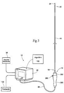
remotely manipulate or actuate the distal end region 20. Image guidance, e.g.,
CT,
radiographic, IVUS, OCT or another suitable guidance modality, or combinations
thereof, can be used to aid the caregiver's manipulation.
[00121] As shown in Fig. 6B, the distal end region 20 of the elongated shaft
16
can flex in a substantial fashion to gain entrance into a respective
left/right renal
artery by manipulation of the elongated shaft 16. As shown in Figs. 28A and
28B,
the distal end region 20 of the elongated shaft 16 can gain entrance to the
renal
artery via passage within a guide catheter 94. The distal end region 20 of the
elongated shaft 16 carries at least one energy delivery element 24 (e.g.,
radiofrequency electrode, electrode, cooled radiofrequency electrode, thermal
element, thermal heating element, electrically resistive heating element,
cryoablation
applicator, microwave antenna, ultrasound transducer, high intensity focused
ultrasound transducer, laser emitter). The energy delivery element 24 is also
specially sized and configured for manipulation and use within a renal artery.
[00122] As Fig. 6B shows, once entrance to a renal artery is gained, further
manipulation of the distal end region 20 and the energy delivery element(s) 24
within
the respective renal artery establishes proximity to and alignment between the
energy delivery element(s) 24 and tissue along an interior wall of the
respective
renal artery. In some embodiments, manipulation of the distal end region 20
will also
facilitate contact between the energy delivery element 24 and a wall of the
renal
artery. In the context of the present application, the phrasing "contact
between an
energy delivery element and a wall of the renal artery" generally means
contiguous
-23-
CA 02796865 2012-10-18
WO 2011/139589 PCT/US2011/033491
physical contact with or without atraumatic distension of the renal artery
wall and
without puncturing or perforating the renal artery wall.
[00123] In the representative embodiment of Fig. 6B, the thermal heating
element 24 of distal end region 20 is positioned along a distal tip or end of
the distal
end region, e.g., at a distal end of an optional third or distal flexure zone
44.
However, it should be understood that the distal end region 20 optionally may
comprise one or more additional thermal heating elements that are positioned
relatively more proximal. When multiple thermal heating elements are provided,
the
thermal heating elements may deliver power independently (i.e., may be used in
a
monopolar fashion), either simultaneously or progressively, and/or may deliver
power between any desired combination of the elements (i.e., may be used in a
bipolar fashion). Furthermore, the caregiver optionally may be capable of
dynamically choosing which thermal heating element(s) are used for power
delivery
in order to form highly customizable lesion(s) within the renal artery, as
desired.
[00124] In one representative embodiment shown in Fig. 6C, one or more
additional thermal heating elements 24a optionally may be positioned
proximally of
thermal heating element 24, e.g., along a third flexure zone 44, at a proximal
region
of the optional third flexure zone 44 and/or at a distal region of an optional
second or
intermediate Flexure zone 34 for contacting an internal wall of the renal
artery at
position(s) longitudinally spaced, but generally in angular alignment, with
the distally
located thermal heating element 24. The spacing of the thermal heating
elements
24 and 24a may be specified to provide a desired spacing between lesions
formed
when using the elements within a renal artery. In one representative
embodiment,
thermal heating elements 24 and 24a are spaced apart as far as about 1 cm. In
other embodiments, the spacing between thermal heating elements 24 and 24a is
in
the range of about 2 mm to about 5 mm. In one representative embodiment, the
thermal heating elements 24 and 24a are spaced apart about 5 mm. In another
representative embodiment, the thermal heating elements 24 and 24a are spaced
apart about 2 mm.
[00125] Additionally or alternatively, as shown in Fig. 6D, one or more
thermal
heating elements 24b may be positioned relatively more proximal for contacting
an
internal wall of the renal artery at position(s) that are longitudinally and
angularly
spaced (e.g., in angular opposition) from the distally located thermal heating
element
-24-
CA 02796865 2012-10-18
WO 2011/139589 PCT/US2011/033491
24. Such thermal heating element(s) 24b may, for example, be positioned at an
apex of a bend formed during deflection of the optional second flexure zone
34, at a
proximal region of the optional second flexure zone 34, and/or at a distal
region of a
first or proximal flexure zone 32. The spacing separating thermal heating
element
24b from thermal heating element 24 and/or from optional thermal heating
element
24a may be specified as desired to provide desired longitudinal and angular
spacing
between lesions formed within renal vasculature. In one representative
embodiment, thermal heating elements 24 and 24b are spaced apart about 5 mm to
about 25 mm. In another representative embodiment, the thermal heating
elements
24 and 24b can be spaced as far as about 30 mm. In another representative
embodiment, the thermal heating elements 24 and 24b are spaced apart about 11
mm. In still another representative embodiment, the thermal heating elements
24
and 24b are spaced apart about 17.5 mm.
[00126] As also will be described in greater detail later, different sections
of the
elongated shaft 16 serve different mechanical functions when in use. The
sections
are thereby desirably differentiated in terms of their size, configuration and
mechanical properties for (i) percutaneous introduction into a femoral artery
through
a small-diameter access site; (ii) atraumatic passage through the tortuous
intravascular path 14 through an iliac artery, into the aorta, and into a
respective
left/right renal artery, including (iii) significant flexure near the junction
of the left/right
renal arteries and aorta to gain entry into the respective left or right renal
artery; (iv)
controlled translation, deflection, rotation and/or actuation within the
respective renal
artery to attain proximity to and a desired alignment with an interior wall of
the
respective renal artery; (v) the placement of at least one energy delivery
element 24
into contact with tissue on the interior wall; (vi) allowing substantially
stable contact
force between the at least one energy delivery element and the interior wall
during
motion of the renal artery with respect to the aorta due to respiration and/or
blood
flow pulsatility; and (vii) repositioning via retraction and/or deflection in
a multiple
directions and/or rotation within the renal artery for subsequent
treatment(s).
[00127] Referring back to Fig. 5, the system 10 also includes an energy
generator 26 (e.g., a radiofrequency generator). Under the control of the
caregiver
or automated control algorithm 102 (as will be described in greater detail
later), the
generator 26 generates a selected form and magnitude of energy. A cable 28
-25-
CA 02796865 2012-10-18
WO 2011/139589 PCT/US2011/033491
operatively attached to the handle assembly 200 electrically connects the
energy
delivery element 24 to the generator 26. At least one supply wire (not shown)
passing along the elongated shaft 16 or through a lumen in the elongated shaft
16
from the handle assembly 200 to the energy delivery element 24 conveys the
treatment energy to the energy delivery element 24. A control mechanism, such
as
foot pedal 100, can be connected (e.g., pneumatically connected or
electrically
connected) to the generator 26 to allow the operator to initiate, terminate
and,
optionally, adjust various operational characteristics of the generator,
including, but
not limited to, power delivery.
[00128] For systems that provide for the delivery of a monopolar electric
field via
the energy delivery element 24, a neutral or dispersive electrode 38 can be
electrically connected to the generator 26 and attached to the exterior of the
patient.
Additionally, one or more sensors 52 (see, e.g., Figs. 9A and 9B), such as one
or
more temperature (e.g., thermocouple, thermistor, etc.), impedance, pressure,
optical, flow, chemical or other sensors, can be located proximate to or
within the
energy delivery element and connected to one or more of the supply wires. For
example, a total of two supply wires can be included, in which both wires
could
transmit the signal from the sensor and one wire could serve dual purpose and
also
convey the energy to the energy delivery element. Alternatively, both wires
could
'transmit energy to the energy delivery element.
[00129] Once proximity between, alignment with, and contact between the
energy delivery element 24 and tissue are established within the respective
renal
artery (as Fig. 6B shows), the purposeful application of energy from the
generator 26
to tissue by the energy delivery element 24 induces one or more desired
neuromodulating effects on localized regions of the renal artery and adjacent
regions
of the renal plexus (RP), which lay intimately within or adjacent to the
adventitia of
the renal artery. The purposeful application of the neuromodulating effects
can
achieve neuromodulation along all or a portion of the RP.
[00130] The neuromodulating effects can include both thermal ablation, non-
ablative thermal alteration or damage (e.g., via sustained heating and/or
resistive
heating), and electromagnetic neuromodulation. Desired thermal heating effects
may include raising the temperature of target neural fibers above a desired
threshold
to achieve non-ablative thermal alteration, or above a higher temperature to
achieve
-26-
CA 02796865 2012-10-18
WO 2011/139589 PCT/US2011/033491
ablative thermal alteration. For example, the target temperature can be above
body
temperature (e.g., approximately 37 C) but less than about 45 C for non-
ablative
thermal alteration, or the target temperature can be about 45 C or higher for
the
ablative thermal alteration. Desired electromagnetic neuromodulation effects
may
include altering the electrical signals transmitted in a nerve.
[00131] Further details of special size, configuration, and mechanical
properties
of the elongated shaft 16, the distal end region 20 and the energy delivery
element
24, as well as other aspects of the system 10, will now be described. In still
other
embodiments, the system 10 may have a different configuration and/or include
different features. For example, alternative multi-energy delivery element
devices,
such as multi-electrode baskets, spirals or lassos, or balloon expandable
devices,
may be implemented to intravascularly deliver neuromodulatory treatment with
or
without contacting the vessel wall.
B. Size and Configuration of the Elongated Shaft for Achieving
Intravascular Access to a Renal Artery
[00132] As explained above, intravascular access to an interior of a renal
artery
can be achieved, for example, through the femoral artery. As Fig. 6A shows,
the
elongated shaft 16 is specially sized and configured to accommodate passage
through this intravascular path 14, which leads from a percutaneous access
site in
the femoral artery to a targeted treatment site within a renal artery. In this
way, the
caregiver is able to orient the energy delivery element 24 within the renal
artery for
its intended purpose.
[00133] For practical purposes, the maximum outer dimension (e.g., diameter)
of
any section of the elongated shaft 16, including the energy delivery element
24 it
carries, is dictated by the inner diameter of the guide catheter or delivery
catheter
through which the elongated shaft 16 is passed. Assuming, for example, that an
8
French guide catheter (which has an inner diameter of approximately 0.091
inches)
would likely be, from a clinical perspective, the largest guide catheter used
to access
the renal artery, and allowing for a reasonable clearance tolerance between
the
energy delivery element 24 and the guide catheter, the maximum outer dimension
can be realistically expressed as being less than or equal to approximately
0.085
inches. However, use of a smaller 5 French guide catheter 94 may require the
use
-27-
CA 02796865 2012-10-18
WO 2011/139589 PCT/US2011/033491
of smaller outer diameters along the elongated shaft 16. For example, an
energy
delivery element 24 that is to be routed within a 5 French guide catheter
would have
an outer dimension of no greater than 0.053 inches. In another example, an
energy
delivery element 24 that is to be routed within a 6 French guide catheter
would have
an outer dimension of no greater than 0.070 inches.
1. Force Transmitting Section
[00134] As Fig. 7A shows, the proximal end region 18 of the elongated shaft 16
includes, coupled to the handle assembly 200, a force transmitting section 30.
The
force transmitting section 30 is sized and configured to possess selected
mechanical
properties that accommodate physical passage through and the transmission of
forces within the intravascular path 14, as it leads from the accessed femoral
artery
(left or right), through the respective iliac branch artery and into the
aorta, and in
proximity to the targeted renal artery (left or right). The mechanical
properties of the
force transmitting section 30 include at least a preferred effective length
(expressed
in inches or centimeters). It should be understood that the term force
transmitting
section can be used interchangeably with elongated tubular shaft or proximal
force
transmitting section.
[00135] As Fig. 7A shows, the force transmitting section 30 includes a
preferred
effective length L1. The preferred effective length L1 is a function of the
anatomic
distance within the intravascular path 14 between the access site and a
location
proximate to the junction of the aorta and renal arteries. The preferred
effective
length L1 can be derived from textbooks of human anatomy, augmented by a
caregiver's knowledge of the targeted site generally or as derived from prior
analysis
of the particular morphology of the targeted site. The preferred effective
length L1 is
also dependent on the length of the guide catheter that is used, if any. In a
representative embodiment, for a normal human, the preferred effective length
L1
comprises about 30 cm to about 110 cm. If no guide catheter is used, then the
preferred effective length L1 comprises about 30 cm to about 35 cm. If a 55 cm
length guide catheter is used, then the preferred effective length L1
comprises about
65 cm to about 70 cm. If a 90 cm length guide catheter is used, then the
preferred
effective length L1 comprises about 95 cm to about 105 cm.
-28-
CA 02796865 2012-10-18
WO 2011/139589 PCT/US2011/033491
[00136] The force transmitting section 30 also includes a preferred axial
stiffness
and a preferred torsional stiffness. The preferred axial stiffness expresses
the
capability of the force transmitting section 30 to be advanced or withdrawn
along the
length of the intravascular path 14 without buckling or substantial
deformation.
Since some axial deformation is necessary for the force transmitting section
30 to
navigate the tortuous intravascular path 14 without providing too much
resistance,
the preferred axial stiffness of the force transmitting section should also
provide this
capability. The preferred torsional stiffness expresses the capability of the
force
transmitting section 30 to rotate the elongated shaft 16 about its
longitudinal axis
along its length without kinking or permanent deformation. As will be
described in
greater detail later, the ability to advance and retract, as well as rotate,
the distal end
region 20 of the elongated shaft 16 within the respective renal artery is
desirable.
[00137] The desired magnitude of axial stiffness and rotational stiffness for
the
force transmitting section 30 can be obtained by selection of constituent
material or
materials to provide a desired elastic modulus (expressed in terms, e.g., of a
Young's Modulus (E)) indicative of axial and torsional stiffnesses, as well as
selecting the construct and configuration of the force transmitted section in
terms of,
e.g., its interior diameter, outer diameter, wall thickness, and structural
features,
including cross-sectional dimensions and geometry. Representative examples are
described in greater detail below.
2. First Flexure Zone
[00138] As Figs. 7A and 7B show, the distal end region 20 of the elongated
shaft
16 is coupled to the force transmitting section 30. The length L1 of the force
transmitting section 30 generally serves to bring the distal end region 20
into the
vicinity of the junction of the respective renal artery and aorta (as Fig. 6B
shows).
The axial stiffness and torsional stiffness of the force transmitting region
transfer
axial and rotation forces from the handle assembly 200 to the distal end
region 20,
as will be described in greater detail later. It should be understood that the
term first
flexure zone can be used interchangeably with flexible tubular structure.
[00139] As shown in Fig. 7B, the distal end region 20 includes a first flexure
zone
32 proximate to the force transmitting section 30. The first flexure zone 32
is sized
and configured to have mechanical properties that accommodate significant
flexure
-29-
CA 02796865 2012-10-18
WO 2011/139589 PCT/US2011/033491
or bending at a prescribed preferred access angle al and provide for the
transmission of torque during rotation, without fracture, collapse,
substantial
distortion, or significant twisting of the elongated shaft 16. The first
flexure zone 32
should accommodate flexure sufficient for the distal end region 20 to advance
via a
guide catheter into the renal artery without substantially straightening out
the guide
catheter.
[00140] Angle al is defined by the angular deviation that the treatment device
12
must navigate to transition between the aorta (along which the force
transmitting
section 30 is aligned) and the targeted renal artery (along which the distal
end region
20 is aligned) (this is also shown in Fig. 6B). This is the angle that the
first flexure
zone 32 must approximate to align the distal end region 20 of the elongated
shaft 16
with the targeted renal artery, while the force transmitting section 30 of the
elongated
shaft 16 remains aligned with the native axis of the aorta (as Fig. 6B shows).
The
more tortuous a vessel, or the more severe the take-off angle between the
renal
artery and the aorta, the greater bend the first flexure zone 32 will need to
make for
the distal end region of the treatment device to access the renal artery and
the
smaller the angle al.
[00141] When the catheter is outside the patient and the first flexure zone 32
is in
a substantially straight, non-deflected configuration, angle al (as shown in
Fig. 7B)
is approximately 180 . Upon full deflection of the first flexure zone 32, the
angle al
is reduced to anywhere between about 30 and 180 . In a representative
embodiment, upon full deflection angle al is about 300 to about 135 . In
another
representative embodiment, upon full deflection angle al is about 90 .
[00142] The first flexure zone 32 is sized and configured to possess
mechanical
properties that accommodate significant, abrupt flexure or bending at the
access
angle al near the junction of the aorta and the renal artery. Due to its size,
configuration, and mechanical properties, the first flexure zone 32 must
resolve
these flexure or bending forces without fracture, collapse, distortion, or
significant
twisting. Such flexure or bending of the first flexure zone may occur at least
in part
within the distal region of a guide catheter without substantially
straightening out the
guide catheter. The resolution of these flexure or bending forces by the first
flexure
zone 32 makes it possible for the distal end region 20 of the elongated shaft
16 to
gain entry along the intravascular path 14 into a targeted left or right renal
artery.
-30-
CA 02796865 2012-10-18
WO 2011/139589 PCT/US2011/033491
[00143] The first flexure zone 32 is sized and configured in length L2 to be
less
than length L1 (see Fig. 7A). That is because the distance between the femoral
access site and the junction of the aorta and renal artery (typically
approximating
about 40 cm to about 55 cm) is generally greater than the length of a renal
artery
between the aorta and the most distal treatment site along the length of the
renal
artery, which is typically less than about 7 cm. The preferred effective
length L2 can
be derived from textbooks of human anatomy, augmented with a caregiver's
knowledge of the site generally or as derived from prior analysis of the
particular
morphology of the targeted site. For example, the length L2 generally may be
less
than about 15 cm, e.g., may be less than about 10 cm. In one representative
embodiment, the length L2 may be about 9 cm.
[00144] Desirably, the length L2 is selected to make it possible to rest a
portion
of the first flexure zone 32 partially in the aorta at or near the length L1,
as well as
rest the remaining portion of the first flexure zone 32 partially within the
renal artery
(as Fig. 6B shows). In this way, the first flexure zone 32 defines a
transitional bend
that is supported and stable within the vasculature.
[00145] In the deflected configuration of Fig. 7B, the first flexure zone 32
comprises a radius of curvature RoC1. In embodiments where the curvature of
first
flexure zone 32 does not vary or is consistent along the length L2, the length
L2 and
the deflection angle al may define the radius of curvature RoC1. It should be
understood that the curvature of first flexure zone 32, and thereby the radius
of
curvature RoC1 of the first flexure zone, alternatively may vary along the
length L2.
[00146] In such embodiments where the curvature does not vary, the length L2
may define a fraction (180 - al)/360 of the circumference C1 of a circle
with an
equivalent radius of curvature RoC1. Thus, the circumference of such an
equivalent
circle is:
3600
C - x L2 = 2;c x Roq (10)
' (180 - al)
Solving for the radius of curvature RoC1:
360 x L2
ROC, = (11)
2,r x (180 -al) -31-
CA 02796865 2012-10-18
WO 2011/139589 PCT/US2011/033491
[00147] Thus, in a representative embodiment of the first flexure zone 32
where
the curvature of the first flexure zone does not vary along the length L2,
where the
length L2 is less than or equal to about 9 cm, and where the angle a1 is about
300 to
about 135 , the radius of curvature RoC1 is about 3.5 cm to about 11.5 cm. In
a
representative embodiment of first flexure zone 32 where the curvature of the
first
flexure zone does not vary along the length L2, where the length L2 is less
than or
equal to about 9 cm, and where the angle al is about 90 , the radius of
curvature
RoC1 is less than or equal to about 5.75 cm.
[00148] As will be apparent, Equation (11) may be rearranged such that the
length L2 and the radius of curvature RoC1 define the angle al. Furthermore,
Equation (11) may be rearranged such that the radius of curvature RoC1 and the
angle al define the length L2. Thus, in embodiments where the curvature of
first
flexure zone 34 does not vary along the length L2, any one of the length L2,
angle
al and radius of curvature RoC1 may be specified by specifying the other two
variables.
[00149] As will be described in greater detail later, and as shown in Fig. 6B,
the
length L2 of the first flexure zone 32 optionally does not extend the full
length of the
targeted length of the renal artery. That is because the distal end region 20
of the
elongated shaft 16 optionally includes one or more additional flexure zones,
distal to
the first flexure zone 32 (toward the substance of the kidney), to accommodate
other
different functions important to the therapeutic objectives of the treatment
device 12.
As will be described later, the ability to transmit torque through the first
flexure zone
32 makes it possible to rotate the thermal heating device to properly position
the
energy delivery element within the renal artery for treatment.
[00150] In terms of axial and torsional stiffness, the mechanical properties
of first
flexure zone 32 can, and desirably do, differ from the mechanical properties
of the
force transmitting section 30. This is because the first flexure zone 32 and
the force
transmitting section serve different functions while in use. Alternatively,
the
mechanical properties of first flexure zone 32 and force transmitting section
30 can
be similar.
[00151] The force transmitting section 30 serves in use to transmit axial load
and
torque over a relatively long length (L1) within the vascular pathway. In
contrast, the
-32-
CA 02796865 2012-10-18
WO 2011/139589 PCT/US2011/033491
first flexure zone 32 needs to transmit axial load and torque over a lesser
length L2
proximate to or within a respective renal artery. Importantly, the first
flexure zone 32
must abruptly conform to an access angle al near the junction of the aorta and
the
respective renal artery, without fracture, collapse, substantial distortion,
or significant
twisting, or straightening a guide catheter imparting the access angle al.
This is a
function that the force transmitting zone need not perform. Accordingly, the
first
flexure zone 32 is sized and configured to be less stiff and to possess
greater
flexibility than the force transmitting section 30.
[00152] Additionally, the first flexure zone 32 may allow energy delivery
element(s) 24 to maintain stable contact with the interior wall of the renal
artery as
the respective kidney moves due to patient respiration. As a patient breathes
the
kidney may move, causing the renal artery to pivot about the ostium, where the
renal
artery joins the aorta. Stable contact between the energy delivery element(s)
24 and
the inner wall of the renal artery is desired during energy delivery.
Therefore, the
energy delivery element(s) 24 must move, along with the renal artery, relative
to the
aorta. The mechanical properties of the first flexure zone 32 that accommodate
significant, abrupt flexure or bending at the access angle al near the
junction of the
aorta and the renal artery also allow the sections of the catheter distal to
the first
flexure zone 32 to pivot about the ostium without significant impediment,
allowing the
energy delivery element to maintain stable contact force with the inner wall
of the
renal artery. In some embodiments, deflectable section 34 distal to first
flexure zone
32 may become stiffer than the first flexure zone 32 when it is controllably
deflected.
The additional stiffness of deflectable section 34 helps maintain a stable
contact
force between the energy delivery element 24 and an inner wall of the renal
artery
and allows the catheter to move with the renal artery relative to the aorta
with
sufficient freedom due to the flexible deformation of the first flexure zone
32. The
renal artery pivots about the juncture with the aorta such that movement of
the renal
artery increases with distance from the juncture with the aorta. The length of
the
distal end region 20 distal to the first flexure zone 32 along with the length
of the first
flexure zone 32 is configured such that an increasing portion of the first
'flexure zone
32 is positioned in the renal artery the more distal the treatment site to
provide
sufficient increased flexibility in the region of the juncture with the aorta
to allow
stable contact force between the energy delivery element 24 and the more
distal
-33-
CA 02796865 2012-10-18
WO 2011/139589 PCT/US2011/033491
treatment site of the inner wall of the renal artery, especially during
increased motion
at the more distal treatment site.
[00153] The desired magnitude of axial stiffness, rotational stiffness, and
flexibility for the first flexure zone 32 can be obtained by selection of
constituent
material or materials to provide a desired elastic modulus (expressed, e.g.,
in terms
of a Young's Modulus (E)) indicative of flexibility, as well as selecting the
construct
and configuration of the force transmitting section, e.g., in terms of its
interior
diameter, outer diameter, wall thickness, and structural features, including
cross-
sectional dimensions and geometry. Representative examples will be described
in
greater detail later.
[00154] Although it is desirable that the force transmitting section 30 and
the first
flexure zone 32 have stiffness and flexibility properties that are unique to
their
respective functions, it is possible that the force transmitting section 30
and the first
flexure zone 32 comprise the same materials, size and geometric configuration
such
that the force transmitting section 30 and the first flexure zone 32
constitute the
same section.
3. Second Flexure Zone
[00155] As shown in Figs. 7A, 7B, and 7C, the distal end region 20 of the
elongated shaft 16 also optionally may include, distal to the first flexure
zone 32, a
second flexure zone 34. In some embodiments, the energy delivery element 24
may
be supported by the second flexure zone 34. It should be understood that the
term
second flexure zone can be used interchangeably with deflectable section or
intermediate flexure zone or deflectable tubular body or multi-directional
deflectable
assembly.
[00156] The second flexure zone 34 is sized, configured, and has the
mechanical
properties that accommodate additional flexure or bending, independent of the
first
flexure zone 32, at a preferred contact angle a2, without fracture, collapse,
substantial distortion, or significant twisting. The second flexure zone 34
should also
accommodate flexure sufficient for the distal end region 20 to advance via a
guide
catheter into the renal artery without straightening out the guide catheter.
The
second flexure zone 34 may be configured in some embodiments for controllable
deflection in multiple directions.
-34-
CA 02796865 2012-10-18
WO 2011/139589 PCT/US2011/033491
[00157] The preferred contact angle a2 is defined by the angle through which
the
energy delivery element 24 can be radially deflected within the renal artery
to
establish contact between the energy delivery element 24 and an inner wall of
the
respective renal artery (as Fig. 6B shows). The magnitude of the contact angle
a2
and the length of the second flexure zone L3 preferably are based on the
native
inside diameter of the respective renal artery where the energy delivery
element 24
rests, which may vary between about 2 mm and about 10 mm, as well as the
diameter of the energy delivery element 24. It is most common for the diameter
of
the renal artery to vary between about 2 mm and about 8 mm, with a mean
diameter
of about 6mm.
[00158] The second flexure zone 34 extends distally from the first flexure
zone
32 for a length L3 into the targeted renal artery (see Fig. 6B). Desirably,
the length
L3 is selected, taking into account the length L2 of the first flexure zone 32
that
extends into the renal artery, as well as the anatomy of the respective renal
artery, to
actively place the energy delivery element 24 (carried at the end of the
distal end
region 20) at or near the targeted treatment site (as Fig. 6B shows). The
length L3
can be derived, taking the length L2 into account, from textbooks of human
anatomy,
together with a caregiver's knowledge of the site generally or as derived from
prior
analysis of the particular morphology of the targeted site.
[00159] As Fig. 7A shows, the second flexure zone 34 is desirably sized and
configured in length L3 to be less than length L2. This is because, in terms
of
length, the distance required for actively deflecting the energy delivery
element 24
into contact with a wall of the renal artery is significantly less than the
distance
required for bending the elongated shaft 16 to gain access from the aorta into
the
renal artery. Thus, the length of the renal artery is occupied in large part
by the
second flexure zone 34 and not as much by the first flexure zone 32.
[00160] In a representative embodiment, L2 is less than or equal to about 9 cm
and L3 is about 5 mm to about 15 mm. In certain embodiments, particularly for
treatments in relatively long blood vessels, L3 can be less than or equal to
about 20
mm. In another representative embodiment, and as described later in greater
detail,
L3 is less than or equal to about 12.5 mm. In another representative
embodiment,
particularly wherein second flexure zone comprises a hinge joint, L3 is no
greater
than 3 mm. about 12.5 mm.
-35-
CA 02796865 2012-10-18
WO 2011/139589 PCT/US2011/033491
[00161] When the catheter is outside the patient and the second flexure zone
34
is in a substantially straight, non-deflected configuration, contact angle a2
(as shown
in Fig. 7C) is approximately 1800. Upon full deflection of the second flexure
zone 34,
the angle a2 is reduced to anywhere between about 450 and 180 . In a
representative embodiment, upon full deflection, angle a2 is about 750 to
about 135 .
In another representative embodiment, upon full deflection, angle a2 is less
than or
equal to about 90 .
[00162] In the deflected configuration of Fig. 7C, the second flexure zone 34
comprises a radius of curvature RoC2. In embodiments where the curvature of
second flexure zone 34 does not vary or is consistent along the length L3, the
length
L3 and the contact angle a2 may define the radius of curvature RoC2. It should
be
understood that the curvature of second flexure zone 34, and thereby the
radius of
curvature RoC2 of the second flexure zone , alternatively may vary along the
length
L3.
[00163] In such embodiments where the curvature does not vary, the length L3
may define a fraction (180 - a2)/360 of the circumference C2 of a circle
with an
equivalent radius of curvature RoC2. Thus, the circumference of such an
equivalent
circle is:
C 360 x L3 = 2,r x RoC (12)
2 (180 - a2) a
[00164] Solving for the radius of curvature RoC2:
RoC = 360 x L3 (13)
2 2;c x (180 - a2)
[00165] Thus, in a representative embodiment of the second flexure zone 34
where the curvature of the second flexure zone does not vary along the length
L3,
where the length L3 is about 5 mm to about 20 mm, and where the contact angle
a2
is about 75 to about 135 , the radius of curvature RoC2 is about 3 rrim to
about 25
mm. In a representative embodiment of second flexure zone 34 where the
curvature
of the second flexure zone does not vary along the length L3, where the length
L3 is
about 12.5mm, for example less than or equal to about 12.5 mm, and where the
angle a2 is about 75 to about 135 , the radius of curvature RoC2 is about 7mm
to
about 16mm, for example less than or equal to about 15mm.. Iln a
representative
-36-
CA 02796865 2012-10-18
WO 2011/139589 PCT/US2011/033491
embodiment of second flexure zone 34 where the curvature of the second flexure
zone does not vary along the length L3, where the length L3 is about 12.5 mm,
and
where the angle a2 is about 900, the radius of curvature RoC2 is about 8 mm.
[00166] As will be apparent, Equation (13) may be rearranged such that the
length L3 and the radius of curvature RoC2 define the contact angle a2.
Furthermore, Equation (13) may be rearranged such that the radius of curvature
RoC2 and the angle a2 define the length L3. Thus, in embodiments where the
curvature of second flexure zone 34 does not vary along the length L3, any one
of
the length L3, angle a2 and radius of curvature RoC2 may be specified by
specifying
the other two variables.
[00167] In the deflected configuration of Fig. 7C, the second flexure zone 34
locates the energy delivery element 24 at a dimension Y from a longitudinal
axis A of
the second flexure zone 34 just distal of the first flexure zone 32. The
dimension Y
can vary from about 2 mm to about 20 mm. In some configurations, and given the
dimension of most renal arteries, the dimension Y can be from about 5mm to
about
15mm. Since the average diameter of most renal arteries is generally less than
10
rnm as described below, it may be desirable for dimension Y to be less than or
equal
to 10 mm. For example the Y dimension can be 6 mm or 8 mm or anywhere
between and including 6 mm to 10 mm.
[00168] By way of example, the average diameter of a human renal artery is
from
about 2mm to about 8mm, but may range from about 2mm to about 10mm.
Accordingly, if the distal end of the first flexure zone 32 were positioned
adjacent to a
wall of an artery having an 8mm diameter, the second flexure zone 34 would be
capable of deflection sufficient for the energy delivery element 24 to contact
the
opposite wall of the artery. In other embodiments, however, the dimension Y
may
have a different value and may be oversized to facilitate contact in a
straight or
curved vessel. The second flexure zone 34 is also configured to locate the
energy
delivery element 24 at a dimension X from a distal end of the 'first 'flexure
zone 32.
The dimension X can vary, e.g., based on the dimension Y and the length L3.
[00169] As Fig. 7C shows, having first and second flexure zones 32 and 34, the
distal end region 20 of the elongated shaft 16 can, in use, be placed into a
complex,
multi-bend structure 36. The complex, multi-bend structure 36 comprises one
deflection region at the access angle al over a length L2 (the first flexure
zone 32)
-37-
CA 02796865 2012-10-18
WO 2011/139589 PCT/US2011/033491
and a second deflection region at the contact angle a2 over a length L3 (the
second
flexure zone 34). In the complex, multi-bend, both L2 and L3 and angle al and
angle
a2 can differ. This is because the angle al and length L2 are specially sized
and
configured to gain access from an aorta into a respective renal artery through
a
femoral artery access point, and the angle a2 and length L3 are specially
sized and
configured to align an energy delivery element 24 with an interior wall inside
the
renal artery.
[00170] In the illustrated embodiment (see, e.g., Fig. 7C), the second flexure
zone 34 is sized and configured to allow a caregiver to remotely deflect the
second
flexure zone 34 within the renal artery, to radially position the energy
delivery
element 24 into contact with an inner wall of the renal artery.
[00171] In the illustrated embodiment, a control mechanism is coupled to the
second flexure zone 34. The control mechanism includes a control wire 40
attached
to the distal end of the second flexure zone 34 (a representative embodiment
is
shown in Figs. 12B and 12C and will be described in greater detail later). It
should
be understood that the term control wire can be used interchangeably with
flexure
control element. The control wire 40 is passed proximally through the
elongated
shaft 16 and coupled to an actuator 260 (also called a flexure controller) on
the
handle assembly 200. Operation of the actuator 260 (e.g., by the caregiver
pulling
proximally on or pushing forward the actuator 260) pulls the control wire 40
back to
apply a compressive and bending force to the second flexure zone 34 (as Figs.
7C
and 12C show) resulting in bending. The compressive force in combination with
the
optional directionally biased stiffness (described further below) of the
second flexure
zone 34 deflects the second flexure zone 34 and, thereby, radially moves the
energy
delivery element 24 toward an interior wall of the renal artery (as Fig. 6B
shows).
[00172] Desirably, as will be described in greater detail later, the distal
end
region 20 of the elongated shaft 16 can be sized and configured to vary the
stiffness
of the second flexure zone 34 about its circumference. The variable
circumferential
stiffness imparts preferential and directional bending to the second flexure
zone 34
(i.e., directionally biased stiffness). In response to operation of the
actuator 260, the
second flexure zone 34 may be configured to bend in a single preferential
direction.
Representative embodiments exemplifying this feature will be described in
greater
-38-
CA 02796865 2012-10-18
WO 2011/139589 PCT/US2011/033491
detail later. Additional representative embodiments depicting multidirectional
bending will also be described later in greater detail.
[00173] The compressive and bending force and resulting directional bending
from the deflection of the second flexure zone 34 has the consequence of
altering
the axial stiffness of the.second flexure zone. The actuation of the control
wire 40
serves to increase the axial stiffness of the second flexure zone. As will be
described later, the axial stiffness of the deflected second flexure zone in
combination with other flexible aspects of the distal end region of the
catheter
treatment device allows for favorable performance in a renal artery
neuromodulation
treatment.
[00174] In terms of axial and torsional stiffnesses, the mechanical properties
of
second flexure zone 34 can, and desirably do, differ from the mechanical
properties
of the first flexure zone 32. This is because the first flexure zone 32 and
the second
flexure zone 34 serve different functions while in use.
[00175] The first flexure zone 32 transmits axial load and torque over a
longer
length (L2) than the second flexure zone 34 (L3). Importantly, the second
flexure
zone 34 is also sized and configured to be deflected remotely within the renal
artery
by the caregiver. In this arrangement, less resistance to deflection is
desirable. This
is a function that the first flexure zone 32 need not perform. Accordingly,
the second
flexure zone 34 is desirably sized and configured to be less stiff (when the
control
wire 40 is not actuated) and, importantly, to possess greater flexibility than
the first
flexure zone 32 in at least one plane of motion.
[00176] Still, because the second 'flexure zone 34, being distal to the first
flexure
zone 32, precedes the first flexure zone 32 through the access angle access
angle
al, the second flexure zone 34 also includes mechanical properties that
accommodate its flexure or bending at the preferred access angle al, without
fracture, collapse, substantial distortion, or significant twisting of the
elongated shaft
16.
[00177] The desired magnitude of axial stiffness, rotational stiffness, and
flexibility for the second flexure zone 34 can be obtained by selection of
constituent
material or materials to provide a desired elastic modulus (expressed, e.g.,
in terms
of a Young's Modulus (E)) indicative of flexibility, as well as by selecting
the
-39-
CA 02796865 2012-10-18
WO 2011/139589 PCT/US2011/033491
construct and configuration of the second flexure zone 34, e.g., in terms of
its interior
diameter, outer diameter, wall thickness, and structural features, including
cross-
sectional dimensions and geometry. Representative examples will be described
in
greater detail later. Axial stiffness, torsional stiffness, and flexibility
are properties
that can be measured and characterized in conventional ways.
[00178] As before described, both the first and second flexure zones 32 and 34
desirably include the mechanical properties of axial stiffness sufficient to
transmit to
the energy delivery element 24 an axial locating force. By pulling back on the
handle
assembly 200, axial forces are transmitted by the force transmitting section
30 and
the first and second flexure zones 32 and 34 to retract the energy delivery
element
24 in a proximal direction (away from the kidney) within the renal artery.
Likewise, by
pushing forward on the handle assembly 200, axial forces are transmitted by
the
force transmitting section 30 and the first and second flexure zones 32 and 34
to
advance the energy delivery element 24 in a distal direction (toward the
kidney)
within the renal artery. Thus, proximal retraction of the distal end region 20
and
energy delivery element 24 within the renal artery can be accomplished by the
caregiver by manipulating the handle assembly 200 or shaft from outside the
intravascular path 14.
[00179] As before described, both the first and second flexure zones 32 and 34
also desirably include torsional strength properties that will allow the
transmission of
sufficient rotational torque to rotate the distal end region 20 of the
treatment device
12 such that the energy delivery element 24 is alongside the circumference of
the
blood vessel wall when the second flexure zone 34 is deflected. By pulling or
pushing on the actuator to deflect the energy delivery element 24 such that it
achieves vessel wall contact, and then rotating the force transmitting section
30 and,
with it, the first and second flexure zones 32 and 34, the energy delivery
element 24
can be rotated in a circumferential path within the renal artery. As described
later,
this rotating feature enables the clinical operator to maintain vessel wall
contact as
the energy delivery element 24 is being relocated to another treatment site.
By
maintaining wall contact in between treatments, the clinical operator is able
to
achieve wall contact in subsequent treatments with higher certainty in
orientations
with poor visualization.
-40-
CA 02796865 2012-10-18
WO 2011/139589 PCT/US2011/033491
4. Third Flexure Zone
[00180] As Figs. 7A, 7B, 7C, and 7D show, the distal end region 20 of the
elongated shaft 16 also optionally may include, distal to the optional second
flexure
zone 34, a third flexure zone 44. Third flexure zone may be used
interchangeably
with distal flexure zone and force dampening section. In this arrangement, the
length
L3 of the second flexure zone 34 may be shortened by a length L4, which
comprises
the length of the third flexure zone 44. In this arrangement, the energy
delivery
element 24 is carried at the end of the third flexure zone 44.
[00181] As Fig. 7D shows, the third flexure zone 44 is sized, configured, and
has
the mechanical properties that accommodate additional flexure or bending,
independent of the first flexure zone 32 and the second flexure zone 34, at a
preferred treatment angle a3. The third flexure zone 44 should also
accommodate
flexure sufficient for the distal end region 20 to advance via a guide
catheter into the
renal artery without straightening out the guide catheter or causing injury to
the blood
vessel. The treatment angle a3 provides for significant flexure about the axis
of the
distal end region 20 (a representative embodiment is shown in Fig. 15C). Not
under
the direct control of the physician, flexure at the third flexure zone occurs
in
response to contact between the energy delivery element 24 and wall tissue
occasioned by the radial deflection of the energy delivery element 24 at the
second
flexure zone 34 (see Fig. 6B). Passive deflection of the third flexure zone
provides
the clinical operator with visual feedback via fluoroscopy or other
angiographic
guidance of vessel wall contact (as shown in Figs. 46A to 46E). Additionally,
the
third flexure zone desirably orients the region of tissue contact along a side
of the
energy delivery element 24, thereby increasing the area of contact. The third
flexure
zone 44 also biases the energy delivery element 24 against tissue, thereby
stabilizing the energy delivery element 24.
[00182] The function of the third flexure zone 44 provides additional benefits
to
the therapy. As actuation of the control wire 40 deflects the second flexure
zone 34,
pressing the energy delivery element 24 against an inner wall of an artery the
third
flexure zone effectively dampens the contact force between the energy delivery
element 24 and the vessel wall. This effect is particularly valuable in a
renal artery
treatment due to movement of the renal artery caused by respiration and/or
pulsatile
flow. While the flexibility of the first flexure zone allows the distal end
region of the
-41-
CA 02796865 2012-10-18
WO 2011/139589 PCT/US2011/033491
treatment catheter to follow movement of the renal artery during respiration,
the
increased axial stiffness of the deflected second flexure zone provides
helpful
integrity to the distal end region to maintain contact between the energy
delivery
element and vessel wall. The third flexure zone helps soften or cushion the
contact
force so that atraumatic contact can be achieved and maintained, particularly
during
movement of the renal artery. By dampening this contact force, the third
flexure
zone minimizes the chance of mechanical injury to the vessel wall and avoids
excessive contact between the energy delivery element and vessel wall (see
discussion of active surface area).
[00183] As Fig. 7A shows, the third flexure zone 44 is desirably sized and
configured in length L4 to be less than length L3. This is because, in terms
of
length, the distance required for orienting and stabilizing the energy
delivery element
24 in contact with a wall of the renal artery is significantly less than the
distance
required for radially deflecting the energy delivery element 24 within the
renal artery.
In some embodiments, length L4 can be as long as about 1 cm. In other
embodiments, the length L4 is from about 2 mm to about 5 mm. In one
representative embodiment, the length L4 is less than or equal to about 5 mm.
In
another representative embodiment, the length L4 is less than or equal to
about 2
mm. In another representative embodiment wherein the deflectable section 34 is
comprised of a hinge joint, the length L4 is less than or equal to about 16
mm, which
in this embodiment can be greater than the length L3 of the deflectable
section 34.
[00184] When the catheter is outside the patient and the third flexure zone 44
is
in a substantially straight, non-deflected configuration, treatment angle a3
(as shown
in Fig. 7D) is approximately 180 . Upon full deflection of the third flexure
zone 44,
the angle a3 is reduced to anywhere between about 45 and 1800. In a
representative embodiment, upon full deflection, angle a3 is about 75 to
about 135 .
In another representative embodiment, upon full deflection, angle a3 is about
900.
[00185] In the passively deflected configuration of Fig. 7D, the third flexure
zone
44 comprises a radius of curvature RoC3. In embodiments where the curvature of
third flexure zone 44 does not vary or is consistent along the length L4, the
length L4
and the contact angle a3 may define the radius of curvature RoC3. It should be
understood that the curvature of third flexure zone 44, and thereby the radius
of
curvature RoC3 of the third flexure zone, alternatively may vary along the
length L4.
-42-
CA 02796865 2012-10-18
WO 2011/139589 PCT/US2011/033491
[00186] In such embodiments where the curvature does not vary, the length L4
may define a fraction (1800 - a3)/360 of the circumference C3 of a circle
with an
equivalent radius of curvature RoC3. Thus, the circumference of such an
equivalent
circle is:
C 360 xL4=2,cxRoC (14)
C3 (180 - a3) s
[00187] Solving for the radius of curvature RoC2:
RoC _ 360 x L4 (15)
s 2z x (180 -a3)
[00188] Thus, in a representative embodiment of the third flexure zone 44
where
the curvature of the third flexure zone does not vary along the length L4,
where the
length L4 is about 2mm to about 5mm, and where the contact angle a3 is about
75
to about 135 , the radius of curvature RoC3 is about 1 mm to about 6mm.
[00189] As will be apparent, Equation (15) may be rearranged such that the
length L4 and the radius of curvature RoC3 define the contact angle a3.
Furthermore, Equation (15) may be rearranged such that the radius of curvature
RoC3 and the angle a3 define the length L4. Thus, in embodiments where the
curvature of third flexure zone 44 does not vary along the length L4, any one
of the
length L4, angle a3 and radius of curvature RoC3 may be specified by
specifying the
other two variables.
[00190] The mechanical properties of third flexure zone 44 and the second
flexure zone 34 in terms of axial stiffness, torsional stiffness, and
flexibility can be
comparable. However, the third flexure zone 44 can be sized and configured to
be
less stiff and, importantly, to possess greater flexibility than the second
flexure zone
34.
[00191] In the embodiment just described (and as shown in Fig. 7D), the distal
end region 20 may comprise a first or proximal flexure zone 32, a second
flexure
zone 34, and a third flexure zone 44. The first, second and third flexure
zones
function independently from each other, so that the distal end region 20 of
the
elongated shaft 16 can, in use, be placed into a more compound, complex, multi-
bend structure 36. The compound, complex, multi-bend structure 36 comprises a
first deflection region at the access angle al over a length L2 (the first
flexure zone
-43-
CA 02796865 2012-10-18
WO 2011/139589 PCT/US2011/033491
32); an second deflection region at the contact angle a2 over a length L3 (the
second flexure zone 34); and a third deflection region at the treatment angle
a3 over
a length L4 (the third flexure zone 44). In the compound, complex, multi-bend
structure 36, all lengths L2, L3, and L4 and all angles al, a2, and a3 can
differ. This
is because the angle al and length L2 are specially sized and configured to
gain
access from an aorta into a respective renal artery through a femoral artery
access
point; the angle a2 and length L3 are specially sized and configured to align
an
energy delivery element 24 element with an interior wall inside the renal
artery; and
the angle a3 and length L4 are specially sized and configured to optimize
surface
contact between tissue and the energy delivery element.
[00192] The composite length of L2, L3 and L4 of the first, second and third
flexure zones, respectively, of the distal end region 20, along with the
length L1 of
the force transmitting section 30 and the length L5 (see Fig. 8A) of the
energy
delivery element 24 (i.e., the composite length equal to L1+L2+L3+L4+L5),
specifies
a working length of the elongated shaft 16 of the treatment device 12. In some
representative embodiments, this working length is about 40 cm to about 125
cm. In
a representative embodiment where no guide catheter is used, then this working
length may be about 40 cm to about 50 cm. If, alternatively, a 55 cm length
guide
catheter is used, then this working length may be about 70 cm to about 80 cm.
If a
90 cm length guide catheter is used, then this working length may be about 105
cm
to about 115 cm.
C. Size and Configuration of the Energy Delivery Element for Achieving
Neuromodulation in a Renal Artery
[00193] In some patients, it may be desirable to create multiple focal lesions
that
are circumferentially spaced along the longitudinal axis of the renal artery.
However,
it should be understood that a single focal lesion with desired longitudinal
and/or
circumferential dimensions, one or more full-circle lesions, multiple
circumferentially
spaced focal lesions at a common longitudinal position, and/or multiple
longitudinally
spaced focal lesions at a common circumferential position alternatively or
additionally may be created.
[00194] Creating multiple focal lesions that are circumferentially spaced
along the
longitudinal axis of the renal artery avoids the creation of a full-circle
lesion, thereby
-44-
CA 02796865 2012-10-18
WO 2011/139589 PCT/US2011/033491
reducing a risk of vessel stenosis, while still providing the opportunity to
circumferentially treat the renal plexus, which is distributed about the renal
artery. It
is desirable for each lesion to cover at least 10% of the vessel circumference
to
increase the probability of affecting the renal plexus. However, it is
important that
each lesion not be too large (e.g., > 60% of vessel circumference) lest the
risk of a
stenotic effect increases (or other undesirable healing responses such as
thrombus
formation or collateral damage). In one embodiment the energy delivery element
24
is configured to create a lesion at least 30% (i.e., greater than or equal to
30%) of
the vessel circumference. In another embodiment, the energy delivery element
24 is
configured to create a lesion of greater than or equal to 30% but less than
60% of
the vessel circumference. It is also important that each lesion be
sufficiently deep to
penetrate into and beyond the adventitia to thereby affect the renal plexus.
However, lesions that are too deep (e.g., >5mm) run the risk of interfering
with non-
target tissue and tissue structures (e.g., renal vein) so a controlled depth
of thermal
treatment is desirable.
[00195] As described in greater detail below, energy delivery element 24 may
be
delivered to a first treatment site within the renal artery such that the
energy delivery
element 24 is positioned in contact with an interior wall of the artery for
treating the
renal plexus (see Fig. 43C). Once positioned within the artery as desired,
energy
may be delivered via the energy delivery element to create a first focal
lesion at this
first treatment site (see Fig. 43D). The first focal lesion creates a first
treatment zone
98a that is not continuous completely around the circumference of the renal
artery in
a radial plane or cross-section normal to the wall or to the longitudinal axis
of the
artery (i.e., the first focal lesion does not extend all the way around the
circumference of the vessel wall). As a result, there is a discrete untreated
zone
about the circumference of the artery in the radial plane of the first
treatment zone
normal to the longitudinal axis of the artery.
[00196] After formation of the first focal lesion at the first treatment zone
98a, the
energy delivery element 24 optionally may be angularly repositioned relative
to the
renal artery (see Figs. 43E and 43F). This angular repositioning may be
achieved,
for example, by angularly rotating the elongated shaft 16 of treatment device
12 via
handle assembly 200 (see Fig. 16A). In addition to angular repositioning of
the
energy delivery element 24, the energy delivery element optionally may be
-45-
CA 02796865 2012-10-18
WO 2011/139589 PCT/US2011/033491
repositioned along the lengthwise or longitudinal dimension of the renal
artery (see
Fig. 43E). This longitudinal repositioning may be achieved, for example, by
translating the elongated shaft 16 of treatment device 12 via handle assembly
200,
and may occur before, after or concurrent with angular repositioning of the
energy
delivery element 24.
[00197] Repositioning the energy delivery element 24 in both the longitudinal
and
angular dimensions places the energy delivery element in contact with the
interior
wall of the renal artery at a second treatment site for treating the renal
plexus (see
Fig. 43E). Energy then may be delivered via the energy delivery element to
form a
second focal lesion at this second treatment site, thereby creating a second
treatment zone 98b and a second untreated zone (see Fig. 43F).
[00198] As with the first treatment zone created by the first focal lesion,
the
second treatment zone is not continuous about the complete circumference of
the
renal artery. However, the first and second treatment zones (as well as the
first and
second untreated zones) are angularly and longitudinally offset from one
another
about the angular and lengthwise dimensions of the renal artery, respectively
(see
Fig. 43G). Superimposing the first and second treatment zones, which are
positioned along different cross-sections or radial planes of the renal
artery, about a
common cross-section provides a composite treatment zone that covers a greater
portion of the circumference of the artery than either treatment zone
individually. As
this composite treatment zone is not continuous (i.e., it is formed from
multiple,
longitudinally and angularly spaced treatment zones), it is expected that a
greater
portion of the circumference of the arterial wall may be treated with reduced
risk of
vessel stenosis, as compared to formation of a single focal lesion covering an
equivalent portion of the arterial circumference at a single treatment site
(i.e., at a
single lengthwise position or about a single cross-section of the renal
artery).
[00199] One or more additional focal lesions optionally may be formed at one
or
more additional angularly and longitudinally spaced treatment sites to created
additional angularly and longitudinally spaced treatment zones (see Figs. 43G-
43K).
In one representative embodiment, superimposition of all or a portion of the
treatment zones provides a composite treatment zone that is non-continuous
(i.e.,
that is broken up along the lengthwise dimension or longitudinal axis of the
renal
artery), yet that is substantially circumferential (i.e., that substantially
extends all the
-46-
CA 02796865 2012-10-18
WO 2011/139589 PCT/US2011/033491
way around the circumference of the renal artery over a lengthwise segment of
the
artery). This superimposed treatment zone beneficially does not create a
continuous
circumferential lesion along any individual radial plane or cross-section
normal to the
artery, which may reduce a risk of acute or late stenosis formation, as
compared to
circumferential treatments that create such continuous circumferential
lesions.
[00200] Non-continuous circumferential treatment by positioning energy
delivery
element(s) at different angular orientations along multiple lengthwise
locations may
preferentially affect anatomical structures that substantially propagate along
the
lengthwise dimension of the artery. Such anatomical structures can be neural
fibers
and/or structures that support the neural fibers (e.g., the renal plexus).
Furthermore,
such non-continuous circumferential treatment may mitigate or reduce
potentially
undesirable effects induced in structures that propagate about the angular
dimension
of the artery, such as smooth muscle cells. Were a continuous circumferential
lesion
alternatively to be formed, the angular or circumferential orientation of the
smooth
muscle cells relative to the artery may increase a risk of acute or late
stenosis or
acute vessel spasm.
[00201] In multiple energy delivery element configurations (e.g., multi-
electrode
configurations), such as in Figs. 6C and 6D, multiple non-continuous
circumferential
treatment zones can be created during a single catheter placement within the
renal
artery. The multiple energy delivery elements can be spaced and located such
that
they are longitudinally and angularly spaced apart From one another and such
that
they create longitudinally offset and angularly opposed or offset treatment
zones.
Retraction and rotation of the treatment device 12 can reposition the energy
delivery
elements to create additional longitudinally and angularly separated treatment
zones, thereby allowing the practitioner the ability to create multiple
treatment zones
per catheter placement and several treatment zones via only two catheter
placements.
[00202] In some embodiments, as discussed later with respect to Figs. 26, the
distal end region 20 of the treatment device 12 may comprise a helical
deflected
configuration with multiple thermal heating elements positioned along its
length.
When positioned in the helical deflected configuration within a renal artery,
the
multiple thermal heating elements 24 may be spaced circumferentially along the
longitudinal length of the distal end region in contact with the wall of the
artery. In
-47-
CA 02796865 2012-10-18
WO 2011/139589 PCT/US2011/033491
some embodiments, a non-continuous circumferential treatment may be achieved
via a single catheter placement without angular or longitudinal repositioning
of the
distal end region 20.
[00203] As described (and as Fig. 8A shows), the energy delivery element 24 is
sized and configured, in use, to contact an internal wall of the renal artery.
In the
illustrated embodiment (see Fig. 8A), the energy delivery element 24 takes the
form
of an electrode 46 sized and configured to apply an electrical field
comprising
radiofrequency (RF) energy from the generator 26 to a vessel wall. In the
illustrated
embodiment, the electrode 46 is operated in a monopolar or unipolar mode. In
this
arrangement, a return path for the applied RF electric field is established,
e.g., by an
external dispersive electrode (shown as 38 in Fig. 6A), also called an
indifferent
electrode or neutral electrode. The monopolar application of RF electric field
energy
serves to ohmically or resistively heat tissue in the vicinity of the
electrode 46. The
application of the RF electrical field thermally injures tissue. The treatment
objective
is to thermally induce neuromodulation (e.g., necrosis, thermal alteration or
ablation)
in the targeted neural fibers. The thermal injury forms a lesion in the vessel
wall,
which is shown, e.g., in Fig. 9B. Alternatively, a RF electrical field can be
delivered
with an oscillating intensity that does not thermally injure the tissue
whereby
neuromodulation in the targeted nerves is accomplished by electrical
modification of
the nerve signals.
[00204] The active surface area of contact (ASA) between the energy delivery
element 24 or electrode 46 and the vessel wall has great bearing on the
efficiency
and control of the transfer of a thermal energy field across the vessel wall
to
thermally affect targeted neural fibers in the renal plexus (RP). The active
surface
area of the energy delivery element 24 and electrode 46 is defined as the
energy
transmitting area of the element 24 or electrode 46 that can be placed in
intimate
contact against tissue. Too much contact between the energy delivery element
and
the vessel wall and/or too much power may create unduly high temperatures at
or
around the interface between the tissue and the energy delivery element,
thereby
creating excessive heat generation at this interface and/or spasm and
contraction of
the vessel wall. This excessive heat can also create a lesion that is
circumferentially
too large, increasing the risk of stenosis. This excessive heat can also lead
to
undesirable thermal damage at the vessel wall, which stiffens and desiccates
the
-48-
CA 02796865 2012-10-18
WO 2011/139589 PCT/US2011/033491
vessel tissue making it more susceptible to puncture and perforation.
Additionally,
the tissue desiccation (i.e., dehydration) reduces the electrical and thermal
conductivity of the tissue. Reduced conductivity may potentially create a
lesion that
is too shallow to reach the neural fibers and may also result in the buildup
of
excessive heat, causing increased and undesirable damage to the vessel wall
and
increasing the likelihood of thrombus formation. Although the risks of
excessive wall
contact and heating are many, too little contact between the energy delivery
element
and the vessel wall may impair the efficacy of the treatment. For example, too
little
contact may result in superficial heating of the vessel wall, thereby creating
a lesion
that is too small (e.g., < 10% of vessel circumference) and/or too shallow to
reach
the target renal neural fibers.
[00205] While the active surface area (ASA) of the energy delivery element 24
and electrode 46 is important to creating lesions of desirable size and depth,
the
ratio between the active surface area (ASA) and total surface area (TSA) of
the
energy delivery element 24 and electrode 46 is also important. The ASA to TSA
ratio influences lesion formation in two ways: (1) the degree of resistive
heating via
the electric field, and (2) the effects of blood flow or other convective
cooling
elements such as injected or infused saline. As discussed above, the RF
electric
field causes lesion formation via resistive heating of tissue exposed to the
electric
field. The higher the ASA to TSA ratio (i.e., the greater the contact between
the
electrode and tissue), the greater the resistive heating. As discussed in
greater
detail below, the flow of blood over the exposed portion of the electrode (TSA
-
ASA) provides conductive and convective cooling of the electrode, thereby
carrying
excess thermal energy away from the interface between the vessel wall and
electrode. If the ratio of ASA to TSA is too high (e.g., 50%), resistive
heating of the
tissue can be too aggressive and not enough excess thermal energy is being
carried
away, resulting in excessive heat generation and increased potential for
stenotic
injury, thrombus formation and undesirable lesion size. If the ratio of ASA to
TSA is
too low (e.g., 10%), then there is too little resistive heating of tissue,
thereby
resulting in superficial heating and smaller and shallower lesions.
[00206] Various size constraints for the energy delivery element 24 may be
imposed for clinical reasons by the maximum desired dimensions of the guide
catheter, as well as by the size and anatomy of the renal artery itself.
Typically, the
-49-
CA 02796865 2012-10-18
WO 2011/139589 PCT/US2011/033491
maximum outer diameter (or cross-sectional dimension for non-circular cross-
section) of the electrode 46 comprises the largest diameter encountered along
the
length of the elongated shaft 16 distal to the handle assembly 200. Thus, the
outer
diameters of the force transmitting section 30, first, second and third
flexure zones
32, 34, and 44 are equal to or (desirably) less than the maximum outer
diameter of
the electrode 46.
[00207] In a representative embodiment shown in Fig. 8A, the electrode 46
takes
the form of a right circular cylinder, possessing a length L5 that is greater
than its
diameter. The electrode 46 further desirably includes a distal region that is
rounded
to form an atraumatic end surface 48. In the representative embodiment shown
in
Fig. 8B, the electrode 46 is spherical in shape, such that the length L5 is
equal to the
electrode's diameter. The spherical shape, too, presents an atraumatic surface
to
the tissue interface.
[00208] As shown in Figs. 8A and 8B, the angle a3 and length L4 of the distal
flexure zone 44 are specially sized and configured, given the TSA of the
respective
electrode, to optimize an active surface area of contact between tissue and
the
respective electrode 46 (ASA). The angle a3 and the length L4 of the distal
flexure
zone 44 make it possible to desirably lay at least a side quadrant 50 of the
electrode
46 against tissue (see Fig. 8C), though it should be understood that the
electrode 46
does not necessarily need to be positioned with its side quadrant 50 against
tissue
prior to power delivery. In a representative embodiment, the active surface
area of
the electrode 46 contacting tissue (ASA) can be expressed as ASA ? 0.25 TSA
and
ASA <_ 0.50 TSA.
[00209] An ASA to TSA ratio of over 50% may be effective with a reduced power
delivery profile. Alternatively, increasing the conductive or convective
cooling of the
electrode (e.g. vie active cooling) can compensate for a higher ASA to TSA
ratio. As
discussed further below, this could be achieved by injecting or infusing
cooling fluids
such as saline (e.g., room temperature saline or chilled saline) over the
electrode
and into the blood stream.
[00210] The stiffnesses of each of the second and third flexure zones 34 and
44
are also selected to apply via the electrode a stabilizing force that
positions the
electrode 46 in substantially secure contact with the vessel wall tissue. This
stabilizing force also influences the amount of wall contact achieved by the
energy
-50-
CA 02796865 2012-10-18
WO 2011/139589 PCT/US2011/033491
delivery element (i.e., the ASA to TSA ratio). With greater stabilizing force,
the
energy delivery element has more wall contact and with less stabilizing force,
less
wall contact is achieved. Additional advantages of the stabilizing force
include, (1)
softening the contact force between the distal end 20 and vessel wall to
minimize
risk of mechanical injury to vessel wall, (2) consistent positioning of the
electrode 46
flat against the vessel wall, and (3) stabilizing the electrode 46 against the
vessel
wall. As discussed above with respect to the combined effect of the first
flexure
zone and second/second flexure zone, this stabilizing force allows the
catheter
treatment device to maintain consistent contact with the vessel wall even
during
motion of the renal artery during respiration. The stabilizing force also
allows the
electrode to return to a neutral position after the electrode is removed from
contact
with the wall.
[00211] As previously discussed, for clinical reasons, the maximum outer
diameter (or cross-sectional dimension) of the electrode 46 is constrained by
the
maximum inner diameter of the guide catheter through which the elongated shaft
16
is to be passed through the intravascular path 14. Assuming that an 8 French
guide
catheter 94 (which has an inner diameter of approximately 0.091 inches) is,
from a
clinical perspective, the largest desired catheter to be used to access the
renal
artery, and allowing for a reasonable clearance tolerance between the
electrode 46
and the guide catheter, the maximum diameter of the electrode 46 is
constrained to
about 0.085 inches. In the event a 6 French guide catheter is used instead of
an 8
French guide catheter, then the maximum diameter of the electrode 46 is
constrained to about 0.070 inches. In the event a 5 French guide catheter is
used,
then maximum diameter of the electrode 46 is constrained to about 0.053
inches.
Based upon these constraints and the aforementioned power delivery
considerations, the electrode 46 desirably has a maximum outer diameter of
from
about 0.049 to about 0.051 inches. The electrode 46 also desirably has a
minimum
outer diameter of about 0.020 inches to provide sufficient cooling and lesion
size. In
some embodiments, the electrode 46 (i.e., the energy delivery element 24) may
have a length of about 1 mm to about 3 mm. In some embodiments in which the
energy delivery element is a resistive heating element, it can have a maximum
outer
diameter from about 0.049 to 0.051 inches and a length of about 10 mm to 30
mm.
_51-
CA 02796865 2012-10-18
WO 2011/139589 PCT/US2011/033491
D. Applying Energy to Tissue Via the Energy Delivery Element
[00212] Referring back to Fig. 5, in the illustrated embodiment, the generator
26
may supply to the electrode 46 a pulsed or continuous RF electric field.
Although a
continuous delivery of RF energy is desirable, the application of thermal
energy in
pulses may allow the application of relatively higher energy levels (e.g.,
higher
power), longer or shorter total duration times, and/or better controlled
intravascular
renal neuromodulation therapy. Pulsed energy may also allow for the use of a
smaller electrode.
[00213] The thermal therapy may be monitored and controlled, for example, via
data collected with one or more sensors 52, such as temperature sensors (e.g.,
thermocouples, thermistors, etc.), impedance sensors, pressure sensors,
optical
sensors, flow sensors, chemical sensors, force sensors, strain sensors, etc.
(see
Figs. 9A and 9B). Sensor(s) 52 may be incorporated into or on electrode 46
and/or
in/on adjacent areas on the distal end region 20.
[00214] Advantageously, since the second flexure zone 34 deflects in a
controlled manner, the surface of electrode 46 that contacts tissue during
treatment
may be known. As such, sensor(s) 52 may be incorporated into the electrode in
a
manner that specifies whether the sensor(s) are in contact with tissue at the
treatment site and/or are facing blood flow. The ability to specify sensor
placement
relative to tissue and blood flow is highly significant, since a temperature
gradient
across the electrode from the side facing blood flow to the side in contact
with the
vessel wall may be up to about 15 C. Significant gradients across the
electrode in
other sensed data (e.g., flow, pressure, impedance, etc.) also are expected.
[00215] The sensor(s) 52 may, for example, be incorporated on the side of the
electrode that contacts the vessel wall at the treatment site during power and
energy
delivery (see Fig. 9B), may be incorporated into the tip of the electrode, may
be
incorporated on the opposing side of the electrode that faces blood flow
during
energy delivery (see Fig. 9A), and/or may be incorporated within certain
regions of
the electrode (e.g., distal, proximal, quadrants, etc.). In some embodiments,
multiple
sensors may be provided at multiple positions along the electrode and/or
relative to
blood flow. For example, a plurality of circumferentially and/or
longitudinally spaced
-52-
CA 02796865 2012-10-18
WO 2011/139589 PCT/US2011/033491
sensors may be provided. In one embodiment, a first sensor may contact the
vessel
wall during treatment, and a second sensor may face blood flow.
[00216] Additionally or alternatively, various microsensors can be used to
acquire
data corresponding to the energy delivery element, the vessel wall and/or the
blood
flowing across the energy delivery element. For example, arrays of micro
thermocouples and/or impedance sensors can be implemented to acquire data
along
the energy delivery element or other parts of the treatment device. Sensor
data can
be acquired or monitored prior to, simultaneous with, or after the delivery of
energy
or in between pulses of energy, when applicable. The monitored data may be
used
in a feedback loop to better control therapy, e.g., to determine whether to
continue or
stop treatment, and it may facilitate controlled delivery of an increased or
reduced
power or a longer or shorter duration therapy.
[00217] Non-target tissue may be protected by blood flow (F) within the
respective renal artery that serves as a conductive and/or convective heat
sink that
carries away excess thermal energy. For example (as Figs. 9A and 9B show),
since
blood flow (F) is not blocked by the elongated shaft 16 and the electrode 46
it
carries, the native circulation of blood in the respective renal artery serves
to remove
excess thermal energy from the non-target tissue and the energy delivery
element.
The removal of excess thermal energy by blood flow also allows for treatments
of
higher power, where more power can be delivered to the target tissue as
thermal
energy is carried away from the electrode and non-target tissue. In this way,
intravascularly-delivered thermal energy heats target neural fibers located
proximate
to the vessel wall to modulate the target neural fibers, while blood flow (F)
within the
respective renal artery protects non-target tissue of the vessel wall from
excessive or
undesirable thermal injury. When energy is delivered in pulses, the time
interval
between delivery of thermal energy pulses may facilitate additional convective
or
other cooling of the non-target tissue of the vessel wall compared to applying
an
equivalent magnitude or duration of continuous thermal energy.
[00218] It may also be desirable to provide enhanced cooling by inducing
additional native blood flow across the energy delivery element. For example,
techniques and/or technologies can be implemented by the caregiver to increase
perfusion through the renal artery or to the energy delivery element itself.
These
techniques include positioning partial occlusion elements (e.g., balloons)
within
-53-
CA 02796865 2012-10-18
WO 2011/139589 PCT/US2011/033491
upstream vascular bodies such as the aorta or proximal portion of the renal
artery to
improve flow across the energy delivery element. Additionally or
alternatively,
autologous blood from another area of the vasculature may be siphoned off and
re-
directed into the renal artery to increase the volumetric flow rate and/or
velocity of
blood flowing through the artery.
[00219] In addition, or as an alternative, to passively utilizing blood flow
(F) as a
heat sink, active cooling may be provided to remove excess thermal energy and
protect non-target tissues. For example, a thermal fluid infusate may be
injected,
infused, or otherwise delivered into the vessel in an open circuit system.
Additionally
or alternatively, the energy delivery element 24 (e.g., electrode 46) may be
actively
cooled in a closed circuit system (i.e., without delivering any agents into
the
bloodstream) to remove excess thermal energy, such as by circulating a thermal
fluid infusate (e.g., a cryogenic or chilled fluid) within the distal end
region 20 or by
some other mechanism.
[00220] Thermal fluid infusates used for active cooling may, for example,
comprise (room temperature or chilled) saline or some other biocompatible
fluid.
The thermal fluid infusate(s) may, for example, be introduced through the
treatment
device 12 via one or more infusion lumens and/or ports. When introduced into
the
bloodstream, the thermal fluid infusate(s) may, for example, be introduced
through a
guide catheter at a location upstream from the energy delivery element 24 or
electrode 46, or at other locations relative to the tissue for which
protection is sought.
The delivery of a thermal fluid infusate in the vicinity of the treatment site
(via an
open circuit system and/or via a closed circuit system) may, for example,
allow for
the application of increased/higher power, may allow for the maintenance of
lower
temperature at the vessel wall during energy delivery, may facilitate the
creation of
deeper or larger lesions, may facilitate a reduction in treatment time, may
allow for
the use of a smaller electrode size, may compensate for acutely reduced blood
flow,
may compensate for anatomical characteristics resulting in relatively low
blood flow
at a treatment site, or a combination thereof.
[00221] Although many of the embodiments described herein pertain to
electrical
systems configured for the delivery of RF energy, it is contemplated that the
desired
treatment can be accomplished by other means, e.g., by coherent or incoherent
light; direct thermal modification (e.g., with a heated or cooled fluid or
resistive
-54-
CA 02796865 2012-10-18
WO 2011/139589 PCT/US2011/033491
heating element); microwave; ultrasound (including high intensity focused
ultrasound); diode laser; radiation; a tissue heating fluid; and/or a
cryogenic fluid.
Ill. Representative Embodiments
A. First Representative Embodiment (First, Second, and Third Flexure
Zones with Distally Carried Energy Delivery Element)
[00222] Figs. 10A to 15H show a representative embodiment of an elongated
shaft 16 that includes a force transmitting section 30, as well as first,
second and
third flexure zones 32, 34, and 44, having the physical and mechanical
features
described above. In this embodiment, the thermal heating element 24 is carried
distally of the third flexure zone 44 (see, e.g., Fig. 11A).
1. Force Transmitting Section
[00223] In the illustrated embodiment, as shown in Figs. 10A and 10B, the
force
transmitting section 30 comprises a first elongated and desirably tubular
structure,
which can take the form of, e.g., a first tubular structure 54. The first
tubular structure
54 is desirably a hypo tube that is made of a metal material, e.g. of
stainless steel, or
a shape memory alloy, e.g., nickel titanium (a.k.a., Nitinol or NiTi), to
possess the
requisite axial stiffness and torsional stiffness, as already described, for
the force
transmitting section 30. As already described, the force transmitting section
30
comprises the most stiff section along the elongated shaft 16, to facilitate
axially
movement of the elongated shaft 16, as well as rotational manipulation of the
elongated shaft 16 within the intravascular path 14. Alternatively, the first
tubular
structure 54 may comprise a hollow coil, hollow cable, solid cable (w/
embedded
wires), a braided or braid reinforced shaft, a coil reinforced polymer shaft,
a
metal/polymer composite, etc.
[00224] The stiffness is a function of material selection as well as
structural
features such as interior diameter, outside diameter, wall thickness, geometry
and
other features that are made by micro-engineering, machining, cutting and/or
skiving
the hypo tube material to provide the desired axial and torsional stiffness
characteristics. For example, the elongated shaft can be a hypo tube that is
laser
cut to various shapes and cross-sectional geometries to achieve the desired
functional properties.
-55-
CA 02796865 2012-10-18
WO 2011/139589 PCT/US2011/033491
[00225] When the first tubular structure 54 is made from an electrically
conductive metal material, the first tubular structure 54 may include a sheath
56 or
covering made from an electrically insulating polymer material or materials,
which is
placed over the outer diameter of the underlying tubular structure. The
polymer
material can also be selected to possess a desired durometer (expressing a
degree
of stiffness or lack thereof) to contribute to the desired overall stiffness
of the first
tubular structure 54. Candidate materials for the polymer material include,
but are
not limited to, polyethylene terephthalate (PET); Pebax material; nylon;
polyurethane, Grilamid material or combinations thereof. The polymer material
can
be laminated, dip-coated, spray-coated, or otherwise deposited/attached to the
outer
diameter of the tube.
2. First Flexure Zone
[00226] As Figs. 11A, 11 B, and 11 C show, the first flexure zone 32 comprises
a
second elongated and desirably tubular structure, which can take the form of,
e.g., a
second tubular structure 58. The second tubular structure 58 can be made from
the
same or different material as the first tubular structure 54. The axial
stiffness and
torsional stiffness of the second tubular structure 58 possesses the requisite
axial
stiffness and torsional stiffness, as already described, for the first flexure
zone 32. As
already described, the first flexure zone 32 may be less stiff and more
flexible than
the force transmitting section 30, to navigate the severe bend at and prior to
the
junction of the aorta and respective renal artery. The second tubular
structure is
desirably a hypo tube, but can alternatively comprise a hollow coil, hollow
cable,
braided shaft, etc.
[00227] It may be desirable for the first and second tubular structures 54 and
58
to share the same material. In this event, the form and physical features of
the
second tubular structure 58 may be altered, compared to the first tubular
structure
54, to achieve the desired stiffness and flexibility differences. For example,
the
interior diameter, outside diameter, wall thickness, and other engineered
features of
the second tubular structure 58 can be tailored to provide the desired axial
and
torsional stiffness and flexibility characteristics. For example, the second
tubular
structure 58 can be laser cut along its length to provide a bendable, spring-
like
structure. Depending on the ease of manufacturability the first and second
tubular
-56-
CA 02796865 2012-10-18
WO 2011/139589 PCT/US2011/033491
structures may be produced from the same piece of material or from two
separate
pieces. In the event the first tubular structure and second tubular structure
are not of
the same material, the outside diameter of the second tubular structure 58 can
be
less than the outer diameter of first tubular structure 54 (or have a smaller
wall
thickness) to create the desired differentiation in stiffness between the
first and
second tubular structures 54 and 58.
[00228] When the second tubular structure 58 is made from an electrically
conductive metal material, the second tubular structure 58, like the first
tubular
structure 54, includes a sheath 60 (see Figs. 11 B and 11 C) or covering made
from
an electrically insulating polymer material or materials, as already
described. The
sheath 60 or covering can also be selected to possess a desired durometer to
contribute to the desired differentiation in stiffness and flexibility between
the first
and second tubular structures 58.
[00229] The second tubular structure 58 can comprise a different material than
the first tubular structure 54 to impart the desired differentiation in
stiffness and
flexibility between the first and second tubular structures 58. For example,
the
second tubular structure 58 can comprise a cobalt-chromium-nickel alloy,
instead of
stainless steel. Alternatively, the second tubular structure 58 can comprise a
less
rigid polymer, a braided or braid-reinforced shaft, a coil reinforced polymer
shaft, a
metal/polymer composite, nitinol or hollow cable-like structure. In addition
to
material selection, the desired differentiation in stiffness and overall
flexibility can be
achieved by selection of the interior diameter, outside diameter, wall
thickness, and
other engineered features of the second tubular structure 58, as already
described.
Further, a sheath 60 or covering made from an electrically insulating polymer
material, as above described, can also be placed over the outer diameter of
the
second tubular structure 58 to impart the desired differentiation between the
first and
second tubular structures 54 and 58.
3. Second Flexure Zone
[00230] As Figs. 12A, 12B, 12C, and 12D show, the second flexure zone 34
comprises a third elongated and desirably tubular structure, which can take
the form
of, e.g., a third tubular structure 62. The third tubular structure 62 can be
made from
the same or different material as the first and/or second tubular structures
54 and 58.
-57-
CA 02796865 2012-10-18
WO 2011/139589 PCT/US2011/033491
The axial stiffness and torsional stiffness of the third tubular structure 62
possesses
the requisite axial stiffness and torsional stiffness, as already described,
for the
second flexure zone 34. As already described, the second flexure zone 34 may
be
less stiff and more flexible than the first flexure zone 32, to facilitate
controlled
deflection of the second flexure zone 34 within the respective renal artery.
[00231] If the second and third tubular structures 58 and 62 share the same
material, the form and physical features of the third tubular structure 62 are
altered,
compared to the second tubular structure 58, to achieve the desired stiffness
and
'flexibility differences. For example, the interior diameter, outside
diameter, wall
thickness, and other engineered features of the third tubular structure 62 can
be
tailored to provide the desired axial and torsional stiffness and flexibility
characteristics. For example, the third tubular structure 62 can be laser cut
along its
length to provide a more bendable, more spring-like structure than the second
tubular structure 58.
[00232] When the third tubular structure 62 is made from an electrically
conductive metal material, the third tubular structure 62 also may include a
sheath
64 (see Figs. 12B, 12C, and 12D) or covering made from an electrically
insulating
polymer material or materials, as already described. The sheath 64 or covering
can
also be selected to possess a desired durometer to contribute to the desired
differentiation in stiffness and flexibility between the second and third
tubular
structure 62.
[00233] The third tubular structure 62 can comprise a different material than
the
second tubular structure to impart the desired differentiation in stiffness
and flexibility
between the second and third tubular structures 62. For example, the third
tubular
structure 62 can include a Nitinol material, to impart the desired
differentiation in
stiffness between the second and third tubular structures 58 and 62. In
addition to
material selection, the desired differentiation in stiffness and overall
flexibility can be
achieved by selection of the interior diameter, outside diameter, wall
thickness, and
other engineered features of the third tubular structure 62, as already
described.
[00234] For example, in diameter, the outside diameter of the third tubular
structure 62 is desirably less than the outer diameter of second tubular
structure 58.
Reduction of outside diameter or wall thickness influences the desired
differentiation
in stiffness between the second and third tubular structures 58 and 62.
-58-
CA 02796865 2012-10-18
WO 2011/139589 PCT/US2011/033491
[00235] As discussed in greater detail above, preferential deflection of the
second flexure zone is desirable. This can be achieved by making the third
tubular
structure 62 compressible in the desired direction of deflection and resilient
to
compression opposite the direction of deflection. For example, as shown in
Figs.
12B and 12C, the third tubular structure 62 (unlike the second tubular
structure 58)
can include a laser-cut pattern that includes a spine 66 with connecting ribs
68. The
pattern biases the deflection of the third tubular structure 62, in response
to pulling
on the control wire 40 coupled to the distal end of the third tubular
structure 62,
toward a desired direction. The control wire 40 is attached to a distal end of
the
deflectable section with solder 130. When the control wire is pulled the third
tubular
structure compresses on the compressible side biasing deflection in the
direction of
the compressible side. The benefits of preferential deflection within a renal
artery
have already been described.
[00236] As also shown in Fig. 12D, a flat ribbon material 70 (e.g., Nitinol,
stainless steel, or spring stainless steel) can be attached to the third
tubular structure
62. When the pulling force is removed from the control wire 40, the flat
ribbon, which
serves to reinforce the deflectable third tubular structure 62, will
elastically straighten
out the deflectable third tubular structure 62.
[00237] Further, a sheath 64 (see Figs. 12B, 12C, and 12D) or covering made
from an electrically insulating polymer material, as above described, and
having a
desired durometer can also be placed over the outer diameter of the second
tubular
structure 58 to impart the desired differentiation between the first and
second tubular
structures 54 and 58.
[00238] In the embodiment of Figs. 12B-12D, the width of the spine 66 (i.e.,
the
radial arc length of the spine 66 at regions along the longitudinal axis of
the third
tubular structure 62 that do not include ribs 68) affects the relative
stiffness and
elasticity of the third tubular structure 62. It should be understood that the
width of
the spine 66 may be specified to provide the third tubular structure 62 with a
desired
relative stiffness and/or elasticity.. Furthermore, the width of the spine 66
may vary
along the longitudinal axis of the third tubular structure 62, thereby
providing the third
tubular structure with a varying relative stiffness and/or elasticity along
its length.
Such variation in the width of the spine 66 may be gradual, continuous,
abrupt,
discontinuous, or combinations thereof.
-59-
CA 02796865 2012-10-18
WO 2011/139589 PCT/US2011/033491
[00239] The length L3 of the deflectable section 34 is between about 5 mm and
20 mm, for example less than or equal to about 12.5 mm. As the distal end
region
20 is advanced from a guide catheter into a renal artery the energy delivery
element
24 contacts the superior surface of the renal artery wall. The length L3
allows the
energy delivery element 24 to be manipulated through deflection of the
deflectable
section 34 to contact dorsal, ventral and inferior surfaces of the renal
artery wall
within a short distance as long as a portion of the deflectable section 34
protrudes
from the guide catheter. Thus the length L3 of the deflectable section 34 is
chosen
to be specially suited for use in a renal artery.
[00240] The width of the ribs 68 (i.e., the distance spanned by each rib along
the
longitudinal axis of the third tubular structure 62), as well as the spacing
of the ribs
68 (i.e., the distance spanned by the spine 66 along the longitudinal axis of
the third
tubular member 62 between adjacent ribs 68), optionally may affect a maximal
preferential deflection achievable by the second flexure zone 34 before
adjacent ribs
68 contact one another, i.e. may limit the maximum amount of compression to
the
side of the third tubular structure that is compressible.. Such contact
between
adjacent ribs 68 optionally may define the radius of curvature and/or the
angle a2
(see Figure 7C) of the deflectable section 34 under such maximal preferential
deflection. The deflectable section is configured for a state of maximum
flexure,
wherein the state of maximum flexure is achieved when the deflectable body
moves
the energy delivery element away from the axis of the elongated tubular body
by a
predetermined distance. The maximum flexure avoids the risk of causing trauma
to
the renal artery wall which could happen if a deflectable section 34 of length
L3 were
deflected significantly more than the diameter of a renal artery. As will be
discussed
in more detail later, the force dampening section 44 is configured to dampen
force
exerted to the artery wall when the deflectable section 34 is deflected.
Stable contact
force between an energy delivery element 24 and an inner wall of a renal
artery can
be created by exerting a force that is greater than an instable force and less
than a
traumatic force. The force dampening section 44 dampens the contact force
keeping it within a stable yet atraumatic range even when the deflectable
section 34
moves the energy delivery element 24 away from the axis of the elongated
tubular
body by a distance greater than the diameter of a renal artery. For example,
the
force dampening section 44 may flex enough for the deflectable section 34 to
be
-60-
CA 02796865 2012-10-18
WO 2011/139589 PCT/US2011/033491
configured for a state of maximum flexure such that the predetermined distance
is
about 4 mm greater than a renal artery diameter. In one embodiment the distal
assembly 53 has a length of about 3 mm to 6 mm (e.g. less than or equal to 5
mm),
the deflectable section 34 has a length L3 of about 8mm to 15 mm (e.g. less
than or
equal to 12.5 mm) and has a maximum flexure displacing the energy delivery
element 24 a predetermined distance of about 10 to 14 mm. Alternatively or
additionally, the predetermined distance can be adjusted by a deflection
limiter in the
handle 200 that limits the actuator 260 to displacing the control wire a
maximum
amount thus limiting the deflection to an adjusted state of maximum flexure.
[00241] It should be understood that the width and/or the spacing of the ribs
68
may be specified as desired to achieve a desired maximal preferential
deflection.
Furthermore, the width and/or the spacing of the ribs 68 may vary along the
longitudinal axis of the third tubular structure 62, thereby providing the
second
flexure zone 34 with a varying radius of curvature under such maximal
preferential
deflection. Such variation in the width and/or spacing of the ribs 68 may be
gradual,
continuous, abrupt, discontinuous, or combinations thereof.
[00242] Preferential deflection from reduced stiffness in the direction of
deflection, as described above, can be achieved in a number of additional
ways. For
example, as Fig. 13B shows, the third tubular structure 62 can comprise a
tubular
polymer or metal/polymer composite having segments with different stiffnesses
D1
and D2, in which D1 >D2 (that is, the segment with D1 is mechanically stiffer
than the
segment with D2). The third tubular structure 62 can also take the form of an
oval, or
rectangular, or flattened metal coil or polymer having segments with different
stiffnesses D1 and D2, in which D1>D2 (as shown in Fig. 13C). In either
arrangement, the segment having the lower stiffness D2 is oriented on the
third
tubular structure 62 on the same side as the actuator wire is attached.
[00243] Alternatively, as Figs. 14B and 14C show, the third tubular structure
62
can comprise an eccentric polymer or metal/polymer composite which can be
braided or coiled. The third tubular structure 62 can also take the form of an
eccentric oval, or rectangular, or flattened metal coil or polymer (as Fig.
14C shows).
In either arrangement, the thinner (less stiff) wall segment 76 is oriented on
the third
tubular structure 62 on the same side as the actuator wire attached.
-61-
CA 02796865 2012-10-18
WO 2011/139589 PCT/US2011/033491
4. Third Flexure Zone
[00244] As shown in Figs. 15A to 15H, the third flexure zone 44 comprises a
flexible tubular structure 74. The flexible structure 74 can comprise a metal,
a
polymer, or a metal/polymer composite. The material and physical features of
the
flexible structure 74 are selected so that the third flexure zone 44 has (1)
sufficient
flexibility to elastically deform when an energy delivery element 24 applies a
pressure to an inner wall of a renal artery that is less than a pressure that
is at high
risk of causing trauma; but (2) sufficient stiffness to create a contact force
or
pressure between the energy delivery element 24 and inner wall of the renal
artery
that allows for energy delivery and stable contact. The flexibility of the
third flexure
zone44 dampens the force applied by the energy delivery element 24 to the
artery
wall so that the force remains in this suitable range as the second flexure
zone34 is
deflected over a wide range. Furthermore, by elastically deforming, a third
flexure
zone 44 aligns an energy delivery element 24 so that its side is in contact
with the
artery wall as previously discussed.
[00245] The material and physical features of the flexible structure 74 could
optionally be selected so that the axial stiffness and torsional stiffness of
the flexible
structure 74 is not greater than the axial stiffness and torsional stiffness
of the third
tubular structure 62. The overall flexibility of the flexible structure 74
could optionally
be at least equal to or greater than the flexibility of third tubular
structure 62 when
the third tubular structure has not been deflected by the control wire 40.
[00246] The flexible structure 74, as a part of the third flexure zone 44, can
be
coupled to the second flexure zone as described above. Alternatively, in
embodiments that do not provide a second flexure zone, the third flexure zone
can
be coupled to the first flexure zone. As shown in Fig. 15B, the energy
delivery
element 24 is carried at the distal end of the flexible structure 74 for
placement in
contact with tissue along a vessel wall of a respective renal artery.
[00247] The material selected for the flexible structure 74 can be radiopaque
or
non-radiopaque. For example, a radiopaque material, e.g., stainless steel,
platinum,
platinum iridium, or gold, can be used to enable visualization and image
guidance.
When using a non-radiopaque material, the material optionally may be doped
with a
-62-
CA 02796865 2012-10-18
WO 2011/139589 PCT/US2011/033491
radiopaque substance, such as barium sulfate, to facilitate visualization and
image
guidance.
[00248] The configuration of the flexible structure 74 can vary. For example,
in
the embodiment depicted in Figs. 15B and 15C, the flexible structure 74
comprises a
thread 104 encased in, or covered with, a polymer coating or wrapping 110. The
thread 104 is routed through a proximal anchor 108, which is attached to the
distal
end of the second flexure zone 34, and a distal anchor 106, which is fixed
within or
integrated into the heating element 24/electrode 46. The distal anchor 106 may
be
fixed within the heating element 24/electrode 46 using, e.g., solder.
Alternatively,
the distal anchor 106 and heating element 24/electrode 46 may fabricated as a
single piece or unitary structure.
[00249] Although various types of materials can be used to construct the
aforementioned structures, in order to have a flexible structure 74 that
securely
connects to the second flexure zone 34 and the energy delivery element 24, it
is
desirable for thread 104 to be comprised of Kevlar or similar polymer thread
and for
the proximal anchor 108 and distal anchor 106 to be comprised of stainless
steel.
While the coating 110 can be comprised of any electrically insulative
material, and
particularly those listed later with respect to sheath 80, it is desirable for
the
structures of the flexible structure 74 to be encased/coated/covered by a low-
durometer polymer such as carbothane laminate 110. As shown in Fig. 15C, one
or
more supply wires 29 may run alongside or within the flexible structure 74. As
previously mentioned these wires may provide the energy delivery element 24
with
electrical current/energy from the generator 26 and also convey data signals
acquired by sensor 52. As depicted in Fig. 15C, the control wire 40 extending
from
the handle actuator 260 can be formed into the proximal anchor 108 and
attached to
the elongated shaft using solder 130.
[00250] One advantage of the above-described configuration of the flexible
structure 74 is that the flexible structure 74 creates a region of electrical
isolation
between the energy delivery element and the rest of the elongated shaft. Both
the
Kevlar thread 104 and laminate 110 are electrically insulative, thereby
providing the
supply wire(s) 29 as the sole means for electrical connectivity. Accordingly,
the
external surface of the flexible structure 74 and third flexure zone 44 is
electrically
inactive.
-63-
CA 02796865 2012-10-18
WO 2011/139589 PCT/US2011/033491
[00251] As shown in Figs. 15D through 15F, the flexible structure 74 allows
considerable passive deflection of the third flexure zone 44 when the energy
delivery
element 24 is put into contact with the vessel wall. As already described,
this
flexibility has several potential benefits. One such benefit may be the
ability of the
third flexure zone 44 to reduce force or stress applied between the energy
delivery
element 24 and the vessel wall when or as the second flexure zone 34 is
deflected,
relative to the force or stress that would be applied to the vessel wall
during second
flexure zone 34 deflection if the third flexure zone 44 were to be removed and
the
energy delivery element were to be coupled directly to the distal end of the
second
flexure zone 34. This may reduce a risk of trauma. Furthermore, the force or
stress
applied by the energy delivery element 24 to the vessel wall may be maintained
in a
consistent range during second flexure zone 34 deflection, particularly during
movement caused by respiration and/or pulsatile Flow, which may facilitate
consistent and/or controlled lesion creation.
[00252] The size and configuration of the flexible structure 74 enables the
energy
delivery element to deflect in many directions because the third flexure zone
may
bend by angle O in any plane through the axis of the distal end region. For
treatments within a peripheral blood vessel such as the renal artery, it is
desirable
that angle O < 90 degrees. Optionally, the flexible structure 74 is not very
resilient,
i.e., does not provide a significant restoring or straightening moment when
deflected.
[00253] The energy delivery element 24 desirably may provide omni-directional
delivery of energy in substantially any or all directions. As the third
flexure zone 44
passively deflects at a treatment site about an angle O appropriate to a given
patient's anatomical geometry, any portion of the energy delivery element 24
may be
aligned with an interior wall of the renal artery for energy delivery to
target renal
nerves. Blood flow may remove heat during such energy delivery, thereby
reducing
or mitigating a need for shielding or other preferential directing of the
energy
delivered to the target renal nerves that could make the third -flexure zone
44
undesirably stiffer or bulkier. Such omni-directional energy delivery without
shielding/preferential directing may facilitate simpler or safer positioning
of the
energy delivery element 24 at a treatment site, as compared to shielded or
directed
energy delivery elements, e.g. energy delivery elements comprising a microwave
or
radioactive power source.
-64-
CA 02796865 2012-10-18
WO 2011/139589 PCT/US2011/033491
[00254] In alternative embodiments of the third flexure zone 44, the flexible
structure 74 can take the form of a tubular metal coil, cable, braid, polymer
or
metal/polymer composite, as Fig. 15H shows. Alternatively, the flexible
structure 74
can take the form of an oval, or rectangular, or flattened metal coil or
polymer, as
Fig. 15G shows. In alternate embodiments, the flexible structure 74 may
comprise
other mechanical structures or systems that allow the energy delivery element
24 to
pivot in at least one plane of movement. For example, the flexible structure
74 may
comprise a hinge or ball/socket combination.
[00255] If the flexible member comprises, in whole or in part, an electrically
conductive material, the third flexure zone 44 desirably includes an outer
sheath 80
(see Figs. 15G and 15H) or covering over the flexible structure 74 made from
an
electrically insulating polymer material. The polymer material also possesses
a
desired durometer for flexibility of the flexible member (e.g., 25D to 55D).
[00256] Candidate materials for the polymer material include polyethylene
terephthalate (PET); Pebax; polyurethane; urethane, carbothane, tecothane, low
density polyethylene (LDPE); silicone; or combinations thereof. The polymer
material can be laminated, dip-coated, spray-coated, or otherwise
deposited/applied
over the flexible structure 74. Alternatively, a thin film of the polymer
material (e.g.,
PTFE) can be wrapped about the flexible structure 74. Alternatively, the
flexible
structure 74 can be inherently insulated, and not require a separate sheath 80
or
covering. For example, the flexible structure can comprise a polymer-coated
coiled
wire.
[00257] Optionally, third flexure zone 44 can include a sensor 42 that
indicates
an amount of deflection of third flexure zone 44 as shown in Fig. 16A. The
sensor
42 can be, for example, a piezo-resistive element that is a full or partial
length of the
third flexure zone 44 and can be mounted to a side of the third flexure zone.
A pair
of conductors (not shown) running through the elongated shaft 16 would connect
the
sensor 42 to an electrical supply and sensing circuit (not shown). When the
third
flexure zone 44 is deflected in response to a force applied to the energy
delivery
element 24 or a portion of the third flexure zone 44 by an inner wall of a
renal artery,
the sensor 42 will deliver a signal that quantifies the amount of deflection.
When the
sensor 42 is a piezo-resistive element its resistance will change proportional
to its
-65-
CA 02796865 2012-10-18
WO 2011/139589 PCT/US2011/033491
strain. The amount of deflection of third flexure zone 44 is an indication of
contact
force with the inner wall of the renal artery.
5. Rotation Controller
[00258] As will be discussed later in greater detail, it is desirable to
rotate the
device within the renal artery after the energy delivery element is in contact
with the
vessel wall. However, it may be cumbersome and awkward for a clinical
practitioner
to rotate the entire handle assembly at the proximal end of the device,
particularly
given the dimensions of the renal anatomy. In one representative embodiment,
as
shown in Figs. 16A and 16B, the proximal end of the shaft 16 is coupled to the
handle assembly 200 by a rotator 230.
[00259] The proximal end of the force transmitting section 30 is attached to a
stationary coupling 88 on the rotator 230. Rotation of the rotator 230 (as
Fig. 16A
shows) thereby rotates the force transmitting section 30, and, with it, the
entire
elongated shaft 16, without rotation of the handle assembly 200. As Fig. 16A
shows,
a caregiver is thereby able to hold the proximal portion of the handle
assembly 200
rotationally stationary in one hand and, with the same or different hand,
apply a
torsional force to the rotator 230 to rotate the elongated shaft 16. This
allows the
actuator to remain easily accessed for controlled deflection.
[00260] Since there are cables and wires running from the handle assembly
through the shaft of the device (e.g., control 40, electrical transmission
wire and/or
sensor/thermocouple wire(s) 29, etc.), it is desirable to limit rotation of
the shaft
relative to these wires in order to avoid unnecessary entanglement and
twisting of
these wires. A rotational limiting element can be incorporated into the handle
assembly and rotator to address this issue. The rotator 230 and handle
assembly
can be configured to allow for the optimal number of revolutions for the
shaft, given
such structural or dimensional constraints (e.g., wires). The components of
the
handle assembly may be configured, for example to allow for a finite number of
revolutions of the shaft (e.g., two) independent of the handle assembly.
Limiting
rotation of the shaft to the optimal number of revolutions may be achieved by
any
number of commonly known mechanical features.
[00261] As has been described and will be described in greater detail later,
by
intravascular access, the caregiver can manipulate the handle assembly 200 to
-66-
CA 02796865 2012-10-18
WO 2011/139589 PCT/US2011/033491
locate the distal end region 20 of the elongated shaft 16 within the
respective renal
artery. The caregiver can then operate the actuator 260 on the handle assembly
200 (see Fig. 16A and 16B) to deflect the energy delivery element 24 about the
second flexure zone 34. The caregiver can then operate the rotator 230 on the
handle assembly 200 (see Figs. 16A and 16B) to apply a rotational force along
the
elongated shaft 16. The rotation of the elongated shaft 16 when the second
flexure
zone 34 is deflected within the respective renal artery rotates the energy
delivery
element 24 within the respective renal artery, making it easier to achieve
contact
with the vessel wall and determine whether there is wall contact, particularly
in
planes where there is poor angiographic visualization.
[00262] In an additional aspect of the disclosed technology, the handle
assembly
200 may be configured to minimize operator/caregiver handling of the device
while it
is within the patient. As shown, for example, in Fig. 16B, the handle assembly
also
comprises one or more surfaces 243 that substantially conform to the surface
beneath (e.g., operating table). This surface 243, which is shown to be
substantially
flat in Fig. 16B, can alternatively be curved, shaped or angled depending on
the
configuration and/or geometry of the beneath surface. The conforming surface
243
enables the clinical operator to keep the handle assembly 200 stable when the
treatment device 12 is within the patient. In order to rotate the device when
it is
inside the patient, the operator can simply dial the rotator 230 without any
need to lift
the handle assembly. When the operator desires to retract the device for
subsequent treatments, the operator can simply slide the handle assembly along
the
beneath surface to the next position. Again, this mitigates the risk of injury
due to
operator error or over handling of the treatment device. Additionally or
alternatively,
the lower surface can engage the surface underneath using clips, texture,
adhesive,
etc.
[00263] Additional enhancements to the rotation mechanism disclosed herein
include providing tactile and/or visual feedback on the rotational fitting so
that the
operator can exercise greater control and care in rotating the device. The
rotator
230 can also be selectively locked to the handle assembly, thereby preventing
further rotation, if the operator wishes to hold the treatment device in a
particular
angular position. Another optional enhancement includes providing distance
-67-
CA 02796865 2012-10-18
WO 2011/139589 PCT/US2011/033491
markers along the shaft/handle assembly to enable the operator to gauge
distance
when retracting the treatment device.
B. Second Representative Embodiment (Third Flexure Zone Comprises a
Flexible Thermal Heating Element)
[00264] Figs. 17A and 17B show a representative embodiment of an elongated
shaft 16 that includes a force transmitting section 30, a first or proximal
flexure zone
32, a second flexure zone 34, and a third flexure zone 44. In this embodiment,
the
materials, size, and configuration of the proximal force transmitting section
30, first
-Flexure zone 32, and second flexure zone 34 are comparable to the respective
counterparts described in the first representative embodiment.
[00265] In this embodiment, however, the third flexure zone 44 is sized and
configured to itself serve as a flexible energy delivery 90. In diameter, the
flexible
energy delivery element 90 is sized and configured to be equal to or greater
than the
second flexure zone 34. The total surface area TSA of the flexible thermal
heating
element 90 is thereby increased, so that the possible active surface area of
the
electrode 46 is increased as well.
[00266] Also, in this arrangement, the entire length of the flexible thermal
heating
element 90 shares the flexibility properties of the third flexure zone 44, as
previously
described. The flexible thermal heating element can be an active flexible
electrode.
Materials are selected that, in addition to imparting the desired flexibility,
are
electrically conductive as well. The active flexible electrode can be made
from a
flexible conductive wire or tube, a laser cut conductive tube, a coiled
conductor, or a
multiple filament brush electrode. Alternatively, the flexible thermal heating
element
90 can be a flexible resistive heating element made from an electrically
insulated
resistive metal that heats when electrical current is delivered through it.
The flexible
thermal heating element 90 is flexible enough along its entire length to
conform
closely against the vessel wall, thereby further increasing the possible
active surface
area of the thermal heating element. The flexible thermal heating element 90
may
also more readily deflect away from the vessel wall when engaging the vessel
wall
head-on, to thereby minimize the forces exerted against the vessel wall as the
flexible thermal heating element 90 is placed into side-on relationship with
the vessel
-68-
CA 02796865 2012-10-18
WO 2011/139589 PCT/US2011/033491
wall. The flexible thermal heating element 90 can thereby be considered more
atraumatic.
[00267] In the illustrated embodiment, the active, flexible electrode 90
further
desirably includes a distal region that is tapered to form a blunt, atraumatic
end
surface 48. The end surface 48 can be formed from metal materials by laser,
resistive welding, or machining techniques. The end surface 48 can also be
formed
from polymer materials by bonding, lamination, or insert molding techniques.
C. Third Representative Embodiment (Third Flexure Zone Includes a
Substantially Spherical Active Electrode)
[00268] Figs. 18A-18C show a representative embodiment of an elongated shaft
16 that includes a proximal force transmitting section 30, a first flexure
zone 32, and
a second flexure zone 34, and a third flexure zone 44. In this embodiment, the
materials, size, and configuration of the proximal force transmitting section
30, first
flexure zone 32, and second flexure zone 34 are comparable to the respective
counterparts in the first and second embodiments.
[00269] In this embodiment, however, the third flexure zone 44 is sized and
configured to carry at least one substantially spherical active electrode 92
at a
location more proximally spaced from its distal end. While the at least one
active
electrode 92 alternatively may comprise a substantially cylindrical
configuration,
such that the active electrode is a band electrode, the preferred
substantially
spherical configuration of the at least one active electrode 92 advantageously
is
expected to reduce electrical edge effects that may be encountered at the more
abrupt transition that exists at relatively sharp edges at the periphery of a
cylindrically shaped electrode. For the purposes of the present invention,
substantially spherical electrodes include electrodes that protrude outward
from
elongated shaft 16 and have rounded edges. Thus, substantially spherical
electrodes may be spherical, oblong, ellipsoid, cylindrical with rounded
edges,
complex contoured, etc.
[00270] In this embodiment, the third flexure zone 44 shares the flexibility
characteristics of the third flexure zone 44 described with respect to the
previous
embodiments. In diameter, the third flexure zone 44 of the third
representative
embodiment shown in Figs. 18 may be sized and configured to be smaller than or
-69-
CA 02796865 2012-10-18
WO 2011/139589 PCT/US2011/033491
approximately equal to the diameter of the second flexure zone 34. In
diameter, the
at least one spherical active electrode 92 is sized to be larger than the
diameter of
the third flexure zone 44. Therefore, flexure of the third flexure zone 44 can
place
the spherical electrode 92 into contact with a greater tissue area, thereby
increasing
the active surface area (ASA) of the electrode.
[00271] In the illustrated embodiment, the third flexure zone 44 desirably
includes
a distal region that is tapered to form a blunt, atraumatic end surface 48.
The end
surface 48 can be formed from metal materials by laser, resistive welding, or
machining techniques. The end surface 48 can also be formed from polymer
materials by bonding, lamination, or insert molding techniques.
[00272] The at least one spherical electrode 92 can be attached to the distal
flexure zone 44 e.g., by crimping, heat shrink, molding, spot welding, laser
welding,
or soldering techniques. The placement of the at least one spherical electrode
92
along the length of the third flexure zone 44 can vary. It can be placed,
e.g., in the
approximate mid-region of the third flexure zone 44, or closer to the distal
end than
the proximal end, or vice versa.
[00273] Figs. 18A and 18B illustrate the third embodiment with a single
spherical
electrode 92. However, any number of additional spherical electrodes 92 may be
provided along the third flexure zone 44, as desired. For example, Fig. 18C
illustrates the third embodiment with three spherical electrodes 92 positioned
along
the length of the third flexure zone 44. In some embodiments, one or more
spherical
electrodes 92 can additionally or alternatively be placed along the second
flexure
zone 34, as described herein below.
D. Fourth Representative Embodiment (Third Flexure Zone Includes a
Substantially Semi-Spherical Active Electrode)
[00274] Figs. 19A-19C show a representative embodiment of an elongated shaft
16 that includes a proximal force transmitting section 30, a first flexure
zone 32, and
a second flexure zone 34, and a third flexure zone 44. In this embodiment, the
materials,. size, and configuration of the proximal force transmitting section
30, first
flexure zone 32, and second flexure zone 34 are comparable to the respective
counterparts in the first, second and third embodiments.
-70-
CA 02796865 2012-10-18
WO 2011/139589 PCT/US2011/033491
[00275] In this embodiment, however, the third flexure zone 44 is sized and
configured to carry at least one substantially semi-spherical active electrode
92a at a
location more proximally spaced from its distal end. The semi-spherical active
electrode is attached to the third flexure zone 44, such that it is directed
toward
target tissue in the deflected configuration of the third flexure zone. While
the at
least one active electrode 92a alternatively may comprise a substantially semi-
cylindrical configuration, the preferred substantially semi-spherical
configuration of
the at least one active electrode 92a advantageously is expected to reduce
electrical
edge effects that may be encountered at relatively sharp edges at the
periphery of a
semi-cylindrically shaped electrode. For the purposes of the present
invention,
substantially semi-spherical electrodes include electrodes that protrude
outward from
one side of elongated shaft 16 and have rounded edges. Thus, substantially
spherical electrodes may be semi-spherical, semi-oblong, semi-ellipsoid, semi-
cylindrical with rounded edges, complex contoured along one side of shaft 16,
etc.
[00276] In this embodiment, the third flexure zone 44 shares the flexibility
characteristics of the third flexure zone 44 described with respect to the
previous
embodiments. In radius, the third flexure zone 44 of the third representative
embodiment shown in Figs. 19 is sized and configured to be approximately equal
to
the radius of the second flexure zone 34. In radius (i.e., from the cross-
sectional
center of the third flexure zone 44), the at least one semi-spherical active
electrode
92a is sized to be larger than the radius of the third flexure zone 44.
Therefore,
flexure of the third flexure zone 44 can place the semi-spherical electrode
92a into
contact with a greater tissue area, thereby increasing the active surface area
(ASA)
of the electrode. Use of semi-spherical electrode 92a, rather than use of
spherical
electrode 92, is expected to increase the ASA to TSA ratio of the electrode.
[00277] In the illustrated embodiment, the third flexure zone 44 desirably
includes
a distal region that is tapered to form a blunt, atraumatic end surface 48.
The end
surface 48 can be formed from metal materials by laser, resistive welding, or
machining techniques. The end surface 48 can also be formed from polymer
materials by bonding, lamination, or insert molding techniques.
[00278] The at least one semi-spherical electrode 92 can be attached to the
distal flexure zone 44 e.g., by spot welding, laser welding, or soldering
techniques.
The placement of the at least one spherical electrode 92 along the length of
the third
-71-
CA 02796865 2012-10-18
WO 2011/139589 PCT/US2011/033491
flexure zone 44 can vary. It can be placed, e.g., in the approximate mid-
region of
the third flexure zone 44, or closer to the distal end than the proximal end,
or vice
versa.
[00279] Figs. 19A and 19B illustrate the fourth embodiment with a single semi-
spherical electrode 92. However, any number of additional semi-spherical
electrodes 92 may be provided along the third flexure zone 44, as desired. For
example, Fig. 18C illustrates the fourth embodiment with three semi-spherical
electrodes 92 positioned along the length of the third flexure zone 44. In
some
embodiments, one or more semi-spherical electrodes 92 can additionally or
alternatively be placed along the second flexure zone 34, as described herein
below
E. Fifth Representative Embodiment (Third Flexure Zone Includes a Multi-
Filament Brush Active Electrode)
[00280] Figs. 20A and 20B show a representative embodiment of an elongated
shaft 16 that includes a proximal force transmitting section 30, a 'first
flexure zone 32,
and a second flexure zone 34, and a third flexure zone 44. In this embodiment,
the
materials, size, and configuration of the proximal force transmitting section
30, first
flexure zone 32, and second flexure zone 34 are comparable to the respective
counterparts in the previously described embodiments.
[00281] In this embodiment, however, the third flexure zone 44 is sized and
configured to carry at its distal end a brush active electrode 96 having a
plurality of
filaments. In diameter, the brush electrode 96 is sized and configured to be
equal to
or greater than the second flexure zone 34. The diameter and multiple
filaments of
the brush electrode 96 increase the total surface area TSA of the brush
electrode 96,
so that the possible active surface area of the electrode 46 is increased as
well.
[00282] Also, in this arrangement, the entire length of the brush electrode 96
shares the flexibility properties of, or is more flexible than, the third
flexure zone 44,
as previously described. Materials are selected that, in addition to imparting
the
desired flexibility, are electrically conductive as well. The brush electrode
96 is
thereby flexible enough along its entire length to conform closely against the
vessel
wall with individual filaments of the electrode independently deflecting and
conforming to the wall, thereby further increasing the possible active surface
area of
the electrode.
-72-
CA 02796865 2012-10-18
WO 2011/139589 PCT/US2011/033491
[00283] As compared to previously described embodiments of the electrode 46,
filaments of the brush electrode 96 more readily may deflect away from the
vessel
wall when engaging the vessel wall head-on, thereby reducing the forces
exerted
against the vessel wall as the electrode 96 is placed into contact with the
vessel wall.
The multi-filament brush electrode 96 thereby may be considered atraumatic and
may mitigate a need for the third flexure zone 44 (e.g., the brush electrode
96 may
be coupled to a distal end of the second flexure zone 34 if the third flexure
zone 44
is not provided). Furthermore, the increased TSA of the brush electrode 96 may
enhance heat transfer due to active (e.g., via an injected thermal fluid) or
passive
(e.g., via blood flow) cooling of the electrode, which may facilitate delivery
of higher
power electrical fields through the electrode for thermally-induced modulation
of
target renal nerves with reduced injury to non-target tissue of the renal
vasculature.
F. Sixth Representative Embodiment (Third Flexure Zone Includes Off-
Axis Force Redistribution)
[00284] Figs. 21A, 21B and 21C show a representative embodiment of an
elongated shaft 16 that includes a force transmitting section 30, a first
flexure zone
32, and a second flexure zone 34, and a third flexure zone 44. In this
embodiment,
the materials, size, and configuration of the proximal force transmitting
section 30,
first flexure zone 32, and second flexure zone 34 are comparable to the
respective
counterparts in the previously described embodiments.
[00285] In this embodiment, however, the third flexure zone 44 is sized and
configured to promote buckling or bending in the first and/or second flexure
zones
with reduced vessel wall contact force, as compared to some previously
described
embodiments. This may be achieved by an off-axis bend 49 that positions the
normal force vector applied between the third flexure zone and the vessel wall
off-
axis from the longitudinal axis of the elongated shaft 16. An off-axis bend 49
can
reduce the risk of trauma by means of i) displacing an axial load on a
catheter
column to an eccentric load and/or a side load to facilitate buckling of the
catheter
shaft, ii) changing the direction of a force applied to a renal artery wall,
iii) reducing
pressure exerted to the renal artery wall by increasing surface area, and/or
iv)
facilitating navigation around a sharp bend. It should be understood that the
term off-
axis bend can be used interchangeably with force redirecting element, or pre-
shaped
geometry.
-73-
CA 02796865 2012-10-18
WO 2011/139589 PCT/US2011/033491
[00286] For example, as seen in Figs. 21, the flexible structure 74 of the
third
flexure zone 44, the polymer coating or wrapping 110, can comprise an off-axis
bend
49 in an unconstrained configuration. The material and physical features of
the
flexible structure 74 are selected so that the axial stiffness and torsional
stiffness of
the flexible structure 74 is not greater than the axial stiffness and
torsional stiffness
of the third tubular structure 62, and the overall flexibility of the flexible
structure 74 is
at least equal to and desirably greater than the flexibility of third tubular
structure 62
when the third tubular structure has not been deflected by the control wire
40.
Alternatively the force redirecting element 49 can be a bend in a third
flexure zone
44 made From a wire or tube with desired flexibility incorporated into the
force
dampening section 44 by means of material selection and dimension. For
example,
the force dampening section 44 can be made from Nitinol wire with a diameter
of
about 0.10 to 0.20 mm.
[00287] The curvature or off-axis bending of the flexible structure 74 in the
unconstrained configuration (as shown in Fig. 21A and 2113) positions the
normal
force vector exerted as the third flexure zone 44 engages the vessel wall out
of
alignment with the axis of the first flexure zone 32 and/or the second flexure
zone 34
upon advancement of the catheter within a renal artery. It is expected that
this
positioning of the normal force vector may reduce the vessel contact force
needed to
cause buckling or bending of the first and/or second flexure zone, which also
may
reduce a risk of traumatic force application to the vessel wall.
Additionally/alternatively, such a second flexure zone may facilitate the
establishment of contact and treatment at angularly opposed luminal surfaces
of the
renal artery without necessitating rotation of elongated shaft 16.
[00288] For purposes of discussing the force interactions between the catheter
and artery wall a simplified example with an effectively stiff and straight
catheter 300
(as shown in Fig. 21 D) follows. As discussed in more detail later, variables
such as
catheter flexibility, dimensions, and geometry as represented by the present
invention modify the force interactions. Every force has both a magnitude and
direction. The magnitude of the force applied by an effectively stiff and
straight
catheter on to the artery wall is essentially equal to the force applied by
the caregiver
advancing the catheter into the body. In this example the essentially straight
and stiff
catheter is advanced into a renal artery by pushing the proximal end of the
catheter,
-74-
CA 02796865 2012-10-18
WO 2011/139589 PCT/US2011/033491
thus the catheter's advancing trajectory is translation along the catheter's
axis.
Therefore, the direction of the force applied by the catheter on the artery
wall is
forward along the catheter's axis. In this simplified example, the artery wall
is
represented by an elastic wall that has a maximum distension and wall
strength.
The force exerted by the artery wall includes a normal force, the component
perpendicular to the surface, which is characterized by the artery wall's
ability to
withstand distension and puncture (a function of elasticity and strength); and
the
component parallel to the artery wall surface, which is characterized by the
friction
between the artery wall and catheter surface.
[00289] A straight catheter shaft is similar to a column which can withstand a
significant load along its axis before deforming. A load applied to the side
of a
column will cause it to bend at a lower force than an axial load. A load
applied
parallel to the column but at a distance from its axis, an eccentric load,
will cause the
column to buckle with a smaller load than an axial load. The more eccentric
the
load, the smaller the force required to buckle the column. A specially
configured
force redirecting element 49 distances the distal tip of the catheter from the
axis
such that as the distal tip is advanced into a renal artery wall the load
applied to all
parts of the distal end region 20 is eccentric. In particular, the load
applied to the
force dampening section 44 is at an angle to the axis, thereby promoting
deformation
or buckling of the force dampening section 44; the load applied to the
deflectable
section is eccentric causing it to buckle as shown in Figure 21 F and 21 G.
Thus, the
distal end region 20 is configured to deform under a load that is less than a
load that
could apply a pressure to an artery wall causing excessive trauma, thereby
reducing
the risk of trauma to the renal artery wall. Examples of distal end regions 20
comprising different embodiments of force redirecting element 49 are shown in
Figs.
21Hto21L.
[00290] Furthermore, the pressure applied by the catheter to the artery wall
is the
force divided by the area of contact. If only the tip of the catheter contacts
the artery
wall, the pressure is equal to the force divided by the contacting surface
area of the
tip. If the catheter contacts the artery wall over a large contacting surface
area SA
such as along the side of an energy delivery element 24 and force dampening
section 44, as shown in Fig 21 F, then the pressure is greatly reduced as the
force is
divided by a much larger area. For example, the pressure exerted by a catheter
with
-75-
CA 02796865 2012-10-18
WO 2011/139589 PCT/US2011/033491
a 0.049" diameter tip is about 75% greater than pressure exerted dispersed
over the
length of a distal assembly with a force redirecting element 49.
[00291] In some embodiments force redirecting element 49 and force dampening
section 44 comprise the same structure wherein the force redirecting element
is a
preformed bend or curve in the force dampening section 44 as shown in Figs. 21
H
and 21J. Alternatively, force redirecting element can be two preformed bends
or
curves in the force dampening section 44 as shown in Figs. 211 and 21 K, or
force
redirecting element can be any number and combination of bends or curves that
distance the distal tip 57 of an energy delivery element 24.from the axis of
the
elongated body 16 as shown in Figure 21 L.
[00292] In other embodiments force redirecting element 49 and force dampening
section 44 can comprise separate structures. For example, as shown in Figure
21 N
force redirecting element 49 is a wire or tube with a preformed angular bend.
The
force redirecting element can be connected to a separate force dampening
section
44, which in Figure 21 N is a spring coil.
[00293] Referring to Fig. 21 H, a force redirecting element 49 can comprise an
angular bend with an angle a4 between about 135 and 170 , for example less
than
or equal to about 160 and a radius of curvature RoC4 between about 0 rrim and
1
mm, for example less than or equal to about .25 mm. The force redirecting
element
49 can be positioned along the force dampening section 44 within about 0 mm to
2
mm From the proximal end of the force dampening section 44, for example less
than
or equal to about .25 mm. The length of the distal assembly 53 distal to the
force
redirecting element 49 can be between 3 mm and 10 mm, for example less than or
equal to about 5 rrim.
[00294] Referring to Fig. 211 a force redirecting element 49 can comprise a
first
angular bend with and angle a5 and radius of curvature RoC5 and a second
angular
bend with and angle a6 and radius of curvature RoC6; wherein the angles a5 and
a6
is between 135 and 170 , for example less than or equal to about 145 , radius
of
curvature RoC5 and RoC6 is between 0mm and 2 mm, for example less than or
equal to about .25 mm.
-76-
CA 02796865 2012-10-18
WO 2011/139589 PCT/US2011/033491
[00295] As shown in Figs. 21J and 21K the force redirecting element 49 of the
first representative embodiment can comprise one or two curves. The force
redirecting element 49 can be a curved force dampening section 44.
[00296] As shown in Fig 21K a force redirecting element 49 can comprise any
pre-formed geometry that places the distal end of a catheter relative to the
axis of
the deflectable section 34 by a preset angle a7 and distance L7, wherein the
preset
angle a7 is between about 15 to 45 , for example less than or equal to about
200,
and the distance L7 is between about 1 mm and 6 mm, for example less than or
equal to about 2 mm.
[00297] The force redirecting elements described above can be oriented such
that the energy delivery element 24 is displaced in a direction that is in
about the
opposite direction and same plane as the predetermined biased flexure of the
deflectable section 34. Alternatively a force redirecting element can be
oriented
such that the energy delivery element 24 is displaced in a direction that is
in about
the same direction and plane as the predetermined biased flexure of the
deflectable
section 34.
G. Seventh Representative Embodiment (Second Flexure Zone Includes a
Pre-Formed Shape)
[00298] Figs. 22A-22K show representative embodiments of the seventh
embodiment with an elongated shaft 16 that includes a force transmitting
section 30,
a first flexure zone 32, a second flexure zone 34, and an optional third
flexure zone
44. In these embodiments, the materials, size, and configuration of the force
transmitting section 30, first flexure zone 32, and optional third flexure
zone 44 are
comparable to their respective counterparts described in any of the previous
embodiments.
[00299] In these embodiments, however, the second flexure zone 34 may
comprise a third tubular structure 62 with a force redirecting element 49
comprising a
pre-formed shape or geometry that, in an unrestrained configuration, is off-
axis or
deflected from the longitudinal axis of the elongated shaft 16 (see, e.g.,
Figs. 22A
and 22B), which may facilitate locating of the energy delivery element 24 into
contact
with a treatment site within a renal artery. The length and diameter of second
flexure
zone 34 may be comparable to those described in any of the previous
embodiments
-77-
CA 02796865 2012-10-18
WO 2011/139589 PCT/US2011/033491
of the second flexure zone 34. In one embodiment, the pre-formed shape of the
third tubular structure 62 may be specified to provide the second flexure zone
34
with a desired radius of curvature RoC2 and angle ^2 (see Fig. 7C), such as
those
described previously. In other embodiments, the pre-formed shape can take
other
geometrical and dimensional forms. The third tubular structure 62 may be
fabricated,
for example, from a shape memory material, such as a nickel-titanium alloy
(i.e.,
Nitinol) or from spring steel, to provide the pre-formed shape.
[00300] When advanced within, and retrieved from, a renal artery via an
intravascular path, the second flexure zone 34 may be positioned within a
guide
catheter, such as guide catheter 96, which may substantially straighten or
constrain
the third tubular structure 62 during such intravascular delivery and
retrieval. After
advancement of the second flexure zone 34 distal of the guide catheter, the
third
tubular structure 62 may re-assume its off-axis, pre-formed shape, e.g., to
bring the
energy delivery element 24 into contact with a wall of the renal artery. The
second
flexure zone 34 optionally may be actively deflected (e.g., as described
previously
via control wire 40 attached to handle actuator 260), in addition to the
passive
deflection provided by the pre-formed shape of the third tubular structure 62.
Alternatively, deflection of the second flexure zone 34 may be entirely
passive (i.e.,
may be entirely due to the pre-formed shape of the third tubular structure),
mitigating
a need for wire 40 and actuator 260.
1. Active Deflection in the Direction of the -Pre-Formed Shape
[00301] When the second flexure zone 34 is configured for both active and
passive deflection, the third tubular structure 62 may be configured such that
active
deflection of the second flexure zone is biased in the direction of the third
tubular
structure's pre-formed shape. This can be achieved by making the third tubular
structure 62 compressible in the direction of the structure's pre-formed shape
and
resilient to compression opposite the structure's pre-formed shape. In such a
configuration, active deflection augments or magnifies the passive deflection
provided by the third tubular structure's pre-formed shape.
[00302] Fig. 22C provides a representative embodiment of a second flexure zone
34 that has a pre-formed shape and that is configured for active deflection in
the
direction of the pre-formed shape. In Fig. 22C, the third tubular structure 62
-78-
CA 02796865 2012-10-18
WO 2011/139589 PCT/US2011/033491
comprises a laser-cut pattern that includes spine 66 with connecting ribs 68.
The
spine 66 comprises a pre-formed shape that positions the second flexure zone
34
off-axis or deflected from the longitudinal axis of the elongated shaft 16 in
an
unrestrained configuration. The direction of the pre-formed shape is such that
the
laser-cut pattern biases active deflection of the third tubular structure 62,
in response
to pulling on the control wire 40 coupled to the distal end of the third
tubular structure
62, toward the direction of the pre-formed shape. The control wire 40 is
attached to
a distal end of the second flexure zone with solder 130.
2. Active Deflection in the Opposite Direction of the Pre-Formed
Shape for Bi-Directional Deflection via a Single Control Wire
[00303] As an alternative to the embodiment of Fig. 22C, when the second
flexure zone 34 is configured for both active and passive deflection, the
third tubular
structure 62 may be configured such that active deflection of the second
flexure
zone is biased in a substantially opposite direction of the third tubular
structure's pre-
formed shape. This can be achieved by making the third tubular structure 62
compressible in the opposite direction of the structure's pre-formed shape and
resilient to compression in the direction of the structure's pre-formed shape.
In such
a configuration, active deflection reduces or reverses the passive deflection
provided
by the third tubular structure's pre-formed shape.
[00304] Fig. 22D provides a representative embodiment of a second flexure zone
34 that has a pre-formed shape and that is configured for active deflection in
the
opposite direction of the pre-formed shape. In Fig. 22D, the third tubular
structure
62 again comprises a laser-cut pattern that includes spine 66 with connecting
ribs
68. As in the embodiment of Fig. 22C, the spine 66 comprises a pre-formed
shape
that positions the second flexure zone 34 off-axis or deflected from the
longitudinal
axis of the elongated shaft 16 in an unrestrained configuration. However, in
contrast
to the embodiment of Fig. 22C, the direction of the pre-formed shape is such
that the
laser-cut pattern biases active deflection of the third tubular structure 62,
in response
to pulling on the control wire 40 coupled to the distal end of the third
tubular structure
62, away from the direction of the pre-formed shape.
[00305] As seen in Figs. 22E-22G, when the second flexure zone 34 has a pre-
formed shape and is configured for active deflection in the opposite direction
of the
-79-
CA 02796865 2012-10-18
WO 2011/139589 PCT/US2011/033491
pre-formed shape, the second Flexure zone desirably may achieve bi-directional
bending via a single control wire 40. As seen in Fig. 22E, in the unrestrained
configuration of the second flexure zone 34 without active deflection (e.g.,
when the
control wire 40 is not being pulled in tension), the second flexure zone 34
assumes
the pre-formed shape of its third tubular structure 62. As seen in Fig. 22F,
tension
applied to control wire 40 partially or completely straightens the bend in the
second
flexure zone 34. As seen in Fig. 22G, in some embodiments additional pulling
(i.e.
proximal retraction) of control wire 40 may deflect the second flexure zone in
the
opposite direction of its pre-formed shape, thereby providing bi-directional
bending of
the second flexure zone with a single control wire 40.
[00306] Optionally, the control wire 40 may be under tension, as in Fig. 22F,
during delivery and/or retrieval of the energy delivery element 24 within a
renal
artery, in order to at least partially straighten the pre-formed shape of the
second
flexure zone 34 during such delivery/retrieval. When positioned within the
renal
artery, tension may be removed from the control wire 40 to deflect the second
flexure zone in the direction of its pre-formed shape, as in Fig. 22E, in
order to bring
the energy delivery element 24 into contact with a wall of the renal artery.
Additionally or alternatively, the control wire 40 may be pulled more
proximally to
deflect the second flexure zone in the opposite direction of its pre-formed
shape, as
in Fig. 22G, in order to bring the energy delivery element 24 into contact
with an
opposing wall of the renal artery without necessitating rotation of the
elongated shaft
16. As discussed previously, the third flexure zone 44 desirably accommodates
contact with any wall of the renal artery and passively deflects to bring the
energy
delivery element 24 into at least partial alignment with the contacted wall of
the
artery, thereby accommodating bi-directional deflection of the second flexure
zone
34.
3. Active Deflection in Any Desired Direction in Combination with
the Pre-Formed Shape
[00307] Figs. 22C-22G illustrate representative embodiments of second flexure
zone 34 that are configured for both active and passive deflection of the
second
flexure zone , wherein the active deflection is either in the direction of, or
opposed to,
the direction of passive deflection (i.e., the direction of the second flexure
zone's pre-
formed shape). However, it should be understood that in other contemplated
-80-
CA 02796865 2012-10-18
WO 2011/139589 PCT/US2011/033491
embodiments active deflection of the second flexure zone may be in any
plane(s), as
desired, and is not limited to active deflection in the direction of pre-
formed shape or
in the opposite direction of pre-formed shape.
4. Active Deflection Longitudinally Offset from the Pre-Formed
Shape
[00308] In Figs. 22C-22G, active deflection and passive deflection of the
second
flexure zone occur along a common longitudinal segment. Active and passive
deflection alternatively/additionally may be longitudinally spaced or offset
from one
another. For example, the second flexure zone 34 may comprise a more proximal
section that is configured for active deflection and a more distal section
that has a
pre-formed shape, or vice versa. Active deflection may occur in the direction
of the
pre-formed shape, in the opposite direction of the pre-formed shape, or in any
other
direction, as desired.
[00309] Fig. 22H illustrates a representative embodiment of a second flexure
zone 34 with a more proximal section configured for active deflection and a
more
distal section that has a pre-formed shape. The more proximal section of the
second
flexure zone 34 illustratively is configured for active deflection in the
opposite
direction of the more distal section's pre-formed shape. However, it should be
understood that the pre-formed shape alternatively may be directed in the
direction
of active deflection or in any other direction.
[00310] As seen in Fig. 22H, the third tubular structure 62 comprises a laser-
cut
pattern that includes spine 66 with connecting ribs 68. In contrast to the
embodiments of Figs. 22A-22G, solder 130 connects control wire 40 to the third
tubular structure 62 proximal of the second flexure zone's distal end, e.g.,
at the
distal end of a more proximal section of the third tubular structure 62 and/or
at the
proximal end of a more distal section of the third tubular structure. Distal
of the
attachment of control wire 40 to the third tubular structure 62, spine 66
comprises a
force redirecting element 49, which comprises a pre-formed, off-axis shape.
The
third tubular structure's laser-cut pattern biases active deflection of the
third tubular
structure 62, in response to pulling on the control wire 40 coupled to the
third tubular
structure 62 proximal of the spine's pre-formed shape, in the opposite
direction of
the pre-formed shape.
-81-
CA 02796865 2012-10-18
WO 2011/139589 PCT/US2011/033491
[00311] With reference now to Figs. 221-22K, when the second flexure zone 34
has a more proximal section configured for active deflection in an opposite
direction
of a more distal section's pre-formed shape, the second flexure zone desirably
may
promote buckling in the first or second flexure zones with reduced contact
force
applied to the vessel wall by the energy delivery element 24 which may provide
a
less traumatic treatment and/or may mitigate a need for the optional third
flexure
zone 44. Additionally/alternatively, such a second flexure zone may facilitate
the
establishment of contact and treatment at angularly opposed luminal surfaces
of the
renal artery without necessitating rotation of elongated shaft 16.
[00312] As seen in Fig. 221, in the unrestrained configuration of the second
flexure zone 34 without active deflection (e.g., when the control wire 40 is
not being
pulled in tension), the more distal section of the second flexure zone 34
assumes the
pre-formed shape of its third tubular structure 62. As discussed previously,
when
positioned within a renal artery, the first flexure zone 32 may lie along or
near a
superior wall surface of the renal artery (see, for example, Fig. 7E). As seen
in Fig.
22J, when not actively deflected, the pre-formed shape of the more distal
section of
the second flexure zone may urge energy delivery element 24 and optional third
flexure zone 44 into contact with that superior wall surface. Previously
described
passive deflection of the optional third flexure zone may at least partially
align the
energy delivery element 24 with the superior wall surface, as shown.
[00313] As seen in Fig. 22K, tension applied to control wire 40 deflects the
more
proximal section of the second flexure zone 34 in the opposite direction of
the more
distal pre-formed shape, e.g., toward an inferior surface of the renal artery.
The pre-
formed shape may cause the energy delivery element 24 to contact the inferior
surface at a lower contact angle (i.e., at an angle less than perpendicular to
the
surface) than it otherwise would without the pre-formed shape, thereby
reducing
buckling forces applied to the heating element (e.g. to the heating element
and/or to
the optional third flexure zone 44), as well as puncture forces applied to the
vessel
wall, which may provide a more atraumatic treatment and/or may mitigate a need
for
the optional third flexure zone 44. Previously described passive deflection of
the
optional third flexure zone may at least partially align the energy delivery
element 24
with the inferior wall surface, as shown. Figs. 22J and 22K illustrate
establishment
-82-
CA 02796865 2012-10-18
WO 2011/139589 PCT/US2011/033491
of contact and treatment at angularly opposed luminal surfaces of the renal
artery
without necessitating rotation of elongated shaft 16.
H. Eighth Representative Embodiment (The Force Redirecting Element is
Configured to Facilitate Multi-Directional Access)
[00314] Figs. 23A-23G show representative embodiments of the eighth
embodiment having an elongated shaft 16 that includes a force transmitting
section
30, a first flexure zone 32, and a force dampening section 44 comprising a
force
redirecting element. In these embodiments, the materials, size, and
configuration of
the force transmitting section 30, first flexure zone 32, force dampening
section 44,
force redirecting elements 49, and energy delivery element 24 are comparable
to
their respective counterparts described in any of the previous embodiments.
[00315] However, in the eighth representative embodiment the force dampening
section 44 and force redirecting element 49 are configured to deflect the
energy
delivery element 24 in multiple directions so that the energy delivery element
24 can
be placed in contact with an inner wall of a renal artery at various
locations. In such
embodiments, the force redirecting element 49 comprises multiple (i.e., more
than
one) bends. For example, as shown in Fig. 23D, bends 49' and 49" are spaced
apart along the axis of the catheter. The eighth embodiment is configured to
be
advanced into a renal artery while retracted in a delivery sheath 95. When the
distal
assembly is retracted in the delivery sheath the force dampening section 44
and
force redirecting element 49 flexibly conform to the delivery sheath (see Fig.
23B).
When the distal assembly is advanced to a desired depth in a renal artery the
delivery sheath is pulled back to expose a first bend 49' of the force
redirecting
element 49 which elastically deforms to deflect the force dampening section 44
a
first angle a8, distancing and energy delivery element 24 from the axis of the
elongated tubular body 16 in a first direction (see Fig. 23C). When the
delivery
sheath is pulled back further to expose a second bend 49" the second bend
elastically deforms deflecting the force dampening section 44 a second angle
a9,
distancing the energy delivery element 24 from the axis of the elongated
tubular
body 16 in a second direction (see Fig. 23D).
[00316] The force redirecting element 49 can be configured with multiple
angles
a8 and a9 as shown in Fig. 23A such that when deployed in a renal artery an
energy
-83-
CA 02796865 2012-10-18
WO 2011/139589 PCT/US2011/033491
delivery element 24 is placed in contact with an inner wall of a renal artery
in multiple
directions dependent on the portion of the force redirecting element 49 that
protrudes from a delivery sheath as shown in Figs. 23E and 23F. The angles a8
and
a9 can be greater than 900 and less than 180 and such that a first angle a8
minus a
second angle a9 is greater than 0 and less than 900, for example first angle
a8 can
be between about 130 and 150 , for example less than or equal to 140 , and
second angle a9 can be between about 90 and 130 , for example less than or
equal
to about 110 . The length of the force dampening section 44 and position of
the force
redirecting element 49 are configured so that the energy delivery element 24
is
placed in contact with an inner wall of a renal artery with stable contact
force. For
example, the length from the distal end of an energy delivery element 24 ,
including
the force dampening section 44 to the first bend 49' can be about 8 mm to 11
mm
(e.g. less than or equal to 9.5 mm); the first angle a8 can be about 130 to
150 (e.g.
less than or equal to 140 ); the length between the first and second angle can
be
about 1.25 mm to 3 mm (e.g. less than or equal to 1.5 mm); and the second
angle
a9 can be about 90 to 130 (e.g. less than or equal to 110 ).
[00317] Alternatively, force redirecting element 49 can be configured with a
gradual curve such as a helical shape as shown in Fig. 23G such the force
dampening section 44 is deflected in multiple three dimensional directions,
depending on the proportion of force redirecting element 49 that is protruded
from a
delivery sheath. The force redirecting element 49 in combination with the
force
dampening section 44 are configured such that as the force redirecting element
49 is
advanced from a delivery sheath in its flexibly conformed retracted state it
elastically
deforms to place an energy delivery element 24, mounted on a distal end of the
force dampening section 44, in contact with an inner wall of a renal artery.
For
example, the force redirecting element 49 can comprise a helical structure
with a
helical angle between about 20 and 50 (e.g. less than or equal to 30 ); a
diameter
of about 2 mm to 4 mm (e.g. less than or equal to 3 mm); and about .5 to 3
turns
(e.g. less than or equal to 1 turn); and the force redirecting element 49 can
be
positioned about 7 mm to 11 mm (e.g. less than or equal to 9.5 mm) from the
distal
end of the energy delivery element 24.
-84-
CA 02796865 2012-10-18
WO 2011/139589 PCT/US2011/033491
1. Ninth Representative Embodiment (The Length of the Force
Dampening Section can be Telescopically Adjusted)
[00318] Figs. 24A-24D show representative embodiments of the ninth
embodiment having an elongated shaft 16 that includes a force transmitting
section
30, a first flexure zone 32, a force redirecting element 49, and a force
dampening
section 44. In these embodiments, the materials, size, and configuration of
the force
transmitting section 30, first flexure zone 32, force redirecting element 49,
force
dampening section 44, and energy delivery element 24, are comparable to their
respective counterparts described in any of the previous embodiments.
[00319] However, in the ninth representative embodiment the force redirecting
element 49 is connected to a first flexure zone 32 and the force dampening
section
44 comprises an elongated flexible wire or tube that is slidably contained in
a lumen
17 passing through the force redirecting element 49 and elongated tubular body
16
such that the force dampening section 44 can be telescopically distanced from
the
distal opening of the lumenl7 by advancing the proximal end of the force
dampening
section 44 through the lumen 17. As with previous embodiments the force
redirecting element 49 is configured to flexibly conform to the inner lumen of
a guide
catheter and elastically deflect to a predetermined angle when not constrained
by
the guide catheter. The force redirecting element 49 comprises an angle as
discussed earlier that distances the energy delivery element 24 from the axis
of the
elongated tubular body 16 such that as the catheter is advanced along an axial
trajectory and a force is applied to the energy delivery element 24 by a
contacting
inner artery wall, the force dampening section 44 and elongated tubular body
are
persuaded to buckle and the trajectory is modified to flow through an artery.
The
telescopically adjustable length of the force dampening section 44 can be
shortened
while the distal assembly 53 is being advanced through a renal artery. When
the
distal assembly is advanced to a desired distance in a renal artery the force
dampening section 44 can be telescopically lengthened to facilitate contact
between
the energy delivery element 24 and an inner wall of the renal artery.
[00320] The force redirecting element 49 can deflect the force dampening
section
44 at angle similar to an angle in previous embodiments (such as angle a4
shown in
Fig. 7B). For example, the angle of the force redirecting element 49 can be
between
about 130 and 170 (e.g. less than or equal to 1600). The minimum length of
the
-85-
CA 02796865 2012-10-18
WO 2011/139589 PCT/US2011/033491
force dampening section 44 protruding distal from the bend of the force
redirecting
element 49 can also be similar to the length L4 of a force dampening section
44 in
previous embodiments (as shown in Fig. 7A). For example, the minimum
protruding
length of the force dampening section 44 can be between about 2mm and 5mm. The
length of the force dampening section 44 protruding from the distal opening of
the
lumen 17 can be telescopically increased to a maximum of between about 5 mm to
30 mm (e.g. less than or equal to 20 mm). Alternatively, a combination of the
angle
a4 and length of the telescopically protruding force dampening section 44 can
distance an energy delivery element 24 from the axis of the elongated tubular
body
16 by a distance of between about 1 mm and 15 mm.
[00321] As shown in Fig. 24D force dampening section 44 can further comprise a
second force redirecting element 49' that distances the distal tip of the
energy
delivery element 24 from the axis of the force dampening section 44 such that
as the
force dampening section 44 is telescopically advanced, a load created by
contact
with the artery is distanced from the axis of the force dampening section 44
promoting buckling of the force dampening section 44.
[00322] Force dampening section 44 can be comprised, for example, of an
electrically insulated Nitinol wire and conducting wires that carry energy and
sensor
signals to and from the energy delivery element 24 and the generator 26 can be
held
in the space between the electrical insulation and the Nitinol wire. The
proximal end
of the force dampening section 44 can extend through a lumen to a proximal
opening in the lumen of the elongated tubular body where it can be manipulated
to
telescopically lengthen the distal portion of the force dampening section 44
that
protrudes from the distal opening of the lumen 17. Alternatively, the proximal
end of
the force dampening section 44 can be manipulated by an actuator 260 in a
handle
200.
J. Tenth Representative Embodiment (Second Flexure Zone Facilitates
Controlled, Multi-Directional Deflection)
[00323] Figs. 25A-25W show representative embodiments having an elongated
shaft 16 that includes a force transmitting section 30, a first flexure zone
32, a
second flexure zone 34, an energy delivery element 24 and an optional third
flexure
zone 44 (see Fig. 25A). In these embodiments, the materials, size, and
configuration
-86-
CA 02796865 2012-10-18
WO 2011/139589 PCT/US2011/033491
of the force transmitting section 30, first flexure zone 32, and optional
third flexure
zone 44 are comparable to their respective counterparts described in any of
the
previous embodiments. Furthermore, the length and diameter of second Flexure
zone 34 in the embodiments of Figs. 25 may be comparable to those described in
any of the previous embodiments of the second flexure zone 34. Also,
controlled
bending of the second flexure zone 34 may provide the second Flexure zone with
a
desired radius of curvature RoC2 and angle ^2 (see Fig. 7C), such as those
described previously.
[00324] However, in this embodiment of the present invention, the second
flexure
zone 34 may facilitate controlled deflection in multiple different directions,
e.g., may
comprise multiple control wires 40 for controllably deflecting the second
flexure zone
in multiple different directions. Controlled, multi-directional bending of the
second
flexure zone may facilitate placement of energy delivery element 24 into
stable
contact with a treatment site or with multiple treatment sites within a renal
artery.
Such control over placement of the energy delivery element may be especially
useful
in patients with relatively tortuous vessels. For example, if placement of the
energy
delivery element 24 into contact with a renal arterial treatment site is sub-
optimal
under controlled bending of the second flexure zone in a first direction, the
second
flexure zone may be controllably deflected in a second direction to more
optimally
place the energy delivery element into contact with the treatment site, or
with an
alternative or additional treatment site. Furthermore, stable contact and
energy
delivery may be achievable at multiple treatment sites via controlled multi-
directional
deflection of the second flexure zone.
[00325] In some representative embodiments, the second flexure zone may
comprise a centrally positioned spine coupled to ribs or surrounded by a coil;
the
centrally positioned spine may possess a geometry that facilitates controlled,
multi-
directional bending. The second flexure zone may comprise multiple
circumferentially positioned spines connected by ribs, or a centrally
positioned spine
to facilitate controlled, multi-directional bending.
-87-
CA 02796865 2012-10-18
WO 2011/139589 PCT/US2011/033491
1. Centrally Positioned Spine
[00326] Figs. 25B-25M provide representative embodiments with a second
flexure zone 34 configured for controlled, multi-directional bending having a
central
spine and multiple control wires.
[00327] In the embodiment of Figs. 25B and 25C, the second flexure zone is
configured for controlled, bi-directional bending. As seen in the cross-
section of Fig.
25B, the third tubular structure 62 of the second flexure zone 34 comprises a
centrally positioned spine 66 having a substantially flat or ribbon shape
(i.e., the
spine's width is significantly greater than its depth) that substantially
divides the third
tubular structure in half. A central lumen of diameter less than the spine's
depth may
be formed through the center of the spine 66 for passage of electrical
transmission
wire(s) and/or sensor/thermocouple wire(s) 29. Alternatively, wire(s) 29 can
pass
through a lumen defined by centrally positioned spine 66 and ribs 68.
[00328] Third tubular structure 62 may be fabricated, for example, via
Electrical
Discharge Machining (EDM), micromachining and/or extrusion, to form a tube
with a
ribbon having a lumen, wherein the ribbon bisects the tube, as in Fig. 25B. As
seen
in Fig. 25C, a laser-cut pattern then may remove sections of the ribboned tube
along
its length to form connecting ribs 68a and 68b at spaced intervals along the
tube's
length that extend on opposing sides of spine 66 about the circumference of
the third
tubular structure 62. Control wires 40a and 40b are attached to a distal end
of the
second flexure zone with solder 130 on opposing sides of spine 66 and travel
along
the length of the third tubular structure radially positioned between the
spine 66 and
the ribs 68.
[00329] Alternatively, the deflectable section 34 may comprise a centrally
positioned spine 66 that is resilient to compression and is surrounded by a
third
tubular structure 62. The third tubular structure is compressible and may
comprise a
laser-cut hypo tube, a hollow coil with a loose pitch, a hollow cable, a
braided shaft,
etc. The spine may be connected to the third tubular structure 62 along its
length,
may be connected to the structure at only one or a few locations (e.g., at its
distal
end), or may float or be friction fit within the coiling third tubular
structure.
[00330] The geometry of spine 66, in combination with the geometry of ribs 68a
and 68b and the distal attachment locations of control wires 40a and 40b,
facilitate
-88-
CA 02796865 2012-10-18
WO 2011/139589 PCT/US2011/033491
controlled, bi-directional bending of the second flexure zone 34, e.g., by
substantially
constraining buckling or bending of the spine 66 in response to pulling of a
wire 40a
or 40b to planes perpendicular to the width of the spine. The second flexure
zone
deflects in a first direction in response to pulling on the control wire 40a
while the
control wire 40b is not under significant tension (see Fig. 25C). The second
flexure
zone deflects in a second, opposing direction in response to pulling on the
control
wire 40b while the control wire 40a is not under significant tension.
[00331] While Figs. 25B and 25C illustrate a bi-directional bending embodiment
of the second flexure zone 34, the third tubular structure 62 may be
fabricated with a
centrally positioned spine that facilitates bending in any number of
directions, as
desired. Figs. 25D-25J illustrate an embodiment of the second flexure zone
with a
centrally positioned spine that is configured for controlled, quad-directional
deflection. As seen in Figs. 25G-251, the third tubular structure 62 comprises
centrally positioned spine 66 with longitudinally-spaced spinal ribbon
sections 66a
and 66b whose widths are angularly offset from one another by about 900 in an
alternating pattern along the length of the third tubular structure. A
centrally-
positioned lumen extends through the ribbon sections along the length of the
third
tubular structure for passage of electrical transmission wire(s) and/or
sensor/thermocouple wire(s) 29. Between each pair of the spinal ribbon
sections
66a and 66b, the spine 66 flares radially outward to form a spinal ribbon
connector
section 66c that connects the pair of spinal ribbon sections.
[00332] In the embodiment of Figs. 25G-251, each connector section 66c has
four
sides or extensions that extend to the circumference of the third tubular
structure 62.
The four sides or extensions have radial-most points that are angularly offset
by
about 45 from the widths of ribbon sections 66a and 66b. Connecting ribs 68a,
68b,
68c and 68d connect each of the four sides or extensions of each connector
section
66c at the radial-most points, forming a circumferential ring or hoop at the
level of
each connector section 66c.
[00333] Third tubular structure 62 thus comprises a series of repeating
segments
along the length of the structure. Each repeating segment has a first
connector
section 66c with ribs 68; followed lengthwise by a ribbon section 66a having a
width
that is 45 angularly offset from the radial-most points of the sides or
extensions of
the first connector section 66c; followed lengthwise by a second connector
section
-89-
CA 02796865 2012-10-18
WO 2011/139589 PCT/US2011/033491
66c with ribs 68, the second connector section having sides or extensions with
radial-most points that are 45 angularly offset from the width of the ribbon
section
66a and that are angularly aligned with the radial-most points of the sides or
extensions of the first connector section 66c; followed lengthwise by a ribbon
section
66b having a width that is 45 angularly offset from the radial-most points of
the
sides or extensions of the second connector section 66c and that is 900
angularly
offset from the width of ribbon section 66a; followed lengthwise by a
repeating first
connector section 66c; etc. The third tubular structure 62 of Figure 25G may,
for
example, be fabricated from a combination of EDM, micromachining and/or
extrusion, as well laser cutting with ribs 68, the repeating first connector
section
having sides or extensions with radial-most points that are 45 angularly
offset from
the width of the ribbon section 66b and that are angularly aligned with the
radial-
most points of the sides or extensions of the second connector section 66c;
etc.
[00334] The ribbon sections 66a and 66b preferably have widths that are less
than the diameter of third tubular structure 62 at the level of each connector
section
66c (e.g., less than the diameter of the rings formed by ribs 68), such that
the
geometry of the repeating segments of the third tubular structure 62 forms
four
lengthwise voids along the length of the third tubular structure. Two of the
voids are
substantially aligned with, but positioned radially outward of, the width of
the spinal
ribbon section 66a, while the remaining two voids are substantially aligned
with, but
positioned radially outward of, the width of the spinal ribbon section 66b.
Thus, the
four voids are about 450 angularly offset from the radial-most points of the
sides or
extensions of the connector sections 66c, i.e. the voids occupy the space
between
the sides or extensions where the sides or extensions extend to the
circumference of
the third tubular structure 62 and are connected by ribs 68.
[00335] A control wire 40a, 40b, 40c or 40d is positioned within each of the
voids
along the length of the third tubular structure and is attached to a distal
end of the
second flexure zone with solder 130. Pulling on any one of the control wires
while
the other three control wires are not under significant tension may provide
controlled
deflection of the second Flexure zone 34 in the direction of the wire being
pulled
(alternatively, any three control wires may be pulled while the fourth control
wire is
not under significant tension in order to provide controlled deflection of the
second
flexure zone in the opposite direction of the control wire not being pulled).
In this
-90-
CA 02796865 2012-10-18
WO 2011/139589 PCT/US2011/033491
manner, the second flexure zone 34 may be configured for controlled, quad-
directional bending in four directions that are about 900 angularly offset or
out of
phase from one another.
[00336] For example, as seen in Fig. 25H, pulling on wire 40a causes ribbon
sections 66a, whose widths are in a plane perpendicular to the plane of wire
40a, to
buckle or bend in the direction of the wire 40a, providing controlled bending
of the
third tubular structure 62 and second flexure zone 34 in the direction of the
wire 40a.
Likewise, as seen in Fig. 251, pulling on wire 40b causes the ribbon sections
66b to
buckle or bend in the direction of the wire 40b, providing controlled bending
of the
second flexure zone 34 in the direction of the wire 40b. Conversely, pulling
on wire
40c would cause the ribbon sections 66a (and thereby the second flexure zone
34)
to buckle or bend in the opposite direction of that achieved with wire 40a
(not
shown), while pulling on wire 40d would cause the ribbon sections 66b (and
thereby
the second flexure zone 34) to buckle or bend in the opposite direction of
that
achieved with wire 40b (not shown).
[00337] In some multi-directional deflection embodiments, such as those shown
in Figs. 25G to 25J, pulling on any two adjacent control wires while the
remaining
control wires are not under significant tension may provide controlled
deflection of
the second flexure zone 34 in additional directions offset or out of phase
from the
directions achieved by pulling on any single control wire 40. When two
adjacent
wires are pulled, substantially all of the alternating ribbon sections 66a and
66b
would be expected to buckle or bend. Ribbon sections 66a would be expected to
bend in their flexibly biased plane in the direction of applied tension by a
first control
wire, while alternate ribbon sections 66b would be expected to bend in their
flexibly
biased plane in the direction of applied tension by a second adjacent control
wire.
The alternating ribbon sections would bend in directions which are about 90
offset
from one another. The amount of bending the alternating ribbon sections 66a
and
66b would be proportionate to the amount of tension applied by each respective
control wire. The cumulative effect, along the total length of the second
flexure zone
34, of bending both alternating ribbon sections would be a bend in the
direction
between the two flexibly biased planes. In this manner, the second flexure
zone 34
may be configured for controlled deflection in four directions by pulling one
of the
-91-
CA 02796865 2012-10-18
WO 2011/139589 PCT/US2011/033491
four control wires 40, and additional directions by pulling two adjacent
control wires
40 with equal or disproportionate tensions.
[00338] As seen in Fig. 25D, the third tubular structure may be fabricated via
EDM, micromachining and/or extrusion with the cross section of spinal
connector
sections 66c. As seen in Fig. 25F, laser cutting in a first side-sectional
plane of the
third tubular member 62 that is about 45 angularly offset from points at
which the
interior portion of the third tubular structure connects to the tubular outer
portion,
may form spinal ribbon sections 66a, as well as diametric narrowing at the
level of
spinal ribbon sections 66b. Likewise, as seen in Fig. 25E, laser cutting in a
second
side-sectional plane of the third tubular member 62 that is perpendicular to
(i.e., that
is 90 angularly offset from) the first side-sectional plane may form spinal
ribbon
sections 66b and spinal connector sections 66c, as well as diametric narrowing
at
the level of the spinal ribbon sections 66a. This provides the spinal
connector
sections 66c with four sides, as in Fig. 25G.
[00339] With reference now to Fig. 25J, an alternative configuration of the
third
tubular structure configured for quad-directional controlled deflection (and
deflection
in additional directions when two adjacent control wires are pulled, as
previously
described) is described. In Fig. 25J, each of the spinal ribbon sections 66a
and 66b
flares radially outward to connect to the spinal connector sections 66c along
only two
sides or extensions that extend to the circumference of the third tubular
structure 62.
The two sides or extensions have radial-most points that are substantially
aligned
with the widths of each of the ribbon sections 66a and 66b, respectively.
Connecting
ribs 68a and 68b, 68c and 68d connect each of the four sides or extensions
found at
each connector section 66c (two such sides or extensions emanating from each
of
the ribbon sections 66a and 66b, respectively, about 90 out of phase with the
other
two sides or extensions), forming a circumferential ring or hoop at the level
of each
connector section 66c.
[00340] Third tubular structure 62 thus comprises a series of repeating
segments
along the length of the structure. Each repeating segment has a first
connector
section 66c with ribs 68; followed lengthwise by a ribbon section 66a having a
width
that is angularly aligned with two of the radial-most points of the sides or
extensions
of the first connector section 66c and about 90 out of phase with the other
two sides
or extensions of the first connector section; followed lengthwise by a second
-92-
CA 02796865 2012-10-18
WO 2011/139589 PCT/US2011/033491
connector section 66c with ribs 68, the second connector section having four
sides
or extensions with four radial-most points, two of which are again aligned
with the
width of the ribbon section 66a and two that are about 900 out of phase with
the
ribbon section 66a; followed lengthwise by a ribbon section 66b having a width
that
is 90 angularly offset from the two radial-most points of the sides or
extensions of
the second connector section 66c that are aligned with the width of ribbon
section
66a, and having a width that is angularly aligned with the remaining two
radial-most
points of the second connector section 66c; followed lengthwise by a repeating
first
connector section 66c with ribs 68, the repeating first connector section
having four
sides or extensions with four radial-most points, two of which are again
aligned with
the width of the ribbon section 66b and two that are about 90 out of phase
with the
ribbon section 66b; etc.
[00341] In the embodiment of Fig. 25J, two lumens extend through each ribbon
section 66a and 66b, respectively, near either end of the width of each ribbon
section (i.e., four such lumens in all, in addition to the centrally-
positioned lumen for
passage of wire 29). Control wires 40 may be routed through these lumens for
controlled quad-directional deflection (and deflection in additional
directions when
two adjacent control wires are pulled) of the second flexure zone 34, as
described
previously.
[00342] Figs. 25B and 25C illustrate a second flexure zone 34 with a centrally
positioned spine 66 configured for bi-directional controlled deflection, while
Figs.
25G to 251 illustrate second flexure zones 34 with a centrally positioned
spine 66
configured for quad-directional controlled deflection (and deflection in
additional
directions when two adjacent control wires are pulled, as previously
described). The
second flexure zone alternatively may comprise a centrally positioned spine 66
configured for deflection in any number of additional directions, as desired.
For
example, additional ribbon sections may be provided at additional angular
offsets
and connected by spinal connector sections having additional sides (e.g., as
seen in
Fig. 25K, for six-directional bending, three alternating spinal ribbon
sections may be
provided at 60 angular offsets, connected by spinal connector sections having
six
sides or extensions whose radial-most points extend to the circumference of
the third
tubular structure 62 in angular alignment with the edges of the spinal ribbon
sections, such that six voids are created that are offset by about 30 from
the width
-93-
CA 02796865 2012-10-18
WO 2011/139589 PCT/US2011/033491
of any spinal ribbon section). When combined with appropriate ribs 68 and
control
wires 40, controlled deflection in any number of directions may be achieved.
However, it is expected that the second flexure zone 34 may become
increasingly
stiff as the number of alternating ribbon sections increases, which may place
a
practical limit on the attainable number of controlled deflection directions.
[00343] Referring now to Figs. 25L and 25M, as an alternative to a second
flexure zone 34 with a third tubular structure 62 comprising a centrally
positioned
spine in combination with a laser-cut pattern that forms connecting ribs, the
second
flexure zone 34 may comprise a centrally positioned spine 66 that is
surrounded by a
coiling third tubular structure 62. A coiling third tubular structure may
increase
Flexibility of the second flexure zone 34. The coiling third tubular structure
may
comprise a laser-cut hypo tube, a hollow coil, a hollow cable, a braided
shaft, etc.
The spine may be connected to the coiling third tubular structure 62 along its
length,
may be connected to the structure at only one or a few locations (e.g., at its
distal
end), or may float or be friction fit within the coiling third tubular
structure.
[00344] The spine 66 may comprise any of the spines seen in Fig. 25B to 25K
(e.g., may be flat or ribbon-like, as in Figs. 25B and 25C; or may comprise
angularly
offset, alternating ribbons, as in Figs. 25G to 25K), or may comprise any
additional
number of alternating ribbons, as desired, to facilitate controlled deflection
in any
number of directions, as desired. The spine may be fabricated, for example,
via
EDM, micromachining and/or extrusion and may comprise a laser-cut pattern
along
its length that increases flexibility. The spine may alternate along its
length, e.g., in a
spiraling laser-cut pattern.
[00345] In Figs. 25L and 25M, second flexure zone 34 illustratively is
configured
for controlled, bi-directional deflection. The spine 66 comprises a flat or
ribbon-
shaped spine, and the coiling third tubular structure 62 surrounds the spine.
Control
wires 40a and 40b are attached to a distal end of the second flexure zone with
solder 130 on opposing sides of the spine 66. As in the embodiment of Figs.
25B
and 25C, pulling on the control wire 40a while the control wire 40b is not
under
significant tension (see Fig. 25M) deflects the second flexure zone 34 in a
first
direction. The second flexure zone deflects in a second, opposing direction in
response to pulling on the control wire 40b while the control wire 40a is not
under
significant tension.
-94-
CA 02796865 2012-10-18
WO 2011/139589 PCT/US2011/033491
[00346] Embodiments comprising multiple-direction deflection may further
comprise a third flexure zone 44 comprising a flexible structure 74 as
previously
described.
[00347] An alternative multi-directional actuator 260 may comprise a
multidirectional joystick coupled to multiple control wires, as in Fig. 25W.
Alternatively, one or more bi-directional actuators, each for actuation in two
directions in a given plane, may be provided.
2. Circumferentially Positioned Spines
[00348] Figs. 25N to 25S show representative embodiments of the second
embodiment with an elongated shaft 16 that includes a proximal force
transmitting
section 30, a first or proximal flexure zone 32, a second or intermediate
flexure zone
34, and an optional third or distal flexure zone 44. In these embodiments, the
materials, size, and configuration of the proximal force transmitting section
30, first
flexure zone 32, and optional third flexure zone 44 are comparable to their
respective counterparts described in any of the previous embodiments.
[00349] In these embodiments, however, the second flexure zone 34 may
comprise a third tubular structure 62 with two or more circumferentially
positioned
spines 66. As discussed in greater detail above, preferential deflection of
the second
flexure zone in multiple directions is desirable. This can be achieved by
making the
third tubular structure 62 compressible in the desired direction of deflection
and
resilient to compression along a plane perpendicular to the deflection. In
this
embodiment such variable compressibility is achieved with two or more
circumferentially positioned spines that are resilient to compression yet are
sufficiently flexible to bend in a direction of biased compressibility. Two
circumferentially positioned spines that are resilient to compression form a
plane that
is resilient to compression and that passes through the two circumferentially
positioned spines. Figs. 25N to 25S illustrate representative embodiments of
the
second embodiment with a second flexure zone 34 having multiple
circumferentially
positioned spines and control wires 40 configured for controlled, multi-
directional
bending.
[00350] In the embodiment of Figs. 25N and 250, the second flexure zone 34 is
configured for controlled, bi-directional bending. As seen in the cross-
section of Fig.
-95-
CA 02796865 2012-10-18
WO 2011/139589 PCT/US2011/033491
25N, the third tubular structure 62 of the second flexure zone 34 comprises a
laser-
cut pattern that forms angularly opposed (i.e., about 180 angularly offset),
circumferentially positioned spines 66a and 66b that divide the circumference
of the
third tubular structure into two halves that are connected by connecting ribs
68a and
68b, respectively, positioned on either side of the third tubular structure
about its
circumference. The connecting ribs 68a and 68b may each span an arcuate
segment of about 180 about the circumference of the third tubular structure.
Control wires 40a and 40b are attached to a distal end of the second flexure
zone
with solder 130a and 130b, respectively on opposing sides of third tubular
structure
62, angularly offset from spines 66a and 66b.
[00351] The width of each spine 66a and 66b is not significantly greater than
the
depth of each spine, respectively (e.g., the width of each spine may be less
than, or
equal to, its depth), in order to facilitate bi-directional deflection of the
third tubular
structure 62 in the directions of the ribs 68a and 68b, while restricting
deflection in
the directions of the spines (i.e., restricting deflection in the plane
including the two
spines). Optionally, ribs 68a on a first side of the third tubular structure
62 may
alternate with ribs 68b on the opposite side of the third tubular structure
along the
length of the structure, which may increase flexibility and/or facilitate
controlled
deflection of the second flexure zone 34.
[00352] The geometry of spines 66a and 66b, as well as of ribs 68a and 68b, in
combination with the distal, angularly offset attachment locations of control
wires 40a
and 40b, facilitate controlled, bi-directional bending of the second flexure
zone 34.
The second flexure zone deflects in a first direction in response to pulling
on the
control wire 40a while the control wire 40b is not under significant tension
(see Fig.
250). The second flexure zone deflects in a second, opposing direction in
response
to pulling on the control wire 40b while the control wire 40a is not under
significant
tension.
[00353] While Figs. 25N and 250 illustrate a bi-directional bending embodiment
of the second flexure zone 34, the third tubular structure 62 may be
fabricated to
facilitate bending in any number of directions, as desired, by adding
additional
circumferentially positioned spines connected by ribs, and by adding
additional
control wires. For example, Figs. 25P and 25Q illustrate an embodiment of the
second flexure zone configured for controlled, tri-directional deflection. In
Figs. 25P
-96-
CA 02796865 2012-10-18
WO 2011/139589 PCT/US2011/033491
and 25Q, the third tubular structure 62 of the second flexure zone 34
comprises a
laser-cut pattern that forms angularly offset, circumferentially positioned
spines 66a,
66b and 66c that divide the circumference of the third tubular structure into
thirds
that are connected by connecting ribs 68a, 68b and 68c, respectively,
positioned
about the circumference of the third tubular structure. The spines may be
angularly
offset by about 1200 from one another about the circumference of the third
tubular
structure.
[00354] The spines comprise longitudinally spaced expansion elements 67, such
as undulating or S-shaped elements, which resist compression of the spines
during
compressive bending while facilitating moderate elongation of the spines
during
tensile bending. When a spine 66 is bent in a manner that elongates the spine
(e.g.,
places the spine in tension), the expansion elements 67 at least partially
straighten
to accommodate such spinal elongation. Conversely, when a spine 66 is bent in
a
manner that shortens the spine (e.g., places the spine in compression), the
expansion elements 67 have a geometry that resists such spinal compression. In
this manner, the expansion elements 67 allow spines 66 to accommodate
controlled
deflection in desired directions, while resisting deflection in other
directions.
Optionally, expansion elements 67 (as well as the spines 66 or the third
tubular
structure 62) may be fabricated from a shape memory alloy, such as Nitinol, so
that
the expansion elements resume their undulating shape after removal of tension
from
a spine 66.
[00355] In each one third arc segment of the circumference of the third
tubular
structure 62 positioned between the spines, a control wire 40a, 40b or 40c is
attached to a distal end of the second flexure zone with solder 130. The
control
wires 40a, 40b, and 40c can held in position relative to the spines 66a, 66b,
and 66c
by a spacing element (not shown) which could be, for example, a flexible
extruded
polymer tube comprising lumens for the control wires. Pulling on any one of
the
control wires while the other two control wires are not under significant
tension may
provide controlled deflection of the second flexure zone 34 in the direction
of the wire
being pulled. For example, when control wire 40c is pulled the two adjacent
spines
66c and 66b resist compression and provide a bending moment. The third tubular
structure 62 compresses on the side of the bending moment where the control
wire
40c is being pulled, and expands on the opposing side of the bending moment.
The
-97-
CA 02796865 2012-10-18
WO 2011/139589 PCT/US2011/033491
expansion elements 67 of the spine 66a that is positioned substantially in
angular
opposition to the control wire 40c being pulled, at least partially expand (at
least
temporarily) to accommodate the bending of third tubular structure. In this
manner,
the second flexure zone 34 may be configured for controlled, tri-directional
bending
in three directions that are about 1200 offset or out of phase from one
another.
[00356] Figs. 25R and 25S illustrate an embodiment of the second flexure zone
configured for controlled, quad-directional deflection. In Figs. 25R and 25S,
the third
tubular structure 62 of the second flexure zone 34 comprises a laser-cut
pattern that
forms angularly offset, circumferentially positioned spines 66a, 66b, 66c and
66d
having expansions elements 67 and that divide the circumference of the third
tubular
structure into quartiles that are connected by connecting ribs 68a, 68b, 68c
and 68d,
respectively, positioned about the circumference of the third tubular
structure. The
spines may be angularly offset by about 900 about the circumference of the
third
tubular structure.
[00357] In each quartile arc segment of the circumference of the third tubular
structure 62 positioned between the spines, a control wire 40a, 40b, 40c or
40d is
attached to a distal end of the second flexure zone with solder 130. Pulling
on any
one of the control wires while the other three control wires are not under
significant
tension may provide controlled deflection of the second flexure zone 34 in the
direction of the wire being pulled. In this manner, the second flexure zone 34
may
be configured for controlled, quad-directional bending in four directions that
are
about 900 offset or out of phase from one another.
[00358] Figs. 25N-25S illustrate a second flexure zone 34 with
circumferentially
positioned spines configured for bi-, tri- , or quad-directional controlled
deflection. As
will be apparent to those of skill in the art, the laser-cut pattern of third
tubular
structure 62 may comprise any number of circumferentially positioned spines 66
having expansion elements 67 and connected by connecting ribs 68 about the
structure's circumference to divide the circumference into any number of arc
segments (e.g., halves, thirds, quartiles, quintiles, sextiles, septiles,
octiles, nontiles,
deciles, etc.), as desired. When combined with appropriate control wires,
controlled
deflection in any number of directions may be achieved. However, it is
expected that
the second flexure zone 34 will become increasingly stiff as the number of arc
segments about its circumference (i.e., as the number of circumferentially
positioned
-98-
CA 02796865 2012-10-18
WO 2011/139589 PCT/US2011/033491
spines) increases, which may place a practical limit on the attainable number
of
controlled deflection directions.
3. Centrally Positioned Spine in Combination with Circumferentially
Positioned Spines
[00359] Figs. 25T-25U illustrate a representative embodiment of the eighth
embodiment with a second flexure zone 34 configured for controlled, multi-
directional deflection having a centrally positioned spine 66, in combination
with
multiple circumferentially positioned spines 66, and multiple control wires
40.
[00360] In the embodiment of Figs. 25T and 25U, the second flexure zone 34
illustratively is configured for controlled, bi-directional deflection. The
centrally
positioned spine 66a is substantially flat or ribbon-shaped, while the third
tubular
structure 62 of the second flexure zone 34 comprises a laser-cut pattern that
forms
circumferentially positioned spines 66b and 66c, which are angularly aligned
with the
edges of the centrally positioned spine 66a (i.e., the spines 66b and 66c may
be
angularly offset by about 180 about the circumference of the third tubular
structure),
and that forms ribs 68a and 68b that connect the circumferentially positioned
spines
66b and 66c about the circumference of the third tubular structure 62 (see
Fig. 25T).
The centrally positioned spine 66a and the circumferentially positioned spines
66b
and 66c connected by connecting ribs 68a and 68b divide the circumference of
the
third tubular structure into two halves. Control wires 40a and 40b are
attached to a
distal end of the second flexure zone with solder 130 on opposing sides of the
centrally positioned spine 66a, angularly offset from the circumferentially
positioned
spines 66b and 66c.
[00361] The width W of the centrally positioned spine 66a is substantially
greater
than the thickness T of the centrally positioned spine 66a. The geometries of
the
centrally positioned spine 66a and the circumferentially positioned spines 66b
and
66c facilitate bi-directional deflection of the third tubular structure 62 in
the directions
of the ribs 68a and 68b (i.e., perpendicular to the width of the centrally
positioned
spine 66a), while restricting deflection in the directions of the
circumferentially
positioned spines 66b and 66c (i.e. parallel to the width of the centrally
positioned
spine 66a).
-99-
CA 02796865 2012-10-18
WO 2011/139589 PCT/US2011/033491
[00362] The edges of centrally positioned spine 66a may be attached to
circumferentially positioned spines 66b and 66c along all or a portion of
their lengths
(e.g. at the distal end of the centrally positioned spine 66a), or the
centrally
positioned spine 66a may substantially float within the third tubular
structure 62.
Alternatively, the edges of centrally positioned spine 66a may be positioned
in
channels or detents (not shown) that are formed with, or in proximity to,
circumferentially positioned spines 66b and 66c to maintain angular alignment
between the edges of the centrally positioned spine and the circumferentially
positioned spines 66b and 66c while facilitating variation in longitudinal
alignment
(this may enhance flexibility of the second flexure zone 34).
[00363] The geometry of centrally positioned spine 66a and circumferentially
positioned spines 66b and 66c, as well as of connecting ribs 68a and 68b, in
combination with the distal, angularly offset attachment locations of control
wires 40a
and 40b, facilitate controlled, bi-directional bending of the second flexure
zone 34.
The second flexure zone deflects in a first direction in response to pulling
on the
control wire 40a while the control wire 40b is not under significant tension
(see Fig.
25U). The second flexure zone deflects in a second, opposing direction in
response
to pulling on the control wire 40b while the control wire 40a is not under
significant
tension.
[00364] While the second flexure zone 34 of Figs. 25T and 25U illustratively
is
configured for controlled, bi-directional deflection, it should be understood
that a
second flexure zone with a centrally positioned spine and multiple
circumferentially
positioned spines alternatively may be configured for controlled deflection in
any
additional number of directions, as desired, by increasing the number of
alternating,
angularly offset spinal ribbon segments about the length of the centrally
positioned
spine; and by increasing the number of circumferentially positioned spines
having
expansion elements 67 and in alignment with the spinal ribbon segments of the
centrally positioned spine. See, for example, the centrally positioned and
circumferentially positioned spines described previously with respect to Figs.
25G-
25K and Figs. 25P-25S. For example, the quad-directional centrally positioned
spines of Figs. 25G-25J may be utilized in combination with the four
circumferentially
positioned spines of Figs. 25R and 25S to achieve controlled, quad-directional
-100-
CA 02796865 2012-10-18
WO 2011/139589 PCT/US2011/033491
deflection of the second flexure zone 34. Additional directional controls may
be
provided, as desired.
4. Handle Actuator for Controlled, Multi-Directional Deflection
[00365] In one representative embodiment, as shown in Fig. 25V, the actuator
260 of handle assembly 200 comprises a ball-and socket joint for controlled
multi-
directional deflection of the second flexure zone 34 via controlled pulling on
one or
more control wires 40 that proximally terminate at the actuator and distally
terminate
in the second flexure zone. Fig. 25V illustratively shows four control wires
40
circumferentially spaced about the handle assembly 200 and that extend
circumferentially to the second flexure zone. The actuator 260 can swivel in
all
directions relative to the handle assembly, allowing any wire (or wires) to be
pulled in
tension, as desired, to deflect the second flexure zone 34 in multiple
directions in a
controlled manner.
[00366] An alternative multi-directional actuator 260 may comprise a
multidirectional joystick coupled to multiple control wires, as in Fig. 25W.
Alternatively, one or more bi-directional actuators, each for actuation in two
directions in a given plane, may be provided.
K. Eleventh Representative Embodiment (Second Flexure Zone
Configured for Helical Deflection)
[00367] Figs. 26A-26L show representative embodiments of the eleventh
embodiment having an elongated shaft 16 that includes a proximal force
transmitting
section 30, a first flexure zone 32, a second or intermediate flexure zone 34,
and an
optional third or distal flexure zone 44 (see Fig. 26A). In these embodiments,
the
materials, size, and configuration of the proximal force transmitting section
30, first
flexure zone 32, and optional third flexure zone 44 are comparable to their
respective counterparts described in any of the previous embodiments.
Furthermore, the length and diameter of second flexure zone 34 in the
embodiments
of Figs. 26 may be comparable to those described in any of the previous
embodiments of the second flexure zone 34.
[00368] However, in the eleventh embodiment of the present invention, the
second flexure zone 34 is configured for helical deflection. Helical
deflection of the
- 101 -
CA 02796865 2012-10-18
WO 2011/139589 PCT/US2011/033491
second flexure zone may facilitate establishment of complete or partial
circumferential contact between the second flexure zone and an interior wall
of a
renal artery. This may reduce an angle of contact between the thermal element
24
and the interior wall of the artery, which may reduce forces applied by the
thermal
element to the vessel well, may provide a less traumatic treatment, may reduce
a
risk of acute dissection of the arterial wall, may provide better apposition
between a
side of the thermal element and the vessel wall and/or may stabilize the
thermal
element in place against the vessel wall for the duration of treatment and
through the
cardiac cycle. Furthermore, the helix may provide longitudinally spaced hoop
strength along its contact path with the interior wall of the renal artery,
which may
reduce a risk of luminal narrowing and blood flow blockage due to vascular
spasm
during treatment.
[00369] In some representative embodiments of the eleventh embodiment,
helical deflection of the second flexure zone 34 may mitigate a need for
optional third
flexure zone 44.
[00370] In some representative embodiments of the eleventh embodiment, one
or more thermal elements 24 may be positioned along the length of the
helically
deflecting second flexure zone 34 and/or at its distal end. When multiple
thermal
elements are provided, the longitudinal and/or circumferential spacing of the
thermal
elements in the deployed configuration may be specified to facilitate creation
of a
treatment zone within a renal artery having desired longitudinal and/or
circumferential spacing, while either avoiding entirely or reducing rotation
and/or
longitudinal repositioning of the thermal elements.
[00371] In some representative embodiments of the eleventh embodiment, one
or more substantially spherical, cylindrical, semi-spherical or semi-
cylindrical
electrodes (as described previously) may be positioned along the length of the
second flexure zone.
[00372] In some representative embodiments of the eleventh embodiment, a long
and substantially continuous thermal element may be positioned along the
length of
the helically deflecting second flexure zone. The thermal element may be
configured
for direct thermal modification of tissue, such as via injection of a thermal
fluid within
the second flexure zone, and/or via resistive heating or via Peltier cooling
of the
helical second flexure zone. Direct heating of thermal element 24 may
facilitate
-102-
CA 02796865 2012-10-18
WO 2011/139589 PCT/US2011/033491
creation of a longer treatment zone than would be achievable with an
electrode, for
example, may facilitate creation of a helical treatment zone that provides a
longitudinally spaced, circumferential treatment zone within a renal artery.
[00373] Direct heating also may facilitate renal neuromodulation via energy
delivery for a relatively longer period of time at a relatively lower energy
density than
would be achievable with an electrode-based thermal element. This may allow
blood flow to remove excess thermal energy from an interior wall of a renal
artery,
thereby reducing a risk of injury to non-target wall tissue, while
facilitating an
alteration in the temperature of target renal nerves (in response to the
direct thermal
modification applied via the thermal element 24) to a temperature sufficient
to
achieve desired renal neuromodulation.
[00374] It should be understood that any of the previously described thermal
elements 24, and not just helical embodiments of the thermal element, may be
long
and continuous and/or may be configured for direct thermal modification of
tissue.
For example, the flexible electrode 90 of the second representative embodiment
seen in Figs. 17 alternatively may comprise a thermal element configured for
the
direct thermal modification of tissue.
[00375] When the second flexure zone 34 is configured for helical deflection,
the
properties of its helix should be appropriate for placement in a target blood
vessel,
such as a renal artery, and for creation of a desired treatment zone. For
example,
the helix should be configured for delivery in a low profile configuration
with a
diameter and longitudinal length comparable to those described in any of the
previous embodiments of the second flexure zone 34 and/or with a longitudinal
length comparable to the previously described length of the renal artery
(e.g., with a
diameter configured for placement within a 5-8 French guide and a length L3
between about 5mm and 30mm, for example, a length L3 of about 10-30mm).
Furthermore, the helix should be configured for deployment to an expanded
configuration with a diameter appropriate for establishment of partial or
complete
circumferential contact with the interior wall of the vessel along a desired
longitudinal
length. As discussed previously, the native inside diameter of a renal artery
may
vary between about 2 mm and about 10 mm, i.e. the native inside radius of the
renal
artery may vary between about 1 mm and 5 mm. It is most common for the
diameter
-103-
CA 02796865 2012-10-18
WO 2011/139589 PCT/US2011/033491
of the renal artery to vary between about 2 mm and about 8 mm, i.e. for the
radius of
the renal artery to vary between about 1 mm and about 4 mm.
[00376] The radius (i.e., '/ the diameter) of a helix, rHelix, is defined as:
2 2
f - LArc - PHelix (16)
Helix -
tHelix 2,r
where LArc is the arc length along the helix, tHelix is the arcuate angle
circumscribed
along the longitudinal length of the helix (i.e., the angle of rotation about
the axis of
elongated shaft 16 along the longitudinal length of the helix), and PHeiix is
the pitch of
the helix. Helical pitch is defined as the longitudinal distance between two
points on
a helix that are separated by one full revolution of the helix.
The longitudinal length of the helix, LLong, is thus defined as:
L- PHelix X tHelix (17)
Long 27C
[00377] By substituting Equation (17) into Equation (16), it can be shown
that:
L2 - L2
_ Arc Long
)
Helix (18)
tHelix
[00378] The second flexure zone's initial helical radius, rHelixl, and initial
longitudinal length, LLongl, in the delivery configuration are constrained by
the
previously described appropriate diameter (radius) and length of the second
flexure
zone 34 and/or by the length of the renal artery. Furthermore, the second
flexure
zone's deflected helical radius, rHelix2, in the deployed configuration is
constrained
by the interior diameter (radius) of the renal artery in which treatment is to
be
conducted. As specified in Equation (18), the second flexure zone's helical
radius
may be reversibly expanded from rHelixl to rHelix2 by reversibly altering the
helix's
tHelix, LArc or LLong (or PHelix), or a combination thereof. Furthermore, from
Equation (17), the helix's longitudinal length may change as the helix's
PHelix and/or
tHelix change.
[00379] When the helically deflecting second flexure zone comprises multiple
thermal elements 24 positioned along its length, the thermal elements may be
positioned along the deployed pitch PHelix2 of the second flexure zone's helix
to
provide desired longitudinal and/or circumferential spacing of the thermal
elements
-104-
CA 02796865 2012-10-18
WO 2011/139589 PCT/US2011/033491
in the deployed configuration of the second flexure zone (e.g., to provide
minimal
desired longitudinal and/or circumferential spacing of the thermal elements at
maximal expected renal arterial radius). This may facilitate the creation of a
treatment zone within a renal artery having desired circumferential and
longitudinal
spacing without necessitating rotation or longitudinal repositioning of the
thermal
elements after initial deployment.
[00380] In some representative embodiments of the eleventh embodiment, the
second flexure zone's helix expands from rHelixl to rHelix2 by reducing the
longitudinal length of the helix from the initial longitudinal length, LLong1,
to a
deflected longitudinal length, LLong2, while tHelix and LArc remain constant
(i.e., by
reducing the pitch of the helix from PHelixl to PHelix2). In some
representative
embodiments of the eleventh embodiment, the second flexure zone helically
expands from rHelixl to rHelix2 by reducing the helix's arcuate angle from an
initial
arcuate angle, tHelix1, to a deflected arcuate angle, tHelix2, while LLong and
LArc
remain constant (i.e., by increasing the pitch of the helix from PHelixl to
PHelix2). In
some representative embodiments of the eleventh embodiment, the second flexure
zone helically expands from rHelix1 to rHelix2 by enlarging the helix's arc
length
from an initial arc length, LArc1, to a deflected arc length, LArc2, while
tHelix and
LLong (and, thus, PHelix) remain constant. In some representative embodiments
of
the eleventh embodiment, the second flexure zone helically expands from
rHelix1 to
rHelix2 via a combination of reduction in the helix's longitudinal length,
reduction in
the helix's arcuate angle, and/or enlargement in the helix's arc length.
1. Helical Radius Expansion via Reduction in Longitudinal Length
[00381] Delivery conditions of the second flexure zone's helix; rHelix1,
LLong1,
tHelix1 and LArc1; are specified or known. For example, rHelixl and LLong1 are
specified by the constraints that guide catheter delivery and the renal
anatomy,
respectively, place on the second flexure zone 34 in the delivery
configuration, while
tHelix1 and LArc1 may be chosen to provide the second flexure zone with
desired
deployed conditions. In this embodiment, the second flexure zone helically
expands
from rHelix1 to rHelix2 (e.g., to the interior radius of a renal artery) by
reducing the
longitudinal length of the helix from LLong1 to LLong2 (i.e., by reducing the
pitch of
- 105 -
CA 02796865 2012-10-18
WO 2011/139589 PCT/US2011/033491
the helix from PHelixl to PHelix2), while tHelix and LArc remain constant
(i.e.,
tHelixl = tHelix2 = tHelix, and LArcl = LArc2 = LArc).
[00382] By rearranging Equation (18), it can be shown that:
LA.c = FrH LLong (17n)
t
Helis tHelis
[00383] Since LArc and tHelix (and, thus, the ratio LArc/tHelix) are held
constant
during radial expansion of the helix, the delivery and deployed conditions of
the helix
are related, as follows:
2 z
j,2Helixl + LLongl _ j,2 Helix2 + LLong2 (20)
tHelix tHelix
[00384] Thus, the longitudinal length in the expanded configuration, LLong2,
is
defined as:
= ~4ongl z (z z LLong2 - -tHelix rHelix2 - tHelixl (21)
[00385] For example, given an initial radius rHelixl of 0.5mm, a desired final
radius rHelix2 of 4mm (e.g., for use in an 8rnm diameter renal artery), an
initial
longitudinal length LLongl of 27mm, and a desired arcuate angle tHelix of 2^
(e.g.,
3601 or one complete revolution of the helix for achieving circumferential
contact
within a renal artery), the second flexure zone's helix should be shortened to
a
deployed longitudinal length LLong2 of slightly more than 10mm.
[00386] Conversely, Equation (21) may be rearranged and utilized to choose a
tHelix (and, thereby, an LArc) that provides a desired rHelix2 and LLong2 in
the
deployed configuration:
- L2
t - Long2 Longl
L2 (22)
Helix 2 2
N tHelixl - rHelix2
[00387] For example, given an initial radius rHelix1 of 0.5mm, a desired final
radius rHelix2 of 4mm (e.g., for use in an 8mm diameter renal artery), an
initial
longitudinal length LLongl of 27mm, and a desired final longitudinal length
LLong2
of 10mm (e.g. to achieve about a 5mm spacing between multiple thermal elements
24 positioned at the proximal end, the midpoint and the distal end of the
helix), the
arcuate angle tHelix circumscribed by the helix along its longitudinal length
should
-106-
CA 02796865 2012-10-18
WO 2011/139589 PCT/US2011/033491
equal slightly more than 2^ (i.e., slightly more than 3600 or 1 complete
revolution of
the helix).
[00388] Advantageously, the deployed radius rHelix2 of the helix during
expansion via longitudinal shortening of the second flexure zone 34 from
LLongl to
LLong2 may be varied dynamically up to a maximum radius where the helix's
deployed longitudinal length LLong2 equals zero:
rMax = LA., (23)
tHelix
[00389] This may facilitate establishment of at least partial circumferential
contact
with the interior wall of renal arteries of various radii up to the maximum
radius.
[00390] When the helically deflecting second flexure zone comprises multiple
thermal elements 24 positioned along its length, Equations (16)-(23) may be
utilized
to provide desired longitudinal and/or circumferential spacing of the thermal
elements in the deployed configuration of the second flexure zone. For
example, the
thermal elements may be positioned as desired along the deployed pitch PHelix2
of
the second flexure zone's helix. This may facilitate the creation of a
treatment zone
within a renal artery having desired circumferential and longitudinal spacing
without
necessitating rotation or longitudinal repositioning of the thermal elements
after initial
deployment.
[00391] In the representative embodiments of Figs. 26B-26G, third tubular
structure 62 of second flexure zone 34 includes a laser-cut pattern that forms
a
helical spine 66 with connecting ribs 68. A control wire 40 is attached to a
distal end
of the second flexure zone 34 with solder 130. The helical spine biases
deflection of
the third tubular structure 62, in response to proximal retraction of the
control wire
40, from a substantially straight configuration (see Figs. 26B and 26C) with
an initial
helical radius rHelix1 (rHelix1 is approximately 1/2 the diameter of the third
tubular
structure 62) and initial longitudinal length LLongl, to a longitudinally
shorter and
radially wider helical configuration (see Fig. 26D) with a deflected helical
radius
rHelix2 and deflected longitudinal length LLong2.
[00392] The arc length LArc and arcuate angle tHelix of helical spine 66 are
specified to achieve desired deployed conditions; they do not vary during
radial
expansion of the helix via longitudinal shortening. However, it is expected
that as
-107-
CA 02796865 2012-10-18
WO 2011/139589 PCT/US2011/033491
the arc length LArc and/or arcuate angle tHelix of the spine 66 are specified
at
relatively greater values, the tensile force applied to control wire 40 that
would be
necessary to helically deflect the second flexure zone into circumferential
contact
with an interior wall of a patient's renal artery (e.g., the force expected to
provide a
helical radius in the range of about 1-5mm) would be increased relative to
when the
arc length LArc and/or arcuate angle tHelix of the spine are specified at
relatively
smaller values.
[00393] The helically deflecting second flexure zone 34 may be positioned
proximal of the thermal heating element 24 carried by the third flexure zone
44.
Alternatively, as seen in Fig. 26E, one or more thermal elements 24, such as
previously described substantially spherical electrodes 92 or substantially
semi-
spherical electrodes 92a, may be positioned along the length of the second
flexure
zone 34. In Fig. 26E, third flexure zone 44 is provided as an atraumatic
distal tip, but
it should be understood that the elongated shaft 16 alternatively may be
provided
without the third flexure zone 44.
[00394] In Fig. 26F, a long and substantially continuous thermal element 24 is
positioned along the length of the helically deflecting second flexure zone
34.
Thermal element 24 of Fig. 26F may be configured for direct thermal
modification of
tissue, such as via injection of a thermal fluid within the second flexure
zone, via
resistive heating and/or via Peltier cooling of the helical second flexure
zone.
Although thermal element 24 in Fig. 26F is shown as long and substantially
continuous, it should be understood that the thermal element alternatively may
comprise multiple thermal elements spaced closely together. Furthermore, in
Fig.
26F, the elongated shaft 16 illustratively is provided without the third
flexure zone 44,
though the third flexure zone alternatively may be provided.
[00395] In Fig. 26G, control wire 40 is positioned external to the third
tubular
structure 62 along the second flexure zone. For example, the control wire may
exit
the elongated shaft at or near a proximal end of the second flexure zone 34,
e.g.,
through a side port or through a longitudinal space between connecting ribs
68.
Positioning control wire 40 external to the second flexure zone along its
length may
reduce the tensile forces applied to the control wire 40 that are necessary to
helically
deflect the second flexure zone. As will be apparent, control wire(s) 40
optionally
may be positioned external to the second flexure zone in any of the previously
-108-
CA 02796865 2012-10-18
WO 2011/139589 PCT/US2011/033491
described second flexure zones, such as any of the previously described planar
bending second flexure zones.
[00396] Referring to Fig. 26H another embodiment of the second flexure zone 34
is described wherein the second flexure zone 34 comprises an elastic or
superelastic
material or wire, such as Nitinol. In Fig. 26H, the second flexure zone 34 is
distally
coupled to control wire 40 and is proximally coupled to the first flexure zone
32, such
that proximal retraction of the control wire longitudinally shortens the
second flexure
zone to the deployed configuration with a radially expanded helix. Removal of
tension from the control wire causes the second flexure zone to resume a
substantially straight delivery configuration.
[00397] Second flexure zone 34 of Fig. 26H alternatively may comprise a
plastically deformable material, such as a polymer or metal wire or coil. In
such an
embodiment, proximal retraction of the control wire 40 may plastically deform
the
second flexure zone during longitudinal shortening and radial expansion of its
helix.
Control wire 40 may be sufficiently stiff that distally pushing the wire
longitudinally
lengthens the second flexure zone and radially collapses its helix for
delivery and/or
retrieval. Alternatively, advancing a sheath, such as the guide catheter, over
the
elongated shaft 16 and expanded second flexure zone 34 may straighten and
lengthen the second flexure zone, thereby radially collapsing its helix for
delivery
and/or retrieval.
2. Helical Radius Expansion via Reduction in Arcuate Angle
[00398] As discussed previously, delivery conditions of the second flexure
zone's
helix; rHelixl, LLongl, tHelixl and LArc1; are specified or known. For
example,
rHelix1 and LLong1 are specified by the constraints that guide catheter
delivery and
the renal anatomy, respectively, place on the second flexure zone 34 in the
delivery
configuration, while tHelixl and LArc1 may be chosen to provide the second -
flexure
zone with desired deployed conditions. In this embodiment, the second flexure
zone
helically expands from rHelix1 to rHelix2 (e.g., to the interior radius of a
renal artery)
by reducing the arcuate angle circumscribed by the helix from tHelix1 to
tHelix2 (i.e.,
by increasing the pitch of the helix from PHelix1 to PHelix2), while LLong and
LArc
remain constant (i.e., LLong1 = LLong2 = LLong, and LArc1 = LArc2 = LArc).
[00399] By rearranging Equation (18), it can be shown that:
-109-
CA 02796865 2012-10-18
WO 2011/139589 PCT/US2011/033491
2 z z 2 (24)
LArc - LLong YHelix X tHelix
[00400] Since LArc and LLong (and, thus, LArc2 - LLong2) are held constant
during radial expansion of the helix, the delivery and deployed conditions of
the helix
are related, as follows:
YHelixl X tHelixl = YHelix2 X tHelix2 (25)
[00401] Thus, the arcuate angle in the delivery configuration, tHelixl, is
defined
as:
tHelixl - YHelix2 X tHelix2 (26)
-
rHelix1
[00402] For example, given an initial radius rHelixl of 0.5mm, a desired
deployed
radius rHelix2 of 4mm (e.g., for use in an 8mm diameter renal artery), a fixed
delivery and deployed longitudinal length (e.g., LLong = 20mm), and a desired
deployed arcuate angle tHelix2 of 211 (i.e., 360 or one complete revolution
of the
helix for achieving circumferential contact within a renal artery), the second
flexure
zone's arcuate angle during delivery tHelixl should be about 1611 (i.e., about
2880
or 8 revolutions). When the desired deployed arcuate angle tHelix2 is reduced
to ^
(i.e., 180 or half a revolution), the second flexure zone's arcuate angle
during
delivery tHelixl is reduced proportionally to about 811 (i.e., about 1440 or
4
revolutions).
[00403] Advantageously, the deployed radius rHelix2 of the helix during
expansion via reduction in the arcuate angle may be varied dynamically to
increase
the radius by decreasing the deployed arcuate angle tHelix2. This may
facilitate
establishment of at least partial circumferential contact with the interior
wall of renal
arteries of various radii.
[00404] When the helically deflecting second flexure zone comprises multiple
thermal elements 24 positioned along its length, Equations (16)-(18) and (24)-
(26)
may be utilized to provide desired longitudinal and/or circumferential spacing
of the
thermal elements in the deployed configuration of the second flexure zone. For
example, the thermal elements may be positioned as desired along the deployed
pitch PHelix2 of the second flexure zone's helix. This may facilitate the
creation of a
treatment zone within a renal artery having desired circumferential and
longitudinal
- 0-
CA 02796865 2012-10-18
WO 2011/139589 PCT/US2011/033491
spacing without necessitating rotation or longitudinal repositioning of the
thermal
elements after initial deployment.
[00405] With reference now to Figs. 261 and 26J, the second flexure zone 34
may be helically wrapped around control wire or shaft 40, which is positioned
external to the second flexure zone. The control wire is connected to the
distal end
of the second flexure zone via solder 130 and may be relatively stiff at least
in the
vicinity of the second flexure zone 34, as compared to previously described
control
wires 40.
[00406] As seen in Fig. 261, in the delivery configuration, the second flexure
zone
is wrapped relatively tightly around the control wire (e.g., rHelixl is
relatively similar
to the diameter of the control wire) during delivery. As seen in Fig. 26J, the
second
flexure zone helically expands from the delivery radius rHelixl to the
deployed radius
rHelix2 via rotation of the control wire 40 along its longitudinal axis, which
untwists
the helix. LArc and LLong remain constant during such expansion, while the
arcuate
angle of the helix is reduced from tHelixl to tHelix2.
3. Helical Radius Expansion via Enlargement in Arc Length
[00407] As discussed previously, delivery conditions of the second flexure
zone's
helix; rHelixl, LLongl, tHelixl and LArcl; are specified or known. In this
embodiment, the second flexure zone helically expands from rHelixl to rHelix2
(e.g.,
to the interior radius of a renal artery) by increasing the arc length of the
helix from
LArcl to LArc2, while LLong and tHelix (and, thus, PHelix) remain constant
(i.e.,
LLong l = LLong2 = LLong, and tHelixl = tHelix2 = tHelix).
[00408] By rearranging Equation (18), it can be shown that:
L2Long = _ L2Arc - r2 2elix /27r.'. X tH
[00409] Thus, the arc length of the helix is defined as:
_ 2 22
)
elix 28
LArc-LLong + rHelix X tH /
[00410] Furthermore, since LLong and tHelix are held constant during radial
expansion of the helix, the delivery and deployed conditions of the helix are
related,
as follows:
E 2 2 2 2 2 (29)
Arcl - rHelixl X tHelix = LArc2 - rHelix2 X tHelix -111-
CA 02796865 2012-10-18
WO 2011/139589 PCT/US2011/033491
[00411] Thus, the arc length in the delivery configuration, LArcl, is defined
as:
LArcl LArc2 + r2 2 2 2 eli11 X GO,., - r..,,,.,2 x tHeliz (30)
[00412] For example, given a desired deployed radius rHelix2 of 4mm (e.g., for
use in an 8mm diameter renal artery), a fixed delivery and deployed
longitudinal
length LLong of 15mm, and a desired delivery and deployed arcuate angle tHelix
of
20 (i.e., 360 or one complete revolution of the helix for achieving
circumferential
contact within a renal artery in the deployed configuration), the desired
deployed arc
length LArc2 from Equation (28) using deployed conditions is slightly more
than
29mm. Thus, given a delivery radius rHelixl of 0.5mm the second flexure zone's
arc
length during delivery LArcl from Equation (28) or Equation (30) should be
slightly
more than 15mm. When the desired deployed arcuate angle tHelix is increased to
411 (720 or 2 revolutions), the second flexure zone's deployed arc length
LArc2 is
increased to about 52.5mm, while the delivery arc length LArcl is increased to
slightly more than 16mm.
[00413] Advantageously, the deployed radius rHelix2 of the helix during
expansion via enlargement of the arc length may be varied dynamically to
increase
the radius as the helix's deployed arc length LArc2 is increased. This may
facilitate
establishment of at least partial circumferential contact with the interior
wall of renal
arteries of various radii.
[00414] When the helically deflecting second flexure zone comprises multiple
thermal elements 24 positioned along its length, Equations (16)-(18) and (27)-
(30)
may be utilized to provide desired longitudinal and/or circumferential spacing
of the
thermal elements in the deployed configuration of the second flexure zone. For
example, the thermal elements may be positioned as desired along the delivery
and
deployed pitch PHelix of the second flexure zone's helix. This may facilitate
the
creation of a treatment zone within a renal artery having desired
circumferential and
longitudinal spacing without necessitating rotation or longitudinal
repositioning of the
thermal elements after initial deployment.
[00415] With reference now to Figs. 26K and 26L, the second flexure zone 34
may be helically wrapped around control wire or shaft 40, which is positioned
external to the second flexure zone. The control wire is connected to the
distal end
of the second flexure zone via solder 130 and may be relatively stiff at least
in the
-112-
CA 02796865 2012-10-18
WO 2011/139589 PCT/US2011/033491
vicinity of the second flexure zone 34, as compared to some of the previously
described control wires 40.
[00416] The second flexure zone may be wrapped relatively tightly around the
control wire (e.g., rHelixl is relatively similar to the diameter of the
control wire)
during delivery. As seen in Fig. 26K, when positioned at a treatment site, the
distal
end of the second flexure zone may be positioned a desired longitudinal
distance
LLong from the guide catheter 94. As seen in Fig. 26L, while keeping LLong and
tHelix (and, thus, PHelix) constant, the elongated shaft 16 may be advanced
distally
relative to the control wire 40 and the guide catheter 94. This increases the
arc
length LArc of the second flexure zone from the delivery arc length LArcl to
the
deployed arc length LArc2, causing radial expansion of the helix from the
delivery
radius rHelixl to the deployed radius rHelix2.
4. Helical Radius Expansion via a Combination of Reduction of
Longitudinal Length, Reduction of Arcuate Angle, Enlargement
of Arc Length, and/or Alteration of Pitch
[00417] As discussed previously, delivery conditions of the second flexure
zone's
helix; rHelixl, LLong1, tHelixl and LArcl; are specified or known. Helical
expansion
from delivery radius rHelixl to deployed radius rHelix2 (e.g., to the interior
radius of
a renal artery) may be achieved via a combination of reduction in the helix's
longitudinal length LLong, reduction of the helix's arcuate angle tHelix,
enlargement
of the helix's arc length Larc, and/or alteration of the helix's pitch PHelix.
Equations
(16)-(30) may be utilized, as appropriate (given the specific mechanism of
radial
expansion), to achieve desired deployed conditions rHelix2, LLong2, tHelix2
and
LArc2. Furthermore, when multiple thermal elements 24 are positioned along the
length of the second flexure zone, the Equations may be used, as appropriate,
to
provide desired longitudinal and/or circumferential spacing of the thermal
elements
in the deployed configuration of the second flexure zone. For example, the
thermal
elements may be positioned as desired along the deployed pitch PHelix2 of the
second flexure zone's helix. This may facilitate the creation of a treatment
zone
within a renal artery having desired circumferential and longitudinal spacing
without
necessitating rotation or longitudinal repositioning of the thermal elements
after initial
deployment.
-113-
CA 02796865 2012-10-18
WO 2011/139589 PCT/US2011/033491
[00418] Various combinations of reduction in the helix's longitudinal length
LLong, reduction in the helix's arcuate angle tHelix, enlargement in the
helix's arc.
length Larc and/or alteration of the helix's pitch PHelix may be utilized to
achieve
radial expansion of the helix. For example, in the embodiment of Figs. 261 and
26J,
the control wire 40 may both be rotated and proximally translated during
deployment
to both reduce the helix's longitudinal length and reduce the helix's arcuate
angle to
achieve desired radial expansion of the helix. As another example, in the
embodiment of Figs. 26K and 26L, the control wire 40 may be proximally
retracted
as the elongated shaft 16 is advanced distally and the guide catheter 94 is
held in
place to both reduce the helix's longitudinal length and increase the helix's
arc length
to achieve desired radial expansion of the helix. As yet another example, in
the
embodiment of Figs. 26K and 26L, the control wire 40 may be rotated about its
longitudinal axis as the elongated shaft 16 is advanced to both reduce the
helix's
arcuate angle and increase the helix's arc length to achieve desired radial
expansion
of the helix. Further still, in the embodiment of Figs. 26K and 26L, the
control wire
40 may be both rotated about its longitudinal axis and proximally translated
as the
elongated shaft 16 is advanced distally and the guide catheter 94 is held in
place to
reduce the helix's arcuate angle, reduce the helix's longitudinal length, and
increase
the helix's arc length to achieve desired radial expansion of the helix.
L. Twelfth Representative Embodiment (Second Flexure Zone Configured
for Complex Deflection)
[00419] Figs. 27A-27D show representative embodiments of the twelfth
embodiment having an elongated shaft 16 that includes a proximal force
transmitting
section 30, a first or proximal flexure zone 32, a second or intermediate
flexure zone
34, and an optional third or distal flexure zone 44 (see Fig. 27A). In these
embodiments, the materials, size, and configuration of the proximal force
transmitting section 30, first flexure zone 32, and optional third flexure
zone 44 are
comparable to their respective counterparts described in any of the previous
embodiments. Furthermore, the length and diameter of second flexure zone 34 in
the embodiments of Figs. 27 may be comparable to those described in any of the
previous embodiments of the second flexure zone 34.
[00420] However, in the twelfth embodiment of the present invention, the
second
flexure zone 34 is configured for complex deflection. Complex deflection may
- 114 -
CA 02796865 2012-10-18
WO 2011/139589 PCT/US2011/033491
comprise, for example, deflection in multiple directions in a single plane,
deflection in
multiple directions in multiple planes, planar deflection in combination with
helical
deflection, etc. Complex deflection of the second flexure zone 34 may
facilitate
placement of one or more thermal elements 24, which may be positioned along
the
length of the second flexure zone and/or coupled to optional third flexure
zone 44,
into contact with the interior wall of a renal artery at desired longitudinal
and/or
circumferential position(s).
[00421] In the representative embodiment of Fig. 27B, the third tubular
structure
62 of the second flexure zone 34 comprises a laser-cut pattern that includes
spine
66 with connecting ribs 68. Control wire 40 is attached to a distal end of the
second
-Flexure zone with solder 130. The spine 66 comprises first segment 66' and
second
segment 66" that are longitudinally spaced and angularly opposed (i.e., about
1800
angularly offset from one another), as well as third segment 66"' that is
longitudinally
spaced and angularly opposed to the second segment 66", but that is angularly
aligned with the first segment 66'. In response to pulling on the control wire
40, the
positioning of the spine's first and second segments is such that the third
tubular
structure's laser-cut pattern biases deflection of the third tubular structure
back and
forth in a plane orthogonal to the first and second segments of the spine,
such that
the second flexure zone has an "S"-shape in the deflected configuration of
Fig. 27C.
As shown, thermal elements 24 optionally may be positioned along the length of
the
second flexure zone, as well as at the distal end of the third flexure zone
44, such
that in the deflected configuration, the thermal elements are positioned at
the apex of
bends that may contact an interior wall of a renal artery at longitudinally
spaced and
circumferentially opposed positions.
[00422] In the representative embodiment of Fig. 27D, the spine 66 comprises
first segment 66' and second segment 66" that are longitudinally spaced and
angularly opposed, as well as third segment 66"' and fourth segment 66"" that
are
longitudinally spaced and angular opposed From one another, as well as
orthogonal
(i.e., about 900 angularly offset) from the first segment 66' and second
segment 66".
In response to pulling on the control wire 40, the positioning of the spine's
first and
second segments is such that the third tubular structure's laser-cut pattern
biases
deflection of the third tubular structure back and forth in each of two
orthogonal
planes, such that the second flexure zone has a "U"-shape in each of the
orthogonal
- 115 -
CA 02796865 2012-10-18
WO 2011/139589 PCT/US2011/033491
planes in the deflected configuration of Fig. 27E. As shown, thermal elements
24
optionally may be positioned along the length of the second flexure zone 34,
as well
as at the distal end of the third flexure zone 44, such that in the deflected
configuration, the thermal elements are positioned at the apex of bends that
may
contact an interior wall of a renal artery at longitudinally spaced
circumferential
positions that are about 900 angularly offset.
[00423] Complex deflection of the second flexure zone 34 optionally may be
achieved using multiple control wires 40. For example, in the embodiment of
Fig.
27B, a first control wire may be distally attached to the distal end of the
first spinal
segment 66', a second control wire may be distally attached to the distal end
of the
second spinal segment 66", and a third control wire may be distally attached
to the
distal end of the third spinal segment 66"'. Pulling on the first control wire
would
deflect just the first spinal segment, while pulling on the second control
wire would
deflect both the first and the second spinal segments, and pulling on the
third control
wire would deflect all three spinal segments (as in Fig. 27B). Use of multiple
pull
wires thereby may facilitate deflection of portions, or all, of the second
flexure zone
34, as desired.
M. Thirteenth Representative Embodiment (Second Flexure Zone
Configured for Electrically-Initiated Deflection)
[00424] Figs. 28A and 28B show representative embodiments of the thirteenth
embodiment having an elongated shaft 16 that includes a proximal force
transmitting
section 30, a first or proximal flexure zone 32, a second or intermediate
flexure zone
34, and an optional third or distal flexure zone 44 (see Fig. 28A). In these
embodiments, the materials, size, and configuration of the proximal force
transmitting section 30, first flexure zone 32, and optional third flexure
zone 44 are
comparable to their respective counterparts described in any of the previous
embodiments. Furthermore, the length and diameter of second flexure zone 34 in
the embodiments of Figs. 28 may be comparable to those described in any of the
previous embodiments of the second flexure zone 34.
[00425] However, in the thirteenth embodiment of the present invention, the
second flexure zone 34 is configured for electrically initiated deflection. In
the
representative embodiment of Fig. 28B, the third tubular structure 62 of the
second
-116-
CA 02796865 2012-10-18
WO 2011/139589 PCT/US2011/033491
flexure zone 34 comprises a laser-cut pattern that includes spine 66 with
connecting
ribs 68, though it should be understood that other patterns, biasing spines or
other
structural designs formed by laser cutting or mechanisms other than laser
cutting
alternatively may be utilized. Control wire 40 is attached to a distal end of
the
second flexure zone with solder 130. As with previous embodiments, in response
to
the control wire 40 pulling proximally on the distal end of the second flexure
zone,
the third tubular structure's laser-cut pattern biases deflection of the third
tubular
structure in a plane orthogonal to the spine. However, unlike in previously
described
embodiments, control wire 40 pulls on the distal end of the second flexure
zone due
to electrically initiated shortening of the control wire rather than
mechanically initiated
tension along its length.
[00426] As seen in Fig. 28B, control wire 40 also is attached to a proximal
end of
the second flexure zone via solder 130'. Unlike in previous embodiments, the
control wire 40 does not extend proximal of the second flexure zone through
the
elongated shaft 16 to handle assembly 200. Rather, electrical supply wires 29
travel
from handle 200 through the elongated shaft and are electrically connected to
the
control wire 40 at solder joints 130 and 130'. Actuator 260 of handle
asserribly 200
applies electrical current to supply wires 29, which transfer the electrical
current to
control wire 40. Control wire 40 is shortened in response to the electrical
current,
which causes deflection of the second flexure zone 34.
[00427] In contrast to this thirteenth embodiment, some of the previously
described embodiments of the present invention have one or more control wires
40
extending all the way through elongated shaft 16 to handle assembly 200.
Tension
is applied to the control wire(s) 40 along their entire lengths via actuator
260 to pull
on the second flexure zone 34 and cause its deflection. To facilitate such
deflection,
the elongated shaft 16 proximal of the second flexure zone is relatively
resistant to
buckling, as the shaft is placed in compression along its length when tension
is
applied to the control wire(s) 40. Thus, the elongated shaft 16 may be
relatively stiff,
and it may become stiffer during deflection of the second flexure zone. In the
thirteenth embodiment, since the control wire 40 does not extend proximal of
the
second flexure zone, the more proximal sections of the elongated shaft 16 are
not
compressed and need not resist buckling as aggressively. Thus, the elongated
shaft
may be fabricated in a manner that provides greater flexibility, which may
enhance
- 117 -
CA 02796865 2012-10-18
WO 2011/139589 PCT/US2011/033491
deliverability and/or may reduce catheter whip during rotation. Electrically
initiated
control wire shape change or shortening may be utilized in conjunction with
any of
the previously described second flexure zones 34 and control wires 40.
[00428] In one representative embodiment of the thirteenth embodiment, control
wire 40 comprises a shape memory material, such as Nitinol. Prior to its
attachment
to the second flexure zone, the control wire's temperature is raised above its
transformation temperature (e.g., in the range of about 20 C to 60 C,
dependent on
the relative proportions of nickel and titanium in the alloy) to place the
Nitinol in an
austenite phase. The control wire is straightened and allowed to cool below
its
transformation temperature, such that the wire is placed in a martensite
phase. The
control wire then is attached to the proximal and distal ends of the second
flexure
zone under significant elastic deformation (e.g., strain in the range of about
6-10%).
Electricity applied to supply wires 29 resistively heats the control wire 40
above its
transformation temperature, causing the control wire to transform back to the
Austenite phase and resume its prior heat-set, substantially unstrained
austenite
configuration. This shortens the control wire, causing the second flexure zone
to
deflect. The control wire or the third tubular structure 62 (which optionally
may be
resistively heated via supply wires 29) alternatively may comprise a bent
austenite
shape that pulls the second Flexure zone in the direction of the bend.
[00429] In one representative embodiment of the thirteenth embodiment, the
control wire 40 comprises an electroactive polymer, commonly referred to as an
artificial muscle. Electricity applied to the electroactive polymer control
wire shortens
the control wire, causing the second flexure zone to deflect. When the
electricity is
turned off, the control wire resumes its initial shape, allowing the second
flexure
zone to straighten (or to be straightened).
N. Fourteenth Representative Embodiment (Second Flexure Zone
Configured for Deflection at a Joint)
[00430] Figs. 29A-29E show representative embodiments of the fourteenth
embodiment having an elongated shaft 16 that includes a proximal force
transmitting
section 30, a first or proximal flexure zone 32, a joint 35, and an optional
third or
distal flexure zone 44 (see Fig. 29A). In these embodiments, the materials,
size, and
configuration of the proximal force transmitting section 30, first flexure
zone 32, and
-118-
CA 02796865 2012-10-18
WO 2011/139589 PCT/US2011/033491
optional third flexure zone 44 are comparable to their respective counterparts
described in any of the previous embodiments.
[00431] However, in the fourteenth embodiment of the present invention, the
second flexure zone 34 is replaced by one or more joints 35 to facilitate
deflection of
the third flexure zone 44. Joints 35 may provide precise deflection control,
as the
joints may exhibit consistent deflection dynamics. Furthermore, joints may
provide a
sharper bend than would be achievable with some of the previously described
embodiments of the second flexure zone since a joint represents a pivot point
as
opposed to a Radius of Curvature. Thus, the length of a jointed second flexure
zone
may be less than the length of a previously described biased spine second
flexure
zone. This may facilitate thermal neuromodulation in shorter renal arteries,
and/or
may facilitate use of a longer third flexure zone 44 as shown in Fig. 29E. A
longer
third flexure zone may dissipate vessel contact force over its longer length
and
resiliently apply pressure to the vessel wall to provide stable electrode
contact during
pulsatile blood flow and respiratory motion. Also, a longer third flexure zone
may be
easier to visualize with fluoroscopy. The third flexure zone 44 may be between
about 6 mm and 16 mm long, for example about less than or equal to 9.5 mm,
which
could be suitable to provide sufficient flexure in renal arteries.
[00432] With reference to Fig. 29B, in one representative embodiment of the
fourteenth embodiment, hinge joint 35 that connects the first flexure zone 32
to the
third flexure zone 44. Control wires 40a and 40b are attached to either side
of the
joint 35 distal to the Axis of Rotation R for rotating the force dampening
section 44
about the Axis of Rotation R of the hinge joint. Alternatively, one control
wire is
attached to a side of a joint 35 distal to the Axis of Rotation R for rotating
the force
dampening section 44 about the Axis of Rotation R of the hinge joint and a
spring
rotates the force dampening section 44 back to its undeflected state when
tension in
the control wire is relieved.
[00433] Alternatively, multiple third flexure zones can be connected to the
first
flexure zone via one or more joints. Each distal flexure zone can be attached
to or
comprise an electrode. Each distal flexure zone can be actuated to rotate
about the
joint independently or together with a single control wire. Alternatively, a
spring can
be positioned in the joint to push the distal flexure zones open and they can
be
closed by being retracted into a delivery sheath. When the distal flexure
zones are
- 119 -
CA 02796865 2012-10-18
WO 2011/139589 PCT/US2011/033491
open the electrodes are moved away from one another and placed in contact with
a
vessel wall.
[00434] Force dampening section 44 comprises, along its longitudinal length, a
force redirecting element 49, which distances the energy delivery element 24
from
the axis of the force dampening section 44 at a similar angle and distance as
described in earlier embodiments. Since the slenderness ratio (length :
diameter) is
greater for a longer force dampening section 44, a longer force dampening
section
44 is more susceptible to buckling especially when a load applied is distanced
from
its axis. As the distal assembly 53 is advanced into a renal artery and the
energy
delivery element 24 contacts a renal artery wall, the load applied to the
energy
delivery element 24 is distanced from the axis of the force dampening section
44 and
could cause the force dampening section 44 to buckle at a load that is lower
than a
traumatic load. A force redirecting element 49 can be located on the force
dampening section 44 longitudinally at about the midpoint. For example, on a
9.5
mm long force dampening section 44 the force redirecting element 49 can be
about
4 to 5 mm proximal to the distal end. With reference to Fig. 29C, in one
representative embodiment of the fourteenth embodiment, the second flexure
zone
34 comprises first hinge joint 35 and second hinge joint 35'. Control wires
40a and
40b are attached to either side of the joint 35 for rotating the distal
flexure zone
about the Axis of Rotation R of the hinge joint 35, while control wires 40c
and 40d
are attached to either side of the second joint 35' for rotating the third
flexure zone
about the Axis of Rotation R' of the hinge joint 35'.The Axis of Rotation R'
of hinge
joint 35' preferably is orthogonal to the Axis of Rotation R of hinge joint 35
to provide
deflection of the distal flexure zone 44 in two orthogonal planes.
[00435] With reference to Fig. 29D, in one representative embodiment of the
fourteenth embodiment, the second flexure zone 34 comprises ball-and-socket
joint
35 that joins proximal and distal flexure zones and that facilitates rotation
in any
plane with a Radius of Curvature RoC of about zero. Any number of control
wires 40
(illustratively four control wires) may be provided for deflecting the second
flexure
zone 34.
-120-
CA 02796865 2012-10-18
WO 2011/139589 PCT/US2011/033491
0. Fifteenth Representative Embodiment (Actively Cooled Energy
Delivery Elements)
1. Energy Application to Intravascular Tissue in Combination with
Active Cooling
[00436] When utilized in a monopolar fashion, the previously described energy
delivery element 24 may comprise an electrode that conducts RF current from
the
electrode through tissue to a return electrode, such as neutral electrode 38,
positioned on the patient's skin. When utilized in a bipolar fashion, the
thermal
heating element 24 may comprise an electrode that conducts RF current from the
active electrode 24 through tissue to a return electrode that is also
positioned on the
elongated shaft 16. RF current conducts through tissue from the active
electrode to
the return electrode along parallel tissue circuits.
[00437] RF current is most concentrated in the tissue near the surface of the
active electrode. A simplified relationship of current density in a
homogeneous
tissue for a relatively spherical electrode describes current density to decay
with
distance from the active electrode surface at a rate of r4. In an environment
that has
substantial differences in electrical and thermal properties (in particular,
differences
in impedance, heat capacity, convection), and as the electrode shape deviates
from
spherical, this relationship becomes more complicated.
[00438] Rapidly alternating current, such as RF, vibrates ions in tissue,
generating heat. The heating effect is proportional to current density, as
well as the
rate of heat transfer due to conduction, convection and/or radiation in the
tissue and
blood. Generally speaking, the hottest tissue is at or near the surface of the
electrode, and temperature decreases quickly with distance.
[00439] With respect to Figs. 30B-30D, it may be beneficial to actively cool
the
thermal heating element and/or non-target tissue in the vicinity of the
thermal heating
element. Such cooling may facilitate formation of lesions with enhanced safety
(e.g.,
lower intraluminal tissue surface temperature), enhanced efficacy, shorter (or
longer)
duration, higher power, greater depth and/or larger size than would be
achievable in
the absence of cooling. As seen in Fig. 30A, when the intravascular treatment
device 12 comprises open circuit active cooling, system 10 may comprise a
fluid
pump 11 for pumping a thermal fluid from a fluid source 11a through a fluid
delivery
- 121 -
CA 02796865 2012-10-18
WO 2011/139589 PCT/US2011/033491
tube 11 b, and through a lumen of the treatment device 12 to its distal end
region 20
in the vicinity of the thermal heating element 24. When the intravascular
treatment
device 12 comprises closed circuit active cooling, for example as shown in
Fig. 30C
and 30D, system 10 may additionally comprise a return lumen 45b of the
treatment
device in fluid communication with a fluid return tube (not shown) which
returns
thermal fluid to a fluid source for recirculation or disposes of the thermal
fluid
external to the patient.
[00440] The velocity, volumetric flow rate and total volume of thermal fluid
pumping via fluid pump 11 may be controlled manually by the caregiver or, as
in Fig.
30A, may be controlled by algorithm 102. The treatment device 12 is provided
with
sufficient strength against bursting to facilitate safe infusion or delivery
of thermal
fluids via pump 11. Furthermore, the treatment device and its distal end
region 20
comprise material and mechanical properties for maintaining the position of
the
thermal heating element 24 while the infusate is infused or delivered.
[00441] For example, infusate, such as a thermal fluid infusate (e.g., room
temperature or chilled saline), may be injected (open circuit system) into the
patient's
blood stream in the vicinity of the treatment site during power or energy
delivery to
act as a conductive and/or convective heat sink that removes thermal energy
(see
Fig. 30B). Infusate injection (e.g., continuous infusate injection) may
provide more -
or more rapid - heat transfer, as well as more uniform and/or predictable heat
transfer dynamics, as compared to the passive cooling provided by pulsatile
blood
flow. Infusate injection also may remove blood proteins from the thermal
heating
element, thereby reducing a risk of coagulum formation. In addition or as an
alternative to infusate injection, active cooling may comprise a closed
circuit system
with a circulating or stationary coolant (e.g., a cryogenic fluid, chilled
saline, etc.) that
removes heat from the thermal heating element, and indirectly from non-target
tissue, during power or energy delivery (see Figs. 30C and 30D).
[00442] Energy is defined as Power x Time. When closed or open circuit active
cooling is provided, if the power and time over which energy is delivered are
not
altered as compared to when active cooling is not provided, then the energy
delivered also is not altered. Thus, as seen in Figs. 31A and 31 B, the active
cooling
may further protect non-target tissue TiNON-TARGET at or near the vessel wall
from
thermal injury, e.g., may lower the surface temperature TsuRF-ACTIVE of the
vessel wall
-122-
CA 02796865 2012-10-18
WO 2011/139589 PCT/US2011/033491
during power delivery as compared to treatment without active cooling TSURF-
INACTIVE,
while maintaining a desired tissue temperature TLESION at a desired treatment
depth
dLESION within target tissue TiTARGET from the luminal surface of the vessel
wall.
[00443] If, however, active cooling is provided in combination with increased
power but consistent duration of power delivery, the energy delivered is
increased,
which may facilitate the safe creation of a deeper or larger lesion than would
be
feasible without active cooling that protects non-target tissue at the vessel
wall.
Likewise, providing active cooling in combination with increased duration of
power
delivery but consistent magnitude of power level would increase the energy
delivered, again potentially facilitating the safe creation of a deeper or
larger lesion
than would be feasible absent active cooling. For example, as seen in Figs.
32A and
32B, increased energy delivery in the presence of active cooling may maintain
a
consistent surface temperature T8 (or may decrease the surface temperature)
with
that achieved with lower energy in the absence of active cooling, while
increasing
the depth of treatment dLESION-ACTIVE at which the target temperature TLESION
is
reached as compared to the depth of treatment at which the target temperature
is
reached when utilizing lower energy in the absence of active cooling dLESION-
INACTIVE.
[00444] Active cooling also may facilitate delivery of energy via an increased
power level in combination with decreased power delivery duration. As seen in
Figs.
33A and 33B, this may facilitate more rapid lesion creation, which could
shorten the
duration of power delivery, which is the time until the power delivery is
turned off,
tFINISH, while maintaining the desired lesion depth dLESION at which the
target
temperature TLESION is achieved, as well as the surface tissue temperature Ts
at or
below a desired level. Depending on the relative degrees of power magnitude
increase and power duration decrease, such alteration also may facilitate the
delivery of more energy in less time, which may facilitate the safe creation
of a
deeper or larger lesion in less time. For example Fig. 33A shows temperature
vs.
time at a distance from a thermal heating element 24 within target tissue
TTARGET. By
cooling the surface of the treatment site power can be increased at a faster
rate
which can raise temperature of target tissue above a target temparture TLESION
sooner, the temperature of the target tissue can be held above the target
temperature for an equal duration, and power can be turned off sooner,
resulting in
an earlier finish time tFINISH-COOLED compared to the finish time without
cooling tFINISH-
-123-
CA 02796865 2012-10-18
WO 2011/139589 PCT/US2011/033491
NON-COOLED. Thus the treatment duration could be reduced. In a different
algorithm,
as shown in Fig. 33B which shows temperature vs. time at a distance from a
thermal
heating element within target tissue, treatment duration can be reduced by
providing
an equivalent thermal dose that is at a higher temperature for a shorter
duration.
[00445] The three effects of cooling previously described (lower surface
temperature, larger/deeper lesions, and faster lesions) were simplified for
purposes
of discussion by keeping a variable constant. Variations or combinations of
these
effects can be achieved by changing variables including: power, rate of power
increase, duration of power delivery, and cooling rate. An algorithm, such as
algorithm 102, optionally may be utilized to control these variables.
2. Volume and Rate of Infusate Infusion During Open Circuit Active
Cooling
[00446] When active cooling is achieved via an open circuit system utilizing
intravascular infusate (e.g., saline) infusion (see, e.g., Fig. 30B), the
volume and rate
of infusate infusion are of note. Intravascular infusate infusion may, for
example, be
provided in the vicinity of a treatment site From between about 0-10 seconds
(e.g.,
about 5 seconds) prior to power delivery, then during power delivery, and for
about
0-10 seconds (e.g., about 5 seconds) after power delivery. In some patients,
intravascular infusion of a significant saline volume may induce pulmonary
edema or
heart failure, and some patient groups may be at higher risk of such
complications.
These higher risk patient groups may include patient groups that are
therapeutically
indicated for renal neuromodulation, including, for example, those with a
history of
heart failure or heart disease, renal insufficiency and/or diabetes mellitus.
[00447] Advantageously, the magnitude of maximum power delivered during
renal neuromodulation treatment in accordance with embodiments described in
the
present application may be relatively low (e.g., less than about 15 Watts, for
example, less than about 10 Watts or less than about 8 Watts) as compared, for
example, to the power levels utilized in electrophysiology treatments to
achieve
cardiac tissue ablation (e.g., power levels greater than about 15 Watts, for
example,
greater than about 30 Watts). Furthermore, the relative volume of an electrode
or
thermal heating element 24 described in embodiments within the present
application
configured for use in the renal vasculature for renal neuromodulation is
expected to
-124-
CA 02796865 2012-10-18
WO 2011/139589 PCT/US2011/033491
be significantly lower than the volume of an electrode utilized to achieve
cardiac
tissue ablation (e.g., -10% relative volume).
[00448] Since relatively low power levels may be utilized in combination with
relative small electrodes to achieve renal neuromodulation, the flow rate
and/or total
volume of intravascular infusate injection needed to maintain the thermal
heating
element and/or non-target tissue at or below a desired temperature during
power
delivery (e.g., at or below about 50 C, for example, at or below about 45 C)
also
may be relatively lower than would be required at the higher power levels
used, for
example, in electrophysiology treatments (e.g., power levels above about 15
Watts).
This relative reduction in 'flow rate and/or total volume of intravascular
infusate
infusion advantageously may facilitate the use of intravascular infusate in
higher risk
patient groups that would be contraindicated were higher power levels and,
thus,
correspondingly higher infusate rates/volumes utilized (e.g., patients with
heart
disease, heart failure, renal insufficiency and/or diabetes mellitus).
[00449] When the intravascular infusate comprises saline, one liter of the
saline
may comprise about 9 grams of sodium chloride, which includes about 3.6 grams
of
sodium. 3.6 grams of sodium is about 150% of the recommended daily allowance
for patients with heart failure or hypertension. Each liter of saline also may
contain
about 1,000 Units of the anti-coagulant heparin. Furthermore, saline injection
increases venous pressure, and thereby capillary pressure, which increases the
amount of fluid leaving the vasculature. If lymphatic drainage and renal
excretion
(urine output) are not able to maintain homeostasis, fluid accumulates and may
cause pulmonary edema or heart failure.
[00450] Based on the foregoing, it may be desirable to limit saline (e.g.,
room
temperature saline) infusion to less than about 1 Liter, for example, less
than about
500 mL, less than about 250 mL or less than about 100 mL. Such limitation of
saline
infusion volume may facilitate infusion in higher risk patient groups, for
example,
those with heart disease, heart failure, diabetes mellitus and/or renal
insufficiency.
When the maximum power level does not exceed about 15 Watts, e.g., does not
exceed about 10 Watts, it is expected that an infusion rate less than or equal
to
about 15 mUminute, e.g., less than or equal to about 10 mUminute, would be
sufficient to maintain the thermal heating element at or below a desired
temperature,
e.g., at or below about 50 C, for example, at or below about 45 C. For
treatment
-125-
CA 02796865 2012-10-18
WO 2011/139589 PCT/US2011/033491
times of two minutes or less, these infusion rates facilitate treatment at
multiple sites
while maintaining a total infusion volume below about 1 Liter, 500 mL, 250 mL
and/or
100 mL. A control algorithm, such as algorithm 102 or a manual controller, may
be
provided to control the infusion rate and/or total infusion volume, while a
fluid pump
may be provided to propel the infusate through the elongated shaft 16 at the
desired
(e.g., controlled) rate.
[00451] As an example, were saline to be injected for 5 seconds pre- and post-
treatment, as well as during 2 minutes of treatment (i.e., were saline to be
injected
for about 130 seconds per treatment site), each treatment at an infusion rate
of 15
mUminute would result in a total infusion volume of about 32.5 mL. Thus,
treatment
may be performed at about 3 treatment sites while maintaining a total infusion
volume below about 100 mL, at over 7 treatment sites while maintaining a total
infusion volume below about 250 mL, at about 15 treatment sites while
maintaining a
total infusion volume below about 500 mL, and at over 30 treatment sites while
maintaining a total infusion volume below about 1 Liter. Treatments of less
than 2
minutes may facilitate total infusion volumes that are even lower for a given
number
of treatment sites and/or may facilitate treatment at more sites while
maintaining total
infusion volume below a desired threshold.
[00452] Likewise, were saline to be injected for 5 seconds pre- and post-
treatment, as well as during 2 minutes of treatment (i.e., were saline to be
injected
for about 130 seconds per treatment site), each treatment at an infusion rate
of 10
mUminute would result in a total infusion volume of about 21.7 mL. Thus,
treatment
may be performed at over 4 treatment sites while maintaining a total infusion
volume
below about 100 mL, at over 11 treatment sites while maintaining a total
infusion
volume below about 250 mL, at about 23 treatment sites while maintaining a
total
infusion volume below about 500 mL, and at about 46 treatment sites while
maintaining a total infusion volume below about 1 Liter. Treatments of less
than 2
minutes may facilitate total infusion volumes that are even lower for a given
number
of treatment sites (and/or may facilitate treatment at more sites while
maintaining
total infusion volume below a desired threshold).
[00453] In addition or as an alternative to limiting the volume of infusate
intrasvascularly infused during renal neuromodulation, urinary catheterization
may
be provided to offload excess fluids. Also, a hybrid open and closed cooling
system
-126-
CA 02796865 2012-10-18
WO 2011/139589 PCT/US2011/033491
may be provided to reduce or limit the volume of infusate by removing at least
a
portion of any excess thermal energy via the closed component of the cooling
system (e.g., via a circulated coolant, as in Figs. 30C and 30D).
[00454] As yet another alternative, rather than infusing saline, the infusate
may
comprise blood, either autologous or from an external donor. When autologous,
the
blood may be arterial or venous and may be withdrawn from some other point in
the
vasculature, such as at or near the femoral artery access point, for injection
within
the renal arteries. In this manner, the total Fluid volume in the patient is
not altered,
while the flow rate and volume through the renal arteries (and thereby the
rate of
thermal heat transfer from the thermal heating element and/or non-target wall
tissue)
is increased.
3. Impact of Open Circuit Cooling on Thermal Heating Element
Contact Stability, and Mitigation Thereof
[00455] Active cooling via infusate injection from an irrigated electrode,
such as
in Fig. 30B, might destabilize stable contact at the interface between tissue
at the
treatment site and the electrode 46. As fluid flows out of the electrode
through ports
47, e.g., radially and/or perpendicular from the electrode, the fluid may urge
the
electrode away from the treatment site tissue.
[00456] As seen in Figs. 34-34L, various embodiments of the irrigated
electrode
46 and/or of ports 47 may be provided that may enhance or facilitate
maintenance of
stable contact between the treatment site tissue and the electrode. In these
embodiments, ports 47 are configured to direct the thermal fluid infusate away
from
the tissue/electrode interface and/or to direct the infusate with a lower
force vector
directed perpendicular to the interface. Since the infusate is not directed at
the
tissue/electrode interface (or is not directed at the interface with as great
a force),
there may be less cooling at the interface than would be achieved if the
infusate
were directed perpendicular to the interface along a common cross-section.
However, the fluid flow through the electrode would still pull heat from the
tissue
through the electrode and into the blood.
[00457] With reference to Fig. 34A, irrigation ports 47 of electrode 46 may be
positioned on the side of the electrode that does not contact tissue in order
to direct
the thermal fluid infusate away from the tissue/electrode interface.
Additionally or
-127-
CA 02796865 2012-10-18
WO 2011/139589 PCT/US2011/033491
alternatively, one or more irrigation ports 47 may be provided at the tip of
the
electrode 46 (e.g., irrigated electrode tip), as in Fig. 34B. In an additional
embodiment seen in Fig. 34C, electrode 46 is wider in diameter than more
proximal
portions of the distal end region 20 of elongated shaft 12, and proximal-
facing port(s)
47 are positioned along a proximal surface of the electrode between the
elongated
shaft and the outer diameter of the electrode. In Fig. 34D, electrode 46
comprises a
contoured shape with at least one reduced diameter waist or groove positioned
along the length of the cylindrical electrode; port(s) 47 are positioned
within the
groove, recessed from the tissue/electrode interface.
[00458] In Fig. 34E-34H, irrigation port(s) 47 direct flow at a smaller angle
to the
electrode surface, such that the perpendicular force vector at the
tissue/electrode
interface is less. In Fig. 34E, the port(s) 47 are angled to deliver distally-
directed
fluid (i.e., antegrade or in the direction of blood flow) at an acute angle to
the vessel
wall. In Fig. 34F, the port(s) 47 are angled to deliver proximally-directed
fluid (i.e.,
retrograde or in the opposite direction of blood flow) at an acute angle to
the vessel
wall. In Fig. 34G, the port(s) 47 are angled to deliver fluid an acute angle
to the
vessel wall in a circumferential direction (i.e., neither antegrade nor
retrograde). In
Fig. 34H, the port(s) 47 are angled and recessed relative to the outermost
diameter
of the electrode 46 for delivering fluid an acute angle to the vessel wall. As
will be
apparent, any combination of distally-directed, proximally-directed and/or
circumferentially-directed fluid infusion at an acute angle to the vessel wall
may be
provided.
[00459] Optionally, port(s) 47 may be utilized to draw blood into the thermal
heating element 24 to increase heat transfer from the thermal heating element
to the
blood. The blood may, for example, be drawn through one or more ports 47, such
as one or more tip ports as in Fig. 34B, and through the elongated shaft 12 to
a
syringe or blood reservoir positioned external to the body. The blood
optionally may
be deposited back into the patient's bloodstream at the same or a different
location,
during or after the procedure. Additionally or alternatively, blood drawn
through
port(s) 47 may be re-routed from the renal artery to a location of lower blood
pressure, such as the femoral artery or a vein.
[00460] As seen in Figs. 341-34L, irrigation port(s) 47 optionally may be
located
proximal of the electrode 46 rather than in or on the electrode itself, such
that the
-128-
CA 02796865 2012-10-18
WO 2011/139589 PCT/US2011/033491
fluid infusate flows in the direction of renal blood flow over the electrode,
rather than
flowing out of the electrode. Blood flow through the renal artery may be
substantially
laminar, and there is much less flow over the electrode positioned at the wall
than
though the center of the vessel. Thus, infusate delivered through irrigation
ports 47
positioned proximal of the electrode 46 (e.g., positioned along the third
flexure zone
44 or the second flexure zone 34) may reduce the temperature of the fluid
flowing
over the electrode and/or may increase flow at the wall.
[00461] In Fig. 341, port(s) 47 positioned proximal of the electrode 46 are
directed
in radial directions for delivering infusate substantially perpendicular to
the elongated
shaft 12. In Fig. 34J, the port(s) are directed in radial directions at
(distally- and/or
proximally-directed) acute angles to the elongated shaft 12 for delivering
infusate at
an acute angle to the shaft. In Fig. 34K, port(s) 47 are directed toward the
center of
the vessel, generally away from the tissue/electrode interface. In Fig. 34L,
the
port(s) are directed circumferentially relative to the vessel, which may
establish
vortices in the vicinity of the electrode 46 and/or redirect blood flow
towards
treatment site.
[00462] Additional techniques for directing the thermal fluid infusate away
from
the tissue/electrode interface and/or for directing the infusate with a lower
force
vector directed perpendicular to the interface may comprise varying the
velocity or
pressure infusion through the port(s) 47. For example, a relatively greater
number of
ports may be provided in all directions, such that each port has less
volumetric flow
through it for a given volumetric flow rate; this may reduce the velocity or
pressure of
flow through each port. Additionally or alternatively, port(s) positioned on
the
interface side of the electrode 46 and/or elongated shaft 12 may be relatively
smaller
than ports positioned on the bloodflow-facing side of the electrode.
Furthermore, the
volumetric flow rate may be controlled, as discussed previously, to provide as
little
flow as is needed to achieve a desired cooling effect. Cooling the electrode
46 with
a hybrid open-circuit and closed-circuit cooling system also may reduce the
volumetric flow rate of infusion needed to achieve the desired cooling effect.
[00463] Maintenance of stable contact between treatment site tissue and the
electrode during delivery of infusate through ports 47 also may be achieved by
providing the distal end region 20 and its complex bend configuration with
sufficient
mechanical stabilization. Mechanical stabilization can be designed into the
device to
-129-
CA 02796865 2012-10-18
WO 2011/139589 PCT/US2011/033491
make up for any destabilization induced by irrigation. For example, a large
force
applied over a large surface area will apply less pressure while providing
stabilization. This can be achieved by making contact between the catheter and
artery wall over a large surface area as in lengthwise contact, helical
contact,
multiple point contact with bends, deployable contact, etc. Furthermore,
contact or
stabilization feedback may be provided (e.g., via one or more sensor(s) 52) to
tell the
caregiver whether or not tissue/electrode interface stability is insufficient
for effective
ablation. Such feedback may, for example, comprise feedback of impedance or
pressure measurements at or in the vicinity of the interface. Further still,
fluid flowing
through lumen 45 during open circuit cooling may stiffen the distal end region
20 of
the elongated shaft 12, which may offset any destabilization induced by
infusion of
such fluid.
[00464] Irrigation-induced orthogonal forces that push the electrode 46 away
from the vessel wall optionally may be utilized to aid delivery of the
electrode into
stable contact with the vessel wall. For example, such forces may reduce
potentially
traumatic forces as the electrode and/or the distal end region 20 is advanced
into the
artery wall. Additionally, the forces and the infusate may establish a
lubricious layer
that aids placement of the electrode and/or reduces friction or scraping
during
placement, repositioning and/or withdrawal of the electrode. When irrigation
or
infusion is directed retrograde at an acute angle to the vessel wall (see Fig.
34F), the
infusate may both push the electrode away from the vessel wall and propel the
electrode forward into the vessel, which may aid delivery and placement of the
electrode.
4. Impact of Active Cooling on Temperature Measurement, and
Mitigation Thereof
[00465] When utilizing active cooling, temperature measurement may be less
accurate or less useful than when cooling is not provided. As seen in Fig. 35,
because the electrode and the tissue surface are cooled (directly in an open
circuit
system, indirectly in a closed circuit system), when a temperature sensor 52
(e.g., a
thermocouple) is provided in or on the electrode, a significant rise in tissue
temperature at lesion depth from the luminal surface TieS10n may correspond to
only a
small rise in electrode and/or surface temperature Ts (and, thus, monitored
temperature sensor temperature). A correlation can be made between the cooled
- 130 -
CA 02796865 2012-10-18
WO 2011/139589 PCT/US2011/033491
electrode temperature and this deeper lesion tissue temperature, but such a
correlation is expected to be significantly less accurate in the presence of
active
cooling.
[00466] In order to account for the decreased accuracy or usefulness of
temperature measurements at the treatment site, a complex algorithm, such as
an
embodiment of algorithm 102, may be provided to enhance the accuracy of the
correlation between the electrode temperature and the deep tissue temperature.
The algorithm may model the complex fluid dynamic and thermodynamic
environment in the vicinity of the temperature sensor and/or the treatment
site.
Variables utilized in such an algorithm may include, for example, flow rate,
infusate
or coolant temperature, blood flow rate, blood temperature, tissue
temperature,
tissue electrical and thermal characteristics, coolant and blood temperature
downstream of the treatment site, etc. Additional and alternative variables
may be
used. Additional sensors may be provided to measure one or more of these
variables.
[00467] Additionally or alternatively, an indicator of treatment efficacy and
safety
other than temperature may be utilized. For example, the relative change in
impedance measured at the electrode over time as a lesion is being created may
be
used an indicator of the lesion formation. Typically, as tissue heats, its
impedance
decreases up to a certain temperature threshold; as tissue properties change
with
increasing temperature, impedance then increases. A suitable lesion size may,
for
example, be correlated to a relative decrease in impedance, a relative change
in
slope in the impedance curve, and/or a relative increase in impedance
following a
decrease, as measured at an impedance sensor 52.
[00468] The placement of temperature sensor(s) 52 relative to electrode 46
and/or irrigation port(s) 47 may be specified to reduce or mitigate the impact
of
active cooling on surface temperature TS measurement accuracy and/or
usefulness
in assessing lesion temperature Tiesion at a desired depth. Temperature
sensor(s) 52
may, for example, be placed externally or remotely relatively to the electrode
46
and/or port(s) 47, such that the temperature sensor(s) are not cooled, or
cooled less,
by delivery of infusate or coolant. For example, a protruding temperature
sensor that
protrudes from the electrode and distends or inserts into the tissue may be
provided.
Additionally or alternatively, a needle temperature sensor may be deployed
from the
-131-
CA 02796865 2012-10-18
WO 2011/139589 PCT/US2011/033491
device to a target depth. When irrigation is directed away from the
tissue/electrode
interface (see, e.g., Fig. 34A), temperature sensor(s) 52 may be located on
the side
of the electrode that contacts tissue.
[00469] Furthermore, as described in more detail below with regard to Fig. 36,
energy can be delivered with an algorithm incorporating intermittent power
delivery
and cooling, which may facilitate more accurate and/or useful temperature
measurement.
5. Control Systems for Energy Delivery During Active Cooling
[00470] As discussed previously, it may be desirable to reduce, control or
minimize the volume of infusate delivered during open circuit cooling. The
control
algorithm, such as previously described algorithm 102, may comprise one or
more
control loops that control or alter the volumetric flow rate of infusate
infusion in
response to one or more monitored parameters of power delivery (e.g., power
magnitude, duration of power delivery, the impact of power delivery on sensor
measurements, such as temperature, flow, pressure, and/or impedance
measurements, etc.). For example, during idle state (i.e., while energy is not
being
delivered), a relatively low volumetric flow rate of infusate infusion may be
provided
(e.g., a rate that is sufficient to prevent blood from clotting within port(s)
47 and/or
lumen(s) 45) in order to reduce/control saline infusion into patient.
Optionally, a low
power, pre-treatment energy pulse may be provided in the presence of low flow
infusion to measure impedance and/or relative impedance to verify stable
contact at
the electrode/tissue interface. When activating energy delivery, power may be
ramped while maintaining the relatively low infusate flow rate until a greater
infusate
flow rate is required. For example, if measured temperature increases above a
pre-
determined level at or below a predetermined power level, e.g. a 5 C or more
increase over baseline at less than or equal to 5 W, then the infusate flow
rate may
be increased. This initial phase of energy delivery with low infusate flow
rate can
provide a more accurate temperature measurement compared to a higher infusate
flow rate. This temperature measurement can be compared to the energy
delivered
to indicate if blood flow is sufficient, too low, or if there is poor contact
with tissue.
For example, a high temperature rise can indicate low blood flow and cooling
can be
increased; an ideal temperature rise can indicate sufficient blood flow; a low
-132-
CA 02796865 2012-10-18
WO 2011/139589 PCT/US2011/033491
temperature rise can indicate poor contact with tissue. Indication of blood
flow can
be incorporated into the energy delivery algorithm in a subsequent phase of
energy
delivery. For example, low blood flow can be compensated with increased
infusate
flow rate or decreased power; ideal blood flow can result in maintaining a low
infusate flow rate; poor tissue contact can result in an message to recheck
electrode
contact and position.
[00471] As previously described the dimensions of an electrode can be
configured such that when placed in contact with an inner wall of a renal
artery an
active surface area to total surface area ratio (ASA:TSA) can produce a
suitable
sized lesion when used in an environment with an appropriate range of
volumetric
blood flow rate. If volumetric blood flow rate is lower than the appropriate
range,
convection of heat from the electrode and tissue surface may not be sufficient
resulting in a higher surface temperature that could cause blood coagulation,
and/or
excessive tissue injury at the surface of the vessel wall, and/or impede the
ability to
effectively raise the temperature of target tissue. An electrode can be
configured to
have an appropriate ASA:TSA with a given power delivery profile to create an
effective lesion with minimal or low active cooling in a majority of patients
having a
renal artery volumetric blood flow within range. Active cooling can be
initiated or
increased as needed if the volumetric blood flow rate in a respective renal
artery is
below the range. For example, volumetric blood flow rate in a renal artery
being
treated can be below range when the artery is stenotic, when more than one
main
renal artery feeds the same kidney, when the main renal artery is very short
and the
blood flow is divided among branches. In situations such as these, when
volumetric
blood flow is lower than a range for which an electrode is configured, active
cooling
can be increased by manual control or automatically. Automatic increase of
active
cooling can be initiated by a control algorithm that responds to monitored
parameters
as previously described.
[00472] Irrigation or infusion also may be used in conjunction with algorithm
control loops that adjust the flow rate to compensate for abrupt changes in
electrode
contact and/or renal blood flow. For example, an abrupt change in renal blood
flow
can be caused by an acute constriction of a renal artery, a change in heart
rate, a
change in renal vasculature resistance. Active cooling can be initiated or
increased
manually or automatically in response to an abrupt change of renal blood flow.
-133-
CA 02796865 2012-10-18
WO 2011/139589 PCT/US2011/033491
[00473] After power delivery reaches steady-state, flow rate and/or power
optionally may be temporarily adjusted within a preset range to allow
temperature to
rise to some measurable amount over normal blood temperature in order to
increase
the utility of temperature measurements, but not so high as to cause potential
heating issues such as blood coagulation or excessive tissue injury. Allowing
temperature to rise may provide additional feedback that an effective lesion
has
been created. After a rise in temperature is detected, then the flow rate
and/or
power may return to levels where the measured temperature does not rise or
decreases back to baseline.
[00474] Algorithm 102 optionally may incorporate intermittent power delivery
and
(open circuit or closed circuit) active cooling. As seen in Fig. 36, when the
cooling,
or power and cooling are intermittently stopped or reduced for brief periods,
heat
conducts from tissue at lesion depth to the tissue surface and the temperature
sensor. The surface temperature Ts may better approximate or may more reliably
correlate with the lesion temperature Tiesion during such intermittent idle
periods.
Thus, the information gathered during the idle periods may be used to
calculate a
more accurate representation of lesion temperature T,esion=
6. Additional Representative Embodiments
i. Open Circuit Embodiment
[00475] When multiple irrigation ports 47 and multiple temperature sensors 52
(e.g., multiple thermocouples) are incorporated into electrode 46, the flow
rate to
each port (or to each group of ports coupled to a common infusion lumen 45)
may be
adjusted based on local temperature measurements. For example, a temperature
sensor that measures cooler temperatures may indicate that that portion of the
electrode is relatively distant from the tissue/electrode interface. Thus, the
irrigation
flow rate to port(s) 47 in the vicinity of that temperature sensor may be
reduced to
reduce the amount of saline infused in the patient and/or to improve
sensitivity to
changes in temperature.
[00476] Likewise, a temperature sensor that measures warmer temperatures
may indicate that that portion of the electrode is relatively near the
tissue/electrode
interface, and the irrigation flow rate to port(s) in the vicinity of the
warmer
-134-
CA 02796865 2012-10-18
WO 2011/139589 PCT/US2011/033491
temperature sensor may be increased to increase heat transfer in the vicinity
of the
tissue/electrode interface. Reducing irrigation or infusion on the side of the
electrode
facing blood flow may facilitate delivery of more irrigation on the
tissue/electrode
interface side of the electrode, while maintaining the total infusate volume
below a
desired threshold.
ii. Closed Circuit Embodiment
[00477] Fig. 37 illustrates an additional embodiment of the present invention.
In
Fig. 37, the distal end region 20 of elongated shaft 12 comprises balloon
catheter
300 with one or more electrodes 46 bonded, coupled or laminated to the
interior or
exterior of the expandable balloon. Electrical connections to each electrode
46 may
be provided by wires 29 or by electrical traces on the surface of the balloon
catheter
300. The wires/electrical traces are electrically coupled to generator 26.
[00478] Balloon catheter 300 is pliable and may conform to a range of expected
anatomies upon inflation within a renal artery. The fluid used to inflate the
balloon
may provide a heat sink for closed circuit cooling of the electrode(s) 46
and/or of
contacted tissue. Optionally, the fluid may be circulated to enhance
convective
cooling and/or to maintain the temperature of the fluid at a desired level.
[00479] Since balloon 300 blocks blood flow, accurate modeling of the complex
thermodynamic and fluid dynamic environment during treatment may be more
tractable, which may facilitate better control of treatment, lower risk
treatment and/or
more efficacious treatment.
[00480] When multiple electrodes 46 are provided, as in Fig. 37, the
longitudinal
and/or circumferential spacing of the electrodes may be specified, as desired,
to
facilitate treatment at multiple longitudinal/circumferential positions
without
necessitating re-positioning of the distal end region 20.
iii. Open Circuit Embodiment with an Occluding Balloon
[00481] Fig. 38 shows an additional embodiment of the present invention. In
Fig.
38 the distal end region 20 of elongated shaft 12 comprises a distal occluding
balloon 301 mounted to the catheter. Proximal to the distal occluding balloon
301 is
an infusion port 49 coupled to a lumen in fluid communication with a source of
infusate and a pumping mechanism with a flow rate monitor. Proximal to the
-135-
CA 02796865 2012-10-18
WO 2011/139589 PCT/US2011/033491
infusion port 49 is one or more electrodes 46 placed on a mechanically or self-
expanding member 35. Electrode 46 may comprise a temperature sensor. A
temperature sensor may be included to measure the temperature of infusate. For
example, a thermocouple or thermistor can be placed in an infusate supply
lumen
(not shown) in the elongate shaft, at the opening of the infusate port 49, in
the
infusate supply source, and/or in the pumping mechanism. The flow rate of
infusate
can be monitored with a flow meter or a flow sensor or be controlled by the
speed of
the pumping mechanism.
[00482] Blood flow though a renal artery flows from the aorta to the kidney,
in
other words, towards the distal end of the elongate shaft 12. In this
embodiment the
distal occluding balloon slows or stops flow temporarily and infusion of
infusate
through infusion port 49 flows in reverse direction across electrode 46 in to
the aorta.
[00483] Since the flow rate and temperature of infusate is known, accurate
modeling of the complex thermodynamic and fluid dynamic environment during
treatment may be more tractable, which may facilitate better control of
treatment,
lower risk treatment and/or more efficacious treatment.
[00484] Fig 39 illustrates an embodiment of the present invention with an
occluding balloon 301 mounted on a balloon catheter 13 that is introduced
through a
lumen 51 in the ablation catheter. The ablation catheter comprises one or more
electrodes 46 placed on a mechanically or self- expanding member 35.
[00485] Similar to the embodiment of Fig. 38 infusate is pumped in to the
renal
artery and flows in a reverse direction over the electrode 46 in to the aorta.
In the
embodiment of Fig. 39 the infusion port 51 can be the same port through which
a
balloon catheter 13 is delivered or it can be a separate lumen and port.
[00486] Fig. 40 illustrates an embodiment of the present invention with an
occluding balloon 301 mounted on the distal end of a guide catheter 95. An
ablation
catheter can be an embodiment shown in Figs. 5 through 34L, with or without a
port
47. The guide catheter 95 of Fig. 40 comprises an inflation lumen in
communication
with the inner volume of the balloon 301 and with an inflation port (not
shown) on the
proximal end of the guide catheter 95. The guide catheter 95 also comprises a
lumen 96 through which an ablation catheter is introduced into a renal artery.
- 136 -
CA 02796865 2012-10-18
WO 2011/139589 PCT/US2011/033491
[00487] Similar to the embodiment of Fig. 38 infusate is pumped in to the
renal
artery. However, in the embodiment of Fig. 40 infusate is delivered through
lumen 96
and flows over electrode 46 in forward direction in to a kidney. Occluding
balloon
301 restricts or stops blood flow from entering the artery.
[00488] Fig. 41 illustrates an embodiment comprising a distal occluding
balloon
301 a and a proximal occluding balloon 301b mounted to elongate shaft 12
distal and
proximal to one or more electrode 46, respectively. Elongate shaft 12 further
comprises infusion port 49 and aspiration port 53 placed distal and proximal
to
electrode 46, respectively. The infusion port 49 is in fluid communication
with a
supply lumen (not shown) running the length of the elongate shaft to an
infusate
supply connector on the proximal end of the catheter. The aspiration port 53
is in
fluid communication with an aspiration lumen (not shown) running the length of
the
elongate shaft to an aspiration connector on the proximal end of the catheter.
One
or more electrodes 46 are placed on a mechanically or self- expanding member
35.
[00489] A supply of infusate is mechanically pumped into the supply connector
and aspirated infusate is disposed of or collected in a collection container.
The
Infusate flows over the electrode 46 and is removed from the artery through
the
aspiration port 53.
iv. Open Circuit Embodiment with a Weeping Balloon/Mesh
[00490] Fig. 42 shows an additional embodiment of the present invention. In
Fig.
42, the distal end region 20 of elongated shaft 12 comprises a weeping balloon
302
with one or more electrodes 46 bonded or laminated to the interior or exterior
of the
expandable weeping balloon. Electrical connections to each electrode 46 may be
provided by wires 29 or by electrical traces on the surface of the weeping
balloon
302. The wires/electrical traces are electrically coupled to generator 26.
[00491] Weeping balloon 302 is pliable and may conform to a range of expected
anatomies upon inflation within a renal artery. Weeping balloon 302 comprises
pores 303 that allow for fluid inside the balloon to pass through. Pores 303
are
positioned near or proximal to electrode(s) 46. The fluid, such as saline,
used to
inflate the weeping balloon and pass through the pores 303 may provide a heat
sink
and/or convective cooling of the electrode(s) 46 and/or of contacted tissue.
Additionally, the fluid could be chilled.
-137-
CA 02796865 2012-10-18
WO 2011/139589 PCT/US2011/033491
[00492] Since weeping balloon 302 blocks blood flow, accurate modeling of the
complex thermodynamic and fluid dynamic environment during treatment may be
more tractable, which may facilitate better control of treatment, lower risk
treatment
and/or more efficacious treatment.
[00493] When multiple electrodes 46 are provided, as in Fig. 42, the
longitudinal
and/or circumferential spacing of the electrodes may be specified, as desired,
to
facilitate treatment at multiple longitudinal/circumferential positions
without
necessitating re-positioning of the distal end region 20.
[00494] Alternatively, an expandable braid, mesh or fabric can be used instead
of
a weeping balloon. The expandable braid, mesh or fabric could be expanded by
injecting fluid into it. Alternatively, it could be expanded by mechanical
means such
as a pull wire that reduces the length of the braid or the expandable braid
could be
self-expanding.
IV. Use of the System
A. Intravascular Delivery, Deflection and Placement of the Treatment
Device
[00495] Any one of the embodiments of the treatment devices 12 described
herein can be delivered over a guide wire using conventional over-the-wire
techniques. When delivered in this manner (not shown), the elongated shaft 16
includes a passage or lumen accommodating passage of a guide wire.
[00496] In one exemplary approach, a guide wire (not shown) is inserted
through
the access site and passed using image guidance through the femoral artery,
into
the iliac artery and aorta, and into either the left or right renal artery. A
guide
catheter can be passed over the guide wire into the accessed renal artery. The
guide wire is then removed.
[00497] In a second exemplary approach, a first guide catheter is placed at
the
entrance of the renal artery (with or without a guide wire). A second guide
catheter
is passed via the first guide catheter (with or without the assistance of a
guide wire)
into the renal artery. The treatment device is then routed via the second
guide
catheter into the renal artery. Once the treatment device is properly
positioned
within the renal artery the second guide catheter is retracted, leaving the
first guide
-138-
CA 02796865 2012-10-18
WO 2011/139589 PCT/US2011/033491
catheter at the entrance to the renal artery. In this approach the first and
second
guide catheters should be sized and configured to accommodate passage of the
second guide catheter within the first guide catheter (i.e., the inner
diameter of the
first guide catheter should be greater than the outer diameter of the second
guide
catheter). For example, the first guide catheter could be 8 French in size and
the
second guide catheter could be 5 French in size.
[00498] In a third exemplary approach, and as shown in Fig. 43A, a renal guide
catheter 94 (e.g. a 6 French renal guide catheter) is positioned within the
abdominal
aorta, just proximal to the entrance of the renal artery. As now shown in Fig.
43B,
the treatment device 12 as described herein is passed through the guide
catheter 94
and into the accessed renal artery. The elongated shaft makes atraumatic
passage
through the guide catheter 94, in response to forces applied to the force
transmitting
section 30 through the handle assembly 200. The first or proximal flexure zone
32
accommodates significant flexure at the junction of the left/right renal
arteries and
aorta to gain entry into the respective left or right renal artery through the
guide
catheter 94 (as Fig. 43B shows).
[00499] As Fig. 43C shows, the second flexure zone 34 on the distal end
portion
of the elongated shaft 16 can now be axially translated into the respective
renal
artery, remotely deflected (illustratively, planar deflection or bending, but
alternatively
any other previously described deflection, such as helical deflection, may be
provided) and/or rotated in a controlled fashion within the respective renal
artery to
attain proximity to and a desired alignment with an interior wall of the
respective
renal artery. As Fig. 43C further shows, the optional third flexure zone 44
bends to
place the thermal energy heating element 24 into contact with tissue on the
interior
wall (alternatively or additionally, one or more energy delivery elements 24
may
positioned along the length of the second flexure zone 34 and brought into
contact
with tissue on the interior wall during remote deflection of the second
flexure zone).
B. Creation of Thermally Affected Tissue Regions
As previously described (and as Fig. 43B shows), the energy delivery element
24
can be positioned by bending along the first flexure zone 32 at a first
desired axial
location within the respective renal artery. As Fig. 43C shows, the energy
delivery
element 24 can be radially positioned by deflection of second flexure zone 34
toward
-139-
CA 02796865 2012-10-18
WO 2011/139589 PCT/US2011/033491
the vessel wall. As Fig. 43C also shows, the energy delivery element 24 can be
placed into a condition of optimal surface area contact with the vessel wall
by further
deflection of the third flexure zone 44.
Once the energy delivery element 24 is positioned in the desired location by a
combination of deflection of the second flexure zone 34, deflection of the
third flexure
zone 44 and/or rotation of the catheter, treatment can be administered.
Optionally,
infusate, such as saline, may be delivered (e.g., may be infused through the
energy
delivery element, as in Fig. 30B) in the vicinity of the treatment site
before, during
and/or after treatment to provide conductive and/or convective cooling in
excess of
that provided by blood flow. By applying energy through the energy delivery
element
24, a first thermally affected tissue region 98(a) can be formed, as Fig. 43D
shows.
In the illustrated embodiment, the thermally affected region 98(a) takes the
form of a
lesion on the vessel wall of the respective renal artery.
After forming the first thermally affected tissue region 98(a), the catheter
optionally
may be repositioned for another thermal treatment. As described above in
greater
detail, it is desirable to create multiple focal lesions that are
circumferentially spaced
along the longitudinal axis of the renal artery. To achieve this result, the
catheter
optionally may be retracted and, optionally, rotated to position the energy
delivery
element proximally along the longitudinal axis of the blood vessel. Rotation
of the
elongated shaft 16 from outside the access site (see Fig. 43E) may
circumferentially
reposition the energy delivery element 24 about the renal artery. Once the
energy
delivery element 24 is positioned at a second axial and circumferential
location within
the renal artery spaced from the first-described axial position, as shown in
Fig. 43E
(e.g., 98(b)), another focal treatment can be administered treatment (with or
without
saline infusion). By repeating the manipulative steps just described (as shown
in
Figs. 43F through 43K), the caregiver can create several thermally affected
tissue
regions 98(a), 98(b), 98(c) and 98(d) on the vessel wall that are axially and
circumferentially spaced apart, with the first thermally affected tissue
region 98(a)
being the most distal and the subsequent thermally affected tissue regions
being
more proximal. Fig. 431 provides a cross-sectional view of the lesions formed
in
several layers of the treated renal artery. This figure shows that several
circumferentially and axially spaced-apart treatments (e.g., 98(a)-98(d)) can
provide
substantial circumferential coverage and, accordingly, cause a neuromodulatory
-140-
CA 02796865 2012-10-18
WO 2011/139589 PCT/US2011/033491
effect to the renal plexus. Clinical investigation indicates that each lesion
will cover
approximately 20 to 30 percent of the circumferential area surrounding the
renal
artery. In other embodiments, the circumferential coverage of each lesion can
be as
much as 50 percent.
In an alternative treatment approach, the treatment device can be administered
to
create a complex pattern/array of thermally affected tissue regions along the
vessel
wall of the renal artery. As Fig. 43L shows, this alternative treatment
approach
provides for multiple circumferential treatments at each axial site (e.g., 98,
99 and
101) along the renal artery. Increasing the density of thermally affected
tissue
regions along the vessel wall of the renal artery using this approach might
increase
the probability of thermally-blocking the neural fibers within the renal
plexus.
The rotation of the energy delivery element 24 within the renal artery as
shown in
Fig. 43G may improve the reliability and consistency of the treatment. Since
angiographic guidance such as fluoroscopy only provides visualization in two
dimensions, it is generally only possible in the anterior/posterior view to
obtain visual
confirmation of wall contact at the superior (vertex) and inferior (bottom) of
the renal
artery. For anterior and posterior treatments, it may be desirable to first
obtain
confirmation of contact at a superior or inferior location and then rotate the
catheter
such that the energy delivery element travels circumferentially along the
vessel wall
until the desired treatment location is reached. Physiologic data such as
impedance
can be concurrently monitored to ensure that wall contact is maintained or
optimized
during catheter rotation. Alternatively, the C-arm of the fluoroscope can be
rotated to
achieve a better angle for determining wall contact.
Figs. 43 illustrate multiple longitudinally and circumferentially spaced focal
lesions
that are created by repositioning an energy delivery element 24 through a
combination of second flexure zone deflection, and elongated shaft rotation
and/or
translation. In some of the previously described embodiments of the treatment
device, such multiple focal lesions may be created with multiple energy
delivery
elements 24 positioned along the length of the distal end region 20.
Additionally or
alternatively, in some of the previously described embodiments of the
treatment
device, such multiple focal lesions may be created by repositioning energy
delivery
element(s) 24 solely through second flexure zone deflection in multiple
planes, solely
through elongated shaft translation, solely through elongated shaft rotation,
or solely
-141 -
CA 02796865 2012-10-18
WO 2011/139589 PCT/US2011/033491
through any subset of second Flexure zone deflection, elongated shaft
translation
and elongated shaft rotation.
Figs. 46A to 46C provide fluoroscopic images of a treatment device within a
renal
artery during an animal study. Fig. 46A shows positioning of the treatment
device
and energy delivery element 24 at a distal treatment location. The second
flexure
zone 34 has been deflected to position the energy delivery element 24 in
contact
with the vessel wall and to cause flexure in the third flexure zone 44. Fig.
46A also
shows contact region 124 where the apex of the bend of the second flexure zone
34
is in contact with the vessel wall in radial or angular opposition to contact
between
the energy delivery element and vessel wall. Fig. 46B shows the placement of
the
treatment device at a more proximal treatment location following
circumferential
rotation and axial retraction. Fig. 46C shows the placement of the treatment
device
at a proximal treatment location just distal to the junction of the aorta and
renal
artery. Figs. 46D and 46E provide analogous fluoroscopic images depicting the
treatment device positioned for treatment within a human renal artery. Fig.
46D
shows the treatment device advanced to a distal treatment location similar to
that
described above with respect to Fig. 46A. Fig. 46E shows the treatment device
in a
proximal treatment position similar to that described above with respect to
Fig. 46C.
Since both the energy delivery element 24 and solder 130 at the distal end of
the
second flexure zone 34 can be radiopaque, as shown in Figs. 46A to 46C, the
operator using angiographic visualization can use the image corresponding to
the
first treatment location to relatively position the treatment device for the
second
treatment. For example, in renal arteries of average length, it is desirable
for the
clinical operator to treat at about every 5 mm along the length of the main
artery. In
embodiments where the length of the third flexure zone 44 is 5 mm, the
operator can
simply retract the device such that the current position of the energy
delivery element
24 is longitudinally aligned with the position of the solder 130 in the
previous
treatment.
In another embodiment, a different type of radiopaque marker can replace
solder
130. For example, a band of platinum can be attached to the distal end of the
second flexure zone to serve as a radiopaque marker.
Since angiographic visualization of the vasculature generally requires
contrast agent
to be infused into the renal artery, it may be desirable to incorporate within
or
-142-
CA 02796865 2012-10-18
WO 2011/139589 PCT/US2011/033491
alongside the treatment device a lumen and/or port for infusing contrast agent
into
the blood stream. Alternatively, the contrast agent can be delivered into the
blood
alongside the treatment device within the annular space between the treatment
device and the guide catheter through which the device is delivered.
Exposure to thermal energy (heat) in excess of a body temperature of about 37
C,
but below a temperature of about 45 C, may induce thermal alteration via
moderate
heating of the target neural fibers or of vascular structures that perfuse the
target
fibers. In cases where vascular structures are affected, the target neural
fibers are
denied perfusion resulting in necrosis of the neural tissue. For example, this
may
induce non-ablative thermal alteration in the fibers or structures. Exposure
to heat
above a temperature of about 45 C, or above about 60 C, may induce thermal
alteration via substantial heating of the fibers or structures. For example,
such higher
temperatures may thermally ablate the target neural fibers or the vascular
structures.
In some patients, it may be desirable to achieve temperatures that thermally
ablate
the target neural fibers or the vascular structures, but that are less than
about 90 C,
or less than about 85 C, or less than about 80 C, and/or less than about 75 C.
Regardless of the type of heat exposure utilized to induce the thermal
neuromodulation, a reduction in renal sympathetic nerve activity ("RSNA") is
expected.
C. Control of Applied Energy
With the treatments disclosed herein for delivering therapy to target tissue,
it may be
beneficial for energy to be delivered to the target neural structures in a
controlled
manner. The controlled delivery of energy will allow the zone of thermal
treatment to
extend into the renal fascia while reducing undesirable energy delivery or
thermal
effects to the vessel wall. A controlled delivery of energy may also result in
a more
consistent, predictable and efficient overall treatment. Accordingly, the
generator 26
desirably includes programmed instructions comprising an algorithm 102 (see
Fig. 5)
for controlling the delivery of power and energy to the thermal heating
device. The
algorithm 102, a representative embodiment of which is shown in Fig. 44, can
be
implemented as a conventional computer program for execution by a processor
coupled to the generator 26. A caregiver using step-by-step instructions can
also
implement the algorithm 102 manually.
-143-
CA 02796865 2012-10-18
WO 2011/139589 PCT/US2011/033491
The operating parameters monitored in accordance with the algorithm may
include,
for example, temperature, time, impedance, power, flow velocity, volumetric
flow
rate, blood pressure, heart rate, etc. Discrete values in temperature may be
used to
trigger changes in power or energy delivery. For example, high values in
temperature (e.g. 85 degrees C.) could indicate tissue desiccation in which
case the
algorithm may decrease or stop the power and energy delivery to prevent
undesirable thermal effects to target or non-target tissue. Time additionally
or
alternatively may be used to prevent undesirable thermal alteration to non-
target
tissue. For each treatment, a set time (e.g., 2 minutes) is checked to prevent
indefinite delivery of power.
Impedance may be used to measure tissue changes. Impedance indicates the
electrical property of the treatment site. If a thermal inductive, electric
field is applied
to the treatment site the impedance will decrease as the tissue cells become
less
resistive to current flow. If too much energy is applied, tissue desiccation
or
coagulation may occur near the electrode, which would increase the impedance
as
the cells lose water retention and/or the electrode surface area decreases
(e.g., via
the accumulation of coagulum). Thus, an increase in tissue impedance may be
indicative or predictive of undesirable thermal alteration to target or non-
target tissue.
Additionally or alternatively, power is an effective parameter to monitor in
controlling
the delivery of therapy. Power is a function of voltage and current. The
algorithm
may tailor the voltage and/or current to achieve a desired power.
Derivatives of the aforementioned parameters (e.g., rates of change) also may
be
used to trigger changes in power or energy delivery. For example, the rate of
change
in temperature could be monitored such that power output is reduced in the
event
that a sudden rise in temperature is detected. Likewise, the rate of change of
impedance could be monitored such that power output is reduced in the event
that a
sudden rise in impedance is detected.
As seen in Fig. 44, when a caregiver initiates treatment (e.g., via the foot
pedal), the
algorithm 102 commands the generator 26 to gradually adjust its power output
to a
first power level P1 (e.g., 5 watts) over a first time period t1 (e.g., 15
seconds). The
power increase during the first time period is generally linear. As a result,
the
generator 26 increases its power output at a generally constant rate of P1/t1.
Alternatively, the power increase can be non-linear (e.g., exponential or
parabolic)
-144-
CA 02796865 2012-10-18
WO 2011/139589 PCT/US2011/033491
with a variable rate of increase. Once P1 and t1 are achieved, the algorithm
can hold
at P1 until a new time t2 for a predetermined period of time t2 - t1 (e.g., 3
seconds).
At t2 power is increased by a predetermined increment (e.g., 1 watt) to P2
over a
predetermined period of time, t3 - t2 (e.g., I second). This power ramp in
predetermined increments of about I watt over predetermined periods of time
can
continue until a maximum power PMAX is achieved or some other condition is
satisfied. In one embodiment, PMAX is 8 watts. In another embodiment PM,ax is
10
watts. Optionally, the power may be maintained at the maximum power PMAX for a
desired period of time or up to the desired total treatment time (e.g., up to
about 120
seconds).
In Fig. 44, algorithm 102 illustratively comprises a power-control algorithm.
However, it should be understood that algorithm 102 alternatively may comprise
a
temperature-control algorithm. For example, power may be gradually increased
until
a desired temperature (or temperatures) is obtained for a desired duration
(durations). In another embodiment, a combination power-control and
temperature-
control algorithm may be provided.
As discussed, the algorithm 102 includes monitoring certain operating
parameters
(e.g., temperature, time, impedance, power, flow velocity, volumetric flow
rate, blood
pressure, heart rate, etc.). The operating parameters can be monitored
continuously
or periodically. The algorithm 102 checks the monitored parameters against
predetermined parameter profiles to determine whether the parameters
individually
or in combination fall within the ranges set by the predetermined parameter
profiles.
If the monitored parameters fall within the ranges set by the predetermined
parameter profiles, then treatment can continue at the commanded power output.
If
monitored parameters fall outside the ranges set by the predetermined
parameter
profiles, the algorithm 102 adjusts the commanded power output accordingly.
For
example, if a target temperature (e.g., 65 degrees C) is achieved, then power
delivery is kept constant until the total treatment time (e.g., 120 seconds)
has
expired. If a first temperature threshold (e.g., 70 degrees C) is achieved or
exceeded, then power is reduced in predetermined increments (e.g., 0.5 watts,
1.0
watts, etc.) until a target temperature is achieved. If a second power
threshold (e.g.,
85 degrees C) is achieved or exceeded, thereby indicating an undesirable
condition,
-145-
CA 02796865 2012-10-18
WO 2011/139589 PCT/US2011/033491
then power delivery can be terminated. The system can be equipped with various
audible and visual alarms to alert the operator of certain conditions.
The following is a non-exhaustive list of events under which algorithm 102 may
adjust and/or terminate/discontinue the commanded power output:
(1) The measured temperature exceeds a maximum temperature threshold
(e.g., about 70 degrees to about 85 degrees C.).
(2) The average temperature derived from the measured temperature
exceeds an average temperature threshold (e.g., about 65 degrees C.).
(3) The rate of change of the measured temperature exceeds a rate of change
threshold.
(4) The temperature rise over a period of time is below a minimum
temperature change threshold while the generator 26 has non-zero output.
Poor contact between the energy delivery element 24 and the arterial wall can
cause such a condition.
(5) A measured impedance exceeds an impedance threshold (e.g., <20
Ohms, or >500 Ohms).
(6) A measured impedance exceeds a relative threshold (e.g., impedance
decreases from a starting or baseline value and then rises above this baseline
value)
(7) A measured power exceeds a power threshold (e.g., >8 Watts or >10
Watts).
(8) A measured duration of power delivery exceeds a time threshold (e.g.,
>120 seconds).
V. Prepackaged Kit for Distribution, Transport and Sale of the Disclosed
Apparatuses and Systems
As shown in Fig. 45, one or more components of the system 10 shown in Fig. 5
can
be packaged together for convenient delivery to and use by the
customer/clinical
operator. Components suitable for packaging include, the treatment device 12,
the
cable 28 for connecting the treatment device 12 to the generator 26, the
neutral or
dispersive electrode 38, and one or more guide catheters 94 (e.g., a renal
guide
-146-
CA 02796865 2012-10-18
WO 2011/139589 PCT/US2011/033491
catheter). Cable 28 can also be integrated into the treatment device 12 such
that
both components are packaged together. Each component may have its own sterile
packaging (for components requiring sterilization) or the components may have
dedicated sterilized compartments within the kit packaging. This kit may also
include
step-by-step instructions for use 126 that provide the operator with technical
product
features and operating instructions for using the system 10 and treatment
device 12,
including all methods of insertion, delivery, placement and use of the
treatment
device disclosed herein.
VI. Additional Clinical Uses of the Disclosed Apparatuses, Methods and Systems
Although much of the disclosure in this Specification relates to at least
partially
denervating a kidney of a patient to block afferent and/or efferent neural
communication from within a renal blood vessel (e.g., renal artery), the
apparatuses,
methods and systems described herein may also be used for other intravascular
treatments. For example, the aforementioned catheter system, or select aspects
of
such system, can be placed in other peripheral blood vessels to deliver energy
and/or electric fields to achieve a neuromodulatory affect by altering nerves
proximate to these other peripheral blood vessels. There are a number of
arterial
vessels arising from the aorta which travel alongside a rich collection of
nerves to
target organs. Utilizing the arteries to access and modulate these nerves may
have
clear therapeutic potential in a number of disease states. Some examples
include
the nerves encircling the celiac trunk, superior mesenteric artery, and
inferior
mesenteric artery.
Sympathetic nerves proximate to or encircling the arterial blood vessel known
as the
celiac trunk may pass through the celiac ganglion and follow branches of the
celiac
trunk to innervate the stomach, small intestine, abdominal blood vessels,
liver, bile
ducts, gallbladder, pancreas, adrenal glands, and kidneys. Modulating these
nerves
either in whole (or in part via selective modulation) may enable treatment of
conditions including (but not limited to) diabetes, pancreatitis, obesity,
hypertension,
obesity related hypertension, hepatitis, hepatorenal syndrome, gastric ulcers,
gastric
motility disorders, irritable bowel syndrome, and autoimmune disorders such as
Crohn's disease.
-147-
CA 02796865 2012-10-18
WO 2011/139589 PCT/US2011/033491
Sympathetic nerves proximate to or encircling the arterial blood vessel known
as the
inferior mesenteric artery may pass through the inferior mesenteric ganglion
and
follow branches of the inferior mesenteric artery to innervate the colon,
rectum,
bladder, sex organs, and external genitalia. Modulating these nerves either in
whole
(or in part via selective modulation) may enable treatment of conditions
including (but
not limited to) GI motility disorders, colitis, urinary retention, hyperactive
bladder,
incontinence, infertility, polycystic ovarian syndrome, premature ejaculation,
erectile
dysfunction, dyspareunia, and vaginismus.
While arterial access and treatments have received attention in this
Specification, the
disclosed apparatuses, methods and systems can also be used to deliver
treatment
from within a peripheral vein or lymphatic vessel.
VII. Conclusion
The above detailed descriptions of embodiments of the invention are not
intended to
be exhaustive or to limit the invention to the precise form disclosed above.
Although
specific embodiments of, and examples for, the invention are described above
for
illustrative purposes, various equivalent modifications are possible within
the scope
of the invention, as those skilled in the relevant art will recognize. For
example,
while steps are presented in a given order, alternative embodiments may
perform
steps in a different order. The various embodiments described herein can also
be
combined to provide further embodiments.
From the foregoing, it will be appreciated that specific embodiments of the
invention
have been described herein for purposes of illustration, but well-known
structures
and functions have not been shown or described in detail to avoid
unnecessarily
obscuring the description of the embodiments of the invention. Where the
context
permits, singular or plural terms may also include the plural or singular
term,
respectively. For example, much of the disclosure herein describes an energy
delivery element 24 or electrode 46 in the singular. It should be understood
that this
application does not exclude two or more energy delivery elements or
electrodes.
It should also be understood that energy delivery element 24 can be an
electrode,
radiofrequency electrode, cooled radiofrequency electrode, thermal element,
thermal
heating element, electrically resistive heating element, cryoablative
applicator,
- 148 -
CA 02796865 2012-10-18
WO 2011/139589 PCT/US2011/033491
microwave antenna, ultrasound transducer, high intensity focused ultrasound
transducer, or laser emitter.
Additionally, other terms used herein may be expressed in different and
interchangeable ways. For example, a force transmitting section can also be an
proximal force transmitting section, elongated tubular shaft; a first flexure
zone can
also be a flexible tubular structure; a deflectable section can also be an
intermediate
flexure zone or a second flexure zone or a deflectable tubular body; a control
wire
can be a flexure control element; a force dampening section can be a third
flexure
zone or distal flexure zone or passively flexible structure; a force
redirecting element
can be a pre-shaped geometry.
Moreover, unless the word "or" is expressly limited to mean only a single item
exclusive from the other items in reference to a list of two or more items,
then the
use of "or" in such a list is to be interpreted as including (a) any single
item in the list,
(b) all of the items in the list, or (c) any combination of the items in the
list.
Additionally, the term "comprising" is used throughout to mean including at
least the
recited feature(s) such that any greater number of the same feature and/or
additional
types of other features are not precluded. It will also be appreciated that
specific
embodiments have been described herein for purposes of illustration, but that
various modifications may be made without deviating from the invention.
Accordingly, the invention is not limited except as by the appended claims.
-149-