Note: Descriptions are shown in the official language in which they were submitted.
CA 2797078 2017-05-04
SYSTEM AND METHOD FOR STIMULATING
SENSORY NERVES
FIELD OF THE INVENTION
[0001] The present invention relates to electrical stimulation of cutaneous
sensory receptors and,
more particularly, to an electrotherapy system for outpatient use having
reusable skin-penetrating
electrodes and surface skin electrodes for stimulating sensory nerves within
skin tissue.
BACKGROUND OF THE INVENTION
[0002] Electroanalgesic therapies are known nonpharmacologic alternatives to
conventional
analgesic drugs for the management of acute and chronic pain. For example,
percutaneous
electrical nerve stimulation (PENS) is a known form of electroanalgesic
therapy typically used
for the treatment of intractable pain associated with chronic low back pain
syndrome by
stimulating the spinal cord using electrodes implanted percutaneously into the
epidural space.
The term PENS has also been used to describe a technique for inserting 32-
gauge acupuncture
needles into soft tissues or muscles to electrically stimulate peripheral
nerve fibers in the
sclerotomal, myotomal, or dermatomal distribution corresponding to a patient's
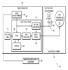
pain symptoms.
Medical devices having arrays of percutaneous electrodes that utilize
microstructure needles,
which are less invasive than deeper-penetrating acupuncture needles, have also
been used for
delivering PENS. The microstructure needles provide sufficient penetration to
overcome the
electrical impedance of the skin tissue for effectively recruiting sensory
fibers.
[0003] As the understanding of the topographical organization of nociceptive
systems becomes
more detailed, the target location of the stimulation, the percutaneous
electrodes' depth of
penetration, and the current amplitude become more exacting. Percutaneous
neuromodulation
therapy (PNT) and cutaneous field stimulation (CFS) are specific forms of PENS
that have been
developed using that understanding. PNT is used for the treatment of cervical
and lumbar pain
1
CA 2797078 2017-05-04
and utilizes longer, acupuncture-type needles having a depth of penetration
into the skin tissue of
up to 3 cm. And, CFS is used more generally to treat pain and itch and
utilizes an array of
microstructure needles introduced close to the nerve endings in the skin.
Because of the
stringent requirements established for needle electrodes by the Food and Drug
Administration
(FDA) regarding the packaging, sterilization, reuse, and disposal of such
electrodes, treatments
utilizing such electrodes have generally been administered under the
supervision of a physician
= (e.g., in a doctor's office or a clinic).
[0004] CFS is used to assist in the management of chronic nociceptive and
neuropathic pain
based on the understanding that specific types of sensory nerves that are
linked to diminishing
the perception of pain can be activated by low amplitude, long duration
electrical stimulation if
electrodes having sharp tips (i.e., microstructure needles) are introduced
close to the nerve
= endings in the skin. CFS treatment also influences specific active
components necessary for
perceiving itch by inducing long lasting inhibitory mechanisms in central
pathways and by
actually normalizing the number of epidermal sensory fibers in itchy skin.
Accordingly, CFS
also provides an alternative to known treatments for localized itch.
[0005] The sensory receptors stimulated by CFS are axons within the skin
tissue known as
nociceptors, specifically M and C nerve fibers. The stimulation of AO and C
nerve fibers,
although effective in diminishing the perceptions of both pain and itch, can
be a relatively
uncomfortable treatment because a prickling and/or burning sensation is
perceived from the
stimulation of the M and C nerve fibers, which can be painful. Because the
aversiveness of M
and C nerve fiber stimulation can be masked by Al3 fiber stimulation, it would
be a considerable
advantage to combine AP fiber stimulation (e.g., transcutaneous electrical
nerve stimulation
(TENS)) and AO and C fiber stimulation (e.g., CFS) in the same equipment.
Accordingly, there
2
CA 2797078 2017-05-04
is a need for a method and device that combines AP fiber stimulation and M and
C fiber
stimulation in one treatment. Moreover, there is a need for a method and
device that combines
TENS and CFS in one treatment.
BRIEF DESCRIPTION OF THE DRAWINGS
[0006] Many aspects of the present invention can be better understood with
reference to the
following drawings, which are part of the specification and represent
preferred embodiments of
the present invention. The components in the drawings are not necessarily to
scale, emphasis
instead being placed upon illustrating the principles of the present
invention. And, in the
drawings, like reference numerals designate corresponding parts throughout the
several views.
[0007] FIG. 1 is a schematic view of an electrotherapy system in accordance
with a non-limiting
embodiment of the present invention;
[0008] FIG. 2 is an isometric view a pulse generator and electrode carrier in
accordance with a
non-limiting embodiment of the invention;
[0009] FIGs. 3A through 3F are elevational views taken in section of different
non-limiting
embodiments of skin-penetrating electrodes with stop nodules according to the
present invention;
[0010] FIG. 4 is an isometric view of an electrode carrier in accordance with
a non-limiting
embodiment of the present invention;
[0011] FIG. 5 is an isometric view of an electrode carrier in accordance with
another non-
limiting embodiment of the present invention;
[0012] FIGs. 6A and 6B are elevational views taken in section of a one-piece
electrode carrier
comprising an antimicrobial agent according to non-limiting embodiments of the
invention;
[0013] FIG. 7 is an elevational view taken in section of a two-piece electrode
carrier comprising
a circuit board and disposable interface in accordance with a non-limiting
embodiment of the
3
CA 2797078 2017-05-04
present invention;
[0014] FIG. 8 is a plan view of the front face of an embodiment of the circuit
board of FIG. 5;
[0015] FIG. 9 is a plan view of the front face of another embodiment of the
circuit board of FIG.
5;
[0016] FIG. 10 is a plan view of the front face of yet another embodiment of
the circuit board of
FIG. 5;
[0017] FIG. 11 is a plan view of the front face of the electrode carrier of
FIG. 5 in accordance
with a non-limiting embodiment of the present invention;
[0018] FIG. 12 is a plan view of the front face of the electrode carrier of
FIG. 5 in accordance
with another non-limiting embodiment of the present invention;
[0019] FIG. 13 is an isometric view of an electrotherapy system in accordance
with a non-
limiting embodiment of the present invention;
[0020] FIG. 14 is a side view taken in section of skin-penetrating and surface
skin electrodes
prior to being applied to a patient's skin;
[0021] FIG. 15 is a side view taken in section of skin-penetrating and surface
skin electrodes
applied to a patient's skin;
[0022] FIG. 16 is a graph illustrating the waveform of a train of pulse bursts
of biphasic pulsed
current in accordance with a non-limiting embodiment of the present invention;
[0023] FIG. 17 is a graph illustrating the waveform of a single monophasic
pulse of electrical
current in accordance with a non-limiting embodiment of the present invention;
[0024] FIG. 18 is a graph illustrating the waveform of a pulse train of
monophasic pulsed current
made up of a plurality of the pulses of FIG. 18; and
[0025] FIG. 19 is a graph illustrating the waveform of a combination of the
waveform of FIG. 16
4
CA 2797078 2017-05-04
and the waveform of FIG. 18 in accordance with a non-limiting embodiment of
the present
invention.
DETAILED DESCRIPTION OF THE PREFERRED EMBODIMENTS
[0026] Non-limiting embodiments of the present invention will now be described
in detail, by
way of example, with reference to the drawings.
[0027] Turning to the figures, FIG. 1 illustrates a non-limiting embodiment of
an electrotherapy
system 100 for stimulating sensory nerves within skin tissue. The
electrotherapy system 100
includes a multi-channel pulse generator 102, an electrode carrier 104, and a
combination
disinfecting/recharging mechanism 106. The pulse generator 102 includes an
electrical circuit
108 that is configured to transmit pulsed currents into a patient's skin via
skin-penetrating
electrodes 110 (e.g., percutaneous electrodes) and surface skin electrode(s)
112 (e.g., conductive
plate electrodes) disposed on the electrode carrier 104. The skin penetrating
electrodes 110 are
configured to apply electrical stimulation (i.e., electro-stimulation)
percutaneously to M and C
nerve fibers, and the surface skin electrode(s) 112 are configured to apply
electrical stimulation
transcutaneously to All nerve fibers prior to and/or overlapping in time with
the electrical
stimulation applied to the M and C nerve fibers. And, the
disinfecting/recharging mechanism
106 functions to reduce microbial reproduction on the skin-penetrating
surfaces of the skin-
penetrating electrodes 110 and the skin-contacting surfaces of the electrode
carrier 104 and the
surface skin electrode(s) 112 in between treatment applications. The
disinfecting/recharging
mechanism 106 may also function to recharge the pulse generator's 102 power
source 114 in
between treatment applications, concurrently with or separate from its
disinfecting operation.
Multi-Channel Pulse Generator 102
[0028] As FIG. 1 illustrates, the multi-channel pulse generator 102 includes
an electrical circuit
108, a power source 114, and a key pad 116. The electrical circuit 108
includes a voltage
CA 2797078 2017-05-04
converter 118, a microcontroller unit (MCU) 120, shift registers 122, an
output stage 124, and an
electrically erasable programmable read-only memory (EEPROM) 126. The voltage
converter
118 is a one Megahertz oscillator that feeds a voltage multiplier circuit (not
shown) to boost the
power source 114 voltage to approximately 50 Volts. Any suitable voltage
converter 118 that
converts complementary metal¨oxide¨semiconductor (CMOS) logic to analog may be
used. The
MCU 120 monitors the key pad 116 input, provides timing sequence to the shift
registers 122,
and executes instructions from the firmware stored in the EEPROM 126. The MCU
120 may be
PIC-based. The shift registers 122 provide the logic used for clocking the
output pulse timing to
the output stage 124. The output stage 124 includes a series of transistors
that couple the power
source 114 voltage to the electrode carrier 104.
[0029] Memory is stored via the EEPROM 126. The EEPROM 126 can be any suitable
nonvolatile memory device. Also, the EEPROM 126 may provide memory storage for
a data
logging function (not shown). The data logging function can be used to record
treatment uses,
durations, amplitude outputs, and other user/patient/subject information, such
that a
manufacturer, a sponsor of a clinical investigation, or a prescribing
physician may query the
EEPROM 126 to obtain that information. Other non-limiting configurations of
the pulse
generator 102 and firmware may also be employed by the present invention. And,
the pulse
generator's 102 components may be further integrated into a field programmable
gate array (not
shown) with internal flash memory.
[0030] As FIG. 1 illustrates, the power source 114 is made up of three AAA 1.5
Volt alkaline
batteries which provide approximately 5 to 50 Volts per channel, but the power
source 114 may
be any conventional rechargeable battery or batteries, including rechargeable
lithium polymer
batteries (not shown). The power source 114 may also be any other suitable
voltage source, such
6
CA 2797078 2017-05-04
as a conventional outlet plug, solar panel, etc.
[0031] As FIG. 1 also illustrates, a pulse conditioning circuit 128 is
provided between the pulse
generator 102 and the surface skin electrode(s) 112. The pulse conditioning
circuit 128 may be
disposed on the electrode carrier 104. The pulse conditioning circuit 128
allows more accurate
positioning of the active portions of the skin-penetrating electrodes 110 for
effectively
stimulating A6 and C nerve fibers. Several factors effect whether the skin-
penetrating electrodes
110 will generate a sufficient voltage gradient to effectively stimulate A8
and C nerve fibers.
For example, load varies based on the skin-penetrating electrode's 110
distance from a nerve,
with impedance decreasing as the needle tip approaches the nerve, and the
resistance/capacitance
of a patient's skin tissue may differ between patients or for different skin
locations on the same
patient. Thus,
the voltage gradient created by the skin-penetrating electrodes 110 is
unpredictable and highly dependent on the positioning of the skin-penetrating
electrodes 110.
[0032] In order to provide a predictable voltage gradient for different loads
and different skin
resistances/capacitances, the pulse conditioning circuit 128 is placed in
series with the electrical
path through a patient's skin to maintain the desired voltage gradient to
effectively stimulate A6
and C nerve fibers. To create that electrical path, one or more surface skin
electrodes 112 can be
employed with the reverse polarity of the skin-penetrating electrodes 110 so
that it operates as a
collector for the skin-penetrating electrodes 110. In that configuration, the
pulse conditioning
circuit 128 is located on the return electrical pathway between the surface
skin electrode 112 and
the pulse generator 102. In the alternative, one or more skin-penetrating
electrodes 110 can
operate as a collector for the other skin-penetrating electrodes 110.
[0033] The pulse conditioning circuit 128 maintains the desired voltage
gradient by maintaining
a constant waveform across the skin-penetrating electrodes 110 and the
collector electrodes.
7
CA 2797078 2017-05-04
Preferably, the pulse conditioning circuit 128 is configured to approximate a
relatively
rectangular waveform (e.g., FIGs. 17 and 18) when delivered through the
patient's skin tissue.
And, the pulse conditioning circuit 128 maintains a constant waveform by
maintaining a linear
relationship between the voltage and current components of the waveform based
on the
impedance characteristics of the patient's skin tissue. But, as discussed
above, the electrical
characteristics of skin tissue may change between patients or even between
locations on a single
patient's skin. Accordingly, the characteristics of the pulse conditioning
circuit 128 may also
need to change.
[0034] Although FIG. 1 illustrates the pulse conditioning circuit 128 as a
capacitor 130 in
parallel with a resistor 132, a more complex circuit can be employed. For
example, the pulse
conditioning circuit may include a semiconductor field effect transistor, a
digital signal
processor, an inductor, and other active semiconductor components so that the
circuit
characteristics of the pulse conditioning circuit 128 can be adjusted to
maintain the desired
= waveform across patients' skin tissue as the electrical characteristics
of the patients' skin tissue
change. Accordingly, the pulse generator 102 may also include a circuit (not
shown) for
measuring values of voltage and current across a patient's skin tissue to
determine the impedance
of the patient's skin. Based=on that measurement, a digital computer (not
shown) in the pulse
generator 102 can be used to automatically adjust the components of the pulse
conditioning
= circuit to maintain the desired waveform through the patient's skin as
the impedance of the skin
tissue fluctuates, thereby maintaining the desired voltage gradient. In the
alternative, the patient
can adjust the circuit characteristics of the pulse conditioning circuit 128
manually.
[0035] The key pad 116 may be any suitable operator key pad for patient input
having a display
to indicate the status and output of the electrotherapy system 100. The key
pad 116 provides a
8
CA 2797078 2017-05-04
user interface to control the programming and function of the pulse generator
102. As
illustrated, for example, in FIG. 2, the key pad 116 may include positive and
negative toggle
keys 200 for controlling the amount of electro-stimulation output, a series of
LEDs 202 for
displaying the level of electro-stimulation output, and a power button 204 for
turning the
electrotherapy system 100 on and off.
[0036] As illustrated in FIG. 2 illustrates, the pulse generator 102 is
provided physically separate
but electrically connected to the electrode carrier 104 by an electrode cable
206. A cable-plug
assembly 208 is provided to detachably connect the pulse generator 102 to the
surface skin
electrode(s) 112 and the skin-penetrating electrodes 110 via the electrode
cables 208. The output
stage 124 of the electrical circuit 108 may be disposed on the electrode
carrier 104 or in the pulse
generator 102. The pulse generator 102 may be constructed in a housing made of
any suitable
material, such as a polycarbonate/ABS blend, when it is provided physically
separate from the
electrode carrier 104. In the alternative, the pulse generator 102 may be
formed or mounted on
the rear face of electrode carrier 104 (e.g. FIG. 13).
[0037] The embodiment of the electrode carrier 104 illustrated in FIG. 2
includes two rows of
three and two rows of four skin-penetrating electrodes 110 with three rows of
three surface skin
electrodes 122 interspersed therebetween. Thus, that electrode carrier 104
includes an array of
fourteen (14) skin-penetrating electrodes 110 and nine (9) surface skin
electrodes 112. Each
individual skin-penetrating electrode 110 and each individual surface skin
electrodes 112 is
electrically connected to the pulse generator via a separate channel for
effecting current transfer
through each of the electrodes 110 and 112. Accordingly, in the embodiment
illustrated in FIG.
2, the multi-channel pulse generator 102 includes at least twenty-three
channels (one for each of
the fourteen skin-penetrating electrodes 110 and one for each of the nine
surface skin electrodes
9
CA 2797078 2017-05-04
112). The pulse generator 102 can be similarly configured for virtually any
number of electrodes
110 and 112 and corresponding channels.
Electrode Carrier 104
[0038] The electrode carrier 104 is made of thin and flexible, but not
extendable or
compressible, polycarbonate. The electrode carrier 104 is substantially flat
yet conformable and
shapeable to the skin tissue such that it can be applied to most body parts.
It is also possible for
the electrode carrier 104 to be made of less pliable, polymer materials in
order to provide more
rigidity. For example, the electrode carrier 104 can be a printed circuit
board (PCB), as
conventionally known in the fabrication and manufacture of appliances for
electro-stimulation
and in the delivery and administration of electrotherapy.
[0039] As FIG. 2-5 illustrate, each of the skin-penetrating electrodes 110 is
embedded in a stop
nodule 210 so that only a skin-penetrating portion 212 extends from the
annular surface of the
stop nodule 210. The stop nodules 210 advance the skin-penetrating portion 212
of the skin-
penetrating electrodes 110 further toward a patient's skin tissue by
functioning as a spacer
between the front side of the electrode carrier 104 and a patient's skin. The
stop nodule 210 also
enables the skin-penetrating portions 212 of the skin-penetrating electrodes
110 to penetrate a
patient's skin a predetermined depth when pressure is applied from above by
providing a blunt
contact surface that makes contact with the patient's skin and stops the skin-
penetrating portion
212 from penetrating the patient's skin any further beyond that point of
contact.
[0040] To provide a blunt enough contact surface to control the depth that the
skin-penetrating
portion 212 of the skin-penetrating electrodes 110 penetrates a patient's
skin, the stop nodules
210 have a cross-sectional surface area of about 0.2 to 25 mm2, preferably
about 3 mm2. The
distal end of each stop nodule 210 is preferably a convex shape to provide the
optimal amount of
skin contact for controlling the depth that the skin-penetrating portion 212
of the skin-penetrating
CA 2797078 2017-05-04
electrodes 110 penetrates a patient's skin. For example, the distal end of the
stop nodule 210
may be domed (e.g., FIGs. 2, 3A, 3B, and 4), conical (e.g., FIGs. 3C and 3D),
or substantially
flat (e.g., FIGs. 3E, 3F, 5, 6A, and 6B). When the distal end of the stop
nodule 210 is conical,
the angle a between the skin-penetrating portion 212 of the skin-penetrating
electrode 110 and
the stop nodule 210 preferably does not exceed 160 for satisfactorily
controlling the depth that
the skin-penetrating portions 212 of the skin-penetrating electrodes 110
penetrate a patient's
skin. Larger angles a result in a greater depth of skin penetration.
[0041] The cross-sectional surface area of each skin-penetrating portion 212
should be
sufficiently small such that it will penetrate a patient's skin under the
exertion of pressure
without causing significant skin injuries. Accordingly, the cross-sectional
surface of the skin-
penetrating electrodes 110 should be about 0.065 to 0.4 mm2. The tip of each
skin-penetrating
portion 212 may be pointed at an angle less than 90 , preferably less than 45
, to further reduce
skin injuries. The tips of the skin-penetrating portions 212 may be perfectly
conical (e.g., FIGs.
2, 3A, 3C, 3E, 4, 5, 6A, and 6B) or convexly/concavely conical pointed (e.g.,
FIGs. 7, 14, and
15), they may have a cutting edge (not shown), or they may have the shape of a
needle or a pin
(e.g., FIGs. 3B, 3D, and 3F).
[0042] The skin-penetrating electrodes 110 are also designed to penetrate a
patient's skin
sufficiently to achieve the desired stimulation of skin receptors. More
particularly, the skin-
penetrating portions 212 of the skin-penetrating electrodes 110 have a
sufficiently small non-
insulated, "active" surface area for providing the high electrical current
density required to
activate and recruit AS and C nerve fibers, but are long enough to reach a
depth of skin
penetration at which AS and C nerve fibers can be activated and recruited.
Accordingly, when
the overall length required to reach the desired depth of skin penetration
results in too much
11
CA 2797078 2017-05-04
active surface area on the skin-penetrating portions 212, it may be necessary
to insulate a portion
of the skin-penetrating portions 212 along their length so that only a small
active surface area is
exposed at their tips (e.g., FIGs. 14 and 15). The active length of the skin-
penetrating portions
212 should be about 0.1 to 0.5 mm.
[0043] The depth of skin penetration desired will depend on the type of skin
being treated and
the location of the M and C nerve fibers being targeted. And, because the stop
nodules 210
advance the skin-penetrating portions 212 of the skin-penetrating electrodes
110 further toward a
patient's skin tissue, different combinations of dimensions for the stop
nodules 210 and the skin-
penetrating portions 212 may be used to achieve that desired depth. For
example, the skin-
penetrating portions 212 of the skin-penetrating electrodes 110 may have a
length from base to
tip of about 0.1 to 5.0 mm, preferably about 0.2 to 3.0 mm, and the stop
nodules 210 may have a
height from base to distal end of about 0.1 to 5.0 mm. Moreover, both the
heights and cross-
sectional surface areas of the stop nodules 210 may be changed depending on
the electrode
density and the curvature of the skin tissue being treated to help achieve the
desired depth of
penetration.
[0044] The stop nodules 210 may be made of non-conductive material, such as UV
stabilized
polycarbonate/ABS, so that current is only transferred to a patient's skin via
the skin-penetrating
portions 212 of the skin-penetrating electrodes 110. If the stop nodules 210
are made of an
electrically conductive material, the skin-penetrating portions 212 of the
skin-penetrating
electrodes 110 should be electrically insulated from the stop nodules 210. The
skin-penetrating
electrodes 110 may be made from silver, platinum and other noble metals,
stainless steel blanks,
commercially available stainless steel hypodermic needles cut and shaped to a
desired length,
and combinations thereof. The skin-penetrating electrodes 110 may further be
plated with
12
CA 2797078 2017-05-04
conductive metals if desired. The stop nodule 210 may be molded around the
skin-penetrating
electrode 110 or formed separately and later assembled with the skin-
penetrating electrode 110
such that the skin-penetrating electrodes 110 are removable and replaceable in
the electrode
carrier 104.
[0045] The surface skin electrodes 112 may be any suitable conventional
surface skin electrode
with an adhesive interface for application to skin tissue. Such surface skin
electrodes 112 are
conventionally known for use in applying transcutaneous electrical nerve
stimulation (TENS).
The surface skin electrodes 112 can be made of metal, carbonized silicon, or
other conductive
polymers. The surface skin electrodes 112 should have a large conductive
diameter to provide
the lower electrical current densities required to activate and recruit Af3
fibers. For example, the
surface skin electrodes 112 should have a surface area, or a combined surface
area for linked
rows H' or columns V' (e.g., FIGs. 9 and 10), of more than 100 mm2. The
surface skin
electrodes 112 can act as return or collector electrodes of opposite polarity
from the skin-
penetrating electrodes 110 or other surface skin electrodes 112 during the
application and
delivery of electrotherapy and electro-stimulation.
[0046] The array of skin-penetrating electrodes 110 may be of substantially
any shape, including
asymmetrical arrangements, and may include one hundred skin-penetrating
electrodes 110 or
more. Such arrays may include a plurality of surface skin electrodes 112
interspersed between
the skin-penetrating electrodes 110 (e.g., FIGs. 2, 4, and 5) on the electrode
carrier 104 or as a
frame surrounding the perimeter of the skin-penetrating electrodes 110 on the
electrode carrier
104 (not shown). The surface skin electrodes 112 should be sized and spaced
relative to the
skin-penetrating electrodes 110 based on the size of the array of skin-
penetrating electrodes 110
and the number of skin-penetrating electrodes 110.
13
CA 2797078 2017-05-04
[0047] As FIGs. 4 and 5 illustrate, the skin-penetrating electrodes 110 may be
disposed on the
electrode carrier 104 in a rectangular array defined by columns V and rows H
that are spaced
apart from one another by about 10 mm or more so as to form a field of
stimulation. The surface
skin electrodes 112 may be disposed on the electrode carrier 104 in a
rectangular array defined
by columns V' and rows H' disposed between the columns V and rows H of skin-
penetrating
electrodes 110. The horizontal distance between each surface skin electrode
112 and its
neighboring skin-penetrating electrode 110 may be about 1 to 30 mm. If the
surface skin
electrode 112 is not disposed on the electrode carrier 104 with the skin-
penetrating electrodes
110, the surface skin electrode 112 should be positioned at a distance close
enough to the array
of skin-penetrating electrodes 110 for the surface skin electrode 112 to serve
as a collector for
the electro-stimulation applied via the array of skin-penetrating electrodes
110. And, instead of
extending from stop nodules 210 to advance the skin-penetrating portions 212
further toward a
patient's skin tissue as illustrated, for example, in FIG. 4, the skin-
penetrating portions 212 may
also extend from raised crest sections 506 as illustrated, for example, in
FIG. 5.
[0048] In FIG. 5, side walls 500 and 502 are formed in the electrode carrier
104 on opposite
sides of each row H of skin-penetrating electrodes 104 so as to form valley
sections 504 and the
crest sections 506. The surface skin electrodes 112 are disposed in the valley
sections 504
between side walls 500 and 502 and the skin-penetrating electrodes 110 are
disposed on the crest
sections 506 above the surface skin electrodes 112. Accordingly, just as with
skin-penetrating
electrodes 110 extending from stop nodules 210 (e.g., FIG. 4), when the
electrode carrier 104 is
applied to a patient's skin by exerting pressure on its rear face, the skin-
penetrating electrodes
110 disposed on the crest sections 506 will extend further toward the surface
of the patient's
skin. The side walls 500 and 502 may be constructed of stretchable material
such that they bend
14
CA 2797078 2017-05-04
and the electrode carrier 104 conforms to the skin tissue of a patient's
various curved body parts,
such as the knees, elbows, feet. And, the side walls 500 and 502 may be
substantially straight
(e.g., FIG. 5) or they may be curved (e.g., FIG. 14).
[0049] As FIGs. 6 and 7 illustrate, the electrode carrier 104 comprises a
circuit board 600
surrounded by a non-conductive coating 602. The skin-penetrating electrodes
110 and surface
skin electrodes 112 are disposed in the non-conductive coating 602 and
electrically coupled to
the circuit board 600. The circuit board 600 electrically couples the skin-
penetrating electrodes
110 and surface skin electrodes 112 to the pulse generator 102 via traces 802
and 804 (FIGs. 8-
10) screen-printed on the circuit board 600, via individually insulated wires,
or via any other
suitable electrical connection. The non-conductive coating 602 is provided as
a substrate pad
surrounding the electrode carrier 104 to prevent current from passing into a
patient's skin from
any conductive element of the electrode carrier 104 other than the skin-
penetrating electrodes
110 and/or surface skin electrodes 112. And, by surrounding the circuit board
600 with a non-
conductive coating 602, any electrical components disposed on the circuit
board 600 are
protected from damage during certain disinfecting operations, such as boiling
or autoclaving.
[0050] The non-conductive coating 602 may be made of any non-conductive
thermoplastic
elastomer material that is suitable for protecting and insulating integrated
circuits and integrated
circuit components and for use in contact with skin tissue during the delivery
and administration
of electro-stimulation and/or electrotherapy. The preferred material should
produce a cleanable,
hypoallergenic substrate that is supple and conformable to the skin tissue.
The preferred material
may also need to be capable of withstanding high temperatures so that the
electrode carrier 104
can be boiled or placed in an autoclave to disinfect it. Such materials
include, but are not limited
to, styrene-ethylene/butylene-styrene (SEBS) polymers.
CA 2797078 2017-05-04
[0051] As FIGs. 6A and 6B illustrate, an antimicrobial agent 604 can be
layered on the non-
conductive coating 602 (FIG. 6A) or infused within the non-conductive coating
602 (FIG. 6B) to
provide an antimicrobial microatmosphere surrounding the skin-penetrating
electrodes 110 and
other skin-contacting surfaces of the electrode carrier 104. The skin-
penetrating electrodes 110
are infused with the antimicrobial agent 604 in either configuration. The
antimicrobial agent 604
can retard, control, kill, and/or prevent microbial contamination in addition
to or in lieu of the
disinfecting/recharging mechanism 106. In FIG. 6A, the top layer of the skin-
contacting surfaces
imparts the antimicrobial properties of the antimicrobial agent 604, while in
FIG. 6B the
antimicrobial properties are concentrated in zones of inhibition Z
specifically surrounding the
skin-penetrating electrodes 110. It is important to concentrate the
antimicrobial agent 604
around the skin-penetrating electrodes 110 in the latter configuration because
the stop nodules
210 may not be infused with the antimicrobial agent 604. The antimicrobial
agent 604 can also
be layered on or infused within the surface skin electrodes 112, whether or
not they are disposed
on the electrode carrier 104 with the skin-penetrating electrodes 110.
[0052] Any one or a number of metal ions that have been shown to possess
antibiotic activity,
including silver, copper, zinc, mercury, tin, lead, bismuth, cadmium,
chromium, and thallium
ions, may be used in the composition of the antimicrobial agent 604.
Preferably, the
antimicrobial agent 604 is composed substantially of silver in concentrations
that allow the
electrodes 110 and 112 to remain conductive without compromising the
insulating structures that
surround them, such as the stop nodules 210 and non-conductive coating 602,
and compromising
the pathway of the electrical circuit 108.
[0053] As FIG. 7 illustrates, the electrode carrier 104 can also be provided
in a two-piece
configuration wherein the skin-penetrating electrodes 110 and surface skin
electrodes 112 are
16
CA 2797078 2017-05-04
=
provided in a disposable interface 700. The disposable interface 700 is formed
of a non-
conductive material and can be operatively connected to and disconnected from
the circuit board
600 such that the skin-contacting surfaces (i.e., the front face of the
electrode carrier 104 and the
surface skin electrodes 112) and the skin-penetrating surfaces (i.e., the skin-
penetrating
electrodes 110) of the electrode carrier 104 can easily be removed from the
circuit board 600 for
disinfecting and/or replacement. Providing a disposable interface 700 provides
an alternative or
additional safety measure for protecting the electronic components on the
circuit board 600 from
damage during disinfecting operations, such as boiling or autoclaving. It also
allows the skin-
contacting surfaces and skin-penetrating surfaces of the electrode carrier 104
to be commercially
replaceable without also requiring replacement of the circuit board 600 or any
of its associated
components.
[0054] As FIGs. 7-10 illustrate, each skin-penetrating electrode 110 within
the disposable
interface 700 may be electrically coupled to the circuit board 600 via a
corresponding first
electrical coupling 702 disposed on the circuit board 600. Accordingly, the
circuit board 600
includes an array of first electrical couplings 702 disposed thereon in a
rectangular array that is
also defined by columns V and rows H so that each skin-penetrating electrode
110 independently
matches up with its corresponding first electrical coupling 702 when the
disposable interface 700
is disposed on the circuit board 600. Similarly, when the surface skin
electrodes 112 are also
provided in the disposable interface 700, the circuit board 600 also includes
an array of second
electrical couplings 800 defined by columns V' and rows H' so that each
surface skin electrode
112 independently matches up with its corresponding second electrical coupling
800 when the
disposable interface 700 is disposed on the circuit board 600. The skin-
penetrating electrodes
110 illustrated in FIG. 7 are disposed on the crest sections 506 of an
electrode carrier 104 (e.g.,
17
CA 2797078 2017-05-04
FIG. 5) rather than in stop nodules 210 (e.g., FIG. 4), but the disposable
interface 700 may be
constructed in either configuration.
[0055] The disposable interface 700 may also be constructed with the surface
skin electrodes
112 disposed on the circuit board 600 rather than on the disposable interface
700. In that
configuration, the disposable interface 700 will include openings (not shown)
defined by
columns V' and rows H' that align with the surface skin electrodes 112 on the
circuit board 600
so that the surface skin electrodes 112 can make electrical contact with a
patient's skin when the
disposable interface 700 is disposed on the circuit board 600. Also in that
configuration, the
surface skin electrodes 112 may be anchored to the circuit board 600 by any
suitable technique,
such as soldering. And, as yet another alternative, the surface skin
electrodes 112 may be
adhesively attached to the front face of the disposable interface 700 between
the rows of skin-
penetrating electrodes 110 so they can be adhered to and subsequently peeled
off of the
disposable interface 700 so as to allow more freedom in the configuration of
the first electrical
couplings 702 and their associated electrical connections 802 on the circuit
board 600. That
configuration also allows the various components of an electrode carrier 104
to be subjected to
certain disinfecting operations, such as boiling or autoclaving, after peeling
off the surface skin
electrodes 112 when all of the other components of the electrode carrier 104
(e.g., the skin-
penetrating electrodes, the circuit board 600, and the non-conductive coating
602) are configured
to be subjected to that disinfecting procedure and the surface skin electrodes
112 are not. There
may be circumstances when it is more economical to make certain portions of
the electrode
carrier 104 disposable and others not.
[0056] To place the skin-penetrating electrodes 110 and the surface skin
electrodes 112 in
electrical communication with the pulse generator 102, the first electrical
couplings 702 and
18
CA 2797078 2017-05-04
second electrical couplings 800 are electrically connected to the pulse
generator 102 via
independent electrical connections 802 and 804, respectively, so as to
separately connect each
independent electrode 110 and 112 to a separate channel of the pulse generator
102. In the
alternative, the skin-penetrating electrodes 110 and the surface skin
electrodes 112 can be
configured such that each skin-penetrating electrode 110 is coupled in series
to an adjacent skin-
penetrating electrode 110 in the same row H and such that each surface skin
electrode 112 is
coupled in series to an adjacent surface skin electrode 112 in the same row H'
(e.g., FIG. 9).
And, as yet another alternative, the skin-penetrating electrodes 110 and the
surface skin
electrodes 112 can be configured such that each skin-penetrating electrode 110
is coupled in
series to an adjacent skin-penetrating electrode 110 in the same column V and
such that each
surface skin electrode 112 is coupled in series to an adjacent surface skin
electrode 112 in the
same column V' (e.g., FIG. 10). Coupling the rows H and H' or columns V and V'
as described
reduces the number of channels required by the pulse generator 102 to operate
the electrodes 110
and 112, with one channel corresponding to each row H and H' or column V and
V'.
[0057] Each independent electrical coupling 702 and 800 on the circuit board
600 is connected
to the pulse generator 102 via a single, bundled electrode cable 206
comprising an insulated wire
for each channel of the pulse generator 102 used to apply electro-stimulation.
An attachment
mechanism 806, such as an interlocking fabric or double stick tape with peel-
away backing, may
be disposed between the skin-penetrating electrodes 110 and/or the surface
skin electrodes 112
to removably attach the disposable interface 700 to the circuit board 600 so
the disposable
interface 700 can be placed on and subsequently peeled off of the circuit
board 600. The
disposable interface 700 also may be attached to the circuit board 600 via a
mechanical
connection, such as clips or clamps. And, when the skin-penetrating electrodes
110 are disposed
19
CA 2797078 2017-05-09
on the crest sections 506 of the disposable interface 700, they may be
configured to include
circular portions 704 (FIG. 7) for providing additional contact area when
electrically coupling
the skin-penetrating electrodes 110 to their respective first electrical
couplings 702.
[0058] As FIGs. 11 and 12 illustrate, a non-conductive adhesive strip 1100 may
be applied in the
valley sections 504 of the electrode carrier 104 illustrated in FIG. 5 to
assist in adhesive fixation
of the surface skin electrodes 112 and the electrode carrier 104 to a
patient's skin tissue during
treatment use. Non-conductive adhesive strips 1100 may also be disposed along
the outside edge
of the electrode carrier 104 to provide additional adhesion (not shown).. And,
the adhesive strips
may also be disposed between the skin-penetrating electrodes 110 and the
surface skin electrodes
112 in a pattern similar to that of the attachment mechanism 806 illustrated
in FIGs. 8-10. In
each of those configurations,. the non-conductive adhesive strips 1100 must be
arranged so they
do not cover the surface skin electrodes 112 and/or interfere with the
transfer of electric
stimulation to a patient's skin via the surface skin electrodes 112. Instead,
the surface skin
electrodes 112 should be covered with electrically conductive gel or hydrogel
or any
conventional coupling medium (e.g., a non-conductive adhesive through which
current can pass
substantially unobstructed) for enhancing uniform conductivity at the
electrode-skin interface,
and for increasing surface area conductivity. The coupling medium may also be
used to
electrically couple adjacent surface skin electrodes 112 with one another
across the front face of
the electrode carrier 104.
[0059] Both the disposable interface 700 and the circuit board 600 may include
a plurality of
venting bores 1200, illustrated as square holes in FIG. 12, that put a
patient's skin at the front
face of the electrode carrier 104 in fluid communication with the atmosphere
at the rear face of
the electrode carrier 104 so as to ventilate moisture and perspiration that
may be released from
CA 2797078 2017-05-04
the patient's skin while the electrode carrier 104 is disposed thereon ¨
particularly while the
patient is receiving electro-stimulation and/or electrotherapy. To facilitate
ventilation through
both the disposable interface 700 and the circuit board 600, the venting bores
1200 in the
disposable interface 700 are configured to align with corresponding venting
bores 1200 in the
circuit board 600 when the disposable interface 700 is attached to the circuit
board 600. That
alignment allows moisture and perspiration that is released from a patient's
skin while the
electrode carrier 104 is disposed on the patient's skin to escape properly
through the venting
bores 1200. In a one-piece electrode carrier 104 (e.g., FIGs. 6A and 6B), the
venting bores 1200
merely extend all of the way through the electrode carrier 104.
[0060] A wearable applicator (not shown), such as a garment fitted for a
particular body
segment, strap, belt, bandage, splint, stabilizer, supporter, brace or cast
may be used to assist in
the proper positioning and placement of the electrode carrier 104 and
electrodes 110 and 112.
Fasteners, including interlocking fabrics, buttons, snaps, zippers, and the
like can be used to join
the electrode carrier 104 with the wearable applicator such that the electrode
carrier 104 can be
anatomically positioned for therapeutic effectiveness on a wide range of body
parts.
Disinfecting/Recharging Mechanism 106
[0061] The disinfecting/recharging mechanism 106 reduces microbial
reproduction on the skin-
penetrating surfaces of the skin-penetrating electrodes 110 and the skin-
contacting surfaces of
the electrode carrier 104 and surface skin electrodes 112 by applying
germicidal radiation to
those surfaces for a sufficient time and strength to inactivate common skin
pathogens, including
bacteria spores, molds, protozoa, viruses and yeast. In a
preferred embodiment, the
disinfecting/recharging mechanism 106 uses germicidal ultraviolet light to
damage the
pathogens' genetic material, thereby inhibiting the pathogens' replication and
colony formation.
The required dose to inactivate 90% of most types of infection-causing
microbes is within a
21
CA 2797078 2017-05-04
range of about 2 to 6 mJ/cm2. Dosages of UV intensity of about 500 to 1500
W/cm2 for up to
about one hour of exposure time can be sufficient to inactivate the microbes
by damaging their
DNA, and can even destroy the microbes by disrupting their cellular processes.
Accordingly, the
disinfecting/recharging mechanism 106 is configured to apply germicidal
radiation up to
approximately 1000 J/cm2 for several sessions per day (in between electro-
stimulation treatment
uses) over periods of an hour or more.
[0062] As FIG. 13 illustrates, the disinfecting/recharging mechanism 106
includes an upper
casing 1300 and a lower casing 1302 that form a disinfecting chamber 1304
therein that can be
closed off with a UV absorbent lid 1306. A UV Lamp 1308 is disposed in the
disinfecting
chamber 1304 applying germicidal ultraviolet light. The disinfecting chamber
1304 is suitably
sized and dimensioned to position the skin-penetrating surfaces of the skin-
penetrating electrodes
110 and the skin-contacting surfaces of the electrode carrier 104 and surface
skin electrodes 112
at an appropriate distance from the UV lamp 1308 to apply the required amount
germicidal
ultraviolet light to disinfect those surfaces. For example, the disinfecting
chamber 1304 may be
sized and dimensioned so that the skin-penetrating surfaces of the skin-
penetrating electrodes
110 are at a distance of approximately 1 to 5 cm from the UV lamp 1308 when
the array of skin-
penetrating electrodes 110 is positioned within the chamber 1304 with the
front face (i.e., the
side placed against a patient's skin) down so the skin-penetrating electrodes
110 extend toward
the UV lamp 1308. In FIG. 13, the electrode carrier 104 is illustrated with
its front face facing
downward and away from the vantage point from which FIG. 13 was taken so that
the skin-
penetrating electrodes 110 and the surface skin electrodes 112 are not
visible. Instead, only the
rear face (i.e., the side facing away from a patient's skin) of the electrode
carrier 104 is shown.
[0063] A reflector 1310 may disposed between the upper casing 1300 and lower
casing 1302 of
22
CA 2797078 2017-05-04
the disinfecting/recharging mechanism 108 to provide the floor and surrounding
surfaces of the
chamber 1304. The reflector 1310 may be made of aluminum or may have an
aluminum surface.
The reflector 1310 and its reflective surface may be made of any suitable
material known for
producing a relatively high reflectivity index for ultraviolet radiation.
[0064] The disinfecting chamber 1304 may also include any suitable number of
platforms 1312
(shown and hidden) to properly support and position the electrode carrier 104
in the
disinfecting/recharging mechanism 106. The platforms 1312 should be
positioned, sized, and
dimensioned such that there is minimum interference with the skin-penetrating
electrodes' 110
exposure to the germicidal radiation.
[0065] Different configurations of the chamber 1304 and UV lamp 1308 may also
be used. For
example, the UV lamp 1308 may be positioned above the skin-penetrating
electrodes 110 to emit
the radiation in a downward direction. In that configuration, the skin-
penetrating electrodes 110
are positioned within the chamber 1304 on a sliding tray (not shown) with the
front face up,
wherein the sliding tray is used to slide the electrode carrier 104 in and out
of the chamber 1304.
The chamber 1304 may also be constructed and configured to allow electrode
carriers 104 of
different shapes and sizes (e.g., FIGS. 4 and 5), and/or more than one
electrode carrier 104 at a
time, to be irradiated and/or recharged in the disinfecting/recharging
mechanism 106.
[0066] The UV lamp 1308 may be any shaped or non-shaped commercially available
germ-
killing lamp configured to generate radiation in the required UV range. The UV
lamp's 1308
shape may be dependent upon the size and shape of the electrode carrier 104
and the chamber
1304 needed to enclose the electrode carrier 104. In a preferred embodiment,
the UV lamp 1308
is a low pressure mercury vapor lamp having a U-shape that is configured to be
an upside-down
U when positioned in the chamber 1304, but any suitable commercially available
UV lamp
23
CA 2797078 2017-05-04
having a Wattage of approximately 2-6 Watts or more and that is configured to
deliver
germicidal radiation may be used. The wavelength of the electromagnetic
radiation delivered by
the UV lamp 1308 is in the range of about 240 to 280 nanometers, preferable
about 254
nanometers. A medium or high pressure mercury vapor lamp, LED, or laser
capable of
generating the preferred 254 nanometers and other known bands of germicidal
light may also be
used. And, more than one lamp and/or type of lamp may be used in combination.
[0067] In addition to or as an alternative to using germicidal ultraviolet
light to disinfect the
electrode carrier 104, boiling water and/or steam may also be used to
disinfect the electrode plate
104. Accordingly, the disinfecting/recharging mechanism 106 may be configured
with
components for introducing boiling water and/or steam into the chamber 1304.
In that
configuration, the upper casing 1300 and the lid 1306 may include sealing
surfaces (not shown)
= to maintain a seal to withstand the high pressures associated with
autoclaving medical devices.
The disinfecting/recharging mechanism 106 may also be configured to use any
other suitable
disinfecting mechanism.
[0068] The disinfecting aspect of the disinfecting/recharging mechanism 106 is
intended to
enhance the electrotherapy system's 100 outpatient reusability. More
particularly, by providing
such disinfecting functionality, the methods and devices of the present
invention can be
employed with portability for outpatient treatment in a manner prescribed by a
physician. And,
although the electrotherapy system 100 is not intended to be shared from
patient to patient, the
disinfecting/recharging mechanism 106 will also minimize the risk of disease
transmission from
one patient to another, while minimizing the risk from environmental sources
to a patient, should
it be used in that manner.
[0069] In addition to the disinfecting function, the disinfecting/recharging
mechanism 106 may
24
CA 2797078 2017-05-09
serve as a recharging station. Accordingly, the disinfecting/recharging
mechanism 106
illustrated in FIG. 13 includes a pair of recharging conductors 1314
configured to mate with a
corresponding pair of recharging conductors 1316 on the electrode carrier 104
for recharging the
power source 114 (FIG. 1) of the pulse generator 102. As illustrated in FIG.
13, the pulse
generator 102 is disposed on the electrode carrier 104 rather than in an
electrically connected but
physically separate device, as illustrated in FIG. 2. The pulse generator 102
is connected to the
recharging conductors 1316 via electrical connections 1318. The conductors
1316 and electrical
connections 1318 are disposed on the rear face of the electrode carrier 104 to
allow electrical
communication with the conductors 1314 on the disinfecting/recharging
mechanism's 106 lid
= 1306 so the pulse generator 102 can be electrically coupled to the
disinfecting/recharging
mechanism 106 for recharging during periods of non-use.
[0070] Other configurations of conductors 1316 and electrical connections 1318
may also be
used depending on the size and shape of the electrode carrier 104 and
disinfecting/recharging
mechanism 106, as well as the type and recharging load of the system's 100
power source 114
(FIG. 1).
In addition, a separate (i.e., not incorporated into the
disinfecting/recharging
mechanism 106) battery charging station may be used in addition to the
disinfecting/recharging
mechanism 106. And, when the pulse generator 102 is electrically connected to
but physically
separate from the electrode carrier 104, as illustrated in FIG. 2, the
disinfecting/recharging
mechanism 106 may be configured to accommodate the electrode cable 208 (dotted-
line) that
provides electrical communication between the pulse generator 102 and the
electrode carrier 104.
[0071] The electronics for the disinfecting/recharging mechanism 106 are
represented by "E"
and may be housed in the lower casing 1302 of the disinfecting/recharging
mechanism 106. The
disinfecting/recharging mechanism 106 may have a number of electronic
features, including the
=
CA 2797078 2017-05-04
display of outputs for apprising a patient of the percentage that the
disinfection and/or recharging
functions are complete. The disinfecting/recharging mechanism 106 may also
have a separate
= indicator or plurality of indicators that display when the disinfection
function and/or the
recharging function are completed. The disinfecting/recharging mechanism 106
may receive
power for each of its functionalities via a conventional outlet plug 1320 or
any other suitable
power source.
Electrotherapy
= [0072] The electrotherapy system 100 of the present invention provides
temporary relief from
the symptoms of chronic pain by targeting cutaneous thin M and C nerve fibers
while
stimulating A13 nerve fibers to help mask the aversive feeling from the A6 and
C nerve fiber
stimulation. For example, the use of TENS to target Al3 nerve fibers can be
combined with CFS
to help reduce and mask the aversive feeling from the Ao and C nerve fiber
stimulation of CFS.
The combination of TENS with CFS is based on the body's response to different
types of pain.
Electrical impulses in response to acute pain sensations are transmitted to
the brain through
peripheral nerves and the spinal cord. At the time point of an injury, the
signal is transmitted by
nociceptive primary afferent nerve fibers to the dorsal horn of the spinal
cord. Nociceptive
primary afferent neurons belong to the M and C nerve fibers. At the dorsal
horn and in the
spinal cord or its trigeminal analogue, secondary neurons take over by
transferring the signal to
the thalamus and finally to the cerebral cortex. Input in tactile AP nerve
fibers is known to
interact with cutaneous nociceptive-input in the spinal cord and higher
centers causing relief of
pain. Therefore, by targeting the Al3 nerve fibers via the use of TENS, the
aversive sensation
caused by stimulation of the A6 and C nerve fibers via CFS can be masked,
resulting in more
tolerable electrotherapy to assist in the symptomatic relief of chronic pain.
[0073] The electrotherapy system 100 also provides an effective alternative to
known treatments
26
CA 2797078 2017-05-04
of localized histamine-induced itching in a similar manner. Accordingly, the
surface skin
electrodes 112 are configured to apply electro-stimulation to Af3 nerve fibers
and the skin-
penetrating electrodes 110 are configured to apply electro-stimulation to A6
and C nerve fibers.
The pulse generator is configured to transmit pulsed currents into a patients
skin via the skin-
penetrating electrodes 110 and the surface skin electrodes 112.
[0074] Figures 14 and 15 illustrate skin-penetrating electrodes 110 and
surface skin electrodes
112 before and during application to a patient's skin tissue, respectively.
The elements
illustrated in those figures are exaggerated for clarity. As FIG. 14
illustrates, the electrode
carrier 104 includes valley sections 504 and crest sections 506 formed by
rounded side walls 500
and 502. The skin-penetrating portions 212 of the skin-penetrating electrodes
110 extend from
the crest sections 506 and are insulated with an insulating material 1400
along the length of the
skin-penetrating portion 212 so that the amount of "active" length exposed to
the patient's
dermis S3 has a sufficiently small surface area to provide the high electrical
current density
required to activate and recruit A6 and C nerve fibers. There may also be
instances where the
skin-penetrating portions 212 need not extend all the way into the dermis S3
to activate and
recruit A6 and C nerve fibers. But, in no case will the skin-penetrating
portions 212 need to
extend any deeper than the dermis S3.
[0075] As FIG. 15 illustrates, the non-conductive coating 602 of the electrode
carrier 104 abuts
the stratum corneum S1 (i.e., the top layer of the epidermis S2) of the skin
tissue when the
electrode carrier 104 is applied to a patient's skin. In that position, the
skin-penetrating portions
212 of the skin-penetrating electrodes 110 penetrate and extend through the
stratum corneum S 1
and the epidermis S2 into the dermis S3, where the active portion can target
the A6 and C nerve
fibers. The valley sections 504 are also compressed so that the surface skin
electrodes 112 are
27
CA 2797078 2017-05-04
placed in contact with the stratum corneum S1 of the epidermis S2, where they
can
transcutaneously target Ap nerve fibers.
[0076] With the skin-penetrating electrodes 110 and the surface skin
electrodes 112 properly
disposed on a patient's skin as illustrated in FIG. 15, electro-stimulation
can be produced through
any of the skin-penetrating electrodes 110 and/or surface skin electrodes 112.
Preferably, one or
more surface skin electrode(s) 112 is used as a collector electrode for the
skin-penetrating
electrodes 110 and/or the surface skin electrodes 112 that are producing
electro-stimulation. To
avoid current always passing through the same surface skin electrode 112 when
electro-
stimulation is applied via the surface skin electrodes 112, the pulse
generator 102 may be
programmed to alternate between the surface skin electrodes 112 through which
electro-
stimulation is being applied, including alternating which surface skin
electrode(s) 112 is being
used as a collector electrode. If no electro-stimulation is being applied via
the surface skin
electrodes 112, all of the surface skin electrodes 112 may be used as
collector electrode for the
electro-stimulation being applied through the skin-penetrating electrodes 110.
[0077] Electro-stimulation may be applied via a surface skin electrode 112
that is phase locked
with the electro-stimulation applied via a neighboring skin-penetrating
electrode 110. The
electro-stimulation applied via the surface skin electrodes 112 generates
signals produced in A13
nerve fibers and the electro-stimulation applied via the skin-penetrating
electrodes 110 generates
signals produced in the AS and C nerve fibers. The two types of electro-
stimulation are phase
locked so that the signals produced in AP nerve fibers will arrive at the
patient's spinal cord prior
to and/or overlapping in time with the signals produced in the AS and C nerve
fibers.
[0078] Pairs and/or other combinations of skin-penetrating electrodes 110 and
surface skin
electrodes 112 can be activated consecutively in either a random or orderly
pattern. For
28
=
CA 2797078 2017-05-04
example, a random, non-consecutive pattern of electro-stimulation can be
applied by alternately
activating one or more column V' or row H' of surface skin electrodes 112
prior to and/or
overlapping in time with a random skin-penetrating electrode 110 or with a
combination of skin-
penetrating electrodes 110. And, an orderly, consecutive pattern of electro-
stimulation can be
applied by consecutively activating phase locked pairs of surface skin
electrodes 112 and skin-
penetrating electrodes 110 in a sequence starting at one side (i.e., an edge)
of the electrode
carrier 104 and proceeding to. the other side of the electrode carrier 104.
[0079] The non-consecutive pattern of electro-stimulation creates a sensation
of massaging
stimulation that is therapeutically effective in providing electroanalgesia
for the treatment of
pain. And, the consecutive pattern of electro-stimulation creates a sensation
of a sweeping
stimulation that mimics the sequence of stimulation that occurs naturally when
scratching or
massaging the skin, which is particularly useful in treating patients
suffering from chronic pain
or itch. Both of those patterns can be achieved with a configuration of skin-
penetrating
electrodes 110 and surface skin electrodes 112 such as that provided for in
FIG. 8, wherein each
of the skin-penetrating electrodes 110 and surface skin electrodes 112 has a
separate electrical
coupling 800 and 702, respectively, such that a different channel of the pulse
generator 102 can
be used to separately control each skin-penetrating electrode 110 and each
surface skin electrode
112.
[0080] Non-consecutive and consecutive patterns of electro-stimulation may
also be achieved
with a configuration of skin-penetrating electrodes 110 and surface skin
electrodes 112 such as
that provided for in FIGs. 9 or 10, wherein the skin-penetrating electrodes
110 and the surface
skin electrodes 112 are connected in series into separate rows H and H' or
columns V and V',
respectively. The non-consecutive pattern of electro-stimulation can be
applied by alternately
29
CA 2797078 2017-05-04
activating one or more column V' or row H' of surface skin electrodes 112
prior to and/or
overlapping in time with a random column V or row H of skin-penetrating
electrodes 110. And,
the consecutive pattern of electro-stimulation can be applied by consecutively
activating paired
columns V and V' or rows H and H' of skin-penetrating electrodes 110 and
surface skin
electrodes 112 in a sequence starting at one side of the electrode carrier 104
and proceeding to
the other side of the electrode carrier 104.
[0081] The surface skin electrode 112 can be used to target AP nerve fibers
within a patient's
skin tissue using a biphasic pulsed current comprising pulse trains with pulse
durations Ti of
about 0.05 to 0.30 milliseconds and a pulse string frequency of about 50 to
400 Hertz. The
biphasic pulsed current may be applied in a continuous pulse string within a
predefined period
(e.g., 100 pulses of 0.25 millisecond duration applied over 1000 milliseconds
at a continuous
frequency of 100 Hz) or broken up into bursts of pulses over a predefined
period. When applied
as bursts of pulses, the biphasic pulsed current has a burst duration of up to
about 100
milliseconds and a burst frequency of about 0.1 to 10 Hertz. The biphasic
pulsed current has a
current amplitude of up to about 50 milliamperes. The waveform of the biphasic
pulsed current
used to target AP nerve fibers may be either symmetric or asymmetric.
[0082] FIG. 16 illustrates an exemplary asymmetric biphasic waveform 1600 that
can be used by
the present invention to target Ap nerve fibers via the surface skin
electrodes 112. That biphasic
waveform 1600 has a period of one second and generates a current with a pulse
duration T1 of
0.15 milliseconds and an interpulse interval T2 of 9.85 milliseconds that form
pulse bursts having
a burst duration T3 of 50 milliseconds and an interburst interval T4 of 200
milliseconds. The
intraburst, or pulse string frequency within the burst, is 100 Hertz (i.e.,
100 pulses of 10
millisecond duration per second), and the burst frequency is 4 Hertz (i.e., 4
bursts of 250
CA 2797078 2017-05-04
millisecond duration per second).
[0083] The skin-penetrating electrodes 110 can be used to target AS and C
nerve fibers within a
patient's skin tissue using a monophasic pulsed current comprising continuous
pulse trains with
pulse durations T1 of about 0.5 to 10.0 milliseconds, a pulse string frequency
of about 0.1 to 10
Hertz, and a current amplitude of up to about 2 milliamperes. The longer pulse
durations T1 are
useful for the recruitment of C nerve fibers. And, by staggering the
monophasic pulsed current
across different skin-penetrating electrodes 110, the overall frequency of
stimulation can be
increased over the field of stimulation. For example, if the monophasic pulsed
current has a
frequency of 4 Hz, an electrotherapy system 100 having fourteen (14) skin-
penetrating electrodes
can apply eleetro-stimulation with a frequency of approximately 56 Hertz
(i.e., 14 electrodes x 4
Hz = 56 Hz).
[0084] FIG. 17 illustrates an exemplary waveform 1700 of an individual
monophasic pulse that
can be used by the present invention to target AS and C nerve fibers within a
patient's skin tissue
via the skin-penetrating electrodes 110. The characteristics of the pulse
conditioning circuit 128
in the return electrical pathway between the skin-penetrating electrodes 110
and surface skin
electrodes 112 cause the waveform 1700 of that pulse to approximate a
rectangular wave. That
waveform 1700 has a pulse duration Ti of 1.0 millisecond and a current
amplitude varying from
about 0.8 to 1.2 milliamperes from the pulse onset. The current amplitude is
at its maximum
value for less than 0.1 milliseconds after the pulse onset. Preferably, the
maximum current
amplitude will be about 0.5 to 2 milliamperes and will last a maximum of about
0.25
milliseconds after the pulse onset. The maximum current amplitude can then be
reduced by
about 5 to 50 percent for the remainder of the pulse duration. The current
amplitude in
milliamperes is measured as a function of time in milliseconds.
31
CA 2797078 2017-05-04
[0085] FIG. 18 illustrates an exemplary waveform 1800 of a train of the
monophasic pulses
illustrated in FIG. 17 with a period of one second. The pulse duration T1 is
1.0 milliseconds, the
interpulse interval T2 is 249 milliseconds, and the frequency is about 4 Hertz
(i.e., 4 pulses per
second). Accordingly, the monophasic pulsed current is applied as a continuous
pulse string
rather than in pulse bursts.
[0086] FIG. 19 illustrates an embodiment of a waveform 1900 of current that
can be driven
through a patient's skin by the electrotherapy system 100 using a combination
of the asymmetric
biphasic waveform 1600 applied by the surface skin electrodes 112 and the
train of monophasic
approximate square waveforms 1800 applied by the skin-penetrating electrodes
110. As
illustrated, the waveforms 1600 and 1800 are applied so that the individual
monophasic pulses
1700 generated with the skin-penetrating electrodes 110 occur alternatively in
time between the
asymmetric biphasic waveform 1600 generated with the surface skin electrodes
112. In the
alternative, the individual monophasic pulses 1700 generated with the skin-
penetrating
electrodes 110 may be applied so as to overlap in time with the asymmetric
biphasic waveform
1600 generated with the surface skin electrodes 112, or the asymmetric
biphasic waveform 1600
generated with the surface skin electrodes 112 can occur prior in time (and/or
subsequent in
time) to the monophasic approximate square waveforms 1800 generated with the
skin-
penetrating electrodes 110. Each of those different combinations of waveforms
1600 and 1800,
and iterations thereof, may also be used in combination with each other. The
illustrated
waveforms 1600, 1800, and 1900 are not to scale, with the size of the
individual monophasic
pulses 1700 being exaggerated for clarity.
[0087] Accordingly, a combination of waveforms 1600 and 1800 that more similar
to the
waveform 1900 illustrated in FIG. 19 may be required to ensure that the
waveforms 1600 and
32
CA 2797078 2017-05-04
1800 from the surface skin electrodes 112 and skin-penetrating electrodes 110,
respectively,
arrive at the patient's spinal cord at the same time and produce the desired
masking effect.
[0088] During a treatment session, a patient can use the pulse generator 102
to begin applying
the asymmetric biphasic waveform 1600 with the surface skin electrodes 112.
While applying
the asymmetric biphasic waveform 1600 with the surface skin electrodes 112,
the patient can
then gradually begin applying the monophasic approximate square waveforms 1800
with the
skin-penetrating electrodes 110. The patient can increase the stimulation
applied with the skin-
penetrating electrodes 110 in gradual steps during the first minutes of a
treatment session using
the toggle keys 200 on the pulse generator, which allows the patient to adapt
to the signals
produced by those pulsed currents to a comfortable level as treatment is
applied. Ultimately, that
allows the patient to achieve a much higher level of comfortable AS and C
nerve fiber
stimulation with the skin-penetrating electrodes 110 than the patient could
otherwise comfortably
= achieve. And, the relative strength of the All nerve fiber stimulation
with the surface skin
electrodes 112 may be reduced over time as the patient adapts to the sensation
of the AS and C
nerve fiber stimulation. In addition, as the patient continues with subsequent
sessions of therapy,
the relative strength of the A13 nerve fiber stimulation can be varied
(reduced or increased)
depending on the patient's adaptation to the AS and C nerve fiber stimulation.
= [0089] After a treatment session using one of the disclosed methods, a
patient can easily
disinfect or cheaply dispose of the skin-contacting and skin-penetrating
portions of the
electrotherapy system 100. The electrode carrier 104 can be disinfected for
reuse by the patient
by placing it in the disinfecting/recharging mechanism 106. The patient can
further minimize the
risk of environmental contaminants by using commercially available detergents,
disinfectants,
and other non-residue cleaners to dampen the skin-contacting and skin-
penetrating surfaces of
33
CA 2797078 2017-05-04
the electrode carrier 104. The surface of each skin-penetrating electrode 110
can then be agitated
and swabbed and, finally, wiped clean with commercially available antiseptic
wipes and
isopropyl alcohol. After the cleaned surfaces are dried, the electrode carrier
104 and/or
disposable interface 700 may be stored in the disinfecting/recharging
mechanism 106 until the
next treatment session. In the alternative, the patient can remove and discard
the disposable
interface 700 and replace it with a new, sterilized disposable interface 700
that is commercially
available.
[0090] Additional advantages and modifications will readily occur to those
skilled in the art.
Therefore, the invention in its broader aspects is not limited to the specific
details and
representative embodiments shown and described herein. Accordingly, various
modifications
= may be made without departing from the spirit or scope of the general
inventive concept as
defined by the appended claims and their equivalents.
34