Note: Descriptions are shown in the official language in which they were submitted.
CA 02797237 2012-10-22
WO 2011/133736 PCT/US2011/033368
FLUOROSCOPY-INDEPENDENT, ENDO VASCULAR AORTIC OCCLUSION
SYSTEM
STATEMENT OF GOVERNMENT INTEREST
[0001] The United States Government has rights to this invention.
FIELD OF THE DISCLOSURE
[0002] This disclosure relates generally to aortic occlusion systems deployed
within the aorta,
i.e. endovascular, used for resuscitation in the setting of profound shock
from hemorrhage or
cardiac or neurogenic causes resulting in severe central aortic hypotension
and pending
cardiovascular collapse. The injury patterns and scenario to which this system
most applies, but
to which this system is not limited, is torso or junctional hemorrhage not
controllable with
manual pressure or a tourniquet device, i.e. non-compressible hemorrhage. This
disclosure
relates further to endovascular resuscitative aortic occlusion systems that
are applied rapidly in
settings in which fluoroscopy is not available, i.e. fluoroscopy-independent,
as a method of
occluding the aorta and increasing central perfusion pressure to the heart and
brain while
controlling hemorrhage distal to the occlusion site..
BACKGROUND OF THE DISCLOSURE
[0003] Non-compressible sites of torso vascular injury remain one of the
leading causes of
potentially preventable death in both active duty troops during wartime
conflict and in civilian
trauma centers. An example of this type of torso vascular injury is a gunshot
wound to the
abdomen with a central site of bleeding and a patient in shock. Unlike an
extremity injury,
wherein a tourniquet could be used for vascular control or direct pressure
could be held at select
arterial pressure points, vascular injuries to the torso require surgical
exposure followed by the
often difficult application of vascular clamps for hemorrhage control. In a
patient group
- 1 -
CA 02797237 2012-10-22
WO 2011/133736 PCT/US2011/033368
presenting in shock, the time it takes to achieve such exposure and control
may mean the
difference between life and death. Specifically, the end stages of shock from
hemorrhage or
cardiac or neurologic causes are accompanied by critically low blood pressure
and circulation to
the brain and heart, which eventually lead to neurological death, cardiac
arrest, or both.
100041 Currently accepted methods of controlling hemorrhage in other areas
of the body
are not effective in treating torso hemorrhage. For example, while tourniquets
have been
developed and used successfully to manage bleeding from injured limbs, they
are not successful
in controlling torso bleeding. Manual pressure with and without new topical
hemostatic agents
and bandages has been taught for extremity and head and neck wounds, but is
not successful for
torso vascular injury. However, without similar expeditious maneuvers to
address uncontrolled
hemorrhage in the torso, this pattern of bleeding remains the leading cause of
potentially
preventable death on the modern battlefield and occurs frequently in civilian
trauma centers.
[0005] Moreover, one currently acceptable method of managing non-
compressible torso
hemorrhage, i.e., open resuscitative thoracotomy with clamping of the thoracic
aorta, has major
limitations. For example, the performance of an emergency or resuscitative
thoracotomy is
maximally invasive as it involves a large opening of the left chest with
retraction of the left lung
and other vital structures to expose the thoracic aorta for clamping. As such,
resuscitative
thoracotomy requires specialized surgical instruments and lighting, and can
only be performed
by a select group of highly trained medical professionals. Patients undergoing
this surgical
maneuver require general anesthesia with endotracheal tube insertion and
mechanical ventilation.
If a thoracotomy with aortic cross-clamp placement is successful, the patient
will have the added
morbidity of an additional, large, cavitary wound from which to recover.
[0006] Thoracotomies are considered one of the most difficult surgical
incisions to manage
post-operatively, as they are extremely painful and frequently lead to lung
complications. Chest
- 2 -
CA 02797237 2012-10-22
WO 2011/133736 PCT/ES2011/033368
wall pain and manipulation of the left lung from the procedure can prevent the
patient from
breathing effectively, and may lead to pneumonia. Notwithstanding these
drawbacks,
resuscitative thoracotomy is the only known and widely accepted method to
control bleeding and
support central blood pressure (i.e., perfusion to the heart and brain) in
this setting.
Acknowledged as an effort of last resort, this complex surgical maneuver is
maintained as
standard, despite the absence of significant tangible advances in the
technique for the last four
decades. Aside from refinements in determining which patients are best suited
for this surgery,
versus those in whom this is futile, the technique of occluding the thoracic
aorta through an open
incision, retracting the lung and clamping the aorta remains substantially the
same in 2010 as it
was in 1970. Further, the supporting literature demonstrates that survival
associated with this
surgery is less than 5%, considering all patients in whom it is performed.
[0007] Despite these substantial drawbacks, the fact that the surgical
maneuver continues
to be pursued, although old, suggests that the purpose behind the surgical
maneuver, i.e.,
resuscitative thoracic aortic occlusion, has physiologic merit. The advantage
of occluding the
thoracic aorta in this setting is further substantiated by documented attempts
at using
rudimentary balloons within the thoracic aorta to accomplish this same result,
i.e., occluding
distal flow to the lower half of the body where the bleeding is occurring, and
to support perfusion
to the brain and myocardium. More specifically, use of a compliant balloon as
a potentially
effective treatment to emergency thoracotomy has been quietly explored for
decades. The
earliest reports describing this exploration in animal models were in the
1950s.
[0008] However, the technique of balloon occlusion in the thoracic aorta of
young
trauma victims was, and continues to be, inadequate because of deficient
balloon design and the
requirement for fluoroscopy in order to deploy any such devices. For example,
currently
marketed compliant occlusion balloons are available for use in ruptured aortic
aneurysms, which
- 3 -
81662383
by necessity has resulted in their extremely large diameter (up to 42mm). Two
examples of such
TM
aortic balloons are the Reliant (Medtronic Vascular), with a recommended
delivery sheath of 12
TM
French, and Coda (Cook Medical), with a recommended delivery sheath of 14
French. Each of
these balloon systems require specialized and often scarce radiographic
imaging (i.e. x-ray or
fluoroscopy) to place and inflate them in the correct position in the thoracic
aorta.
[0009] These large balloons require large diameter sheaths (12-14 French)
which must be
placed inside of the femoral and external iliac artery, and have not been
designed for use
specifically in the setting of non-compressible torso hemorrhage. In other
words, the occlusion
balloons have a large diameter design made for use in elderly individuals
affected by aneurysm
disease, and not for the normal aorta of young adult civilian trauma victims
or injured military
troops. Also, the delivery shafts of currently available balloons are too
flexible to remain in
position without a supporting sheath. As such, available occlusion balloons
required very large
and extended length sheaths in order to be delivered to and maintained or
fixed at the desired
position in the thoracic aorta.
[0010] Further, the balloons mentioned as examples above do not have a
mechanism for
safeguarding from over-inflation, which is why each must be inflated while
being directly
visualized using x-ray or fluoroscopy to prevent aortic rupture. For example,
U.S. Pat. No.
6,719,720 discloses a two-balloon catheter system having a balloon-within-a-
balloon that is
designed to limit high arterial pressures to a defined location at the central
site of ballooning.
However, there is nothing that prevents over-pressurization of the internal
aortic balloon.
[0011] The conventional technique of balloon occlusion is also limited by
reliance upon x-ray
or fluoroscopy to deliver and inflate the balloon within the correct position.
For example, each
of the balloons mentioned above can occlude an aorta, but each needs to be
inflated under
fluoroscopy to prevent aortic rupture. U.S. Pat. No. 5,738,652 discloses a
catheter for use with
- 4 -
CA 2797237 2017-07-24
CA 02797237 2012-10-22
WO 2011/133736 PCT/US2011/033368
inducing cardioplegic arrest in a patient that includes at its distal end a
balloon "configured to
occlude the coronary sinus of a patient's heart, and has a length and
flexibility which allow the
distal end to be positioned in the coronary sinus with the proximal end
extending transluminally
to a peripheral vein.., and out of the body through a puncture therein." See
U.S. Pat. No.
5,738,652 (Abstract). However, fluoroscopy is required to use this balloon
catheter for such
procedures. See U.S. Pat. No. 5,738,652, col. 4, lines 10-16 ("a body of clear
fluid can be
maintained in the aortic region upstream from the expanded distal end of the
aortic catheter to
facilitate imaging, e.g., angioscopic observation, of the cardiac procedure")
and col. 8, lines 25-
27 ("Shaft 122 is preferably radiopaque to permit fluoroscopic observation
thereof to facilitate
positioning."). Thus, the requirement of x-ray or fluoroscopy to use currently
available balloon
occlusion systems restricts performance of this procedure to fixed operating
rooms with C-arm
capabilities or fixed imaging suites, both of which are typically not
available in trauma or
emergency settings.
[0012] In addition to balloon occlusion, various other endovascular procedures
are predicted
on, or tied to, the use of real time fluoroscopy to visualize devices within
the torso vessels.
Although fluoroscopy affords visualization of endovascular procedures, the
need for this
modality carries a significant burden. Specifically, fluoroscopic imaging is
costly and its
requirement severely limits where catheter-based endovascular procedures can
be performed and
who can perform them. The requirement for fluoroscopy means that valuable and
potentially
lifesaving interventions can only be performed by a select number of trained
providers in
adequately equipped facilities often hours from a point of injury. Even
routine or elective
endovascular procedures may be delayed as they compete in a resource limited
environment
among a pool of procedures to be completed using fluoroscopic equipment in the
intensive care
unit, operating room or endovascular imaging suite. In addition, in emergency,
intensive care or
- 5 -
CA 02797237 2012-10-22
WO 2011/133736 PCT/US2011/033368
surgical environments, fluoroscopy is often not readily available or the
environments in which
the patients are positioned, e.g., an intensive care unit (ICU) bed or
operating room (OR) table,
are not specifically made for imaging, thereby impeding the use of
fluoroscopy.
[0013] U.S. Pat. No. 4,713,888 to Broselow discloses a pediatric emergency
tape that
informs a physician of equipment lengths and sizes to perform emergency
resuscitation on a
child. The tape also provides references at each weight zone on the tape
corresponding to pre-
calculated medication dosages. However, there is no similar device for adult
torso vascular
anatomy, i.e. morphometry, which will facilitate or guide endovascular
procedures of the torso.
[0014] In sum, existing and related technologies differ from the system and
method of the
present disclosure in function and form. Regarding function, current
technologies were designed
and approved for use in the temporary occlusion of large blood vessels, or to
expand vascular
prostheses (e.g., endovascular stent grafts in the elderly). In form, however,
current related
technologies were designed and approved for use with fluoroscopy, for both
device positioning
and device inflation. In contrast, the system and method of the present
disclosure are designed
specifically for use in a young adult population exposed to non-compressible
torso hemorrhage
from trauma or other forms of cardiogenic or neurogenic shock, who have normal
aortic
diameters, and importantly, without dependence on fluoroscopy.
SUMMARY OF THE DISCLOSURE
100151 Using a sufficiently broad pool of human patients from which
statistically reliable data
may be drawn, it is possible to mathematically derive a correlation (i.e.,
nomogram) between
readily measurable external torso landmarks and the dimensions of the human
aorta within the
abdomen and thorax. As used herein, the term nomogram includes one or more
tables, charts,
graphs, or other visual depiction of a correlation of data. More specifically,
it is possible to
- 6 -
CA 02797237 2012-10-22
WO 2011/133736 PCT/US2011/033368
define, using this easily discernable and consistently located external
measure of torso extent, the
anticipated lengths or distances of arterial anatomy, i.e. arterial
morphometry, between
functionally important locations within the torso. This mathematical
correlation or nomogram
will allow determination of the appropriate distance with which to insert an
endovascular wire
and aortic occlusion balloon into the torso aorta without the need for
fluoroscopy (x-ray). In
other words, the nomogram will allow a rapid measure of external torso extent
in an injured
individual or in an individual suffering from cardiogenic or neurogenic shock
which will then
provide the correlating distance to which the endovascular wire and
resuscitative aortic occlusion
balloon should be inserted. The endovascular wire and resuscitative aortic
occlusion balloon are
inserted through a puncture in the femoral artery to the standard location
below the left
subclavian artery at which point inflation of the balloon and occlusion of the
aorta can be
accomplished. The system of the present disclosure employs such data and
provides a self-
centering endovascular wire having a J tip sheath, introduced through a
transdermal or
percutaneous sheath (bridging the skin and subcutaneous tissue) to the torso
arterial tree at the
femoral artery, to deliver a sufficiently compliant aortic occlusion balloon
to a location within
the thoracic aorta below the left subclavian artery at the aortic arch. This
technology enables
aortic occlusion to augment heart and brain perfusion in response to non-
compressible torso
hemorrhage or other forms of shock, even in semi-austere treatment settings
that lack access to
fluoroscopy. This technology also offers a much less invasive and viable
alternative to current
procedures for arresting hemorrhage, such as thoracotomy. Once the arterial
occlusion balloon is
inflated, blood pressure to the lower extremities and less critical organs is
reduced, while blood
pressure to the brain and heart is increased, thereby supporting the vital
functions of life while
corrective actions can be taken.
- 7 -
CA 02797237 2012-10-22
WO 2011/133736 PCT/US2011/033368
[0016] As used herein, the terms proximal and distal are from the perspective
of the physician
or other medical professional, such that proximal describes a direction away
from a patient,
while distal describes a direction toward the patient.
[0017] The self-centering endovascular wire of the present disclosure is
biocompatible and is
provided with calibration indicia, such as major length markers in 5cm
increments and minor
length markers in lcm increments along the shaft. The J tip is provided at a
leading (distal) end
of the self-centering endovascular wire to prevent vessel perforation as the
wire is advanced
along the torso arterial tree toward the thoracic aorta.
[0018] Immediately below (i.e., just proximately of) the J tip, four self-
expanding nitenol wire
projections are provided encircling the endovascular wire, which can move
along the
endovascular wire as they expand or contract. Two beads are provided to anchor
the four self-
expanding projections relative to the endovascular wire, with one of the beads
at a leading or
distal end of the four self-expanding projections, and the other of the beads
at the trailing or
proximate end of the four self-expanding nitenol wire projections. The beads
are of a diameter
sufficiently small to pass through the transdermal or percutaneous sheath, yet
large enough to
prevent movement of an arterial occlusion balloon, delivered on the
endovascular wire, past the
four self-expanding nitenol wire projections.
[0019] The transdermal or percutaneous sheath, by way of example, may be a 6
French sheath
having a length of about 10cm. Upon insertion and advancement of the
percutaneous sheath into
the femoral artery at the femoral head, the distal outlet end of the sheath is
open to an interior of
the external iliac artery. When inserted into the sheath, the four self-
expanding nitenol wire
projections of the self-centering endovascular wire are in their unexpanded
state. Each of the
nitenol wires has a diameter of approximately 0.014 inch. When advanced
outside the sheath
into the external ileac artery, each of the four nitenol wire projections
reacts to human body
- 8 -
CA 02797237 2012-10-22
WO 2011/133736 PCT/US2011/033368
temperature and expands, until the strut portion (between the anchoring beads)
reaches an overall
cross-sectional dimension within a range of about 5mm to as much as about 25
mm, with the
wire projections opposing the arterial wall in all directions. The expanded
state of the wire
projections causes the shaft of the lead portion of the endovascular wire to
be centered in the
arterial lumen, helping prevent the endovascular wire from inadvertently
diverting into an
undesired arterial branch, such as the kidney arteries arising from the
abdominal aorta, along the
course of its travel toward the thoracic aorta inferior to the left subclavian
artery. The thermal
expansion of the four nitenol wire projections, or struts, is a reversible
process, such that when it
is desired to remove the endovascular wire from the patient, the four nitenol
wire projections can
be re-constrained or collapsed as they re-enter the transdermal sheath.
[0020] The aortic occlusion balloon includes a 2-lumen hollow balloon shaft.
The distal
lumen extends the length of the catheter, including a tapered tip to prevent
the balloon from
passing over the proximal bead anchor of the self-centering nitenol wire strut
mechanism on the
self-centering endovascular wire. The balloon shaft may include pre-calibrated
length markers.
The other lumen communicates with the balloon and is used to expand and
deflate the balloon.
A terminating port with a one-way valve may be provided to be engaged by a
fluid source, such
as a syringe, for selective inflation and deflation of the arterial occlusion
balloon. A pressure
gauge may be provided in communication with the valve at the terminating port,
which may be
calibrated to alert the physician when sufficient pressure has been attained
to adequately inflate
the aortic occlusion balloon. The self-centering wire and occlusion balloon
may be inserted as a
single unit or device.
[0021] In another aspect of the disclosure, an apparatus and method of
determining
endovascular insertion lengths and diameters based upon external torso anatomy
without the use
of fluoroscopy is provided. The apparatus and method are particularly useful
in emergency,
- 9 -
81662383
intensive care unit, or surgical environments where apparatus insertion into
the vascular tree,
be it arterial or venous, has been dependent upon fluoroscopy that is now not
readily
available.
[0022] In such emergency settings, for example, vascular lengths may be
estimated based
on carefully prescribed algorithms that correlate these average vascular
lengths and diameters
for men and women to external torso extent, e.g., the distance between the
sternal notch to the
symphisis pubis. This distance can be easily measured by drawing an extendible
tool, such as
a tape or telescoping measuring device, across a patient's body and holding
the tool between
these two external points of torso measurement.
[0022a] According to an embodiment, there is provided a thoracic aortic
occlusion system
comprising: an endovascular wire; a balloon shaft comprising an aortic
occlusion balloon on a
distal end of the balloon shaft and a lumen to permit the balloon shaft to
pass over the
endovascular wire when the endovascular wire and the balloon shaft are
inserted into a
femoral artery at a femoral head of a patient; and a nomogram comprising one
of a table, chart
or graph correlating distances between at least two external anatomical
landmarks of each
human of a pool of humans to centerline distances from the femoral artery at
the femoral head
to a location within a thoracic aorta of each of the humans, where a distance
between at least
two anatomical landmarks is measured on a patient and compared to the
distances between the
at least two external anatomical landmarks on the nomogram to calculate a
length to which the
endovascular wire and the balloon shaft are to be inserted in the patient such
that the aortic
occlusion balloon is positioned at a desired location within the thoracic
aorta without using
fluoroscopy.
BRIEF DESCRIPTION OF THE SEVERAL VIEWS OF THE DRAWINGS
[0023] Fig. 1 is an anatomical representation of a human body,
illustrating the arterial path
from the common femoral artery to the thoracic aorta, inferior to the left
subclavian artery at
the aortic arch;
[0024] Fig. IA is an anatomical representation of a human body,
illustrating consistently
identifiable external bony landmarks of the torso;
- 10 -
CA 2797237 2017-07-24
81662383
100251 Fig. 2, embedded within Fig. 1, illustrates hollow needle entry into
the left common
femoral artery at the femoral head to allow first placement of a wire through
the hollow
needle into the common femoral artery in the direction of the external iliac
artery or torso, and
then, after removal of the needle from over the wire, placement of a
transdermal sheath over
the wire and into the artery, establishing a working port within the lumen of
the blood vessel;
100261 Fig. 3 is an enlarged plan view of a self-centering endovascular
wire of the present
disclosure;
- 10a -
CA 2797237 2017-07-24
CA 02797237 2012-10-22
WO 2011/133736 PCT/US2011/033368
[0027] Fig. 4 is a perspective view of the self-centering endovascular wire of
Fig. 3 as it is
extended in its constrained or collapsed form through a transdermal sheath
from the femoral
artery into the external iliac artery;
[0028] Fig. 5 is a perspective view of the thermally-activated self-centering
endovascular wire
and transdermal sheath similar to Fig. 4, illustrating the self-centering
nitenol wire struts on the
endovascular wire in an expanded condition or form after exiting the
transdermal sheath within
the external iliac artery;
[0029] Fig. 6 is a perspective view of the self-centering endovascular wire
with an arterial
occlusion balloon disposed proximally of the self-centering nitenol wire
struts, illustrating the
arterial occlusion balloon in an uninflated condition;
[0030] Fig. 7 is a plan view of the self-centering endovascular wire and
arterial occlusion
balloon of Fig. 6 in an implanted and inflated condition within the thoracic
aorta, inferior to the
left subclavian artery;
[0031] Fig. 8 is a cross-sectional view, taken along lines 8-8 of Fig. 4;
[0032] Fig. 9 is a cross-sectional view, taken along lines 9-9 of Fig. 5;
[0033] Fig. 10 is a perspective view of a multi-way port affixed to a balloon
shaft of the
arterial occlusion balloon of Fig. 6, illustrating a one-way valve associated
with a main port of
the multi-way port in a closed position, preventing fluid communication
between a fluid source,
such as a syringe, and the balloon shaft and preventing the passage of an
inflation medium
through the port;
[0034] Fig. 11 is a perspective view of the multi-way port of Fig. 10,
illustrating the one-way
valve in an open position, permitting fluid communication between a fluid
source, such as a
-11-
CA 02797237 2012-10-22
WO 2011/133736 PCT/US2011/033368
syringe, and the balloon shaft to achieve inflation or deflation of the
arterial occlusion balloon
and an inflation medium through the port and into the balloon shaft;
[0035] Fig. 12 is a flow chart illustrating endovascular procedure categories
in large axial
vessels of a human torso;
[0036] Fig. 13 is an anatomical representation of the human body, illustrating
arterial torso
vascular anatomy;
[0037] Fig. 14 is another anatomical representation of the human body,
illustrating venous
torso vascular anatomy;
[0038] Fig. 15 is a front perspective view of an exemplary torso vascular
insertion tool;
[0039] Fig.16 is a back perspective view of the exemplary torso vascular
insertion tool;
[0040] Fig. 17 is a front perspective view of a second embodiment of an
exemplary torso
vascular insertion tool; and
[0041] Fig. 18 is a front perspective view of the second embodiment of the
exemplary torso
vascular insertion tool having a cover disposed thereon.
DETAILED DESCRIPTION OF THE PREFERRED EMBODIMENTS
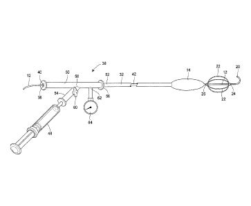
[0042] A thoracic aortic occlusion system 10 of the present disclosure is
illustrated in Fig. 6.
This thoracic occlusion system 10, and method of using the same, employs
correlation data
extracted from a statistically reliable pool of human patients. As used
herein, even reference
numerals denote structural features of the thoracic aortic occlusion system
10, while odd
reference numerals denote anatomic locations of a human. The system 10 relies
upon this data to
predict the arterial measurement of a normal torso arterial tree 11 from the
femoral artery 13 at
the level of the femoral head 15 to a level just below 21 the left subclavian
artery 17 at the aortic
- 12 -
CA 02797237 2012-10-22
WO 2011/133736 PCT/US2011/033368
arch 19 (or other relevant locations), each of which is illustrated in Fig. 1.
Using this prediction
model or nomogram, a trained medical professional can derive the distance to
which a calibrated,
self-centering endovascular wire 12 of the present disclosure should be
advanced from the
femoral artery 13 into the descending thoracic aorta 29 to a level just below
21 the left
subclavian artery 17 and the aortic arch 19 before deploying an occlusion
balloon 14 (Fig. 6)
over this endovascular wire 12 to the same position. More specifically, the
occlusion balloon 14
is deployed at a location 21 inferior of the left subclavian artery 17 at the
aortic arch 19, in an
effort to augment or support heart and brain profusion in the setting of end-
stage shock resulting
from non-compressible torso hemorrhage.
[0043] The prediction model or nomogram may be developed from, by way of
example, a
population of male and female trauma patients between the ages of 18-45 years.
Computed
tomographic measurements are made from the pool of patients to develop
statistical associations
between distances separating consistently located, external anatomical or
boney landmarks and
measurements (namely length and diameter data) within the central vascular
anatomy.
[0044] A first anatomical landmark distance measured for each patient is a
torso extent (in
cm), from the symphysis pubis 23 to the sternal notch 25, as illustrated in
Fig. 1A. Center-line
measurements are also taken (in cm) from the femoral artery 13 at the level of
the femoral head
15 to the left subclavian artery 17. These measurements are supplemented with
center-line
measurements (in cm) from the femoral artery 13 at the level of the femoral
head 15 to seven
additional key points of anatomical interest, including: (a) the left
subclavian artery origin 27;
(b) the artery of Adamkiewics origin 29; (c) the celiac artery origin 31; (d)
the left renal artery
origin 33; (c) the right renal artery origin 35; (f) the aortic bifurcation
37; and (g) the iliac artery
bifurcation 39. In addition to measuring the center-line distance from the
femoral artery 13 at
the level of the femoral head 15 to these various locations, cross-sectional
diameter (in mm) and
- 13 -
CA 02797237 2012-10-22
WO 2011/133736 PCT/US2011/033368
cross-sectional area (in mm2) measurements are also determined for each
respective vessel. A
measurement is also taken of the distance (in mm) a hollow tip access needle
would traverse at a
450 insertion angle from the epidermis layer of the skin 41 to an initial
entry point of the femoral
artery 13, which may be referred to as a percutaneous access length
measurement.
100451 For each of the measurements described above, data is collected and
means, standard
deviations, and 95th % confidence intervals are calculated, by gender, for
minimum, maximum,
st, thl0', 25th, median, 75th, 90th, 95th
and 99th percentiles. Based on these calculations, a
mathematical model employing height and gender as covariates defining the
statistical
association between the external measure of torso extent (i.e., distance from
the symphysis pubis
23 to the sternal notch 25) and the central vasculature anatomy data is
created. This
mathematical model or nomogram may be the basis for a conversion chart that a
medical
professional, unaided by fluoroscopic guidance (i.e. fluoroscopy-independent),
may use to
calculate the proper insertion distance of the calibrated endovascular wire 12
of the thoracic
aortic occlusion system 10 for a given patient.
100461 Turning now to Figs. 3-11, the thoracic aortic occlusion system 10 is
illustrated.
Referring now to Fig. 3, the system includes a self-centering endovascular
wire 12, preferably
made of a biocompatible wire having calibration indicia thereon, such as pre-
calibrated minor
length markers 16 provided at 1 cm intervals, and major length markers 18
provided at 5 cm
intervals along the length of the self-centering endovascular wire 12. By way
of example, the
overall length of the self-centering endovascular wire 12 may be 180 cm and
have a diameter of
approximately 0.035 inch. The self-centering endovascular wire 12 includes a J
tip 20 at a distal
end thereof The J tip 20 is used to minimize trauma to or perforation of the
arterial vessels as
the endovascular wire 12 is advanced along the torso arterial tree 11. The J
tip 20 is also
- 14 -
CA 02797237 2012-10-22
WO 2011/133736 PCT/US2011/033368
sufficiently flexible to unfold in the event the J tip 20 was to hook onto an
arterial branch, such
as during withdrawal of the endovascular wire 12.
[0047] Immediately proximate of the J tip 20 is a plurality of self-expanding
wire struts 22.
The wire struts 22 extend between a leading securement bead 24 and a trailing
securement bead
26, both of which secure the wire struts 22 to the endovascular wire 12. The
self-expanding wire
struts 22 are made of a material that expands upon exposure to fluid at body
temperature, such as
nitenol, and are disposed at sufficient intervals about the endovascular wire
12, such as four self-
expanding wire struts 22 at 90 intervals. These struts 22, when in their
collapsed state such as
during insertion through a transdermal sheath 28, will span the length of the
securement beads 24
and 26. However, the struts 22 will shorten in the length as they extend over
the endovascular
wire 12 when in their expanded state, such as within the descending thoracic
aorta 21, and as
such the wire struts 22 will be movable to some extent axially relative to the
endovascular wire
12, but between the securement beads 24 and 26. In this manner, the self-
expanding wire struts
22 serve a self-centering function, keeping the tip or leading end of the
endovascular wire 12
away from the sidewalls of the arterial vessels, helping to prevent the
endovascular wire 12 from
turning down an unintended branch. For example, the left renal artery origin
33 (Fig. 1), the
right renal artery origin 35 (Fig. 1), and the celiac artery origin 31 (Fig.
1) each can branch off
from the abdominal aorta 43 at a 90 angle thereto. The nitenol self-expanding
wire struts 22
serve to maintain the endovascular wire 12 within the abdominal aorta 43 (Fig.
1).
[0048] Referring now to Fig. 4, a transdermal or percutaneous sheath 28,
preferably 6 French,
and by way of example having a length of approximately 10 cm with an inner
diameter of
approximately 0.087 inches is illustrated. The sheath 28 is inserted into the
femoral artery 13
through a puncture in the skin 41 (Fig. 2) with a hollow tip needle to provide
an access port by
- 15 -
CA 02797237 2012-10-22
WO 2011/133736 PCT/US2011/033368
way of first a wire and then the sheath 28. The distal end 30 of the sheath 28
is positioned within
the external iliac artery 45 (Fig. 1).
[0049] The endovascular wire 12, the J tip 20, the leading securement bead 24,
the trailing
securement bead 26, and the unexpanded occlusion balloon 14 (which is disposed
proximate to
the trailing securement bead 26 and illustrated in Fig. 6) are all of
sufficiently small cross-
sectional dimensions to pass through the sheath 28. The leading securement
bead 24 and the
trailing securement bead 26 are also of a sufficient diameter so as to prevent
the occlusion
balloon 14 from migrating distally over the wire struts 22 and past the J tip
20.
[0050] Referring now to Fig. 6, the aortic occlusion balloon 14 is carried on
a balloon shaft 32
having pre-calibrated length indicia 34 thereon. The pre-calibrated length
indicia 34 may include
minor length markers 34a in lcm increments, and major length markers 34b in
5cm increments.
The balloon shaft 32 preferably has a length of approximately 90cm (35.43
inches) and an outer
diameter less than approximately 1.98 mm (0.087 inches), so as to fit through
the sheath 28. The
balloon shaft 32 includes a lumen 42 (see also Figs. 10 and 11), permitting
the balloon shaft 32
to pass over the endovascular wire 12. A tapered distal end portion 44 of the
balloon shaft 32
prevents the balloon shaft 32 from being inserted past the trailing securement
bead 26. The
balloon shaft 32 will remain over the self-centering endovascular wire 12
during occlusion
balloon inflation to provide a rigidity that is sufficient to permit the
balloon to be manually
maintained at a desired location within the thoracic aorta 47 (Fig. 1),
resisting distal or caudal
migration, such as might otherwise result from aortic pulsation.
[0051] The aortic occlusion balloon 14 has a length of approximately 3cm, and
is affixed to
the end of the balloon shaft 32 less than 1 cm below (proximally of) the
tapered distal end
portion of the balloon shaft 44. As indicated above, the aortic occlusion
balloon 14 is inserted
- 16 -
CA 02797237 2012-10-22
WO 2011/133736 PCT/US2011/033368
(in a collapsed state) through the transdermal or percutaneous sheath 28 with
the endovascular
wire 12 and the balloon shaft 32.
[0052] Upon navigating the endovascular wire to the desired location within
the thoracic aorta
47, an inflation fluid is introduced through the balloon shaft 32 to the
aortic occlusion balloon
14, causing the aortic occlusion balloon 14 to inflate, as illustrated in Fig.
7. By way of example,
the aortic occlusion balloon 14 may, upon inflation to approximately 1 atm,
expand to a
maximum diameter of approximately 26mm, conforming to the shape of the
thoracic aorta 47,
thereby obstructing blood flow through the thoracic aorta 47 inferiorly of the
inflated aortic
occlusion balloon 14 and promptly augmenting heart and brain profusion. The
inflation fluid is a
sterile biocompatible fluid introduced to the multi-port and valve assembly 36
(Figs. 10 and 11)
using a fluid source, such as a syringe 48. Upon removal of the sterile
inflation fluid, the aortic
occlusion balloon 14 deflates, permitting withdrawal thereof through the
transdermal or
percutaneous sheath 28.
[0053] As illustrated in Figs. 10 and 11, the aortic occlusion balloon 14 is
inflated and deflated
via a multi-port and valve assembly 36. The multi-port and valve assembly 36
includes a one-
way valve, which is preferably pressure gauge calibrated so as to alert a
physician when the
aortic occlusion balloon 14 has reached its desired inflation pressure. The
multi-port and valve
assembly 36 further includes a terminating port 38, with a diaphragm 40. The
endovascular wire
12 can extend through the diaphragm 40, while maintaining fluid-tight
communication between
the multi-port and valve assembly 36 and the aortic occlusion balloon 14. The
multi-way port
and valve assembly 36 further includes an elongate tubular barrel section 50
affixed to a
proximal end 52 of the balloon shaft 32. The elongate tubular barrel section
50 may have a
length of approximately 10 to 15 cm. The elongate tubular barrel section 50
does not pass
through the transdermal sheath 28 or enter the body of the patient. In
addition to the terminating
- 17 -
CA 02797237 2012-10-22
WO 2011/133736 PCT/US2011/033368
port 38, which may be considered a first port, the multi-way port and valve
assembly 36 includes
a second port 56 at a distal end. As discussed above, the multi-way port and
valve assembly 36
further includes a diaphragm 40, which is disposed at a proximal end of the
elongate tubular
barrel section 50. The diaphragm 40 permits the endovascular wire 12 to pass
through and
extend externally of the first port 38, while maintaining a fluid-tight
connection, thereby
avoiding leakage of bodily fluid through the first port 38.
[0054] A side port 58 and a branch 54 off the elongate tubular barrel section
50, intermediate
the first port 38 and second port 56, enables attachment of the syringe 48 to
the multi-way port
and valve assembly 36. A one-way valve 60 is actuable between an open
condition (permitting
passage of inflation fluid therethrough), as illustrated in Fig. 11, and a
closed condition
(preventing passage of inflation fluid), as illustrated in Fig. 10.
Preferably, the one-way valve 60
is in an open condition when oriented parallel to the branch 54 off the
elongate tubular barrel
section 50 (Fig. 11), and is in a closed condition when rotated to a position
perpendicular to the
branch 54. An additional port 62, provided intermediate the one-way valve and
the proximal end
52 of the balloon shaft 32 to which the multi-way port and valve assembly 36
is affixed, is
provided with a pressure monitoring device 64. A physician may monitor the
pressure
monitoring device 64 during inflation, enabling the physician to determine
when the pressure
within the occlusion balloon 14 and the balloon shaft 32 has reached a
pressure of, for example,
2 atm, so as to avoid over-inflation and potential injury to the thoracic
aorta 47.
[0055] Using a correlation chart or nomogram derived from the statistical data
regarding the
measurements between the consistently identifiable external measures or
landmarks of torso
extent (e.g. the symphysis pubis 23 and the sternal notch 25), the physician
calculates the proper
distance to which the endovascular wire 12 and balloon shaft 32 are to be
inserted into the
transdermal sheath 28 through a puncture in the skin 41 and into the femoral
artery 13 at the
- 18-
CA 02797237 2012-10-22
WO 2011/133736 PCT/US2011/033368
location of the femoral head 15, thereby positioning the aortic occlusion
balloon 14 at the desired
location 21 within the thoracic aorta 47, inferiorly of the left subclavian
artery 17 without the aid
of fluoroscopy (i.e. fluoroscopy-independent). The major length markers 18
along the
endovascular wire 12 may be annotated with length-identifying numbers to
facilitate
determination of the length to which the self-centering endovascular wire 12
has been advanced
within the torso arterial tree 11. Upon insertion to the desired length, with
the one-way valve 60
in the open condition, the physician actuates a piston of the syringe 48,
thereby introducing
inflation fluid through the balloon shaft 32 and into the occlusion balloon
14, inflating the
occlusion balloon 14 to a volume sufficient to block the thoracic aorta 47.
[0056] The endovascular, fluoroscopy-independent resuscitative thoracic aortic
occlusion
system 10 of the present disclosure may be provided to users in the form of a
kit, enabling
assembly of the same at, by way of example only, a forward surgical hospital
close to a
battlefield in a civilian trauma setting either outside of a hospital or in a
resuscitation room of an
emergency department. The system 10 may be applied in clinical scenarios other
than traumas
addressed in such urgent care settings, such as cardiac arrest, neurogenic
shock, or post-partum
hemorrhage that may occur in operating or delivery rooms. The kit may include
the
endovascular wire 12 having the J tip 20 and at least one wire strut 22
disposed proximally to the
J tip 20. The kit may further include the occlusion balloon 14 that may be
disposed proximally
on the balloon shaft 32, and advanced over the endovascular wire 12 until it
reaches the trailing
securement bead 26. The transdermal sheath 28 may also be a part of the kit,
such that the
transdermal or percutaneous sheath 28 is able to receive each of the
endovascular wire 12, the
occlusion balloon 14 when in an uninflated condition, and the balloon shaft
32, as described
above. When assembled with the endovascular wire 12, the balloon shaft 32, and
the
transdermal sheath 28, the occlusion balloon 14 may be selectively inflated
and deflated at a
- 19 -
CA 02797237 2012-10-22
WO 2011/133736 PCT/US2011/033368
desired location within the thoracic aorta 47 of a patient to treat vascular
injury without the aid of
fluoroscopy.
[0057] The kit may further include the multi-port and valve assembly 36 in
fluid
communication with both the proximal end of the balloon shaft 32 and a fluid
source, such as the
syringe 48, as also described above. One of a table, a nomogram, a chart or a
graph correlating
distances between at least readily externally identifiable anatomical
landmarks of a pool of
humans to distances from the femoral artery 13 to a location within the
thoracic aorta 47 to
which the endovascular wire 12 and the balloon shaft 32 are to be inserted may
also be included
in the kit.
[0058] Referring now to Figs. 12-18, additional embodiments of the present
disclosure are
illustrated. Referring to Fig. 12, endovascular procedures in large axial
vessels of the human
torso may be considered as narrow margin procedures 110 or wide margin
procedures 112. In
other words, many catheter-based procedures in the aorta and vena cava require
exact positioning
of devices to be safe and effective, and, therefore, have a narrow margin of
error during
procedures. The narrow margin procedures 110 require standard fluoroscopy to
effectively and
accurately direct the procedure. Examples of narrow margin procedures 110
include: (1)
placement of stent grafts to treat age-related aneurysms near major branch
vessels of the thoracic
or abdominal aorta; or (2) treatment of local or localized disease processes
such as arterial
stenoses caused by atherosclerosis with balloon angioplasty and bare metal
stents. Because these
procedures entail placing devices at the exact location, i.e., within
millimeters, of vital branch or
vein locations, they require real time visualization using contrast agents and
fluoroscopy.
[0059] Other catheter-based endovascular procedures have a wider margin of
error. Examples
of wide margin procedures 112 include positioning of occlusion balloons to
control torso
hemorrhage, vena cava filter devices to prevent pulmonary embolus, and stent
grafts to treat
- 20 -
CA 02797237 2012-10-22
WO 2011/133736 PCT/US2011/033368
vessel disruptions localized by computed tomography (CT). In the case of
balloon occlusion, the
goal is to temporarily halt flow beyond a certain point in the vena cava or
aorta to aid with
hemorrhage control. Additionally, in the setting of aortic occlusion, life-
preserving blood
pressure above, or proximal to the balloon occlusion, is maintained or
supported. In these
instances, the balloon may be positioned anywhere over a much longer length of
vessel, e.g.,
within several centimeters, prior to inflation. Similarly, positioning and
placement of
thromboembolic filter devices in the vena cava may occur over a relatively
wide distance of
vessel, e.g., between the iliac vein confluence and the renal veins. Thus, in
such wide margin
procedures, fluoroscopy is not required.
[0060] While fluoroscopy is not required, a detailed characterization of the
axial vessels of the
human torso is necessary to accomplish such procedures in fluoroscopy free
environments.
Contrast-enhanced computer tomography (CT) using 64-panel detectors and
special measuring
software allows for such detailed characterization. More specifically,
software programs placed
in or alongside CT units allow precise centerline measurements within the
axial vessels as well
as determination of their diameter. Application of centerline measurements
allows definition of
clinically relevant distances between a standard vascular entry point, i.e.,
femoral vessels and
major branch artery points within the vessels.
[0061] Referring back to Fig. 1, the normal torso arterial tree 11 with major
branch artery
points is illustrated. The major branch artery points include a left femoral
artery 13, an external
iliac artery 45, and a left subclavian artery 17. Center-line measurements are
taken (in cm), for
example, from the femoral artery 13 at the level of the femoral head 15 to the
left subclavian
artery 17, as the left femoral artery 13 is a common vascular entry point. As
also illustrated in
Fig. 1, other major branch artery points include the artery of Adamkiewics
origin 21, the celiac
artery origin 31, the left renal artery origin 33, the right renal artery
origin 35, the aortic
- 21 -
CA 02797237 2012-10-22
WO 2011/133736 PCT/US2011/033368
bifurcation 37, and the iliac artery bifurcation 39. In addition to measuring
the center-line
distance from the femoral artery 13 at the level of the femoral head 15 to
these various locations,
cross-sectional diameter, e.g., in mm, and cross-sectional area, e.g., in mm2,
measurements are
also determined for each respective vessel.
100621 Referring now to Fig. 13, arterial torso vascular anatomy is
illustrated with various
landing zones within the thoracic aorta 47 for wide margin procedures 112. For
example, a
thoracic aortic zone 137 is disposed below a region adjacent to the left
subclavian artery 17 along
a descending thoracic aorta 47. An infrarenal aortic zone 139 is disposed
between left renal
artery 25 and the iliac artery 45, and a common iliac artery zone 141 is
disposed between the
aortic bifurcation 37 and a distal end of the femoral artery 13.
100631 Referring now to Fig. 14, venous torso vascular anatomy is illustrated
with various
landing zones within the vena cava for wide margin procedures. For example, a
retro-hepatic
inferior vena cava zone 143 is disposed along a descending thoracic aorta 47.
An infrarenal IVC
zone 145 is disposed below the retro-hepatic inferior vena cava zone 143, and
a common iliac
vein zone 147 is disposed below the infrarenal IVC zone 145.
[0064] To further characterize torso vascular morphometry, it is necessary to
account for the
relationship between vascular lengths and diameters and an individual's length
or height.
Because patient height is not consistently available, especially in the
context of emergencies or
trauma, an external measure of torso extent is needed.
[0065] Referring back now to Fig. 1A, an external measure of torso extent is
illustrated. This
measure extends from a sternal notch 25 to the symphisis pubis 23 and can be
readily palpated
and recorded, even in emergency and trauma settings. Not only is the external
measure of torso
- 22 -
CA 02797237 2012-10-22
WO 2011/133736 PCT/US2011/033368
extent readily available, but it provides a measure which is specific to the
torso that houses
vascular anatomy of interest.
[0066] Determination of vascular lengths or distances within the torso from a
readily available
external measure of torso extent requires correlation of this data to form a
nomogram. The
nomogram defines, with a predetermined confidence interval, the relationship
between the
external measure of torso extent and distances within the axial vessels of the
torso, as well as the
specific vessel diameters. If considered from the perspective of a common
vascular entry point
such as the femoral vessels, to clinically important branch points or landing
zones 137-147
within the aorta or vena cava, the nomogram is relevant to the performance of
wide margin
endovascular procedures. The nomogram allows a provider to quickly estimate
from a basic
external measure, e.g., the distance between the sternal notch 25 and the
symphysis pubis 23
(Fig. 1A), the distance from the femoral vessels to landing zones in the
descending thoracic aorta
47 or the inferior vena cava.
[0067] Referring now to Figs. 15 and 16, an exemplary torso vascular insertion
tool 200 is
illustrated. The tool 200 translates data from the above-described correlation
and nomogram,
making such information useful in a clinical setting. More specifically, and
referring now to Fig.
15, a first side 210 of the tool 200 is illustrated. The first side 210
includes an edge 214 and a
calibrated ruler 218 disposed on the edge 214 of the first side 210 for use in
measuring a torso
extent length, i.e., the length between the sternal notch 25 and the symphisis
pubis 23 (Fig. 1A),
on a patient. Adjacent to the calibrated ruler 218 on the first side 210 of
the tool 200 is a first
chart 220 providing a listing, by way of example, of male safe zone arterial
insertion lengths
from the femoral artery 13 vessels to relevant landing zones within the aorta.
Such landing
zones include the thoracic aortic zone 137, the infrarenal aortic zone 139,
and the common iliac
artery (CIA) zone 141. Adjacent to the first chart 220 disposed on the first
side 210 of the tool
- 23 -
CA 02797237 2012-10-22
WO 2011/133736 PCT/ES2011/033368
200 is a second chart 222. The second chart 222 provides a listing, by way of
example, of male
safe zone venuous insertion lengths from the femoral artery 13 vessels to
relevant landing zones
within the vena cava. Such landing zones include the retro-hepatic inferior
vena cava (IVC)
zone 143, the infrarenal IVC zone 145, and the common iliac vein (CIV) zone
147. For the torso
arterial segment, the tool 200 provides distances from the femoral artery 13
to the aortic
bifurcation 37, the lowest renal artery 33, the celiac artery 31, and the left
subclavian artery 17
(see Figs. 1 and 13). Venous insertion distances are provided from the femoral
vein 153 to the
bifurcation of the vena cava 155, the lowest renal vein 157, and the hepatic
vein 159 (see Fig.
14).
[0068] Referring now to Fig. 16, a second side 212 of the exemplary torso
vascular insertion
tool 200 is illustrated. The second side 212 includes an edge 216 and a
calibrated ruler 218
disposed on the edge 216 also for use in measuring on a patient the external
torso extent between
the sternal notch 25 and the symphisis pubis 23 (Fig. 1A), but this time for a
female patient. A
third chart 224 is disposed adjacent to the calibrated ruler 218 on the second
side 212 of the tool
200. The third chart 224 provides, by way of example, a listing of female safe
zone arterial
insertion lengths from the femoral artery 13 vessels to relevant landing zones
within the aorta.
Such landing zones also include the thoracic aortic zone 137, the infrarenal
aortic zone 139, and
the common iliac artery (CIA) zone 141. Adjacent the third chart 224 is a
fourth chart 226. The
fourth chart 226 provides, by way of example, a listing of female safe zone
venuous insertion
lengths from the femoral artery 13 vessels to relevant landing zones within
the vena cava. Such
landing zones include the retro-hepatic inferior vena cava (IVC) zone 143, the
infrarenal IVC
zone 145, and the common iliac vein (CIV) zone 147.
[0069] The second side 212 may also include a listing of the diameter of the
torso axial vessels
at clinically important locations. More specifically, the back side 212 of the
tool 200 provides
- 24 -
CA 02797237 2012-10-22
WO 2011/133736 PCT/US2011/033368
the diameter of the iliac artery 39, the thoracic aorta 47, for the torso
axial vessels. The venous
diameters include the iliac vein and infrarenal and suprarenal vena cava. The
second side 212
may also include clinically relevant specifications for endovascular devices
commonly used in
wide margin endovascular procedures 112, such as compliant balloons, basic
stent grafts, and
vena cava filters.
[0070] While various numerical indices and zones are included in the exemplary
tool 200, the
tool 200 is but one example; actual devices could use different numerical
indices and zones than
those provided in the exemplary tool 200 and still be within the scope of the
appended claims.
[0071] Referring now to Fig. 17, a second embodiment of another exemplary
torso vascular
insertion tool 300 is illustrated. The torso vascular insertion tool 300
includes a data-wheel 310
having a first side 312 and a second side 314 (not shown) and a tape 316 wound
between the first
and second sides 312, 314 of the data-wheel 310, a portion of the tape 316
extending along one
side of the data-wheel 310. The tape 316 is used to measure on a patient the
external measure of
torso extent between the sternal notch 25 and the symphisis pubis 23 (Fig.
1A). By way of
example, the first side 312 of the data-wheel 310 provides listings of male
safe zone arterial
insertion lengths from the femoral artery 13 vessels to relevant landing zones
within the aorta
corresponding to a circular listing of measured torso extent lengths. More
specifically, the first
side 312 of the data-wheel 310 includes an inner row 318, a middle row 320,
and an outer row
322. The inner row 318 provides a circular listing of male torso extent
lengths, e.g., in
centimeters. The middle row 320 provides a circular listing of the thoracic
aortic zone 137
lengths corresponding to the circular listing of male torso extent lengths;
and the outer row 322
provides a circular listing of the infrarenal aortic zone 139 lengths also
corresponding to the
circular listing of male torso extent lengths. It will be understood by one of
skill in the art that
the first side 312 of the data-wheel 310 may alternatively provide listings of
male safe zone
- 25 -
CA 02797237 2012-10-22
WO 2011/133736 PCT/US2011/033368
venuous insertion lengths, for example. In addition, many other variations or
combinations of
correlation data between the measured torso extent length and new devices,
different landing
zones, and vessel sizes, for example, may also be provided on the data-wheel
310.
[0072] Referring now to Fig. 18, the second embodiment of the exemplary torso
vascular
insertion tool 300 is also illustrated, here with a cover 324 over the first
side 312 of the data-
wheel 310 of the tool 300. After a user draws the tape 316 over a patient to
measure the external
measure of torso extent between the sternal notch 25 and the symphisis pubis
23 (Fig. 1A), the
cover 324 of the data-wheel 310 rotates to a value of the measured torso
extent length displayed
on the inner row 318 of the data-wheel 310. The user is then able to compare
the torso extent
length measured or shown on the inner row 318 of the data-wheel to one or more
of
corresponding safe zones displayed on the middle row 320, e.g., the thoracic
aortic zone 137
insertion length, or the outer row 322, e.g., the infrarenal aortic zone 139
insertion length of the
data-wheel 310 to calculate a length to which the endovascular device is to be
inserted.
[0073] A second side 314 (not shown) of the data-wheel 310 may include a
circular listing of
female safe zone arterial insertion lengths from the femoral artery 13 vessels
to relevant landing
zones within the aorta. Such landing zones also include the thoracic aortic
zone 137, the
infrarenal aortic zone 139, and the common iliac artery (CIA) zone 141. The
second side 314 of
the data-wheel 310 may alternatively include a circular listing of female safe
zone venous
insertion lengths from the femoral artery 13 vessels to relevant landing zones
within the vena
cava. Such landing zones include the retro-hepatic inferior vena cava (IVC)
zone 143, the
infrarenal IVC zone 145, and the common iliac vein (CIV) zone 147.
[0074] Like the first side 312, the second side 314 of the data-wheel 310 and
tape 316
combination of the second embodiment of the torso vascular insertion tool 300
may be easily
- 26 -
CA 02797237 2012-10-22
WO 2011/133736 PCT/US2011/033368
expanded or changed to alternatively include various other correlation data
between the
measured torso extent length and new devices and vessels sizes, for example.
[0075] While various numerical indices and zones are included in the exemplary
tool 300, the
tool 300 is also but one example; actual devices could use different numerical
indices and zones
than those provided in the exemplary tool 300 and still be within the scope of
the appended
claims.
100761 Both embodiments of the torso vascular insertion tool 200, 300 may also
include
calipers or rods to facilitate measuring depending upon a patient's shape.
More specifically, in
patients having larger abdominal areas, the calipers or rods are needed to
provide an accurate
linear measurement of the patient's torso extent length because the larger
abdominal areas would
otherwise impede an accurate measurement. For example, when the torso vascular
insertion
tools 200, 300 are tape, and the tape is flexible and plastic, the caliper or
rod may be disposed on
one or both ends of the tape. The calipers or rods may also be retractable and
extendible to help
facilitate more accurate locating of externally-identifiable anatomic
landmarks and measuring of
the torso extent length in such patients.
[0077] While preferred embodiments of the present disclosure have been
described above,
variations may be made that are still within the scope of the appended claims.
- 27 -