Note: Descriptions are shown in the official language in which they were submitted.
CA 02797247 2016-04-26
- 1 -
METHODS AND USES OF T1E2 BINDING AND/OR ACTIVATING AGENTS
[0001]
Field of the disclosure
[0002] The disclosure relates to methods and uses of Tie2 binding
and/or activating agents. In particular, the disclosure relates to methods and
uses for inhibiting the expansion of granulocyte colony forming unit (CFU-G),
for reducing eosinophils and basophils and for treating allergic diseases.
Background of the disclosure
[0003] Angiopoietins (Ang) 1-4 have all been shown to bind to and
activate Tie2 receptor tyrosine kinase activity to differing extents. All the
Angs
are characterized structurally by an N-terminal super clustering domain (SCD)
followed by a coiled-coil domain (CCD) and a C-terminal fibrinogen-like
domain (FLD) (Ward and Dumont 2002; Tsigkos et al. 2003), Functional
studies have highlighted a role for the SCD and COD's in forming high order
homotypic multimers of Ang (Procopio et at. 1999). The specific nature of
these multimers is variable and is unique to each Ang family member. Binding
specificity of the Angs for the Tie2 receptor has been ascribed to the FLD
(Tisgkos et at. 2003; Procopio et al. 1999). Taken together, it is the unique
structural attributes of each Ang family member that promotes binding and
differential clustering of Tie2. The pleiotropic physiological effects of Angs
1-4
are thought to at least in part be mediated by appropriate and specific
clustering of the receptor. For instance mice engineered to overexpress the
CCD of Ang 1, capable of multimerizing with endogenous Ang1 produced in
the same cell, caused improper patterning of the coronary vessels (Ward et
at. 2004). Furthermore, chimeric forms of Ang 1 engineered to contain the C-
terminal FLD and one of several different CODs differed in their ability to
activate the Tie2 receptor (Cho at at. 2004a; Cho et at. 2004b).
CA 02797247 2012-10-23
WO 2011/134056
PCT/CA2011/000473
- 2 -
[0004] Based on
this information, the present inventors previously
designed a peptide mimetic, called Vasculotide, that binds to Tie2 and when
configured as a tetramer results in the clustering of the receptor and its
activation.
[0005] Activating
Tie2 through the tetramerization of high affinity Tie2
binding peptides using the biotin/avidin model (Van Slyke et al. 2009) has
established the use of the peptide as an agonist to the Tie2 receptor to
promote angiogenesis for applications in diabetic wound healing and other
cardiovascular indications.
[0006] Patients
with allergic diseases respond to an allergen with a
systemic response that initiates the production of specific inflammatory
cells,
eosinaphils and basophils and their progenitors, from the bone marrow
(Denburg and Keith 2008; Hogan et al. 2008; Rothenberg and Hogan 2006).
Upon release of these cells from the bone marrow they usually target the
respiratory mucosa and other tissues and once activated eosinophils and
basophils are one of the key immunomodulatory cells that sustain the allergic
response (Rothenberg and Hogan 2006; Barrett and Austen 2009; Gauvreau
et al. 2009; Schroeder 2009). These cells are known to secrete TGF-p, which
is known to be an extremely potent pro-fibrogenic factor (Jacobsen et al.
2007; Hogan 2007; Raap and Ward law 2008).
Summary of the disclosure
[0007] The
present inventors have shown that administration of a
multimeric form of a Tie2 binding peptide called "Vasculotide" is able to
inhibit
the expansion of CFU-G cells, resulting in a reduction in eosinophils and
basophils, without a more general immunosuppression. The present inventors
have also shown that in a transgenic mouse model of atopic dermatitis, triple
transgenic mice which express Ang-1 showed amelioration of disease
whereas triple transgenic mice, which express Ang-2, showed deterioration of
disease.
CA 02797247 2012-10-23
WO 2011/134056
PCT/CA2011/000473
- 3 -
[0008] Accordingly, the
present disclosure provides a method of
inhibiting the expansion of CFU-G cells in an animal or cell in need thereof
comprising administering a Tie2 binding and/or activating agent. The
disclosure also provides use of a Tie2 binding and/or activating agent for
inhibiting the expansion of CFU-G cells in an animal or cell in need thereof.
Also provided is use of a Tie2 binding and/or activating agent in the
preparation of a medicament for inhibiting the expansion of CFU-G cells in an
animal or cell in need thereof. Further provided is a Tie2 binding and/or
activating agent for use in inhibiting the expansion of CFU-G cells in an
animal or cell in need thereof.
[0009] The present
disclosure also provides a method of reducing
eosinophils and/or basophils in an animal or cell in need thereof comprising
administering a Tie2 binding and/or activating agent. The disclosure also
provides use of a Tie2 binding and/or activating agent for reducing
eosinophils
and/or basophils in an animal or cell in need thereof. Also provided is use of
a
Tie2 binding and/or activating agent in the preparation of a medicament for
reducing eosinophils and/or basophils in an animal or cell in need thereof.
Further provided is a Tie2 binding and/or activating agent for use in reducing
eosinophils and/or basophils in an animal or cell in need thereof.
[0010] The present
disclosure further provides a method of treating
allergic disease or response in an animal or cell in need thereof comprising
administering a Tie2 binding and/or activating agent. The disclosure also
provides use of a Tie2 binding and/or activating agent for treating allergic
disease or response in an animal or cell in need thereof. Also provided is use
of a Tie2 binding and/or activating agent in the preparation of a medicament
for treating allergic disease or response in an animal or cell in need
thereof.
Further provided is a Tie2 binding and/or activating agent for use in treating
allergic disease or response in an animal or cell in need thereof. In one
embodiment, the allergic disease is an atopic allergic disease, such as atopic
dermatitis, asthma or allergic rhinitis.
CA 02797247 2012-10-23
WO 2011/134056
PCT/CA2011/000473
- 4 -
[0011] The
present disclosure also provides a method of treating a
condition associated with eosinophils and/or basophils in an animal or cell in
need thereof comprising administering a Tie2 binding and/or activating agent.
The disclosure also provides use of a Tie2 binding and/or activating agent for
treating a condition associated with eosinophils and/or basophils in an animal
or cell in need thereof. Also provided is use of a Tie2 binding and/or
activating
agent in the preparation of a medicament for treating a condition associated
with eosinophils and/or basophils in an animal or cell in need thereof.
Further
provided is a Tie2 binding and/or activating agent for use in treating a
condition associated with eosinophils and/or basophils in an animal or cell in
need thereof. In one embodiment, the condition associated with eosinophils
and/or basophils is a myelodysplastic syndrome. In one embodiment, the
condition associated with eosinophils and/or basophils is a leukemia of
eosinophil and/or basophil origin, such as chronic myeloid leukemia, acute
myeloid leukemia, chronic eosinophilc leukemia, acute eosinophilic leukemia,
chronic myelomonocytic leukemia with eosinophilia, and acute basophilic
leukemia. In another embodiment, the condition associated with eosinophils
and/or basophils is inflammatory bowel disease. In yet another embodiment,
the condition associated with eosinophils and/or basophils is a parasitic
infection. In yet another embodiment, the condition associated with
eosinophils and/or basophils is idiopathic hypereosinophilic syndrome (HES).
[0012] The
present disclosure also provides a method of reducing
inflammatory cytokine and/or chemokine levels in an animal or cell in need
thereof comprising administering a Tie2 binding and/or activating agent. The
disclosure also provides use of a Tie2 binding and/or activating agent for
reducing inflammatory cytokine and/or chemokine levels in an animal or cell in
need thereof. Also provided is use of a Tie2 binding and/or activating agent
in
the preparation of a medicament for reducing inflammatory cytokine and/or
chemokine levels in an animal or cell in need thereof. Further provided is a
Tie2 binding and/or activating agent for use in reducing inflammatory cytokine
and/or chemokine levels in an animal or cell in need thereof. In one
embodiment, the inflammatory cytokine and/or chemokine levels are serum
CA 02797247 2012-10-23
WO 2011/134056
PCT/CA2011/000473
- 5 -
inflammatory cytokine and/or chemokine levels. In one embodiment, the
inflammatory cytokines and/or chemokines comprise at least one of eotaxin,
IL-17, MIG, IL12/1L23 (p40), IL-9, MIP-1 a, MIP-1b, RANTES, TNF-a, IL-113, IL-
5, IL-13, and MCP-1. In another embodiment, the inflammatory cytokines and
chemokines comprise IL-17, MIG, IL12/11_23 (p40), IL-9, MIP-1 a, M1P-1b,
RANTES, TNF-a, IL-1(3, IL-5, IL-13, and MCP-1. In yet another embodiment,
the inflammatory cytokines and/or chemokines comprise eotaxin.
[0013] The agent may be administered in any suitable manner,
including without limitation, topically, systemically, orally, intranasally or
by
inhalation.
[0014] In one embodiment, the agent is an angiopoietin-1 or a nucleic
acid encoding angiopoietin-1. In another embodiment, the agent is an
inhibitor of angiopoietin-2, such as a blocking antibody against angiopoietin-
2
or an antisense nucleic acid against angiopoietin-2.
[0015] In another embodiment, the agent comprises a Tie2 binding
peptide monomer or a multimeric form of a Tie2 binding peptide monomer.
[0016] The multimeric form can be, for example, a dimer, tetramer, or
a
multimeric form that comprises six, eight, ten or twelve units of the monomer.
In another embodiment, the multimeric form comprises an odd number of
units, such as three, five, seven, nine or eleven units.
[0017] In yet another embodiment, the Tie2 binding peptide monomer
comprises a structure: A-B-C, wherein A comprises a Tie2 binding peptide, B
comprises a spacer and C comprises a multimerizing group, wherein C has
affinity for D, a multimer agent comprising multiple binding sites for C. For
example, the multimer agent D can have four binding sites for the
multimerizing group C such that a tetramer is formed when four Tie2 binding
peptide monomers, A-B-C, interact with the multimer agent D. In an
embodiment, C comprises a biotin group and D comprises an agent selected
from the group consisting of avidin, streptavidin and neutravidin. In yet
another embodiment, B comprises polyethylene glycol (PEG).
CA 02797247 2012-10-23
WO 2011/134056
PCT/CA2011/000473
- 6 -
[0018] In a further embodiment, the Tie2 binding peptide monomer
comprises a structure: A-B, wherein A comprises a Tie2 binding peptide and
B comprises a spacer, wherein the multimeric form is created by covalent
linkage of multiple Tie2 binding peptide monomers via the spacer B. In
anembodiment, B comprises polyethylene glycol (PEG).
[0019] Tie2 binding peptides for use in the monomers include, but are
not limited to, a T7 peptide as shown in SEQ ID NOs: 1 or 2, a GA3 peptide
as shown in SEQ ID NOs: 3 or 4, a T6 peptide as shown in SEQ ID NOs: 7 or
8 or a T8 peptide as shown in SEQ ID NOs: 5 or 6. In an alternative
embodiment, the Tie2 binding peptide is a T4 peptide as shown in SEQ ID
NOs: 9 or 10.
[0020] In another embodiment, the multimeric form is a dimer,
comprising: (a) a first peptide chain; (b) a second peptide chain; and (c) a
linking moiety connecting said first and second peptide chains, wherein said
peptide dimer binds to and activates the Tie2 receptor. In one embodiment,
the first peptide chain is a T7 peptide (SEQ ID NOs: 1 or 2) and/or the second
peptide chain is a T7 peptide (SEQ ID NOs: 1 or 2). Optionally, the linking
moiety comprises one or more water soluble polymers covalently bound to the
first peptide chain and the second peptide chain. The one or more water
soluble polymers may be linear polymers. In one embodiment, the water
soluble polymer is a polyethylene glycol (PEG), optionally having a molecular
weight in the range of about 3,000 Daltons to 50,000 Daltons. In various
embodiments, the PEG has a molecular weight of about 3,000, about 3,400,
about 5,000, about 10,000, about 15,000, about 20,000, about 25,000, about
30,000 or about 40,000 Daltons.
[0021] In yet another embodiment, the multimeric form comprises a
peptide tetramer, comprising: (a) a first peptide chain; (b) a second peptide
chain; (c) a third peptide chain; (d) a fourth peptide chain; and (e) a
linking
moiety connecting said first, second, third and fourth peptide chains, wherein
said peptide tetramer binds to and activates the Tie2 receptor. Optionally,
the
first, second, third and fourth peptide chains are T7 peptides (SEQ ID NOs: 1
CA 02797247 2016-04-26
- 7 -
or 2). The linking moiety may comprise one or more water soluble polymers
covalently bound to the first, second, third and fourth peptide chains. In one
embodiment, the water soluble polymer is a branched chain water soluble
polymer, such as PEG. The branched PEG may have a molecular weight in a
range of about 3,000 Daltons to about 50,000 Daltons. In various
embodiments, the PEG has a molecular weight of about 3,000, about 3,400,
about 5,000, about 10,000, about 15,000, about 20,000, about 25,000, about
30,000 or about 40,000 Daltons.
[0022] The multimeric forms described herein exhibit Tie2 agonist
activity. For example, the multimeric form stimulates Tie2 phosphorylation or
stimulates phosphorylation of MAPK, AKT and eNOS.
[0023] In a particular embodiment, the multimeric form is a tetramer
and the Tie2 binding peptide monomer comprises a structure: A-B-C, wherein:
A comprises a Tie2 binding peptide selected from a T7 peptide (SEQ
ID NOs: 1 or 2) and a GA3 peptide (SEQ ID NOs: 3 or 4);
B comprises a polyethylene glycol spacer; and
C comprises a biotin group,
wherein four copies of A-B-C are associated with a tetramer agent, D, to
create the tetramer form, the tetramer agent, D, being selected from the group
consisting of avidin, streptavidin and neutravidin,
[0024] Other features and advantages of the present disclosure will
become apparent from the following detailed description.
Brief description of the drawings
[0025] The disclosure will now be described in relation to the drawings
in which:
CA 02797247 2012-10-23
WO 2011/134056 PCT/CA2011/000473
- 8 -
[0026] Figure 1A shows Vasculotide-dependent activation of the Tie2
receptor in vivo. CD1 mice were injected intraperitoneally (I P.) with
indicated
doses of Vasc-PEG. PBS was used as a control. The compound was allowed
to circulate for 1 hour prior to sacrificing. Immunoprecipitation (IP) of Tie2
from
lung lysates and western blot analysis of Tie2 activation were performed
(indicated by phosphotyrosine (pY) Tie2). Total levels of Tie2 are indicated
in
the lower panel (pan Tie2).
[0027] Figure 1B shows Vasculotide-driven Tie2 receptor activation in
vivo. Tie2 activation (pY Tie2) was quantified at various times following
intraperitoneal injection (I.P.) or gavage (P.O.) of Vasc-PEG into CD1 mice.
Lysates were extracted from the lungs and Tie2 phosphorylation analysis
performed on pooled lysates from three animals subjected to
immunoprecipitation. Increased Tie2 phosphorylation is apparent for at least
72hrs following I.P. injection.
[0028] Figure 1C shows detection and quantification of Vasculotide by
ELISA. A typical standard curve is shown in which a defined amount of
compound is spiked into diluted mouse plasma. PBS is used as a negative
control. Vasculotide concentrations examined ranged from 97pg to 400ng.
Concentrations of Vasculotide falling within this range displayed a high
degree
of linearity.
[0029] Figure 1D shows results of Vasculotide-specific ELISA to assess
Vasc-PEG persistence in circulation. Circulating Vasc-PEG (plasma) was
quantified at various times following intraperitoneal injection (I.P.) or
gavage
(P.O.) into CD1 mice. At 24 hours post injection, a statistically detectable
level
of Vasc-PEG (determined by unpaired student t-test) was observed in the
plasma (compare PBS to 24 hrs or 24hrs to 96hrs). Oral delivery did not result
in any detectable levels of Vasc-PEG when compared to PBS.
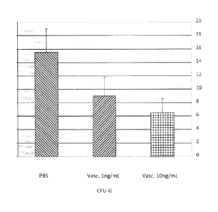
[0030] Figure 2 shows inhibition of cellular proliferation and/or
differentiation by Vasculotide in vitro. Dissociated bone marrow from CD1
mice was cultured in Methocult M3434 media for 7 days. Hematopoietic
colonies (CFU-G) were then counted. A dose dependent, statistically
CA 02797247 2012-10-23
WO 2011/134056
PCT/CA2011/000473
- 9 -
significant reduction in CFU-G numbers was noted in response to Vasculotide
treatment.
[00311 Figure 3A shows gross phenotypic appearance of neonatal wild-
type (left) and pTek-tTA:pTet0S-Tek Atopic dermatitis (AT Derm) mice (right).
Note the scaly skin, delayed fur growth and slightly reduced size of the
transgenic mouse compared to wild-type littermate.
[0032] Figure 3B is a schematic of transgenic mouse breeding
approaches. Schema of the four transgenes used in these studies to produce
the driver line (top left, minimal Tie2 promoter¨ tTa) and the responder lines
(top right, tet0S-Tie2, tet0S-Ang1, and tet0S-Ang2) are indicated. Mice
express the responder transgene only when double transgenic for the driver
and responder or triple transgenic for the driver and two responder genes
(lower schema). Expression of the responder genes can be silenced through
addition of doxycycline to the drinking water.
[0033] Figure 3C shows the effect of Angiopoietin 1 (Ang1) in a mouse
model of atopic dermatitis. Ang1 expression ameliorates the experimental
atopic dermatitis phenotype. Gross phenotypic improvement is noted when
experimental atopic dermatitis (pTek-tTA pTet0S-Tek) mice (DT(Tie-2)) are
crossed to Ang1 mice, triple transgenic (TT)(pTek-tTA : pTet0S-Tek :
pTet0S-Angl) (TT (Tie2+Ang-1)). The effects are evidenced by a decrease in
erythema around the nose and eyes. Also noted is a reduction in ear
microhaemorrhages (left images) and tail scaly plaque formation.
[0034] Figure 3D shows the effect of Angiopoietin 2 (Ang2) in a mouse
model of atopic dermatitis. Ang2 expression exacerbates all aspects of the
experimental atopic dermatitis phenotype. Ang2 triple transgenic (TT) mice
(TT)Tie-2iAng-2 present with increased erythema and tail plaque compared
to experimental atopic dermatitis animals (DT (Tie-2)).
[0035] Figure 3E shows Angiopoietin 1 ameliorates experimental atopic
dermatitis while Angiopoietin 2 exacerbates all aspects of the phenotype.
Enforced expression of Ang2 by the minimal Tie2 promoter (DT (Ang-2)) does
CA 02797247 2012-10-23
WO 2011/134056
PCT/CA2011/000473
- 10 -
,
not induce atopic dermatitis or erythema compared to wild-type (WI) animals.
Only in triple transgenics (pTek-tTA : pTet0S-Tek : pTet0S-Ang2) is it
observed that Ang2 exacerbates the AT dernn phenotype. These effects are
evidenced by an increase in erythema around the nose and eyes. Also noted
is an increase in ear microhaemorrhages and tail scaly plaque formation in
the triple transgenic (pTek-tTA : pTet0S-Tek : pTet0S-Ang2).
[0036] Figure 4 shows the effects of Vasculotide treatment on
mouse
body weight. Following a 30-day treatment with Vasculotide, body weight was
shown to be comparable to control mice. Mice were weighed at day zero and
day 30 of the study. Percent change in body weight during 30-day treatment is
shown. There was no statistically detectable alteration in body weight of mice
receiving Vasculotide (40pg/kg every 3 days) compared to vehicle treated
mice.
[0037] Figure 5 shows the effects of Vasculotide treatment on a
mouse
model of atopic dermatitis. Vasculotide treatment (D and F) was shown to
ameliorate the superficial features of the atopic dermatitis phenotype. Gross
phenotypic improvement was noted when experimental atopic dermatitis mice
(pTek-tTA : pTet0S-Tek) (AT-Derm) were treated with Vasculotide (40pg/kg
every 3 days for 30 days) regardless of the initial severity of the phenotype
(Moderate (C, D) or Severe (E, F)). Wild-type mice (Control) treated with
Vasculotide (B) were indistinguishable from PBS-treated (A) littermates.
Decreased erythema around the eyes and snout was observed in
Vasculotide-treated moderate (D) and severe (F) mice with an overall
reduction in ear microhaemorrhages compared to corresponding PBS-treated
animals (C and E respectively).
[0038] Figure 6 shows the effects of Vasculotide treatment on
epidermal hyperplasia and immune cell infiltrate in a murine model of atopic
dermatitis. Vasculotide treatment was shown to resolve atopic dermatitis-
related epidermal hyperplasia and to reduce immune cell infiltrate in the
dermis. Skin tissue samples were obtained both before (pre-treatment biopsy)
and following treatment (30 days treatment) from wild-type (Control, A),
CA 02797247 2012-10-23
WO 2011/134056
PCT/CA2011/000473
- 11 -
moderate phenotype (AT-Derm: Moderate, B) and severe phenotype (AT-
Derm: Severe, C) mice treated with either PBS (PBS) or Vasculotide (Vasc
40mg/kg). Tissues were subjected to hematoxylin and eosin staining,
revealing a marked reduction in both epidermal hyperplasia and immune cell
infiltrate in treated tissues. Stained pretreatment biopsies illustrate the
severity
of the phenotype. Corresponding post-treatment samples were obtained from
the same mouse and are shown for consistency.
[0039] Figure 7 shows the effect of Vasculotide on the expression of E-
selectin, a pro-inflammatory endothelial cell adhesion protein, in the skin
and
underlying muscle layers. Immunohistochemistry was performed on tissues
taken from wild-type (Control) and atopic dermatitis (AT-Derm: Severe) mice
and revealed a profound upregulation of E-selectin (C,G) compared to wild-
type controls (A,E). AT-Derm: Severe mice treated with Vasculotide at a dose
of 40pg/kg every 3 days for 30 days (D,H) showed a dramatic reduction in E-
selectin expression. E-selectin expression in AT-Dern Severe mice treated
with Vasculotide (D,H) was indistinguishable from expression in wild-type
animals treated with Vasculotide (B,F) or PBS (A,E).
[0040] Figure 8 shows the effect of Vasculotide on the expression of
ICAM1, a pro-inflammatory endothelial cell adhesion protein, in the skin and
underlying muscle layers. Similar to results obtained for E-selectin, ICAM1
expression was upregulated in AT-Derm: Severe animals (C,G) compared to
wild-type mice (A,E). Treatment of the AT-Derm: Severe mice with
Vasculotide at a dose of 40pg/kg every 3 days for 30 days (D,H) showed a
dramatic reduction in ICAM1 expression. ICAM1 expression in AT-Derm:
Severe mice treated with Vasculotide (D,H) was indistinguishable from
expression in wild-type animals treated with Vasculotide (B,F) or PBS (A,E).
[0041] Figure 9 shows the effect of Vasculotide on the expression of
VCAM1, a pro-inflammatory endothelial cell adhesion protein, in the skin and
underlying muscle layers. Similar to results obtained for E-selectin and
ICAM1, VCAM1 expression was upregulated in AT-Derm: Severe animals
(C,G) compared to wild-type mice (A,E). Treatment of the AT-Derm: Severe
CA 02797247 2012-10-23
WO 2011/134056
PCT/CA2011/000473
- 12
mice with Vasculotide at a dose of 40pg/kg every 3 days for 30 days (D,H)
showed a dramatic reduction in VCAM1 expression. VCAM1 expression in
AT-Derm: Severe mice treated with Vasculotide (D,H) was indistinguishable
from expression in wild-type animals treated with Vasculotide (B,F) or PBS
(A,E).
[0042] Figure 10 shows the effects of Vasculotide treatment on the
number of circulating eosinophils and basophils in wild-type (Control) and AT-
Derm mice. Peripheral blood was drawn from normal CD-1 mice (Control) and
atopic dermatitis mice (AT-Derm) treated with either vehicle (PBS) or
Vasculotide for 30 days. Blood was processed for FACS analysis for surface
IgE and FITC absorption. Decreased numbers of basophils and eosinophils
were observed in Vasculotide treated control and AT-Derm mice compared to
PBS-treated mice. No effects were observed with respect to neutrophils.
[0043] Figure 11 shows the effects Vasculotide treatment on the
circulating levels of a panel of cytokines and chemokines. Plasma was
collected from all mice following 30 days of treatment with Vasculotide or
PBS. Cytometric bead array (CBA) analysis was conducted on twelve
analytes: IL-17 (A), MIG (B), IL12/1L23 (p40) (C), IL-9 (D), MIP-1 a (E), MIP-
1b
(F), RANTES (G), TNF-a (H), IL-113 (I), IL-5 (J), IL-13 (K), and MCP-1 (L).
Statistically significant results (unpaired student t-test were p=<0.05) are
indicated on each graph.
[0044] Figure 12 shows the effects of Vasculotide treatment on eotaxin
levels in wild-type (Control) and AT-Derm mice determined by ELISA. AT-
Derm mice treated with Vasculotide showed a statistically significant decrease
in eotaxin levels compared to PBS treated AT-Derm mice (p=0.0208,
unpaired student t-test).
[0045] Figure 13 shows schematics of treatment paradigms in a chronic
allergen challenge model. Eight week old female Balb/c mice were sensitized
to ovalbumin (Ova) by intraperitoneal (IP) systemic injection of Ova in the
presence of aluminum hydroxide (Al(OH)3) (Alum). Post-sensitization, mice
were challenged with Ova delivered in a full body aerosolization chamber
CA 02797247 2012-10-23
WO 2011/134056 PCT/CA2011/000473
- 13 -
three times a week starting on day 21. Vasculotide or saline vehicle (PBS)
was delivered to the mice via IP injections every 2 days, starting at day 21.
Room air control mice did not receive sensitization or aerosolization
procedures, while the IP immunization group received 2 IP injections of Ova
(day 0 and 7) in the presence of aluminum hydroxide (A1(OH)3) (Alum) with no
subsequent aerosolization exposure to Ova.
[0046] Figure 14
shows schematics of treatment paradigms in acute
allergen challenge models. Post-sensitization (days 0 and 7) mice were
challenged intranasally with one of either PBS (vehicle control, groups 1 and
2, no asthma) or PBS-Ova solution every day from day 20 to day 24 (groups
3,4,5). Two separate regimens of Vasculotide or PBS administration via IP
injections were followed: 1) five injections, starting on Day 15 and repeated
every second day up to day 24 (groups 3 and 4); and 2) the same sequence
as in 1) but initiated on day 19 for a total of 3 treatments (group 5).
[0047] Figure 15
shows reduction in lung inflammation in asthmatic
mice treated with Vasculotide. The total number of cells present in the
bronchial alveolar lavage (BAL) of mice was enumerated by flow cytometry
using flow-set beads to allow sample volume calculation. Flow cytometry
analysis was gated on FSC/SSC to eliminate debris and dead cells. The
concentration of cells (cells/ml) was then calculated from the number of
microsphere events (7000/sample) counted compared to cell events.
[0048] Figure
16A shows a statistical reduction of eosinophils, with a
non-statistical trend towards decreased lymphocytes and neutrophils in the
BAL of asthmatic mice treated with Vasculotide in the acute setting (day 24).
The differential cell count of the BAL was determined by flow cytometry and
counting beads using surface markers to identify: eosinophils (SSChi
FITCmed IgEmed), neutrophils (SSCmed FITChi IgEmed), monocytes
(SSCmed CD11b+ CD115+) and lymphocytes (SSClo and CD4+ or CD8+).
Statistical analysis was performed by way of unpaired students T-test.
Differences were deemed significant when p<0.05. B) The same analysis was
performed for BAL collected from the chronically asthmatic mice. Cell counts
CA 02797247 2012-10-23
WO 2011/134056
PCT/CA2011/000473
- 14 -
were performed on day 42 and day 62. A statistical reduction in monocytes,
eosinophils and lymphocytes was noted in response to Vasculotide treatment.
C) Analysis of neutrophil numbers revealed a very small increase in BAL
numbers in d62 mice treated with Vsaculotide. PBS was used as a control in
all experiments.
[0049] Figure 17 shows reduction of the number of eosinophils in
asthmatic mice treated with Vasculotide. BAL was drawn from acute asthma
mice treated with PBS (data in histograms represented by thin line) or
Vasculotide (data in histograms represented by thick line) at day 24 and from
chronic asthma mice at day 42 or 62. A) Flow cytometry analysis showing the
identification of eosinophils and neutrophils based on the differential
binding
of free FITC and expression of IgE at the cell surface. Flow cytometry
histograms showing Tie2 expression at the surface of B) eosinophils
(FITCmed IgEmed) and C) neutrophils (FITChi IgEmed). (Center and Right
panel) Also indicated is TLR4 expression at the surface of Tie2 expressing
eosinophils (FITCmed IgEmed) (B) and neutrophils (FITChi IgEmed) (C).
[0050] Figure 18 shows reduction of the number of monocytes in
asthmatic mice treated with Vasculotide. BAL was drawn from acute asthma
mice treated with PBS (data in histograms represented by thin line) or
Vasculotide (data in histograms represented by thick line) at day 24 (d24) and
from chronic asthma mice at day 42 (d42) and 62 (d62). Flow cytometry
analysis was used to identify monocytes based on the differential expression
of CD11 b (horizontal axis) and CD115 (vertical axis) at the cell surface. Two
subpopulations of monocytes were further distinguished by the differential
expression of Ly6C (right panel). Classical resident monocytes (CD11b+
CD115+ Ly6C-) and inflammatory monocytes (CD11 b+ CD115+ Ly6C+) were
present in the BAL of mice. Inflammatory moncytes were decreased and
classical monocytes were increased in all groups that received Vasculotide.
[0051] Figure 19 shows reduction in the number of peripheral blood
mononuclear cells (PBMNC) following Vasculotide treatment in murine
asthma models. Peripheral blood was drawn from acute asthma mice treated
CA 02797247 2012-10-23
WO 2011/134056 PCT/CA2011/000473
- 15 -
with PBS or Vasculotide at d24 and from chronic asthma mice at d42 or d62.
Red blood cells were lysed and mononuclear cells were fixed. The total
number of mononuclear cells present in blood of mice was obtained by flow
cytometry using flow-set beads to allow sample volume calculation. Flow
cytonnetry analysis was gated on FSC/SSC to eliminate debris and dead cells
and was stopped at 5000 beads. The concentration of cells (cells/m1) was
then calculated using the number of events recorded. Statistical analysis was
performed by way of unpaired student T-test. Differences were deemed
significant when p<0.05.
[0052] Figure 20 shows decreases in inflammatory mediator plasma
levels following Vasculotide treatment in murine models of asthma. A) 1L-5
(pg/ml); B) IL-4 (pg/ml); C) MIP-1 a; D) TNF-a; E) MIG; F) MIP-1 b; G)
RANTES; H) IL-9; I) IL-13; J) MCP-1; K) IL-17; and L) IL-1 p. Plasma was
collected from non-asthmatic controls, acute asthma mice at d24 and from
chronic asthma mice at d42 or d62. Animals were treated with PBS vehicle or
Vasculotide. Cytometric bead array was used to simultaneously and
quantitatively measure multiple cytokines/chemokines in a single sample. The
exact quantity (pg/ml) of each cytokine/chemokine in plasma was obtained by
including serial dilutions of a standard mixture of cytokine/chemokine protein
with known concentration. * p<0.05 ** p<0.01, ' p<0.001. ND = none
detected.
[0053] Figure 21 shows decreased lung fibrosis in d62 chronic asthma
mice following Vasculotide treatment. A) Lungs of chronic asthma mice (d62)
treated with PBS or Vasculotide were prepared for histology and stained with
Masson's Trichrome (n=8-10). Two different representative microscopic fields
of view are shown for each of PBS or Vasculotide-treated lungs. Masson's
Trichrome produces red keratin or muscle fibers, blue collagen, pink
cytoplasm and black cell nuclei. Analysis of blue collagen fibers was
performed on 5 separate images from each lung section and quantified using
ImageJ software and a Masson's Trichrome macro. All analyses were
performed by blinded observer. B) Quantification is presented in table format
CA 02797247 2012-10-23
WO 2011/134056
PCT/CA2011/000473
- 16 -
as the average % area positive blue staining in a given microscope field for
each of PBS (control) and Vasculotide treatment.
Detailed description of the disclosure
[0054] The present inventors have shown that Vasculotide, a Tie2
binding peptide agent, inhibits the expansion of granulocyte colony forming
units, resulting in reduced numbers of basophils and eosinophils, both of
which are involved in allergic disease and response. Further, the inventors
have shown that expression of Angiopoietin-1 (Angl) improves the phenotype
of mice with atopic dermatitis while expression of Angiopoietin-2 (Ang2)
exacerbates the condition.
Definitions:
[0055] As used herein, the term "Tie2" refers to a receptor protein
tyrosine kinase that is expressed almost exclusively on endothelial and
progenitor cells and that is also known in the art as TEK, p140 TEK, CD202B
and VMCM. The term "Tie2" is intended to encompass the receptor from any
species that expresses this receptor. In one embodiment, Tie2 is a human
Tie2. The mRNA and protein sequences of human Tie2 are set forth at
GenBank Accession Nos. NM_000459 and NP_000450, respectively.
[0056] As used herein, the term "angiopoietin" is intended to refer to
any one of a family of protein growth factors known to be ligands for Tie2,
including angiopoietin 1 (or Ang 1), angiopoietin 2 (or Ang 2), angiopoietin 3
(or Ang 3) and angiopoietin 4 (or Ang 4). The term "angiopoietin" is intended
to encompass the growth factor from any species that expresses the growth
factor, optionally human angiopoietin family members. The mRNA and
protein sequences of human Ang 1 are set forth at GenBank Accession Nos.
NM_001146 and NP_001137, respectively. The mRNA and protein
sequences of human Ang 2 are set forth at GenBank Accession Nos.
NM_001147 and NP_001138, respectively. The mRNA and protein
sequences of human Ang 4 are set forth at GenBank Accession Nos.
NM 015985 and NP 057069, respectively.
CA 02797247 2012-10-23
WO 2011/134056
PCT/CA2011/000473
- 17 -
[0057] As used herein, the term "MAPK" is intended to refer to mitogen
activated protein kinase, also known as ERK or extracellular signal-regulated
kinase, an intracellular kinase that is phosphorylated upon activation of
Tie2.
The term "MAPK" is intended to encompass the kinase from any species that
expresses the kinase, optionally human MAPK. The mRNA and protein
sequences of human MAPK are set forth at GenBank Accession Nos.
NM 002736 and NP 002745, respectively.
[0058] As used herein, the term "AKT" is intended to refer to a protein
kinase also known as v-akt murine thymoma viral oncogene homolog, an
intracellular kinase that is phosphorylated upon activation of Tie2. The term
"AKT" is intended to encompass the kinase from any species that expresses
the kinase, optionally human AKT. The mRNA and protein sequences of
human AKT are set forth at GenBank Accession Nos. NM_001014431 and
NP_001014431, respectively.
[0059] As used herein, the term "eNOS" is intended to refer to
endothelial cell nitric oxide synthetase, also known as NOS 3, NOS III or
ECNOS, an intracellular enzyme that is phosphorylated upon activation of
Tie2. The term "eNOS" is intended to encompass the enzyme from any
species that expresses the enzyme, optionally human eNOS. The mRNA and
protein sequences of human eNOS are set forth at GenBank Accession Nos.
NM 000603 and NP 000594, respectively.
[0060] As used herein, the term "Tie2 binding peptide" is intended to
encompass peptides at least two amino acids in length and optionally no more
than 100 amino acids in length that have binding affinity for Tie2. The term
"Tie2 binding peptide" is not intended to encompass naturally occurring
ligands for Tie2, such as native, full-length angiopoietin proteins.
Furthermore, the term "Tie2 binding peptide" is intended to encompass
peptides comprised in whole or in part of L-amino acids, peptides comprised
in whole or in part of D-amino acids and peptides comprised of both L- and D-
amino acids. Still further, the term "Tie2 binding peptide" is intended to
encompass peptides comprised in whole or in part of the 20 naturally-
CA 02797247 2016-04-26
- 18 -
occurring amino acid residues, peptides comprised in whole or in part of non-
naturally-occurring amino acid residues and peptide comprised of both
naturally-occurring and non-naturally-occurring amino acid residues.
[0061] As used herein, the term "Tie2 binding peptide monomer" is
intended to refer to a single unit of a Tie2 binding peptide compound. The
Tie2 binding peptide compound, or monomer, comprises the Tie2 binding
peptide, and may comprise other chemical moieties (e,g., spacers,
multimerizing groups and the like), but the Tie2 binding peptide monomer
comprises only one copy (or unit) of the Tie2 binding peptide and thus has a
single valency for the Tie2 receptor.
(0062] As used herein, the term "multimeric form" of a Tie2 binding
peptide monomer is intended to refer to forms that contain more than one unit
of the Tie2 binding peptide monomer such that the multimeric form (e.g.,
dimer, tetramer and the like) comprises more than one copy (or unit) of the
Tie2 binding peptide and thus has multivalency for the Tie2 receptor. In a
particular embodiment, the multimeric form is a tetramer. Multimeric forms of
Tie2 binding peptides have been previously described in WO 2007/001903.
(0063] As used herein, the term "high affinity", as used with respect to
binding of a Tie2 binding peptide to the Tie2 receptor, is intended to mean
binding of the peptide to the receptor with Kd of about 10-3 M or less, 10-4 M
or
less, or 10-5 M or less.
[0064] As used herein, the term "Tie2 agonist activity" is intended to
refer to stimulating, enhancing, increasing or upregulating Tie2 receptor
activity, as measured by any method, technique, signal, detector or indicator
that is known in the art to be indicative of Tie2 receptor activity. Non-
limiting
examples of such indicators of Tie2 activity include phosphorylation of human
Tie2 at amino acid residue Y897, Y992, Y1048, Y1102, Y1108 or Y1113, or at
amino acid Y1100, Y1106, or Y1106, 1111 of mouse Tie2, or phosphorylation
of one or more of MAPK, AKT and eN0S. Also included as indicators are the
CA 02797247 2012-10-23
WO 2011/134056
PCT/CA2011/000473
- 19 -
ability to reduce the expansion of CFU-G cells in vitro and to reduce
circulating basophils and/or eosinophils.
Methods and Uses
[0065] The present inventors have shown that administration of a
multimeric form of a Tie2 binding peptide, called Vasculotide, is able to
inhibit
the expansion of CFU-G cells.
[0066] Accordingly, the present disclosure provides a method of
inhibiting the expansion of CFU-G cells comprising administering a Tie2
binding and/or activating agent. The disclosure also provides use of a Tie2
binding and/or activating agent for inhibiting the expansion of CFU-G cells in
an animal or cell in need thereof. Also provided is use of a Tie2 binding
and/or
activating agent in the preparation of a medicament for inhibiting the
expansion of CFU-G cells in an animal or cell in need thereof. Further
provided is a Tie2 binding and/or activating agent for use in inhibiting the
expansion of CFU-G cells in an animal or cell in need thereof.
[0067] The term "CFU-G" as used herein refers to colony-forming unit-
granulocyte cells, which is a type of blood-forming cell that produces
granulocytes, such as eosinophils, basophils and neutrophils. "Inhibition of
expansion" as used herein refers to a decrease of at least 5%, 10%, 20%,
30%, 40%, 50%õ 60%, 70%, 80% or more in the number of granulocyte
colony-forming cells as compared to an untreated control.
[0068] The present inventors have shown that the administration of
Vasculotide results in a reduction in circulating eosinophils and basophils,
without a more general immunosuppression of T cells, B cells, monocytes or
neutrophils. Accordingly, the present disclosure also provides a method of
reducing eosinophils and/or basophils in an animal or cell in need thereof
comprising administering a Tie2 binding and/or activating agent. The
disclosure also provides use of a Tie2 binding and/or activating agent for
reducing eosinophils and/or basophils in an animal or cell in need thereof.
Also provided is use of a Tie2 binding and/or activating agent in the
CA 02797247 2012-10-23
WO 2011/134056
PCT/CA2011/000473
- 20 -
preparation of a medicament for reducing eosinophils and/or basophils in an
animal or cell in need thereof. Further provided is a Tie2 binding and/or
activating agent for use in reducing eosinophils and/or basophils in an animal
or cell in need thereof.
[0069] The phrase "reducing eosinophils and/or basophils" as used
herein refers to a reduction in the number of circulating eosinophils and/or
basophils wherein at least 5%, 10%, 20%, 30%, 40%, 50%, 60%, 70%, or
80% less eosinophils and/or basophils are circulating compared to control.
Further, reduction of basophils leads to reduction of mast cells, thus
reduction
of basophils, includes reduction of mast cells.
[0070] Eosinophils and basophils are implicated in the allergic
response. Accordingly, the present disclosure also provides a method of
treating an allergic disease or response in an animal or cell in need thereof
comprising administering a Tie2 binding and/or activating agent. The
disclosure also provides use of a Tie2 binding and/or activating agent for
treating an allergic disease or response in an animal or cell in need thereof.
Also provided is use of a Tie2 binding and/or activating agent in the
preparation of a medicament for treating an allergic disease or response in an
animal or cell in need thereof. Further provided is a Tie2 binding and/or
activating agent for use in treating an allergic disease or response in an
animal or cell in need thereof.
[0071] The term "treatment or treating" as used herein means an
approach for obtaining beneficial or desired results, including clinical
results.
Beneficial or desired clinical results can include, but are not limited to,
alleviation or amelioration of one or more symptoms or conditions,
diminishment of extent of disease, stabilized (i.e. not worsening) state of
disease, preventing spread of disease, delay or slowing of disease
progression, amelioration or palliation of the disease state, and remission
(whether partial or total), whether detectable or undetectable.
[0072] In an embodiment, the allergic disease or response is atopic
disease. The term "atopic disease" as used herein refers to an allergic
CA 02797247 2012-10-23
WO 2011/134056
PCT/CA2011/000473
- 21
sensitivity affecting parts of the body not in direct contact with an allergen
and
is defined by an increase in levels of IgE in the serum of the animal. In one
embodiment, the atopic disease is atopic dermatitis/eczema, asthma,
conjunctivitis, chronic sinusitis, eosinophil esophagitis, food allergies or
allergic rhinitis/hay fever. Asthma, allergic rhinitis and atopic dermatitis
are
commonly referred to as the atopic triad wherein in many cases atopic
dermatitis is the first to manifest itself (Eichenfield et al. 2003) and is
commonly followed by either the development of asthma and/or allergic
rhinitis. Accordingly, in one embodiment, the atopic disease is atopic
dermatitis. In another embodiment, the atopic disease is asthma.
[0073] In
another embodiment, the present disclosure also provides a
method of treating a condition associated with eosinophils and/or basophils in
an animal or cell in need thereof comprising administering a Tie2 binding
and/or activating agent. The disclosure also provides use of a Tie2 binding
and/or activating agent for treating a condition associated with eosinophils
and/or basophils in an animal or cell in need thereof. Also provided is use of
a
Tie2 binding and/or activating agent in the preparation of a medicament for
treating a condition associated with eosinophils and/or basophils in an animal
or cell in need thereof. Further provided is a Tie2 binding and/or activating
agent for use in treating a condition associated with eosinophils and/or
basophils in an animal or cell in need thereof. In one embodiment, the
condition associated with eosinophils and/or basophils is a myelodysplastic
syndrome. In another embodiment, the condition associated with eosinophils
and/or basophils is a leukemia of eosinophil and/or basophil origin such as
chronic myeloid leukemia, acute myeloid leukemia, chronic eosinophilc
leukemia, acute eosinophilic leukemia, chronic myelomonocytic leukemia with
eosinophilia, and acute basophilic leukemia. In another embodiment, the
condition associated with eosinophils and/or basophils is inflammatory bowel
disease. In yet another embodiment, the condition associated with eosinophils
and/or basophils is a parasitic infection. In yet another embodiment, the
condition associated with eosinophils and/or basophils is idiopathic
hypereosinophilic syndrome (H ES).
CA 02797247 2012-10-23
WO 2011/134056
PCT/CA2011/000473
- 22 -
[0074] The
present disclosure also provides a method of reducing
inflammatory cytokine and/or chemokine levels in an animal or cell in need
thereof comprising administering a Tie2 binding and/or activating agent. The
disclosure also provides use of a Tie2 binding and/or activating agent for
reducing inflammatory cytokine and/or chemokine levels in an animal or cell in
need thereof. Also provided is use of a Tie2 binding and/or activating agent
in
the preparation of a medicament for reducing inflammatory cytokine and/or
chemokine levels in an animal or cell in need thereof. Further provided is a
Tie2 binding and/or activating agent for use in reducing inflammatory cytokine
and/or chemokine levels in an animal or cell in need thereof. In one
embodiment, the inflammatory cytokine and/or chemokine levels are serum
inflammatory cytokine and/or chemokine levels. In one embodiment, the
inflammatory cytokines and/or chemokines comprise at least one of eotaxin,
IL-17, MIG, IL12/1L23 (p40), IL-9, MIP-1 a, MIP-1b, RANTES, TNF-a, IL-113, IL-
5, IL-13, and MCP-1. In another embodiment, the inflammatory cytokines and
chemokines comprise IL-17, MIG, IL12/1L23 (p40), IL-9, MIP-la, MIP-1b,
RANTES, TNF-a, 1L-113, IL-5, IL-13, and MCP-1. In yet another embodiment,
the inflammatory cytokines and/or chemokines comprise eotaxin. Such
methods and uses have therapeutic applications in treating diseases and
conditions associated with increased inflammatory cytokines and/or
chemokines.
[0075] In an
embodiment, the methods and uses further comprise
administration or use of an immunomodulator or corticosteroid in combination
with the Tie2 binding and/or activating agent.
[0076] The Tie2 binding and/or activating agent may be administered
by any suitable method, including topically, systemically, orally,
intranasally or
by inhalation.
[0077] The term
"administering" includes the administration of the
multimeric form to an animal or to a cell in vitro or in vivo.
CA 02797247 2012-10-23
WO 2011/134056
PCT/CA2011/000473
- 23 -
[0078] The term "a cell" includes a single cell as well as a plurality
or
population of cells. Administering to a cell includes administering in vitro
(or
ex vivo) as well as in vivo.
[0079] Administration of an "effective amount" of the agents described
herein is defined as an amount effective, at dosages and for periods of time
necessary to achieve the desired result. The effective amount of the Tie2
binding and/or activating agent may vary according to factors such as the
disease state, age, sex, and weight of the animal. Dosage regimens may be
adjusted to provide the optimum therapeutic response. For example, several
divided doses may be administered daily or the dose may be proportionally
reduced as indicated by the exigencies of the therapeutic situation. The mode
of administration (e.g. in vivo by injection or topical application or ex vivo
in
culture) will also impact the dosage regime.
[0080] The term "animal" as used herein includes all members of the
animal kingdom including humans.
[0081] The methods and uses described herein include administration
or use of the Tie2 binding and/or activating agent alone or as part of a
pharmaceutical composition comprising the Tie2 binding and/or activating
agent. Such pharmaceutical compositions can be for intralesional,
intravenous, topical, rectal, parenteral, local, inhalant, intranasal or
subcutaneous, intradermal, intramuscular, intrathecal, transperitoneal, oral,
and intracerebral use. The composition can be in liquid, solid or semisolid
form, for example pills, tablets, creams, gelatin capsules, capsules,
suppositories, soft gelatin capsules, gels, membranes, tubelets, solutions or
suspensions.
[0082] The pharmaceutical compositions can be prepared by per se
known methods for the preparation of pharmaceutically acceptable
compositions which can be administered to patients, and such that an
effective quantity of the active substance is combined in a mixture with a
pharmaceutically acceptable vehicle. Suitable vehicles are described, for
example, in Remington's Pharmaceutical Sciences (Remington's
CA 02797247 2012-10-23
WO 2011/134056
PCT/CA2011/000473
- 24 -
Pharmaceutical Sciences, Mack Publishing Company, Easton, Pa., USA 2003
¨ 20th Edition) and in The United States Pharmacopeia: The National
Formulary (USP 24 NF19) published in 1999).
[0083] On this basis, the pharmaceutical compositions for use in the
methods and/or uses described herein include, albeit not exclusively, the
active compound or substance in association with one or more
pharmaceutically acceptable vehicles or diluents, and contained in buffered
solutions with a suitable pH and iso-osmotic with the physiological fluids.
The
pharmaceutical compositions may additionally contain other agents such as
corticosteroids and immune modulators.
Tie2 binding and/or activating agents for use in the Methods and Uses
described herein
Anqi000ietin-1
[0084] In one embodiment, the Tie2 binding and/or activating agent
comprises an angiopoietin-1 protein or a variant thereof, In one embodiment,
the angiopoietin-1 protein comprises the amino acid sequence as shown in
NP 00137 or a variant thereof.
[0085] In another embodiment, the Tie2 binding and/or activating agent
comprises a nucleic acid encoding an angiopoietin-1 protein or variant
thereof. In one embodiment, the angiopoietin-1 nucleic acid molecule
comprises the amino acid sequence as shown in NM_00146 or a variant
thereof.
[0086] The term "nucleic acid" as used herein refers to a sequence of
nucleotide or nucleoside monomers consisting of naturally occurring bases,
sugars and intersugar (backbone) linkages. The nucleic acid sequences may
be ribonucleic (RNA) or deoxyribonucleic acids (DNA).
[0087] The term "variant" as used herein includes modifications,
substitutions, additions, derivatives, analogs, fragments or chemical
equivalents of the angiopoietin amino acid sequences that perform
substantially the same function as the angiopoietin peptides disclosed herein
- 25 -
in substantially the same way. For instance, the variants of the angiopoietin
peptides would have the same function of being able to bind to and/or activate
Tie2.
[0088] Variants also include peptides with amino acid sequences that
are substantially or essentially identical to the angiopoietin sequences.
[0089] The term "substantially identical" or "essentially identical"
as
used herein means an amino acid sequence that, when optimally aligned, for
example using the methods described herein, share at least 75%, 80%, 85%,
90%, 95%, 96%, 97%, 98%, 99%, or 100% sequence identity with a second
amino acid sequence.
[0090] The term "angiopoietin-1 fragment" as used herein means a
portion of the angiopoietin-1 peptide that contains at least 10%, 20%, 30%,
40%, 50%, 60%, 70%, 80%, 90%, 95%, or more of the entire length of the
angiopoietin-1 polypeptide that is able to bind and/or activate Tie2.
[0091] The term "homolog" means those amino acid or nucleic acid
sequences which have slight or inconsequential sequence variations from
angiopoietin-1, i.e., the sequences function in substantially the same manner.
The variations may be attributable to local mutations or structural
modifications. Sequences having substantial homology include nucleic acid
sequences having at least 65%, at least 85%, or 90-95% identity with
angiopoietin-1 sequences. Sequence identity can be calculated according to
methods known in the art. Nucleic acid sequence identity can be assessed by
the algorithm of BLAST version 2.1 advanced search.
The advanced blast search
is set to default parameters. (ie Matrix BLOSUM62;
Gap existence cost 11; Per residue gap cost 1; Lambda ratio 0.85 default).
References to BLAST searches are: Altschul, S.F., Gish, W., Miller, W.,
Myers, E.W. & Lipman, D.J. (1990) "Basic local alignment search tool." J. Mol.
Biol. 215:403410; Gish, W. & States, D.J. (1993) "Identification of protein
coding regions by database similarity search." Nature Genet. 3:266272;
CA 2797247 2018-09-17
CA 02797247 2012-10-23
WO 2011/134056 PCT/CA2011/000473
- 26 -
Madden, T.L., Tatusov, RI. & Zhang, J. (1996) "Applications of network
BLAST server" Meth. Enzymol. 266:131_141; Altschul, S.F., Madden, T.L.,
Schaffer, A.A., Zhang, J., Zhang, Z., Miller, W. & Lipman, D.J. (1997)
"Gapped BLAST and PSI_BLAST: a new generation of protein database
search programs." Nucleic Acids Res. 25:33893402; Zhang, J. & Madden,
T.L. (1997) "PowerBLAST: A new network BLAST application for interactive
or automated sequence analysis and annotation." Genome Res. 7:649656.
[0092] The term "analog" means an amino acid or nucleic acid
sequence which has been modified as compared to the angiopoietin-1
sequences wherein the modification does not alter the utility of the sequence
(e.g. as a Tie2 binding and/or activating agent) as described herein. The
modified sequence or analog may have improved properties over the
angiopoietin-1 sequences. One example of a nucleic acid modification to
prepare an analog is to replace one of the naturally occurring bases (i.e.
adenine, guanine, cytosine or thymidine) of the sequence with a modified
base such as xanthine, hypoxanthine, 2-aminoadenine, 6-methyl, 2-propyl
and other alkyl adenines, 5-halo uracil, 5-halo cytosine, 6-aza uracil, 6-aza
cytosine and 6-aza thymine, pseudo uracil, 4-thiouracil, 8-halo adenine, 8-
aminoadenine, 8-thiol adenine, 8-thiolalkyl adenines, 8-hydroxyl adenine and
other 8-substituted adenines, 8-halo guanines, 8 amino guanine, 8-thiol
guanine, 8-thiolalkyl guanines, 8-hydroxyl guanine and other 8-substituted
guanines, other aza and deaza uracils, thymidines, cytosines, adenines, or
guanines, 5-trifluoromethyl uracil and 5-trifluoro cytosine.
[0093] Another example of a modification is to include modified
phosphorous or oxygen heteroatoms in the phosphate backbone, short chain
alkyl or cycloalkyl intersugar linkages or short chain heteroatomic or
heterocyclic intersugar linkages in the nucleic acid molecules. For example,
the nucleic acid sequences may contain phosphorothioates, phosphotriesters,
methyl phosphonates, and phosphorodithioates.
[0094] A further example of an analog of a nucleic acid molecule of the
disclosure is a peptide nucleic acid (PNA) wherein the deoxyribose (or ribose)
CA 02797247 2012-10-23
WO 2011/134056
PCT/CA2011/000473
- 27 -
phosphate backbone in the DNA (or RNA), is replaced with a polyamide
backbone which is similar to that found in peptides (RE. Nielsen, et al
Science 1991, 254, 1497). PNA analogs have been shown to be resistant to
degradation by enzymes and to have extended lives in vivo and in vitro.
PNAs also bind stronger to a complementary DNA sequence due to the lack
of charge repulsion between the PNA strand and the DNA strand. Other
nucleic acid analogs may contain nucleotides containing polymer backbones,
cyclic backbones, or acyclic backbones. For example, the nucleotides may
have morpholino backbone structures (U.S. Pat. No. 5,034,506). The analogs
may also contain groups such as reporter groups, a group for improving the
pharmacokinetic or pharmacodynamic properties of nucleic acid sequence.
[0095] The disclosure also includes sequences that hybridize to the
angiopoietin-1 sequences or a fragment thereof and maintain the property of
binding and/or activating Tie2. The term "sequence that hybridizes" means a
nucleic acid sequence that can hybridize to a sequence under stringent
hybridization conditions. Appropriate "stringent hybridization conditions"
which promote DNA hybridization are known to those skilled in the art, or may
be found in Current Protocols in Molecular Biology, John Wiley & Sons, N.Y.
(1989), 6.3.1-6.3.6. The term "stringent hybridization conditions" as used
herein means that conditions are selected which promote selective
hybridization between two complementary nucleic acid molecules in solution.
Hybridization may occur to all or a portion of a nucleic acid sequence
molecule. The hybridizing portion is at least 50% the length with respect to
one of the polynucleotide sequences encoding a polypeptide. In this regard,
the stability of a nucleic acid duplex, or hybrids, is determined by the Tm,
which in sodium containing buffers is a function of the sodium ion
concentration, G/C content of labeled nucleic acid, length of nucleic acid
probe (I), and temperature (Tm 81.5 C ¨ 16.6 (Log10 [Na+J) + 0.41(%(G+C)
¨ 600/I), Accordingly, the parameters in the wash conditions that determine
hybrid stability are sodium ion concentration and temperature. In order to
identify molecules that are similar, but not identical, to a known nucleic
acid
molecule a 1% mismatch may be assumed to result in about a 1 C decrease
CA 02797247 2012-10-23
WO 2011/134056
PCT/CA2011/000473
- 28
in Tm, for example if nucleic acid molecules are sought that have a greater
than 95% identity, the final wash will be reduced by 5 C. Based on these
considerations stringent hybridization conditions shall be defined as:
hybridization at 5 x sodium chloride/sodium citrate (SSC)/5 x Denhardt's
solution/1.0% SDS at Tm (based on the above equation) - 5 C, followed by a
wash of 0.2 x SSC/0.1% SDS at 60 C.
[0096]
Angiopoietin-1 may be modified to contain amino acid
substitutions, insertions and/or deletions that do not alter the binding
and/or
activating properties of the protein. Conserved amino acid substitutions
involve replacing one or more amino acids of the protein with amino acids of
similar charge, size, and/or hydrophobicity characteristics. When only
conserved substitutions are made the resulting analog should be functionally
equivalent to angiopoietin-1. Non-conserved substitutions involve replacing
one or more amino acids of the conjugate protein with one or more amino
acids which possess dissimilar charge, size, and/or hydrophobicity
characteristics.
[0097]
Administration or use of a nucleic acid encoding Angiopoietin-1
or variant thereof includes administration or use of a vector containing the
nucleic acid molecule and the necessary regulatory sequences for the
transcription and translation of the inserted sequence.
[0098] Suitable
regulatory sequences may be derived from a variety of
sources, including bacterial, fungal, viral, mammalian, or insect genes (for
example, see the regulatory sequences described in Goedde), Gene
Expression Technology: Methods in Enzymology 185, Academic Press, San
Diego, CA (1990)). Selection of
appropriate regulatory sequences is
dependent on the host cell chosen as discussed below, and may be readily
accomplished by one of ordinary skill in the art. Examples of such regulatory
sequences include: a transcriptional promoter and enhancer or RNA
polymerase binding sequence, a ribosomal binding sequence, including a
translation initiation signal. Additionally, depending on the host cell chosen
and the vector employed, other sequences, such as an origin of replication,
CA 02797247 2012-10-23
WO 2011/134056
PCT/CA2011/000473
- 29 -
additional DNA restriction sites, enhancers, and sequences conferring
inducibility of transcription may be incorporated into the expression vector.
It
will also be appreciated that the necessary regulatory sequences may be
supplied by angiopoietin-1 sequences and/or its flanking regions.
[0099] The
recombinant expression vectors used in the methods and
uses described herein may also contain a selectable marker gene which
facilitates the selection of host cells transformed or transfected with a
recombinant molecule described herein. Examples of selectable marker
genes are genes encoding a protein such as G418 and hygromycin which
confer resistance to certain drugs, 13-galactosidase, chloramphenicol
acetyltransferase, firefly luciferase, or an immunoglobulin or portion thereof
such as the Fc portion of an immunoglobulin optionally IgG. Transcription of
the selectable marker gene is monitored by changes in the concentration of
the selectable marker protein such as (3-galactosidase, chloramphenicol
acetyltransferase, or firefly luciferase. If the selectable marker gene
encodes
a protein conferring antibiotic resistance such as neomycin resistance
transformant cells can be selected with G418. Cells that have incorporated
the selectable marker gene will survive, while the other cells die. This makes
it possible to visualize and assay for expression of recombinant expression
vectors and in particular to determine the effect of a mutation on expression
and phenotype. It will be appreciated that selectable markers can be
introduced on a separate vector from the nucleic acid of interest.
[00100]
Recombinant expression vectors can be introduced into host
cells to produce a transformed host cell. The term "transformed host cell" is
intended to include cells that are capable of being transformed or transfected
with a recombinant expression vector of the disclosure. The terms
"transduced", "transformed with", "transfected with", "transformation" and
"transfection" are intended to encompass introduction of nucleic acid (e.g. a
vector or naked RNA or DNA) into a cell by one of many possible techniques
known in the art. Prokaryotic cells can be transformed with nucleic acid by,
for example, electroporation or calcium-chloride mediated transformation. For
CA 02797247 2012-10-23
WO 2011/134056
PCT/CA2011/000473
- 30 -
example, nucleic acid can be introduced into mammalian cells via
conventional techniques such as calcium phosphate or calcium chloride co-
precipitation, DEAE-dextran mediated transfection, lipofectin,
electroporation,
microinjection, RNA transfer, DNA transfer, artificial chromosomes, viral
vectors and any emerging gene transfer technologies. Suitable methods for
transforming and transfecting host cells can be found in Sambrook et al.
(Molecular Cloning: A Laboratory Manual, 2nd Edition, Cold Spring Harbor
Laboratory press (1989)), and other laboratory textbooks.
[00101] Suitable host cells include a wide variety of eukaryotic host cells
and prokaryotic cells. For example, the proteins may be expressed in yeast
cells or mammalian cells. Other suitable host cells can be found in Goeddel,
Gene Expression Technology: Methods in Enzymology 185, Academic Press,
San Diego, CA (1991). In addition, the proteins of the disclosure may be
expressed in prokaryotic cells, such as Escherichia coil (Zhang et al.,
Science
303(5656): 371-3 (2004)).
[00102] Suitable mammalian cells include, among others: 293T cells,
COS (e.g., ATCC No. CRL 1650 or 1651), BHK (e.g. ATCC No. CRL 6281),
CHO (ATCC No. CCL 61), HeLa (e.g., ATCC No. CCL 2), 293 (ATCC No.
1573) and NS-1 cells.
[00103] Suitable expression vectors for directing expression in
mammalian cells generally include a promoter (e.g., derived from viral
material such as polyoma, Adenovirus 2, cytomegalovirus and Simian Virus
40), as well as other transcriptional and translational control sequences.
Examples of mammalian expression vectors include pCDM8 (Seed, B.,
Nature 329:840 (1987)), pMT2PC (Kaufman et al., EMBO J. 6:187-195
(1987)) and pCMV (Clontech, California, U.S.A.).
Angiopoietin-2 inhibitors
[00104] In another embodiment, the Tie2 binding and/or activating agent
comprises an inhibitor of angiopoietin-2.
CA 02797247 2012-10-23
WO 2011/134056
PCT/CA2011/000473
- 31 -
[00106] An "angiopoietin-2 inhibitor" as used herein includes any
substance that is capable of inhibiting the expression or activity of
angiopoietin-2 and thus, includes substances that inhibit angiopoietin-2 or
the
interaction of angiopoietin-2 with the Tie2 receptor. Such inhibitors
optionally
include antisense nucleic acid molecules, siRNAs, proteins, antibodies (and
fragments thereof), aptamers, peptibodies, small molecule inhibitors and other
substances. In an embodiment, the inhibitor is a blocking antibody or fragment
thereof against angiopoietin-2. In one embodiment, the angiopoietin-2 has the
amino acid sequence as shown in NP_001138. In another embodiment, the
inhibitor is an antisense nucleic acid or an siRNA against an angiopoietin-2
nucleic acid molecule. In one embodiment, the angiopoietin-2 nucleic acid
molecule has the nucleic acid sequence as shown in NM_001147.
[00106] The term "antisense nucleic acid" as used herein means a
nucleic acid that is produced from a sequence that is inverted relative to its
normal presentation for transcription. Antisense nucleic acid molecules may
be chemically synthesized using naturally occurring nucleotides or variously
modified nucleotides designed to increase the biological stability of the
molecules or to increase the physical stability of the duplex formed with
mRNA or the native gene e.g. phosphorothioate derivatives and acridine
substituted nucleotides. The antisense sequences may be produced
biologically using an expression vector introduced into cells in the form of a
recombinant plasmid, phagemid or attenuated virus in which antisense
sequences are produced under the control of a high efficiency regulatory
region, the activity of which may be determined by the cell type into which
the
vector is introduced.
[00107] The term "siRNA" refers to a short inhibitory RNA that can be
used to silence gene expression of a specific gene. The siRNA can be a short
RNA hairpin (e.g. shRNA) that activates a cellular degradation pathway
directed at mRNAs corresponding to the siRNA. Methods of designing specific
siRNA molecules and administering them are known to a person skilled in the
art. It is known in the art that efficient silencing is obtained with siRNA
duplex
CA 02797247 2012-10-23
WO 2011/134056
PCT/CA2011/000473
- 32
complexes paired to have a two nucleotide 3' overhang. Adding two thymidine
nucleotides is thought to add nuclease resistance. A person skilled in the art
will recognize that other nucleotides can also be added.
[00108] The term "aptamer" as used herein refers to short strands of
nucleic acids that can adopt highly specific 3-dimensional conformations.
Aptamers can exhibit high binding affinity and specificity to a target
molecule.
These properties allow such molecules to specifically inhibit the functional
activity of proteins. Thus, in another embodiment, the Ang2 inhibitor is an
aptamer that binds and inhibits Ang2 activity.
[00109] .. The term "peptibody" as used herein refers to a recombinant
protein that fuses a peptide region with the Fc region of IgG. Thus, in
another
embodiment, the Ang2 inhibitor is an Ang2 peptide inhibitor fused with the Fc
region of IgG.
[00110] The term "antibody" as used herein is intended to include
monoclonal antibodies, polyclonal antibodies, and chimeric antibodies. The
antibody may be from recombinant sources and/or produced in transgenic
animals. The term "antibody fragment" as used herein is intended to include
without limitations Fab, Fab', F(ab')2, scFv, dsFv, ds-scFv, dimers,
minibodies, diabodies, and multimers thereof, multispecific antibody
fragments and domain antibodies. Antibodies can be fragmented using
conventional techniques. For example, F(ab')2 fragments can be generated
by treating the antibody with pepsin. The resulting F(ab')2 fragment can be
treated to reduce disulfide bridges to produce Fab' fragments. Papain
digestion can lead to the formation of Fab fragments. Fab, Fab' and F(ab')2,
scFv, dsFv, ds-scFv, dimers, minibodies, diabodies, bispecific antibody
fragments and other fragments can also be synthesized by recombinant
techniques.
[00111] Conventional methods can be used to prepare antibodies. For
example, by using a peptide from angiopoietin or Tie2, polyclonal antisera or
monoclonal antibodies can be made using standard methods. A mammal,
(e.g., a mouse, hamster, or rabbit) can be immunized with an immunogenic
CA 02797247 2012-10-23
WO 2011/134056
PCT/CA2011/000473
- 33 -
form of the peptide which elicits an antibody response in the mammal.
Techniques for conferring immunogenicity on a peptide include conjugation to
carriers or other techniques well known in the art. For example, the peptide
can be administered in the presence of adjuvant. The progress of
immunization can be monitored by detection of antibody titers in plasma or
serum. Standard ELISA or other immunoassay procedures can be used with
the immunogen as antigen to assess the levels of antibodies. Following
immunization, antisera can be obtained and, if desired, polyclonal antibodies
isolated from the sera.
[00112] To produce
monoclonal antibodies, antibody producing cells
(lymphocytes) can be harvested from an immunized animal and fused with
myeloma cells by standard somatic cell fusion procedures thus immortalizing
these cells and yielding hybridoma cells. Such techniques are well known in
the art, (e.g., the hybridoma technique originally developed by Kohler and
Milstein (Nature 256:495-497, 1975) as well as other techniques such as the
human B-cell hybridoma technique (Kozbor and Roder, Immunology Today
4:3, 72-79, 1983), the EBV-hybridoma technique to produce human
monoclonal antibodies (Cole et al., "The EBV-Hybridoma Technique and its
Application to Human Lung Cancer" in "Monoclonal Antibodies in Cancer
Therapy", Allen R. Bliss, Inc. (1985), pages 77-96) and screening of
combinatorial antibody libraries (Huse et a). Science 246:4935, 1275-1282,
1989). Hybridoma cells can be screened immunochemically for production of
antibodies specifically reactive with the peptide and the monoclonal
antibodies
can be isolated. Therefore, the disclosure also contemplates hybridoma cells
secreting monoclonal antibodies with specificity for angiopoietin-2 or Tie2.
[00113] Chimeric
antibody derivatives, i.e., antibody molecules that
combine a non-human animal variable region and a human constant region
are also contemplated. Chimeric
antibody molecules can include, for
example, the antigen binding domain from an antibody of a mouse, rat, or
other species, with human constant regions. Conventional methods may be
used to make chimeric antibodies containing the immunoglobulin variable
CA 02797247 2012-10-23
WO 2011/134056
PCT/CA2011/000473
- 34 -
region which recognizes angiopoietin-2 or Tie2 protein (See, for example,
Morrison et al. (PNAS 81:21, 6851-6855, 1984), and Takeda et al. (Nature
314:452-454), and the patents of Cabilly et al., U.S. Patent No. 4,816,567;
Boss et al., U.S. Patent No. 4,816,397; Tanaguchi et al., European Patent
Publication No. EP171496; European Patent Publication No. 0173494, United
Kingdom patent GB 21770966).
[00114] Monoclonal or chimeric antibodies specifically reactive with
angiopoietin-2 or Tie2 as described herein can be further humanized by
producing human constant region chimeras, in which parts of the variable
regions, particularly the conserved framework regions of the antigen-binding
domain, are of human origin and only the hypervariable regions are of non-
human origin. Such immunoglobulin molecules may be made by techniques
known in the art, (e.g., Teng et al. (1983) Proc. Natl. Acad. Sci. 80:12, 7308-
7312), Kozbor and Roder (1983) Immunology Today 4:3, 72-79; Olsson et al.
(1982) Methods in Enzymol. 92, 3-16, PCT Patent Application Publication No.
W092/06193 and EP Patent Application Publication No. 0 239 400).
Humanized antibodies can also be commercially produced (Scotgen Limited,
2 Holly Road, Twickenham, Middlesex, Great Britain.)
[00115] Specific antibodies, or antibody fragments, reactive against
angiopoietin-2 or Tie2 may also be generated by screening expression
libraries encoding immunoglobulin genes, or portions thereof, expressed in
bacteria with peptides produced from the nucleic acid molecules encoding a
angiopoietin-2 or Tie2. For example, complete Fab fragments, VH regions
and FV regions can be expressed in bacteria using phage expression libraries
(See for example Ward et al. (1989) Nature 348:544-546, Huse et al. (1989)
Science 246:4935, 1275-1282, and McCafferty et al. (1989) Nature 348, 552-
555).
[00116] Antibodies may also be prepared using DNA immunization. For
example, an expression vector containing a nucleic acid encoding
angiopoietin-2 may be injected into a suitable animal such as mouse. The
protein will therefore be expressed in vivo and antibodies will be induced.
The
CA 02797247 2012-10-23
WO 2011/134056
PCT/CA2011/000473
- 35 -
antibodies can be isolated and prepared as described above for protein
immunization.
[00117] The angipoietin-2 inhibitors, the angiopoietin-1 peptides or
the
Tie2 binding peptides described herein may also contain or be used to obtain
or design "peptide mimetics". For example, a peptide mimetic may be made to
mimic the function of an angiopoietin-2 inhibitor. "Peptide mimetics" are
structures which serve as substitutes for peptides in interactions between
molecules (See Morgan et al. (1989), Ann. Reports Med. Chem. 24, 243-252
for a review). Peptide mimetics include synthetic structures which may or
may not contain amino acids and/or peptide bonds but retain the structural
and functional features of the protein, including binding to and/or activating
Tie2. Peptide mimetics also include peptoids, oligopeptoids (Simon et al.
(1992) Proc. Natl. Acad. Sci. 89, 9367-9371).
[00118] Peptide mimetics may be designed based on information
obtained by systematic replacement of L-amino acids by D-amino acids,
replacement of side chains with groups having different electronic properties,
and by systematic replacement of peptide bonds with amide bond
replacements. Local conformational constraints can also be introduced to
determine conformational requirements for activity of a candidate peptide
mimetic. The mimetics may include isosteric amide bonds, or D-amino acids
to stabilize or promote reverse turn conformations and to help stabilize the
molecule. Cyclic amino acid analogues may be used to constrain amino acid
residues to particular conformational states. The mimetics can also include
mimics of the secondary structures of the proteins described herein. These
structures can model the 3-dimensional orientation of amino acid residues into
the known secondary conformations of proteins. Peptoids may also be used
which are oligomers of N-substituted amino acids and can be used as motifs
for the generation of chemically diverse libraries of novel molecules.
Tie2 Binding Peptides Monomers and Multimeric Forms
CA 02797247 2012-10-23
WO 2011/134056
PCT/CA2011/000473
- 36 -
[00119] In another embodiment, the Tie2 binding and/or activating agent
for use in the methods and uses described herein comprises a Tie2 binding
peptide monomer or a multimeric form thereof.
[00120] In one embodiment, the multimeric form comprises an even
number of units of the monomer. In another embodiment, the multimeric form
is a tetramer. In yet another embodiment, the multimeric form is a dimer. In
yet other embodiments, the multimeric form comprises six, eight, ten or twelve
units of the Tie2 binding peptide monomer. In another embodiment, the
multimeric form comprises an odd number of units of the monomer. For
example, the multimeric form can be a trimer or the multimeric form can
comprise five, seven, nine or eleven units of the Tie2 binding peptide
monomer. In a particular embodiment, the multimeric form is a tetramer.
[00121] The Tie2 binding peptide contained within the monomer is at
least two amino acids in length, is at least five amino acids in length or is
at
least seven amino acids in length. An optional size range for the peptide is 7-
amino acids in length, or 7-15 amino acids in length. Other size ranges
include 5-30 amino acids in length, 5-40 amino acids in length, 5-50 amino
acids in length, 5-60 amino acids in length, 5-70 amino acids in length, 5-80
amino acids in length, 5-90 amino acids in length or 5-100 amino acids in
20 length. Optionally, the peptide is no more than 100 amino acids in
length.
[00122] In one embodiment, the Tie2 binding peptide within the
monomer comprises an amino acid sequence that is present in a native Tie2
ligand (e.g., an angiopoietin, such as Ang 1 or Ang 2). For example, a
fragment of an angiopoietin that retains the ability to bind to Tie2 can be
used
25 as the Tie2 binding peptide. Alternatively, in another embodiment, the Tie2
binding peptide within the monomer comprises an amino acid sequence that
is not present in a native Tie2 ligand. It has been shown that peptides having
amino acid sequences that differ from the primary sequence of angiopoietins
can be selected that have affinity for Tie2 (see e.g., Tournaire, R. et al.
(2004)
EMBO Reports 5, 262-267). Such peptides can be identified, for example, by
screening of a phage displayed peptide library (e.g., a random 7-mer library)
CA 02797247 2012-10-23
WO 2011/134056
PCT/CA2011/000473
- 37 -
for peptides that bind to Tie2 (e.g., a Tie2-Fc fusion protein), with
confirmation
of peptide binding to Tie2 by screening of the selected peptide for binding to
Tie2 using an ELISA assay (e.g., as described in Tournaire, R. et al. (2004)
supra).
[00123] In an
embodiment, the Tie2 binding peptide used in the
monomer binds to Tie2 with high affinity but does not substantially inhibit
binding of an angiopoietin to Tie2. In such an embodiment, the multimeric
form does not compete with native angiopoietins for binding to Tie2. For
example, the Tie2 binding peptide binds to Tie2 with high affinity but does
not
substantially inhibit the binding of Ang 1 to Tie2. Additionally or
alternatively,
the Tie2 binding peptide binds to Tie2 with high affinity but does not
substantially inhibit the binding of, for example, Ang 2 or Ang 4, to Tie2.
[00124] In an
embodiment, the Tie2 binding peptide monomer comprises
a T7 peptide, which T7 peptide comprises an amino acid sequence: His-His-
His-Arg-His-Ser-Phe (SEQ ID NO: 1). In one embodiment, the T7 peptide has
an amino terminal cysteine residue added to it and, thus, in this embodiment,
the T7 peptide comprises an amino acid sequence: Cys-His-His-His-Arg-His-
Ser-Phe (SEQ ID NO: 2).
[00125] In
another embodiment, the Tie2 binding peptide monomer
comprises a GA3 peptide, which GA3 peptide comprises an amino acid
sequence: Trp-Thr-Ile-Ile-Gln-Arg-Arg-Glu-Asp-Gly-Ser-Val-Asp-Phe-Gln-Arg-
Thr-Trp-Lys-Glu-Tyr-Lys (SEQ ID NO: 3). In one embodiment, the GA3
peptide has an amino terminal cysteine residue added to it and, thus, in this
embodiment, the GA3 peptide comprises an amino acid sequence: Cys-Trp-
Thr-Ile-Ile-Gln-Arg-Arg-Glu-Asp-Gly-Ser-Val-Asp-Phe-Gln-Arg-Thr-Trp-Lys-
Glu-Tyr-Lys (SEQ ID NO: 4).
[00126] In yet
another embodiment, the Tie2 binding peptide monomer
comprises a T8 peptide, which T8 peptide comprises an amino acid
sequence: His-Pro-Trp-Leu-Thr-Arg-His (SEQ ID NO: 5). In one
embodiment, the T8 peptide has an amino terminal cysteine residue added to
CA 02797247 2012-10-23
WO 2011/134056
PCT/CA2011/000473
- 38 -
it and, thus, in this embodiment, the T8 peptide comprises an amino acid
sequence: Cys-His-Pro-Trp-Leu-Thr-Arg-His (SEQ ID NO: 6).
[00127] In yet another embodiment, the Tie2 binding peptide monomer
comprises a T6 peptide, which T6 peptide comprises an amino acid
sequence: Lys-Leu-Trp-Val-Ile-Pro-Lys (SEQ ID NO: 7). In one embodiment,
the T6 peptide has an amino terminal cysteine residue added to it and, thus,
in this embodiment, the T6 peptide comprises an amino acid sequence: Cys-
Lys-Leu-Trp-Val-Ile-Pro-Lys (SEQ ID NO: 8).
[00128] In another embodiment, the Tie2 peptide binding monomer
comprises a T4 peptide, which T4 peptide comprises an amino acid
sequence: Asn-Leu-Leu-Met-Ala-Ala-Ser (SEQ ID NO: 9). In one
embodiment, the T4 peptide has an amino terminal cysteine residue added to
it and, thus, in this embodiment, the T4 peptide comprises an amino acid
sequence: Cys-Asn-Leu-Leu-Met-Ala-Ala-Ser (SEQ ID NO: 10).
[00129] The Tie2 binding peptides T4, T6, T7 and T8 also are described
in Tournaire, R. et al. (2004) EMBO Reports 5, 262-267. The Tie2 binding
peptide GA3 also is described in Wu, X. et al. (2004) Biochem. Biophys. Res.
Commun. 315, 1004-1010.
[00130] The Tie2 binding peptides described herein may be modified to
contain amino acid substitutions, insertions and/or deletions that do not
alter
the peptides ability to bind and/or activate Tie2. Conserved amino acid
substitutions involve replacing one or more amino acids of the peptide with
amino acids of similar charge, size, and/or hydrophobicity characteristics.
When only conserved substitutions are made the resulting analog should be
functionally equivalent to the peptide. Non-conserved substitutions involve
replacing one or more amino acids of the peptide with one or more amino
acids which possess dissimilar charge, size, and/or hydrophobicity
characteristics.
[00131] The Tie2 binding peptides described herein may be modified to
make them more therapeutically effective or suitable. For example, the
CA 02797247 2012-10-23
WO 2011/134056 PCT/CA2011/000473
- 39 -
peptides may be converted into pharmaceutical salts by reacting with
inorganic acids including hydrochloric acid, sulphuric acid, hydrobromic acid,
phosphoric acid, etc., or organic acids including formic acid, acetic acid,
propionic acid, glycolic acid, lactic acid, pyruvic acid, oxalic acid,
succinic
acid, malic acid, tartaric acid, citric acid, benzoic acid, salicylic acid,
benzenesulphonic acid, and tolunesulphonic acids.
[00132] In addition to the Tie2 binding peptide, the Tie2 binding
peptide
monomer can comprise other chemical moieties or groups, such as spacers
and/or multimerizing groups. For example, the Tie2 binding peptide can be
linked to a spacer, which may serve one or more functionalities. The spacer
can, for example, function to increase the distance between the monomers
when they are multimerized to facilitate interaction of the multimeric form
with
the Tie2 receptor (e.g., reduce steric hindrance). Additionally or
alternatively,
the spacer can, for example, serve as a chemical group by which the
monomers can be multimerized and/or can contribute to the
pharmacodynamics/pharmacokinetics of the compound. Moreover, the Tie2
binding peptide monomer can comprise one or more multimerizing groups,
chemical moieties that function to facilitate multimerization of the monomers.
A particular multimerizing group is a biotin group, which has affinity for
avidin,
streptavidin and neutravidin such that any of the three latter compounds can
be used for multimerization of monomers comprising a biotin group. Another
example of a multimerizing group is a coiled coil domain, which can be linked
to the amino terminus of the peptide through standard recombinant DNA
engineering techniques and which self-assembles into oligomeric structures
(see e.g., U.S. Patent Application Publication Nos. 2003/0220476 and
2006/0074230 for further description of the use of coiled coil domains for
multimerization). Non-limiting examples of coiled coil domains suitable for
use are the coiled coil domains from the yeast transcription factor GCN4, from
cartilage matrix protein (CMP) or from cartilage oligomeric matrix protein
(COMP).
CA 02797247 2012-10-23
WO 2011/134056 PCT/CA2011/000473
-40 -
[00133] In one
embodiment, the spacer is a polyethylene glycol (PEG)
spacer, which is a polymeric molecule that can contain different numbers of
units, such as 2,4, 6, 8, 10, 11 or 12 units. PEG polymers are also known in
the art as polyethylene oxide (PEO) polymers and thus the terms PEG and
PEO as used herein are intended to be equivalent. Numerous other suitable
spacers (also known as linkers) are well known in the art, non-limiting
examples of which include other polyalkylene glycols, polyesters and
polyalkylene amines. Moreover, a wide variety of spacers linked on one end
to a reactive moiety and on the other end to a biotin group are commercially
available (EZ-Link Biotin reagents available from Pierce Chemical Co.,
Rockford, IL, USA) and can be used in the preparation of the Tie2 binding
peptide monomers used with the methods and uses described herein. Non-
limiting examples of commercially available reagents of the structure:
reactive
moiety-spacer-biotin include:
Sulfhydryl Reactive Reagents:
EZ-Link Biotin-BMCC (1-Biotinamido-4-(4'-[maleimidoethyl-cyclohexane]-
carboxamido)butane)
EZ-Link Biotin-HPDP (N-(6-
(Biotinamido)hexyl)-3'-(2'-pyridyldithio)-
propionamide
EZ-Link lodoacetyl-LC-Biotin (N-iodoacetyl-N-biotinylhexylenediamine)
EZ-Link lodoacetyl-PEO2 Biotin ((+)-Biotinyl-iodoacetamidy1-3, 6-
dioxaoctanediamine)
EZ-Link Maleimide PEOn-Biotin (n = 2 or 11)
Amine Reactive Reagents:
EZ-Link NHS-PEO-Biotin (n = 4 or 12)
EZ-Link NHS-SS-Biotin (succinimidyl 2-
(biotinamido)-ethy1-1,3' ¨
dithiopropionate)
EZ-Link Sulfo-NHS-LC-Biotin (Sulfosuccinimidy1-6-(biotinamido) hexanoate)
EZ-Link TFP-PE03-Biotin (Tetrafluorophenyl Ester PE03- biotin)
Carboxyl Reactive Reagents:
EZ-Link 5-(Biotinamido)pentylamine
EZ-Link Amine-PE02-Biotin Labeling Reagent
((+)-Biotiny1-3,6-
dioxaoctanediamine)
EZ-Link Amine-PE03-Biotin Labeling Reagent ((+)-Biotiny1-3,6,9-
trioxaundecanediamine)
EZ-Link Biotin PEO-Amine ((+)-Biotiny1-3, 6-dioxaoctanediamine)
EZ-Link Biotin-PEO-LC-Amine ((+)-Biotiny1-3, 6, 9-trioxaundecanediamine)
CA 02797247 2012-10-23
WO 2011/134056
PCT/CA2011/000473
- 41 -
[00134]
Furthermore, a branched arm spacer can be linked to multiple
copies of the Tie2 binding peptide as a means to multimerize the peptide.
Non-limiting examples include 2 and 4 armed activated branched PEG
spacers, although spacers with more arms, such as 8 or 12 armed activated
branched PEG spacers also can be used. Branched activated PEG spacers
(e.g., activated with maleimide) are commercially available (e.g., NOF
Corporation, Tokyo, Japan).
[00135] In an
embodiment, the Tie2 binding peptide monomer comprises
a structure: A-B-C, wherein A comprises a Tie2 binding peptide, B comprises
a spacer and C comprises a multimerizing group, wherein C has affinity for D,
a multimer agent comprising multiple binding sites for C. In one embodiment,
the multimer agent D has four binding sites for the multimerizing group C such
that a tetramer is formed when four Tie2 binding peptide monomers, A-B-C,
interact with the multimer agent D. In an
embodiment, the multimerizing
group, C, for use in creating tetramers is a biotin group. Optional multimer
agents, D, for use in creating tetramers are avidin, streptavidin and
neutravidin. It is well known in the art that avidin, streptavidin and
neutravidin
have four binding sites for biotin and that biotin binds with high affinity to
each
of avidin, streptavidin and neutravidin. An optional spacer, B, for use in a
monomer of the structure A-B-C is a polyethylene glycol (PEG) spacer.
[00136] In
another embodiment, the Tie2 binding peptide monomer
comprises a structure: A-B, wherein A comprises a Tie2 binding peptide and
B comprises a spacer, wherein the multimeric form is created by covalent
linkage of multiple Tie2 binding peptide monomers via the spacer B. An
optional spacer, B, for use in a monomer of the structure A-B is a
polyethylene glycol (PEG) spacer.
[00137] In yet
another embodiment, the Tie2 binding peptide monomer
comprises a structure: A-B-C, wherein:
A comprises a Tie2 binding peptide selected from a T7 peptide and a
GA3 peptide;
B comprises a polyethylene glycol spacer; and
CA 02797247 2012-10-23
WO 2011/134056
PCT/CA2011/000473
-42 -
C comprises a biotin group,
wherein four copies of A-B-C are associated with a tetramer agent, D, to
create the tetramer form, the tetramer agent, D, being selected from the group
consisting of avidin, streptavidin and neutravidin. A specific example of this
embodiment is a compound in which A comprises a T7 peptide, B comprises
a polyethylene glycol spacer and C comprises a biotin group, and wherein the
tetramer agent D comprises avidin.
[00138] In yet another embodiment, the Tie2 binding peptide monomer
comprises a structure A-B-C, wherein:
A comprises a Tie2 binding peptide;
B comprises a spacer; and
C comprises a multimerizing group.
[00139] .. Optionally, the Tie2 binding peptide, A, comprises a T7 peptide
or a GA3 peptide. Alternatively, the Tie2 binding peptide can comprise, for
example, a 18 peptide, a T6 peptide or a T4 peptide. In an embodiment, the
spacer, B, comprises a polyethylene glycol spacer. In another embodiment,
the multimerizing group, C, comprises a biotin group.
[00140] In yet a further embodiment, the multimeric form for use in the
methods and uses described herein comprises a peptide dimer, comprising:
(a) a first peptide chain; (b) a second peptide chain; and (c) a linking
moiety
connecting said first and second peptide chains, wherein said peptide dimer
binds to and activates the Tie2 receptor. Optionally, the first peptide chain
is
a T7 peptide and/or the second peptide chain is a T7 peptide. In an
embodiment, both the first and second peptide chains are T7 peptides.
Alternatively, the first and second peptide chains independently can be
selected from the group consisting of a 17 peptide, a GA3 peptide, a 14
peptide, a T6 peptide and a T8 peptide. In an embodiment, the first and
second peptide chains are both the same type of peptide chain. Additional
Tie2 binding peptides that can be used are described in further detail above.
[00141] Optionally, the linking moiety comprises one or more water
soluble polymers covalently bound to the first peptide chain and the second
CA 02797247 2012-10-23
WO 2011/134056 PCT/CA2011/000473
- 43 -
peptide chain. In one embodiment, the one or more water soluble polymers
are linear polymers. Optionally, the water soluble polymer is a polyethylene
glycol (PEG) (e.g., a linear PEG molecule). The PEG can have a molecular
weight of less than about 50,000 Daltons. In one embodiment, the linear PEG
has a molecular weight in the range of about 3,000 Daltons to about 20,000
Daltons. In various embodiments, the linear PEG has a molecular weight of
about 3,000 Daltons, about 3,400 Daltons, about 5,000 Daltons or about
10,000 Daltons. It is understood that in a given preparation of PEG, the
molecular weights will typically vary among individual molecules. Some
molecules will weigh more, and some less, than the stated molecular weight.
Such variation is generally reflected by use of the word "about" to describe
the
molecular weights of the PEG molecules.
[00142] In
another embodiment, the multimeric form comprises dimers
utilizing a linear PEG linker having a molecular weight less than about 20,000
Da, or having a molecular weight in the range of about 3,000 Daltons to about
10, 000 Da.
[00143] In
another embodiment, the multimeric form comprises a peptide
tetramer, comprising: (a) a first peptide chain; (b) a second peptide chain;
(c)
a third peptide chain; (d) a fourth peptide chain; and (e) a linking moiety
connecting said first, second, third and fourth peptide chains, wherein said
peptide tetramer binds to and activates the Tie2 receptor. In one
embodiment, the first, second, third and fourth peptide chains are T7
peptides. Alternatively, the first, second, third and fourth peptide chains
independently can be selected from the group consisting of a T7 peptide, a
GA3 peptide, a T4 peptide, a T6 peptide and a 18 peptide, and optionally the
first, second, third and fourth peptide chains are all the same type of
peptide
chain. Additional Tie2 binding peptides that can be used are described in
further detail above.
[00144] In such
an embodiment, the linking moiety comprises one or
more water soluble polymers covalently bound to the first, second, third and
fourth peptide chains. In one embodiment, the one or more water soluble
CA 02797247 2012-10-23
WO 2011/134056
PCT/CA2011/000473
- 44 -
polymers are branched chain polymers, such as a polyethylene glycol (PEG)
(e.g., a branched chain PEG molecule). Optionally, the branched PEG has a
molecular weight in the range of about 3,000 Daltons to about 50,000 Daltons.
In various embodiments, the branched PEG has a molecular weight of about
3,000 Daltons, about 3,400 Daltons, about 5,000 Daltons, about 10,000
Daltons, about 20,000 Daltons, about 25,000 Daltons, about 30,000 Daltons,
or about 40,000 Daltons. It is understood that in a given preparation of PEG,
the molecular weights will typically vary among individual molecules. Some
molecules will weigh more, and some less, than the stated molecular weight.
Such variation is generally reflected by use of the word "about" to describe
the
molecular weights of the PEG molecules.
[00145] In the PEG-containing dimers, a single, optionally linear, PEG
moiety is simultaneously attached to the termini (e.g., the N-termini) of both
peptide chains of the peptide dimer. In the PEG-containing tetramers, a
single, branched chain PEG moiety is simultaneously attached to the termini
of the four peptide chains of the peptide tetramer. To prepare the PEG-
containing dimeric and tetrameric compounds described above, Tie2 binding
peptides can be reacted with activated PEG linkers (e.g., PEG dimaleimide for
preparation of dimers, PEG tetramaleimide for preparation of tetramer. Such
activated PEG linkers (linear or branched chain) are commercially available
(e.g., from NOF America Corporation).
[00146] In addition to the dimers and tetramers described above, other
multimeric forms comprising two or more Tie2 binding peptides linked by a
linking moiety can be used, such as those containing three, five, six, seven,
eight, nine, ten, eleven or twelve Tie2 binding peptides covalently linked to
a
linking moiety, optionally a branched linking moiety, such as a branched chain
PEG molecule. Such alternative multimeric forms can be prepared as
described for the dimers and tetramers, using linker moieties having the
appropriate number of reactive ends (e.g., six reactive ends for a multimer
containing six peptide chains) and the appropriate ratio of peptide to linker
(e.g., 6:1 for a multimer containing six peptide chains).
CA 02797247 2016-04-26
-45 -
[00147] Alternative
water soluble polymer linkers include, but are not
limited to, copolymers of ethylene glycol/propylene glycol,
carboxymethylcellulose, dextran, polyvinyl alcohol, polyvinyl pyrrolidone,
poly-
1,3-dioxolane, poly-1,3,6-trioxane, ethylene/maleic anhydride copolymer,
polyaminoacids (either homopolymers or random copolymers), poly(n-vinyl
pyrrolidone)polyethylene glycol, propropylene glycol homopolymers,
polypropylene oxide/ethylene oxide copolymers, and polyoxyethylated
polyols. For peptide dimers, the polymer linker can have a molecular weight
of less than 20,000 Da. In one embodiment, the molecular weight is about
10,000 Da. For peptide tetramers, the polymer linker has a molecular weight
of about 20,000 Da.
[00148] Other types
of linking moieties known in the art can be used to
join the peptide chains in the multimers (e.g., two peptide chains in the
dimer,
four peptide chains in the tetramer). Non-limiting examples of additional
suitable linker moieties that can be used to join multiple peptide chains to
form
multimers include those described in US Patent Application Publication Nos.
2007/0104704 and US Publication 2007/0027074.
[00149] The above
disclosure generally describes the present
disclosure. A more complete understanding can be obtained by reference to
the following specific examples. These examples are described solely for the
purpose of illustration and are not intended to limit the scope of the
disclosure.
Changes in form and substitution of equivalents are contemplated as
circumstances might suggest or render expedient. Although specific terms
have been employed herein, such terms are intended in a descriptive sense
and not for purposes of limitation.
[00150] The following
non-limiting examples are illustrative of the
present disclosure:
EXAMPLES
Example 1
CA 02797247 2012-10-23
WO 2011/134056
PCT/CA2011/000473
- 46 -
Materials and Methods:
Generation, Genotyping and Treatment of Trans genic Mice
[00151] Briefly, construction of the driver transgene, pTek-tTA, and the
responder transgenes, pTet0S-Tek, pTetos-Ang1, and pTetos-Ang2 have
been described previously. All transgenic lines were maintained and
genotyped as previously described (Voskas et al. (2005) Am. J. Pathol.
166(3):843-855, Ward et al. (2004) Am. J. Pathol. 165(3):889-899, Bureau et
al. (2006) Am. J. Physiol. Heart Circ. Physiol. 291(2):H948-965). Males were
housed with a single female for 14 to 20 days and then separated into
individual cages. CD1 mice (an outbred mouse line) were maintained in a
barrier facility. All adult mice used in these studies were 2 to 6 months old.
Morphological, Histochemical and lmmunohistochemical Analysis
[00152] Mouse skin tissue was fixed in 4% paraformaldehyde (Sigma,
St. Louis, MO) for 16 hours at 4 degrees Celsius, and transferred to 70%
ethanol. Tissues were subsequently paraffin embedded and sectioned at 4pM
on SuperFrost glass slides. Tissue slides were stained for hematoxylin and
eosin using standard techniques. For immunohistochemistry, anti-E-Selectin
(rat; Pharmingen, La Jolla, CA), anti-VCAM1 (rat; Pharmingen), anti-ICAM1
(Hamster; Pharmingen, La Jolla, CA), primary antibodies were detected using
biotinylated secondary antibodies as described in the Vectastain Elite ABC kit
(Vector Laboratories, Burlingame, CA). Slides were processed and
counterstained with methyl green using standard techniques. Slides were
analyzed and photos were processed using a Leica compound light
microscope coupled to a Leica DFC300FX camera (Wetzlar, Germany). Skin
tissue was taken (biopsy) at day 0, prior to initiation of treatment and at 30
days. Gross phenotypic examination was conducted and mice were
photographed in a standard format using a Digital Canon Eos Rebel.
Methocuft Colony Assay
[00153] Bone marrow was flushed from the femur bones of two month
old CD1 mice and was suspended in lscove's Modified Dulbecco's Media
CA 02797247 2012-10-23
WO 2011/134056
PCT/CA2011/000473
- 47 -
(Sigma, St. Louis, MO) at a concentration of 1X10⁵ per ml. Cells were
thoroughly suspended in methocellulose M3434 media (Stem Cell
Technologies, Vancouver, BC) such that 1X10⁴ cells per 35mm tissue culture
dish were dispensed. Prior to dispensing into the tissue culture dishes either
vehicle (PBS) or Vasculotide 1 ng/ml or 1Ong/m1 was mixed in. Plates were
placed in an incubator at 37 C, 5% CO2 for 7 days without replenishment of
vehicle or Vasculotide. After 7 days the plates were enumerated for CFU-G.
Blood Collection and FACs Analysis
[00154] Blood was
collected from all mice on day 0 (prior to treatment)
and day 30 (after 30 days of treatment). On day 0, approximately 50p1 was
collected via tail vein puncture into a lithium heparin micro tube. Day 30
blood
was collected via cardiac puncture into a lithium heparin micro tube. Both
blood samples were prepared for storage and subsequent analysis using
Phosflow Lyse/Fix Buffer (BD Biosciences, Franklin Lakes, NJ), according to
manufacturers instructions. Cells were stained for IgE versus free FITC and
characteristic populations of basophils and eosinophils were identified.
Drug Preparation (Vasculotide) and Analysis
[00155] Modified T7
peptide (CHHHRHSF) (SEQ ID NO:2) was reacted
at room temperature with a 10kDa tetrameric polyethylene glycol-maleimide
(NOF corporation, Japan) in a 12:1 molar ratio (w/w) for 4hrs suspended in
phosphate buffered saline (PBS) (Sigma, St. Louis, MO) pH 6.8. Reactants
were dialysed in a 7,000 Da molecular weight dialysis cassette (Slide-A-Lyzer,
Thermo Scientific, Rockford, IL). Dialysis was with PBS, pH 7.4 initially (2
exchanges at 800X volume, every 4 hours) and then 6 exchanges of double
distilled water over a 48 hour period (6 exchanges at 800X volume). Dialysed
product was frozen at -80 degrees Celsius and then lyophilized. The dried
product was resuspended in sterile PBS, pH 7.4. A small portion of the
product was analysed by MALDI TOF to determine the efficiency of T7
peptide conjugation to the 10kDa tetrameric polyethylene glycol-maleimide.
Drug Treatments
CA 02797247 2012-10-23
WO 2011/134056
PCT/CA2011/000473
- 48 -
[00156] Normal and double transgenic (pTek-tTA : pTet0S-Tek) CD1
mice were injected intra peritoneally with either sterile PBS, pH 7.4
(vehicle)
or Vasculotide (40pg/kg) every three days for the period of 30 days. Mice
were observed closely during this period for phenotypic resolution and/or any
potential ill effects.
Cytometric Bead Array Analysis
[00157] Plasma was collected in lithium/heparin microtubes from all
mice
following 30 days of treatment with Vasculotide or vehicle. Plasma was diluted
1:8 in sample diluent (BD Biosciences, Franklin Lakes, NJ). A multiplex
analysis of 12 different mouse cytokines was examined according to
manufacturer's instructions. Analysis and cytokine/chemokine quantification
was performed using the manufacturers FCAP software package (BD
Biosciences, Franklin Lakes, NJ).
Antibodies for western blotting and immunoprecipitation
[00158] The following commercially available antibodies were used:
monoclonal anti-Tie2 (Pharminogen), polyclonal anti-Tie2 (Santa Cruz
Biotechnology) and monoclonal anti-pY (clone 4G10, Upstate Biotechnology).
Lung lysates were prepared and immunoprecipitated in full RIPA buffer (50
mM Tris-HCI, pH 7.4, 150 mM NaCI, 1 mM PMSF, 1 mM EDTA, 5 pg/ml
Aprotinin 5 pg/ml Leupeptin, 1% Triton x-100, 1% Sodium deoxycholate, 0.1%
SDS). Proteins were resolved in 10% PAGE according to Laemlli and
transferred to PVDF membrane for subsequent antibody probing.
Eotaxin quantification (ELISA)
[00159] Blood was collected from mice as previously described.
Separated plasma was diluted 1:4 in calibrator diluent according to
manufacturer's specifications (R&D Biosystems, Minneapolis, MN USA). All
aspects of the assay were conducted according to manufacturer's directions.
Quantification of plasma eotaxin concentrations was performed by linear
regression analysis from assessment of defined amounts of eotaxin standard.
Results:
CA 02797247 2012-10-23
WO 2011/134056 PCT/CA2011/000473
-49 -
Vasculotide activates Tie2 in vivo
[00160] Purified Vasculotide was examined for its ability to activate
the
Tie2 receptor tyrosine kinase in CD1 mice (Figure 1A). Three mice per group
were injected intraperitoneally (IP) with either Vasculotide (500ng-12.5n) or
vehicle (PBS) control. After one hour, mice were sacrificed and lung
homogenates were prepared for immunoblot analysis. A dose of 500ng/25-
30g mouse of Vasculotide activated Tie2 optimally (see pY Tie2, upper panel)
in CD1 mice after 1 hour in circulation while higher doses appeared to have
reduced receptor-activating activity. This effect is particularly evident when
total protein loading is accounted for (see pan Tie2, lower panel). While this
may seem somewhat paradoxical, this phenomenon has also been reported
by others while investigating the effect of Ang1 on Tie2 activation (Brkovic
et
al. (2007) J. Leukoc. Biol. 81(4), 1093-1101), Sturn at al. (2005)
Microcirculation 12(5), 393-403, Murdoch et al. (2007) J Immunol 178(11),
7405-7411, van der Geer at al. (1994) Ann. Rev. Cell Biol. 10, 251-337,
Gruber et al. (1995) Blood 86(7), 2488-2493, Maliba et al. (2008) J. Leukoc.
Biol. 83(2), 352-360, and Van Slyke et al. (2009) Tissue Eng. Part A 15(6),
1269-1280).
Assessment of Vasculotide-dependent Tie2 activation kinetics
[00161] To assess the duration of Tie2 activation following Vasculotide
administration, three mice per time point were injected with 21.,tg
intraperitoneally (I.P.) or by gavage (P.O.). Lung lysates were prepared from
each of the mice at the indicated times following initial dosing (Figure 1B).
Equal quantities of protein from each group were pooled and
immunoprecipitated for Tie2 to establish an average Tie2 activation for each
group. Activation of Tie2 (see pY Tie2, upper panels) was evident at time
points ranging from 24-72 hours in the I.P. injected mice. By 96 hours post
injection, the Tie2 receptor phosphorylation had returned to basal (PBS
stimulated) levels. Assessment of the oral availability of Vasculotide did not
yield increased activation of Tie2 phosphorylation two hours post gavage.
CA 02797247 2012-10-23
WO 2011/134056 PCT/CA2011/000473
- 50 -
These results suggest that Vasculotide, in its current formulation, is not
compatible with oral delivery but is deliverable intraperitoneally.
Detection of circulating Vasculotide in mouse plasma
[00162] Several monoclonal antibodies have been raised against the
modified T7 peptide. These antibodies have been extensively validated and
used to develop an ELISA which is capable of measuring picogram quantities
of Vasculotide in mouse plasma or serum. There is an excellent linear
relationship between ELISA signal and Vasculotide concentration with little
interference from mouse plasma/serum proteins (Figure 1C).
[00163] The developed ELISA was used to track circulating levels of
Vasculotide in the plasma of mice over time. Results showed that despite
activation of Tie2 at 72 hours following a 2[4,g dose of Vasculotide
detectable
by immunoblot analysis (Figure 1B), the ELISA is only capable of detecting
significant (p=0.0149, PBS vs Vasc) levels of circulating Vasculotide up to 24
hours (Figure 1D). The incongruence of these findings is likely due to a
paucity of sensitivity in the ELISA-based detection of Vasculotide.
Furthermore, these findings suggest that minute quantities of Vasculotide may
be all that is required to drive sustained Tie2 activation. Consistent with
the
lack of Tie2 activation noted in mice that received 2lag of Vasculotide P.O.,
there were no significant levels of Vasculotide detectable in the plasma of
these mice.
Vasculotide decreases the proliferation and/or differentiation of CFU-G
[00164] Angiopoietin 1 has been shown to provide quiescence and pro-
survival signals to stem cells of the bone marrow (Lee et al. (2008) J.
Radiat.
Res. (Tokyo) 49(3), 313-320, Kim et at. (2008) FEBS Lett. 582(23-24), 3509-
3514, and Gomei et al. (2010) Exp. Hematol. 38(2), 82-89). Using an ex vivo
model of bone marrow proliferation and differentiation (methocult), the
influence of Vasculotide on bone marrow extracted from the femurs of normal
CD1 mice was observed. Bone marrow was disaggregated and resuspended
in methocuft M3434 media plus Vasculotide (Vasc 1 ng/ml or 1Ong/m1) or
CA 02797247 2012-10-23
WO 2011/134056 PCT/CA2011/000473
- 51 -
control (PBS). Enumeration of colonies at the 7 day time point revealed a
statistically significant, dose-dependent reduction in total CFU-G as
determined by gross appearance in duplicate plates treated with Vasculotide
(Figure 2). All other populations of cells were statistically
indistinguishable
from PBS control plates.
Angl Ang2 and Vasculotide modulate an inflammatory skin disease
resembling human Atopic Dermatitis/Psoriasis-gross phenotypic observations
[00165] The present inventors have previously shown that enforced
overexpression of the Tie2 receptor in the vasculature results in an
inflammatory skin phenotype in mice (Figure 3A) that is consistent with many
hallmarks of human atopic dermatitis/psoriasis (Voskas et al. (2005) Am. J.
Pathol. 166(3), 843-855, Voskas et al. (2009) J. Leukoc. Biol. 84(1), 59-67).
Importantly, an improvement in this phenotype was observed following
treatment with classic anti-psoriasis interventions such as cyclosporine A and
targeted ablation of T-cells (Voskas et al. (2005) Am. J. Pathol. 166(3), 843-
855, Voskas et al. (2009) J. Leukoc. Biol. 84(1), 59-67) suggesting that this
transgenic model shares many common elements with the human condition.
Using common mouse transgenic approaches, a system was devised in which
the impact of enforced expression of Ang1 or Ang2 could be assessed in this
mouse model of inflammatory skin disease. This is the first such system
where the implications of Ang1 or Ang2 expression can be simultaneously
addressed in a single pathological state. A schematic of the genetic crosses
is
provided in Figure 3B. Atopic dermatitis (AT derm) mice (pTek-tTA:pTet0S-
Tek) engineered to overexpress Ang1 (pTet0S-Angl ) in the vasculature
displayed a dramatic amelioration of disease. This was marked by a gross
reduction in snout, conjunctiva, ear and paw erythema (Figures 30-E).
Additionally, there was a marked reduction in micro-haemorrhages of the
ears, and a loss of scaly plaque on the tail (Figures 3C-E). Histologically,
the
triple transgenic Ang1 mice displayed a resolution of epidermal hyperplasia
and a reduction in dermal leukocyte infiltration.
CA 02797247 2012-10-23
WO 2011/134056 PCT/CA2011/000473
- 52 -
[00166] In contrast to the effects of Ang1 overexpression in the AT
derm
mice, Ang2 overexpression exacerbated all of the noted phenotypic hallmarks
(Figures 3C-E). The results of the Ang1 and Ang2 triple transgenic AT derm
mouse studies served an important role in evaluating the therapeutic
properties of Vasculotide in modulating Tie2 activity. Double transgenic AT
derm mice (pTek4TA:pTet0S-Tek), and their single transgenic or wild-type
littermates were given Vasculotide (40g/kg) or PBS every three days for the
period of 30 days. Mice were weighed prior to the study and at the end of the
study to track potential toxicity issues that may result in weight loss or a
delay
in weight gain. Results showed no statistically significant alteration in the
weight of mice treated with Vasculotide or vehicle control (PBS) (Figure 4).
[00167] After the third dose of Vasculotide (9 days), a reduction in
erythema was noted in the AT derm mice by two independent observers while
no overt effects were noted in the single transgenic or wild-type litter mates
receiving Vasculotide. Upon completion of the 30 day trial, mice were
photographed to capture gross phenotypic changes. AT derm mice that had
received Vasculotide displayed complete or almost complete resolution of
erythema, micro-haemorrhages of the ears, and scaly plaque formation on the
tail (Figures 5D, F). The degree of resolution of the phenotype post-treatment
did not seem to correspond to the severity of the phenotype upon initiation
(Figure 5D shows the effects of Vasculotide on the AT-Derm: moderate
phenotype while Figure 5F shows the effects of Vasculotide on the AT-Derm:
severe phenotype).
Histological and lmmunohistological examination of Vasculotide-treated AT
derm mice
[00168] Skin biopsies were taken from all mice prior to initiation of
the
study and at the end of the study to track epidermal and dermal changes in
response to Vasculotide. AT derm mice treated with Vasculotide displayed a
dramatic reduction in epidermal hyperplasia and infiltration of leukocytes as
revealed by H&E staining of tissues (Figure 6; compare PBS treated to Vasc
treated). Thin sections of skin tissue were H&E stained both prior to and
CA 02797247 2012-10-23
WO 2011/134056 PCT/CA2011/000473
- 53 -
following treatment with one of PBS or Vasculotide. Staining revealed a
marked reduction in both epidermal hyperplasia and immune cell infiltrate in
tissues treated with Vasculotide.
[00169] It has been suggested that the mechanism for generation of
tissue eosinophilia is via upregulation of VCAM-1 (Bochner et al. (1995) J.
Immunol. 154, 799-803, Adamko et al. (2004) Curr. Allergy Asthma Rep. 4,
149-158). Paraffin embedded thin sections were stained for markers of
activated and/or inflamed endothelium including E-selectin (Figure 7A-H),
ICAM1 (Figure 8A-H) and VCAM1 (Figure 9A-H). Strikingly, AT derm mice
displayed a high degree of positive staining for these markers (Figures 7C
and G, Figures 8C and G, and Figures 9C and G) while those mice treated
with Vasculotide display very low or absent levels of positive staining
(Figures
7D and H, Figures 8D and H, and Figures 9D and H). These results suggest
Vasculotide possesses robust anti-inflammatory properties on the vasculature
of afflicted mice (Figures 7D, 8D and 9D). Examination of deeper tissue, the
underlying skeletal muscle layer, demonstrated that the anti-inflammatory
properties of Vasculotide identified in the skin are not restricted to the
dermal
layers (Figures 71-1, 8H and 9H).
[00170] A central role for eosinophils has been identified in atopic
conditions (Denburg and Keith (2008) Chest 134(5), 1037-1043, Hogan et al.
(2008) Clin. Exp. Allergy 38(5), 709-750, Rothenberg and Hogan (2006) Ann.
Rev. lmmunol. 24, 147-174, Gauvreau et al. (2009) Clin. Exp. Allergy 39(9),
1297-1306). Previously, double transgenic AT derm mice were observed to
present with grossly increased eosinophil numbers, both in the systemic
circulation and as dermal infiltrates (Voskas et al. (2005) Am. J. Pathol.
166(3), 843-855, Voskas et al. (2008) J. Leukoc. Biol. 84(1), 59-67). In
addition, morphological differences have been observed between circulating
eosinophils and tissue-dwelling eosinophils (Malm-Erjefalt et al. (2005) Clin.
Exp. Allergy 35, 1334-1340). In response to an inflammatory stimulus,
circulating blood eosinophils interact with endothelial cells initially then
enter
the tissue. Eosinophils must be activated by chemoattractant molecules such
CA 02797247 2012-10-23
WO 2011/134056 PCT/CA2011/000473
- 54 -
as chemokines prior to entry into tissue (Teixeira et at. (1995) Trends
Pharmacol. Sci. 16, 418-423; Teixeira et al. (1997) Blood 89, 4566-4573).
There are additional factors that control the accumulation of eosinophils in
vivo, including their release from the bone marrow and circulation in blood
(Teixeira et al. (1995) Trends Pharmacol. Sci. 16:418-423). Clinical trials
have
demonstrated differential effects of anti-1L5 antibody treatment on blood
eosinophilia and tissue eosinophilia suggesting that distinct mechanisms
control the relative number of these cells in specific compartments (Menzies-
Gow et al. (2003) J. Allergy Clin. Immunol. 111, 714-719, Leckie et al. (2000)
Lancet 356, 2144-2148, Flood-Page et al. (2003) Am. J. Respir. Crit. Care
Med. 167, 199-204). Additional studies have suggested roles for IL-4 and IL-
13 in the generation of blood eosinophilia and the entrance of eosinophils
into
tissue (Webb et al. (2000) J. lmmunol. 165, 108-113, Bochner et al. (1995) J.
Immunol. 154, 799-803, and Matsukura et al. (2001) Am. J. Respir. Cell Mol.
Biol. 24, 755:761). These observations led to the examination of the potential
for Vasculotide to improve the AT derm phenotype by reducing the level of
circulating eosinophils. This would also be consistent with results shown in
Figure 2 in which a Vasculotide-dependent reduction in CFU-G was observed
in CD1 bone marrow in an ex vivo methocult assay. After 30 days of
treatment, blood, spleen, lymph nodes and bone marrow were collected from
each mouse and a comprehensive FACS analysis was conducted to examine
numbers of T cells (CD4/CD8), B cells, monocytes, stem cells, eosinophils,
basophils, neutrophils, and macrophages. Of the cell populations examined, a
marked reduction in basophil (Figure 10) and eosinophil numbers were
observed in the peripheral blood. Interestingly, noted reductions in both
basophils and eosinophils occurred irrespective of disease state (AT derm vs.
wild-type) suggesting that Vasculotide reduces the circulating numbers of
these cells regardless of the inflammatory state. Of note, the number of
granulocytes in the bone marrow of AT derm and wild-type mice, treated with
Vasculotide or untreated, were unchanged. Without wishing to be bound by
any one theory, this finding points to the potential that Vasculotide is
modulating the differentiation, proliferation and/or the release of these cell
CA 02797247 2012-10-23
WO 2011/134056 PCT/CA2011/000473
- 55 -
populations from the bone marrow into the peripheral circulation. Consistent
with this observation, the common CFU-G has been shown to express the
defined target of Vasculotide, Tie2 (Gupta et al. (2006) Blood 107(5):1837-
1846).
Cytokine/Chemokine analysis of Vasculotide-treated AT derm and wild-type
littermates
[00171] Blood taken from all animals at the completion of the study was
used to examine the levels of various pro-inflammatory cytokines/chemokines.
Clarified plasma was analysed via cytometric bead array (BD Bioscience) for
12 different cytokines/chemokines. Results are shown in Figure 11. In wild-
type (or single transgenic) mice, a statistically significant decrease in the
following analytes were observed post-Vasculotide treatment: IL-17, p=0.0492
(Figure 11A); MIG, p=0.0117 (Figure 11B); IL-12/1L-23 p40, p=0.0494, (Figure
11C); IL-9, p=0.0079 (Figure 11D); MIP1a, p=0.0473 (Figure 11E); MIP1b,
p=0.003 (Figure 11F); TNF-a, p=0.0309 (Figure 11H); IL-5, p=0.0027 (Figure
11J); IL-13, p=0.0027 (Figure 11K); MCP1, p=0.0116 (Figure 11L).
Reductions were also observed in wild-type (or single transgenic) compared
to AT Derm mice treated with PBS for the following: IL-9, p=0.0227 (Figure
11D) and MIP1b, p=0.0144 (Figure 11F). AT derm mice treated with
Vasculotide displayed a reduction in IL-113, p=0.0492 (Figure 111) when
compared to PBS-treated AT derm mice.
Vasculotide reduces the level of circulating eotaxin in wild type and AT derm
mice
[00172] All mouse plasma was analysed for eotaxin 1-3 levels by way of
ELISA (R&D Biosystems, Minneapolis, MN USA). PBS-treated mice in the AT
Derm group had non-statistically elevated eotaxin levels when compared to
normal control mice treated with PBS (Figure 12). The cohort of AT Derm
mice that were treated with Vasculotide showed a statistically significant
(p=0.0208, unpaired student t-test) decrease compared to PBS treated AT
Derm mice (Figure 12).
CA 02797247 2012-10-23
WO 2011/134056 PCT/CA2011/000473
- 56 -
Example 2,
Effects of Vasculotide treatment in acute and chronic models asthma
Material and Methods
[00173] Induction of experimental allergic asthma: (Chronic asthma
model) Seven week old female Balb/c mice were injected intraperitoneally
(IP) with 20[Ig of chicken ovalbumin (OVA) absorbed with 1mg aluminum
hydroxide, an adjuvant known to promote the development of a Th2 response
by the immune system, on day 0 (denoted by first day injection) and day 7.
Full body inhaled exposure to OVA was initiated at day 21. Briefly, these
treatments utilized an InExpose full body aerosol chamber (Scireq, Montreal,
Canada) where a 1% OVA solution, suspended in PBS, pH 7.45, was
nebulized at 0.2mL/min with a filtered room air flow rate of 2400mUmin for a
total period of 30 minutes, 3 times a week for either 42 (Figure 13A) or 62
days (Figure 138). (Acute asthma model) Seven week old female Balb/c
mice were injected intraperitoneally with 50[tg of chicken OVA absorbed with
1mg aluminum hydroxide on day 0 (denoted by first day injection) and day 7.
Aqueous OVA (100mg in PBS) was delivered intranasally daily from day 20 to
day 24 (Figure 2), Vasculotide or saline vehicle (control, PBS) was delivered
to the mice via IP injections.
[00174] Blood collection: Blood was collected from the left ventricle of
anaesthetized mice into heparinized 1cc syringes. Following collection, blood
was placed into Li-heparin microtubes. Subsequently, the tubes were
centrifuged at 3000 RPM for 15 minutes at 4 C. Plasma was collected from
the top of the samples and frozen at -80 C for later analysis. The remaining
buffy coat and red blood cells were lysed then fixed in 1X lyse/fix buffer (BD
Biosciences, Mississauga, Canada) according to manufacturer's instructions.
[00175] Bronchial alveolar lavage: Twenty-four hours after last aerosol
exposure and immediately following exsanguinations, tissue was dissected
from neck to expose trachea. A small lateral incision was made in the trachea
CA 02797247 2012-10-23
WO 2011/134056 PCT/CA2011/000473
- 57 -
to allow passage of 23 gauge lavage catheter into trachea. The catheter,
attached to a 3cc syringe containing warmed Hanks buffer, was carefully
passed into the lumen of the trachea. A suture was secured around the
trachea with a knot to secure the catheter in place. Buffer (1cc) was injected
slowly into the lungs and fully aspirated three times. The volume of recovered
buffer was recorded for each animal to facilitate calculation of BAL
infiltrate.
[00176] Cytometic Bead Array: Analysis of plasma samples generated
for the asthma study was the same as that outlined for the atopic dermatitis
study with one small modification. Plasma from mice used in the asthma study
was diluted 1:4 instead of 1:8 for the atopic dermatitis samples.
[00177] FACS analysis of leukocyte populations: (BAL analysis)
Collected BAL fluid was centrifuged at 1000 RPM for 10 minutes at 4 C. BAL
fluid was removed and collected for subsequent CBA analysis. The BAL cell
pellet was lysed/fixed in 1X lyse/fix buffer according to manufacturer's
instructions (BD Biosciences, Mississauga, Canada). The total number of
cells contained in each sample was determined by flow cytometry using flow-
set beads (Count Bright Beads, lnvitrogen), to facilitate sample volume and
cell count calculations. A total of 7000 beads were counted for each sample.
Flow cytometry analysis was gated on FSC/SSC to eliminate debris and dead
cells. Differential cell count analysis was also performed by FACS where
surface marker antibodies were used as follows: eosinophils (SSChl FITCmed
Igred), neutrophils (SSCmed FITChl lgred), monocytes (SSCmed CD11b+
CD115+) and lymphocytes (SSCI and CD4+ or CD8+). In addition to monocyte
markers (SSCmed CD11b+ CD115+), these cell populations were further
characterized using antibodies specific for Tie2, TLR4, and Ly6C. Ly6CI0' and
Ly6ChI9h further define monocytes as classical, resident monocytes or
inflammatory monocytes, respectively. Identified neutrophils were further
labelled with antibodies to Tie2 and TLR4. All cell suspensions were
incubated with purified anti-CD16/CD32 mAb (BD Biosciences, Mississauga,
ON, Canada) to block non-specific background caused by Fc receptors,
followed by incubation with the specific antibodies. Cells were incubated at
CA 02797247 2012-10-23
WO 2011/134056 PCT/CA2011/000473
- 58 -
4 C for 30 min with optimal dilution of antibodies experimentally determined
previously. Data acquisition was performed using a FACS LSRII flow
cytometer (BD Biosciences, Mississauga, ON, Canada), and analysis was
performed using FlowJo software (Tree Star, Inc., Ashland, OR).
[00178] (Peripheral blood
analysis) Peripheral blood was prepared for
FACS analysis by lysing then fixing in 1X lyse/fix buffer according to
manufacturer's instructions (BD Biosciences, Mississauga, Canada). The total
number of mononuclear cells was obtained by FACS in the same manner as
was the case for BAL enumeration.
[00179] Quantification of
fibrosis: Lungs were fixed in 4%
paraformaldehyde for 16 hours and then transferred to 70% ethanol. Thin
sections (51,n) were cut from paraffin-embedded tissue according to standard
laboratory practice. Slides were stained with Masson's trichrome and were
examined by light microscopy (Leica DM LB2 compound microscope). A total
of 5 random fields of view for each sample (n=8-10) were photographed.
Blinded quantification of blue collagen fibers was performed using NIH
ImageJ software and a specific Masson's trichrome macro. Data was reported
according to the percent blue positive staining area for a given microscopic
field of view.
Results
[00180] Several mouse
models have been developed that recapitulate
specific clinically relevant features of asthma. Acute and chronic allergen
challenge models have been extensively used to investigate key questions
relating to the pathogenesis of asthma and potential approaches to novel
therapies. To compare and contrast the results from these different models,
female Balb/c mice challenged with Ova, an allergen known to induce a
robust pulmonary allergic reaction in mice, were used in both acute and
chronic experimental paradigms (Figures 13 and 14).
[00181] Outcome ¨ Acute
asthma model: The acute allergen
challenge model reproduced many key features of clinical asthma by inducing
CA 02797247 2012-10-23
WO 2011/134056 PCT/CA2011/000473
- 59 -
an extremely quick and robust allergic response in female Balb/c mice
challenged with Ova intranasally. This response is characterized by high
serum IgE levels, pulmonary airway inflammation marked by infiltration of
immune cells, goblet cell hyperplasia, changes in cytokine production, airway
epithelial cell hypertrophy, and airway hyperresponsiveness (AHR) to specific
stimuli.
[00182] Outcome ¨ Chronic asthma model: The chronic allergen
challenge models used, which involved nebulization of Ova following initial
sensitization, reproduced similar features of clinical asthma including a Th2-
dependent allergic inflammatory response characterized by increased
eosinophils in the pulmonary airway mucosa, and evidence of more
permanent changes in the lung tissue commonly referred to as "tissue
remodeling". For these experiments, female Balb/c mice were challenged with
nebulized Ova at day 42 or day 62. This model is thought to better
recapitulate and follow the development of these long-term remodeling effects
on tissues.
Impact of Vasculotide
[00183] To evaluate the effect of Vasculotide in the setting of
established
pulmonary inflammation, chronic asthma models lasting 42 and 62 days were
performed. Additionally, the inflammatory status of acute asthma models (24
days) treated with Vasculotide was compared and contrasted with those noted
for the chronic asthma studies. The effects of Vasculotide administration on
several key features of clinical asthma, including airway and systemic
inflammation and production of inflammatory mediators, were measured. A
detailed analysis, by flow cytometry, of the cellular content of
bronchoalveolar
lavage (BAL) collected from both acute and chronic asthmatic mice was
performed. For each sample, a precise absolute mononuclear cell count was
obtained by adding counting beads during flow cytometry analysis. In acute
and day 42 (d42) chronic asthma experiments, both intranasal and nebulized.
Ova challenges caused a significant increase in BAL cellularity compared to
CA 02797247 2012-10-23
WO 2011/134056 PCT/CA2011/000473
- 60 -
non-asthmatic animal controls (Figure 15). In contrast, day 62 (d62) chronic
asthma mice had low BAL cellularity, reflecting established chronic asthma
characterized by low airway inflammation (Figure 15). Systemic treatment of
asthmatic mice with Vasculotide significantly reduced total absolute BAL
counts by 25% in acute asthma experiments, and by 45% in the d42 chronic
asthma model (Figure 15). Importantly, Vasculotide treatment in the absence
of asthma in mice had no effect on BAL cell numbers (Figure 15).
[00184] Characterization of BAL cellular content by flow cytometry
revealed that increased BAL counts in asthmatic mice were mostly due to
infiltration of eosinophils (SSChl FITCmed IgEmed) or neutrophils (SSCmed
FITCh
Ig Emed) (Figures 16 and 17), and to a lesser extent classical monocytes
(SSCmed CD11b+ CD115+ Ly6C") (Figure 18), inflammatory monocytes
(SSCmed CD1113+ CD115+ Ly6C+) (Figure 18) and lymphocytes (SSCI and
CD4+ or CD8+) (Figure 16B). Treatment of acutely asthmatic mice with
Vasculotide only reduced BAL eosinophil absolute counts, and did not affect
the number of neutrophils, monocytes, and lymphocytes (Figures 16A, 17
and 18). In chronic asthma experiments, Vasculotide significantly decreased
BAL eosinophil numbers, as well as monocyte infiltration (Figures 16B-C, 17
and 18). At d42, the neutrophil count was unchanged, whereas a slight
increase was seen at d62 (Figures 16C and 17C). Tie2 is expressed at the
surface of eosinophils and neutrophils as shown by a range of mean
fluorescence intensity (MFI) of 350-570 and 100-135, respectively (Figure
17B and C). The cell surface levels of Tie2 on eosinophils were decreased in
d42 chronic asthma animals treated with Vasculotide (MFI 566 PBS vs 205
Vasculotide) (Figure 17B). Tie2 levels were unchanged in neutrophils (Figure
17C). Decreased Tie2 receptor expression coincides with a marked increase
in Toll-like receptor 4 (TLR4) expression on eosinophils (MFI 166 PBS vs 758
Vasculotide) that is sustained under established chronic asthma conditions
(d62 chronic asthma experiments; MFI 118 PBS vs 132 Vasculotide) (Figure
17B). An increase in TLR4 expression has been associated with an anti-
inflammatory response in the lung (Zhao et al. 2010). Treatment of asthmatic
mice with Vasculotide also had an effect on monocytes isolated from BAL
CA 02797247 2012-10-23
WO 2011/134056 PCT/CA2011/000473
- 61 -
samples. Whereas the overall number of monocytes was unchanged,
significantly fewer inflammatory monocytes (Ly6C+) than classical resident
monocytes (Ly6C-) were found in the BAL of acute asthma animals compared
to PBS asthmatic controls (Figures 16A and 18 (top panel; d24)). In chronic
asthma experiments (d42 and d62), this trend was accentuated, with a
decreased absolute number of monocytes in the BAL of animals treated with
Vasculotide (Figures 16B and 18 (d42 and d62)).
[00185] In both acute and chronic asthma models employed, systemic
inflammation was associated with an increased number of inflammatory
mononuclear cells in the peripheral blood of mice as determined by flow
cytometry. Treatment of acute asthmatic mice with Vasculotide prevented a
significant increase in the number of peripheral blood cells, maintaining
values
close to normal non-asthmatic levels (Figure 19). In chronic asthmatic
animals, although there was a clear trend toward a reduction in the absolute
number of peripheral blood mononuclear cells, results did not reach
significance (Figure 19). To investigate the possible mechanisms associated
with the decreased pulmonary and systemic inflammatory phenotypes
observed upon treatment of asthmatic mice with Vasculotide, a cytometric
bead array was used to measure plasma levels of 12 different cytokines and
chemokines (Figure 20). Many cytokines directly implicated in the
pathogenesis of asthma were decreased in the serum of acute asthmatic mice
treated with Vasculotide, including IL-5 (Figure 20A), IL-4 (Figure 20B), IL-
13
(Figure 201), TNF (Figure 20D), RANTES (Figure 20G), MCP-1 (Figure
20J), MIPla (Figure 20C), and MIP113 (Figure 20F). Other cytokines
evaluated were MIG (Figure 20E), IL-9 (Figure 20H), IL-17 (Figure 20K), and
IL-113 (Figure 20L). Interestingly, in chronic asthma experiments, sustained
effects of Vasculotide on decreasing IL-5, TNF and RANTES only were
observed (Figure 20). These cytokines and chemokines are known to
influence eosinophils and other inflammatory cells in asthma (Kim et al.
2010).
[00186] A feature of chronic asthma is lung tissue remodeling.
Histological analysis of lung tissue sections taken from d62 chronic asthma
CA 02797247 2012-10-23
WO 2011/134056 PCT/CA2011/000473
- 62 -
animals and stained with Masson's trichrome revealed a considerable amount
of collagen deposition in the peribronchiole and perivascular lung areas in
PBS-treated asthmatic animals (Figure 21A). In animals treated with
Vasculotide, a marked decrease in collagen deposition was observed (Figure
21A). The amount of collagen in lung sections was quantified using computer
analysis. Results demonstrated a striking 4-fold decrease in collagen
deposition, revealing a clear improvement in lung histology in asthmatic
animals following treatment with Vasculotide (Figure 21B).
Discussion
[00187] The molecular target for Vasculotide, Tie2, is expressed on key
cell types that are thought to play pivotal role(s) in driving the remodeling
response of the lung in the asthmatic patient, including: the vascular
endothelium, eosinophils, neutrophils and monocytes. Pre-clinical and clinical
studies have pointed towards a significant imbalance between Ang1 and its
natural antagonist, Ang2, as an important pathophysiological mechanism
underlying the development of asthma. An appropriate re-balancing of the
Ang1iTie2 signaling pathway via Vasculotide delivery was shown to have a
dramatic impact on many hallmarks of asthma, including systemic
inflammatory cytokine and chemokine levels, proliferation of mononuclear
cells, and lung fibrosis.
Example 3
In vitro modulation of eosinophillbasophil progenitors
[00188] Eosinophils/basophils (Eo/B) from mice and humans exhibit
several common properties, but also behave somewhat different. Thus, Eo/B
from mice and healthy donors are isolated. Eosinophils from the periphery or
bone marrow are enriched by magnetic bead sorting using anti C0125 (IL-
5Ra) conjugated to magnetic particles. These cells preparations are
stimulated with either recombinant Ang1 or Vasculotide and different cellular
parameters are measured, including cellular growth, apoptosis, migration and
Tie2-specific cellular signaling.
CA 02797247 2016-04-26
- 63
[00189] To examine the effect of Vasculotide on Eo/B progenitors, bone
marrow colony assays are performed. Human bone marrow preparations are
purchased from Stem Cell Technologies (Vancouver, BC) and murine bone
marrow are obtained by flushing femurs of normal mice. Non-adherent
mononuclear cells are cultured in MethoCult supplemented with IL-5 to
optimize for eosinophil/basophil colony formation (Eo/B-CFU). These cultures
are treated with Vasculotide at differing concentrations and at different
times
post plating in Meth Cult. The appearance of Eo/B-CFU is measured.
In vivo modulation of eosinophil/basophil progenitors
[00190] Eo/Bs and their progenitors are harvested from Vasculotide
treated asthmatic animals from the periphery or bone marrow. In order to
establish an Eo/B cellular profile treated and non-treated animals are
harvested on week 1, 2, 4 and 6 and the number of Eo/Bs in the periphery,
bone marrow and progenitors is determined. The number of peripheral and
bone marrow eosinophils is determined by flow cytometry analysis using
Ly6G, CD11b, IgE and free FITC markers. Eo/B progenitors in the bone
marrow are measured by Eo/B-CFU assay.
[00191] Preliminary data suggests that Vasculotide impacts the Eo/B
progenitors directly.
[00192] While the present disclosure has been described with reference
to what are presently considered to be the examples, it is to be understood
that the disclosure is not limited to the disclosed examples. The scope of
the claims should not be limited by the preferred embodiments and
examples, but should be given the broadest interpretation consistent with
the description as a whole.
CA 02797247 2012-10-23
WO 2011/134056 PCT/CA2011/000473
- 64 -
References:
Adamko D., Lacy P., and Moqbel R. Eosinophil function in allergic
inflammation: from
bone marrow to tissue response. Curr. Allergy Asthma Rep. 4, 149-158 (2004).
Barrett,N.A. & Austen,K.F. Innate cells and T helper 2 cell immunity in airway
inflammation Immunity. 31, 425-437 (2009).
Bochner B.S., Klunk D.A., Sterbinsky S.A., Coffman R.L. and Schleimer R.P. IL-
13
selectively induces vascular cell adhesion molecule-1 expression in human
endothelial cells. J. lmmunol. 154, 799-803 (1995).
Brkovic Aõ Pelletier Mõ Girard Dõ & Sirois MG. Angiopoietin chemotactic
activities
on neutrophils are regulated by PI-3K activation. J Leukoc Biol 81(4), 1093-
1101
(2007).
Bureau Wõ Van Slyke Põ Jones Jõ Han RNõ Ward NLõ Stewart DJõ & Dumont DJ.
Chronic systemic delivery of angiopoietin-2 reveals a possible independent
angiogenic effect. Am. J. Physiol. Heart Circ. Physiol. 291(2), H948-965
(2006).
Cho,C.H., Kammerer,R.A., Lee,H.J., Yasunaga,K., Kim,K.T., Choi,H.H., Kim,W.,
Kim,S.H., Park,S.K., Lee,G.M. & Koh,G.Y. Designed angiopoietin-1 variant, COMP-
Ang1, protects against radiation-induced endothelial cell apoptosis. Proc.
Natl. Acad.
Sci. U.S.A 101, 5553-5558 (2004a).
Cho,C.H., Kammerer,R.A., Lee,H.J., Steinmetz,M.O., Ryu,Y.S., Lee,S.H.,
Yasunaga,K., Kim,K.T., Kim,I., Choi,H.H., Kim,W., Kim,S.H., Park,S.K.,
Lee,G.M. &
Koh,G.Y. COMP-Ang1: a designed angiopoietin-1 variant with nonleaky angiogenic
activity. Proc. Natl. Acad. Sci. U.S.A 101, 5547-5552 (2004b).
Denburg,J.A. & Keith,P.K. Eosinophil progenitors in airway diseases: clinical
implications. Chest 134, 1037-1043 (2008).
Eichenfield,L.F., Hanifin,J.M., Beck,L.A., Lemanske,R.F., Jr., Sampson,H.A.,
Weiss,S.T. & Leung,D.Y. Atopic dermatitis and asthma: parallels in the
evolution of
treatment. Pediatrics. 111, 608-616 (2003).
Flood-Page P.T., Menzies-Gow A.N., Kay A.B. and Robinson D.S. Eosinophil's
role
remains uncertain as anti-interleukn-5 only partially depletes numbers in
asthmatic
airway. Am. J. Respir. Grit. Care Med. 167, 199-204 (2003).
Gauvreau,G.M., Ellis,A.K. & Denburg,J.A. Haemopoietic processes in allergic
disease: eosinophil/basophil development. Clin. Exp. Allergy 39, 1297-1306
(2009).
Gomei Yõ Nakamura Y., Yoshihara Hõ Hosokawa Kõ Iwasaki Hõ Suda T, & Arai F.
Functional differences between two Tie2 ligands, angiopoietin-1 and -2, in
regulation
of adult bone marrow hematopoietic stem cells. Exp Hematol 38(2), 82-89
(2010).
Gruber B.L. Marchese M.J, & Kew R. Angiogenic factors stimulate mast-cell
migration. Blood 86(7), 2488-2493(1995).
CA 02797247 2012-10-23
WO 2011/134056 PCT/CA2011/000473
- 65 -
Gupta Rõ Karpatkin S, & Basch RS. Hematopoiesis and stem cell renewal in long-
term bone marrow cultures containing catalase. Blood 107(5), 1837-1846 (2006).
Hogan,S.P. Recent advances in eosinophil biology. Int. Arch. Allergy lmmunol.
143
Suppl 1, 3-14 (2007).
Hogan S.P., Rosenberg H.F., Moqbel R., Phipps S., Foster P.S., Lacy P., Kay
A.B.,
and Rothenberg M.E. Eosinophils: biological properties and role in health and
disease. Clin Exp Allergy. 38(5), 709-750 (2008).
Huse W.D., Sastry L., Iverson S.A., Kang A.S., Alting-Mees M., Burton D.R.,
Benkovic S.J., and Lerner R.A. Generation of a large combinatorial library of
the
immunoglobulin repertoire in phage lambda. Science 246:4935, 1275-1282 (1989).
Jacobsen E.A., Taranova A.G., Lee N.A. & Lee J.J. Eosinophils: singularly
destructive effector cells or purveyors of immunoregulation? J. Allergy Clin.
lmmunol.
119, 1313-1320 (2007).
Kaufman R.J., Murtha P., Davies M.V. Translational efficiency of polycistronic
mRNAs and their utilization to express heterologous genes in mammalian cells.
EMBO J. 6:187-195 (1987).
Kim Hõ Choi J.Yõ Lee J.Mõ Park Y.Sõ Suh Hõ Song H.Rõ Jo S.A, & Jo I.
Dexamethasone increases angiopoietin-1 and quiescent hematopoietic stem cells:
a
novel mechanism of dexamethasone-induced hematoprotection.
FEBS Lett 582(23-24), 3509-3514 (2008).
Kim,H.Y., DeKruyff,R.H. & Umetsu,D.T. The many paths to asthma: phenotype
shaped by innate and adaptive immunity. Natimmunol. 11, 577-584 (2010).
Kohler G. and Milstein C. Continuous cultures of fused cells secreting
antibody of
predefined specificity. Nature 256, 495-497 (1975).
Kozbor and Roder (1983) Immunology Today 4:3, 72-79
Leckie M.J., ten Brinke A., Khan J., Diamant Z., O'Connor B.J., Walls C.M.,
Mathur
AK., Cowley I-1.C., Chung K.F., Djukanovic R., Hansel T.T., Holgate S.T.,
Sterk P.J.,
and Barnes P.J. Effects of an interleukin-5 blocking monoclonal antibody on
eosinophils, airway hyper-responsiveness, and the late asthmatic response.
Lancet.
356(9248), 2144-8 (2000).
Lee H.Jõ Bae S.Wõ Koh G.Y, & Lee Y.S. COMP-Ang1, angiopoietin-1 variant
protects radiation-induced bone marrow damage in C57BL/6 mice. J Radiat Res
(Tokyo) 49(3), 313-320 (2008).
Melba Rõ Brkovic Aõ Neagoe P.Eõ Villeneuve L.R, & Sirois M.G. Angiopoietin-
mediated endothelial P-selectin translocation: cell signaling mechanisms. J
Leukoc
Biol 83(2), 352-360 (2008).
Malm-Erjefalt M., Greiff L., Ankerst J., Andersson M., Wallengrana J., Cardell
L.-0.,
Rak S., Persson C.G.A. and Erjefalt JS. Circulating eosinophils in asthma,
allergic
CA 02797247 2012-10-23
WO 2011/134056
PCT/CA2011/000473
- 66 -
rhinitis, and atopic dermatitis lack morphological signs of degranulation.
Clin. Exp.
Allergy 35, 1334-1340 (2005).
Matsukura S., Stellato C., Georas S.N., Casolaro V., Pith J.R., Miura K.,
Kurosawa
S., Schindler U., and Schleimer R.P. Interleukin-13 upregulates eotaxin
expression in
airway epithelial cells by a STAT6-dependent mechanism. Am J Respir Cell Mol
Biol.
24(6), 755-61 (2001).
McCafferty J., Griffiths A.D., Winter G., and Chiswell D.J. Phage antibodies:
filamentous phage displaying antibody variable domains. Nature 348, 552-555
(1989).
Menzies-Gow A., Flood-Page P., Sehmi R., Burman J., Hamid Q., Robinson D.S.,
Kay A.B., and Denburg J. Anti-IL-5 (mepolizumab) therapy induces bone marrow
eosinophil maturational arrest and decreases eosinophil progenitors in the
bronchial
mucosa of atopic asthmatics. J Allergy Clin lmmunol. 111(4), 714-9 (2003).
Morgan et al. Ann. Reports Med. Chem. 24,243-252 (1989).
Morrison S.L., Johnson M.J., Herzenberg L.A., Oi V.T. Chimeric human antibody
molecules: mouse antigen-binding domains with human constant region domains.
Proc. Natl. Acad. Sci. 81:21, 6851-6855 (1984).
Murdoch Cõ Tazzyman Sõ Webster S, & Lewis C.E. Expression of Tie-2 by human
monocytes and their responses to angiopoietin-2. J Immunol 178(11):7405-7411
(2007).
Nielsen PE, Egholm M, Berg RH, Buchardt 0. Sequence-selective recognition of
DNA by strand displacement with a thymine-substituted polyamide. Science
254(5037),1497-500 (1991).
Olsson L. and Kaplan H.S. Human--human monoclonal antibody-producing
hybridomas: technical aspects. Methods in Enzymol. 92, 3-16 (1983).
Procopio,W.N., Pelavin,P.I., Lee,W.M. & Yeilding,N.M. Angiopoietin-1 and -2
coiled
coil domains mediate distinct homo- oligomerization patterns, but fibrinogen-
like
domains mediate ligand activity. J. Biol. Chem. 274, 30196-30201 (1999).
Raap,U. & Wardlaw,A.J. A new paradigm of eosinophil granulocytes: neuroimmune
interactions. Exp. Dermatol. 17, 731-738 (2008).
Rothenberg,M.E. & Hogan,S.P. The eosinophil. Annu. Rev. Immunol. 24, 147-174
(2006).
Rothenberg,M.E. Eosinophils: biological properties and role in health and
disease.
Clin. Exp. Allergy 38, 709-750 (2008).
Schroeder,J.T. Basophils beyond effector cells of allergic inflammation. Adv.
Immunol. 101, 123-61, (2009).
Seed, B. An LFA-3 cDNA encodes a phospholipid-linked membrane protein
homologous to its receptor CD2. Nature 329(6142), 840-2 (1987).
CA 02797247 2012-10-23
WO 2011/134056
PCT/CA2011/000473
- 67 -
Simon R.J., Kania R.S., Zuckermann R.N., Huebner V.D., Jewell D.A., Banville
S.,
Ng S, Wang L., Rosenberg S., Marlowe C.K. Peptoids: a modular approach to drug
discovery. Proc. Natl. Acad. Sci. 89, 9367-9371 (1992).
Sturn D.Hõ Feistritzer C,. Mosheimer BA,. Djanani A,. Bijuklic K,. Patsch J,R,
&
Wiedermann C.J. Angiopoietin affects neutrophil migration. Microcirculation
12(5),
393-403 (2005),
Takeda S., Naito T., Hama K., Noma T., Honjo T. Construction of chimaeric
processed immunoglobulin genes containing mouse variable and human constant
region sequences. Nature 314, 452-454 (1985).
Teixeira M.M., Williams T.J., and Hellewell P.G. Mechanisms and
pharmacological
modulation of eosniophil accumulation in vivo. Trends Pharmacol. Sci. 16, 418-
423
(1995).
Teixeira MM., Giembycz M.A., Lindsay M.A. and Hellewell P.G. Pertussis toxin
reveals distinct eearly signaling events in platelet-activating factor-,
leukotriene B4-,
and C5a-induced eosinophil homotypic aggregation in vitro and recruitment in
vivo.
Blood 89, 4566-4573 (1997).
Teng N.N., Lam KS., CaIvo Riera F., Kaplan H.S. Construction and testing of
mouse--human heteromyelomas for human monoclonal antibody production. Proc.
Natl. Acad. Sci. 80:23, 7308-7312 (1983).
Tournaire Rõ Simon M.Põ le Noble F,. Eichmann A,. England P, & Pouyssegur J. A
short synthetic peptide inhibits signal transduction, migration and
angiogenesis
mediated by Tie2 receptor. EMBO Reports 5, 262-267 (2004).
Tsigkos,S., Koutsilieris,M. & Papapetropoulos,A. Angiopoietins in angiogenesis
and
beyond. Expert Opin. Investig. Drugs. 12, 933-941 (2003).
van der Geer P, Hunter T, Lindberg RA. Receptor protein-tyrosine kinases and
their
signal transduction pathways. Ann Rev Cell Biol 10, 251-337 (1994).
VanSlyke,P., Alami,J.M.D., Kuliszewski,M.A., Leong-Poi,H., Sefton,M. &
Dumont,D.J. Acceleration of diabetic wound healing by an angiopoietin
eptidemimetic. eptide mimetic. Tissue Eng Part A 15(6), 1269-1280 (2009).
Voskas Dõ Jones Nõ Van Slyke Põ Sturk Cõ Chang Wõ Haninec Aõ Babichev Y.0,,
Iran Jõ Master Zõ Chen 5õ Ward Nõ Cruz Mõ Jones Jõ Kerbel R.Sõ Jothy 5õ
Dagnino L,, Arbiser Jõ Klement G, & Dumont D.J. A cyclosporine-sensitive
psoriasis-like disease produced in Tie2 transgenic mice. Am J Pathol 166(3),
843-
855 (2005).
Voskas Dõ Babichev Yõ Ling L.Sõ Alami Jõ Shaked Yõ Kerbel R.Sõ Ciruna B, &
Dumont D.J. An eosinophil immune response characterizes the inflammatory skin
disease observed in Tie-2 transgenic mice. J Leukoc Biol 84(1):59-67 (2008).
CA 02797247 2012-10-23
WO 2011/134056
PCT/CA2011/000473
- 68 -
Ward ES., GOssow D., Griffiths A.D., Jones PT., Winter GA. Binding activities
of a
repertoire of single immunoglobulin variable domains secreted from Escherichia
coil. Nature 341(6242):544-6 (1989).
Ward N.L. & Dumont,D.J. The angiopoietins and Tie2/Tek: adding to the
complexity
of cardiovascular development. Semin. Cell Dev. Biol. 13, 19-27(2002).
Ward,N.L., Van Slyke,P. & Dumont,D.J. Functional inhibition of secreted
angiopoietin: a novel role for angiopoietin 1 in coronary vessel patterning.
Biochem.
Biophys. Res. Commun. 323, 937-946 (2004).
Ward N.L., Haninec A.L., Van Slyke P., Sled J.G., Sturk C., Henkelman R.M.,
Wanless I.R. & Dumont D.J. Angiopoietin-1 causes reversible degradation of the
portal microcirculation in mice: implications for treatment of liver disease.
Am. J.
Pathol. 165(3), 889-899 (2004).
Webb D.C., McKenzie A.N., Koskinen A.M.. Yang M., Mattes J., and Foster P.S.
Integrated signals between IL-13, IL-4, and IL-5 regulate airways
hyperreactivity. J.
Immuno1.165(1), 108-13 (2000).
Wu X., Zhao R., Li Z., Yao M., Wang H., Han J., Qu S., Chen X., Qian L., Sun
Y., Xu
Y., Gu J. A novel small peptide as a targeting ligand for receptor tyrosine
kinase
Tie2. Biochem. Biophys. Res. Commun. 315, 1004-1010 (2004).
Zhao,H., Leu,S.W., Shi,L., Dedaj,R., Zhao,G., Garg,H.G., Shen,L., Lien,E.,
Fitzgerald,K.A., Shiedlin,A., Shen,H., Quinn,D.A. & Hales,C.A. TLR4 is a
negative
regulator in noninfectious lung inflammation. J.Immunol. 184, 5308-5314
(2010).