Note: Descriptions are shown in the official language in which they were submitted.
CA 02797268 2012-10-23
WO 2011/137235
PCT/US2011/034308
ALGORITHM FOR DETECTING A SEIZURE FROM CARDIAC
DATA
1. FIELD OF THE INVENTION
This invention relates to medical device systems and methods capable of
detecting
and, in some embodiments, treating an occurring, impending, or recently
occurred seizure.
2. DESCRIPTION OF THE RELATED ART
Of the approximately 60 million people worldwide affected with epilepsy,
roughly 23
million people suffer from epilepsy resistant to multiple medications. In the
USA alone, the
annual cost of epilepsy care is USD 12 billion (in 1995 dollars), most of
which is attributable
to subjects with pharmaco-resistant seizures. Pharmaco-resistant seizures are
associated with
an increase mortality and morbidity (compared to the general population and to
epileptics
whose seizures are controlled by medications) and with markedly degraded
quality of life for
patients. Seizures may impair motor control, responsiveness to a wide class of
stimuli, and
other cognitive functions. The sudden onset of a patient's impairment of motor
control,
responsiveness, and other cognitive functions precludes the performance of
necessary and
even simple daily life tasks such as driving a vehicle, cooking, or operating
machinery, as
well as more complex tasks such as acquiring knowledge and socializing.
Therapies using electrical currents or fields to provide a therapy to a
patient
(electrotherapy) are beneficial for certain neurological disorders, such as
epilepsy.
Implantable medical devices have been effectively used to deliver therapeutic
electrical
stimulation to various portions of the human body (e.g., the vagus nerve) for
treating
epilepsy. As used herein, "stimulation," "neurostimulation," "stimulation
signal,"
"therapeutic signal," or "neurostimulation signal" refers to the direct or
indirect application of
1
CA 02797268 2012-10-23
WO 2011/137235
PCT/US2011/034308
an electrical, mechanical, magnetic, electro-magnetic, photonic, acoustic,
cognitive, and/or
chemical signal to an organ or a neural structure in the patient's body. The
signal is an
exogenous signal that is distinct from the endogenous electro-chemical
activity inherent to
the patient's body and also from that found in the environment. In other
words, the
stimulation signal (whether electrical, mechanical, magnetic, electro-
magnetic, photonic,
acoustic, cognitive, and/or chemical in nature) applied to a cranial nerve or
to other nervous
tissue structure in the present invention is a signal applied from a medical
device, e.g., a
neurostimulator.
A "therapeutic signal" refers to a stimulation signal delivered to a patient's
body with
the intent of treating a medical condition through a suppressing (blocking) or
modulating
effect to neural tissue. The effect of a stimulation signal on neuronal
activity may be
suppressing or modulating; however, for simplicity, the terms "stimulating",
suppressing, and
modulating, and variants thereof, are sometimes used interchangeably herein.
In general,
however, the delivery of an exogenous signal itself refers to "stimulation" of
an organ or a
neural structure, while the effects of that signal, if any, on the electrical
activity of the neural
structure are properly referred to as suppression or modulation.
Depending upon myriad factors such as the history (recent and distant) of the
nervous
system, stimulation parameters and time of day, to name a few, the effects of
stimulation
upon the neural tissue may be excitatory or inhibitory, facilitatory or
disfacilitatory and may
suppress, enhance, or leave unaltered neuronal activity. For example, the
suppressing effect
of a stimulation signal on neural tissue would manifest as the blockage of
abnormal activity
(e.g., epileptic seizures) see Osorio et al., Ann Neurol 2005; Osorio & Frei
IJNS 2009) The
mechanisms thorough which this suppressing effect takes place are described in
the foregoing
articles. Suppression of abnormal neural activity is generally a threshold or
suprathreshold
2
CA 02797268 2012-10-23
WO 2011/137235
PCT/US2011/034308
process and the temporal scale over which it occurs is usually in the order of
tens or hundreds
of milliseconds. Modulation of abnormal or undesirable neural activity is
typically a "sub-
threshold" process in the spatio-temporal domain that may summate and result
under certain
conditions, in threshold or suprathreshold neural events. The temporal scale
of modulation is
usually longer than that of suppression, encompassing seconds to hours, even
months. In
addition to inhibition or dysfacilitation, modification of neural activity
(wave annihilation)
may be exerted through collision with identical, similar or dissimilar waves,
a concept
borrowed from wave mechanics, or through phase resetting (Winfree).
In some cases, electrotherapy may be provided by implanting an electrical
device, i.e.,
an implantable medical device (IMD), inside a patient's body for stimulation
of a nervous
tissue, such as a cranial nerve. Generally, electrotherapy signals that
suppress or modulate
neural activity are delivered by the IMD via one or more leads. When
applicable, the leads
generally terminate at their distal ends in one or more electrodes, and the
electrodes, in turn,
are coupled to a target tissue in the patient's body. For example, a number of
electrodes may
be attached to various points of a nerve or other tissue inside a human body
for delivery of a
neurostimulation signal.
While contingent (also referred to as "closed-loop," "active," or "feedback"
stimulation (i.e., electrotherapy applied in response to sensed information,
such as heart rate))
stimulation schemes have been proposed, non-contingent, programmed periodic
stimulation
is the prevailing modality. For example, vagus nerve stimulation for the
treatment of
epilepsy usually involves a series of grouped electrical pulses defined by an
"on-time" (such
as 30 sec) and an "off-time" (such as 5 min). This type of stimulation is also
referred to as
"open-loop," "passive," or "non-feedback" stimulation. Each sequence of pulses
during an
on-time may be referred to as a "pulse burst." The burst is followed by the
off-time period in
3
CA 02797268 2012-10-23
WO 2011/137235
PCT/US2011/034308
which no signals are applied to the nerve. During the on-time, electrical
pulses of a defined
electrical current (e.g., 0.5 - 3.5 milliamps) and pulse width (e.g., 0.25 ¨
1.0 milliseconds) are
delivered at a defined frequency (e.g., 20 ¨ 30 Hz) for a certain duration
(e.g., 10 - 60
seconds). The on-time and off-time parameters together define a duty cycle,
which is the
ratio of the on-time to the sum of the on-time and off-time, and which
describes the fraction
of time that the electrical signal is applied to the nerve.
In VNS, the on-time and off-time may be programmed to define an intermittent
pattern in which a repeating series of electrical pulse bursts are generated
and applied to a
cranial nerve such as the vagus nerve. The off-time is provided to minimize
adverse effects
and conserve power. If the off-time is set at zero, the electrical signal in
conventional VNS
may provide continuous stimulation to the vagus nerve. Alternatively, the off
time may be as
long as one day or more, in which case the pulse bursts are provided only once
per day or at
even longer intervals. Typically, however, the ratio of "off-time" to "on-
time" may range
from about 0.5 to about 10.
In addition to the on-time and off-time, the other parameters defining the
electrical
signal in VNS may be programmed over a range of values. The pulse width for
the pulses in
a pulse burst of conventional VNS may be set to a value not greater than about
1 msec, such
as about 250-500 sec, and the number of pulses in a pulse burst is typically
set by
programming a frequency in a range of about 20-300 Hz (i.e., 20 pulses per
second to 300
pulses per second). A non-uniform frequency may also be used. Frequency may be
altered
during a pulse burst by either a frequency sweep from a low frequency to a
high frequency, or
vice versa. Alternatively, the timing between adjacent individual signals
within a burst may
be randomly changed such that two adjacent signals may be generated at any
frequency
within a range of frequencies.
4
CA 02797268 2012-10-23
WO 2011/137235
PCT/US2011/034308
Although neurostimulation has proven effective in the treatment of a number of
medical conditions, it would be desirable to further enhance and optimize
neurostimulation-
based therapy for this purpose. For example, it may be desirable to detect an
occurring or
impending seizure. Such detection may be useful in triggering a therapy,
monitoring the
course of a patient's disease, or the progress of his or her treatment thereof
Alternatively or
in addition, such detection may be useful in warning the patient of an
impending seizure or
alerting the patient, a physician, a caregiver, or a suitably programmed
computer in order for
that person or computer program to take action intended to reduce the
likelihood, duration, or
severity of the seizure or impending seizure, or to facilitate further medical
treatment or
intervention for the patient. In particular, detection of an occurring or
impending seizure
enables the use of contingent neurostimulation. The state of the art does not
provide an
efficient and effective means for performing such detection and/or warning.
Conventional
VNS stimulation as described above does not detect occurring or impending
seizures.
Closed-loop neurostimulation therapies for treating epilepsy have been
proposed in
which stimulation is triggered based upon factors including EEG activity (see,
e.g., US
5,995,868 and US 7,280,867) as well as cardiac-based activity (see., e.g., US
6,961,618 and
US 5,928,272). EEG-based approaches involve determination of one or more
parameters
from brain electrical activity that indicate a seizure. Such approaches have
met with limited
success, but have a number of drawbacks, including highly invasive and
technically
demanding surgery for implanted systems, and the poor patient compliance for
external
systems (which require the patient to wear electrodes on the scalp for
extended periods).
Cardiac-based systems could remedy some of the difficulties of EEG-based
systems,
but to date no such systems have been commercialized. Cardiac-based detection
takes
advantage of the fact that certain brain structures (central autonomic system)
exert cardiac
5
CA 02797268 2012-10-23
WO 2011/137235
PCT/US2011/034308
control and depending on the structure, heart rate may be increased
(tachycardia) or
decreased (bradycardia). It has been established that seizures in humans
originating from, or
spreading to central autonomic structures induce changes in heart rate, among
other cardiac
indices. It must be stated that seizure-induced tachycardia is not the result
of increased motor
activity or of changes in blood gases, but a neurogenic phenomenon. In the
present
invention, a highly robust and reliable system is provided for detecting
epileptic seizures
based upon cardiac data. Systems of the present invention are suitable for
commercial, long-
term implants and provide reliable and accurate indications of seizure events
for a wide
variety of epilepsy patients.
SUMMARY OF THE INVENTION
In one aspect, the present invention relates to methods for detecting an
epileptic
seizure based upon a time of beat sequence of the patient's heart. In one
embodiment, such a
method comprises:
obtaining a time series of fiducial time markers for patient heart beats; and
detecting an epileptic seizure by at least one of:
(1) forming a second window for each patient heart beat, said second window
comprising a first heart beat and at least one prior heart beat;
determining a foreground heart rate parameter comprising a statistical measure
of
central tendency of heart rate in said second window;
forming a third window for each patient heart beat, said third window
comprising said
first heart beat from said second window and at least two prior heart beats;
determining a background heart rate parameter comprising a statistical measure
of
central tendency of heart rate in said third window;
6
CA 02797268 2012-10-23
WO 2011/137235
PCT/US2011/034308
determining a relative heart rate comprising at least one of the ratio of said
foreground
and said background heart rate parameters and the ratio of said background and
said
foreground heart rate parameters; and
comparing said relative heart rate to a first seizure threshold value
associated with an
epileptic seizure event;
(2) a) determining at least one short-term heart rate comprising at least one
of
i) a first instantaneous heart rate from said first heart beat and the
immediately preceding heart beat, or
ii) a fourth window heart rate comprising a statistical measure of central
tendency of heart rate using said heart beats in said fourth window; and
b) comparing said at least one short-term heart rate to a short-
term heart rate
threshold associated with an epileptic seizure event;
and
(3) a) determining a fifth window heart rate comprising a statistical measure
of
central tendency of heart rate using said heart beats in said fifth window;
b) determining a slope of the least squares linear fit of the beats in said
fifth
window;
c) comparing said fifth window heart rate to at least one of an upper fifth
window
heart rate threshold and a lower fifth window heart rate threshold associated
with an epileptic
seizure event; and
d) comparing said slope of the least squares linear fit of said heart beats
in said
fifth window to at least one of a lower slope threshold and an upper slope
threshold
associated with an epileptic seizure event;
further comprising, if at least one of said relative heart rate exceeds said
first seizure
threshold value, said short-term heart rate exceeds said short-term heart rate
seizure threshold
7
CA 02797268 2012-10-23
WO 2011/137235
PCT/US2011/034308
value, said fifth window heart rate is below said upper fifth window heart
rate threshold, said
fifth window heart rate exceeds said lower fifth window heart rate threshold,
said slope of
said least squares linear fit is below said upper slope threshold, or said
slope of said least
squares linear fit exceeds said lower slope threshold, indicating the
occurrence of a seizure
event.
In one aspect, the present invention relates to methods for quantifying the
quality of a
candidate heart beat. In one embodiment, such a method comprises:
obtaining a fiducial time marker for a candidate heart beat;
testing said candidate heart beat with a first beat validity test;
setting said beat quality index to a first value indicative of whether said
first beat
validity test was passed;
testing said candidate heart beat with a second beat validity test;
setting said beat quality index to a second value indicative of whether said
second
beat validity test was passed; and
performing at least one responsive action based upon the value of said beat
quality
index, the responsive action selected from the group consisting of: indicating
the occurrence
of an epileptic seizure event; delivering a neurostimulation therapy to the
patient to treat a
medical condition; warning at least one of a caregiver, the patient, or a
physician; and logging
said beat quality index to a memory.
In one aspect, the present invention relates to methods for detecting an
epileptic
seizure based upon a time of beat sequence of the patient's heart. In one
embodiment, such a
method comprises:
obtaining a time series of fiducial time markers for candidate heart beats;
8
CA 02797268 2012-10-23
WO 2011/137235
PCT/US2011/034308
identifying valid heart beats from said candidate heart beats by subjecting a
plurality
of said candidate heart beats to at least one beat validity test, said at
least one beat validity
test comprising at least one interbeat interval test applied to a candidate
heart beat interval
derived from a candidate heart beat and at least one prior heart beat;
accepting as valid heart beats the candidate heart beats that pass said at
least one beat
validity test, wherein a constraint defining said pass is modified after the
most recent prior
valid heart beat that is greater than a constraint modification time
threshold; and
performing at least one responsive action based upon at least one said valid
heart beat,
the responsive action selected from the group consisting of issuing a
detection for an epileptic
seizure event; delivering a neurostimulation therapy to the patient to treat
an epileptic seizure
event; warning at least one of a caregiver, the patient, or a physician; and
logging said
modified constraint to a memory.
In yet another aspect of the present invention, a computer readable program
storage
device is provided that is encoded with instructions that, when executed by a
computer,
perform one or more methods described above.
In one embodiment, a medical device is provided comprising a computer readable
program storage device as described above.
In another embodiment, a medical device is provided comprising a processor,
and a
cardiac data collector adapted to collect cardiac data.
9
CA 02797268 2012-10-23
WO 2011/137235
PCT/US2011/034308
BRIEF DESCRIPTION OF THE DRAWINGS
The invention may be understood by reference to the following description
taken in
conjunction with the accompanying drawings, in which like reference numerals
identify like
elements, and in which:
Figure lA provides a stylized diagram of a medical device implanted into a
patient's
body for providing a therapeutic electrical signal to a neural structure of
the patient's body, in
accordance with one illustrative embodiment of the present invention;
Figure 1B provides a stylized diagram of a medical device implanted into a
patient's
body for providing a therapeutic electrical signal to a neural structure of
the patient's body, in
accordance with one illustrative embodiment of the present invention;
Figure 1C provides a stylized diagram of a medical device implanted into a
patient's
body for providing a therapeutic electrical signal to a neural structure of
the patient's body, in
accordance with one illustrative embodiment of the present invention;
Figure 2A provides a stylized block diagram of a medical device system that
includes
a medical device and an external unit, in accordance with one illustrative
embodiment of the
present invention;
Figure 2B provides another stylized block diagram of a medical device system
that
includes a medical device and an external unit, in accordance with one
illustrative
embodiment of the present invention;
Figure 2C is a block diagram of a medical device system that includes a
medical
device and an external unit, in accordance with one illustrative embodiment of
the present
invention;
CA 02797268 2012-10-23
WO 2011/137235
PCT/US2011/034308
Figure 2D is a block diagram of a medical device system that includes a
medical
device and an external unit, in accordance with one illustrative embodiment of
the present
invention;
Figure 2E is a block diagram of a medical device system that includes a
medical
device and an external unit, in accordance with one illustrative embodiment of
the present
invention;
Figure 2F is a block diagram of a medical device system that includes a
medical
device and an external unit, in accordance with one illustrative embodiment of
the present
invention;
Figure 3A is a stylized block diagram of a cardiac data collection module of a
medical
device, in accordance with one illustrative embodiment of the present
invention;
Figure 3B is a stylized block diagram of an heart beat/interval determination
module
of a medical device, in accordance with one illustrative embodiment of the
present invention;
Figure 3C is a stylized block diagram of a heart beat validation module of a
medical
device, in accordance with one illustrative embodiment of the present
invention;
Figure 3D is a stylized block diagram of a window analysis module of a medical
device, in accordance with one illustrative embodiment of the present
invention;
Figure 3E is a stylized block diagram of a heart beat validation module of a
medical
device, in accordance with one illustrative embodiment of the present
invention;
Figure 3F is a stylized block diagram of a foreground/background module of a
medical device, in accordance with one illustrative embodiment of the present
invention;
11
CA 02797268 2015-05-13
WO 2011/137235
PCT/US2011/034308
Figure 3G is a stylized block diagram of a seizure detection module of a
medical device, in
accordance with one illustrative embodiment of the present invention;
Figure 4 is a block diagram of a monitoring and treatment unit, in accordance
with one
illustrative embodiment of the present invention;
Figure 5 illustrates a flowchart depiction of a method for detecting a seizure
event and taking
one or more responsive actions, in accordance with an illustrative embodiment
of the present
invention;
Figure 6 illustrates a flowchart depiction of a treatment step of the method
depicted in Figure
5, in accordance with an illustrative embodiment of the present invention;
Figure 7 illustrates a flowchart depiction of a method for detecting a seizure
event and
reporting a beat quality parameter, in accordance with an illustrative
embodiment of the present
invention; and
Figure 8 graphically depicts a constraint relaxation, according to one
embodiment of the
present invention.
While the invention is susceptible to various modifications and alternative
forms, specific
embodiments thereof have been shown by way of example in the drawings and are
herein described
in detail. It should be understood, however, that the description herein of
specific embodiments is not
intended to limit the invention to the particular forms disclosed, but on the
contrary, the intention is to
cover all modifications, equivalents, and alternatives falling within the
scope of the invention as
defined by a purposive construction of the appended claims as required by
Canadian Law.
12
CA 02797268 2012-10-23
WO 2011/137235
PCT/US2011/034308
DETAILED DESCRIPTION OF SPECIFIC EMBODIMENTS
Illustrative embodiments of the invention are described herein. In the
interest of
clarity, not all features of an actual implementation are described in this
specification. In the
development of any such actual embodiment, numerous implementation-specific
decisions
must be made to achieve the design-specific goals, which will vary from one
implementation
to another. It will be appreciated that such a development effort, while
possibly complex and
time-consuming, would nevertheless be a routine undertaking for persons of
ordinary skill in
the art having the benefit of this disclosure.
This document does not intend to distinguish between components that differ in
name
but not function. In the following discussion and in the claims, the terms
"including" and
"includes" are used in an open-ended fashion, and thus should be interpreted
to mean
"including, but not limited to." Also, the term "couple" or "couples" is
intended to mean
either a direct or an indirect electrical connection. "Direct contact,"
"direct attachment," or
providing a "direct coupling" indicates that a surface of a first element
contacts the surface of
a second element with no substantial attenuating medium there between. The
presence of
small quantities of substances, such as bodily fluids, that do not
substantially attenuate
electrical connections does not vitiate direct contact. The word "or" is used
in the inclusive
sense (i.e., "and/or") unless a specific use to the contrary is explicitly
stated.
The term "electrode" or "electrodes" described herein may refer to one or more
stimulation electrodes (i.e., electrodes for delivering a therapeutic signal
generated by an
IMD to a tissue), sensing electrodes (i.e., electrodes for sensing a
physiological indication of
a state of a patient's body), and/or electrodes that are capable of delivering
a therapeutic
signal, as well as performing a sensing function.
13
CA 02797268 2012-10-23
WO 2011/137235
PCT/US2011/034308
The term "beat validity test" (BVT) is intended to describe a test or
evaluation of a
sensor signal (or portion thereof) indicative of a candidate heart beat to
determine whether the
candidate beat is a true beat that is actually indicative of a heart beat of
the patient, or is
instead a spurious signal that does not actually indicate a heart beat of the
patient. The sensor
signal may be, for example, a portion of an EKG signal corresponding to an R-
wave peak,
another electrical signal indicative of a heart beat, a phonocardiogram (PKG)
signal, or
another signal used for sensing heart beats. In some embodiments, the signal
may be pre-
processed and/or filtered to remove extraneous noise before being subjected to
a BVT.
BVTs according to some embodiments of the invention operate on a single
candidate
beat or a single instant of time (e.g., a timestamp for a single candidate
beat). It will be
appreciated, however, that a BVT (which may be, for example, an interbeat
interval test or a
window test) may involve additional beats near the candidate beat. Thus, a BVT
that is used
to score (or update a beat quality index for) a single beat may incorporate
prior information
(such as timestamps for prior heart beats or candidate heart beats. While the
term "window
test" refers to a test that incorporates information beyond a single timestamp
and involves
candidate heart beats within an interval of time, the window test may be used
to score a single
heart beat (such as the most recent candidate beat in the window) or multiple
beats.
The term "beat quality index" (BQI) is a measure of the results of one or more
BVTs
applied to a candidate heart beat or, in some instances, a plurality of
candidate heart beats
such as a BQI for a window of time. A time series of BQIs for individual heart
beats may be
developed to indicate periods in which sensed heart beat data is highly
reliable (i.e., instances
in which many individual heart beats in a series have high BQI scores) or is
poor (i.e., when
many beats in a stream show relatively low BQI scores indicative of having
failed one or
more BVTs). BQI scores may also be developed for particular periods or windows
of
interest, such as a period encompassing some time prior, during and/or after
an epileptic
14
CA 02797268 2012-10-23
WO 2011/137235
PCT/US2011/034308
seizure event, discussed more fully hereinafter. In some embodiments, the BQI
may
comprise a single value. In alternative embodiments, the BQI may comprise a
matrix of
multiple values.
In one embodiment, the present invention provides a method of detecting a
seizure
event based upon heart activity, such as a time of beat sequence of the
patient's heart beat.
The seizure event can be, for example, at least one of an unstable brain
state, a brain state
indicative of an elevated probability of a seizure, a brain state indicative
of an impending
seizure, or a seizure, among others.
In one embodiment, the present invention comprises a method for quantifying
the
quality of a candidate heart beat in a time series of candidate heart beats.
The method
involves obtaining fiducial time markers for candidate heart beats in a time
series of such
beats, testing at least some of the beats with a plurality of beat quality
tests, and setting a beat
quality index parameter to a value indicative of whether the candidate beat
passes the beat
quality tests. In a particular embodiment, the method comprises obtaining a
fiducial time
marker for a candidate heart beat in a first time series of candidate heart
beats; setting a beat
quality index for the candidate heart beat to a first value; testing the
candidate beat with a
first beat validity test; setting the beat quality index to a second value
indicative of whether
the candidate beat passed the first beat validity test; testing the candidate
heart beat with a
second beat validity test; setting the beat quality index to a third value
indicative of whether
the candidate beat passed or failed the second beat validity test; and
performing at least one
action in response to setting the beat quality index to the third value.
Responsive actions may
include storing the BQI value in a log; sending a signal indicative of the BQI
value; providing
a warning of a low BQI value, initiating a therapy for a medical condition,
notifying a third
CA 02797268 2012-10-23
WO 2011/137235
PCT/US2011/034308
party of the BQI value (such as a caregiver, physician, the patient, or EMS
service); and
initiating a seizure severity index scoring routine.
In one embodiment, a First Module may be capable of receiving a signal
relating to a
heart activity and deciphering at least a portion of the signal for
identifying one or more
candidate heart beats. The First Module may be capable of identifying wholly
or in part, the
quality of one or more candidate heart beats. In one embodiment, the step of
identifying
valid beats from candidate heart beats may be performed in the First Module
that also
performs quality analysis on the candidate heart beats to distinguish
physiologically plausible
from physiologically implausible candidate heart beats. In a further
embodiment, the First
Module can quantify, wholly or in part, the quality of one or more candidate
heart beats.
In one embodiment, a Second Module is capable of an independent evaluation of
the
signal relating to a heart activity, and/or a further refinement of the
process of identifying
candidate heart beat or validating a candidate heart beat as a valid heart
beat. The evaluation
of the signal relating to a heart activity may include quantifying wholly, or
in part, the quality
of one or more candidate heart beats. In a particular embodiment, the Second
Module is
capable of updating a BQI index score to reflect the results of a window test.
In one
embodiment, the step of identifying valid beats suitable for seizure detection
may be
performed in the Second Module by performing a dispersion analysis on a window
formed to
test each of the valid beats to ensure that the valid beats are acceptable for
use in detecting
epileptic seizure events. In a further embodiment, the Second Module can
quantify wholly or
in part, the quality of one or more candidate heart beats.
In one embodiment, a Third Module is capable of detecting an epileptic seizure
event
based upon one or more indications provided by the signal relating to a heart
activity. In one
embodiment, detection of an epileptic seizure event may be performed by the
Third Module
16
CA 02797268 2012-10-23
WO 2011/137235
PCT/US2011/034308
that detects an epileptic seizure event based upon a ratio of a measure of
central tendency of
valid beats in a first, relatively short window and a measure of central
tendency of valid beats
in a second window longer than the first window. In other embodiments, a Third
Module
may alternatively or in addition detect an epileptic seizure event based on
other parameters
calculated from valid beats.
FIRST MODULE
The first module is capable of receiving a heart signal representative or
relating to the
heart activity of a patient. The first module is capable of processing the
heart signal and
deriving information such as probable heart beats from the heart signal. These
probable or
candidate heart beats may be tested with one or more beat validity tests to
determine how
likely they are to be a true heart beat, as opposed to a spurious or false
heart beat. The results
of such tests may be quantified (e.g., the quality of the candidate heart
beats, the validity of
the heart beats, etc.) by the first module as a beat quality index. Heart
beats deemed valid
may be identified in the method by subjecting a plurality of candidate beats
to at least one
beat validity test in which at least one candidate beat interval is derived
from a candidate
heart beat and at least one preceding heart beat, and subjected to a test to
determine its
validity. In one embodiment the validity test comprises a test to determine if
the candidate
beat interval is physiologically plausible. Regardless of which test is used,
candidate beats
that pass the at least one beat validity test are accepted as valid.
In one embodiment of the invention, all candidate heart beats may be
considered as
valid beats. For extremely reliable sensing elements, or for embodiments with
low noise
(e.g., intracardiac electrodes such as those used in pacemakers), candidate
heart beats may be
so reliable that beat validity testing may be omitted.
17
CA 02797268 2012-10-23
WO 2011/137235
PCT/US2011/034308
Identifying valid beats from candidate heart beats may involve declaring
invalid
certain candidate heart beats if the candidate beat interval relating to those
beats is not
physiologically valid or plausible. In one embodiment, being physiologically
invalid may
mean that a candidate beat, in conjunction with a prior heart beat, indicates
a heart rate (HR)
that is outside of physiologically plausible upper and lower HR limits. In a
particular
embodiment, candidate heart beats are discarded if the candidate beat and a
prior beat
correspond to a heart rate that is below 35 beats per minute (BPM) or above
180 BPM. In
other embodiments, candidate beats may be discarded for other reasons
including: being so
long as to suggest sinus arrest (e.g., a missed heart beat), being so short as
to appear to be due
to noise, having a slope (in conjunction with a prior heart beat) that is too
large to be
physiologically plausible (in other words, the candidate heart beat would mean
that the heart
rate has experienced a sudden acceleration or deceleration that is
physiologically
implausible), or two or more of the foregoing.
Other embodiments of the present application may provide for utilizing one or
more
of the beat validity tests described herein to perform additional functions,
such as quantifying
the robustness and/or reliability of a candidate heart beat or a fiducial or
reference time
marker therefor. This may involve setting a beat quality index associated with
a candidate
heart beat to a value based on whether the candidate heart beat passes the one
or more beat
validity tests. In one embodiment, the beat quality index may be set to first
value, such as an
integer, and set to another value based upon the outcome of the one or more
beat validity
tests.
A beat quality index may be determined for each candidate heart beat in a
first time
series of candidate heart beats to provide a second time series of candidate
beat quality
indices. The second time series of beat quality indices may indicate periods
of high and low
robustness and/or reliability for candidate heart beats.
18
CA 02797268 2015-05-13
WO 2011/137235
PCT/US2011/034308
In one embodiment, the beat validity tests used to determine the beat quality
index for a
candidate heart beat may be manually selected by a physician. In another
embodiment, a library of
BVTs may be maintained and used to determine which BVTs to use for a
particular patient to
optimize accuracy of beat detections. For example, BVTs applied to individual
beats, interbeat
intervals, and/or windows may be used to analyze historical data for a
particular patient or group of
patients. One or more BVTs for use in determining BQls may be determined
automatically by testing
historical data with the BVTs from the library over a baseline analysis
period, for example one week
to six months. An analysis program may determine, from historical time markers
of candidate heart
beats, which BVTs provide more reliable indications of true beats and the
lowest indication of
spurious beats, and these BVTs may be used to determine BQI values and seizure
events. The BVTs
may be periodically re-evaluated and changed to maintain maximum efficiency in
beat identification.
The BVTs may be selected by a physician or selected automatically, based upon
analysis of the
patient's heart beats and/or BQls.
The library of BVTs may also or alternatively include one or more window
tests, as described
herein.
Any BVTs referred to herein or in USSN 12/770,562 may be used. In one
embodiment, the
BVT makes use of a match filter derived from one or more previously observed
patterns of candidate
beats and applied to a window of cardiac data.
From the data stream of individual beat quality indices, window beat quality
indices may also
be determined by providing a statistical measure of central tendency for the
individual beat quality
index values of the candidate beats in the window. In one embodiment, moving
windows may be
determined for each candidate beat that extends from the candidate beat to a
desired period prior to
the beat, such as 5 minutes to 24 hours. A
19
CA 02797268 2012-10-23
WO 2011/137235
PCT/US2011/034308
window beat quality index may be determined as a statistical measure of
central tendency,
such the 50th percentile in a uniform distribution percentile tracking filter,
for the candidate
beats in the window, for example a five minute moving window. When the value
of the
window beat quality index is below a threshold value for window beat quality,
the value of
the beat quality index may be logged and used to indicate that a period of low
data quality has
occurred.
As used herein, the term "statistical measure of central tendency" refers to
any
statistical measure of a location within a distribution, and not necessarily a
mean, median, or
50th percentile value. For example, in one embodiment, the statistical measure
of central
tendency is the 30th percentile in a uniform distribution percentile tracking
filter
Beat quality indices for particular windows of interest may also be created,
for
example, for a window based upon an indication of an epileptic seizure event
as determined
from one or more cardiac parameters. The window based upon an indication of an
epileptic
seizure event may be termed a "seizure window." The window may begin at any
desired
time before or after the indication of a seizure, and may have a defined
duration. In one
embodiment, the time window may begin at a time between 30 minutes before the
seizure
and 30 minutes after the seizure, and the window may have a duration of from
about 5
minutes to about 2 hours. A seizure window beat quality index may be
determined from a
statistical measure of central tendency from the individual beat quality
indices for the
candidate heart beats in the seizure window.
In one embodiment the beat quality index may be incremented by a particular
value
(which may be unique for each beat validity test, thus allowing certain tests
to be weighted
more than other tests) based upon the outcome of the test. In still another
embodiment, a
unique value may be provided based upon the outcome of each of the beat
validity tests. For
example, the BQI may comprise a binary number having a number of digits equal
to the
CA 02797268 2012-10-23
WO 2011/137235
PCT/US2011/034308
number of beat validity tests. For each test that is failed a 0 may be entered
for the digit
associated with that test, and a 1 may be entered for each test that is
passed. Thus, a unique
beat quality index may be provided for each candidate heart beat that
indicates for each BVT
whether the test was passed or not.
Alternatively or in addition, in a further embodiment, valid heart beats can
be
identified from candidate heart beats by subjecting a plurality of candidate
beats to at least
one beat validity test as referred to above, and accepting as valid beats the
candidate beats
that pass said at least one beat validity test, wherein a constraint defining
said pass is
modified at one or more times after the most recent prior valid heart beat
that is greater than a
constraint modification time threshold.
In another embodiment, a constraint modification time threshold can be used in
conjunction with one or more beat validity tests. In this embodiment, even if
a candidate
heart beat passes the at least one beat validity test after a constraint is
modified, the value of
the beat quality may, but need not, be reset to a value indicative of a pass.
In other words,
beat quality can be defined independently of whether a beat was found valid,
valid and
suitable for seizure detection, or neither.
The constraint modification time threshold may be a constant, such as 3 sec, 5
sec, 7
sec, 10 sec, 30 sec, or 60 sec, among even shorter or longer times. In another
embodiment,
the constraint modification time threshold is set at an initial value when a
valid beat is
accepted and decreases with each consecutive candidate beat not accepted as
valid. In other
words, the constraint modification time threshold may, but need not, be
adaptive. For
example, the initial value may be one of those stated above, and the
constraint modification
time threshold may be decreased linearly (e.g., n sec per consecutive
candidate beat not
accepted as valid), exponentially (e.g., by thresholdne, = k*thresholdoid,
where k < 1), or by
other formulas. The constraint modification time threshold may also be
adaptive based on
21
CA 02797268 2012-10-23
WO 2011/137235
PCT/US2011/034308
observations of the patient, i.e., set to longer or shorter values at
different times of day, week,
month, or year; different states of the patient's medical condition; etc.
"Constraint modification" encompasses relaxation of the constraint, tightening
of the
constraint, other changes which simply change the set of candidates that may
be considered
passing, without necessarily relaxing or tightening, and two or more thereof
Constraint
relaxation refers to the alteration of one or more parameters used to define
the constraint in a
way that makes the constraint/validity test more likely to be satisfied, i.e.,
enlarging the set of
candidate beats which would be considered valid. Similarly, constraint
tightening refers to
the alteration of one or more parameters used to define the constraint in a
way that makes the
constraint/validity test less likely to be satisfied. As an illustrative
example, in one
embodiment, the beat validity test is a test whether the instantaneous heart
rate (IHR)
calculated from a candidate heart beat and the most recent prior beat falls
within a range
bounded by minimum IHR (m) = 35 bpm and maximum IHR (M) = 180 bpm. Altering
the
parameters to m = 20 bpm, M = 220 bpm is considered constraint relaxation (by
about 38%,
i.e., (220-20)/(180-35)). Altering the parameters to m = 40 bpm, M = 180 bpm
is tightening
the constraint by about 3%. Altering the parameters to m = 50 bpm, M = 195 bpm
is
constraint modification but not necessarily relaxation or tightening.
In one embodiment, the constraint is modified by relaxing the constraint by
from
about 1% to about 50%. By this is meant raising or lowering the constraint by
from about
1% to about 50% above or below its initial value. In another embodiment, the
constraint is
modified by relaxing the constraint by greater than about 50%, such as from
about 50% to
infinity. As will be apparent to the person of ordinary skill in the art
having the benefit of the
present disclosure, relaxing a constraint to infinity will result in the
constraint always being
met, i.e., the candidate heart beat always passing the beat validity test. In
yet another
embodiment, the constraint is modified by tightening said constraint by from
about 1% to
22
CA 02797268 2012-10-23
WO 2011/137235
PCT/US2011/034308
about 50%, or even greater than about 50%, such as from about 50% to infinity.
As will be
apparent to the person of ordinary skill in the art having the benefit of the
present disclosure,
tightening a constraint to infinity will result in the constraint never being
met, i.e., the
candidate heart beat never passing the beat validity test.
The constraint may be modified once or at a plurality of times after the
constraint
modification time threshold is passed. Regardless of how often the constraints
is modified,
the constraint may be modified according to a step function, a linear
function, or a non-linear
function over a range of times after the constraint modification time
threshold is passed.
Regardless of the function defining constraint modification, the constraint
may be modified
to no more or less than a finite maximum or minimum value, respectively, or
the constraint
may be modified up to infinity or negative infinity.
For a particular example of constraint relaxation, in one embodiment, a first
beat
validity test requires a candidate heart beat to correspond to an
instantaneous heart rate (IHR)
of between 35 bpm (m) and 180 bpm (M) to be considered valid. In this example,
these
limits [m,M] are time-varying and set to
[m,M]=min(275,max(20, [35,180]+[-2,5 ]*max(0,(time_since_last_valid_beat-
5)))).
This formula keeps the original constraint (m=35,M=180) in place for 5
seconds, then
lowers the bottom limit, m, by 2 bpm/sec and raises the upper limit, M, by 5
bpm/sec, but
never lets the lower limit fall below 20 bpm or the upper limit exceed 275
bpm. For example,
after 6 s without a valid beat detection, the test would be whether the
candidate beat
corresponds to an IHR between 33 bpm and 185 bpm, after 7 s the test would be
whether the
candidate beat corresponds to an IHR between 31 bpm and 190 bpm, etc. (Figure
8 depicts
the formula's output graphically). In other words, this example relaxes the
constraint
according to a linear function up to finite maximum and minimum values.
23
CA 02797268 2012-10-23
WO 2011/137235
PCT/US2011/034308
For a particular example of constraint tightening, it may be learned from a
particular
subject that his or her heart rate is always between 50 bpm and 170 bpm (even
during
seizures) unless the signal encounters movement artifacts (e.g., during
exercise). This could
be learned over time so that after a period of monitoring and logging this
information, the
original (generic) [35, 180] bpm constraints could be tightened for this
individual to [50,170]
bpm, which would perform better for this subject. Tests could be tightened
during detected
artifacts not associated with seizures to avoid false positive detections, or
relaxed during
artifact-free periods to track wider ranges in cardiac dynamics (such as
higher or lower IHRs
than allowed through the generic constraint settings).
SECOND MODULE
In one embodiment, the second module is capable of evaluating heart beat
information derived from the heart signal. In another embodiment, the second
module is
operatively coupled with the first module and is capable of further processing
of heart beat
information from the first module. In one embodiment, the second module may
independently (with reference to the operation of the first module) analyze
candidate heart
beat to quantify beat quality and/or validate candidate heart beats. In
another embodiment,
the second module is capable of performing further quantification of beat
quality or
validation of heart beats performed by the first module.
In one embodiment, from the valid beats identified by the beat interval test,
valid
beats suitable for seizure detection may be identified by further testing
performed by the
second module. The testing may involve forming a first window (which may be a
time
window or a number-of-beats window) for each valid beat that includes both a
first valid beat
and at least one preceding heart beat. In one embodiment, the window is a
backward-looking
24
CA 02797268 2015-05-13
WO 2011/137235
PCT/US2011/034308
time window bounded at one end by the first valid beat. The first window is
tested with at least one
window test, and if the first window passes the at least one window test, the
first valid beat frotn the
window is accepted as suitable for seizure detection.
Any window referred to herein may comprise a time window or a number-of-beats
window.
The window may be a simple window (of finite length and with equal weighting
for each time unit or
beat unit in the window). In one embodiment, any window referred to herein may
also be of infinite
length, utilizing any non-negative function with unit area under the curve as
a time-weight. In one
embodiment, any window referred to herein may be an exponential moving window
with time
constant T and corresponding timescale 1/T, which preferably weights more
recent information over
"exponentially forgotten" prior information. The time constant determines how
rapidly information is
forgotten by controlling the decay rate of the exponential time weight.
Exponential moving windows can be easily used and readily implemented in
analog. More
detail on the types of windows usable according to the present application can
be found in US Patents
Nos. 6,768,969; 6,904,390; and 7, 188,053.
In some embodiments of the invention, it may be unnecessary to distinguish
between valid
beats and valid beats suitable for seizure detection. In such embodiments, all
valid beats may be
considered as suitable for seizure detection. Where this is the case,
fonnation of a first window, and
performing dispersion and/or other tests on the first window, may be omitted.
Identifying valid beats that are suitable for seizure detection may involve
forming a time-
based or number-of-beats first window from a first valid beat and at least one
preceding heart beat,
testing the first window, and accepting the first valid beat as suitable for
seizure detection if the first
window passes the test. The first window may be, as a nonlimiting
CA 02797268 2012-10-23
WO 2011/137235
PCT/US2011/034308
example, a 5 second window bounded on the present side by the first valid beat
(i.e., the
window extends 5 seconds back in time from the first valid beat). Such a
window may
comprise, for example, from 2-15 beats in the window depending upon the
patient's heart
rate. In one embodiment, the first window has a duration of from about 2
seconds to about 30
seconds. Testing the first window may involve applying one or more dispersion
tests to the
beats in the window. Such tests allow the first valid beat to be reviewed in
the context of
neighboring valid beats, and thus recent cardiac activity, to determine its
suitability for use in
seizure detection calculations. In one embodiment, the dispersion test may
involve a short-
term heart rate variability (HRV) measure of the beats in the window. In a
particular
embodiment, the HRV may be calculated as the mean squared error of a least-
squares linear
fit of the heart beats in the first window. Other HRV tests may also¨or
alternatively¨be
used. Additional dispersion tests such as upper and/or lower limits for the
number of beats in
the window may also be used in some embodiments.
Either or both of the First Module and/or the Second Module may be used to
define
valid beats, such as valid beats suitable for seizure detection. The selection
of either or both
of the First and/or Second Modules may be performed according to any decision
criterion or
criteria. In one embodiment, the decision criteria is responsive to at least
one parameter
related to the patient's seizure history. For example, it may be appropriate
to use both
Modules if the use of only one Module is associated with an increase in the
number of false
negative seizure identifications and/or an increase in the severity of the
patient's seizures. For
another example, it may be appropriate to use only one Module if the use of
both Modules is
associated with an increase in the number of false positive seizure
identifications. Other
decision criteria for using one or both of the First Module and/or the Second
Module can be
determined as a routine matter by the person of ordinary skill in the art
having the benefit of
the present disclosure.
26
CA 02797268 2012-10-23
WO 2011/137235
PCT/US2011/034308
In another embodiment, valid heart beats are subjected to one or more
homogeneity
tests to ensure that the candidate heart beat data is composed exclusively of
cardiac data, and
to eliminate data that is not of cardiac origin. In one embodiment, the data
in the first
window may be tested to identify data with excessive variation from a central
tendency
measure such as a median, mean, or an adaptive uniform distribution-based
Percentile
Tracking Filter, discussed more fully hereinafter. In a specific embodiment,
the homogeneity
test comprises (i) determining the median of a plurality of data points (e.g.,
interpulse
intervals) in the window; (ii) subtracting the median from each data point;
(iii) determining
the number of data points above and below the median (i.e., persistence of
positive or
negative values; (iv) comparing the persistence of positive and negative
values to at least one
homogeneity threshold; and (v) rejecting data points exceeding the homogeneity
threshold.
Homogeneity thresholds may be identified by a mathematical function, or by
significance
tables stored in a memory.
In one embodiment, the result(s) of the at least one window test can be used
to
quantify the quality of a candidate heart beat or a fiducial time marker for a
candidate heart
beat. This may involve setting (e.g., by incrementing a counter) a beat
quality index
associated with a candidate heart beat.
Regardless of whether quantification of beat quality is performed by the first
module
the second module, or both, in one embodiment, the invention comprises
performing a
responsive action based upon the value of the beat quality index. The
responsive action may
be selected from the group consisting of:
indicating the occurrence of an epileptic seizure event;
delivering a neurostimulation therapy to the patient to treat a medical
condition;
warning at least one of a caregiver, the patient, or a physician; and
logging the beat quality index to a memory.
27
CA 02797268 2012-10-23
WO 2011/137235
PCT/US2011/034308
In one embodiment, delivering a neurostimulation therapy may comprise
initiating a
programmed neurostimulation therapy. In another embodiment, providing a
neurostimulation
therapy may involve modifying a programmed neurostimulation therapy to obtain
a second
neurostimulation therapy and applying the second neurostimulation therapy to a
target neural
structure. "Modifying a neurostimulation" or similar language refers to one of
a) changing a
at least one parameter defining an electrical stimulation signal applied to a
target body tissue
of a patient, b)_switching from non-responsive or open-loop to contingent or
closed-loop
stimulation, or vice versa. In one embodiment, the stimulation comprises one
or more
electrical signals administered to a neural structure of a patient, such as a
vagus nerve of the
patient.
The "parameters derivable from said one or more of said candidate heart beats"
include valid beats, valid beats suitable for seizure detection, interbeat
intervals derived from
candidate heart beats, valid beats, or valid beats suitable for seizure
detection by the formula:
interbeat interval (in seconds) = heart rate (in BPM)/60), heart rate, and
heart rate variability
(HRV), among others. The "parameters derivable from said beat quality index"
include a
mean, median, and other measures of central tendency, among other statistical
or other
parameters.
In a further embodiment, one or more of the parameters derivable from said one
or
more of said candidate heart beats comprise one or more heart rate parameters
or heart rate
variability parameters, and modifying said neurostimulation comprises
identifying a seizure
from said one or more heart rate parameters or heart rate variability
parameters and
administering one or more electrical signals to a neural structure of a
patient based on said
identification of said seizure. One embodiment of identifying a seizure will
be described in
more detail with reference to the Third Module, below.
28
CA 02797268 2012-10-23
WO 2011/137235
PCT/US2011/034308
In one embodiment, the beat quality index can be further used to modify an
earlier
responsive action. For example, after a first value of the beat quality index
is used as a basis
for performing a responsive action, if a later, second value of the beat
quality index indicates
a decline in beat quality, the responsive action may be modified in
consideration of the
possibility that the first value of the beat quality index failed to reflect
one or more changes in
the quality of the beat data that may have begun at the time the first value
was calculated.
For example, if the second value of the beat quality index indicates a decline
in beat
quality and the responsive action was indicating the occurrence of an
epileptic seizure event,
the indication may be retrospectively changed to an indication of no
occurrence, flagged as
based on potentially poor beat data, or the like.
For another example, if the responsive action was delivering a
neurostimulation
therapy to the patient to treat a medical condition, the neurostimulation
therapy may be
discontinued, a decision criterion for a future delivery of the
neurostimulation therapy may be
tightened, or the like.
For yet another example, if the responsive action was warning at least one of
a
caregiver, the patient, or a physician, a communication of a possibly
erroneous warning may
be made, or the like.
For yet another example, if the responsive action was logging the beat quality
index to
a memory, the logged beat quality index value may be changed, flagged as based
on
potentially poor beat data, or the like.
The time between taking the responsive action and modifying it based on the
second
value of the beat quality index can vary based on the responsive action, the
difference in the
first value and the second value of the beat quality index, and/or other
parameters that will
occur to the person of ordinary skill in the art having the benefit of the
present disclosure.
For example, if the responsive action was delivering a neurostimulation
therapy to the patient
29
CA 02797268 2012-10-23
WO 2011/137235
PCT/US2011/034308
to treat a medical condition, wherein the neurostimulation therapy comprises a
series of on-
times and off-times over a therapy delivery period of 5 minutes, a second
value of the beat
quality index determined at 6 minutes after the neurostimulation therapy
delivery begins can
be ignored, and only second values determined at less than 5 minutes after the
neurostimulation therapy delivery begins may be considered.
THIRD MODULE
In one embodiment, the third module may be operatively coupled to the second
module and/or to the first module. The third module may utilize quantified
and/or validated
beat information from the first and/or second modules to detect a seizure
event. In one
embodiment, the epileptic seizure events are detected using valid beats, and
in one
embodiment valid beats accepted as suitable for seizure detection. The
detection involves
forming a second and a third window and determining a relative heart rate
(RHR) based upon
a ratio of statistical measures determined for each of the windows. The RHR is
then
compared to a threshold value for the RHR, and whether the RHR exceeds the RHR
threshold
is determined. An indication of the occurrence of a seizure event is provided
based upon the
comparison.
A second window (which may be a time window or a number-of-beats window) is
formed for each of the valid beats suitable for seizure detection. In one
embodiment, the
second window is a backward-looking time window bounded at one end by a first
valid beat
suitable for seizure detection and including at least one prior valid beat
suitable for seizure
detection. In one embodiment, the second window may be the same size is the
first time
window, except that valid beats that have been identified as suitable for
seizure detection are
used in it instead of simply valid beats. In a particular embodiment, the
window is a three
CA 02797268 2012-10-23
WO 2011/137235
PCT/US2011/034308
second, backward-looking window. In another embodiment, the second window has
a
duration of from about 30 seconds to about 86,400 seconds. A foreground heart
rate (FHR)
parameter for the second window is determined using a statistical measure of
central
tendency of heart rate for the beats in the second window.
The second window may comprise a time window or a number-of-beats window. The
window may be a simple window (with equal weighting for each time unit or beat
unit in the
window) or an exponentially forgetting window (with an unequal weighting for
each time
unit or beat unit in the window, with the most recent time unit or beat unit
having the highest
weighting and previous time units or beat units having lower weightings taking
the form of
an exponential decay function). In one embodiment, the second window is a
backward-
looking, relatively short time window bounded at the present end by a first
valid beat, and
including at least one prior valid beat. In a particular embodiment, the
second window is a
three second window bounded by the first valid beat on the present side. In
another
embodiment, the second window is a three-beat window bounded by the first
valid beat on
the present side. In another embodiment, the second window is an exponentially
forgetting
time window weighted to have a decay rate so that the window emphasizes
information from
a particular time duration (the timescale) or a particular number of beats.
A foreground heart rate parameter for the second window is determined using a
statistical measure of central tendency of heart rate or interbeat intervals
(which are inversely
related to heart rate by the formula: interbeat interval (in seconds) = heart
rate (in BPM)/60
for the beats in the second window. While a number of measures (e.g., mean,
median) may
be used and remain within the scope of the invention, in one embodiment, a
target percentile
value in a uniform distribution Percentile Tracking Filter applied to the
valid beats in the
second window is used as the measure of central tendency. In a particular
embodiment, the
thirtieth (30th) percentile of a uniform distribution Percentile Tracking
Filter is used as the
31
CA 02797268 2012-10-23
WO 2011/137235
PCT/US2011/034308
measure of central tendency. By using a percentile smaller than the 50th
percentile, the
second window will more quickly track decreases in beat interval values, which
corresponds
to increases in heart rate. Thus, in certain embodiments, this choice of a
Percentile Tracking
Filter may more quickly identify HR increases than other higher percentile
choices (such as
the median, the 50th percentile) and more quickly and robustly than other
measures of central
tendency, such as the mean, regardless whether the mean is computed with or
without time-
weighting of information.
In one particular embodiment, the Percentile Tracking Filter is an
exponentially
forgetting Percentile Tracking Filter. Use of exponential forgetting or other
time-weighting
methods in the measure of central tendency may also provide faster
identification of HR
changes. Other types of forgetting, non-forgetting, weighted, and unweighted
Percentile
Tracking Filters (or other measures of central tendency) may also be used.
Examples of such
filters include, by way of nonlimiting example, order statistic filters and
weighted moving
average filters. In one embodiment, upper and lower limits or bounds for the
uniform
distribution used in the foreground Percentile Tracking Filter may be
provided. In some
embodiments these limits may be adaptively determined based upon the maximum
and
minimum value of the beat intervals in the second window (i.e., an "adaptive
uniform
distribution-based Percentile Tracking Filter"), or in another window that may
be larger or
smaller than the second window.
In another embodiment, the statistical measure of central tendency used for
determining the foreground heart rate parameter is a Trimean. The Trimean was
developed
by Tukey and is defined by the formula TM = 1/4 (Q1 + 2M + Q3) where M is the
median and
Q1 and Q3 are the first and third quartiles. More generally, trimean values
using different
percentiles than the first and third quartiles may be used through the formula
TM = 1/4 (H1 +
2M + H2), where M is again the median and H1 and H2 are lower and upper values
known as
32
CA 02797268 2012-10-23
WO 2011/137235
PCT/US2011/034308
the hinges. In one example, the lower hinge H1 may comprise the 20th
percentile and the
upper hinge H2 may comprise the 80th percentile.
The third window is next formed for each of the valid beats suitable for
seizure
detection. The third window is formed using the first valid beat suitable for
seizure detection
from the second window, and at least two prior valid beats. The third window
may, like the
second window, comprise a time or number-of-beats window. In one embodiment,
the third
window is a backward-looking time window that is longer than the second
window, bounded
at the present end by the first valid beat from the second window, and
includes at least two
prior valid beats. In a particular embodiment, the third window is a 500
second window
bounded on the present side by the first valid beat from the second window,
which in a
specific embodiment may be implemented as an exponentially-weighted window
with a 500
second timescale, such as may be used in applying a PTF to the time series. In
another
embodiment, the third window is a 500 beat window bounded on the present side
by the first
valid beat from the second window. In general, the third window has a larger
number of
beats than the second window.
A background heart rate (BHR) parameter is determined using a statistical
measure of
central tendency of heart rate for the beats in the third window. As with the
FHR parameter
previously discussed, a number of measures of central tendency (e.g., mean,
median) may be
used and remain within the scope of the invention. In one embodiment, in one
embodiment,
a target percentile value in a uniform distribution Percentile Tracking Filter
applied to the
valid beats in the second window is used as the measure of central tendency.
In a particular
embodiment, the fiftieth (50th) percentile of an adaptive, uniform
distribution-based
Percentile Tracking Filter is used as the measure of central tendency. In one
particular
embodiment, the Percentile Tracking Filter is an exponentially forgetting
Percentile Tracking
Filter. Other types of forgetting, non-forgetting, weighted and unweighted
Percentile
33
CA 02797268 2012-10-23
WO 2011/137235
PCT/US2011/034308
Tracking Filters or other measures of central tendency may be used. Examples
of such filters
include, by way of nonlimiting example, order statistic filters and weighted
moving average
filters. Upper and lower limits or bounds for the uniform distribution used in
the background
Percentile Tracking Filter may be provided. In some embodiments these limits
may be
adaptively determined based upon the maximum and minimum value of the beat
intervals in
the second window, or in another window that may be larger or smaller than the
second
window.
A relative heart rate (RHR) is determined by the ratio of either the FHR and
BHR
parameters, or the BHR and FHR parameters. The RHR is then compared to a
seizure
threshold value associated with an epileptic seizure event and it is
determined whether the
RHR exceeds the seizure threshold. The method further involves indicating the
occurrence of
a seizure event based upon whether the RHR exceeds the threshold. In some
embodiments, a
duration constraint may also be imposed and the seizure event is indicated
only if the RHR
exceeds the threshold for a prescribed period of time (the duration
constraint).
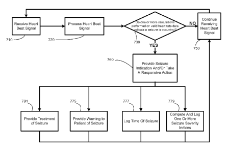
In an exemplary embodiment of the present invention, the method further
comprises
taking a responsive action based upon the identifying the seizure event. The
responsive
action may include providing a warning and/or notifying the patient or a
caregiver, logging
the time of a seizure, computing and storing one or more seizure severity
indices, or treating
the seizure event.
In one embodiment of the present invention, treating the seizure event
comprises
providing a neurostimulation therapy. The neurostimulation therapy may involve
applying an
electrical, mechanical, magnetic, electro-magnetic, photonic, acoustic, and/or
chemical signal
to a neural structure of the body. The neural structure may be a brain, a
spinal cord, a
peripheral nerve, a cranial nerve, or another neural structure. In a
particular embodiment, the
34
CA 02797268 2012-10-23
WO 2011/137235
PCT/US2011/034308
responsive action comprises treating the seizure by providing a cranial nerve
stimulation
therapy. Cranial nerve stimulation has been proposed to treat a number of
medical conditions
pertaining to or mediated by one or more structures of the nervous system,
including
epilepsy, movement disorders, depression, anxiety disorders and other
neuropsychiatric
disorders, dementia, traumatic brain injury, coma, migraine headache, obesity,
eating
disorders, sleep disorders, cardiac disorders (such as congestive heart
failure and atrial
fibrillation), hypertension, endocrine disorders (such as diabetes and
hypoglycemia), and pain
(including neuropathic pain and fibromyalgia), among others. See, e.g., U.S.
Pats. Nos.
4,867,164; 5,299,569; 5,269,303; 5,571,150; 5,215,086; 5,188,104; 5,263,480;
6,587,719;
6,609,025; 5,335,657; 6,622,041; 5,916,239; 5,707,400; 5,231,988; and
5,330,515. Despite
the numerous disorders for which cranial nerve stimulation has been proposed
or suggested
as a treatment option, the fact that detailed neural pathways and/or
mechanisms of action of
stimulation for many (if not all) cranial nerves, and/or the response of such
nerves to
exogenous stimulation, remain relatively poorly understood, which makes
predictions of
efficacy and identification of candidates for a therapy for any given disorder
difficult or
impossible.
In some embodiments, electrical neurostimulation may be provided by implanting
an
electrical device underneath the skin of a patient and delivering an
electrical signal to a nerve
such as a cranial nerve. In another alternative embodiment, the signal may be
generated by
an external pulse generator outside the patient's body, coupled by an RF or
wireless link to an
implanted electrode.
The cardiac data comprising a fiducial time marker for each of a plurality of
heart
beats can be gathered by any of a number of techniques. For example, the
cardiac data may
be gathered by an electrocardiogram (ECG) device. For another example, the
cardiac data
CA 02797268 2015-05-13
=
WO 2011/137235
PCT/US2011/034308
may be gathered by a cranial nerve stimulator device. In one embodiment, the
cardiac data may be
related to the R-waves of the beat sequence, such as a time series of R-waves
or a series of R-R
intervals. Those skilled in the art having benefit of the present disclosure
would appreciate that other
time series of cardiac waves and/or their fiducial points (e.g., P waves, T
waves, etc.) may be used
and still remain within the spirit and scope of the present invention.
Data relating to R-waves may be gathered by an ECG device or, in one
embodiment, by a
vagus nerve stimulator, such as described in U.S. Patent 5,928,272.
Receiving the cardiac data may comprise sensing a time of beat sequence of a
patient's heart
and generating a time series data stream from said time of the beat sequence.
In a further
embodiment, receiving the cardiac data of the patient's heart may comprise
sensing and time-
stamping a plurality of R waves, and generating the time series data stream
may comprise
determining a series of R-R intervals from the time stamps of the sensed R
waves.
In one embodiment, the fiducial time marker is an R wave peak or threshold
crossing. The
amplitude or height of one or more representative R waves may be used to set a
threshold that, when
reached or crossed, is registered as a fiducial time marker of a heart beat.
An interbeat interval can be calculated from a pair of said fiducial time
markers by any
appropriate technique. In one embodiment, the interbeat interval can be
calculated by subtracting the
time stamp of a first fiducial time marker from the time stamp of a second
fiducial time marker
following the first. In most existing HR sensing devices (for example,
exercise HR monitors), the
sensor element involves sensing R-wave peaks, and interbeat
36
CA 02797268 2012-10-23
WO 2011/137235
PCT/US2011/034308
intervals comprise R-R intervals constructed from the time stamps for the R-
wave peaks. In
the present invention, R-R intervals may be used, although any consistently
used fiducial
marker may be employed, such as P-P intervals, T-T intervals, etc.
Under certain recording conditions, cardiac data may be relatively noisy, and
thus,
spurious fiducial time markers may be collected and, as a result, spurious
interbeat intervals
may be generated. Including spurious heart beats and/or interbeat intervals in
later steps of
the method may lead to erroneous calculations of heart rate, which may in turn
result in
misidentifications of seizures (false positive detections) and/or failures to
identify seizures
(false negative detections). As a result, it is desirable to perform one or
more data quality
checks or routines to eliminate spurious heart beats or interbeat intervals
from consideration
by later steps of the method. Even where beats are not spurious, not all valid
beats may yield
acceptable results in cardiac-based seizure detection methods. Accordingly, in
some
embodiments, valid beats may be subjected to further testing (e.g., dispersion
testing in a test
window) to determine whether they are suitable for use in detecting seizures.
In one embodiment, a candidate heart beat is subjected to one or more quality
tests to
determine whether or not it is a valid beat suitable for further analysis, or
is an invalid beat
and should be ignored. While any number of beat quality tests may be employed,
the effects
of additional processing time and energy usage upon the power supply in
implantable devices
may result in a more limited number of tests. In one embodiment, a first beat
quality test may
be performed by determining if the candidate heart beat reflects an interbeat
interval (that is,
the interbeat interval calculated as the difference between the time stamp for
the candidate
heart beat and an immediately preceding heart beat) is not physiologically
plausible. A
second beat quality test may comprise determining if the candidate heart beat
reflects an
interbeat interval is so long as to appear to be due to a sinus arrest. A
third beat quality test
may involve determining if the candidate heart beat reflects an interbeat
interval is so short as
37
CA 02797268 2012-10-23
WO 2011/137235
PCT/US2011/034308
to appear to be due to noise. A fourth beat quality test may involve
calculating the absolute
value of the slope of the interbeat interval for the candidate beat and the
interbeat interval for
the immediately preceding valid beat (defined as the difference between the
candidate beat
interbeat interval and the interbeat interval defined by the immediately
preceding valid beat
and the 2nd immediately preceding valid beat, divided by the time difference
between the
candidate beat and the immediately preceding valid beat), and determining
whether that slope
is too large to be physiologically plausible. In one embodiment, the slope is
declared
implausible (and the candidate beat declared invalid) if the absolute value of
the slope is less
than or equal to 0.3. Other thresholds, such as an adaptable threshold, may be
used instead of
the fixed threshold of 0.3. It will be appreciated that additional or other
beat quality tests
may be applied, and that only some of the foregoing quality tests may be used.
Additionally,
it should be noted that HR and R-R intervals can be used interchangeably,
since they are
related by the simple formula RRi = 60/HR, where the R-R interval (RRi) is in
seconds and
HR is in BPM.
In one embodiment of the invention, the candidate heart beat is declared
invalid (in
other words, in some embodiments, not used for further analysis) if the
interbeat interval
between the candidate beat and the immediately preceding beat is so short that
the
instantaneous heart rate (IHR), defined as 60/interbeat interval, is greater
than about 180
beats per minute (BPM), 200 BPM, or 220 BPM, on the grounds that the human
heart cannot
have so high an IHR even in intense exertion. In one embodiment, the maximum
possible
heart rate is calculated as (220-patient's age in years) BPM, from which an
interbeat interval
corresponding thereto can be calculated and used as the minimum interbeat
interval for future
calculations for that patient. In one embodiment, the maximum heart rate is
defined as 180
BPM. In other embodiments, the patient's actual maximum HR (HRmax) is
determined
38
CA 02797268 2012-10-23
WO 2011/137235
PCT/US2011/034308
empirically by testing and the minimum interbeat interval can be either stored
directly from
the fiducial time marker stream or determined by the formula RRõõ. = 60/(HR.).
Conversely, if the interbeat interval associated with a candidate heart beat
is so long
that the IHR is less than an appropriate lower limit, then the candidate heart
beat can be
declared invalid, on the grounds the human heart cannot have so low an IHR,
even for the
heart of a person with extreme cardiovascular fitness at rest. Although 35 BPM
is an
appropriate lower limit on IHR for the vast majority of epilepsy patients,
another lower limit
can be established by the physician in consultation with the patient, or
determined empirically
from recording the patient's actual heart rate from which an interbeat
interval can be
calculated and used as the maximum interbeat interval for future calculations
for that patient.
Where maximum and/or minimum HR or RRi values are determined empirically for
an individual patient, appropriate values may be determined, for example, by
using a long-
term time window (e.g., 6 months, one month, two weeks, or other time period).
The
maximum rate may be an appropriate function of the actual measured heart rate
in the time
window. In one embodiment, this may be a target percentage in a Percentile
Tracking Filter
applied to the beats in the time window.
In another embodiment, if the interbeat interval is so long as to appear to be
due to
sinus arrest, a candidate heart beat associated with the interbeat interval
can be declared
invalid on the grounds the IHR cannot decelerate so rapidly from one beat to
the next.
Generally, if the interbeat interval is more than about 115% of the
immediately preceding
interbeat interval, then it may likely be that the interbeat interval is too
long to reflect a valid
beat and thus it can be concluded the candidate heart beat reflects a missed
heart beat.
A measured interbeat interval is especially likely to result from one or more
missed
heartbeats if its duration is some integer (or near-integer) multiple of 100%
times the
previous valid interbeat interval (e.g., 200% for one missed beat, 300% for
two missed beats,
39
CA 02797268 2012-10-23
WO 2011/137235
PCT/US2011/034308
etc.). This type of mathematical analysis may also be incorporated into the
beat validity
testing to specifically identify likely missed heartbeats (e.g. any measured
RRi that is within
200% +/- 10% (or other variation threshold) may be identified and logged as
being a likely
missed beat).
In another embodiment, if the interbeat interval is so short as to appear to
be due to
noise, a candidate heart beat associated with the interbeat interval can be
declared invalid on
the grounds the IHR cannot increase so rapidly from one beat to the next.
Generally, if the
interbeat interval is less than about 65% of the immediately preceding
interbeat interval, then
it is almost certainly the case that the interbeat interval is too short to
reflect a valid beat and
thus is can be concluded the candidate heart beat is due to noise or is
otherwise not valid.
In another embodiment, if the candidate heart beat yields an interbeat
interval having
an absolute value of the slope that is too large to be physiologically valid,
it can be declared
invalid on the grounds the IHR cannot accelerate or decelerate so rapidly.
In one embodiment, identifying valid beats comprises determining if each of
the
plurality of candidate beats falls within a plausibly physiological interval.
In one embodiment, the beat validity test comprises comparing the candidate
beat
interval to at least one of an upper and a lower beat interval seizure
duration threshold.
In one embodiment, the upper and lower beat interval seizure duration
thresholds are
derived from at least one of the patient's own heart beat data, and heart beat
data from a
sample patient population based upon one or more of brain state, sex, age,
weight, level of
activity, time of day, type of epilepsy, use of drugs or substances (such as
food) that affect
cardiac function, ambient temperature, body temperature, respiration, and
blood pressure,
among others.
In one embodiment, the at least one beat interval test comprises:
CA 02797268 2012-10-23
WO 2011/137235
PCT/US2011/034308
a) determining that the candidate beat interval corresponds to a heart rate
within
a range bound by a minimum heart rate and a maximum heart rate;
b) determining that the candidate beat interval is within an acceptable
percentage
of at least one of the immediately preceding valid beat interval or a recent
baseline heart rate
in a predetermined time window;
c) determining that the absolute slope of the current candidate beat
interval does
not correspond to a rate of acceleration or deceleration of heart rate that is
physiologically
improbable.
In a further embodiment, in the at least one beat interval test, the minimum
heart rate
is about 35 beats per minute and the maximum heart rate is about 180 beats per
minute; the
candidate beat interval is not more than about 115 percent of the greater of
the immediately
preceding valid beat interval or a recent baseline heart rate in a 30 second
time window, and
is at least about 65 percent of the immediately preceding valid beat interval;
and the absolute
slope of the current candidate beat interval is < 0.3. Other thresholds may be
used, and
thresholds may be altered over time according to the cardiac function of the
patient.
Although any one of the grounds set forth above may be sufficient to declare
invalid a
spurious candidate interbeat interval, and it is most computationally
efficient to declare
invalid a spurious candidate interbeat interval on the basis of a single
ground, two or more of
these grounds may be used to declare invalid a spurious candidate interbeat
interval to ensure
extremely high levels of data reliability for use in a seizure detection
algorithm. Noise and
artifacts are often coincident with seizures, so the aspects of this invention
that enable robust
identification of relevant cardiac rate changes in the presence of noise
and/or artifacts provide
improved methods for ensuring accurate and rapid identification of seizures.
In one embodiment, an index of beat quality may be established to characterize
the
reliability or robustness of candidate heart beats. The index of beat quality
is intended to
41
CA 02797268 2012-10-23
WO 2011/137235
PCT/US2011/034308
quantify how well a candidate heart beat has passed one or more beat tests.
The index can be
used to determine whether a particular test is useful or not (for example,
tests that are always
passed or always failed may not provide useful information on the whether
candidate heart
beats are reliable, and may be discontinued or replaced by other tests). The
index can also be
used as an indication (either alone or with other cardiac parameters) of a
seizure. In some
patients, seizures are associated with changes in sympathetic and
parasympathetic inputs to
the heart, as well as muscle artifacts in the case of tonic, clonic, and/or
tonic/clonic seizures.
Beat quality is lower because of these influences, and a decrease in beat
quality index may, in
at least some patients, be used to indicate or confirm the occurrence of a
seizure.
The index of beat quality may include one or more parameter(s) that may be
used to
quantify the quality of a candidate beat and/or or a validated beat. The index
of beat quality
may be used to track data quality to identify periods of time involving
relatively good quality
or relatively poor data quality. The beat quality tests described herein may
also be used to
determine the index of beat quality. In one embodiment, a counter may be
formed for each
candidate heart beat, and the counter is incremented for each test that the
candidate heart beat
passes. A time stream of data quality points for each beat, or an average or
other statistical
measure for a plurality of beats, may be used to indicate periods of time in
which data quality
is high, low, or otherwise within or outside of acceptable limits. In other
embodiments, a
warning or notification may be provided to a user interface to indicate
periods when data
quality may need to be addressed, such as when a sensor element or lead has
moved or
broken. In some cases, seizure detection, logging, or therapy delivery may be
automatically
disabled until the data quality returns to a certain level.
In one embodiment, the result(s) of the at least one beat validity test can be
used to
quantify the quality of a candidate heart beat or a fiducial time marker
therefor; setting a beat
quality index of said candidate heart beat to a first value; testing said
candidate heart beat
42
CA 02797268 2012-10-23
WO 2011/137235
PCT/US2011/034308
with at least one beat validity test; setting said beat quality index to at
least a second value,
wherein said second value is indicative of whether said at least one beat
validity test was
passed or failed; testing said candidate heart beat with at least a second
heart beat validity
test; setting said beat quality index to at least a third value, wherein said
third value is
indicative of whether said at least a second heart beat validity test was
passed or failed; and
performing a responsive action based upon the value of the beat quality index.
The
responsive action may be selected from the group consisting of:
indicating the occurrence of an epileptic seizure event;
delivering a neurostimulation therapy to the patient to treat a medical
condition;
warning at least one of a caregiver, the patient, or a physician; and
logging the beat quality index to a memory.
In a particular embodiment, the beat quality index is initialized to a first
value (in one
example -1 is used as the initial value) prior to receiving any information
about a candidate
beat. Upon the detection of a candidate beat, the resulting candidate
interbeat interval is
analyzed using a sequential set of beat interval tests and the beat quality
index is increased by
1 for each test passed. Consequently, if there are a total of 5 tests and each
are passed, the
beat quality index achieves a maximum score of 4. If the first 3 tests are
passed and the
fourth is failed, the candidate beat can be rejected and given a beat quality
score of 2. Then
by analyzing the sequence of beat quality indices, one may obtain a wealth of
useful
information, such as (i) the average beat quality index over a moving window
of time, (ii)
how often each applied test is passed and failed (providing information about
the importance
of such test relative to the others, which may be used to optimize
computational efficiency of
the algorithm for a particular beat detector and typical levels of noise in
the sequence of beat
detections), (iii) the identification of (and possible warning/logging/other
action taken due to)
periods of time when heart beat detection has poor accuracy, such as when a
long interval of
43
CA 02797268 2012-10-23
WO 2011/137235
PCT/US2011/034308
time occurs without any (or many) detected beats being considered valid (i.e.,
reaching beat
quality index of 4), (iv) intervals of time with very good heart beat
detection accuracy may be
similarly identified.
In another embodiment, a unique value may be provided based upon the outcome
of
each of the beat validity tests. For example, the BQI may comprise a matrix of
binary values
having a number of elements equal to the number of beat validity tests. For
each test that is
failed a 0 may be entered for the element associated with that test, and a 1
may be entered for
each test that is passed. Thus, a unique beat quality matrix may be provided
for each
candidate heart beat that indicates whether each BVT was passed or not.
In one embodiment, the method further comprises storing a time series of beat
quality
indices of a plurality of said candidate heart beats. In a further embodiment,
the method
further comprises determining a window beat quality window index comprising a
statistical
measure of central tendency for the individual beat quality index values of
the candidate beats
in at least a second window. The statistical measure of said beat quality
indices can be a
median, a mean, a trimean, a mode, a simple or exponentially-forgetting
Percentile Tracking
Filter such as described herein, among others. When the value of the window
beat quality
index is below a threshold value for window beat quality, the value of the
beat quality index
may be logged and used to indicate that a period of low data quality has
occurred.
In one embodiment, the second window can be chosen as a matter of routine
experimentation by the person of ordinary skill in the art having the benefit
of the present
disclosure. In one embodiment, the second window can be essentially the entire
time during
which the seizure detection method is being performed. In another embodiment,
the second
window can be a shorter time window. For example, it may be desirable to have
the beat
quality parameter for a time period encompassing a seizure event as well as
relatively short
time periods before and/or after a seizure event determined from one or more
cardiac
44
CA 02797268 2012-10-23
WO 2011/137235
PCT/US2011/034308
parameters. In one embodiment, the second window begins between 30 minutes
before a
seizure event and 30 minutes after a seizure event and has a duration from
about 5 seconds to
about 2 hours. The time periods can be optimize by a number of methods,
including manual
adjustment by a physician, automatically based upon one or more cardiac
parameters used for
detecting a seizure, or automatically based upon changes (e.g., a decrease) in
the beat quality
index itself The seizure window beat quality index may be determined from a
statistical
measure of central tendency from the individual beat quality indices for the
candidate heart
beats in the seizure window. In one embodiment, a method for quantifying the
quality of a
candidate heart beat comprises receiving a reference time marker for a signal
portion
representative of a candidate heart beat; determining a first quality
threshold for validating
said candidate heart beat; testing said candidate heart beat with a first beat
validity test using
at least said first quality threshold; determining a second quality threshold
for validating said
candidate heart beat; testing said candidate heart beat with a second beat
validity test using at
least said second quality threshold in response to a determination that said
first beat validity
test was satisfied by said candidate heart beat; and associating a beat
quality certification to
said candidate heart beat in response to a determination that said second beat
validity test was
satisfied by said candidate heart beat.
The information about quality, reliability, and robustness of the heart beat
detection
information, which results from quantifying beat quality, can also be used in
adapting the
cardiac-based seizure detection algorithm to improve its performance (e.g.,
sensitivity,
specificity, speed, and information yield). For example, in periods of time
that have
relatively high beat detection quality index, the system may have high
confidence in
information regarding cardiac dynamics that is extracted. This may be used to
adjust
detection thresholds and better learn both physiologic and abnormal cardiac
activity patterns
for the subject. Other periods in which the beat detection is operating
relatively poorly (as
CA 02797268 2012-10-23
WO 2011/137235
PCT/US2011/034308
measured by the statistics of beat quality index during a moving window), may
be avoided
for learning such information about cardiac activity patterns. Detection
decisions may also
be tempered during these times, for example, by raising the detection
thresholds to avoid
potential detections that could be due to noise in the beat detection system.
In another embodiment, there may be situations where it is desirable to have
data,
even if of possible low quality, to perform at least some calculations. Thus,
in one
embodiment, a constraint removal timer is used for at least one of the at
least one beat
validity tests. As described above, the constraint removal timer sets a
threshold for the time
since the last valid beat that, if exceeded, leads to a finding of validity of
the candidate heart
beat.
Even though declaring invalid spurious candidate heart beats by performing one
or
more of the above techniques may provide reliable data in most instances,
additional testing
of candidate heart beats may provide greater reliability and accuracy in
cardiac seizure
detection algorithms (CSDAs). Because CSDAs must discriminate between heart
rate
changes associated with seizures and similar increases associated with non-
pathologic events
(e.g., exercise, state changes such as standing, sitting, or lying down, etc),
the accuracy of the
CSDA depends in part upon highly accurate heart beat detection. Accordingly,
the present
invention may involve testing candidate heart beats beyond the immediate
interbeat interval.
In one embodiment, the invention involves testing candidate heart beats in a
time or number-
of-beats window to determine if the candidate heart beat would result in
excessive dispersion
of heart rate within the window. A candidate heart beat forming part of a
first window may
be discarded if the numbers of beats in the window, the fit error of the beats
in the window,
or both fall outside of acceptable limits.
For example, if the first time window is five seconds, and the number of heart
beats in
that time window is greater than about 15 or less than about 3, the number of
purported heart
46
CA 02797268 2012-10-23
WO 2011/137235
PCT/US2011/034308
beats indicates that the most recent purported heart beat (which may be, for
example, the
heart beat forming the present side of the window) should be flagged as
unsuitable for seizure
detection.
In some embodiments, the determination that a particular valid beat is not
suitable for
seizure detection is only temporary and is limited to a particular window
under analysis. That
is, if a valid first beat (the most recent beat in the window) fails to pass a
window analysis
test (along with one or more prior valid beats), the valid first beat may be
discarded only in
the sense that a detection decision is suspended for the immediate window. The
very next
valid beat, however (which is now a "new first beat" in a new window under
analysis) may
result in a window that passes the window analysis test, and a detection
decision (using the
"new" first beat and the previously "discarded" valid beat) may be allowed.
For another example, a least squares fit may be performed on a candidate heart
beat
and one or more prior beats in a window. A measure of short-term heart rate
variability
(HRVst) may be calculated, for example, as the mean square error of the least-
squares fit of
the heart beats in the window. If the mean squared error of the least squares
fit exceeds a
threshold, then the purported interbeat intervals can be concluded to possess
a fit error so
high that the interbeat intervals contain one or more artifacts, and thus the
candidate heart
beat (which in some embodiments is the only new data point in the window)
should be
ignored. The person of ordinary skill in the art can perform and/or program a
computer to
perform a least squares fit as a routine matter. In one embodiment, the
threshold is 0.25.
Other measures of short-term heart rate variability may also be used. For
example,
the absolute prediction error obtained by comparing the current interbeat
interval to its
predicted value, obtained using past interbeat intervals, can be used as a
measure of short-
term heart rate variability. The predicted value used for this process could
be a constant
predictor, a linear predictor, or a nonlinear predictor. A preferred predictor
would take into
47
CA 02797268 2012-10-23
WO 2011/137235
PCT/US2011/034308
account the distribution of interbeat intervals that have previously occurred
when preceded by
a sequence of interbeat intervals similar to those measured immediately
preceding the
moment at which the prediction is to be made. Measures correlated to the
amount of
curvature present in the interbeat interval sequence (or its counterpart¨
heart rate sequence)
may also be used and correspond to short time action by the body (e.g.,
sympathetic and
parasympathetic activity) to intervene to change the interbeat interval
(either by speeding up
or slowing down the heart). It is the short-time quantification of changes in
the interbeat
interval sequence which the window analysis measures are designed to
illuminate.
In one embodiment, the first window comprises one of:
a) a time window of from 1 to 10 seconds, bounded on the most recent end by
the first valid beat;
b) a number beats window comprising the first valid beat and a number of
immediately preceding beats ranging from 1-10; or
c) an exponentially forgetting window heavily weighted to the most recent 1
to
10 seconds, bounded on the most recent end by the first valid beat, or the
most recent 2-11
beats.
In a further embodiment, the at least one window test comprises at least one
of:
a) determining whether the mean square error of a least squares
linear fit of the
beats in the first window is < a predetermined heart rate variability
threshold.
In another further embodiment, the first window comprises a time window and
the at
least one window test comprises determining that the number of valid beats in
the window
exceeds a lower number of beats threshold.
The at least one window test described above may be used as part of a method
of
quantifying beat quality, as described above.
48
CA 02797268 2012-10-23
WO 2011/137235
PCT/US2011/034308
Indicating the occurrence of an epileptic seizure event can be based upon at
least one
of: a) the result of at least one of a first and a second beat validity tests;
or b) a first heart rate
parameter determined from candidate heart beats in a first window, and a
second heart rate
parameter determined from candidate heart beats in a second window. The first
heart rate
parameter can comprise a statistical measure of central tendency of heart rate
in a first time
window, which can have a duration of from about 2 seconds to about 30 seconds;
and the
second heart rate parameter can comprise a statistical measure of central
tendency of heart
rate within said second time window, which can have a duration of from about
30 seconds to
about 86,400 seconds.
An interbeat interval between the candidate heart beat and an immediately
preceding
candidate heart beat can be calculated; a first beat validity test can be
selected from the group
consisting of: a) testing whether the interbeat interval falls within a range
of physiologically
plausible interbeat intervals; b) testing whether the interbeat interval is
less than an upper
interbeat interval threshold; c) testing whether the interbeat interval is
greater than a lower
interbeat interval threshold; and d) testing whether a slope of change between
the interbeat
interval and one or more preceding interbeat intervals falls within a range of
physiologically
plausible slopes of change; and setting a beat quality index to a second value
can comprise
setting the beat quality index to a value indicative of the outcome of the
first beat validity
test.
The first value of the beat quality index can be set to a first integer; and
setting the
beat quality index to the second value can comprise adding 1 to the first
value.
An earlier responsive action can be modified based upon a value of a second
beat
quality index.
The first beat validity test and the second beat validity test each can be
selected from a
library of beat validity tests. Alternatively or in addition, each of the
first beat validity test
49
CA 02797268 2012-10-23
WO 2011/137235
PCT/US2011/034308
and the second beat validity test can comprise a plurality of tests. For
example, a beat
validity test can comprise a window test on at least one of a time window, a
number of beats
window, or an exponentially forgetting window having a first timeframe. The
second beat
validity test can determine at least one of a) whether a first heart rate
variability measure of a
plurality of candidate heart beats in the window is less than or equal to a
predetermined heart
rate variability threshold; or b) whether the window contains a sufficient
number of candidate
heart beats determined to be within a range of physiologically plausible heart
beats. Setting a
beat quality index to a third value can comprises adding 1 to the beat quality
index if the
window passes the at least one window test.
Beat quality index values for a plurality of candidate heart beats in a first
time series
can be stored as a second time series.
A third window can be determined. In one embodiment, the third time window
begins at a time between 30 minutes before said seizure and 30 minutes after
said seizure and
has a duration from about 5 seconds to about 2 hours.
Regardless of when the third time window begins and how long it lasts, a beat
quality
index for each candidate heart beat in the third window can be determined. A
window beat
quality index can be determined. The window beat quality index can comprise a
statistical
measure of central tendency of the beat quality indices for the candidate
heart beats in the
third window.
Any statistical measure of central tendency can be logged.
A beat quality index for a candidate heart beat can be set to an initial value
prior to
testing with the first beat validity test.
In one embodiment, the present invention relates to a method for quantifying
the
quality of a candidate heart beat, comprising receiving a reference time
marker for a signal
portion representative of a candidate heart beat; determining a first quality
criterion for
CA 02797268 2012-10-23
WO 2011/137235
PCT/US2011/034308
validating the candidate heart beat; testing the candidate heart beat with a
first beat validity
test using at least the first quality criterion; determining a second quality
criterion for
validating the candidate heart beat; testing the candidate heart beat with a
second beat validity
test using at least the second quality criterion; associating a beat quality
certification to the
candidate heart beat based on the first and second beat validity tests; and
performing at least
one responsive action based upon the value of the beat quality index, the
responsive action
selected from the group consisting of: indicating the occurrence of an
epileptic seizure event;
delivering a neurostimulation therapy to the patient to treat a medical
condition; warning at
least one of a caregiver, the patient, or a physician; and logging the beat
quality index to a
memory.
The first and second quality criteria can comprise first and second
thresholds.
The method can further comprise calculating an interbeat interval between the
candidate heart beat and an immediately preceding candidate heart beat;
wherein the first and
second second beat validity tests are selected from the group consisting of:
a) testing whether
the interbeat interval falls within a range of physiologically plausible
interbeat intervals; b)
testing whether the interbeat interval is less than an upper interbeat
interval threshold; c)
testing whether the interbeat interval is greater than a lower interbeat
interval threshold; and
d) testing whether a slope of change between the interbeat interval and one or
more preceding
interbeat intervals falls within a range of physiologically plausible slopes
of change; wherein
associating a beat quality certification to the candidate heart beat based on
the first and
second beat validity tests comprises setting the beat quality index to a value
indicative of the
outcome of the first and the second beat validity tests.
The candidate heart beat can be tested with a third beat validity test, and
associating a
beat quality certification to the candidate heart beat can comprise setting
the beat quality
index to a value indicative of the outcome of the first, the second, and the
third beat validity
51
CA 02797268 2012-10-23
WO 2011/137235
PCT/US2011/034308
tests. Alternatively, the beat quality index can be set to a unique value
indicative of the
outcome of each of the beat validity tests.
MODULE 3
Valid data suitable for seizure detection, as found by the above techniques,
is
available for further calculations to detect a seizure event. These further
calculations can
include the following Submodules of the Third Module.
Submodule 3A:
One series of further calculations comprises:
forming a second window for each valid beat suitable for seizure detection,
said
second window comprising a first valid beat suitable for seizure detection and
at least one
prior valid beat suitable for seizure detection;
determining a foreground heart rate parameter comprising a statistical measure
of
central tendency of heart rate in said second window;
forming a third window for each valid beat suitable for seizure detection,
said third
window comprising said first valid beat suitable for seizure detection from
said second
window and at least two prior valid beats suitable for seizure detection;
determining a background heart rate parameter comprising a statistical measure
of
central tendency of heart rate in said third window;
determining a relative heart rate comprising at least one of the ratio of said
foreground
and said background heart rate parameters and the ratio of said background and
said
foreground heart rate parameters; and
comparing said relative heart rate to a seizure threshold value associated
with an
epileptic seizure event.
A second window heart rate can be calculated from a second plurality of
interbeat
intervals calculated from data collected in a second time window. The second
time window
52
CA 02797268 2012-10-23
WO 2011/137235
PCT/US2011/034308
can be of any length. In one embodiment, the second time window is a time
window having
a duration of from about 3 sec to about 5 sec. In another embodiment, the
second window is
a number-of-beats window comprising from about 3 to about 15 beats. In another
embodiment, the second time window is an exponentially forgetting time window
having a
timescale from about 3 sec to about 5 sec or from about 3 beats to about 15
beats. In another
embodiment, the timescale or exponential forgetting decay factor can be made
adaptive,
increasing with increasing interbeat interval and decreasing with decreasing
interbeat
interval, or the reverse.
The foreground heart rate (FHR or FG HR) can be calculated as a statistical
measure
of central tendency of HR from the beats in the second window. Although
foreground heart
rate is used for purposes of discussion, it may be more accurate in some
instances to calculate
the foreground measure of central tendency of interbeat intervals in the
window instead of
heart rate. For example, one technique that may be used is calculation of the
mean of the
interbeat intervals determined from the beats in the second window. In another
embodiment,
the median may be used as the measure of central tendency. In a still further
embodiment,
the FHR may be a weighted heart rate, such as an exponentially forgetting
heart rate
determined from a statistical measure of central tendency of the beats in the
window.
In one embodiment, the foreground heart rate can be calculated from the
interbeat
intervals of the heart beats in the second window by use of a percentile
tracking filter (PTF).
A PTF has the advantage that any outliers that may pass the declaring invalid
steps, but
which would skew a calculation of the mean (sum of data points/number of data
points),
would be ignored. Generally, a PTF is used to track the (typically time-
varying) n-th
percentile of a set of data values in a moving window. When n=50, the 50-th
percentile of
the data is tracked, making the PTF output in this case somewhat akin to that
of a median
filter. Although the PTF is not an order statistic filter (order statistics
are much less
53
CA 02797268 2012-10-23
WO 2011/137235
PCT/US2011/034308
computationally efficient and more memory intensive and involve "sorting" of
data which the
PTF does not require), it manages to produce an output more quickly and
efficiently while
retaining the desirable outlier insensitivity of a (comparable percentile)
order statistic filter.
While the median filter is mentioned above because of its familiarity, as with
order statistic
filters (a.k.a. rank filters), the PTF may track percentiles of the set of
data values in a moving
window besides the 50th percentile. In particular, the PTF may take values
representing the
30th percentile. .
The PTF may use a simple set of values, a weighted set of values, or the other
techniques known to the person of ordinary skill in the art. More information
on PTFs is
given by US 6,768,968, issued July 27, 2004, US 6,904,390, issued June 7,
2005, US and US
7,188,053, issued March 6, 2007.
In addition to the foreground heart rate, embodiments of the invention further
comprise determining a background heart rate (BHR or BG HR) in a third window
larger
than the second window. Alternatively or in addition, a third time window
heart rate can be
calculated from a third plurality of interbeat intervals calculated from data
collected in a third
time window longer than the second time window.
In one embodiment, the third time window is 300 sec, or an exponentially
forgetting
window heavily weighted to the most recent 300 sec.
The third time window heart rate can be calculated from the mean or the PTF of
the
third plurality of interbeat intervals using techniques discussed above.
Alternatively or in addition, an instantaneous heart rate (IHR) can be
calculated from
the most recent interbeat interval. This can be routinely done as 60/interbeat
interval, with
resulting units of beats per minute (BPM or bpm).
The background heart rate can be calculated from the interbeat intervals of
the valid
beats in the third window. In one embodiment, the third window is a time
window longer
54
CA 02797268 2012-10-23
WO 2011/137235
PCT/US2011/034308
than the second window, and in a particular embodiment the third window is a
time window
having a duration of from 10 seconds to 86,400 seconds. In a more particular
embodiment
the third window is a 500 second time window. In another embodiment, the third
window is
a number-of-beats window comprising from about 30 to about 175,000 beats, and
in a
particular embodiment the third window is a 500 beat window. In another
embodiment, the
third window is an exponentially forgetting window with a timescale of about
10 sec to about
86,400 sec or about 30 beats to about 175,000 beats.
The BHR can be calculated as a statistical measure of central tendency of HR
from
the beats in the third window by techniques similar to those discussed
previously for the
foreground heart rate. Although BHR is used for purposes of discussion, it may
be more
accurate in some instances to calculate the background median value (as
measured, for
example, by a 50th percentile PTF) of interbeat intervals in the window
instead of heart rate.
For example, one technique that may be used is calculation of the mean of the
interbeat
intervals determined from the beats in the window. In another embodiment, the
median may
be used as the measure of central tendency. In a still further embodiment, the
BHR may be a
weighted heart rate, such as an exponentially forgetting heart rate determined
from a
statistical measure of central tendency of the beats in the window. In a
particular
embodiment, the BHR is calculated as a target percentile value (e.g., the 50th
percentile) of a
Percentile Tracking Filter applied to the beats in the third window.
In many embodiments, indicating the occurrence of a seizure event may be made
whenever it is determined that the RHR exceeds (i.e., first crosses) the
threshold associated
with a seizure event. However, in some embodiments, the indicating is made
only after one
or more additional requirements are satisfied. Such additional constraints,
which may include
a multiple additional constraints, may be helpful in eliminating or reducing
false positive
and/or false negative event detection indications.
CA 02797268 2012-10-23
WO 2011/137235
PCT/US2011/034308
For example, indicating the occurrence of a seizure may require, in addition
to RHR
instantaneously exceeding the seizure threshold (i.e., crossing the threshold
for a single heart
beat), that the RHR exceed the threshold for a desired duration, which defines
a seizure
duration threshold or duration constant. Thus, in one nonlimiting example, a
seizure
indication may only be generated when the RHR exceeds the threshold
continuously for 5
seconds.
The seizure duration threshold may also vary depending upon factors such as
those
discussed previously as providing a basis for making the first seizure
threshold itself adaptive
(i.e., the threshold is automatically modified based on the patient's level of
exertion, the time
of day, week, or month, false positive or negative seizure detections, changes
in the state,
high-risk activities such as swimming or driving, etc.). For example, the
exertional state of
the patient may be used to impose a duration constraint where none is usually
present (or to
remove a duration threshold otherwise present), such as a duration constraint
only during
periods of exercise, or during sleep or rest periods. In another nonlimiting
example, an
indication of a seizure is made only if RHR exceeds the seizure threshold for
15 seconds, but
if the patient has experienced a seizure event within the last hour, the
duration constraint is
reduced or eliminated altogether. In another non-limiting example, the
threshold value
associated with a seizure event is decreased if the subject's heart rate
following a seizure
remains above the subject's heart rate baseline for a given activity level,
time of day, etc. In
other instances, a duration constraint may be imposed or increased depending
upon particular
conditions.
In another embodiment, in addition to the FHR and BHR, one or more additional
heart rates may be calculated similarly to the foreground and background heart
rates
discussed above, differing primarily in the length of the time window used to
calculate these
56
CA 02797268 2012-10-23
WO 2011/137235
PCT/US2011/034308
additional heart rates. These additional heart rates may be termed "midground
heart rates" or
"medium-term heart rates" if the time window is intermediate in length to the
second window
and the third window, "ultra-foreground heart rates" or "very-short-term heart
rates" if the
time window is shorter than the second window, "ultra-background heart rates"
or "very-
long-term heart rates" if the time window is longer than the third window.
In another embodiment, the FHR, BHR, or RHR can be analyzed for patterns
indicative of a seizure event.
Submodule 3B:
Another series of further calculations comprises:
a) determining at least one short-term heart rate comprising at least one of
i) a first instantaneous heart rate from a first valid beat and the
immediately
preceding valid beat, or
ii) a fourth window heart rate comprising a statistical measure of central
tendency
of heart rate using said valid beats in said fourth window; and
b) comparing said at least one short-term heart rate to a short-
term heart rate
threshold associated with a seizure event.
In one embodiment, a short-term HR may comprise a first instantaneous HR using
the
first valid beat in the background HR window and the valid beat immediately
preceding the
first valid beat. In another embodiment, a short-term HR may comprise the
median HR for
the foreground HR window. In this embodiment, an indication of seizure
occurrence may be
made only if both the RHR seizure threshold is exceeded and the short-term HR
(however
measured) exceeds the short-term HR threshold.
57
CA 02797268 2012-10-23
WO 2011/137235
PCT/US2011/034308
A short-term HR threshold may be useful to a physician who finds, e.g., that
the RHR
threshold constraint alone yields an unacceptably high a number of false
positive seizure
indications. For example, if the short-term HR threshold value is set at 100
BPM, and is
combined with requirement that the RHR exceed 1.3, then a patient with a
resting heart rate
of 60 performing some mild exertion (climbing stairs, engaged in an
emotionally charged
encounter, etc.) which raises his heart rate to 80 (60 times 1.333) would not
be flagged as
having a seizure event because the short-term HR threshold constrain of 100
BPM was not
met.
Submodule 3C:
A third series of further calculations comprises:
a) determining a fifth window heart rate comprising a statistical measure of
central
tendency of heart rate using said valid beats in said fifth window;
b) determining a slope of the least squares linear fit of the beats in said
fifth
window;
c) comparing said fifth window heart rate to at least one of an upper fifth
window
heart rate threshold and a lower fifth window heart rate threshold associated
with a seizure
event;
d) comparing said slope of the least squares linear fit to at least one of
a lower
slope threshold and an upper slope threshold associated with a seizure event.
In one embodiment, detecting an epileptic seizure comprises the use of
Submodule
3A as discussed above.
58
CA 02797268 2012-10-23
WO 2011/137235
PCT/US2011/034308
In one embodiment of Submodule 3A, determining a foreground heart rate
parameter
comprises determining, from the valid beats in the second window, a target
percentile value
in a uniform distribution Percentile Tracking Filter.
In a further embodiment, the upper and lower bounds for the uniform
distribution are
adaptively determined for each second window based upon the maximum and
minimum beat
intervals in the second window.
In another further embodiment, the target percent value of the Percentile
Tracking
filter comprises a value in the range of 20 percent to 80 percent. In an even
further
embodiment, the Percentile Tracking Filter comprises an adaptive uniform
distribution with
endpoint parameters determined using exponential forgetting factor, with a
prescribed
forgetting factor corresponding to a 5 sec timescale, and the target percent
value of the
Percentile Tracking Filter is 30 percent.
In one embodiment, determining a background heart rate parameter comprises
determining, from the valid beats in the third window, a target percentile
value in an adaptive
uniform distribution Percentile Tracking Filter.
In one embodiment, the method further comprises determining a duration of time
that
the relative heart rate exceeds the first seizure threshold, and wherein
indicating the
occurrence of a seizure event is only performed if the duration exceeds a
seizure duration
threshold.
In one embodiment, indicating the occurrence of a seizure event comprises
generating
a signal if the relative heart rate exceeds the first seizure threshold if one
or more of
a) said relative heart rate exceeds said first seizure threshold;
b) said fifth window heart rate is below said upper fifth window heart rate
threshold and exceeds said lower fifth window heart rate threshold; or
c) said slope of the least squares linear fit at least one of
59
CA 02797268 2012-10-23
WO 2011/137235
PCT/US2011/034308
i) exceeds said lower slope threshold and
ii) is below said upper slope threshold.
In some embodiments, one or more of the above thresholds may, in certain
conditions,
be set to +/- infinity (to either block detection or make this detection
component always in a
state of detection). More generally, if the relative heart rate is within a
prescribed range or
takes on certain values, as represented by appropriate thresholds, all these
measures together
may be considered with a appropriate weighting factors to determine whether or
not to issue a
detection decision.
In particular embodiments, one, two or all three of the foregoing conditions
may be
required to issue a seizure detection indication. In alternative embodiments,
additional
constraints may also be required to be met before issuing an indication of a
seizure detection.
Thus, anyone or more of the three series of calculations set forth above may
be combined by
any appropriate logical operator, e.g., AND, OR, XOR, NOT, or the like, and/or
grouped
together (e.g., of the form "x AND (y OR z)," among others). For example, a
short term heart
rate more than, e.g., 120% of the background HR for more than 15 sec may be
indicative of a
seizure, alone or in combination with a RHR threshold constraint. For another
nonlimiting
example, a slope of heart rate more than, e.g., 0.03 beats/second for 5 sec
may be indicative
of a seizure. The precise values of the thresholds and durations can be set by
the physician in
consultation with the patient or adaptively.
However the seizure event is identified, upon detection of the seizure event,
in some
embodiments, a responsive action may be taken selected from warning, logging
the time of a
seizure, computing and storing one or more seizure severity indices, or
treating the seizure.
A seizure event warning may be given as, for example, a warning tone or light
implemented by a medical device or a device adapted to receive indications of
the seizure; as
CA 02797268 2012-10-23
WO 2011/137235
PCT/US2011/034308
an automated email, text message, telephone call, or video message sent from a
medical
device or a unit in communication with a medical device to the patient's
cellular telephone,
PDA, computer, television, 911 or another emergency contact number for
paramedic/EMT
services, etc. Such a warning may allow the patient or his or her caregivers
to take measures
protective of patient's well-being and those of others, e.g., pulling out of
traffic and turning
off a car, when the patient is driving; stopping the use of machinery,
contacting another adult
if the patient is providing childcare, removing the patient from a swimming
pool or bathtub,
lying down or sitting if the patient is standing, etc.
The time may be logged by receiving an indication of the current time and
associating
the indication of the current time with an indication of the seizure.
Seizure severity indices may be calculated and stored by appropriate
techniques and
apparatus.
The seizure may be treated by appropriate techniques, such as those discussed
below.
The treatment may be one or more treatments known in the art. In one
embodiment, the
treatment comprises at least one of applying an electrical signal to a neural
structure of a
patient; delivering a drug to a patient; or cooling a neural structure of a
patient. When the
treatment comprises applying an electrical signal to a portion of a neural
structure of a
patient, the neural structure may be at least one of a portion of a brain
structure of the patient,
a portion of a cranial nerve of a patient, a portion of a spinal cord of a
patient, a portion of a
sympathetic nerve structure of the patient, a portion of a parasympathetic
nerve structure of
the patient, and/or a portion of a peripheral nerve of the patient.
Though not intended to be bound by theory, in certain circumstances, a seizure
may
be identified from heart rate data at a time before seizure onset as
determined by
61
CA 02797268 2012-10-23
WO 2011/137235
PCT/US2011/034308
electroencephalography, observation by a physician or knowledgeable layman, or
both. The
time before onset may range from a few seconds up to a few minutes. As such,
certain
embodiments of the method may be considered to yield a prediction of a
seizure. It should be
noted that the prediction may sometimes be a false positive. However,
depending on a
physician's judgment, his or her understanding of the devices in use, and the
patient's
condition, a certain amount of false positives may be tolerable.
The above method can be performed alone. In one embodiment, the above method
can be performed in combination with a continuous or open-loop therapy for
epilepsy. In one
embodiment, the above method is performed to take action in response to the
detection of the
seizure, and at all or most other times, a chronic therapy signal is applied
to a target structure
in the patient's body. In one embodiment, the target structure is a cranial
nerve, such as the
vagus nerve.
In one embodiment, the method described above comprises:
obtaining a time series of fiducial time markers for candidate heart beats;
identifying valid beats from the candidate heart beats, as described above;
accepting as valid beats the candidate beats that pass the at least one beat
validity test;
and
detecting an epileptic seizure by at least one of the series of calculations
described
above. In other words, in this embodiment, the method can be performed without
performing
a window test.
In one embodiment, the method described above comprises:
obtaining a time series of fiducial time markers for candidate heart beats;
and
62
CA 02797268 2012-10-23
WO 2011/137235
PCT/US2011/034308
detecting an epileptic seizure by at least one of the series of calculations
described
above. In other words, in this embodiment, the method can be performed without
performing
both a beat validity test and a window test.
Embodiments wherein a beat validity test and/or a window test are not
performed may
be particularly suitable for situations wherein the quality of fiducial time
markers for
candidate heart beats is very high, or where the practitioner would find
acceptable a possible
higher rate of false positive seizure event determinations resulting from
"noisy" data.
Although not limited to the following, an exemplary system capable of
implementing
embodiments of the present invention is described below. Figure lA depicts a
stylized
implantable medical system (IMD) 100 for implementing one or more embodiments
of the
present invention. An electrical signal generator 110 is provided, having a
main body 112
comprising a case or shell with a header 116 for connecting to an insulated,
electrically
conductive lead assembly 122. The generator 110 is implanted in the patient's
chest in a
pocket or cavity formed by the implanting surgeon just below the skin
(indicated by a dotted
line 145), similar to the implantation procedure for a pacemaker pulse
generator.
A nerve electrode assembly 125, preferably comprising a plurality of
electrodes
having at least an electrode pair, is conductively connected to the distal end
of the lead
assembly 122, which preferably comprises a plurality of lead wires (one wire
for each
electrode). Each electrode in the electrode assembly 125 may operate
independently or
alternatively, may operate in conjunction with the other electrodes. In one
embodiment, the
electrode assembly 125 comprises at least a cathode and an anode. In another
embodiment,
the electrode assembly comprises one or more unipolar electrodes.
63
CA 02797268 2012-10-23
WO 2011/137235
PCT/US2011/034308
Lead assembly 122 is attached at its proximal end to connectors on the header
116 of
generator 110. The electrode assembly 125 may be surgically coupled to the
vagus nerve 127
in the patient's neck or at another location, e.g., near the patient's
diaphragm or at the
esophagus/stomach junction. Other (or additional) cranial nerves such as the
trigeminal
and/or glossopharyngeal nerves may also be used to deliver the electrical
signal in particular
alternative embodiments. In one embodiment, the electrode assembly 125
comprises a
bipolar stimulating electrode pair 126, 128 (i.e., a cathode and an anode).
Suitable electrode
assemblies are available from Cyberonics, Inc., Houston, Texas, USA as the
Model 302
electrode assembly. However, persons of skill in the art will appreciate that
many electrode
designs could be used in the present invention. In one embodiment, the two
electrodes are
wrapped about the vagus nerve, and the electrode assembly 125 may be secured
to the vagus
nerve 127 by a spiral anchoring tether 130 such as that disclosed in U.S. Pat.
No. 4,979,511
issued Dec. 25, 1990 to Reese S. Terry, Jr. Lead assembly 122 may be secured,
while
retaining the ability to flex with movement of the chest and neck, by a suture
connection to
nearby tissue (not shown).
In alternative embodiments, the electrode assembly 125 may comprise
temperature
sensing elements, blood pressure sensing elements, and/or heart rate sensor
elements. Other
sensors for other body parameters may also be employed. Both passive and
active
stimulation may be combined or delivered by a single IMD according to the
present
invention. Either or both modes may be appropriate to treat a specific patient
under
observation.
In alternative embodiments, the implantable medical device system further
comprises
an electrical stimulator comprising an electrode 160 (not to scale) adapted to
be coupled to
the spinal cord 180 (Figure 1B) or to a region of the brain 190 (Figure 1C).
The physician
64
CA 02797268 2012-10-23
WO 2011/137235
PCT/US2011/034308
can select precise locations for coupling to the spinal cord 180 or brain 190
based on his or
her observations of the patient's medical condition, among other values. In
various
embodiments, the implantable medical device system may comprise one, two, or
three of the
IMD 100, the spinal cord stimulator, and the brain stimulator.
The electrical pulse generator 110 may be programmed with an external device
(ED)
such as computer 150 using programming software known in the art. A
programming wand
155 may be coupled to the computer 150 as part of the ED to facilitate radio
frequency (RF)
communication between the computer 150 and the pulse generator 110. The
programming
wand 155 and computer 150 permit non-invasive communication with the generator
110 after
the latter is implanted. In systems where the computer 150 uses one or more
channels in the
Medical Implant Communications Service (MICS) bandwidths, the programming wand
155
may be omitted to permit more convenient communication directly between the
computer
150 and the pulse generator 110.
Turning now to Figures 2A-2F, block diagram depictions of exemplary medical
devices 200 are provided, in accordance with various illustrative embodiments
of the present
invention.
In some embodiments, the medical device 200 may be implantable (such as
implantable electrical signal generator 110 from Figure 1), while in other
embodiments the
medical device 200 may be completely external to the body of the patient.
The medical device 200 (such as generator 110 from Figure 1) may comprise a
controller 210 capable of controlling various aspects of the operation of the
medical device
200. The controller 210 is capable of receiving internal data or external
data, and in one
embodiment, is capable of causing a stimulation unit 220 (Figures 2B, 2D, 2F)
to generate
CA 02797268 2012-10-23
WO 2011/137235
PCT/US2011/034308
and deliver an electrical signal to target tissues of the patient's body for
treating a medical
condition. For example, the controller 210 may receive manual instructions
from an operator
externally, or may cause the electrical signal to be generated and delivered
based on internal
calculations and programming. In other embodiments, the medical device 200
does not
comprise a stimulation unit 220 (Figures 2A, 2C, 2E). In either embodiment,
the controller
210 is capable of affecting substantially all functions of the medical device
200.
The controller 210 may comprise various components, such as a processor 215, a
memory 217, etc. The processor 215 may comprise one or more microcontrollers,
microprocessors, etc., capable of performing various executions of software
components.
The memory 217 may comprise various memory portions where a number of types of
data
(e.g., internal data, external data instructions, software codes, status data,
diagnostic data,
etc.) may be stored. The memory 217 may comprise one or more of random access
memory
(RAM), dynamic random access memory (DRAM), electrically erasable programmable
read-
only memory (EEPROM), flash memory, etc.
As stated above, in one embodiment, the medical device 200 may also comprise a
stimulation unit 220 capable of generating and delivering electrical signals
to one or more
electrodes 126, 128 via leads 201 (Figures 2B, 2D, 2F). A lead assembly such
as lead
assembly 122 (Figure 1) may be coupled to the medical device 200. Therapy may
be
delivered to the leads 201 comprising the lead assembly 122 by the stimulation
unit 220
based upon instructions from the controller 210. The stimulation unit 220 may
comprise
various circuitry, such as electrical signal generators, impedance control
circuitry to control
the impedance "seen" by the leads, and other circuitry that receives
instructions relating to
the delivery of the electrical signal to tissue. The stimulation unit 220 is
capable of
delivering electrical signals over the leads 201 comprising the lead assembly
122. As should
66
CA 02797268 2012-10-23
WO 2011/137235
PCT/US2011/034308
be apparent, in certain embodiments, the medical device 200 does not comprise
a stimulation
unit 220, lead assembly 122, or leads 201.
In other embodiments, a lead 201 is operatively coupled to an electrode,
wherein the
electrode is adapted to couple to at least one of a portion of a brain
structure of the patient, a
cranial nerve of a patient, a spinal cord of a patient, a sympathetic nerve
structure of the
patient, or a peripheral nerve of the patient.
The medical device 200 may also comprise a power supply 230. The power supply
230 may comprise a battery, voltage regulators, capacitors, etc., to provide
power for the
operation of the medical device 200, including delivering the therapeutic
electrical signal.
The power supply 230 comprises a power source that in some embodiments may be
rechargeable. In other embodiments, a non-rechargeable power source may be
used. The
power supply 230 provides power for the operation of the medical device 200,
including
electronic operations and the electrical signal generation and delivery
functions. The power
supply 230 may comprise a lithium/thionyl chloride cell or a lithium/carbon
monofluoride
(LiCFx) cell if the medical device 200 is implantable, or may comprise
conventional watch or
9V batteries for external (i.e., non-implantable) embodiments. Other battery
types known in
the art of medical devices may also be used.
The medical device 200 may also comprise a communication unit 260 capable of
facilitating communications between the medical device 200 and various
devices. In
particular, the communication unit 260 is capable of providing transmission
and reception of
electronic signals to and from a monitoring unit 270, such as a handheld
computer or PDA
that can communicate with the medical device 200 wirelessely or by cable. The
communication unit 260 may include hardware, software, firmware, or any
combination
thereof
67
CA 02797268 2012-10-23
WO 2011/137235
PCT/US2011/034308
The medical device 200 may also comprise one or more sensor(s) 212 coupled via
sensor lead(s) 211 to the medical device 200. The sensor(s) 212 are capable of
receiving
signals related to a physiological parameter, such as the patient's heart
beat, blood pressure,
and/or temperature, and delivering the signals to the medical device 200. In
one
embodiment, the sensor(s) 212 may be the same as implanted electrode(s) 126,
128 (Figure
1). In other embodiments, the sensor(s) 212 are external structures that may
be placed on the
patient's skin, such as over the patient's heart or elsewhere on the patient's
torso.
In one embodiment, the medical device 200 may comprise a cardiac data
collection
module 265 that is capable of collecting cardiac data comprising fiducial time
markers of
each of a plurality of heart beats. The cardiac data collection module 265 may
also process or
condition the cardiac data. The cardiac data may be provided by the sensor(s)
212. The
cardiac data collection module 265 may be capable of performing any necessary
or suitable
amplifying, filtering, and performing analog-to-digital (A/D) conversions to
prepare the
signals for downstream processing. The cardiac data collection module, in one
embodiment,
may comprise software module(s) that are capable of performing various
interface functions,
filtering functions, etc., to process fiducial time markers of each of a
plurality of heart beats.
In another embodiment the cardiac data collection module 265 may comprise
hardware
circuitry that is capable of performing these functions. In yet another
embodiment, the
cardiac data collection module 265 may comprise hardware, firmware, software
and/or any
combination thereof A more detailed illustration of the cardiac data
collection module 265 is
provided in Figure 3A and accompanying description below.
The cardiac data collection module 265 is capable of collecting cardiac data
comprising fiducial time markers of each of a plurality of candidate heart
beats and providing
the collected cardiac data to an heart beat/interval determination module 275.
Based upon the
68
CA 02797268 2012-10-23
WO 2011/137235
PCT/US2011/034308
signals processed by the cardiac data collection module 265, the heart
beat/interval
determination module 275 may calculate an interbeat interval from a
consecutive pair of said
fiducial time markers and store such interbeat interval or forward it on for
further
processing/analysis. The heart beat/interval determination module 275 may
comprise
software module(s) that are capable of performing various interface functions,
filtering
functions, etc., to calculate interbeat intervals. In another embodiment the
heart beat/interval
determination module 275 may comprise hardware circuitry that is capable of
performing
these functions. In yet another embodiment, the heart beat/interval
determination module 275
may comprise hardware, firmware, software and/or any combination thereof
Further
description of the heart beat/interval determination module 275 is provided in
Figure 3B and
accompanying description below.
The heart beat/interval determination module 275 is capable of calculating an
interbeat interval from a consecutive pair of said fiducial time markers and
providing the
interbeat interval to the heart beat validation module 285. Based upon the
interbeat interval
received by the heart beat validation module 285, it performs any operations
desired to
identify invalid interbeat intervals and discard them. For example, the heart
beat validation
module 285 may discard the candidate heart beat if the interbeat interval
formed from the
candidate heart beat and the immediately preceding valid beat is not
physiologically valid, is
so long as to appear to be due to a missed heart beat, is so short as to
appear to be due to
noise, has an absolute value of the slope of the interbeat interval that is
too large to be
physiologically valid, or two or more thereof The heart beat validation module
285 may
comprise software module(s) that are capable of performing various interface
functions,
filtering functions, etc., to discard invalid beats. In another embodiment the
heart beat
validation module 285 may comprise hardware circuitry that is capable of
performing these
69
CA 02797268 2012-10-23
WO 2011/137235
PCT/US2011/034308
functions. In yet another embodiment, the heart beat validation module 285 may
comprise
hardware, firmware, software and/or any combination thereof Further
description of the
heart beat validation module 285 is provided in Figure 3C and accompanying
description
below.
The heart beat validation module 285 is capable of declaring invalid beats and
forwarding a plurality of heart beats accepted as valid to window analysis
module 295.
Based upon the plurality of valid beats received by the window analysis module
295, it
performs any operations desired to perform further testing of the valid beats
in one or more
heart beat windows to identify valid beats suitable for seizure detection. For
example, the
window analysis module 295 may discard a plurality of valid beats as
unsuitable for seizure
detection if the number, the heart rate variability, or both of a window
analysis performed on
the valid beat in a backward-looking window fail to pass a number-of-beats
threshold and/or
a HRV threshold. The window analysis module 295 may comprise software
module(s) that
are capable of performing various interface functions, filtering functions,
etc., to reject valid
beats as unsuitable for seizure detection. In another embodiment the window
analysis
module 295 may comprise hardware circuitry that is capable of performing these
functions.
In yet another embodiment, the window analysis module 295 may comprise
hardware,
firmware, software and/or any combination thereof Further description of the
window
analysis module 295 is provided in Figure 3D and accompanying description
below.
The window analysis module 295 is capable of ignoring one or more valid beats
that
are unsuitable for seizure detection, and forwarding a plurality of valid
interbeat intervals that
are suitable for seizure detection to foreground/background module 297 (Figure
3F). (The
terms "flagging," "ignoring," and "discarding" may be used herein to refer to
not using one or
more valid beats for seizure detection). Based upon the plurality of interbeat
intervals
CA 02797268 2012-10-23
WO 2011/137235
PCT/US2011/034308
received from the window analysis module 295, the foreground/background module
297
performs any operations not performed in prior modules (such as interbeat
interval
calculation module 275, heart beat validation module 285, and window analysis
module 295)
and necessary or desirable for use in detection of seizures. In addition to
determination of
foreground HR and background HR, foreground/background module 297 may
calculate, for
example, various heart rates, durations, slopes, HRV measures, or other
parameters
associated with the foreground and background windows., For example, the
foreground/background module 297 may calculate a second time window heart rate
from a
plurality of consecutive interbeat intervals calculated from data collected in
a second time
window, a third time window heart rate from a plurality of consecutive
interbeat intervals
calculated from data collected in a third time window, a first instantaneous
heart rate from
said first valid beat and the immediately preceding valid beat, a fourth
window heart rate
comprising a statistical measure of central tendency of heart rate using said
valid beats in said
first window, a fifth window heart rate comprising a statistical measure of
central tendency of
heart rate using valid beats in a fifth window, slope of the least squares
linear fit of the beats
in said fifth window, or two or more thereof
The foreground/background module 297 may comprise software module(s) that are
capable of performing various interface functions, filtering functions, etc.,
to calculate the
various heart rates, slopes of heart rate series, durations of heart rates or
slopes of heart rates
above various seizure threshold values, or the like. In another embodiment
the
foreground/background module 297 may comprise hardware circuitry that is
capable of
performing these functions. In yet another embodiment, the
foreground/background module
297 may comprise hardware, firmware, software and/or any combination thereof
Further
description of the foreground/background module 297 is provided in Figure 3F
and
accompanying description below.
71
CA 02797268 2012-10-23
WO 2011/137235
PCT/US2011/034308
The foreground/background module 297 is capable of calculating various heart
rates,
slopes of heart rate series, durations of heart rates or slopes of heart rates
above various
seizure threshold values, or the like and forwarding the calculated
information to seizure
detection module 299 (Figure 3G). Based upon the calculated information
received by the
seizure detection module 299, it performs any operations desired to identify a
seizure event.
For example, the seizure detection module 299 may identify a seizure event
based on one or
more of a foreground HR from the second window heart rate from a plurality of
valid beats
suitable for seizure detection in the second window, the background heart rate
from a
plurality of valid beats suitable for seizure detection in the third window, a
duration that a
short-term heart rate measure exceeds a short-term heart rate threshold, a
duration that a slope
of a short-term heart rate , whether a short-term HRV measure exceeds an HRV
threshold,
two or more of the foregoing, or at least one or more relationships between
two or more of
the foregoing. The seizure detection module 299 may comprise software
module(s) that are
capable of performing various interface functions, filtering functions, etc.,
to identify a
seizure event. In another embodiment the seizure detection module 299 may
comprise
hardware circuitry that is capable of performing these functions. In yet
another embodiment,
the seizure detection module 299 may comprise hardware, firmware, software
and/or any
combination thereof Further description of the seizure detection module 299 is
provided in
Figure 3G and accompanying description below.
Figures 2C-2D depict the heart beat/interval determination module 275, the
heart beat
validation module 285, the window analysis module 295, along with a beat
quality index
module 286, as components of a beat quality analysis module 276. The beat
quality index
module 286 is capable of setting an initial value of a beat quality index for
a candidate heart
beat, receiving information from heart beat validation module 285 about beat
validity test(s)
passed and/or failed by a candidate heart beat, receiving information from
window analysis
72
CA 02797268 2012-10-23
WO 2011/137235
PCT/US2011/034308
module 295 about window test(s) passed and/or failed by a window comprising a
valid beat,
and adjusting the value of the beat quality index based on the received
information. As
should be apparent, even though various modules are shown as components of
beat quality
analysis module 276, they may also be capable of functions not related to
quantifying beat
quality.
In another embodiment, in addition to the heart beat/interval determination
module
275, the heart beat validation module 285, and the window analysis module 295,
Figures 2E-
2F depict a constraint modification unit 287. The constraint modification unit
287 modifies
at least one constraint for one or more of the at least one beat validity
tests performed by the
heart beat/interbeat interval determination module 275 and determines, in the
event of a
finding of an invalid beat by the beat validity test(s) (i.e., failure of a
candidate heart beat to
pass one or more tests), if the time since the last valid beat is greater than
the threshold. If the
time since the last valid beat is greater than the threshold, the constraint
modification unit 287
modifies the constraint, such as by relaxing the constraint or tightening the
constraint. This
may make the test easier or more difficult, respectively, for future candidate
beats to pass. If
the candidate heart beat was found valid by the test(s), the constraint
modification unit 287
resets the timer since the last valid beat (because the candidate heart beat
has become the last
valid beat) and, if the constraint is modified during timing by the timer,
resets the constraint
to its initial value.
In addition to components of the medical device 200 described above, an
implantable
medical system may comprise a storage unit to store an indication of at least
one of seizure or
an increased risk of a seizure or an index of beat quality. The storage unit
may be the
memory 217 of the medical device 200, another storage unit of the medical
device 200, or an
external database, such as the local database unit 255 or a remote database
unit 250. The
73
CA 02797268 2012-10-23
WO 2011/137235
PCT/US2011/034308
medical device 200 may communicate the indication via the communications unit
260.
Alternatively or in addition to an external database, the medical device 200
may be adapted to
communicate the indication to at least one of a patient, a caregiver, or a
healthcare provider.
In various embodiments, one or more of the units or modules described above
may be
located in a monitoring unit 270 or a remote device 292, with communications
between that
unit or module and a unit or module located in the medical device 200 taking
place via
communication unit 260. For example, in one embodiment, one or more of the
cardiac data
collection module 265, the heart beat/interval determination module 275, the
heart beat
validation module 285, the window analysis module 295, the
foreground/background module
297, the beat quality index module 286, the beat quality analysis module 276,
the constraint
modification unit 287, or the seizure detection module 299 may be external to
the medical
device 200, e.g., in a monitoring unit 270. Locating one or more of the
cardiac data
collection module 265, the heart beat/interval determination module 275, the
heart beat
validation module 285, the window analysis module 295, the
foreground/background module
297, the beat quality index module 286, the beat quality analysis module 276,
the constraint
modification unit 287, or the seizure detection module 299 outside the medical
device 200
may be advantageous if the calculation(s) is/are computationally intensive, in
order to reduce
energy expenditure and heat generation in the medical device 200 or to
expedite calculation.
The monitoring unit 270 may be a device that is capable of transmitting and
receiving
data to and from the medical device 200. In one embodiment, the monitoring
unit 270 is a
computer system capable of executing a data-acquisition program. The
monitoring unit 270
may be controlled by a healthcare provider, such as a physician, at a base
station in, for
example, a doctor's office. In alternative embodiments, the monitoring unit
270 may be
controlled by a patient in a system providing less interactive communication
with the medical
74
CA 02797268 2012-10-23
WO 2011/137235
PCT/US2011/034308
device 200 than another monitoring unit 270 controlled by a healthcare
provider. Whether
controlled by the patient or by a healthcare provider, the monitoring unit 270
may be a
computer, preferably a handheld computer or PDA, but may alternatively
comprise any other
device that is capable of electronic communications and programming, e.g.,
hand-held
computer system, a PC computer system, a laptop computer system, a server, a
personal
digital assistant (PDA), an Apple-based computer system, a cellular telephone,
etc. The
monitoring unit 270 may download various parameters and program software into
the
medical device 200 for programming the operation of the medical device, and
may also
receive and upload various status conditions and other data from the medical
device 200.
Communications between the monitoring unit 270 and the communication unit 260
in the
medical device 200 may occur via a wireless or other type of communication,
represented
generally by line 277 in Figure 2. This may occur using, e.g., wand 155
(Figure 1) to
communicate by RF energy with an implantable signal generator 110.
Alternatively, the
wand may be omitted in some systems, e.g., systems in which the MD 200 is non-
implantable, or implantable systems in which monitoring unit 270 and MD 200
operate in the
MICS bandwidths.
In one embodiment, the monitoring unit 270 may comprise a local database unit
255.
Optionally or alternatively, the monitoring unit 270 may also be coupled to a
database unit
250, which may be separate from monitoring unit 270 (e.g., a centralized
database wirelessly
linked to a handheld monitoring unit 270). The database unit 250 and/or the
local database
unit 255 are capable of storing various patient data. These data may comprise
patient
parameter data acquired from a patient's body, therapy parameter data, seizure
severity data,
and/or therapeutic efficacy data. The database unit 250 and/or the local
database unit 255
may comprise data for a plurality of patients, and may be organized and stored
in a variety of
CA 02797268 2012-10-23
WO 2011/137235
PCT/US2011/034308
manners, such as in date format, severity of disease format, etc. The database
unit 250 and/or
the local database unit 255 may be relational databases in one embodiment. A
physician may
perform various patient management functions (e.g., programming parameters for
a
responsive therapy and/or setting thresholds for one or more detection
parameters) using the
monitoring unit 270, which may include obtaining and/or analyzing data from
the medical
device 200 and/or data from the database unit 250 and/or the local database
unit 255. The
database unit 250 and/or the local database unit 255 may store various patient
data.
One or more of the blocks illustrated in the block diagram of the medical
device 200
in Figures 2A-2F, may comprise hardware units, software units, firmware units,
or any
combination thereof Additionally, one or more blocks illustrated in Figure 2A-
2F may be
combined with other blocks, which may represent circuit hardware units,
software
algorithms, etc. Additionally, any number of the circuitry or software units
associated with
the various blocks illustrated in Figure 2A-2F may be combined into a
programmable device,
such as a field programmable gate array, an ASIC device, etc.
The medical device system of one embodiment of the present invention provides
for
software module(s) that are capable of acquiring, storing, and processing
various forms of
data, such as patient data/parameters (e.g., physiological data, side-effects
data, heart rate
data, breathing rate data, brain-activity parameters, disease progression or
regression data,
quality of life data, etc.) and therapy parameter data. Therapy parameters may
include, but
are not limited to, electrical signal parameters (e.g., frequency, pulse
width, wave shape,
polarity, on-time, off-time, etc.) that define therapeutic electrical signals
delivered by the
medical device in response to the detection of the seizure, medication type,
dose, or other
parameters, and/or any other therapeutic treatment parameter.
76
CA 02797268 2012-10-23
WO 2011/137235
PCT/US2011/034308
In one embodiment, the present invention may include coupling of at least one
electrode to each of two or more cranial nerves. (In this context, two or more
cranial nerves
mean two or more nerves having different names or numerical designations, and
do not refer
to the left and right versions of a particular nerve). In one embodiment, at
least one electrode
may be coupled to either or both vagus nerves or a branch of either or both
vagus nerves.
The term "operatively" coupled may include directly or indirectly coupling.
Each of the
nerves in this embodiment or others involving two or more cranial nerves may
be stimulated
according to particular activation modalities that may be independent between
the two
nerves.
Returning to systems for providing cranial nerve stimulation, such as that
shown in
Figure 1, and as stated above, alternatively or in addition to a responsive
treatment, if any,
cranial nerve stimulation may be provided on a continuous basis to alleviate
chronic aspects
of the patient's medical disorder. Where cranial nerve stimulation is provided
based solely on
programmed off-times and on-times, the stimulation may be referred to as
passive, inactive,
open-loop, non-feedback, or non-contingent stimulation. In contrast,
stimulation may be
triggered by one or more feedback loops according to changes in the body or
brain of the
patient. This stimulation may be referred to as active, closed-loop, feedback-
loop, or
contingent stimulation. In
one embodiment, feedback-loop stimulation may be
manually-triggered stimulation, in which the patient manually causes the
activation of a pulse
burst outside of the programmed on-time/off-time cycle at a time of the
patient's choosing,
for example, in response to a sensation of an impending seizure. The patient
may manually
activate an implantable signal generator110 to stimulate the cranial nerve,
such as vagus
nerve 127, to treat an acute episode of a medical condition, e.g., a seizure.
The patient may
77
CA 02797268 2012-10-23
WO 2011/137235
PCT/US2011/034308
also be permitted to alter the intensity of the signals applied to the cranial
nerve within limits
established by the physician.
Patient activation of a medical device 100 may involve use of an external
control
magnet for operating a reed switch in an implanted device, for example.
Certain other
techniques of manual and automatic activation of implantable medical devices
are disclosed
in U.S. Pat. No. 5,304,206 to Baker, Jr., et al. ("the '206 patent").
According to the '206
patent, means for manually activating or deactivating the electrical signal
generator 110 may
include a sensor such as piezoelectric element mounted to the inner surface of
the generator
case and adapted to detect light taps by the patient on the implant site. One
or more taps
applied in fast sequence to the skin above the location of the electrical
signal generator 110 in
the patient's body may be programmed into the implanted medical device 100 as
a signal for
intensification of the electrical signal. Two taps spaced apart by a slightly
longer duration of
time may be programmed into the medical device 100 to indicate a desire to de-
intensify the
electrical signal. The patient may be given limited control over operation of
the device to an
extent which may be determined by the program or entered by the attending
physician. The
patient may also activate the medical device 100 using other suitable
techniques or apparatus.
In one embodiment, the medical device 200 may also be capable of detecting a
manual input from the patient. The manual input may include a magnetic signal
input, a tap
input, a wireless data input to the medical device 200, etc.
Turning now to Figure 3A, a more detailed stylized depiction of the cardiac
data
collection module 265 of Figure 2, in accordance with one illustrative
embodiment of the
present invention is depicted. In one embodiment, the cardiac data collection
module 265
comprises a cardiac data signal receiver 410, an analog-to-digital converter
(A/D Converter)
420, and a cardiac data forwarding unit 425. The cardiac data signal receiver
410 is capable
78
CA 02797268 2012-10-23
WO 2011/137235
PCT/US2011/034308
of receiving the signals from the sensor(s) 212 via receiver circuit 412. The
signal that is
received by the receiver circuit 412 is processed and filtered to enable the
data to be further
analyzed and/or processed for determining a fiducial time marker of a heart
beat.
The cardiac data signal receiver 410 may comprise amplifier(s) 414 and
filter(s) 416.
The amplifiers 414 are capable of buffering and amplifying the input signals
received by the
receiver circuit 412. In many cases, the heart beat signal may be attenuated
and may be
characterized by significantly low amplitude responses and signal noise. The
amplifier(s)
414 are capable of buffering (amplification by unity) and amplifying the
signals for further
processing. In one embodiment, the amplifier 414 may comprise op amp
circuit(s), digital
amplifier(s), buffer amplifiers, and/or the like.
The cardiac data signal receiver 410 may also comprise one or more filters
416. The
filters 416 may comprise analog filter(s), digital filter(s), filters
implemented by digital signal
processing (DSP) means or methods, etc. The amplified and buffered heart beat
signal may
be filtered to remove various noise signals residing on the heart beat signal.
The filter 416,
for example, is capable of filtering out various noise signals caused by
external magnetic
fields, electrical fields, noise resulting from physiological activity, etc.
Filtering, signal noise
due to breathing or other signals produced by the patient's body may be
filtered.
The cardiac data signal receiver 410 provides amplified, filtered signals to
the A/D
converter 420. The A/D converter 420 performs an analog-to-digital conversion
for further
processing. The A/D converter 420 may be one type of a plurality of converter
types with
various accuracies, such as an 8-bit converter, a 12-bit converter, a 24-bit
converter, a 32-bit
converter, a 64-bit converter, a 128-bit converter, a 256-bit converter, etc.
The converted
digital signal is then provided to a cardiac data forwarding unit 425. In an
alternative
embodiment, the A/D conversion may be performed prior to filtering or signal
processing of
79
CA 02797268 2012-10-23
WO 2011/137235
PCT/US2011/034308
the heart beat signal. The converted digital signal is then provided to a
cardiac data
forwarding unit 425.
The cardiac data forwarding unit 425 is capable of organizing, correlating,
stacking,
and otherwise processing the digitized, buffered, and filtered cardiac data
and forwarding it to
the heart beat/interval determination module 275. The cardiac data forwarding
unit 425 may
correlate various time stamps with the heart beat signal to provide a time of
beat sequence of
the patient's heart, or more accurately a time of beat sequence of candidate
heart beats subject
to further processing and/or testing in, e.g., subsequent modules 275, 285,
297, 295, and 299.
The digital signals issuing from the cardiac data forwarding unit 425,
comprising a time
stamp sequence of candidate heart beats, may then be forwarded to the heart
beat/interval
calculation module 275.
Turning now to Figure 3B, a more detailed stylized depiction of the heart
beat/interval
determination module 275 of Figure 2, in accordance with one illustrative
embodiment of the
present invention, is depicted. The heart beat/interval determination module
275 may
comprise a cardiac data receiving module 430, for receiving a time stamp
sequence of
candidate heart beats, a heart beat interval determination module 440, and a
heart
beat/interval time series storage unit 450. The heart beat/interval
determination module 275
may determine interbeat intervals for adjacent candidate heart beats as they
appear in the time
series of signals via the cardiac data receiving module 430. For example,
cardiac data
receiving module 430 may characterize certain data points in the time series
of signals as
being fiducial time markers corresponding to, for example, the start, the
peak, or the end of
an R-wave of a patient's cardiac cycle.
Once fiducial time markers are determined from the time series of signals, the
heart
beat interval determination module 440 may determine the interval between
consecutive
CA 02797268 2012-10-23
WO 2011/137235
PCT/US2011/034308
beats ("interbeat interval") and forward this information to heart
beat/interval time series
storage 450, which may store one or both of a time stamp series associated
with fiducial
markers indicating of an individual heart beat and a time stamp series of
adjacent interbeat
intervals. In some embodiments, beat interval determination module 440, may
also calculate
other values from the time sequence of candidate heart beats, such as an
instantaneous HR.
Turning now to Figure 3C, a more detailed stylized depiction of the heart beat
validation module 285 of Figure 2, in accordance with one illustrative
embodiment of the
present invention, is depicted. The heart beat validation module 285 may
receive various
data from the heart beat/interval determination module 275. Heat beat
validation module 285
tests candidate heart beats and/or intervals received from heart beat/interval
calculation
module 275 to one or more beat validity tests. The outcome of the beat
validity tests may be
used to discard candidate heart beats from further analysis, and the valid
beats are then passed
to window analysis module 295.
In the depiction shown in Figure 3C, data received from the heart
beat/interval
determination module 275 is forwarded to heart beat validation module 285. The
data is
initially sent to a physiologically plausible heart beat interval unit 505,
which determines
whether the interbeat interval is physiologically plausible (i.e., whether an
interbeat interval
determined from a candidate heart beat and an immediately preceding beat is
within a range
typically seen in human physiology). In one embodiment, this may involve
comparing the
interbeat interval for the candidate heart beat with an upper and a lower beat
interval duration
threshold and declaring invalid the candidate heart beat as invalid if it lies
outside of the
upper and lower thresholds. In one embodiment this corresponds to ensuring
that the
interbeat interval corresponds to a heart rate of between about 35 BPM and
about 180 BPM.
81
CA 02797268 2012-10-23
WO 2011/137235
PCT/US2011/034308
Valid beats may from physiologically plausible heart beat interval unit 505
are then
be forwarded to apparent missed heartbeat unit 510, which determines whether
the interbeat
interval is so long as to appear to be due to a missed heartbeat and therefore
invalid. In one
embodiment, the apparent missed heartbeat unit 510 tests the interbeat
interval for the
candidate heart beat to ensure that the interval is not more than about 115
percent (or another
acceptable percentage exceeding 100%) of the greater of 1) the immediately
preceding valid
beat interval or 2) a recent baseline heart rate. Other tests may used, so
long as the test
ensures that the beat interval is not excessively long and likely due to a
missed beat.
Candidate beats failing the test(s) of the apparent missed heart beat unit 510
are discarded,
and the remaining beats are forwarded to apparent noise unit 515.
In contrast to apparent missed heart beat unit 510¨which tests candidate heart
beat to
ensure that the interbeat interval is not excessively long¨apparent noise unit
515 tests
candidate heart beats to ensure that the interbeat interval associated with
the candidate beat is
not so short as to appear to be due to noise, in which case the candidate
heart beat is
discarded. In one embodiment, the apparent noise unit 515 tests the interbeat
interval for the
candidate heart beat to ensure that the interval is at least a certain minimum
length. In
particular embodiments, the minimum length may be a fixed duration, such as
about 1/3 sec
(i.e., corresponding to an IHR of at most 180 BPM), or at least a target
minimum percentage
of a recent target interbeat interval, such as at least 65 percent of the
smaller of either 1) the
immediately preceding valid interbeat interval or 2) an interbeat interval
corresponding to a
recent baseline heart rate. If not, the candidate heart beat is declared
invalid.
Candidate beats passing the "short duration" tests of the apparent noise unit
515 are
forwarded to slope unit 520, which determines whether the absolute value of
the slope of a
plurality of interbeat intervals preceding the candidate heart beat interval
is so large as to be
82
CA 02797268 2012-10-23
WO 2011/137235
PCT/US2011/034308
outside the range of physiologically valid slopes and therefore invalid. In
one embodiment,
the slope unit determines the slope of the interbeat intervals for the
candidate beat and the
immediately preceding valid beat, and compares the slope to an upper slope
threshold. In a
particular embodiment, the slope unit determines if the absolute value of the
slope is less than
or equal to 0.3, although other thresholds, such as an adaptable threshold,
may be used
instead of a fixed threshold. If the slope exceeds the slope threshold, the
candidate heart beat
is discarded as invalid.
The candidate heart beats passing the one or more tests of heart beat
validation unit
285 (e.g., physiologically plausible heart beat interval unit 505, apparent
missed heart beat
unit 510, apparent noise unit 515, and slope unit 520) are accepted as valid
beats. Data for
the valid beats (which may comprise time stamps of the valid beats and/or
interbeat interval
durations for each candidate beat and an immediately preceding beat, is
forwarded to window
analysis module 295.
In one embodiment, a finding of validity by any one or more of the units 505,
510,
515, and 520 can be used by a beat quality index module 286 to adjust the
value of a beat
quality index, as described above.
In one embodiment, as shown in Figure 3E, if a candidate heart beat is found
to be
invalid by (i.e., failed a test associated with) one or more of the units 505,
510, 515, and 520,
a constraint modification unit 287 determines if the candidate heart beat
occurred at a time
after the most recent prior valid heart beat that is greater than a constraint
modification time
threshold for the determination made by one or more of units 505, 510, 515,
520. For
example, if the physiologically plausible heart beat interval unit 505 has a
constraint
modification time threshold of 5 sec, and at least 5 sec have elapsed since
the last valid beat
83
CA 02797268 2012-10-23
WO 2011/137235
PCT/US2011/034308
when a candidate heart beat is declared invalid, the constraint modification
unit 287 would
modify the constraint, such as by relaxing the constraint or tightening the
constraint.
Turning now to Figure 3D, a more detailed stylized depiction of the window
analysis
module 295 of Figure 2, in accordance with one illustrative embodiment of the
present
invention, is depicted. The window analysis module 295 may receive various
data from the
heart beat validation module 285, such as time stamps of valid beats and/or
interbeat interval
durations. Based upon data from the heart beat validation module 285, the
window analysis
module 295 further tests the valid interbeat intervals to determine if they
are suitable for use
to detect seizure events. Valid beats suitable for use in detecting seizures
(which, as noted
earlier, may change from one window to another window) are provided to the
foreground/background module 297.
In the depiction shown in Figure 3D, data received from the heart beat
validation
module 285 is forwarded to first window unit 555 in window analysis module
295. First
window unit 555 forms a first window for each valid beat, the window including
the valid
beat and one or more preceding beats. In some embodiments the first window is
a time
window, and in other embodiments the first window is a number-of-beats window.
In a
particular embodiment, first window unit 555 uses a 5 second, backward-looking
time
window bounded on the present end by a first valid beat being tested. First
window unit 555
may also, in some embodiments, determine the number of beats in the window.
The first window from unit 555 is tested with one or more window tests in
first
window analysis unit 560. The window tests determine whether the first valid
beat shows
excessive dispersion in the context of previous heart beats. In one
embodiment, the
dispersion tests include at least one short-term HRV test of the first window.
In one
particular embodiment, the first window analysis unit 560 calculates a least-
squares linear fit
84
CA 02797268 2012-10-23
WO 2011/137235
PCT/US2011/034308
of the beats in the window, and also calculates the mean squared error of the
least squares
linear fit. The mean squared error (MSE) can be used as a short-term HRV
measure and is
compared to at least one HRV threshold. If the MSE exceeds the threshold, then
the first
valid beat is discarded as unsuitable for use in seizure detection because it
produces
unacceptably high dispersion when added to a stream of valid beats. In one
embodiment, a
fixed HRV threshold of 0.25 is used, and the first valid beat is discarded if
the HRV exceeds
0.25. Other HRV thresholds, including adaptive thresholds that vary with time,
patient status
or environmental conditions, may also be used. In particular, nonlinear least
squares fits of
the data may be used instead of the linear least-squares fit, and other models
of fitting data
may also be used depending upon the computational constraints applicable to a
particular
medical device. Whatever the fit chosen, short-term HRV may be measured from
the MSE,
the rate of change in heart rate may be estimated from the slope of the fit,
and the interbeat
intervals in the window may be estimated from the fit. Any of these parameters
can be
measured as a simple measurement, with equal weighting to all time units or
beat units in the
window, or as an exponentially forgetting measurement.
First window analysis unit 560 may also perform additional window tests to
assess the
valid beats in the window. In one embodiment, the number of beats in the
window is tested
to determine whether the number of beats in the window exceeds a minimum
number of beats
threshold for the window. Since only valid beats passing the HRV test may be
used to detect
seizure events, there may be instances when declaring invalid of one or more
beats
compromises the accuracy of a seizure detection algorithm. Enforcing a minimum
number of
beats threshold may help to ensure that only periods with relatively good data
are used for
seizure detection. In addition to disqualified data, the number of beats
threshold may also be
used where noise or skipped beats occur but are not filtered with the testing
performed in
CA 02797268 2012-10-23
WO 2011/137235
PCT/US2011/034308
heart beat validation module 285. In one embodiment, the number of beats
threshold may
comprise a fixed integer number of beats, for example 2 or 3 beats. In another
embodiment, a
fractional threshold may be used corresponding to, for example, 50 BPM. In a
still further
embodiment, an adaptive threshold may be used that varies with time, patient
status or
-- condition, and environmental factors.
If the valid beat passes the one or more window tests of first window analysis
unit
560, the beat is accepted as suitable for seizure detection and forwarded to
foreground/background module 297. Otherwise, the valid beat is discarded and
not used for
seizure detection.
Turning now to Figure 3F, a more detailed stylized depiction of the
foreground/background module 297 of Figure 2, in accordance with one
illustrative
embodiment of the present invention, is depicted. In
one embodiment, the
foreground/background module 297 may receive various data indicative of valid
beats
suitable for seizure detection from the window analysis module 295. This may
include time
stamp data for valid beats suitable for seizure detection, and/or fiducial
time markers
associated with such beats. Based upon data from the window analysis module
295, the
foreground/background module 297 is capable of calculating a short-term
indication of HR
and a longer-term indication of HR for ultimate use in seizure identification
module 299 to
detect a seizure event.
Data from window analysis module 295 is received by foreground HR unit 565,
which forms a second window for each of the valid beats suitable for seizure
detection. The
second window includes a first valid beat suitable for seizure detection and
at least one prior
valid beat suitable for seizure detection. The second window preferably
includes a plurality
of consecutive interbeat intervals. In one embodiment, the second window is a
backward-
86
CA 02797268 2012-10-23
WO 2011/137235
PCT/US2011/034308
looking time window bounded at one end by a first valid beat suitable for
seizure detection
and including at least one prior valid beat suitable for seizure detection. In
one embodiment,
the second window may be the same size is the first time window from module
555. In a
particular embodiment, the window is a three second, backward-looking window,
or an
exponentially-forgetting window with comparable weighting. A relatively short
foreground
window advantageously tracks heart changes quickly, enabling faster detection
of epileptic
seizures. The window size may be optimized to balance the desire for fast
seizure detection
against potential false positive detection from relatively short-lived
tachycardia phenomenon
such as standing or sitting upright, climbing a flight of stairs, or sudden
and transient
exertion.
A foreground heart rate parameter for the second window is determined using a
statistical measure of central tendency of heart rate (or interbeat intervals)
for the beats (or
intervals) in the second window. Commonly known measures of central tendency
such as
moving average, mean or median may be used in some embodiments. However, the
present
inventors have determined that an improved algorithm may be obtained by using
as the
measure of central tendency a target percentile value, for example a
percentile in the range of
the 20th to the 80th percentile, in a uniform distribution-based Percentile
Tracking Filter
applied to the valid beats in the second window. In a particular embodiment,
the thirtieth
(30ih) percentile of a uniform distribution Percentile Tracking Filter is used
as the measure of
central tendency for the sequence of successive valid interbeat intervals,
updated each time
the window validity test is satisfied for the moving window ending at the
heart beat that was
most recently determined valid. By using a percentile smaller than the 50th
percentile, the
second window will more quickly track decreasing interbeat intervals, which
corresponds to
increases in heart rate (i.e. tachycardia) that are frequently associated with
epileptic seizures.
87
CA 02797268 2012-10-23
WO 2011/137235
PCT/US2011/034308
It should be noted that if the FHR parameter uses heart rate instead of
interbeat intervals, the
heart rate would track the 70th percentile to track increases in heart rate
faster than decreases
because, as noted previously, heart rate and interbeat intervals are inversely
related.
Parameters may also be used to describe the uniform distribution used in the
Percentile Tracking Filter to improve performance of the algorithm. In
particular, upper and
lower bounds for the uniform distribution may be specified to improve the
ability of the
algorithm to more accurately track the target percentile used in the PTF.
Additional
parameters may also be used to provide a weighting factor to the PTF,
including for example
forgetting factors used to weight the HR to emphasize more recent heart beats
more than
prior beats. Such exponential forgetting may be used to adaptively track the
recent minimum
and maximum valid interbeat intervals and use these as adaptive parameters
describing the
uniform distribution used by the PTF. Persons of skill in the art, provided
with the present
disclosure and a knowledge of the prior art, will appreciate that other models
may be used in
the PTF for the time-varying distribution of interbeat intervals, as described
in US 6,768,968,
US 6,904,390, and US 7,188,053.
Returning to Figure 3E, data from window analysis module 295 is also used in
background HR unit 567, which forms a third window for each of the valid beats
suitable for
seizure detection. The third window includes the first valid beat suitable for
seizure detection
from the second window and at least two prior valid beat suitable for seizure
detection, and is
used to provide a longer-term ("background") measure of HR than the second
(foreground)
window. In one embodiment, the third window is a backward-looking time window
that is
longer than the second window, bounded at the present end by the first valid
beat from the
second window.
88
CA 02797268 2012-10-23
WO 2011/137235
PCT/US2011/034308
In a particular embodiment, the third window is a 500 second window, or an
exponentially forgetting window with time weighting on a timescale of 500 sec,
bounded on
the present side by the first valid beat from the second window. The third
window size may
be made larger or smaller to smooth out or reveal local perturbations of
patient HR. In many
embodiments, it may be desirable to smooth small-scale fluctuations in HR,
such as those
associated with transient tachycardia events previously discussed (e.g.,
standing, sitting
upright, climbing stairs, sudden exertion).
A background HR parameter for the third window is obtained using a statistical
measure of central tendency of heart rate for the beats in the third window.
As noted
regarding the foreground HR parameter, a number of measures of central
tendency (e.g.,
mean, median) may be used. In one embodiment, a target percentile value (for
example, a
value in the range from the 30th percentile to the 70th percentile) in a
uniform distribution
Percentile Tracking Filter applied to the valid beats in the second window is
used as the
measure of central tendency. In a particular embodiment, the fiftieth (50th)
percentile of a
uniform distribution Percentile Tracking Filter is used as the measure of
central tendency. In
one particular embodiment, the Percentile Tracking Filter is an exponentially
forgetting
Percentile Tracking Filter. Other types weighted and unweighted Percentile
Tracking Filters
or other measures of central tendency may be used.
Upper and lower limits or bounds for the uniform distribution used in the
background
Percentile Tracking Filter may be provided. In some embodiments these limits
may be
adaptively determined based upon the maximum and minimum value of the beat
intervals in
the second relatively short window such a moving average (mean) or median.
The foreground HR and background HR values determined in units 565 and 567 may
in some embodiments be forwarded to seizure identification module 299 without
further
89
CA 02797268 2012-10-23
WO 2011/137235
PCT/US2011/034308
processing in foreground/background module 297. In other embodiments,
foreground/background module 297 performs additional calculations. In
particular, an
instantaneous heart rate calculation unit 569 may determine an IHR for every
valid beat
suitable for seizure detection. Certain embodiments of the invention may also
include a
short-term HR threshold unit 571 which determines a time duration that the IHR
determined
by calculation unit 569 continuously exceeds a short-term HR threshold. If the
IHR
continuously exceeds the short-term HR threshold for a short-term duration
threshold, a
seizure event may be declared as occurring. Alternatively, the IHR
continuously exceeds the
short-term HR threshold for the short-term duration threshold may be required
in addition to
the RHR threshold determined in module 299.
In certain embodiments, the invention may also comprise a slope duration
calculation
unit 573. This unit determines the instantaneous slope of HR (ISHR) and
compares the slope
to a short-term HR slope threshold. If the ISHR exceeds the short-term HR
slope threshold
for a slope duration threshold, a seizure event may be declared on that basis
alone, or may be
required in addition to the RHR exceeding its threshold determined in module
299. The
foreground/background module 297 need not perform all steps 565-573. Any steps
the
foreground/background module 297 performs may be in any order, not necessarily
that
shown.
Although the IHR calculation unit 569, the short-term heart rate threshold
unit 571,
and the slope duration calculation unit 573 are shown in Figure 3E as
components of
foreground/background module 297, in various other embodiments, one or more of
these
units can be included in other modules, such as window analysis module 295.
Turning now to Figure 3F, a more detailed stylized depiction of the seizure
detection
module 299 of Figure 2, in accordance with one illustrative embodiment of the
present
CA 02797268 2012-10-23
WO 2011/137235
PCT/US2011/034308
invention, is depicted. The seizure detection module 299 may receive various
data from the
foreground/background module 297, including, for example, the foreground HR
parameter
and the background HR parameter. Based upon data from the
foreground/background
module 297, the seizure detection module 299 is capable of identifying a
seizure event, such
as described above.
In the exemplary depiction shown in Figure 3F, data received from the
foreground/background module 297 is forwarded to a relative heart rate (RHR)
determination
unit 587, which determines one or more relationships between two or more of
the FHR, the
BHR, the instantaneous heart rate (IHR), the short-term heart rate threshold,
the short-term
heart rate duration threshold, the ISHR, the short-term HR slope threshold,
and the slope
duration threshold. In a preferred embodiment, the RHR determination unit
determines at
least a RHR, although as discussed above any number of additional HR
parameters and
thresholds for such parameters may be determined and forwarded to seizure
identification
unit 589, which determines from one or more of the calculated values, the
relationships, or
both whether a seizure is identified.
In one embodiment, seizure identification unit 589 determines whether or not a
seizure has based upon whether the RHR exceeds a seizure threshold value. The
RHR is
compared to the seizure threshold, and whether the RHR exceeds the RHR
threshold is
determined. A signal indicative of the occurrence of a seizure event is
provided based upon
the comparison. In one embodiment, the threshold may be a fixed numerical
threshold. The
seizure threshold value is preferably one that reflects heart rate changes
typically seen for the
patient's seizure. In patients whose seizures are accompanied with tachycardia
(accelerated
heart rate), the seizure threshold is greater than one. Because the foreground
heart rate is
collected over a shorter time window than the background heart rate, a
threshold greater than
91
CA 02797268 2012-10-23
WO 2011/137235
PCT/US2011/034308
one reflects an increase in a short term heart rate over a baseline, long term
heart rate. In one
embodiment, the seizure threshold value is 1.3. For patients experiencing
bradycardia in
conjunction with seizures the threshold is less than one (or alternatively the
BHR/FHR ratio
is used instead of FHR/BHR). On the other hand, if the patient's seizures are
accompanied
with bradycardia (reduced heart rate), the threshold is generally less than
one. The precise
value of the threshold can be set by a physician in consultation with the
patient, and may be
periodically adjusted. It will be appreciated that for bradycardia-based
detection, detections
occur when the FHR/BHR ratio is below the threshold (or BHR/FHR is above the
threshold)
and the duration constraint is time spent at or below the threshold.
In one embodiment, an adaptive seizure threshold is used, and is determined
based
upon the actual ratio of the FHR and the BHR experienced by the patient during
one or more
seizure events. In another embodiment, the threshold may adaptively change
based upon one
or more variables such as the patient's level of exertion, the time of day,
the number of false
positive seizure detections (i.e., detection events that do not correspond to
an actual seizure
event), the number of false negative seizure detections (i.e., actual seizures
for which no
corresponding detection event occurred, changes in the patient's disease
state, whether the
patient is engaged in a high-risk activity such as swimming or driving, etc.
As noted above, generation of a seizure occurrence signal may depend upon more
than the RHR alone exceeding a threshold. For example, the logic associated
with generating
the seizure occurrence signal may require that the RHR exceed the seizure
threshold for a
specified duration. In addition or alternatively, the seizure detection logic
may require that a
short-term HR parameter (such as IHR or the FHR) must exceed a short-term HR
threshold
(e.g., a fixed threshold of 110 BPM or an adaptive short-term threshold of an
increase of 30
BPM from the BHR value at the time the RHR exceeded its seizure threshold)
before the
92
CA 02797268 2012-10-23
WO 2011/137235
PCT/US2011/034308
signal is generated. Additional thresholds for instantaneous slope and the
duration of an
instantaneous slope measurement exceeding a threshold may also be required.
If a seizure is identified by seizure identification module 299, in one
embodiment, a
response may be implemented. Based upon the identification, the medical device
200 may
initiate one or more of several responsive actions, including generating an
indication of at
least one of a seizure or an impending seizure. This indication may be stored
internally
and/or externally, e.g., in the memory 217 (Figure 2). This indication may
also be
transmitted to an entity separate from the medical device 200, e.g., to the
monitoring unit 270
or monitoring and treatment unit 610 (Figure 4), and stored, e.g., into the
local database unit
255 and/or the database unit 250 (Figure 2). The medical device 200 may
initiate other
responsive actions such as providing an audible, visible, or tactile alert to
the patient or a
caregiver; logging a timestamp of the seizure; initiation of a seizure
severity determination
routine based upon data from the heart beat/interval determination module 275,
the
foreground/background module 297, and/or the seizure detection module 299;
communicating with one or more of database unit 250 or remote device 292, or
notifying
emergency services via email or autophone communications. It may be
appreciated that,
based upon the identification of a seizure by the seizure detection module
299, responsive
action(s) may be performed by either the MD 200, monitoring unit 270, or other
devices such
as remote device 292.
In another embodiment, a preventive therapy or an interventive therapy may be
performed as a responsive action. The therapy may comprise, for example, an
electrical
stimulation of the vagus nerve 127.
Alternatively or in addition to detecting a seizure and providing a signal
indicating its
occurrence, according to one embodiment of the present invention as shown in
Figure 4, a
93
CA 02797268 2012-10-23
WO 2011/137235
PCT/US2011/034308
monitoring and treatment unit 610, which may be a monitoring unit 270 or a
unit other than
medical device 200 implanted in or attached to an external portion of the
patient's body, is
provided. The monitoring and treatment unit 610 may comprise a reporting
module 620 to
receive an indication of an occurring or impending epileptic event from the
medical device
200 and a treatment unit 630 that can provide a therapy, such as an electrical
signal to a
neural structure of a patient, a drug delivery device, or a device that can
cool a neural
structure of a patient. In one embodiment, the medical device 200 may be
external to the
patient's body and the monitoring and treatment unit 610 may comprise a wholly
or partially
implanted system. More specifically, treatment unit 630 may be an implanted
unit with
programmed electrical parameters (e.g., amplitude, pulse width, frequency, on-
time, off-time,
etc.) that defines a therapeutic stimulation signal provided by a stimulation
unit 220 (Figures
2B, 2D, 2F) to the electrodes 128 via the leads 201 (Figures 2B, 2D, 2F).
Reporting module
620 may be implanted or external to the patient's body.
Turning now to Figure 5, a stylized flowchart depiction of detecting a seizure
event,
in accordance with one illustrative embodiment of the present invention, is
provided. The
medical device 200 receives a heart beat signal (block 710). Typically, the
cardiac data
collection module 265 (Figures 2 and 3A) of the medical device 200 receives
the heart beat
signal. After performing buffering, amplification, filtering, and A/D
conversion of the heart
beat signal, the heart beat/interval determination module 275 and window
analysis module
295 processes the heart beat signal to derive valid beat data (block 720).
From the valid beat
data, it is decided from one or more calculations if seizure is occurring
(block 730). This
decision may be performed by seizure identification module 299. A more
detailed
description of the step of deciding if the seizure is occurring is provided in
Figure 6 and the
accompanying description below.
94
CA 02797268 2012-10-23
WO 2011/137235
PCT/US2011/034308
Based upon the decision (block 730), if no seizure is occurring, the medical
device
200 continues to receive the heart beat signal (block 750, returning flow to
block 710).
However, if the medical device 200 decides a seizure is occurring in block
730, the
medical device 200 or an external unit 270 may provide an indication of the
seizure
occurrence and/or take a responsive action (block 760), such as providing a
warning to the
patient or his or her caregivers, physician, etc. (block 775); logging a time
of seizure (block
777); computing and optionally logging one or more seizure severity indices
(block 779);
and/or providing treatment of the seizure (block 781).
The warning 775 may manifest as a warning tone or light implemented by a
nearby
object adapted to receive the indication of a seizure event from the medical
device 200; an
automated email, text message, telephone call, or video message sent from the
medical device
200, either directly or via an monitoring unit 270, to the patient's cellular
telephone, PDA,
computer, television, etc. Such a warning may allow the patient or his or her
caregivers to
take measures protective of patient's well-being and those of others, e.g.,
pulling out of traffic
and turning off a car, when the patient is driving; stopping the use of
machinery, contacting
another adult if the patient is providing childcare, removing the patient from
a swimming
pool or bathtub, lying down or sitting if the patient is standing, etc.
The time of the seizure event may be logged 777 by taking a time stamp of the
decision 730 and storing it in a memory of the medical device 200 or the
external unit 270.
One or more seizure severity indices may be computed and logged 779 from valid
beat data, such as the duration of elevation of a shorter time window heart
rate above a
baseline heart rate, a slope of a short time window heart rate, heart rate
variability or the
slope of heart rate variability in one or more time or number-of-beat windows,
and/or an area
CA 02797268 2012-10-23
WO 2011/137235
PCT/US2011/034308
under the curve of a short time window heart rate relative to a baseline heart
rate, among
others. Though not to be bound by theory, it is reasonable to conclude that,
for at least some
types of seizures, an increase in heart rate, slope of heart rate, heart rate
variability, slope of
heart rate variability, area under the curve of heart rate relative to
baseline, etc. and the
absolute and/or relative durations of such changes provide a reasonable
approximation of
seizure severity as it would be measured electroencephalographically, without
the difficulty
in collecting, storing, and analyzing the volume of EEG data required to
calculate seizure
severity under traditional measures of seizure severity. The seizure severity
index or indices
may be logged 779 as well.
Turning now to Figure 6, a stylized flowchart depiction of providing a
treatment
based upon identifying a seizure (blocks 760 and 781 of Figure 5), according
to one
embodiment of the invention, is provided. In some embodiments, upon
identifying a seizure,
the medical device 200 determines which of a plurality of treatment(s) to
perform (block
910). This determination is made based upon predetermined rules set up by a
healthcare
professional. The treatments may be one or more of electrical signal therapy,
drug therapy,
and/or neural cooling therapy.
With regard to an electrical stimulation treatment, the parameters of
electrical signal
therapy (including an "on time" of zero milliseconds, i.e., the application of
no electrical
signals) are selected (block 920). Similarly, the drug and dosage of drug
therapy (including a
dosage of zero milligrams, i.e., the application of no drugs) are selected
(block 930) and the
parameters of cooling a neural structure (including the maintenance of the
ambient
temperature of the neural structure, i.e., no cooling) are selected (block
940). Thereafter, the
electrical signal, drug, or cooling are applied, delivered, or performed
(blocks 950, 960, and
970). The combination of treatment, if any, may be determined based upon one
or more
96
CA 02797268 2015-05-13
WO 2011/137235
PCT/US2011/034308
values determined by the heart beat/interval determination module 275, heart
beat validation module
285, the foreground/background module 297, or the seizure detection module
299.
Particular embodiments may combine or eliminate one or more of the treatment
therapies
available. Thus, a given device may comprise only electrical signal therapy,
only drug delivery
therapy, or combinations of any of the foregoing therapies.
Turning to Figure 7, a stylized flowchart depiction of detecting a seizure
event, in accordance
with one illustrative embodiment of the present invention, is provided. Many
elements of Figure 7 are
similar to like-numbered elements of Figure 5, and the description of Figure 5
is incorporated by
reference in the context of Figure 7. After performing buffering,
amplification, filtering, and A/D
conversion of the heart beat signal, the heart beat/interval determination
module 275 and window
analysis module 295 process the heart beat signal to derive valid beat data
(block 720). From the valid
beat data, the beat quality analysis module 276 calculates and reports a beat
quality parameter (block
722).
The above methods may be performed by a computer readable program storage
device
encoded with instructions that, when executed by a computer, perform the
method described herein.
All of the methods and apparatuses disclosed and claimed herein may be made
and executed
without undue experimentation in light of the present disclosure. While the
methods and apparatus of
this invention have been described in terms of particular embodiments, it will
be apparent to those
skilled in the art that variations may be applied to the methods and apparatus
and in the steps, or in
the sequence of steps, of the method described herein without departing from
the scope of the
invention, as defined by a purposive construction of the appended claims as
required by Canadian
Law. It should be especially apparent that the principles of the
97
CA 02797268 2015-05-13
WO 2011/137235
PCT/US2011/034308
invention may be applied to selected cranial nerves other than, or in addition
to, the vagus nerve to
achieve particular results in treating patients having epilepsy, depression,
or other medical conditions.
The particular embodiments disclosed above are illustrative only as the
invention may be modified
and practiced in different but equivalent manners apparent to those skilled in
the art having the
benefit of the teachings herein. Furthermore, no limitations are intended to
the details of construction
or design herein shown other than as described in the claims below. It is,
therefore, evident that the
particular embodiments disclosed above may be altered or modified and all such
variations are
considered within the scope of the claimed invention. Accordingly, the
protection sought herein is as
set forth in the claims below.
98