Note: Descriptions are shown in the official language in which they were submitted.
CA 02797846 2012-10-29
WO 2011/140522 PCT/US2011/035646
Methods for the treatment of IL-1p related conditions
RELATED APPLICATIONS
[0001] This application claims the benefit of U.S. Provisional Application No.
61/444,638 filed February 18, 2011, U.S. Provisional Application No.
61/334,125 filed May
12, 2010, and U.S. Provisional Application No. 61/332,658 filed May 7, 2010,
the
disclosures of which are incorporated by reference herein in their entirety.
FIELD OF INVENTION
[0002] The present disclosure relates to methods and materials for treating or
preventing uveitis in a subject, including treatment refractory uveitis.
BACKGROUND OF THE INVENTION
[0003] Uveitis generally refers to intraocular inflammation and may, for
example,
affect the anterior portion of the uvea and/or the posterior portion of the
uvea. Uveitis is a
prevalent cause of visual impairment in many countries. The anterior portion
of the uvea
includes the iris and ciliary body. The posterior portion of the uvea includes
the choroid. In
addition to providing most of the blood supply of the intraocular structures,
the uveal coat
acts as a conduit for immune cells, particularly lymphocytes, to enter the
eye. Consequently,
it is directly involved in many intraocular inflammatory processes.
[0004] The International Uveitis Study Group classifies uveitis in terms of
the
eye(s) involved (i.e., unilateral or bilateral), course (i.e., acute, lasting
less than 12 weeks, or
chronic, lasting more than 12 weeks), and anatomical location in the eye
(Bloch-Michel et
al., Am J Ophthalmol., 103:234-235, 1987). Further standardization of the
characterization
and nomenclature of uveitis is provided by the SUN working group (Jabs, et
al., Am J
Ophtalmol., 140:509-516, 2005). Anterior uveitis includes, for example,
iritis, anterior
cyclitis, and iridocyclitis involving the iris and/or pars plicata (anterior
ciliary body).
Intermediate uveitis includes, for example, pars planitis, posterior cyclitis,
hyalitis, and
basal retinochoroiditis, referring to inflammation of the pars plana
(posterior ciliary body)
and/or adjacent peripheral retina. Posterior uveitis includes focal,
multifocal, or diffuse
choroiditis; retinitis; neuroretinitis, retinochoroiditis; and
chorioretinitis; the latter 2 terms
indicate which tissue appears primarily involved. Panuveitis refers to
inflammation that
1
CA 02797846 2012-10-29
WO 2011/140522 PCT/US2011/035646
involves both the anterior and posterior segments. Uveitis may be further
classified on the
presence or absence of granulomatous inflammation, marked by "mutton fat"
keratic
precipitates, iris nodules, and/or choroidal granulomas.
[0005] Estimates indicate that uveitis may account for about 10% of the visual
handicaps in the western world (Nussenblatt, Int Ophthalmol., 14:303-308,
1990) and up to
15% of all cases of total blindness in the United States (Rothova et al., Br J
Ophthalmol.,
80:332-336, 1996). Legal blindness develops in at least one eye in 22% of all
uveitis
patients and in about 23% of all who require intraocular surgery. In addition,
visual acuity
loss to worse than 6/18 in at least one eye occurs in 35% of patients with
uveitis, mainly as
a result of persistent macular edema (Rothova et al., ibid. The ocular
complications of
uveitis are usually involved in the decrease in visual acuity.
[0006] IL-1(3 is a pro-inflammatory cytokine secreted by a number of different
cell
types including monocytes and macrophages. When released as part of an
inflammatory
reaction, IL-1 (3 produces a range of biological effects, mainly mediated
through induction of
other inflammatory mediators such as corticotrophin, platelet factor-4,
prostaglandin E2
(PGE2), IL-6, and IL-8. IL-1(3 induces both local and systemic inflammatory
effects
through the activation of the IL-1 receptor found on almost all cell types.
The interleukin-1
(IL-1) family of cytokines has been implicated in a number of disease states.
IL-1 family
members include IL-1 a, IL-1 (3, and IL-1Ra. Although related by their ability
to bind to IL-
1 receptors (IL-1R1 and IL-1R2), each of these cytokines is different, being
expressed by a
different gene and having a different primary amino acid sequence.
Furthermore, the
physiological activities of these cytokines can be distinguished from each
other.
[0007] Effective treatment of uveitis, including an acute uveitis exacerbation
(e.g.,
uveitis flare, uveitis attack), with a complete resolution of inflammatory
findings, is
important for a better visual outcome. The longer a uveitis exacerbation goes
unresolved,
the greater are the chances of more severe sequela, incomplete resolution,
and/or loss of
vision. There remains a need for effective methods of treating and preventing
uveitis,
including treatment of refractory uveitis and prevention of uveitis
exacerbations including in
at risk subjects.
SUMMARY OF THE INVENTION
[0008] The present disclosure relates to materials and methods for inhibiting
(e.g.,
treating or preventing) uveitis in a subject, comprising administering to the
subject an
2
CA 02797846 2012-10-29
WO 2011/140522 PCT/US2011/035646
effective amount of anti-IL-10 antibody or binding fragment thereof.
Surprisingly, the
methods disclosed herein provide an effective means for inhibiting (e.g.,
treating or
preventing) treatment refractory (e.g., treatment resistant) uveitis, with or
without the use of
additional pharmaceutical compositions, such as for example a non-steroid
immunosuppressant, a non-steroid anti-inflammatory and/or a steroid. Such
materials and
methods may be used to treat a mammalian (e.g., human) subject suffering from
uveitis
disease (e.g., treatment refractory uveitis) or to prevent occurrence or
reduce the frequency
and/or severity of same in an at risk subject.
[0009] The present disclosure provides a method of inhibiting uveitis in a
subject,
the method comprising administering to the subject an effective amount of anti-
IL-10
antibody or binding fragment thereof, wherein the uveitis is treatment
refractory (e.g.,
treatment resistant) uveitis. In some embodiments, the method of inhibiting
uveitis in a
subject is a method of preventing uveitis in the subject. In some embodiments,
the method
of inhibiting uveitis in a subject is a method of treating uveitis in the
subject.
[0010] In some embodiments of each or any of the aforementioned methods,
inhibiting uveitis in a subject is inhibiting an acute uveitis exacerbation.
[0011] In some embodiments of each or any of the aforementioned methods,
inhibiting uveitis in a subject increases in the interval between acute
uveitis exacerbations.
[0012] In some embodiments of each or any of the aforementioned methods,
inhibiting uveitis in a subject decreases the frequency of acute uveitis
exacerbations.
[0013] In some embodiments of each or any of the aforementioned methods,
inhibiting uveitis in a subject decreases the likelihood of experiencing an
acute uveitis
exacerbation.
[0014] In some embodiments of each or any of the aforementioned methods,
inhibiting uveitis in a subject prevents an acute uveitis exacerbation.
[0015] In some embodiments of each or any of the aforementioned methods,
inhibiting uveitis in a subject treats an acute uveitis exacerbation. In some
embodiments of
each or any of the aforementioned methods, inhibiting uveitis in a subject
decreases the
severity of an acute uveitis exacerbation.
[0016] In some embodiments of each or any of the aforementioned methods, the
treatment refractory uveitis is uveitis that is refractory to treatment with a
pharmaceutical
composition comprising a non-steroid immunosuppressant, a non-steroid anti-
inflammatory
or a steroid. In some embodiments, the non-steroid immunosuppressant is a DNA
synthesis
inhibitor, a cyclosporine, a mycophenolate or a colchicine. In some
embodiments, the DNA
3
CA 02797846 2012-10-29
WO 2011/140522 PCT/US2011/035646
synthesis inhibitor is azathioprine, an alkylating agent, an anti-metabolite
(e.g.,
methotrexate), X-ray therapy, chlorambucil or cyclophosphamide. In some
embodiments,
the non-steroid anti-inflammatory is a TNF inhibitor, an IL-6 inhibitor or an
IL-17 inhibitor.
In some embodiments, the steroid is a steroid hormone selected from the group
consisting of
prednisone, methylprenisolone, prednisolone, a cortisol, an
andrenocorticotrophic hormone
and a glucocorticoid (e.g., dexamethasone).
[0017] In some embodiments of each or any of the aforementioned methods, the
subject is receiving concurrently for the inhibition of said uveitis at least
one (e.g., one or
two) pharmaceutical compositions comprising a non-steroid immunosuppressant, a
non-
steroid anti-inflammatory or a steroid. In some embodiments, the subject is
receiving
concurrently for the inhibition of said uveitis one pharmaceutical composition
comprising a
non-steroid immunosuppressant, a non-steroid anti-inflammatory or a steroid.
[0018] In some embodiments of each or any of the aforementioned methods, the
subject is not receiving concurrently for the inhibition of said uveitis a
pharmaceutical
composition selected from the group consisting of a pharmaceutical composition
comprising a non-steroid immunosuppressant, a pharmaceutical composition
comprising a
non-steroid anti-inflammatory and a pharmaceutical composition comprising a
steroid. In
some embodiments, the subject is not receiving concurrently for the treatment
or prevention
of said uveitis a pharmaceutical composition comprising a non-steroid
immunosuppressant.
In some embodiments, the subject is not receiving concurrently for the
treatment or
prevention of said uveitis a pharmaceutical composition comprising a non-
steroid anti-
inflammatory.
[0019] In some embodiments of each or any of the aforementioned methods, the
subject has received prior treatment for uveitis with one or more
pharmaceutical
compositions comprising a non-steroid immunosuppressant, a non-steroid anti-
inflammatory or a steroid. In some embodiments, the subject had an adverse
reaction or
hypersensitivity to said prior treatment of uveitis with one or more
pharmaceutical
compositions comprising a non-steroid immunosuppressant, a non-steroid anti-
inflammatory or a steroid. In some embodiments, the subject failed said prior
treatment of
uveitis with one or more pharmaceutical compositions comprising a non-steroid
immunosuppressant, a non-steroid anti-inflammatory or a steroid.
[0020] In some embodiments of each or any of the aforementioned methods, the
non-steroid immunosuppressant is a DNA synthesis inhibitor, a cyclosporine, a
mycophenolate or a colchicine. In some embodiments, the DNA synthesis
inhibitor is
4
CA 02797846 2012-10-29
WO 2011/140522 PCT/US2011/035646
azathioprine, an alkylating agent, an anti-metabolite (e.g., methotrexate), X-
ray therapy,
chlorambucil or cyclophosphamide. In some embodiments, the non-steroid anti-
inflammatory is a TNF inhibitor, an IL-6 inhibitor or an IL-17 inhibitor. In
some
embodiments, the steroid is a steroid hormone selected from the group
consisting of
prednisone, methylprenisolone, prednisolone, a cortisol, an
andrenocorticotrophic hormone
and a glucocorticoid (e.g., dexamethasone).
[0021] In some embodiments of each or any of the aforementioned methods, the
subject is receiving concurrently for the treatment or prevention of said
uveitis at least one
pharmaceutical composition comprising a non-steroid immunosuppressant, a non-
steroid
anti-inflammatory or a steroid, and wherein said method provides a reduction
in the dosage
of said at least one pharmaceutical composition. In some embodiments, the
reduction in the
dosage is a reduction in the dose of said at least one pharmaceutical
composition, as
compared to the dose prior to administering the anti-IL-10 antibody or binding
fragment
thereof. In some embodiments, the reduction in the dosage is a reduction in
the frequency
of doses of said at least one pharmaceutical composition, as compared to the
frequency of
doses prior to administering the anti-IL-10 antibody or binding fragment
thereof. In some
embodiments, the dosage of a pharmaceutical composition comprising a non-
steroid
immunosuppressant is reduced. In some embodiments, the non-steroid
immunosuppressant
is a DNA synthesis inhibitor, a cyclosporine, mycophenolate or a colchicine.
In some
embodiments, the DNA synthesis inhibitor is azathioprine or methotrexate. In
some
embodiments, the dosage of a pharmaceutical composition comprising a steroid
is reduced.
In some embodiments, the steroid is a steroid hormone selected from the group
consisting of
prednisone, prednisolone, methylprednisolone, a cortisol, an
andrenocorticotrophic hormone
and a glucocorticoid.
[0022] In some embodiments of each or any of the aforementioned methods, the
method is a method of inhibiting an acute uveitis exacerbation in a subject
diagnosed with
uveitis, and wherein the acute uveitis exacerbation has a severity grade of at
least a 2 step
increase in intraocular inflammation according to SUN criteria.
[0023] The present disclosure provides a method of treating uveitis in a
subject,
the method comprising administering to the subject an effective amount of anti-
IL-10
antibody or binding fragment thereof, wherein the uveitis is treatment
refractory (e.g.,
treatment resistant) uveitis. In some embodiments, the treatment refractory
uveitis is uveitis
that is refractory to treatment with a pharmaceutical composition comprising a
non-steroid
immunosuppressant, a non-steroid anti-inflammatory or a steroid. In some
embodiments,
5
CA 02797846 2012-10-29
WO 2011/140522 PCT/US2011/035646
the non-steroid immunosuppressant is a DNA synthesis inhibitor, a
cyclosporine, a
mycophenolate or a colchicine. In some embodiments, the DNA synthesis
inhibitor is
azathioprine, an alkylating agent, an anti-metabolite (e.g., methotrexate), X-
ray therapy,
chlorambucil or cyclophosphamide. In some embodiments, the non-steroid anti-
inflammatory is a TNF inhibitor, an IL-6 inhibitor or an IL-17 inhibitor. In
some
embodiments, the steroid is a steroid hormone selected from the group
consisting of
prednisone (e.g., methylprenisolone, prednisolone), cortisol,
andrenocorticotrophic hormone
and a glucocorticoid.
[0024] The present disclosure also provides a method of treating or preventing
uveitis in a subject, the method comprising administering to the subject an
effective amount
of anti-IL-10 antibody or binding fragment thereof, wherein the subject is
receiving
concurrently for the treatment or prevention of said uveitis at least one
(e.g., one or two)
pharmaceutical compositions comprising a non-steroid immunosuppressant, a non-
steroid
anti-inflammatory or a steroid. In some embodiments, the subject is receiving
concurrently
for the treatment or prevention said uveitis one pharmaceutical composition
comprising a
non-steroid immunosuppressant, a non-steroid anti-inflammatory or a steroid.
In some
embodiments, the non-steroid immunosuppressant is a DNA synthesis inhibitor, a
cyclosporine, a mycophenolate or a colchicine. In some embodiments, the DNA
synthesis
inhibitor is azathioprine, an alkylating agent, an anti-metabolite (e.g.,
methotrexate), X-ray
therapy, chlorambucil or cyclophosphamide. In some embodiments, the non-
steroid anti-
inflammatory is a TNF inhibitor, an IL-6 inhibitor or an IL-17 inhibitor. In
some
embodiments, the steroid is a steroid hormone selected from the group
consisting of
prednisone (e.g., methylprenisolone, prednisolone), cortisol,
andrenocorticotrophic hormone
and a glucocorticoid.
[0025] The present disclosure also provides a method of treating or preventing
uveitis in a subject, the method comprising administering to the subject an
effective amount
of anti-IL-10 antibody or binding fragment thereof, wherein the subject is
receiving
concurrently for the treatment or prevention of said uveitis a pharmaceutical
composition
comprising an interferon (e.g., IFN-a)
[0026] The present disclosure also provides a method of treating or preventing
uveitis in a subject, the method comprising administering to the subject an
effective amount
of anti-IL-10 antibody or binding fragment thereof, wherein the uveitis is
treatment
refractory (e.g., treatment resistant) uveitis and wherein the subject is not
receiving
concurrently for the treatment or prevention of said uveitis a pharmaceutical
composition
6
CA 02797846 2012-10-29
WO 2011/140522 PCT/US2011/035646
selected from the group consisting of a pharmaceutical composition comprising
a non-
steroid immunosuppressant, a pharmaceutical composition comprising a non-
steroid anti-
inflammatory and a pharmaceutical composition comprising a steroid. In some
embodiments, the subject is not receiving concurrently for the treatment or
prevention of
said uveitis a pharmaceutical composition comprising a non-steroid
immunosuppressant. In
some embodiments, the subject is not receiving concurrently for the treatment
or prevention
of said uveitis a pharmaceutical composition comprising a non-steroid anti-
inflammatory.
In some embodiments, the subject is not receiving concurrently for the
treatment or
prevention of said uveitis a pharmaceutical composition comprising a steroid.
In some
embodiments, the subject is not receiving concurrently for the treatment or
prevention of
said uveitis a pharmaceutical composition comprising a non steroid
immunosuppressant and
a pharmaceutical composition comprising a non-steroid anti-inflammatory. In
some
embodiments, the subject is not receiving concurrently for the treatment or
prevention of
said uveitis a pharmaceutical composition comprising a non steroid
immunosuppressant and
a pharmaceutical composition comprising a steroid. In some embodiments, the
subject is
not receiving concurrently for the treatment or prevention of said uveitis a
pharmaceutical
composition comprising a non-steroid anti-inflammatory and a pharmaceutical
composition
comprising a steroid. In some embodiments, the subject is not receiving
concurrently for
the treatment or prevention of said uveitis any of a pharmaceutical
composition comprising
a non steroid immunosuppressant, a pharmaceutical composition comprising a non-
steroid
anti-inflammatory and a pharmaceutical composition comprising a steroid. In
some
embodiments, the non-steroid immunosuppressant is a DNA synthesis inhibitor, a
cyclosporine, a mycophenolate or a colchicine. In some embodiments, the DNA
synthesis
inhibitor is azathioprine, an alkylating agent, an anti-metabolite (e.g.,
methotrexate), X-ray
therapy, chlorambucil or cyclophosphamide. In some embodiments, the non-
steroid anti-
inflammatory is a TNF inhibitor, an IL-6 inhibitor or an IL-17 inhibitor. In
some
embodiments, the steroid is a steroid hormone selected from the group
consisting of
prednisone (e.g., methylprenisolone, prednisolone), cortisol,
andrenocorticotrophic hormone
and a glucocorticoid.
[0027] In some embodiments of each or any of the aforementioned methods, the
treatment refractory uveitis is uveitis that is refractory to treatment with a
pharmaceutical
composition selected from the group consisting of a pharmaceutical composition
comprising a non-steroid immunosuppressant, a pharmaceutical composition
comprising a
non-steroid anti-inflammatory and a pharmaceutical composition comprising a
steroid. In
7
CA 02797846 2012-10-29
WO 2011/140522 PCT/US2011/035646
some embodiments, the treatment refractory uveitis is uveitis that is
refractory to treatment
with a pharmaceutical composition comprising a non-steroid immunosuppressant.
In some
embodiments, the non-steroid immunosuppressant is a DNA synthesis inhibitor, a
cyclosporine, a mycophenolate or a colchicine. In some embodiments, the DNA
synthesis
inhibitor is azathioprine, an alkylating agent, an anti-metabolite (e.g.,
methotrexate), X-ray
therapy, chlorambucil or cyclophosphamide. In some embodiments, the treatment
refractory uveitis is uveitis that is refractory to treatment with a
pharmaceutical composition
comprising a non-steroid anti-inflammatory. In some embodiment, the non-
steroid anti-
inflammatory is a TNF inhibitor, an IL-6 inhibitor or an IL-17 inhibitor. In
some
embodiments, the treatment refractory uveitis is uveitis that is refractory to
treatment with a
pharmaceutical composition comprising a steroid. In some embodiments, the
steroid is a
steroid hormone selected from the group consisting of prednisone (e.g.,
methylprenisolone,
prednisolone), cortisol, andrenocorticotrophic hormone and a glucocorticoid.
[0028] The present disclosure also provides a method of inhibiting an acute
uveitis
exacerbation (e.g., uveitis flare) in a subject, the method comprising
administering to the
subject an effective amount of anti-IL-1(3 antibody or binding fragment
thereof, wherein the
subject has received prior treatment for uveitis with one or more
pharmaceutical
compositions comprising a non-steroid immunosuppressant, a non-steroid anti-
inflammatory or a steroid. In some embodiments, the non-steroid
immunosuppressant is a
DNA synthesis inhibitor, a cyclosporine, a mycophenolate or a colchicine. In
some
embodiments, the DNA synthesis inhibitor is azathioprine, an alkylating agent,
an anti-
metabolite (e.g., methotrexate), X-ray therapy, chlorambucil or
cyclophosphamide. In some
embodiments, the non-steroid anti-inflammatory is a TNF inhibitor, an IL-6
inhibitor or an
IL-17 inhibitor. In some embodiments, the steroid is a steroid hormone
selected from the
group consisting prednisone (e.g., methylprenisolone, prednisolone), cortisol,
andrenocorticotrophic hormone and a glucocorticoid. In some embodiments, the
subject
had an adverse reaction or hypersensitivity to said prior treatment of uveitis
with one or
more pharmaceutical compositions comprising a non-steroid immunosuppressant, a
non-
steroid anti-inflammatory or a steroid. In some embodiments, the subject
failed said prior
treatment of uveitis with one or more pharmaceutical compositions comprising a
non-
steroid immunosuppressant, a non-steroid anti-inflammatory or a steroid. In
some
embodiments, the subject partially responded to said prior treatment of
uveitis with one or
more pharmaceutical compositions comprising a non-steroid immunosuppressant, a
non-
steroid anti-inflammatory or a steroid. In some embodiments, the acute uveitis
exacerbation
8
CA 02797846 2012-10-29
WO 2011/140522 PCT/US2011/035646
has a severity grade of at least a 2 step increase in intraocular inflammation
according to
SUN criteria.
[0029] The disclosure also provides a method of inhibiting an acute uveitis
exacerbation (e.g., uveitis flare) in a subject, the method comprising
administering to the
subject an effective amount of anti-IL-1R antibody or binding fragment
thereof, wherein the
subject is receiving concurrent treatment for said uveitis with at least one
(e.g., one or two)
pharmaceutical compositions comprising a non-steroid immunosuppressant, a non-
steroid
anti-inflammatory or a steroid. In some embodiments, the non-steroid
immunosuppressant
is a DNA synthesis inhibitor, a cyclosporine, a mycophenolate or a colchicine.
In some
embodiments, the DNA synthesis inhibitor is azathioprine, an alkylating agent,
an anti-
metabolite (e.g., methotrexate), X-ray therapy, chlorambucil or
cyclophosphamide. In some
embodiments, the non-steroid anti-inflammatory is a TNF inhibitor, an IL-6
inhibitor or an
IL-17 inhibitor. In some embodiments, the steroid is a steroid hormone
selected from the
group consisting of prednisone (e.g., methylprenisolone, prednisolone),
cortisol,
andrenocorticotrophic hormone and a glucocorticoid. In some embodiments, the
acute
uveitis exacerbation has a severity grade of at least a 2 step increase in
intraocular
inflammation according to SUN criteria.
[0030] The disclosure also provides a method of inhibiting an acute uveitis
exacerbation in a subject, the method comprising administering to the subject
an effective
amount of anti-IL-1R antibody or binding fragment thereof, wherein the subject
is receiving
concurrently for the treatment or prevention of said uveitis a pharmaceutical
composition
comprising an interferon (e.g., IFN-a)
[0031] In some embodiments of each or any of the aforementioned methods, said
inhibiting an acute uveitis exacerbation is an increase in the interval
between acute uveitis
exacerbations (e.g., between two or more acute uveitis exacerbations). In some
embodiments, said inhibiting an acute uveitis exacerbation is a decrease in
the frequency of
acute uveitis exacerbations. In some embodiments, said inhibiting an acute
uveitis
exacerbation is a decrease in the likelihood of experiencing an acute uveitis
exacerbation.
In some embodiments, said inhibiting an acute uveitis exacerbation is
preventing an acute
uveitis exacerbation. In some embodiments, said inhibiting an acute uveitis
exacerbation is
treating an acute uveitis exacerbation. In some embodiments, said inhibiting
an acute
uveitis exacerbation is decreasing the severity of an acute uveitis
exacerbation.
[0032] The disclosure also provides a method of treating or preventing uveitis
in a
subject, the method comprising administering to the subject an effective
amount of anti-IL-
9
CA 02797846 2012-10-29
WO 2011/140522 PCT/US2011/035646
antibody or binding fragment thereof, wherein the subject is receiving
concurrently for
the treatment or prevention of said uveitis at least one pharmaceutical
composition
comprising a non-steroid immunosuppressant, a non-steroid anti-inflammatory or
a steroid,
and wherein said method provides a reduction (e.g., tapering) in the dosage of
said at least
5 one pharmaceutical composition. In some embodiments, the reduction in dosage
is a
reduction in the dose of said at least one pharmaceutical composition, as
compared to the
dose prior to administering the anti-IL-10 antibody or binding fragment
thereof. In some
embodiments, the reduction in dosage is a reduction in the frequency of doses
of said at
least one pharmaceutical composition, as compared to the frequency of doses
prior to
10 administering the anti-IL-1(3 antibody or binding fragment thereof. In some
embodiments,
the reduction in dosage is a reduction in cumulative exposure to said at least
one
pharmaceutical composition over a period of time (e.g., days, weeks, months)
after
administering the anti-IL-10 antibody or binding fragment thereof, as compared
to the
cumulative exposure over a similar period of time prior to administering the
anti-IL-10
antibody or binding fragment thereof. In some embodiments, reduction in
cumulative
exposure is a reduction in area under the curve (e.g., AUC). In some
embodiments, the
reduction in area under the curve is shown by reduced average blood
concentration of the at
least one pharmaceutical composition over a time-adjusted integrated average
(e.g., for a
time vs. drug dose). In some embodiments, the dosage of a pharmaceutical
composition
comprising a steroid is reduced. In some embodiments, the dosage of a
pharmaceutical
composition comprising a non-steroid immunosuppressant is reduced. In some
embodiments, the dosage of a pharmaceutical composition comprising a non-
steroid anti-
inflammatory is reduced. In some embodiments, the dosage of at least two
pharmaceutical
compositions comprising a steroid, non-steroid immunosuppressant or a non-
steroid anti-
inflammatory is reduced. In some embodiments, the non-steroid
immunosuppressant is a
DNA synthesis inhibitor, a cyclosporine, a mycophenolate or a colchicine. In
some
embodiments, the DNA synthesis inhibitor is azathioprine, an alkylating agent,
an anti-
metabolite (e.g., methotrexate), X-ray therapy, chlorambucil or
cyclophosphamide. In some
embodiments, the non-steroid anti-inflammatory is a TNF inhibitor, an IL-6
inhibitor or an
IL-17 inhibitor. In some embodiments, the steroid is a steroid hormone
selected from the
group consisting of prednisone (e.g., methylprenisolone, prednisolone),
cortisol,
andrenocorticotrophic hormone and a glucocorticoid (e.g., dexamethasone).
[0033] The disclosure also provides a method of inhibiting an acute uveitis
exacerbation (e.g., uveitis flare) in a subject diagnosed with uveitis, the
method comprising
CA 02797846 2012-10-29
WO 2011/140522 PCT/US2011/035646
administering to the subject an effective amount of anti-IL-1(3 antibody or
binding fragment
thereof, wherein the uveitis is treatment refractory (e.g., treatment
resistant) uveitis and
wherein the acute uveitis exacerbation has a severity grade of at least a 2
step increase in
intraocular inflammation according to the SUN criteria.
[0034] The disclosure also provides a method of inhibiting an acute uveitis
exacerbation (e.g., uveitis flare) in a subject diagnosed with uveitis, the
method comprising
administering to the subject an effective amount of anti-IL-1(3 antibody or
binding fragment
thereof, wherein the uveitis is treatment refractory (e.g., treatment
resistant) uveitis and
wherein the acute uveitis exacerbation has a new area of retinitis.
[0035] In some embodiments of each or any of the aforementioned methods, the
subject is a subject at risk for an acute uveitis exacerbation.
[0036] The disclosure also provides a method of inhibiting (e.g., treating,
preventing) uveitis in a subject, the method comprising administering to the
subject an anti-
IL-10 antibody or binding fragment thereof in a dose amount and frequency
sufficient to
maintain a systemic trough serum concentration of at least about 0.5 gg/mL, at
least about
1.0 gg/mL, at least about 1.5 gg/mL, at least about 2.0 gg/mL, at least about
3.0 gg/mL, at
least about 4.0 gg/mL or at least about 5.0 gg/mL of anti-IL-10 antibody or
binding
fragment thereof. In some embodiments, the anti-IL-10 antibody or binding
fragment
thereof is administered in a dose amount and frequency sufficient to maintain
a systemic
trough serum concentration between about 0.5 gg/mL and about 5 gg/mL, between
about 1
gg/mL and 5 gg/mL. or between about 2 gg/mL and 5 gg/mL.
[0037] The disclosure also provides a method of treating uveitis in a subject,
the
method comprising: 1) diagnosing uveitis in the subject, and 2) administering
to the subject
of step 1) an effective amount of anti-IL-1(3 antibody or binding fragment
thereof, wherein
said method results in an improvement in anterior uveitis or posterior
uveitis. In some
embodiments, the method results in an improvement in both anterior uveitis and
posterior
uveitis. In some embodiments, the subject diagnosed with uveitis is a subject
diagnosed
with panuveitis. In some embodiments, the method further results in an
improvement in
intermediate uveitis.
[0038] The disclosure also provides a method of treating uveitis in a subject,
the
method comprising: 1) diagnosing uveitis in the subject, and 2) administering
to the subject
of step 1) an effective amount of anti-IL-1(3 antibody or binding fragment
thereof, wherein
said method results in an improvement in at least one or two parameters (e.g.,
at least three
parameters, at least four parameters, at least five parameters) selected from
visual acuity,
11
CA 02797846 2012-10-29
WO 2011/140522 PCT/US2011/035646
vitreous haze, anterior chamber cell score, macular edema, laser flare cell
count (e.g., flare
score), subretinal pooling, epiretinal membrane formation, hypopyon,
subretinal
neovascularization, optic disc neovascularization, retinal neovascularization,
retinal
infiltrates, retinal vasculitis, occlusive vasculitis, peripheral vascular
sheathing,
inflammatory sheathing, branch retinal vein occlusion, vascular leakage (e.g.,
fundus
fluorescein angiography leakage score, dual fluorescein angiography and
indocyanine green
angiography score), optic disc hyperfluorescence, disc margin staining, optic
disc leakage,
cystic pooling, posterior pole arcades, retinal capillary nonperfusion,
macular ischemia,
pinpoint leaks, retinal staining, iritis, iridocyclitis, anterior cyclitis,
pars planitis, posterior
cyclitis, focal choroiditis, multifocal choroiditis, diffuse choroiditis,
chorioretinitis,
retinochoroiditis, retinitis, neuroretinitis, retinal dysfunction and elevated
intraocular
pressure.
[0039] In some embodiments of each or any of the aforementioned methods, the
method results in an improvement in at least one or two parameters (e.g., at
least three
parameters, at least four parameters, at least five parameters) selected from
visual acuity,
vitreous haze, anterior chamber cell score, macular edema, laser flare cell
count (e.g., flare
score), subretinal pooling, epiretinal membrane formation, hypopyon,
subretinal
neovascularization, optic disc neovascularization, retinal neovascularization,
retinal
infiltrates, retinal vasculitis, occlusive vasculitis, peripheral vascular
sheathing,
inflammatory sheathing, branch retinal vein occlusion, fundus fluorescein
angiography
leakage score, optic disc hyperfluorescence, disc margin staining, optic disc
leakage, cystic
pooling, posterior pole arcades, retinal capillary nonperfusion, macular
ischemia, pinpoint
leaks, retinal staining, iritis, iridocyclitis, anterior cyclitis, pars
planitis, posterior cyclitis,
focal choroiditis, multifocal choroiditis, diffuse choroiditis,
chorioretinitis, retinochoroiditis,
retinitis and neuroretinitis. In some embodiments, the method results in an
improvement in
Ben Ezra score.
[0040] In some embodiments of each or any of the aforementioned methods, the
method results in an improvement in at least one or two parameters (e.g., at
least three
parameters, at least four parameters, at least five parameters) selected from
visual acuity,
vitreous haze, anterior chamber cell score, macular edema, laser flare cell
count (e.g., flare
score), subretinal pooling, epiretinal membrane formation, hypopyon,
subretinal
neovascularization, optic disc neovascularization, retinal neovascularization,
retinal
infiltrates, retinal vasculitis, occlusive vasculitis, peripheral vascular
sheathing,
inflammatory sheathing, branch retinal vein occlusion, fundus fluorescein
angiography
12
CA 02797846 2012-10-29
WO 2011/140522 PCT/US2011/035646
leakage score, optic disc hyperfluorescence, disc margin staining, optic disc
leakage, cystic
pooling, posterior pole arcades, retinal capillary nonperfusion, macular
ischemia, pinpoint
leaks and retinal staining. In some embodiments, the method results in an
improvement in
Ben Ezra score.
[0041] In some embodiments of each or any of the aforementioned methods, the
method results in an improvement in at least one or two parameters (e.g., at
least three
parameters, at least four parameters, at least five parameters) selected from
visual acuity,
vitreous haze, laser flare cell count (e.g., flare score), retinal
infiltrates, retinal vasculitis and
optic disk hyperfluorescence. In some embodiments, the method results in an
improvement
in Ben Ezra score.
[0042] In some embodiments of each or any of the aforementioned methods, the
method results in an improvement in at least one or two parameters (e.g., at
least three
parameters, at least four parameters) selected from visual acuity, vitreous
haze, laser flare
cell count (e.g., flare score) and retinal vasculitis. In some embodiments,
the method results
in an improvement in at least two parameters selected from visual acuity,
vitreous haze,
laser flare cell count (e.g., flare score) and retinal vasculitis. For
example, an improvement
in one parameter may be an improvement in visual acuity, vitreous haze, laser
flare cell
count (e.g., flare score) or retinal vasculitis. For example, an improvement
in two
parameters may be an improvement in two of visual acuity, vitreous haze, laser
flare cell
count (e.g., flare score) or retinal vasculitis. In some embodiments of each
or any of the
aforementioned methods, the method results in an improvement in Ben Ezra
score.
[0043] The disclosure also provides a method of treating uveitis in a subject,
the
method comprising: 1) diagnosing uveitis in the subject, and 2) administering
to the subject
of step 1) an effective amount of anti-IL-1R antibody or binding fragment
thereof, wherein
said method results in an improvement in at least one of visual acuity,
vitreous haze, laser
flare cell count (e.g., flare score) and retinal vasculitis.
[0044] In some embodiments of each or any of the aforementioned methods, the
uveitis in non-infectious uveitis.
[0045] In some embodiments of each or any of the aforementioned methods, the
subject has been diagnosed with a disease or condition selected from Behcet's
disease,
spondyloarthritides (e.g., ankylosing spondylitis, reactive arthritis),
psoriatic arthritis,
psoriasis, inflammatory bowel disease, ulcerative colitis, sarcoidosis,
tubulointerstitial
nephritis and uveitis (TINU) syndrome, rheumatoid arthritis, Kawasaki disease,
Sjogren's
syndrome, systemic lupus erythematosus, polyarteritis, Reiter disease,
Wegener's
13
CA 02797846 2012-10-29
WO 2011/140522 PCT/US2011/035646
granulomatosis, Vogt-Koyanagi-Harada syndrome, systemic juvenile idiopathic
arthritis and
granulomatous angiitis.
[0046] In some embodiments of each or any of the aforementioned methods, the
subject has been diagnosed with cytomegalovirus infection, toxoplasmosis,
syphilis,
tuberculosis, cat scratch disease, Lyme disease, West Nile virus infection,
herpes simplex
virus infection, human immunodeficiency virus infection, fungal infection or
varicella-
zoster infection.
[0047] In some embodiments of each or any of the aforementioned methods, the
subject has been diagnosed with a disease or condition selected from pars
planitis, multiple
sclerosis, sympathetic ophthalmia, birdshot choroidopathy, immune recovery
uveitis (e.g.,
immune reconstitution inflammatory syndrome), lymphoma and idiopathic uveitis.
[0048] The present disclosure also provides a method of treating Behcet's
disease
in a subject, the method comprising administering to the subject an effective
amount of anti-
IL-10 antibody or binding fragment thereof, wherein the subject has been
diagnosed with
uveitis and said uveitis is treatment refractory (e.g., treatment resistant)
uveitis. In some
embodiments, the treatment refractory uveitis is uveitis that is refractory to
treatment with a
pharmaceutical composition comprising a non-steroid immunosuppressant, a non-
steroid
anti-inflammatory or a steroid. In some embodiments, the non-steroid
immunosuppressant
is a DNA synthesis inhibitor, a cyclosporine, a mycophenolate or a colchicine.
In some
embodiments, the DNA synthesis inhibitor is azathioprine, an alkylating agent,
an anti-
metabolite (e.g., methotrexate), X-ray therapy, chlorambucil or
cyclophosphamide. In some
embodiments, the non-steroid anti-inflammatory is a TNF inhibitor, an IL-6
inhibitor or an
IL-17 inhibitor. In some embodiments, the steroid is a steroid hormone
selected from the
group consisting of prednisone (e.g., methylprenisolone, prednisolone),
cortisol,
andrenocorticotrophic hormone and a glucocorticoid.
[0049] The disclosure also provides a method of treating Behcet's disease in a
subject, the method comprising administering to the subject an effective
amount of anti-IL-
10 antibody or binding fragment thereof, said method further comprising
treating an acute
uveitis exacerbation (e.g., uveitis flare), wherein the subject has been
diagnosed with uveitis
and said uveitis is treatment refractory uveitis, and wherein the subject is
receiving
concurrent treatment for said acute uveitis exacerbation with one or two
pharmaceutical
compositions comprising a non-steroid immunosuppressant, a non-steroid anti-
inflammatory or a steroid. In some embodiments, the non-steroid
immunosuppressant is a
DNA synthesis inhibitor, a cyclosporine, a mycophenolate or a colchicine. In
some
14
CA 02797846 2012-10-29
WO 2011/140522 PCT/US2011/035646
embodiments, the DNA synthesis inhibitor is azathioprine, an alkylating agent,
an anti-
metabolite (e.g., methotrexate), X-ray therapy, chlorambucil or
cyclophosphamide. In some
embodiments, the non-steroid anti-inflammatory is a TNF inhibitor, an IL-6
inhibitor or an
IL-17 inhibitor. In some embodiments, the steroid is a steroid hormone
selected from the
group consisting of prednisone (e.g., methylprenisolone, prednisolone),
cortisol,
andrenocorticotrophic hormone and a glucocorticoid. In some embodiments, the
acute
uveitis exacerbation has a severity grade of at least a 2 step increase in
intraocular
inflammation according to SUN criteria.
[0050] In some embodiments of each or any of the aforementioned methods, the
method results in an improvement in anterior uveitis or posterior uveitis. In
some
embodiments, the method results in an improvement in both anterior uveitis and
posterior
uveitis.
[0051] In some embodiments of each or any of the aforementioned methods, the
method results in an improvement in at least one or two parameters (e.g., at
least three
parameters, at least four parameters, at least five parameters) selected from
visual acuity,
vitreous haze, anterior chamber cell score, macular edema, laser flare cell
count (e.g., flare
score), subretinal pooling, epiretinal membrane formation, hypopyon,
subretinal
neovascularization, optic disc neovascularization, retinal neovascularization,
retinal
infiltrates, retinal vasculitis, occlusive vasculitis, peripheral vascular
sheathing,
inflammatory sheathing, branch retinal vein occlusion, vascular leakage (e.g.,
fundus
fluorescein angiography leakage score, dual fluorescein angiography and
indocyanine green
angiography score), optic disc hyperfluorescence, disc margin staining, optic
disc leakage,
cystic pooling, posterior pole arcades, retinal capillary nonperfusion,
macular ischemia,
pinpoint leaks, retinal staining, iritis, iridocyclitis, anterior cyclitis,
pars planitis, posterior
cyclitis, focal choroiditis, multifocal choroiditis, diffuse choroiditis,
chorioretinitis,
retinochoroiditis, retinitis, neuroretinitis, retinal dysfunction and elevated
intraocular
pressure.
[0052] In some embodiments of each or any of the aforementioned methods, the
method results in an improvement in at least one or two parameters (e.g., at
least three
parameters, at least four parameters, at least five parameters) selected from
visual acuity,
vitreous haze, anterior chamber cell score, macular edema, laser flare cell
count (e.g., flare
score), subretinal pooling, epiretinal membrane formation, hypopyon,
subretinal
neovascularization, optic disc neovascularization, retinal neovascularization,
retinal
infiltrates, retinal vasculitis, occlusive vasculitis, peripheral vascular
sheathing,
CA 02797846 2012-10-29
WO 2011/140522 PCT/US2011/035646
inflammatory sheathing, branch retinal vein occlusion, fundus fluorescein
angiography
leakage score, optic disc hyperfluorescence, disc margin staining, optic disc
leakage, cystic
pooling, posterior pole arcades, retinal capillary nonperfusion, macular
ischemia, pinpoint
leaks, retinal staining, iritis, iridocyclitis, anterior cyclitis, pars
planitis, posterior cyclitis,
focal choroiditis, multifocal choroiditis, diffuse choroiditis,
chorioretinitis, retinochoroiditis,
retinitis and neuroretinitis.
[0053] In some embodiments of each or any of the aforementioned methods, the
method results in an improvement in at least one or two parameters (e.g., at
least three
parameters, at least four parameters, at least five parameters) selected from
visual acuity,
vitreous haze, anterior chamber cell score, macular edema, laser flare cell
count (e.g., flare
score), subretinal pooling, epiretinal membrane formation, hypopyon,
subretinal
neovascularization, optic disc neovascularization, retinal neovascularization,
retinal
infiltrates, retinal vasculitis, occlusive vasculitis, peripheral vascular
sheathing,
inflammatory sheathing, branch retinal vein occlusion, fundus fluorescein
angiography
leakage score, optic disc hyperfluorescence, disc margin staining, optic disc
leakage, cystic
pooling, posterior pole arcades, retinal capillary nonperfusion, macular
ischemia, pinpoint
leaks and retinal staining.
[0054] In some embodiments of each or any of the aforementioned methods, the
method results in an improvement in at least one or two parameters (e.g., at
least three
parameters, at least four parameters, at least five parameters) selected from
visual acuity,
vitreous haze, laser flare cell count (e.g., flare score), retinal
infiltrates, retinal vasculitis and
optic disk hyperfluorescence.
[0055] In some embodiments of each or any of the aforementioned methods, the
method results in an improvement in at least one or two parameters (e.g., at
least three
parameters, at least four parameters) selected from visual acuity, vitreous
haze, laser flare
cell count (e.g., flare score) and retinal vasculitis. In some embodiments,
the method results
in an improvement in at least two parameters selected from visual acuity,
vitreous haze,
laser flare cell count (e.g., flare score) and retinal vasculitis. For
example, an improvement
in one parameter may be an improvement in visual acuity, vitreous haze, laser
flare cell
count (e.g., flare score) or retinal vasculitis. For example, an improvement
in two
parameters may be an improvement in two of visual acuity, vitreous haze, laser
flare cell
count (e.g., flare score) or retinal vasculitis.
[0056] In some embodiments each or any of the aforementioned methods, the
method results in an improvement in Ben Ezra score.
16
CA 02797846 2012-10-29
WO 2011/140522 PCT/US2011/035646
[0057] In some embodiments of each or any of the aforementioned methods, the
antibody or antibody fragment binds to human IL-1(3 with a dissociation
constant of about 1
nM or less. In some embodiments, the antibody or antibody fragment binds to
human IL-1(3
with a dissociation constant of about 250 pM or less. In some embodiments, the
antibody or
antibody fragment binds to human IL-10 with a dissociation constant of about
50 pM or
less. In some embodiments, the antibody or antibody fragment binds to human IL-
10 with a
dissociation constant of about 10 pM or less. In some embodiments, the
antibody or
antibody fragment binds to human IL-1(3 with a dissociation constant of about
1 pM or less.
In some embodiments, the antibody or antibody fragment binds to human IL-10
with a
dissociation constant of about 0.3 pM or less.
[0058] In some embodiments of each or any of the aforementioned methods, the
anti-IL-10 antibody or binding fragment thereof is a neutralizing antibody.
[0059] In some embodiments of each or any of the aforementioned methods, the
anti-IL-10 antibody or binding fragment thereof binds to an IL-10 epitope such
that the
bound antibody or fragment substantially permits the binding of IL-10 to IL-1
receptor I
(IL-1 RI).
[0060] In some embodiments of each or any of the aforementioned methods, the
anti-IL-1R antibody or binding fragment thereof does not detestably bind to IL-
1 a, IL-1R or
IL-1 Ra.
[0061] In some embodiments of each or any of the aforementioned methods, the
anti-IL-1R antibody or binding fragment thereof competes with the binding of
an antibody
having the light chain variable region of SEQ ID NO:5 and the heavy chain
variable region
of SEQ ID NO:6.
[0062] In some embodiments of each or any of the aforementioned methods, the
anti-IL-10 antibody or binding fragment thereof binds to an epitope of IL-10
that is
substantially the same as the epitope bound by an antibody having the light
chain variable
region of SEQ ID NO:5 and the heavy chain variable region of SEQ ID NO:6.
[0063] In some embodiments of each or any of the aforementioned methods, the
anti-IL-1R antibody or binding fragment thereof binds to an epitope
incorporating G1u64 of
IL-1(3.
[0064] In some embodiments of each or any of the aforementioned methods, the
antibody or antibody fragment binds to amino acids 1-34 of the N terminus of
IL-1(3.
[0065] In some embodiments, each or any of the anti-IL-1R antibody or binding
fragment thereof is Human Engineered or humanized.
17
CA 02797846 2012-10-29
WO 2011/140522 PCT/US2011/035646
[0066] In some embodiments, each or any of the anti-IL-1(3 antibody or binding
fragment thereof is human.
[0067] In some embodiments of each or any of the aforementioned methods, the
anti-IL-10 antibody or binding fragment thereof is administered in one or more
doses of
about 3 mg/kg or less of antibody or fragment. In some embodiments, the
antibody or
antibody fragment is administered in one or more doses of about 1 mg/kg or
less of
antibody or fragment. In some embodiments, the antibody or antibody fragment
is
administered in one or more doses of about 0.3 mg/kg or less of antibody or
fragment. In
some embodiments, the antibody or antibody fragment is administered in one or
more doses
of about 0.1 mg/kg or less of antibody or fragment. In some embodiments, the
antibody or
antibody fragment is administered in one or more doses of about 0.03 mg/kg or
less of
antibody or fragment. In some embodiments, the antibody or antibody fragment
is
administered in one or more doses of about 0.01 mg/kg or less of antibody or
fragment. In
some embodiments, the one or more doses are at least about 0.01 mg/kg of
antibody or
fragment.
[0068] In some embodiments of each or any of the aforementioned methods, the
anti-IL-10 antibody or binding fragment thereof is administered as a fixed
dose,
independent of a dose per subject weight ratio. In some embodiments, the
antibody or
fragment is administered in one or more doses of 500 mg or less of antibody or
fragment.
In some embodiments, the antibody or fragment is administered in one or more
doses of 250
mg or less of antibody or fragment. In some embodiments, the antibody or
fragment is
administered in one or more doses of 100 mg or less of antibody or fragment.
In some
embodiments, the antibody or fragment is administered in one or more doses of
50 mg or
less of antibody or fragment. In some embodiments, the antibody or fragment is
administered in one or more doses of 25 mg or less of antibody or fragment. In
some
embodiments, the antibody or fragment is administered in one or more doses of
10 mg or
less of antibody or fragment. In some embodiments, the antibody or fragment is
administered in one or more doses of 1.0 mg or less of antibody or fragment.
In some
embodiments, the antibody or fragment is administered in one or more doses of
at least 1.0
mg of antibody or fragment. In some embodiments, the antibody or fragment is
administered in one or more doses of at least 10 mg of antibody or fragment.
In some
embodiments, the antibody or fragment is administered in one or more doses of
about 5 mg
to about 150 mg of antibody or fragment. In some embodiments, the antibody or
fragment
is administered in one or more doses of about 10 mg to about 75 mg of antibody
or
18
CA 02797846 2012-10-29
WO 2011/140522 PCT/US2011/035646
fragment (e.g., 20 mg, 30 mg, 40 mg, 50 mg, 60 mg or 70 mg). In some
embodiments, the
antibody or fragment is administered in one or more doses of about 20 mg to
about 50 mg
of antibody or fragment. In some embodiments, the antibody or fragment is
administered in
one or more doses of about 30 mg of antibody or fragment.
[0069] In some embodiments of each or any of the aforementioned methods,
administration of an initial dose of the antibody or antibody fragment is
followed by the
administration of one or more subsequent doses. In some embodiments,
administration of
an initial dose of the antibody or antibody fragment is followed by the
administration of one
or more subsequent doses, and wherein said one or more subsequent doses are in
an amount
that is approximately the same or less than the initial dose. In some
embodiments,
administration of an initial dose of the antibody or antibody fragment is
followed by the
administration of one or more subsequent doses, and wherein said one or more
subsequent
doses are in an amount that is approximately 10% less than an initial dose,
20% less than
the initial dose, 30% less than the initial dose, 40% less than the initial
dose, 50% less
than the initial dose, 60% less than the initial dose, 70% less than the
initial dose, 80%
less than the initial dose, or 90% less than the initial dose. For example,
when an initial
dose of 40 mg is given, one or more subsequent doses may be 20% less (32 mg),
30% less
(28 mg), 40% less (24 mg), 50% less (20 mg), 60% less (16 mg), etc. As another
example,
when an initial dose of 50 mg is given, one or more subsequent doses may be
20% less (40
mg), 30% less (35 mg), 40% less (30 mg), 50% less (25 mg), 60% (20 mg), etc.
As yet
another example, when an initial dose of 60 mg is given, one or more
subsequent doses may
be 20% less (48 mg), 30% less (42 mg), 40% less (36 mg), 50% less (30 mg), 60%
(24 mg),
etc. In some embodiments, administration of an initial dose of the antibody or
antibody
fragment is followed by the administration of one or more subsequent doses,
and wherein at
least one of the subsequent doses is in an amount that is more than the
initial dose. In some
embodiments, administration of an initial dose of the antibody or antibody
fragment is
followed by the administration of one or more subsequent doses, and wherein at
least one of
the subsequent doses is in an amount that is at least 10% more, 20% more, 30%
more, 40%
more 50% more, 75% more or 100% more than the initial dose. For example, when
an
initial dose of 20 mg is given, one or more subsequent doses may be 20% more
(24 mg),
30% more (26 mg), 40% more (28 mg), 50% more (30 mg), 100% more (40 mg), etc.
As
another example, when an initial dose of 30 mg is given, one or more
subsequent doses may
be 20% more (36 mg), 30% more (39 mg), 40% more (42 mg), 50% more (45 mg),
100%
more (60 mg), etc. As yet another example, when an initial dose of 40 mg is
given, one or
19
CA 02797846 2012-10-29
WO 2011/140522 PCT/US2011/035646
more subsequent doses may be 20% more (48 mg), 30% more (52 mg), 40% more (56
mg),
50% more (60 mg), 100% more (80 mg), etc.
[0070] In some embodiments of each or any of the aforementioned methods, the
anti-IL-10 antibody or binding fragment thereof is administered in a dose
amount and
frequency sufficient to maintain a systemic trough serum concentration of at
least about 0.5
gg/mL, at least about 1.0 gg/mL, at least about 1.5 gg/mL, at least about 2.0
gg/mL, at least
about 3.0 gg/mL, at least about 4.0 gg/mL or at least about 5.0 gg/mL of anti-
IL-10
antibody or binding fragment thereof. In some embodiments, the anti-IL-10
antibody or
binding fragment thereof is administered in a dose amount and frequency
sufficient to
maintain a systemic trough serum concentration between about 0.5 gg/mL and
about 5
gg/mL, between about 1 gg/mL and 5 gg/mL. or between about 2 gg/mL and 5
gg/mL. In
some embodiments of each or any of the aforementioned methods, the anti-IL-10
antibody
or binding fragment thereof has a lower IC50 than an IL-1(3 receptor
antagonist in a human
whole blood IL-10 inhibition assay that measures IL-10 induced production of
IL-8. In
some embodiments, the IL-1(3 receptor antagonist is anakinra.
[0071] The disclosure also provides for use of an anti-IL-10 antibody or
binding
fragment thereof which has a lower IC50 than an IL-10 receptor antagonist in a
human
whole blood IL-10 inhibition assay that measures IL-10 induced production of
IL-8, in the
manufacture of a composition for use in the treatment of uveitis, wherein the
uveitis is
treatment refractory (e.g., treatment resistant) uveitis. In some embodiments,
the IL-10
receptor antagonist is anakinra.
[0072] It is to be understood that where the present specification mentions
methods of treatments making use of antibodies or binding fragments thereof
with certain
properties (such as Kd values or IC50 values), this also means to embody the
use of such
antibodies or fragments thereof in the manufacture of a medicament for use in
these
methods. Further, the invention also encompasses antibodies or binding
fragments thereof
having these properties as well as pharmaceutical compositions comprising
these antibodies
or binding fragments thereof for use in the methods of treatment discussed
hereinafter.
BRIEF DESCRIPTION OF THE DRAWINGS
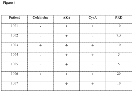
[0073] Fig. 1 is a table showing seven subjects from the IL-1(3 antibody
clinical trial, with
information about treatment medicines received prior to enrollment in the
study.
CA 02797846 2012-10-29
WO 2011/140522 PCT/US2011/035646
[0074] Fig. 2 is a table showing clinical data for subject 1001 treated with
an IL-10
antibody.
[0075] Fig. 3 is a table showing clinical data for subject 1002 treated with
an IL-10
antibody.
[0076] Fig. 4 is a table showing clinical data for subject 1003 treated with
an IL-10
antibody.
[0077] Fig. 5 is a table showing clinical data for subject 1004 treated with
an IL-10
antibody.
[0078] Fig. 6 is a table showing clinical data for subject 1005 treated with
an IL-10
1o antibody.
[0079] Fig. 7 is a table showing clinical data for subject 1006 treated with
an IL-10
antibody.
[0080] Fig. 8 is a table showing clinical data for subject 1007 treated with
an IL-10
antibody.
[0081] Fig. 9 is images showing resolution of a hypopyon following treatment
with an IL-
10 antibody.
[0082] Fig. 10 is images showing resolution of vitreous haze following
treatment with an
IL-1(3 antibody.
DETAILED DESCRIPTION
[0083] Effective therapies for use in treating or preventing uveitis have
remained
an important medical need. The present disclosure provides methods and
materials, and
related articles of manufacture, for treating or preventing uveitis in a
subject, including
treatment refractory (e.g., treatment resistant) uveitis, comprising
administering to the
subject an effective amount of anti-IL-10 antibody or binding fragment
thereof. Such
materials and methods may be used to replace or complement other
pharmaceutical
approaches as provided herein.
21
CA 02797846 2012-10-29
WO 2011/140522 PCT/US2011/035646
[0084] IL-1(3 is a pro-inflammatory cytokine secreted by a number of different
cell
types including monocytes and macrophages. When released as part of an
inflammatory
reaction, IL-1 (3 produces a range of biological effects, mainly mediated
through induction of
other inflammatory mediators such as corticotrophin, platelet factor-4,
prostaglandin E2
(PGE2), IL-6, and IL-8. IL-1(3 induces both local and systemic inflammatory
effects
through the activation of the IL-1 receptor found on almost all cell types.
[0085] The interleukin-1 (IL-1) family of cytokines has been implicated in
several
disease states such as rheumatoid arthritis (RA), osteoarthritis, Crohn's
disease, ulcerative
colitis (UC), septic shock, chronic obstructive pulmonary disease (COPD),
asthma, graft
versus host disease, atherosclerosis, adult T-cell leukemia, multiple myeloma,
multiple
sclerosis, stroke, and Alzheimer's disease. IL-1 family members include IL-la,
IL-1(3, and
IL-1Ra. Although related by their ability to bind to IL-1 receptors (IL-1R1,
IL-1R2), each
of these cytokines is expressed by a different gene and has a different
primary amino acid
sequence. Furthermore, the physiological activities of these cytokines can be
distinguished
from each other.
[0086] Compounds that disrupt IL-1 receptor signaling have been investigated
as
therapeutic agents to treat IL-1 mediated diseases, such as for example some
of the
aforementioned diseases. These compounds include recombinant IL-1Ra (Amgen
Inc.,
Thousand Oaks, CA), IL-1 receptor "trap" peptide (Regeneron Inc., Tarrytown,
NY), as well
as animal-derived IL-1(3 antibodies and recombinant IL-1(3 antibodies and
fragments
thereof.
[0087] As noted above, IL-1 receptor antagonist (IL-1Ra) polypeptide has been
suggested for use in the treatment of gout (So et al., 2007, ibid; McGonagle
et al., 2007,
ibid), but there remains a need for effective means to treat gout,
particularly those that do
not require daily, repeated injections. An additional challenge for IL-1
receptor antagonist-
based therapeutics is the need for such therapeutics to occupy a large number
of receptors,
which is a formidable task since these receptors are widely expressed on all
cells except red
blood cells (Dinarello, Curr. Opin. Pharmacol. 4:378-385, 2004). In most
immune-
mediated diseases, such as the diseases disclosed herein, the amount of IL-1(3
cytokine that
is measurable in body fluids or associated with activated cells is relatively
low. Thus, a
method of treatment and/or prevention that directly targets the IL-1(3 ligand
is a superior
strategy, particularly when administering an IL-1 (3 antibody with high
affinity.
22
CA 02797846 2012-10-29
WO 2011/140522 PCT/US2011/035646
[0088] The present invention provides methods and related compositions and
articles of manufacture for the treatment and/or prevention of gout in a
subject (e.g.,
mammalian, human), using an antibody or fragment thereof specific for IL-1 R.
[0089] As shown in Example 1 below, we have surprisingly found that such an
antibody (e.g., with very high affinity) can be far more potent an inhibitor
of the IL-1
pathway than is IL-Ra (e.g., Kineret ), and provides an opportunity to achieve
a therapeutic
effect at a lower dose and/or with less frequent administration than necessary
for other
drugs, such as recombinant IL-1Ra.
[0090] Such methods as described herein with an IL-10 antibody or fragment may
include the treatment of a subject suffering from gout (e.g., acute gout,
chronic gout,
refractory gout). The methods also may include preventing the occurrence of
gout (e.g.,
acute gout, chronic gout, refractory gout) in an at risk subject.
Antibodies, Humanized Antibodies, and Human Engineered Antibodies
[0091] The IL-1 (e.g., IL-1(3) binding antibodies of the present disclosure
may be
provided as polyclonal antibodies, monoclonal antibodies (mAbs), recombinant
antibodies,
chimeric antibodies, CDR-grafted antibodies, fully human antibodies, single
chain
antibodies, and/or bispecific antibodies, as well as fragments, including
variants and
derivatives thereof, provided by known techniques, including, but not limited
to enzymatic
cleavage, peptide synthesis or recombinant techniques.
[0092] Antibodies generally comprise two heavy chain polypeptides and two
light
chain polypeptides, though single domain antibodies having one heavy chain and
one light
chain, and heavy chain antibodies devoid of light chains are also
contemplated. There are
five types of heavy chains, called alpha, delta, epsilon, gamma and mu, based
on the amino
acid sequence of the heavy chain constant domain. These different types of
heavy chains
give rise to five classes of antibodies, IgA (including IgA1 and IgA2), IgD,
IgE, IgG and
IgM, respectively, including four subclasses of IgG, namely IgGi, IgG2, IgG3
and IgG4.
There are also two types of light chains, called kappa (K) or lambda (X) based
on the amino
acid sequence of the constant domains. A full-length antibody includes a
constant domain
and a variable domain. The constant region need not be present in an antigen
binding
fragment of an antibody. Antigen binding fragments of an antibody disclosed
herein can
include Fab, Fab', F(ab')2, and F(v) antibody fragments. As discussed in more
detail
below, IL-10 binding fragments encompass antibody fragments and antigen-
binding
polypeptides that will bind IL-1(3.
23
CA 02797846 2012-10-29
WO 2011/140522 PCT/US2011/035646
[0093] Each of the heavy chain and light chain sequences of an antibody, or
antigen binding fragment thereof, includes a variable region with three
complementarity
determining regions (CDRs) as well as non-CDR framework regions (FRs). The
terms
"heavy chain" and "light chain," as used herein, mean the heavy chain variable
region and
the light chain variable region, respectively, unless otherwise noted. Heavy
chain CDRs are
referred to herein as CDR-H1, CDR-H2, and CDR-H3. Light chain CDRs are
referred to
herein as CDR-L1, CDR-L2, and CDR-L3. Variable regions and CDRs in an antibody
sequence can be identified (i) according to general rules that have been
developed in the art
or (ii) by aligning the sequences against a database of known variable
regions. Methods for
identifying these regions are described in Kontermann and Dubel, eds.,
Antibody
Engineering, Springer, New York, NY, 2001, and Dinarello et al., Current
Protocols in
Immunology, John Wiley and Sons Inc., Hoboken, NJ, 2000. Databases of antibody
sequences are described in and can be accessed through "The Kabatman" database
at
www.bioinf.org.uk/abs (maintained by A.C. Martin in the Department of
Biochemistry &
Molecular Biology University College London, London, England) and VBASE2 at
www.vbase2.org, as described in Retter et al., Nucl. Acids Res., 33(Database
issue): D671-
D674 (2005). The "Kabatman" database web site also includes general rules of
thumb for
identifying CDRs. The term "CDR," as used herein, is as defined in Kabat et
al., Sequences
of Immunological Interest, 5th ed., U.S. Department of Health and Human
Services, 1991,
unless otherwise indicated.
[0094] Polyclonal antibodies are preferably raised in animals by multiple
subcutaneous (sc) or intraperitoneal (ip) injections of the relevant antigen
and an adjuvant.
An improved antibody response may be obtained by conjugating the relevant
antigen to a
protein that is immunogenic in the species to be immunized, e.g., keyhole
limpet
hemocyanin, serum albumin, bovine thyroglobulin, or soybean trypsin inhibitor
using a
bifunctional or derivatizing agent, for example, maleimidobenzoyl
sulfosuccinimide ester
(conjugation through cysteine residues), N-hydroxysuccinimide (through lysine
residues),
glutaraldehyde, succinic anhydride or other agents known in the art.
[0095] Animals are immunized against the antigen, immunogenic conjugates, or
derivatives by combining, e.g., 100 g or 5 g of the protein or conjugate
(for rabbits or
mice, respectively) with 3 volumes of Freund's complete adjuvant and injecting
the solution
intradermally at multiple sites. One month later, the animals are boosted with
1/5 to
{fraction (1/10)} the original amount of peptide or conjugate in Freund's
complete adjuvant
by subcutaneous injection at multiple sites. At 7-14 days post-booster
injection, the animals
24
CA 02797846 2012-10-29
WO 2011/140522 PCT/US2011/035646
are bled and the serum is assayed for antibody titer. Animals are boosted
until the titer
plateaus. Preferably, the animal is boosted with the conjugate of the same
antigen, but
conjugated to a different protein and/or through a different cross-linking
reagent.
Conjugates also can be made in recombinant cell culture as protein fusions.
Also,
aggregating agents such as alum are suitably used to enhance the immune
response.
[0096] Monoclonal antibody refers to an antibody obtained from a population of
substantially homogeneous antibodies. Monoclonal antibodies are generally
highly specific,
and may be directed against a single antigenic site, in contrast to
conventional (polyclonal)
antibody preparations that typically include different antibodies directed
against different
determinants (epitopes). In addition to their specificity, the monoclonal
antibodies are
advantageous in that they are synthesized by the homogeneous culture,
uncontaminated by
other immunoglobulins with different specificities and characteristics.
[0097] Monoclonal antibodies may be made by the hybridoma method first
described by Kohler et al., (Nature, 256:495-7, 1975), or may be made by
recombinant
DNA methods (see, e.g., U.S. Patent No. 4,816,567). The monoclonal antibodies
may also
be isolated from display libraries (e.g., yeast libraries, phage antibody
libraries) using the
techniques described in, for example, Clackson et al., (Nature 352:624-628,
1991), Marks et
al., (J. Mol. Biol. 222:581-597, 1991) Hoogenboom (Nat Biotechnol. 23:1105-16,
2005) and
Mondon et al., (Front Biosci., 13:1117-1129, 2008).
[0098] In the hybridoma method, a mouse or other appropriate host animal, such
as a hamster or macaque monkey, is immunized as herein described to elicit
lymphocytes
that produce or are capable of producing antibodies that will specifically
bind to the protein
used for immunization. Alternatively, lymphocytes may be immunized in vitro.
Lymphocytes then are fused with myeloma cells using a suitable fusing agent,
such as
polyethylene glycol, to form a hybridoma cell (Goding, Monoclonal Antibodies:
Principles
and Practice, pp. 59-103 (Academic Press, 1986)).
[0099] The hybridoma cells thus prepared are seeded and grown in a suitable
culture medium that preferably contains one or more substances that inhibit
the growth or
survival of the unfused, parental myeloma cells. For example, if the parental
myeloma cells
lack the enzyme hypoxanthine guanine phosphoribosyl transferase (HGPRT or
HPRT), the
culture medium for the hybridomas typically will include hypoxanthine,
aminopterin, and
thymidine (HAT medium), which substances prevent the growth of HGPRT-deficient
cells.
[0100] Preferred myeloma cells are those that fuse efficiently, support stable
high-
level production of antibody by the selected antibody-producing cells, and are
sensitive to a
CA 02797846 2012-10-29
WO 2011/140522 PCT/US2011/035646
medium. Human myeloma and mouse-human heteromyeloma cell lines also have been
described for the production of human monoclonal antibodies (Kozbor, J.
Immunol., 133:
3001 (1984); Brodeur et al., Monoclonal Antibody Production Techniques and
Applications, pp. 51-63 (Marcel Dekker, Inc., New York, 1987)). Exemplary
murine
myeloma lines include those derived from MOP-21 and M.C.-11 mouse tumors
available
from the Salk Institute Cell Distribution Center, San Diego, Calif. USA, and
SP-2 or X63-
Ag8-653 cells available from the American Type Culture Collection, Rockville,
Md. USA.
[0101] Culture medium in which hybridoma cells are growing is assayed for
production of monoclonal antibodies directed against the antigen. Preferably,
the binding
specificity of monoclonal antibodies produced by hybridoma cells is determined
by
immunoprecipitation or by an in vitro binding assay, such as radioimmunoassay
(RIA) or
enzyme-linked immunoabsorbent assay (ELISA). The binding affinity of the
monoclonal
antibody can, for example, be determined by Scatchard analysis (Munson et al.,
Anal.
Biochem., 107:220 (1980)).
[0102] After hybridoma cells are identified that produce antibodies of the
desired
specificity, affinity, and/or activity, the clones may be subcloned by
limiting dilution
procedures and grown by standard methods (Goding, Monoclonal Antibodies:
Principles
and Practice, pp. 59-103 (Academic Press, 1986)). Suitable culture media for
this purpose
include, for example, DMEM or RPMI-1640 medium. In addition, the hybridoma
cells may
be grown in vivo as ascites tumors in an animal. The monoclonal antibodies
secreted by the
subclones are suitably separated from the culture medium, ascites fluid, or
serum by
conventional immunoglobulin purification procedures such as, for example,
protein A-
Sepharose, hydroxylapatite chromatography, gel electrophoresis, dialysis, or
affinity
chromatography.
[0103] It is further contemplated that antibodies may be used as smaller
antigen
binding fragments of the antibody well-known in the art and described herein.
[0104] The present disclosure encompasses IL-1 (e.g., IL-1(3) binding
antibodies
that include two full length heavy chains and two full length light chains.
Alternatively, the
IL-10 binding antibodies can be constructs such as single chain antibodies or
"mini"
antibodies that retain binding activity to IL-10. Such constructs can be
prepared by
methods known in the art such as, for example, the PCR mediated cloning and
assembly of
single chain antibodies for expression in E. coli (as described in Antibody
Engineering, The
practical approach series, J. McCafferty, H. R. Hoogenboom, and D. J.
Chiswell, editors,
Oxford University Press, 1996). In this type of construct, the variable
portions of the heavy
26
CA 02797846 2012-10-29
WO 2011/140522 PCT/US2011/035646
and light chains of an antibody molecule are PCR amplified from cDNA. The
resulting
amplicons are then assembled, for example, in a second PCR step, through a
linker DNA
that encodes a flexible protein linker composed of the amino acids Gly and
Ser. This linker
allows the variable heavy and light chain portions to fold in such a way that
the antigen
binding pocket is regenerated and antigen is bound with affinities often
comparable to the
parent full-length dimeric immunoglobulin molecule.
[0105] The IL-1 (e.g., IL-1(3) binding antibodies and binding fragments of the
present disclosure encompass variants of the exemplary antibodies, fragments
and
sequences disclosed herein. Variants include peptides and polypeptides
comprising one or
more amino acid sequence substitutions, deletions, and/or additions that have
the same or
substantially the same affinity and specificity of epitope binding as one or
more of the
exemplary antibodies, fragments and sequences disclosed herein. Thus, variants
include
peptides and polypeptides comprising one or more amino acid sequence
substitutions,
deletions, and/or additions to the exemplary antibodies, fragments and
sequences disclosed
herein where such substitutions, deletions and/or additions do not cause
substantial changes
in affinity and specificity of epitope binding. For example, a variant of an
antibody or
fragment may result from one or more changes to an antibody or fragment, where
the
changed antibody or fragment has the same or substantially the same affinity
and specificity
of epitope binding as the starting sequence. Variants may be naturally
occurring, such as
allelic or splice variants, or may be artificially constructed. Variants may
be prepared from
the corresponding nucleic acid molecules encoding said variants. Variants of
the present
antibodies and IL-10 binding fragments may have changes in light and/or heavy
chain
amino acid sequences that are naturally occurring or are introduced by in
vitro engineering
of native sequences using recombinant DNA techniques. Naturally occurring
variants
include "somatic" variants which are generated in vivo in the corresponding
germ line
nucleotide sequences during the generation of an antibody response to a
foreign antigen.
[0106] Variants of IL-1 (e.g., IL-1(3) binding antibodies and binding
fragments
may also be prepared by mutagenesis techniques. For example, amino acid
changes may be
introduced at random throughout an antibody coding region and the resulting
variants may
be screened for binding affinity for IL-1(3 or for another property.
Alternatively, amino acid
changes may be introduced in selected regions of an IL-10 antibody, such as in
the light
and/or heavy chain CDRs, and/or in the framework regions, and the resulting
antibodies
may be screened for binding to IL-10 or some other activity. Amino acid
changes
encompass one or more amino acid substitutions in a CDR, ranging from a single
amino
27
CA 02797846 2012-10-29
WO 2011/140522 PCT/US2011/035646
acid difference to the introduction of multiple permutations of amino acids
within a given
CDR, such as CDR3. In another method, the contribution of each residue within
a CDR to
IL-10 binding may be assessed by substituting at least one residue within the
CDR with
alanine. Lewis et at. (1995), Mol. Immunol. 32: 1065-72. Residues which are
not optimal
for binding to IL-1(3 may then be changed in order to determine a more optimum
sequence.
Also encompassed are variants generated by insertion of amino acids to
increase the size of
a CDR, such as CDR3. For example, most light chain CDR3 sequences are nine
amino
acids in length. Light chain sequences in an antibody which are shorter than
nine residues
may be optimized for binding to IL-1 0 by insertion of appropriate amino acids
to increase
the length of the CDR.
[0107] Variants may also be prepared by "chain shuffling" of light or heavy
chains. Marks et al. (1992), Biotechnology 10: 779-83. A single light (or
heavy) chain can
be combined with a library having a repertoire of heavy (or light) chains and
the resulting
population is screened for a desired activity, such as binding to IL-10. This
permits
screening of a greater sample of different heavy (or light) chains in
combination with a
single light (or heavy) chain than is possible with libraries comprising
repertoires of both
heavy and light chains.
[0108] The IL-1 (e.g., IL-1(3) binding antibodies and binding fragments of the
present disclosure encompass derivatives of the exemplary antibodies,
fragments and
sequences disclosed herein. Derivatives include polypeptides or peptides, or
variants,
fragments or derivatives thereof, which have been chemically modified.
Examples include
covalent attachment of one or more polymers, such as water soluble polymers, N-
linked, or
O-linked carbohydrates, sugars, phosphates, and/or other such molecules. The
derivatives
are modified in a manner that is different from naturally occurring or
starting peptide or
polypeptides, either in the type or location of the molecules attached.
Derivatives further
include deletion of one or more chemical groups which are naturally present on
the peptide
or polypeptide.
[0109] The IL-10 binding antibodies and binding fragments can be bispecific.
Bispecific antibodies or fragments can be of several configurations. For
example, bispecific
antibodies may resemble single antibodies (or antibody fragments) but have two
different
antigen binding sites (variable regions). Bispecific antibodies can be
produced by chemical
techniques (Kranz et al. (1981), Proc. Natl. Acad. Sci. USA, 78: 5807), by
"polydoma"
techniques (U.S. Pat. No. 4,474,893) or by recombinant DNA techniques.
Bispecific
antibodies can have binding specificities for at least two different epitopes,
at least one of
28
CA 02797846 2012-10-29
WO 2011/140522 PCT/US2011/035646
which is an epitope of IL-1 R. The IL-1(3 binding antibodies and binding
fragments can also
be heteroantibodies. Heteroantibodies are two or more antibodies, or antibody
binding
fragments (Fab) linked together, each antibody or fragment having a different
specificity.
[0110] Techniques for creating recombinant DNA versions of the antigen-binding
regions of antibody molecules which bypass the generation of monoclonal
antibodies are
contemplated for the present IL-1 (e.g., IL-1(3) binding antibodies and
binding fragments.
DNA is cloned into a bacterial expression system. One example of such a
technique suitable
for the practice of this invention uses a bacteriophage lambda vector system
having a leader
sequence that causes the expressed Fab protein to migrate to the periplasmic
space (between
the bacterial cell membrane and the cell wall) or to be secreted. One can
rapidly generate
and screen great numbers of functional Fab fragments for those which bind IL-
10. Such IL-
1(3 binding agents (Fab fragments with specificity for an IL-1(3 polypeptide)
are specifically
encompassed within the IL-10 binding antibodies and binding fragments of the
present
disclosure.
[0111] The present IL-1 (e.g., IL-1(3) binding antibodies and binding
fragments
can be humanized or human engineered antibodies. As used herein, a humanized
antibody,
or antigen binding fragment thereof, is a recombinant polypeptide that
comprises a portion
of an antigen binding site from a non-human antibody and a portion of the
framework
and/or constant regions of a human antibody. A human engineered antibody or
antibody
fragment is a non-human (e.g., mouse) antibody that has been engineered by
modifying
(e.g., deleting, inserting, or substituting) amino acids at specific positions
so as to reduce or
eliminate any detectable immunogenicity of the modified antibody in a human.
[0112] Humanized antibodies include chimeric antibodies and CDR-grafted
antibodies. Chimeric antibodies are antibodies that include a non-human
antibody variable
region linked to a human constant region. Thus, in chimeric antibodies, the
variable region
is mostly non-human, and the constant region is human. Chimeric antibodies and
methods
for making them are described in Morrison, et al., Proc. Natl. Acad. Sci. USA,
81: 6841-
6855 (1984), Boulianne, et al., Nature, 312: 643-646 (1984), and PCT
Application
Publication WO 86/01533. Although, they can be less immunogenic than a mouse
monoclonal antibody, administrations of chimeric antibodies have been
associated with
human anti-mouse antibody responses (HAMA) to the non-human portion of the
antibodies.
Chimeric antibodies can also be produced by splicing the genes from a mouse
antibody
molecule of appropriate antigen-binding specificity together with genes from a
human
antibody molecule of appropriate biological activity, such as the ability to
activate human
29
CA 02797846 2012-10-29
WO 2011/140522 PCT/US2011/035646
complement and mediate ADCC. Morrison et al. (1984), Proc. Natl. Acad. Sci.,
81: 6851;
Neuberger et al. (1984), Nature, 312: 604. One example is the replacement of a
Fc region
with that of a different isotype.
[0113] CDR-grafted antibodies are antibodies that include the CDRs from a non-
human "donor" antibody linked to the framework region from a human "recipient"
antibody. Generally, CDR-grafted antibodies include more human antibody
sequences than
chimeric antibodies because they include both constant region sequences and
variable
region (framework) sequences from human antibodies. Thus, for example, a CDR-
grafted
humanized antibody of the invention can comprise a heavy chain that comprises
a
contiguous amino acid sequence (e.g., about 5 or more, 10 or more, or even 15
or more
contiguous amino acid residues) from the framework region of a human antibody
(e.g., FR-
l, FR-2, or FR-3 of a human antibody) or, optionally, most or all of the
entire framework
region of a human antibody. CDR-grafted antibodies and methods for making them
are
described in, Jones et al., Nature, 321: 522-525 (1986), Riechmann et al.,
Nature, 332:
323-327 (1988), and Verhoeyen et al., Science, 239: 1534-1536 (1988)). Methods
that can
be used to produce humanized antibodies also are described in U.S. Patents
4,816,567,
5,721,367, 5,837,243, and 6,180,377. CDR-grafted antibodies are considered
less likely
than chimeric antibodies to induce an immune reaction against non-human
antibody
portions. However, it has been reported that framework sequences from the
donor
antibodies are required for the binding affinity and/or specificity of the
donor antibody,
presumably because these framework sequences affect the folding of the antigen-
binding
portion of the donor antibody. Therefore, when donor, non-human CDR sequences
are
grafted onto unaltered human framework sequences, the resulting CDR-grafted
antibody
can exhibit, in some cases, loss of binding avidity relative to the original
non-human donor
antibody. See, e.g., Riechmann et al., Nature, 332: 323-327 (1988), and
Verhoeyen et al.,
Science, 239: 1534-1536 (1988).
[0114] Human engineered antibodies include for example "veneered" antibodies
and antibodies prepared using HUMAN ENGINEERINGTM technology (see for example,
U.S.
Patents 5,766,886 and 5,869,619). HUMAN ENGINEERINGTM technology is
commercially
available, and involves altering an non-human antibody or antibody fragment,
such as a
mouse or chimeric antibody or antibody fragment, by making specific changes to
the amino
acid sequence of the antibody so as to produce a modified antibody with
reduced
immunogenicity in a human that nonetheless retains the desirable binding
properties of the
original non-human antibodies. Generally, the technique involves classifying
amino acid
CA 02797846 2012-10-29
WO 2011/140522 PCT/US2011/035646
residues of a non-human (e.g., mouse) antibody as "low risk", "moderate risk",
or "high
risk" residues. The classification is performed using a global risk/reward
calculation that
evaluates the predicted benefits of making particular substitution (e.g., for
immunogenicity
in humans) against the risk that the substitution will affect the resulting
antibody's folding
and/or antigen-binding properties. Thus, a low risk position is one for which
a substitution
is predicted to be beneficial because it is predicted to reduce immunogenicity
without
significantly affecting antigen binding properties. A moderate risk position
is one for which
a substitution is predicted to reduce immunogenicity, but is more likely to
affect protein
folding and/or antigen binding. High risk positions contain residues most
likely to be
involved in proper folding or antigen binding. Generally, low risk positions
in a non-human
antibody are substituted with human residues, high risk positions are rarely
substituted, and
humanizing substitutions at moderate risk positions are sometimes made,
although not
indiscriminately. Positions with prolines in the non-human antibody variable
region
sequence are usually classified as at least moderate risk positions.
[0115] The particular human amino acid residue to be substituted at a given
low or
moderate risk position of a non-human (e.g., mouse) antibody sequence can be
selected by
aligning an amino acid sequence from the non-human antibody's variable regions
with the
corresponding region of a specific or consensus human antibody sequence. The
amino acid
residues at low or moderate risk positions in the non-human sequence can be
substituted for
the corresponding residues in the human antibody sequence according to the
alignment.
Techniques for making human engineered proteins are described in greater
detail in
Studnicka et al., Protein Engineering, 7: 805-814 (1994), U.S. Patents
5,766,886,
5,770,196, 5,821,123, and 5,869,619, and PCT Application Publication WO
93/11794.
[0116] "Veneered" antibodies are non-human or humanized (e.g., chimeric or
CDR-grafted antibodies) antibodies that have been engineered to replace
certain solvent-
exposed amino acid residues so as to further reduce their immunogenicity or
enhance their
function. As surface residues of a chimeric antibody are presumed to be less
likely to affect
proper antibody folding and more likely to elicit an immune reaction,
veneering of a
chimeric antibody can include, for instance, identifying solvent-exposed
residues in the non-
human framework region of a chimeric antibody and replacing at least one of
them with the
corresponding surface residues from a human framework region. Veneering can be
accomplished by any suitable engineering technique, including the use of the
above-
described HUMAN ENGINEERINGTM technology.
31
CA 02797846 2012-10-29
WO 2011/140522 PCT/US2011/035646
[0117] In a different approach, a recovery of binding avidity can be achieved
by
"de-humanizing" a CDR-grafted antibody. De-humanizing can include restoring
residues
from the donor antibody's framework regions to the CDR grafted antibody,
thereby
restoring proper folding. Similar "de-humanization" can be achieved by (i)
including
portions of the "donor" framework region in the "recipient" antibody or (ii)
grafting
portions of the "donor" antibody framework region into the recipient antibody
(along with
the grafted donor CDRs).
[0118] For a further discussion of antibodies, humanized antibodies, human
engineered, and methods for their preparation, see Kontermann and Dubel, eds.,
Antibody
Engineering, Springer, New York, NY, 2001.
[0119] Exemplary humanized or human engineered antibodies include IgG, IgM,
IgE, IgA, and IgD antibodies. The present antibodies can be of any class (IgG,
IgA, IgM,
IgE, IgD, etc.) or isotype and can comprise a kappa or lambda light chain. For
example, a
human antibody can comprise an IgG heavy chain or defined fragment, such as at
least one
of isotypes, IgGi, IgG2, IgG3 or IgG4. As a further example, the present
antibodies or
fragments can comprise an IgGI heavy chain and an IgGI light chain.
[0120] The present antibodies and fragments can be human antibodies, such as
antibodies which bind IL-1(3 polypeptides and are encoded by nucleic acid
sequences which
are naturally occurring somatic variants of human germline immunoglobulin
nucleic acid
sequence, and fragments, synthetic variants, derivatives and fusions thereof.
Such
antibodies may be produced by any method known in the art, such as through the
use of
transgenic mammals (such as transgenic mice) in which the native
immunoglobulin
repertoire has been replaced with human V-genes in the mammal chromosome. Such
mammals appear to carry out VDJ recombination and somatic hypermutation of the
human
germline antibody genes in a normal fashion, thus producing high affinity
antibodies with
completely human sequences.
[0121] Human antibodies to target protein can also be produced using
transgenic
animals that have no endogenous immunoglobulin production and are engineered
to contain
human immunoglobulin loci. For example, WO 98/24893 discloses transgenic
animals
having a human Ig locus wherein the animals do not produce functional
endogenous
immunoglobulins due to the inactivation of endogenous heavy and light chain
loci. WO
91/00906 also discloses transgenic non-primate mammalian hosts capable of
mounting an
immune response to an immunogen, wherein the antibodies have primate constant
and/or
variable regions, and wherein the endogenous immunoglobulin encoding loci are
substituted
32
CA 02797846 2012-10-29
WO 2011/140522 PCT/US2011/035646
or inactivated. WO 96/30498 and US Patent No. 6,091,001 disclose the use of
the Cre/Lox
system to modify the immunoglobulin locus in a mammal, such as to replace all
or a portion
of the constant or variable region to form a modified antibody molecule. WO
94/02602
discloses non-human mammalian hosts having inactivated endogenous Ig loci and
functional human Ig loci. U.S. Patent No. 5,939,598 discloses methods of
making
transgenic mice in which the mice lack endogenous heavy chains, and express an
exogenous
immunoglobulin locus comprising one or more xenogeneic constant regions. See
also, U.S.
Patent Nos. 6,114,598 6,657,103 and 6,833,268.
[0122] Using a transgenic animal described above, an immune response can be
produced to a selected antigenic molecule, and antibody producing cells can be
removed
from the animal and used to produce hybridomas that secrete human monoclonal
antibodies.
Immunization protocols, adjuvants, and the like are known in the art, and are
used in
immunization of, for example, a transgenic mouse as described in WO 96/33735.
This
publication discloses monoclonal antibodies against a variety of antigenic
molecules
including IL-6, IL-8, TNFa, human CD4, L selectin, gp39, and tetanus toxin.
The
monoclonal antibodies can be tested for the ability to inhibit or neutralize
the biological
activity or physiological effect of the corresponding protein. WO 96/33735
discloses that
monoclonal antibodies against IL-8, derived from immune cells of transgenic
mice
immunized with IL-8, blocked IL-8 induced functions of neutrophils. Human
monoclonal
antibodies with specificity for the antigen used to immunize transgenic
animals are also
disclosed in WO 96/34096 and U.S. patent application no. 20030194404; and U.S.
patent
application no. 20030031667.
[0123] Additional transgenic animals useful to make monoclonal antibodies
include the Medarex HuMAb-MOUSE , described in U.S. Pat. No. 5,770,429 and
Fishwild, et al. (Nat. Biotechnol. 14:845-851, 1996), which contains gene
sequences from
unrearranged human antibody genes that code for the heavy and light chains of
human
antibodies. Immunization of a HuMAb-MOUSE enables the production of fully
human
monoclonal antibodies to the target protein.
[0124] Also, Ishida et al. (Cloning Stem Cells. 4:91-102, 2002) describes the
TransChromo Mouse (TCMOUSETM) which comprises megabase-sized segments of human
DNA and which incorporates the entire human immunoglobulin (hIg) loci. The
TCMOUSETM has a fully diverse repertoire of hIgs, including all the subclasses
of IgGs
(IgGl-G4). Immunization of the TC MOUSETM with various human antigens produces
antibody responses comprising human antibodies.
33
CA 02797846 2012-10-29
WO 2011/140522 PCT/US2011/035646
[0125] See also Jakobovits et al., Proc. Natl. Acad. Sci. USA, 90:2551 (1993);
Jakobovits et al., Nature, 362:255-258 (1993); Bruggermann et al., Year in
Immunol., 7:33
(1993); and U.S. Pat. No. 5,591,669, U.S. Patent No. 5,589,369, U.S. Patent
No. 5,545,807;
and U.S Patent Publication No. 20020199213. U.S. Patent Publication No.
20030092125
describes methods for biasing the immune response of an animal to the desired
epitope.
Human antibodies may also be generated by in vitro activated B cells (see U.S.
Pat. Nos.
5,567,610 and 5,229,275).
[0126] Human antibodies can also be generated through the in vitro screening
of
antibody display libraries. See Hoogenboom et al. (1991), J. Mol. Biol. 227:
381; and Marks
et al. (1991), J. Mol. Biol. 222: 581. Various antibody-containing phage
display libraries
have been described and may be readily prepared. Libraries may contain a
diversity of
human antibody sequences, such as human Fab, Fv, and scFv fragments, that may
be
screened against an appropriate target. Phage display libraries may comprise
peptides or
proteins other than antibodies which may be screened to identify selective
binding agents of
IL-1[3.
[0127] The development of technologies for making repertoires of recombinant
human antibody genes, and the display of the encoded antibody fragments on the
surface of
filamentous bacteriophage, has provided a means for making human antibodies
directly.
The antibodies produced by phage technology are produced as antigen binding
fragments-
usually Fv or Fab fragments-in bacteria and thus lack effector functions.
Effector functions
can be introduced by one of two strategies: The fragments can be engineered
either into
complete antibodies for expression in mammalian cells, or into bispecific
antibody
fragments with a second binding site capable of triggering an effector
function.
[0128] The disclosure contemplates a method for producing target-specific
antibody or antigen-binding portion thereof comprising the steps of
synthesizing a library of
human antibodies on phage, screening the library with target protein or a
portion thereof,
isolating phage that bind target, and obtaining the antibody from the phage.
By way of
example, one method for preparing the library of antibodies for use in phage
display
techniques comprises the steps of immunizing a non-human animal comprising
human
immunoglobulin loci with target antigen or an antigenic portion thereof to
create an immune
response, extracting antibody producing cells from the immunized animal;
isolating RNA
from the extracted cells, reverse transcribing the RNA to produce cDNA,
amplifying the
cDNA using a primer, and inserting the cDNA into a phage display vector such
that
34
CA 02797846 2012-10-29
WO 2011/140522 PCT/US2011/035646
antibodies are expressed on the phage. Recombinant target-specific antibodies
of the
invention may be obtained in this way.
[0129] Phage-display processes mimic immune selection through the display of
antibody repertoires on the surface of filamentous bacteriophage, and
subsequent selection
of phage by their binding to an antigen of choice. One such technique is
described in WO
99/10494, which describes the isolation of high affinity and functional
agonistic antibodies
for MPL and msk receptors using such an approach. Antibodies of the invention
can be
isolated by screening of a recombinant combinatorial antibody library,
preferably a scFv
phage display library, prepared using human VL and VH cDNAs prepared from mRNA
derived from human lymphocytes. Methodologies for preparing and screening such
libraries are known in the art. See e.g., U.S. Patent No. 5,969,108. There are
commercially
available kits for generating phage display libraries (e.g., the Pharmacia
Recombinant Phage
Antibody System, catalog no. 27-9400-01; and the Stratagene SurfZAP.TM. phage
display
kit, catalog no. 240612). There are also other methods and reagents that can
be used in
generating and screening antibody display libraries (see, e.g., Ladner et al.
U.S. Pat. No.
5,223,409; Kang et al. PCT Publication No. WO 92/18619; Dower et al. PCT
Publication
No. WO 91/17271; Winter et al. PCT Publication No. WO 92/20791; Markland et
al. PCT
Publication No. WO 92/15679; Breitling et al. PCT Publication No. WO 93/01288;
McCafferty et al. PCT Publication No. WO 92/01047; Garrard et al. PCT
Publication No.
WO 92/09690; Fuchs et al. (1991) Bio/Technology 9:1370-1372; Hay et al. (1992)
Hum.
Antibod. Hybridomas 3:81-85; Huse et al. (1989) Science 246:1275-1281;
McCafferty et al.,
Nature (1990) 348:552-554; Griffiths et al. (1993) EMBO J 12:725-734; Hawkins
et al.
(1992) J. Mol. Biol. 226:889-896; Clackson et al. (1991) Nature 352:624-628;
Gram et al.
(1992) Proc. Natl. Acad. Sci. USA 89:3576-3580; Garrad et al. (1991)
Bio/Technology
9:1373-1377; Hoogenboom et al. (1991) Nuc Acid Res 19:4133-4137; and Barbas et
al.
(1991) Proc. Natl. Acad. Sci. USA 88:7978-7982.
[0130] In one embodiment, to isolate human antibodies specific for the target
antigen with the desired characteristics, a human VH and VL library are
screened to select
for antibody fragments having the desired specificity. The antibody libraries
used in this
method are preferably scFv libraries prepared and screened as described herein
and in the
art (McCafferty et al., PCT Publication No. WO 92/01047, McCafferty et al.,
(Nature
348:552-554, 1990); and Griffiths et al., (EMBO J 12:725-734, 1993). The scFv
antibody
libraries preferably are screened using target protein as the antigen.
CA 02797846 2012-10-29
WO 2011/140522 PCT/US2011/035646
[0131] Alternatively, the Fd fragment (VH-CH1) and light chain (VL-CL) of
antibodies are separately cloned by PCR and recombined randomly in
combinatorial phage
display libraries, which can then be selected for binding to a particular
antigen. The Fab
fragments are expressed on the phage surface, i.e., physically linked to the
genes that
encode them. Thus, selection of Fab by antigen binding co-selects for the Fab
encoding
sequences, which can be amplified subsequently. Through several rounds of
antigen
binding and re-amplification, a procedure termed panning, Fab specific for the
antigen are
enriched and finally isolated.
[0132] In 1994, an approach for the humanization of antibodies, called "guided
selection", was described. Guided selection utilizes the power of the phage
display
technique for the humanization of mouse monoclonal antibody (See Jespers, L.
S., et al.,
Bio/Technology 12, 899-903 (1994)). For this, the Fd fragment of the mouse
monoclonal
antibody can be displayed in combination with a human light chain library, and
the resulting
hybrid Fab library may then be selected with antigen. The mouse Fd fragment
thereby
provides a template to guide the selection. Subsequently, the selected human
light chains
are combined with a human Fd fragment library. Selection of the resulting
library yields
entirely human Fab.
[0133] A variety of procedures have been described for deriving human
antibodies
from phage-display libraries (See, for example, Hoogenboom et al., J. Mol.
Biol., 227:381
(1991); Marks et al., J. Mol. Biol, 222:581-597 (1991); U.S. Pat. Nos.
5,565,332 and
5,573,905; Clackson, T., and Wells, J. A., TIBTECH 12, 173-184 (1994)). In
particular, in
vitro selection and evolution of antibodies derived from phage display
libraries has become
a powerful tool (See Burton, D. R., and Barbas III, C. F., Adv. Immunol. 57,
191-280
(1994); Winter, G., et al., Annu. Rev. Immunol. 12, 433-455 (1994); U.S.
patent publication
no. 20020004215 and WO 92/01047; U.S. patent publication no. 20030190317; and
U.S.
Patent Nos. 6,054,287 and 5,877,293.
[0134] Watkins, "Screening of Phage-Expressed Antibody Libraries by Capture
Lift," Methods in Molecular Biology, Antibody Phage Display: Methods and
Protocols 178:
187-193 (2002), and U.S. patent publication no. 20030044772, published March
6, 2003,
describe methods for screening phage-expressed antibody libraries or other
binding
molecules by capture lift, a method involving immobilization of the candidate
binding
molecules on a solid support.
[0135] Fv fragments are displayed on the surface of phage, by the association
of
one chain expressed as a phage protein fusion (e.g., with M13 gene III) with
the
36
CA 02797846 2012-10-29
WO 2011/140522 PCT/US2011/035646
complementary chain expressed as a soluble fragment. It is contemplated that
the phage
may be a filamentous phage such as one of the class I phages: fd, M13, fl,
Ifl, Ike, ZJ/Z, Ff
and one of the class II phages Xf, Pfl and Pf3. The phage may be M13, or fd or
a
derivative thereof.
[0136] Once initial human VL and VH segments are selected, "mix and match"
experiments, in which different pairs of the initially selected VL and VH
segments are
screened for target binding, are performed to select preferred VL/VH pair
combinations.
Additionally, to further improve the quality of the antibody, the VL and VH
segments of the
preferred VL/VH pair(s) can be randomly mutated, preferably within the any of
the CDR1,
CDR2 or CDR3 region of VH and/or VL, in a process analogous to the in vivo
somatic
mutation process responsible for affinity maturation of antibodies during a
natural immune
response. This in vitro affinity maturation can be accomplished by amplifying
VL and VH
regions using PCR primers complimentary to the VH CDR1, CDR2, and CDR3, or VL
CDR1, CDR2, and CDR3, respectively, which primers have been "spiked" with a
random
mixture of the four nucleotide bases at certain positions such that the
resultant PCR
products encode VL and VH segments into which random mutations have been
introduced
into the VH and/or VL CDR3 regions. These randomly mutated VL and VH segments
can be
rescreened for binding to target antigen.
[0137] Following screening and isolation of an target specific antibody from a
recombinant immunoglobulin display library, nucleic acid encoding the selected
antibody
can be recovered from the display package (e.g., from the phage genome) and
subcloned
into other expression vectors by standard recombinant DNA techniques. If
desired, the
nucleic acid can be further manipulated to create other antibody forms of the
invention, as
described below. To express a recombinant human antibody isolated by screening
of a
combinatorial library, the DNA encoding the antibody is cloned into a
recombinant
expression vector and introduced into a mammalian host cell, as described
herein.
[0138] It is contemplated that the phage display method may be carried out in
a
mutator strain of bacteria or host cell. A mutator strain is a host cell which
has a genetic
defect which causes DNA replicated within it to be mutated with respect to its
parent DNA.
Example mutator strains are NR9046mutD5 and NR9046 mut Ti.
[0139] It is also contemplated that the phage display method may be carried
out
using a helper phage. This is a phage which is used to infect cells containing
a defective
phage genome and which functions to complement the defect. The defective phage
genome
can be a phagemid or a phage with some function encoding gene sequences
removed.
37
CA 02797846 2012-10-29
WO 2011/140522 PCT/US2011/035646
Examples of helper phages are M13K07, M13K07 gene III no. 3; and phage
displaying or
encoding a binding molecule fused to a capsid protein.
[0140] Antibodies are also generated via phage display screening methods using
the hierarchical dual combinatorial approach as disclosed in WO 92/01047 in
which an
individual colony containing either an H or L chain clone is used to infect a
complete
library of clones encoding the other chain (L or H) and the resulting two-
chain specific
binding member is selected in accordance with phage display techniques such as
those
described therein. This technique is also disclosed in Marks et at,
(Bio/Technology, 10:779-
783, 1992).
[0141] Methods for display of peptides on the surface of yeast and microbial
cells
have also been used to identify antigen specific antibodies. See, for example,
U.S. Patent
No. 6,699,658. Antibody libraries may be attached to yeast proteins, such as
agglutinin,
effectively mimicking the cell surface display of antibodies by B cells in the
immune
system.
[0142] In addition to phage display methods, antibodies may be isolated using
ribosome mRNA display methods and microbial cell display methods. Selection of
polypeptide using ribosome display is described in Hanes et al., (Proc. Natl
Acad Sci USA,
94:4937-4942, 1997) and U.S. Pat. Nos. 5,643,768 and 5,658,754 issued to
Kawasaki.
Ribosome display is also useful for rapid large scale mutational analysis of
antibodies. The
selective mutagenesis approach also provides a method of producing antibodies
with
improved activities that can be selected using ribosomal display techniques.
[0143] The IL-1 (e.g., IL-1(3) binding antibodies and binding fragments may
comprise one or more portions that do not bind IL-1(3 but instead are
responsible for other
functions, such as circulating half-life, direct cytotoxic effect, detectable
labeling, or
activation of the recipient's endogenous complement cascade or endogenous
cellular
cytotoxicity. The antibodies or fragments may comprise all or a portion of the
constant
region and may be of any isotype, including IgA (e.g., IgAl or IgA2), IgD,
IgE, IgG (e.g.
IgGi, IgG2, IgG3 or IgG4), or IgM. In addition to, or instead of, comprising a
constant
region, antigen-binding compounds of the invention may include an epitope tag,
a salvage
receptor epitope, a label moiety for diagnostic or purification purposes, or a
cytotoxic
moiety such as a radionuclide or toxin.
[0144] The constant region (when present) of the present antibodies and
fragments
may be of the yl, y2, y3, y4, , 02, or 6 or r, type, preferably of the y
type, more preferably
38
CA 02797846 2012-10-29
WO 2011/140522 PCT/US2011/035646
of the y, type, whereas the constant part of a human light chain may be of the
K or k type
(which includes the Xi, k2 and k3 subtypes) but is preferably of the K type.
[0145] Variants also include antibodies or fragments comprising a modified Fc
region, wherein the modified Fc region comprises at least one amino acid
modification
relative to a wild-type Fc region. The variant Fc region may be designed,
relative to a
comparable molecule comprising the wild-type Fc region, so as to bind Fc
receptors with a
greater or lesser affinity.
[0146] For example, the present IL-10 binding antibodies and binding fragments
may comprise a modified Fc region. Fc region refers to naturally-occurring or
synthetic
polypeptides homologous to the IgG C-terminal domain that is produced upon
papain
digestion of IgG. IgG Fc has a molecular weight of approximately 50 kD. In the
present
antibodies and fragments, an entire Fc region can be used, or only a half-life
enhancing
portion. In addition, many modifications in amino acid sequence are
acceptable, as native
activity is not in all cases necessary or desired.
[0147] The Fc region can be mutated, if desired, to inhibit its ability to fix
complement and bind the Fc receptor with high affinity. For murine IgG Fc,
substitution of
Ala residues for Glu 318, Lys 320, and Lys 322 renders the protein unable to
direct ADCC.
Substitution of Glu for Leu 235 inhibits the ability of the protein to bind
the Fc receptor
with high affinity. Various mutations for human IgG also are known (see, e.g.,
Morrison et
al., 1994, The Immunologist 2: 119 124 and Brekke et al., 1994, The
Immunologist 2: 125).
[0148] In some embodiments, the present antibodies or fragments are provided
with a modified Fc region where a naturally-occurring Fc region is modified to
increase the
half-life of the antibody or fragment in a biological environment, for
example, the serum
half-life or a half-life measured by an in vitro assay. Methods for altering
the original form
of a Fc region of an IgG also are described in U.S. Patent No. 6,998,253.
[0149] In certain embodiments, it may be desirable to modify the antibody or
fragment in order to increase its serum half-life, for example, adding
molecules such as
PEG or other water soluble polymers, including polysaccharide polymers, to
antibody
fragments to increase the half-life. This may also be achieved, for example,
by
incorporation of a salvage receptor binding epitope into the antibody fragment
(e.g., by
mutation of the appropriate region in the antibody fragment or by
incorporating the epitope
into a peptide tag that is then fused to the antibody fragment at either end
or in the middle,
e.g., by DNA or peptide synthesis) (see, International Publication No.
W096/32478).
Salvage receptor binding epitope refers to an epitope of the Fc region of an
IgG molecule
39
CA 02797846 2012-10-29
WO 2011/140522 PCT/US2011/035646
(e.g., IgG1, IgG2, IgG3, or IgG4) that is responsible for increasing the in
vivo serum half-life
of the IgG molecule.
[0150] A salvage receptor binding epitope can include a region wherein any one
or
more amino acid residues from one or two loops of a Fc domain are transferred
to an
analogous position of the antibody fragment. Even more preferably, three or
more residues
from one or two loops of the Fc domain are transferred. Still more preferred,
the epitope is
taken from the CH2 domain of the Fc region (e.g., of an IgG) and transferred
to the CH1,
CH3, or VH region, or more than one such region, of the antibody.
Alternatively, the
epitope is taken from the CH2 domain of the Fc region and transferred to the
CL region or
VL region, or both, of the antibody fragment. See also International
applications WO
97/34631 and WO 96/32478 which describe Fc variants and their interaction with
the
salvage receptor.
[0151] Mutation of residues within Fc receptor binding sites can result in
altered
effector function, such as altered ADCC or CDC activity, or altered half-life.
Potential
mutations include insertion, deletion or substitution of one or more residues,
including
substitution with alanine, a conservative substitution, a non-conservative
substitution, or
replacement with a corresponding amino acid residue at the same position from
a different
IgG subclass (e.g. replacing an IgGi residue with a corresponding IgG2 residue
at that
position). For example it has been reported that mutating the serine at amino
acid position
241 in IgG4 to proline (found at that position in IgGi and IgG2) led to the
production of a
homogeneous antibody, as well as extending serum half-life and improving
tissue
distribution compared to the original chimeric IgG4. (Angal et at., Mol
Immunol. 30:105-8,
1993).
[0152] Antibody fragments are portions of an intact full length antibody, such
as
an antigen binding or variable region of the intact antibody. Examples of
antibody
fragments include Fab, Fab', F(ab')2, and Fv fragments; diabodies; linear
antibodies; single-
chain antibody molecules (e.g., scFv); multispecific antibody fragments such
as bispecific,
trispecific, and multispecific antibodies (e.g., diabodies, triabodies,
tetrabodies);
minibodies; chelating recombinant antibodies; tribodies or bibodies;
intrabodies;
nanobodies; small modular immunopharmaceuticals (SMIP), adnectins, binding-
domain
immunoglobulin fusion proteins; camelized antibodies; VHH containing
antibodies; and any
other polypeptides formed from antibody fragments.
[0153] The present disclosure includes IL-10 binding antibody fragments
comprising any of the foregoing heavy or light chain sequences and which bind
IL-1(3. The
CA 02797846 2012-10-29
WO 2011/140522 PCT/US2011/035646
term fragments as used herein refers to any 3 or more contiguous amino acids
(e.g., 4 or
more, 5 or more 6 or more, 8 or more, or even 10 or more contiguous amino
acids) of the
antibody and encompasses Fab, Fab', F(ab')2, and F(v) fragments, or the
individual light or
heavy chain variable regions or portion thereof. IL-10 binding fragments
include, for
example, Fab, Fab', F(ab')2, Fv and scFv. These fragments lack the Fc fragment
of an intact
antibody, clear more rapidly from the circulation, and can have less non-
specific tissue
binding than an intact antibody. See Wahl et al. (1983), J. Nucl. Med., 24:
316-25. These
fragments can be produced from intact antibodies using well known methods, for
example
by proteolytic cleavage with enzymes such as papain (to produce Fab fragments)
or pepsin
(to produce F(ab')2 fragments).
[0154] In vitro and cell based assays are well described in the art for use in
determining binding of IL-10 to IL-1 receptor type I (IL-1R1), including
assays that
determining in the presence of molecules (such as antibodies, antagonists, or
other
inhibitors) that bind to IL-10 or IL-1RI. (see for example Evans et al.,
(1995), J. Biol.
Chem. 270:11477-11483; Vigers et al., (2000), J. Biol. Chem. 275:36927-36933;
Yanofsky
et al., (1996), Proc. Natl. Acad. Sci. USA 93:7381-7386; Fredericks et al.,
(2004), Protein
Eng. Des. Sel. 17:95-106; Slack et al., (1993), J. Biol. Chem. 268:2513-2524;
Smith et al.,
(2003), Immunity 18:87-96; Vigers et al., (1997), Nature 386:190-194; Ruggiero
et al.,
(1997), J. Immunol. 158:3881-3887; Guo et al., (1995), J. Biol. Chem.
270:27562-27568;
Svenson et al., (1995), Eur. J. Immunol. 25:2842-2850; Arend et al., (1994),
J. Immunol.
153:4766-4774). Recombinant IL-1 receptor type I, including human IL-1
receptor type I,
for such assays is readily available from a variety of commercial sources (see
for example
R&D Systems, SIGMA). IL-1 receptor type I also can be expressed from an
expression
construct or vector introduced into an appropriate host cell using standard
molecular
biology and transfection techniques known in the art. The expressed IL-1
receptor type I
may then be isolated and purified for use in binding assays, or alternatively
used directly in
a cell associated form.
[0155] For example, the binding of IL-10 to IL-1 receptor type I may be
determined by immobilizing an IL-10 binding antibody, contacting IL-10 with
the
immobilized antibody and determining whether the IL-10 was bound to the
antibody, and
contacting a soluble form of IL-1RI with the bound IL-10/antibody complex and
determining whether the soluble IL-1RI was bound to the complex. The protocol
may also
include contacting the soluble IL-1 RI with the immobilized antibody before
the contact with
IL-10, to confirm that the soluble IL-1RI does not bind to the immobilized
antibody. This
41
CA 02797846 2012-10-29
WO 2011/140522 PCT/US2011/035646
protocol can be performed using a Biacore instrument for kinetic analysis of
binding
interactions. Such a protocol can also be employed to determine whether an
antibody or
other molecule permits or blocks the binding of IL-1(3 to IL-1 receptor type
I.
[0156] For other IL-1(3 / IL-1RI binding assays, the permitting or blocking of
IL-
10 binding to IL-1 receptor type I may be determined by comparing the binding
of IL-1(3 to
IL-1 RI in the presence or absence of IL-1(3 antibodies or IL-1(3 binding
fragments thereof.
Blocking is identified in the assay readout as a designated reduction of IL-
1(3 binding to IL-
1 receptor type I in the presence of anti-IL-10 antibodies or IL-10 binding
fragments
thereof, as compared to a control sample that contains the corresponding
buffer or diluent
but not an IL-10 antibody or IL-10 binding fragment thereof. The assay readout
may be
qualitatively viewed as indicating the presence or absence of blocking, or may
be
quantitatively viewed as indicating a percent or fold reduction in binding due
to the
presence of the antibody or fragment.
[0157] Alternatively or additionally, when an IL-1(3 binding antibody or IL-
1(3
binding fragment substantially blocks IL-1(3 binding to IL-1 RI, the IL-1(3
binding to IL-1 RI
is reduced by at least 10-fold, alternatively at least about 20-fold,
alternatively at least about
50-fold, alternatively at least about 100-fold, alternatively at least about
1000-fold,
alternatively at least about 10000-fold, or more, compared to binding of the
same
concentrations of IL-1(3 and IL-1 RI in the absence of the antibody or
fragment. As another
example, when an IL-1(3 binding antibody or IL-1(3 binding fragment
substantially permits
IL-1(3 binding to IL-1RI, the IL-1(3 binding to IL-1RI is at least about 90%,
alternatively at
least about 95%, alternatively at least about 99%, alternatively at least
about 99.9%,
alternatively at least about 99.99%, alternatively at least about 99.999%,
alternatively at
least about 99.9999%, alternatively substantially identical to binding of the
same
concentrations of IL-1(3 and IL-1 RI in the absence of the antibody or
fragment.
[0158] The present disclosure may in certain embodiments encompass IL-10
binding antibodies or IL-1(3 binding fragments that bind to the same epitope
or substantially
the same epitope as one or more of the exemplary antibodies described herein.
Alternatively or additionally, the IL-10 binding antibodies or IL-10 binding
fragments
compete with the binding of an antibody having variable region sequences of
AB7,
described in US application number 11/472813 or WO 2007/002261 (sequences
shown
below). As an example, when an IL-10 binding antibody or IL-10 binding
fragment
competes with the binding of an antibody having the light chain variable
region of SEQ ID
NO:5 and the heavy chain variable region of SEQ ID NO:6, binding of an
antibody having
42
CA 02797846 2012-10-29
WO 2011/140522 PCT/US2011/035646
the light chain variable region of SEQ ID NO:5 and the heavy chain variable
region of SEQ
ID NO:6 to IL-10 may be reduced by at least about 2-fold, alternatively at
least about 5-
fold, alternatively at least about 10-fold, alternatively at least about 20-
fold, alternatively at
least about 50-fold, alternatively at least about 100-fold, alternatively at
least about 1000-
fold, alternatively at least about 10000-fold, or more, if the binding is
measured in the
presence of the IL-10 binding antibody or IL-10 binding fragment. The IL-10
binding
antibody or IL-10 binding fragment may be present in excess of the antibody
having the
light chain variable region of SEQ ID NO:5 and the heavy chain variable region
of SEQ ID
NO:6, for example an excess of least about 2-fold, alternatively at least
about 5-fold,
alternatively at least about 10-fold, alternatively at least about 20-fold,
alternatively at least
about 50-fold, alternatively at least about 100-fold, alternatively at least
about 1000-fold,
alternatively at least about 10000-fold. Alternatively or additionally, the
present disclosure
encompasses IL-10 binding antibodies and fragments that bind to an epitope
contained in
the amino acid sequence ESVDPKNYPKKKMEKRFVFNKIE (SEQ ID NO: 1) (US
application number 11/472813, WO 2007/002261) which corresponds to residues 83-
105 of
the mature IL-10 protein. As contemplated herein, one can readily determine if
an IL-10
binding antibody or fragment binds to the same epitope or substantially the
same epitope as
one or more of the exemplary antibodies, such as for example the antibody
designated AB7,
using any of several known methods in the art.
[0159] For example, the key amino acid residues (epitope) bound by an IL-10
binding antibody or fragment may be determined using a peptide array, such as
for example,
a PepSpotTM peptide array (JPT Peptide Technologies, Berlin, Germany), wherein
a scan of
twelve amino-acid peptides, spanning the entire IL-l (3 amino acid sequence,
each peptide
overlapping by 11 amino acid to the previous one, is synthesized directly on a
membrane.
The membrane carrying the peptides is then probed with the antibody for which
epitope
binding information is sought, for example at a concentration of 2 g/ml, for
2 hr at room
temperature. Binding of antibody to membrane bound peptides may be detected
using a
secondary HRP-conjugated goat anti-human (or mouse, when appropriate)
antibody,
followed by enhanced chemiluminescence (ECL). The peptides spot(s)
corresponding to
particular amino acid residues or sequences of the mature IL-1(3 protein, and
which score
positive for antibody binding, are indicative of the epitope bound by the
particular antibody.
[0160] Alternatively or in addition, antibody competition experiments may be
performed and such assays are well known in the art. For example, an antibody
of unknown
specificity may be compared with any of the exemplary of antibodies (e.g.,
AB7) of the
43
CA 02797846 2012-10-29
WO 2011/140522 PCT/US2011/035646
present disclosure. Binding competition assays may be performed, for example,
using a
Biacore instrument for kinetic analysis of binding interactions or by ELISA.
In such an
assay, the antibody of unknown epitope specificity is evaluated for its
ability to compete for
binding against the known comparator antibody (e.g., AB7). Competition for
binding to a
particular epitope is determined by a reduction in binding to the IL-10
epitope of at least
about 50%, or at least about 70%, or at least about 80%, or at least about
90%, or at least
about 95%, or at least about 99% or about 100% for the known comparator
antibody (e.g.,
AB7) and is indicative of binding to substantially the same epitope.
[0161] In view of the identification in this disclosure of IL-1(3 binding
regions in
exemplary antibodies and/or epitopes recognized by the disclosed antibodies,
it is
contemplated that additional antibodies with similar binding characteristics
and therapeutic
or diagnostic utility can be generated that parallel the embodiments of this
disclosure.
[0162] Antigen-binding fragments of an antibody include fragments that retain
the
ability to specifically bind to an antigen, generally by retaining the antigen-
binding portion
of the antibody. It is well established that the antigen-binding function of
an antibody can
be performed by fragments of a full-length antibody. Examples of antigen-
binding portions
include (i) a Fab fragment, which is a monovalent fragment consisting of the
VL, VH, CL
and CH1 domains; (ii) a F(ab')2 fragment, which is a bivalent fragment
comprising two Fab
fragments linked by a disulfide bridge at the hinge region; (iii) a Fd
fragment which is the
VH and CH1 domains; (iv) a Fv fragment which is the VL and VH domains of a
single arm
of an antibody, (v) a dAb fragment (Ward et al., (1989) Nature 341:544-546),
which is a
VH domain; and (vi) an isolated complementarity determining region (CDR).
Single chain
antibodies are also encompassed within the term antigen-binding portion of an
antibody.
The IL-10 binding antibodies and fragments of the present invention also
encompass
monovalent or multivalent, or monomeric or multimeric (e.g. tetrameric), CDR-
derived
binding domains with or without a scaffold (for example, protein or
carbohydrate
scaffolding).
[0163] The present IL-1(3 binding antibodies or binding fragments may be part
of
larger immunoadhesion molecules, formed by covalent or non-covalent
association of the
antibody or antibody portion with one or more other proteins or peptides.
Examples of such
immunoadhesion molecules include use of the streptavidin core region to make a
tetrameric
scFv molecule (Kipriyanov, S. M., et al. (1995) Human Antibodies and
Hybridomas 6:93-
101) and use of a cysteine residue, a marker peptide and a C-terminal
polyhistidine tag to
make bivalent and biotinylated scFv molecules (Kipriyanov, S. M., et al.
(1994) Mol.
44
CA 02797846 2012-10-29
WO 2011/140522 PCT/US2011/035646
Immunol. 31:1047-1058). Antibodies and fragments comprising immunoadhesion
molecules can be obtained using standard recombinant DNA techniques, as
described
herein. Preferred antigen binding portions are complete domains or pairs of
complete
domains.
[0164] The IL-10 binding antibodies and binding fragments may also encompass
domain antibody (dAb) fragments (Ward et at., Nature 341:544-546, 1989) which
consist of
a VH domain. The IL-10 binding antibodies and fragments of the present
invention also
encompass diabodies, which are bivalent antibodies in which VH and VL domains
are
expressed on a single polypeptide chain, but using a linker that is too short
to allow for
pairing between the two domains on the same chain, thereby forcing the domains
to pair
with complementary domains of another chain and creating two antigen binding
sites (see
e.g., EP 404,097; WO 93/11161; Holliger et at., Proc. Natl. Acad. Sci. USA
90:6444-6448,
1993, and Poljak et at., Structure 2:1121-1123, 1994). Diabodies can be
bispecific or
monospecific.
[0165] The IL-10 binding antibodies and binding fragments of the present
disclosure also encompass single-chain antibody fragments (scFv) that bind to
IL-10. An
scFv comprises an antibody heavy chain variable region (VH) operably linked to
an
antibody light chain variable region (VL) wherein the heavy chain variable
region and the
light chain variable region, together or individually, form a binding site
that binds IL-1(3. An
scFv may comprise a VH region at the amino-terminal end and a VL region at the
carboxy-
terminal end. Alternatively, scFv may comprise a VL region at the amino-
terminal end and a
VH region at the carboxy-terminal end. Furthermore, although the two domains
of the Fv
fragment, VL and VH, are coded for by separate genes, they can be joined,
using
recombinant methods, by a synthetic linker that enables them to be made as a
single protein
chain in which the VL and VH regions pair to form monovalent molecules (known
as single
chain Fv (scFv); see e.g., Bird et al. (1988) Science 242:423-426; and Huston
et al. (1988)
Proc. Natl. Acad. Sci. USA 85:5879-5883).
[0166] An scFv may optionally further comprise a polypeptide linker between
the
heavy chain variable region and the light chain variable region. Such
polypeptide linkers
generally comprise between 1 and 50 amino acids, alternatively between 3 and
12 amino
acids, alternatively 2 amino acids. An example of a linker peptide for linking
heavy and
light chains in an scFv comprises the 5 amino acid sequence Gly-Gly-Gly-Gly-
Ser (SEQ ID
NO: 2). Other examples comprise one or more tandem repeats of this sequence
(for
CA 02797846 2012-10-29
WO 2011/140522 PCT/US2011/035646
example, a polypeptide comprising two to four repeats of Gly-Gly-Gly-Gly-Ser
(SEQ ID
NO: 2) to create linkers.
[0167] The IL-10 binding antibodies and binding fragments of the present
invention also encompass heavy chain antibodies (HCAb). Exceptions to the H2L2
structure
of conventional antibodies occur in some isotypes of the immunoglobulins found
in
camelids (camels, dromedaries and llamas; Hamers-Casterman et at., 1993 Nature
363: 446;
Nguyen et at., 1998 J. Mol. Biol. 275: 413), wobbegong sharks (Nuttall et at.,
Mol
Immunol. 38:313-26, 2001), nurse sharks (Greenberg et at., Nature 374:168-73,
1995; Roux
et at., 1998 Proc. Nat. Acad. Sci. USA 95: 11804), and in the spotted ratfish
(Nguyen, et at.,
"Heavy-chain antibodies in Camelidae; a case of evolutionary innovation," 2002
Immunogenetics 54(1): 39-47). These antibodies can apparently form antigen-
binding
regions using only heavy chain variable regions, in that these functional
antibodies are
dimers of heavy chains only (referred to as "heavy-chain antibodies" or
"HCAbs").
Accordingly, some embodiments of the present IL-10 binding antibodies and
fragments
may be heavy chain antibodies that specifically bind to IL-10. For example,
heavy chain
antibodies that are a class of IgG and devoid of light chains are produced by
animals of the
genus Camelidae which includes camels, dromedaries and llamas (Hamers-
Casterman et al.,
Nature 363:446-448 (1993)). HCAbs have a molecular weight of about 95 kDa
instead of
the about 160 kDa molecular weight of conventional IgG antibodies. Their
binding domains
consist only of the heavy-chain variable domains, often referred to as VHH to
distinguish
them from conventional VH. Muyldermans et at., J. Mol. Recognit. 12:131-140
(1999). The
variable domain of the heavy-chain antibodies is sometimes referred to as a
nanobody
(Cortez-Retamozo et at., Cancer Research 64:2853-57, 2004). A nanobody library
may be
generated from an immunized dromedary as described in Conrath et at.,
(Antimicrob Agents
Chemother 45: 2807-12, 2001) or using recombinant methods.
[0168] Since the first constant domain (CHI) is absent (spliced out during
mRNA
processing due to loss of a splice consensus signal), the variable domain
(VHH) is
immediately followed by the hinge region, the CH2 and the CH3 domains (Nguyen
et at.,
Mol. Immunol. 36:515-524 (1999); Woolven et at., Immunogenetics 50:98-101
(1999)).
Camelid VHH reportedly recombines with IgG2 and IgG3 constant regions that
contain
hinge, CH2, and CH3 domains and lack a CH1 domain (Hamers-Casterman et at.,
supra).
For example, llama IgGI is a conventional (H2L2) antibody isotype in which VH
recombines
with a constant region that contains hinge, CH1, CH2 and CH3 domains, whereas
the llama
46
CA 02797846 2012-10-29
WO 2011/140522 PCT/US2011/035646
IgG2 and IgG3 are heavy chain-only isotypes that lack CH1 domains and that
contain no
light chains.
[0169] Although the HCAbs are devoid of light chains, they have an antigen-
binding repertoire. The genetic generation mechanism of HCAbs is reviewed in
Nguyen et
al. Adv. Immunol 79:261-296 (2001) and Nguyen et at., Immunogenetics 54:39-47
(2002).
Sharks, including the nurse shark, display similar antigen receptor-containing
single
monomeric V-domains. Irving et at., J. Immunol. Methods 248:31-45 (2001); Roux
et at.,
Proc. Natl. Acad. Sci. USA 95:11804 (1998).
[0170] VHHS comprise small intact antigen-binding fragments (for example,
fragments that are about 15 kDa, 118-136 residues). Camelid VHH domains have
been found
to bind to antigen with high affinity (Desmyter et at., J. Biol. Chem.
276:26285-90, 2001),
with VHH affinities typically in the nanomolar range and comparable with those
of Fab and
scFv fragments. VHHS are highly soluble and more stable than the corresponding
derivatives
of scFv and Fab fragments. VH fragments have been relatively difficult to
produce in
soluble form, but improvements in solubility and specific binding can be
obtained when
framework residues are altered to be more VHH-like. (See, for example,
Reichman et at., J
Immunol Methods 1999, 231:25-38.) VHHS carry amino acid substitutions that
make them
more hydrophilic and prevent prolonged interaction with BiP (immunoglobulin
heavy-chain
binding protein), which normally binds to the H-chain in the Endoplasmic
Reticulum (ER)
during folding and assembly, until it is displaced by the L-chain. Because of
the VHHS'
increased hydrophilicity, secretion from the ER is improved.
[0171] Functional VHHS may be obtained by proteolytic cleavage of HCAb of an
immunized camelid, by direct cloning of VHH genes from B-cells of an immunized
camelid
resulting in recombinant VHHS, or from naive or synthetic libraries. VHHS with
desired
antigen specificity may also be obtained through phage display methodology.
Using VHHS in
phage display is much simpler and more efficient compared to Fabs or scFvs,
since only one
domain needs to be cloned and expressed to obtain a functional antigen-binding
fragment.
Muyldermans, Biotechnol. 74:277-302 (2001); Ghahroudi et at., FEBS Lett.
414:521-526
(1997); and van der Linden et at., J. Biotechnol. 80:261-270 (2000). Methods
for
generating antibodies having camelid heavy chains are also described in U.S.
Patent
Publication Nos. 20050136049 and 20050037421.
[0172] Ribosome display methods maybe used to identify and isolate scFv and/or
VHH molecules having the desired binding activity and affinity. Irving et at.,
J. Immunol.
47
CA 02797846 2012-10-29
WO 2011/140522 PCT/US2011/035646
Methods 248:31-45 (2001). Ribosome display and selection has the potential to
generate
and display large libraries (1014)
[0173] Other embodiments provide VHH-like molecules generated through the
process of camelisation, by modifying non-Camelidae VHS, such as human VHHS,
to
improve their solubility and prevent non-specific binding. This is achieved by
replacing
residues on the VLS side of VHS with VHH-like residues, thereby mimicking the
more soluble
VHH fragments. Camelised VH fragments, particularly those based on the human
framework,
are expected to exhibit a greatly reduced immune response when administered in
vivo to a
patient and, accordingly, are expected to have significant advantages for
therapeutic
applications. Davies et at., FEBS Lett. 339:285-290 (1994); Davies et at.,
Protein Eng.
9:531-537 (1996); Tanha et at., J. Biol. Chem. 276:24774-24780 (2001); and
Riechmann et
at., Immunol. Methods 231:25-38 (1999).
[0174] A wide variety of expression systems are available for the production
of
IL-1(3 binding fragments including Fab fragments, scFv, and VHHS. For example,
expression
systems of both prokaryotic and eukaryotic origin may be used for the large-
scale
production of antibody fragments and antibody fusion proteins. Particularly
advantageous
are expression systems that permit the secretion of large amounts of antibody
fragments into
the culture medium.
[0175] Production of bispecific Fab-scFv ("bibody") and trispecific Fab-
(scFv)(2)
("tribody") are described in Schoonjans et al. (Jlmmunol. 165:7050-57, 2000)
and Willems
et al. (J Chromatogr B Analyt Technol Biomed Life Sci. 786:161-76, 2003). For
bibodies or
tribodies, a scFv molecule is fused to one or both of the VL-CL (L) and VH-CH1
(Fd)
chains, e.g., to produce a tribody two scFvs are fused to C-term of Fab while
in a bibody
one scFv is fused to C-term of Fab. A "minibody" consisting of scFv fused to
CH3 via a
peptide linker (hingeless) or via an IgG hinge has been described in Olafsen,
et at., Protein
Eng Des Sel. 2004 Apr;17(4):315-23.
[0176] Intrabodies are single chain antibodies which demonstrate intracellular
expression and can manipulate intracellular protein function (Biocca, et at.,
EMBO J.
9:101-108, 1990; Colby et at., Proc Natl Acad Sci U S A. 101:17616-21, 2004).
Intrabodies, which comprise cell signal sequences which retain the antibody
construct in
intracellular regions, may be produced as described in Mhashilkar et al (EMBO
J 14:1542-
51, 1995) and Wheeler et al. (FASEB J. 17:1733-5. 2003). Transbodies are cell-
permeable
antibodies in which a protein transduction domains (PTD) is fused with single
chain
variable fragment (scFv) antibodies Heng et at., (Med Hypotheses. 64:1105-8,
2005).
48
CA 02797846 2012-10-29
WO 2011/140522 PCT/US2011/035646
[0177] The IL-10 binding antibodies and binding fragments also encompass
antibodies that are SMIPs or binding domain immunoglobulin fusion proteins
specific for
target protein. These constructs are single-chain polypeptides comprising
antigen binding
domains fused to immunoglobulin domains necessary to carry out antibody
effector
functions. See e.g., WO03/041600, U.S. Patent publication 20030133939 and US
Patent
Publication 20030118592.
[0178] The IL-10 binding antibodies and binding fragments of the present
disclosure also encompass immunoadhesins. One or more CDRs may be incorporated
into
a molecule either covalently or noncovalently to make it an immunoadhesin. An
immunoadhesin may incorporate the CDR(s) as part of a larger polypeptide
chain, may
covalently link the CDR(s) to another polypeptide chain, or may incorporate
the CDR(s)
noncovalently. The CDRs disclosed herein permit the immunoadhesin to
specifically bind
to IL-1(3.
[0179] The IL-10 binding antibodies and fragments also encompass antibody
mimics comprising one or more IL-10 binding portions built on an organic or
molecular
scaffold (such as a protein or carbohydrate scaffold). Proteins having
relatively defined
three-dimensional structures, commonly referred to as protein scaffolds, may
be used as
reagents for the design of antibody mimics. These scaffolds typically contain
one or more
regions which are amenable to specific or random sequence variation, and such
sequence
randomization is often carried out to produce libraries of proteins from which
desired
products may be selected. For example, an antibody mimic can comprise a
chimeric non-
immunoglobulin binding polypeptide having an immunoglobulin-like domain
containing
scaffold having two or more solvent exposed loops containing a different CDR
from a
parent antibody inserted into each of the loops and exhibiting selective
binding activity
toward a ligand bound by the parent antibody. Non-immunoglobulin protein
scaffolds have
been proposed for obtaining proteins with novel binding properties.
(Tramontano et at., J.
Mol. Recognit. 7:9, 1994; McConnell and Hoess, J. Mol. Biol. 250:460, 1995).
Other
proteins have been tested as frameworks and have been used to display
randomized residues
on alpha helical surfaces (Nord et at., Nat. Biotechnol. 15:772, 1997; Nord et
at., Protein
Eng. 8:601, 1995), loops between alpha helices in alpha helix bundles (Ku and
Schultz,
Proc. Natl. Acad. Sci. USA 92:6552, 1995), and loops constrained by disulfide
bridges,
such as those of the small protease inhibitors (Markland et at., Biochemistry
35:8045, 1996;
Markland et at., Biochemistry 35:8058, 1996; Rottgen and Collins, Gene
164:243, 1995;
Wang et at., J. Biol. Chem. 270:12250, 1995). Methods for employing scaffolds
for
49
CA 02797846 2012-10-29
WO 2011/140522 PCT/US2011/035646
antibody mimics are disclosed in US Patent 5,770,380 and US Patent
Publications
2004/0171116, 2004/0266993, and 2005/0038229.
[0180] Preferred IL-10 antibodies or antibody fragments for use in accordance
with the invention generally bind to human IL-10 with high affinity (e.g., as
determined
with BIACORE, as determined by KinExA), such as for example with an
equilibrium
binding dissociation constant (KD) for IL-10 of about 10 nM or less, about 5
nM or less,
about 2nM or less, or preferably about 1 nM or less, about 500 pM or less, or
more
preferably about 250 pM or less, about 100 pM or less, about 50 pM or less,
about 25 pM or
less, about 10 pM or less, about 5 pM or less, about 3 pM or less about 1 pM
or less, about
0.75 pM or less, about 0.5 pM or less, or about 0.3 pM or less. The
dissociation constant
may be measured, for example, using Biacore (GE Healthcare), and measurement
using
Biacore may be preferred when the dissociation constant is greater than about
10 pM.
Alternatively or in addition, the dissociation constant may be measured using
KinExA
(Sapidyne Instruments, Inc), and measurement using KinExA may be preferred
when the
dissociation constant is less than about 10 pM.
[0181] Antibodies or fragments of the present invention may, for example, bind
to
IL-1(3 with an ICS0 of about 10 nM or less, about 5 nM or less, about 2 nM or
less, about 1
nM or less, about 0.75 nM or less, about 0.5 nM or less, about 0.4 nM or less,
about 0.3 nM
or less, or even about 0.2 nM or less, as determined by enzyme linked
immunosorbent assay
(ELISA). Preferably, the antibody or antibody fragment of the present
invention does not
cross-react with any target other than IL-1. For example, the present
antibodies and
fragments may bind to IL-1(3, but do not detestably bind to IL-1 a, or have at
least about 100
times (e.g., at least about 150 times, at least about 200 times, or even at
least about 250
times) greater selectivity in its binding of IL-1(3 relative to its binding of
IL-la. Antibodies
or fragments used according to the invention may, in certain embodiments,
inhibit IL-10
induced expression of serum IL-6 in an animal by at least 50% (e.g., at least
60%, at least
70%, or even at least 80%) as compared to the level of serum IL-6 in an IL-10
stimulated
animal that has not been administered an antibody or fragment of the
invention. Antibodies
may bind IL-1(3 but permit or substantially permit the binding of the bound IL-
1(3 ligand to
IL-1 receptor type I (IL-1RI). In contrast to many known IL-10 binding
antibodies that
block or substantially interfere with binding of IL-10 to IL-1RI, the
antibodies designated
AB5 and AB7 (U.S. 7,531,166) selectively bind to the IL-10 ligand, but permit
the binding
of the bound IL-1(3 ligand to IL-1RI. For example, the antibody designated AB7
binds to an
IL-10 epitope but still permits the bound IL-10 to bind to IL-1RI. In certain
embodiments,
CA 02797846 2012-10-29
WO 2011/140522 PCT/US2011/035646
the antibody may decrease the affinity of interaction of bound IL-10 to bind
to IL-1RI.
Accordingly, the disclosure provides, in a related aspect, use of an IL-10
binding antibody
or IL-10 binding antibody fragment that has at least one of the aforementioned
characteristics. Any of the foregoing antibodies, antibody fragments, or
polypeptides of the
invention can be humanized or human engineered, as described herein.
[0182] A variety of IL-1 (e.g., IL-1(3) antibodies and fragments known in the
art
may be used as provided by the disclosure herein, including for example
antibodies and
antibody binding fragments (e.g., V-region sequences) described in, or derived
using
methods described in the following patents and patent applications: US
4,935,343; US
2003/0026806; US 2003/0124617 (e.g., antibody AAL160); U.S. 7,566,772 (e.g.,
antibody
9.5.2); WO 03/034984; WO 95/01997 (e.g., antibody SK48-E26 VTKY); U.S.
7,446,175
(e.g., antibody ACZ 885); WO 03/010282 (e.g., antibody Hu007); WO 03/073982
(e.g.,
antibody N55S), U.S. 7,541,033 (e.g., W17, U43, W13, W18, W20), U.S.
7,491,392, WO
2004/072116, WO 2004/067568, EP 0 267 611 Bl, EP 0 364 778 Bl, and US
application
number 11/472813. As a non-limiting example, antibodies AB5 and AB7 (U.S.
7,531,166)
may be used in accordance with the invention. Variable region sequences of AB5
and AB7
(also referred to as XOMA 052) are as follows:
[0183] AB5
LIGHT CHAIN
DIQMTQTTSSLSASLGDRVTISCRASQDISNYLSWYQQKPDGTVKLLIYYTSKLHSG
VPSRFSGSGSGTDYSLTISNLEQEDIATYFCLQGKMLPWTFGGGTKLEIK (SEQ ID
NO: 3)
[0184] The underlined sequences depict (from left to right) CDR1, 2 and 3.
[0185] HEAVYCHAIN
QVTLKESGPGILKPSQTLSLTCSFSGFSLSTSGMGVGWIRQPSGKGLEWLAHIWWD
GDESYNPSLKTQLTISKDTSRNQVFLKITSVDTVDTATYFCARNRYDPPWFVDWGQ
3o GTLVTVSS (SEQ ID NO: 4)
[0186] The underlined sequences depict (from left to right) CDR1, 2 and 3.
51
CA 02797846 2012-10-29
WO 2011/140522 PCT/US2011/035646
[0187] AB7
LIGHT CHAIN
DIQMTQSTS SLSASVGDRVTITCRASQDISNYLS WYQQKPGKAVKLLIYYTSKLHSG
VPSRFSGSGSGTDYTLTISSLQQEDFATYFCLQGKMLPWTFGQGTKLEIK (SEQ ID
NO: 5)
[0188] The underlined sequences depict (from left to right) CDR1, 2 and 3.
[0189] HEAVY CHAIN
1o QVQLQESGPGLVKPSQTLSLTCSFSGFSLSTSGMGVGWIRQPSGKGLEWLAHIWWD
GDESYNPSLKSRLTISKDTSKNQVSLKITSVTAADTAVYFCARNRYDPPWFVDWGQ
GTLVTVSS (SEQ ID NO: 6)
[0190] The underlined sequences depict (from left to right) CDR1, 2 and 3.
[0191] The antibodies and antibody fragments described herein can be prepared
by
any suitable method. Suitable methods for preparing such antibodies and
antibody
fragments are known in the art. Other methods for preparing the antibodies and
antibody
fragments are as described herein as part of the invention. The antibody,
antibody fragment,
or polypeptide of the invention, as described herein, can be isolated or
purified to any
degree. As used herein, an isolated compound is a compound that has been
removed from
its natural environment. A purified compound is a compound that has been
increased in
purity, such that the compound exists in a form that is more pure than it
exists (i) in its
natural environment or (ii) when initially synthesized and/or amplified under
laboratory
conditions, wherein "purity" is a relative term and does not necessarily mean
"absolute
purity."
[0192] Compositions
[0193] IL-1 (e.g., IL-1(3) binding antibodies and binding fragments can be
formulated in compositions, especially pharmaceutical compositions, for use in
the methods
disclosed herein. Such compositions comprise a therapeutically or
prophylactically
effective amount of an IL-10 binding antibody or antibody fragment in
admixture with a
suitable carrier, e.g., a pharmaceutically acceptable agent. Typically, IL-10
binding
52
CA 02797846 2012-10-29
WO 2011/140522 PCT/US2011/035646
antibodies and binding fragments are sufficiently purified for administration
to an animal
(e.g., human) before formulation in a pharmaceutical composition.
[0194] Pharmaceutically acceptable agents include for example, carriers,
excipients, diluents, antioxidants, preservatives, coloring, flavoring and
diluting agents,
emulsifying agents, suspending agents, solvents, fillers, bulking agents,
buffers, delivery
vehicles, tonicity agents, cosolvents, wetting agents, complexing agents,
buffering agents,
antimicrobials, and surfactants.
[0195] Neutral buffered saline or saline mixed with albumin are exemplary
appropriate carriers. The pharmaceutical compositions can include antioxidants
such as
ascorbic acid; low molecular weight polypeptides; proteins, such as serum
albumin, gelatin,
or immunoglobulins; hydrophilic polymers such as polyvinylpyrrolidone; amino
acids such
as glycine, glutamine, asparagine, arginine or lysine; monosaccharides,
disaccharides, and
other carbohydrates including glucose, mannose, or dextrins; chelating agents
such as
EDTA; sugar alcohols such as mannitol or sorbitol; salt-forming counterions
such as
sodium; and/or nonionic surfactants such as Tween, pluronics, or polyethylene
glycol
(PEG). Also by way of example, suitable tonicity enhancing agents include
alkali metal
halides (preferably sodium or potassium chloride), mannitol, sorbitol, and the
like. Suitable
preservatives include benzalkonium chloride, thimerosal, phenethyl alcohol,
methylparaben,
propylparaben, chlorhexidine, sorbic acid and the like. Hydrogen peroxide also
can be used
as preservative. Suitable cosolvents include glycerin, propylene glycol, and
PEG. Suitable
complexing agents include caffeine, polyvinylpyrrolidone, beta-cyclodextrin or
hydroxy-
propyl-beta-cyclodextrin. Suitable surfactants or wetting agents include
sorbitan esters,
polysorbates such as polysorbate 80, tromethamine, lecithin, cholesterol,
tyloxapal, and the
like. The buffers can be conventional buffers such as acetate, borate,
citrate, phosphate,
bicarbonate, or Tris-HC1. Acetate buffer may be about pH 4-5.5, and Tris
buffer can be
about pH 7-8.5. Additional pharmaceutical agents are set forth in Remington's
Pharmaceutical Sciences, 18th Edition, A. R. Gennaro, ed., Mack Publishing
Company,
1990.
[0196] The composition can be in liquid form or in a lyophilized or freeze-
dried
form and may include one or more lyoprotectants, excipients, surfactants, high
molecular
weight structural additives and/or bulking agents (see for example US Patents
6,685,940,
6,566,329, and 6,372,716). In one embodiment, a lyoprotectant is included,
which is a non-
reducing sugar such as sucrose, lactose or trehalose. The amount of
lyoprotectant generally
included is such that, upon reconstitution, the resulting formulation will be
isotonic,
53
CA 02797846 2012-10-29
WO 2011/140522 PCT/US2011/035646
although hypertonic or slightly hypotonic formulations also may be suitable.
In addition, the
amount of lyoprotectant should be sufficient to prevent an unacceptable amount
of
degradation and/or aggregation of the protein upon lyophilization. Exemplary
lyoprotectant
concentrations for sugars (e.g., sucrose, lactose, trehalose) in the pre-
lyophilized
formulation are from about 10 mM to about 400 mM. In another embodiment, a
surfactant
is included, such as for example, nonionic surfactants and ionic surfactants
such as
polysorbates (e.g. polysorbate 20, polysorbate 80); poloxamers (e.g. poloxamer
188); poly
(ethylene glycol) phenyl ethers (e.g. Triton); sodium dodecyl sulfate (SDS);
sodium laurel
sulfate; sodium octyl glycoside; lauryl-, myristyl-, linoleyl-, or stearyl-
sulfobetaine; lauryl-,
myristyl-, linoleyl- or stearyl-sarcosine; linoleyl-, myristyl-, or cetyl-
betaine;
lauroamidopropyl-, cocamidopropyl-, linoleamidopropyl-, myristamidopropyl-,
palmidopropyl-, or isostearamidopropyl-betaine (e.g. lauroamidopropyl);
myristaridopropyl-, palmidopropyl-, or isostearamidopropyl-dimethylamine;
sodium
methyl cocoyl-, or disodium methyl ofeyl-taurate; and the MONAQUATTM. series
(Mona
Industries, Inc., Paterson, N.J.), polyethyl glycol, polypropyl glycol, and
copolymers of
ethylene and propylene glycol (e.g. Pluronics, PF68 etc). Exemplary amounts of
surfactant
that may be present in the pre-lyophilized formulation are from about 0.001-
0.5%. High
molecular weight structural additives (e.g. fillers, binders) may include for
example, acacia,
albumin, alginic acid, calcium phosphate (dibasic), cellulose,
carboxymethylcellulose,
carboxymethylcellulose sodium, hydroxyethylcellulose, hydroxypropylcellulose,
hydroxypropylmethylcellulose, microcrystalline cellulose, dextran, dextrin,
dextrates,
sucrose, tylose, pregelatinized starch, calcium sulfate, amylose, glycine,
bentonite, maltose,
sorbitol, ethylcellulose, disodium hydrogen phosphate, disodium phosphate,
disodium
pyrosulfite, polyvinyl alcohol, gelatin, glucose, guar gum, liquid glucose,
compressible
sugar, magnesium aluminum silicate, maltodextrin, polyethylene oxide,
polymethacrylates,
povidone, sodium alginate, tragacanth microcrystalline cellulose, starch, and
zein.
Exemplary concentrations of high molecular weight structural additives are
from 0.1% to
10% by weight. In other embodiments, a bulking agent (e.g., mannitol, glycine)
may be
included.
[0197] Compositions can be suitable for parenteral administration. Exemplary
compositions are suitable for injection or infusion into an animal by any
route available to
the skilled worker, such as intraarticular, subcutaneous, intravenous,
intramuscular,
intraperitoneal, intracerebral (intraparenchymal), intracerebroventricular,
intramuscular,
intraocular, intraarterial, intralesional, intrarectal, transdermal, oral, and
inhaled routes. A
54
CA 02797846 2012-10-29
WO 2011/140522 PCT/US2011/035646
parenteral formulation typically will be a sterile, pyrogen-free, isotonic
aqueous solution,
optionally containing pharmaceutically acceptable preservatives.
[0198] Examples of non-aqueous solvents are propylene glycol, polyethylene
glycol, vegetable oils such as olive oil, and injectable organic esters such
as ethyl oleate.
Aqueous carriers include water, alcoholic/aqueous solutions, emulsions or
suspensions,
including saline and buffered media. Parenteral vehicles include sodium
chloride solution,
Ringers' dextrose, dextrose and sodium chloride, lactated Ringer's, or fixed
oils. Intravenous
vehicles include fluid and nutrient replenishers, electrolyte replenishers,
such as those based
on Ringer's dextrose, and the like. Preservatives and other additives may also
be present,
such as, for example, anti-microbials, anti-oxidants, chelating agents, inert
gases and the
like. See generally, Remington's Pharmaceutical Science, 16th Ed., Mack Eds.,
1980, which
is incorporated herein by reference.
[0199] Pharmaceutical compositions described herein can be formulated for
controlled or sustained delivery in a manner that provides local concentration
of the product
(e.g., bolus, depot effect) sustained release and/or increased stability or
half-life in a
particular local environment. The invention contemplates that in certain
embodiments such
compositions may include a significantly larger amount of antibody or fragment
in the
initial deposit, while the effective amount of antibody or fragment actually
released and
available at any point in time for is in accordance with the disclosure herein
an amount
much lower than the initial deposit. The compositions can include the
formulation of IL-1(3
binding antibodies, antibody fragments, nucleic acids, or vectors of the
invention with
particulate preparations of polymeric compounds such as polylactic acid,
polyglycolic acid,
etc., as well as agents such as a biodegradable matrix, injectable
microspheres,
microcapsular particles, microcapsules, bioerodible particles beads,
liposomes, and
implantable delivery devices that provide for the controlled or sustained
release of the active
agent which then can be delivered as a depot injection. Techniques for
formulating such
sustained- or controlled-delivery means are known and a variety of polymers
have been
developed and used for the controlled release and delivery of drugs. Such
polymers are
typically biodegradable and biocompatible. Polymer hydrogels, including those
formed by
complexation of enantiomeric polymer or polypeptide segments, and hydrogels
with
temperature or pH sensitive properties, may be desirable for providing drug
depot effect
because of the mild and aqueous conditions involved in trapping bioactive
protein agents
(e.g., antibodies). See, for example, the description of controlled release
porous polymeric
CA 02797846 2012-10-29
WO 2011/140522 PCT/US2011/035646
microparticles for the delivery of pharmaceutical compositions in PCT
Application
Publication WO 93/15722.
[0200] Suitable materials for this purpose include polylactides (see, e.g.,
U.S.
Patent 3,773,919), polymers of poly-(a-hydroxycarboxylic acids), such as poly-
D-(-)-3-
hydroxybutyric acid (EP 133,988A), copolymers of L-glutamic acid and gamma
ethyl-L-
glutamate (Sidman et al., Biopolymers, 22: 547-556 (1983)), poly (2-
hydroxyethyl-
methacrylate) (Langer et al., J. Biomed. Mater. Res., 15: 167-277 (1981), and
Langer,
Chem. Tech., 12: 98-105 (1982)), ethylene vinyl acetate, or poly-D(-)-3-
hydroxybutyric
acid. Other biodegradable polymers include poly(lactones), poly(acetals),
poly(orthoesters),
and poly(orthocarbonates). Sustained-release compositions also may include
liposomes,
which can be prepared by any of several methods known in the art (see, e.g.,
Eppstein et al.,
Proc. Natl. Acad. Sci. USA, 82: 3688-92 (1985)). The carrier itself, or its
degradation
products, should be nontoxic in the target tissue and should not further
aggravate the
condition. This can be determined by routine screening in animal models of the
target
disorder or, if such models are unavailable, in normal animals.
[0201] Microencapsulation of recombinant proteins for sustained release has
been
performed successfully with human growth hormone (rhGH), interferon- (rhIFN--
),
interleukin-2, and MN rgpl20. Johnson et al., Nat. Med., 2:795-799 (1996);
Yasuda,
Biomed. Ther., 27:1221-1223 (1993); Hora et al., Bio/Technologv. 8:755-758
(1990);
Cleland, "Design and Production of Single Immunization Vaccines Using
Polylactide
Polyglycolide Microsphere Systems," in Vaccine Design: The Subunit and
Adjuvant
Approach, Powell and Newman, eds, (Plenum Press: New York, 1995), pp. 439-462;
WO
97/03692, WO 96/40072, WO 96/07399; and U.S. Pat. No. 5,654,010. The sustained-
release formulations of these proteins were developed using poly-lactic-
coglycolic acid
(PLGA) polymer due to its biocompatibility and wide range of biodegradable
properties.
The degradation products of PLGA, lactic and glycolic acids can be cleared
quickly within
the human body. Moreover, the degradability of this polymer can be depending
on its
molecular weight and composition. Lewis, "Controlled release of bioactive
agents from
lactide/glycolide polymer," in: M. Chasin and R. Langer (Eds.), Biodegradable
Polymers as
Drug Delivery Systems (Marcel Dekker: New York, 1990), pp. 1-41. Additional
examples
of sustained release compositions include, for example, EP 58,481A, U.S. Pat.
No.
3,887,699, EP 158,277A, Canadian Patent No. 1176565, U. Sidman et al.,
Biopolymers 22,
547 [1983], R. Langer et al., Chem. Tech. 12, 98 [1982], Sinha et al., J.
Control. Release 90,
56
CA 02797846 2012-10-29
WO 2011/140522 PCT/US2011/035646
261 [2003], Zhu et al., Nat. Biotechnol. 18, 24 [2000], and Dai et al.,
Colloids Surf B
Biointerfaces 41, 117 [2005].
[0202] Bioadhesive polymers are also contemplated for use in or with
compositions of the present invention. Bioadhesives are synthetic and
naturally occurring
materials able to adhere to biological substrates for extended time periods.
For example,
Carbopol and polycarbophil are both synthetic cross-linked derivatives of
poly(acrylic acid).
Bioadhesive delivery systems based on naturally occurring substances include
for example
hyaluronic acid, also known as hyaluronan. Hyaluronic acid is a naturally
occurring
mucopolysaccharide consisting of residues of D-glucuronic and N-acetyl-D-
glucosamine.
Hyaluronic acid is found in the extracellular tissue matrix of vertebrates,
including in
connective tissues, as well as in synovial fluid and in the vitreous and
aqueous humour of
the eye. Esterified derivatives of hyaluronic acid have been used to produce
microspheres
for use in delivery that are biocompatible and biodegrable (see for example,
Cortivo et al.,
Biomaterials (1991) 12:727-730; European Publication No. 517,565;
International
Publication No. WO 96/29998; Illum et al., J. Controlled Rel. (1994) 29:133-
141).
Exemplary hyaluronic acid containing compositions of the present invention
comprise a
hyaluronic acid ester polymer in an amount of approximately 0.1 % to about 40%
(w/w) of
an IL-1(3 binding antibody or fragment to hyaluronic acid polymer.
[0203] Both biodegradable and non-biodegradable polymeric matrices can be used
to deliver compositions in accordance with the invention, and such polymeric
matrices may
comprise natural or synthetic polymers. Biodegradable matrices are preferred.
The period
of time over which release occurs is based on selection of the polymer.
Typically, release
over a period ranging from between a few hours and three to twelve months is
most
desirable. Exemplary synthetic polymers which can be used to form the
biodegradable
delivery system include: polymers of lactic acid and glycolic acid,
polyamides,
polycarbonates, polyalkylenes, polyalkylene glycols, polyalkylene oxides,
polyalkylene
terepthalates, polyvinyl alcohols, polyvinyl ethers, polyvinyl esters, poly-
vinyl halides,
polyvinylpyrrolidone, polyglycolides, polysiloxanes, polyanhydrides,
polyurethanes and co-
polymers thereof, poly(butic acid), poly(valeric acid), alkyl cellulose,
hydroxyalkyl
celluloses, cellulose ethers, cellulose esters, nitro celluloses, polymers of
acrylic and
methacrylic esters, methyl cellulose, ethyl cellulose, hydroxypropyl
cellulose, hydroxy-
propyl methyl cellulose, hydroxybutyl methyl cellulose, cellulose acetate,
cellulose
propionate, cellulose acetate butyrate, cellulose acetate phthalate,
carboxylethyl cellulose,
cellulose triacetate, cellulose sulphate sodium salt, poly(methyl
methacrylate), poly(ethyl
57
CA 02797846 2012-10-29
WO 2011/140522 PCT/US2011/035646
methacrylate), poly(butylmethacrylate), poly(isobutyl methacrylate),
poly(hexylmethacrylate), poly(isodecyl methacrylate), poly(lauryl
methacrylate),
poly(phenyl methacrylate), poly(methyl acrylate), poly(isopropyl acrylate),
poly(isobutyl
acrylate), poly(octadecyl acrylate), polyethylene, polypropylene,
poly(ethylene glycol),
poly(ethylene oxide), poly(ethylene terephthalate), poly(vinyl alcohols),
polyvinyl acetate,
poly vinyl chloride, polystyrene and polyvinylpyrrolidone. Exemplary natural
polymers
include alginate and other polysaccharides including dextran and cellulose,
collagen,
chemical derivatives thereof (substitutions, additions of chemical groups, for
example,
alkyl, alkylene, hydroxylations, oxidations, and other modifications routinely
made by those
skilled in the art), albumin and other hydrophilic proteins, zein and other
prolamines and
hydrophobic proteins, copolymers and mixtures thereof. In general, these
materials degrade
either by enzymatic hydrolysis or exposure to water in vivo, by surface or
bulk erosion.
The polymer optionally is in the form of a hydrogel (see for example WO
04/009664, WO
05/087201, Sawhney, et al., Macromolecules, 1993, 26, 581-587,) that can
absorb up to
about 90% of its weight in water and further, optionally is cross-linked with
multi-valent
ions or other polymers.
[0204] Delivery systems also include non-polymer systems that are lipids
including sterols such as cholesterol, cholesterol esters and fatty acids or
neutral fats such as
mono- di- and tri-glycerides; hydrogel release systems; silastic systems;
peptide based
systems; wax coatings; compressed tablets using conventional binders and
excipients;
partially fused implants; and the like. Specific examples include, but are not
limited to: (a)
erosional systems in which the product is contained in a form within a matrix
such as those
described in U.S. Pat. Nos. 4,452,775, 4,675,189 and 5,736,152 and (b)
diffusional systems
in which a product permeates at a controlled rate from a polymer such as
described in U.S.
Pat. Nos. 3,854,480, 5,133,974 and 5,407,686. Liposomes containing the product
may be
prepared by methods known methods, such as for example (DE 3,218,121; Epstein
et al.,
Proc. Natl. Acad. Sci. USA, 82: 3688-3692 (1985); Hwang et al., Proc. Natl.
Acad. Sci.
USA, 77: 4030-4034 (1980); EP 52,322; EP 36,676; EP 88,046; EP 143,949; EP
142,641;
Japanese patent application 83-118008; U.S. Pat. Nos. 4,485,045 and 4,544,545;
and EP
102,324).
[0205] A pharmaceutical composition comprising an IL-10 binding antibody or
binding fragment can be formulated for inhalation, such as for example, as a
dry powder.
Inhalation solutions also can be formulated in a liquefied propellant for
aerosol delivery. In
yet another formulation, solutions may be nebulized. Additional pharmaceutical
58
CA 02797846 2012-10-29
WO 2011/140522 PCT/US2011/035646
composition for pulmonary administration include, those described, for
example, in PCT
Application Publication WO 94/20069, which discloses pulmonary delivery of
chemically
modified proteins. For pulmonary delivery, the particle size should be
suitable for delivery
to the distal lung. For example, the particle size can be from 1 m to 5 m;
however, larger
particles may be used, for example, if each particle is fairly porous.
[0206] Certain formulations containing IL-10 binding antibodies or antibody
fragments can be administered orally. Formulations administered in this
fashion can be
formulated with or without those carriers customarily used in the compounding
of solid
dosage forms such as tablets and capsules. For example, a capsule can be
designed to
release the active portion of the formulation at the point in the
gastrointestinal tract when
bioavailability is maximized and pre-systemic degradation is minimized.
Additional agents
can be included to facilitate absorption of a selective binding agent.
Diluents, flavorings,
low melting point waxes, vegetable oils, lubricants, suspending agents, tablet
disintegrating
agents, and binders also can be employed.
[0207] Another preparation can involve an effective quantity of an IL-1(3
binding
antibody or binding fragment in a mixture with non-toxic excipients which are
suitable for
the manufacture of tablets. By dissolving the tablets in sterile water, or
another appropriate
vehicle, solutions can be prepared in unit dose form. Suitable excipients
include, but are not
limited to, inert diluents, such as calcium carbonate, sodium carbonate or
bicarbonate,
lactose, or calcium phosphate; or binding agents, such as starch, gelatin, or
acacia; or
lubricating agents such as magnesium stearate, stearic acid, or talc.
[0208] Suitable and/or preferred pharmaceutical formulations can be determined
in
view of the present disclosure and general knowledge of formulation
technology, depending
upon the intended route of administration, delivery format, and desired
dosage. Regardless
of the manner of administration, an effective dose can be calculated according
to patient
body weight, body surface area, or organ size. Further refinement of the
calculations for
determining the appropriate dosage for treatment involving each of the
formulations
described herein are routinely made in the art and is within the ambit of
tasks routinely
performed in the art. Appropriate dosages can be ascertained through use of
appropriate
dose-response data.
[0209] Additional formulations will be evident in light of the present
disclosure,
including formulations involving IL-10 binding antibodies and antibody
fragments in
combination with one or more other therapeutic agents. For example, in some
formulations,
an IL-10 binding antibody, antibody fragment (e.g., binding fragment), nucleic
acid, or
59
CA 02797846 2012-10-29
WO 2011/140522 PCT/US2011/035646
vector of the invention is formulated with a second inhibitor of an IL-1
signaling pathway.
Representative second inhibitors include, but are not limited to, antibodies,
antibody
fragments, peptides, polypeptides, compounds, nucleic acids, vectors and
pharmaceutical
compositions, such as, for example, those described in US 6899878, US
2003022869, US
20060094663, US 20050186615, US 20030166069, WO/04022718, WO/05084696,
WO/05019259. For example, a composition may comprise an IL-10 binding
antibody,
antibody fragment, nucleic acid, or vector of the invention in combination
with another IL-
binding antibody, fragment, or a nucleic acid or vector encoding such an
antibody or
fragment.
10 [0210] The pharmaceutical compositions can comprise IL-10 binding
antibodies or
binding fragments thereof in combination with other active agents (e.g., other
than IL-10
binding antibodies or binding fragments). Alternatively, the pharmaceutical
compositions
can comprise IL-10 binding antibodies or binding fragments thereof in
combination with
other pharmaceutical compositions, including, for example, pharmaceutical
compositions
comprising one or more active agents (e.g., other than IL-1(3 binding
antibodies or binding
fragments). Such combinations are those useful for their intended purpose. The
combinations which are part of this invention can be IL-10 antibodies and
fragments, such
as for example those described herein, and at least one additional agent.
Examples of active
agents that may be used in combination set forth below are illustrative for
purposes and not
intended to be limited. The combination can also include more than one
additional agent,
e.g., two or three additional agents if the combination is such that the
formed composition
can perform its intended function.
[0211] The disclosure further contemplates that additional pharmaceutical
compositions comprising one or more other active agents may be administered
separately
from the IL-1(3 binding antibodies or fragments (e.g., concurrent treatment
regimen, subject
receiving concurrent treatment), and such separate administrations may be
performed at the
same time or at different times, such as for example the same or different
days, or different
times of the same day. Administration of the other pharmaceutical compositions
and/or
active agents may be according to standard medical practices known in the art,
or the
administration may be modified (e.g., longer intervals between doses, smaller
dosage levels,
delayed initiation) when used in conjunction with administration of IL-10
binding
antibodies or binding fragments, such as disclosed herein.
[0212] Pharmaceutical compositions contemplated in the present disclosure
include, for example, pharmaceutical compositions comprising one or more
active agents,
CA 02797846 2012-10-29
WO 2011/140522 PCT/US2011/035646
including for example, a non-steroid immunosuppressant, a non-steroid anti-
inflammatory
and/or a steroid. In some embodiments, the non-steroid immunosuppressant is
selected
from a nucleic acid (e.g., DNA) synthesis inhibitor, a cyclosporine, a
mycophenolate and a
colchicine. In some embodiments, the nucleic acid (e.g., DNA) synthesis
inhibitor is
azathioprine, an alkylating agent, an anti-metabolite (e.g., methotrexate), X-
ray therapy,
chlorambucil or cyclophosphamide. In some embodiments, the steroid is a
steroid hormone
selected from prednisone (e.g., methylprenisolone, prednisolone), cortisol,
andrenocorticotrophic hormone and a glucocorticoid. In some embodiments, the
non-
steroid anti-inflammatory is a TNF inhibitor, an IL-6 inhibitor or an IL- 17
inhibitor.
[0213] Other active agents may include, for example, indomethacin, non-
steroidal
anti-inflammatory drugs (NSAIDs) such as aspirin, ibuprofen, and other
propionic acid
derivatives (alminoprofen, benoxaprofen, bucloxic acid, carprofen, fenbufen,
fenoprofen,
fluprofen, flurbiprofen, indoprofen, ketoprofen, miroprofen, naproxen,
oxaprozin, pirprofen,
pranoprofen, suprofen, tiaprofenic acid, and tioxaprofen), acetic acid
derivatives
(indomethacin, acemetacin, alclofenac, clidanac, diclofenac, fenclofenac,
fenclozic acid,
fentiazac, fuirofenac, ibufenac, isoxepac, oxpinac, sulindac, tiopinac,
tolmetin, zidometacin,
and zomepirac), fenamic acid derivatives (flufenamic acid, meclofenamic acid,
mefenamic
acid, niflumic acid and tolfenamic acid), biphenylcarboxylic acid derivatives
(diflunisal and
flufenisal), oxicams (isoxicam, piroxicam, sudoxicam and tenoxican),
salicylates (acetyl
salicylic acid, sulfasalazine) and the pyrazolones (apazone, bezpiperylon,
feprazone,
mofebutazone, oxyphenbutazone, phenylbutazone). Other combinations include
cyclooxygenase-2 (COX-2) inhibitors, aquaretics, oral glucocorticoids, intra-
articular
glucocorticoids, colchicine, xanthine-oxidase inhibitors, allopurinol,
uricosuric agents,
sulfinpyrazone, febuxostat, probenecid, fenofibrate, benemid, angiotensin II
receptor
antagonists, losartan, thiazides, PEG-uricase, sodium bicarbonate,
ethylenediaminetetraacetic acid. Other active agents for combination include
steroids such
as prednisolone, prednisone, methylprednisolone, betamethasone, dexamethasone,
or
hydrocortisone. Such a combination may be especially advantageous, since one
or more
side-effects of the steroid can be reduced or even eliminated by tapering the
steroid dose
required when treating patients in combination with the present antibodies and
fragments.
[0214] Pharmaceutical compositions (e.g., comprising an active agent) may
include, for example antimetabolites, such as for example, methotrexate,
azathioprine,
mycophenolate mofetil, pyrimidine analogues, purine analogues, folate
antagonists; T-cell
inhibitors/calcineurin inhibitors, such as for example, cyclosporine,
mycophenolate,
61
CA 02797846 2012-10-29
WO 2011/140522 PCT/US2011/035646
FK506/Tacrolimus; alkylating/cytotoxic agents, such as for example,
cyclophosphamide,
chlorambucil; Intravenous Immunoglobulin; biologic agents, such as for
example,
Infliximab, Adalimumab, Etanercept; Interleukin-2 receptor antagonists, such
as for
example, Daclizumab; and Interferon-alpha.
[0215] It is further contemplated that an anti-IL-1R antibody or binding
fragment
administered to a subject in accordance with the disclosure may be
administered in
combination (e.g., concurrently) with treatment with at least one additional
pharmaceutical
composition (e.g., comprising an active agent), such as for example any of the
aforementioned active agents. In one embodiment, treatment with the at least
one active
agent is maintained. In another embodiment, treatment with the at least one
active agent is
reduced (e.g., tapered) or discontinued (e.g., when the subject is stable)
during the course of
IL-10 antibody treatment (e.g., with the anti-IL-10 antibody or fragment
maintained at a
constant dosing regimen). In another embodiment, treatment with the at least
one active
agent is reduced (e.g., tapered) or discontinued (e.g., when the subject is
stable), and
treatment with the anti-IL-10 antibody or fragment is reduced (e.g., lower
dose, less
frequent dosing, shorter treatment regimen). In another embodiment, treatment
with the at
least one active agent is reduced (e.g., tapered) or discontinued (e.g., when
the subject is
stable), and treatment with the anti-IL-10 antibody or fragment is increased
(e.g., higher
dose, more frequent dosing, longer treatment regimen). In yet another
embodiment,
treatment with the at least one active agent is maintained and treatment with
the anti-IL-1R
antibody or fragment is reduced or discontinued (e.g., lower dose, less
frequent dosing,
shorter treatment regimen). In yet another embodiment, treatment with the at
least one
active agent and treatment with the anti-IL-10 antibody or fragment are
reduced or
discontinued (e.g., lower dose, less frequent dosing, shorter treatment
regimen).
[0216] In some embodiments, reducing the treatment with at least one active
agent
(e.g., other than anti-IL-1R antibody or binding fragment) is a reduction in
the cumulative
amount of active agent administered during a course of treatment. In some
embodiments,
reducing the treatment with at least one active agent (e.g., other than anti-
IL-1R antibody or
binding fragment) is a reduction in the actual dose amount of active agent
administered. In
some embodiments, reducing the treatment with at least one active agent
provides a
reduction in systemic immunosuppression.
[0217] The pharmaceutical compositions used in the disclosure may include a
therapeutically effective amount or a prophylactically effective amount of the
IL-1(3 binding
antibodies or binding fragments. A therapeutically effective amount refers to
an amount
62
CA 02797846 2012-10-29
WO 2011/140522 PCT/US2011/035646
effective, at dosages and for periods of time necessary, to achieve the
desired therapeutic
result. A therapeutically effective amount of the antibody or antibody portion
may vary
according to factors such as the disease state, age, sex, and weight of the
individual, and the
ability of the antibody or antibody portion to elicit a desired response in
the individual. A
therapeutically effective amount is also one in which any toxic or detrimental
effects of the
antibody or antibody portion are outweighed by the therapeutically beneficial
effects. A
prophylactically effective amount refers to an amount effective, at dosages
and for periods
of time necessary, to achieve the desired prophylactic result.
[0218] A therapeutically or prophylactically effective amount of a
pharmaceutical
composition comprising an IL-10 binding antibody or fragment will depend, for
example,
upon the therapeutic objectives such as the indication for which the
composition is being
used, the route of administration, and the condition of the subject.
Pharmaceutical
compositions are administered in a therapeutically or prophylactically
effective amount to
treat an IL-1 related condition.
[0219] Methods of Use
[0220] Anti-IL-10 binding antibodies or binding fragments thereof in a
therapeutically effective amount may be used as disclosed herein for the
treatment and/or
prevention of uveitis, including, for example, refractory uveitis. The present
disclosure also
contemplates the use of other IL-1 pathway inhibitors, as an alternative or in
addition to the
anti-IL-1R antibodies or fragments.
[0221] The terms "prevention", "prevent", "preventing", "suppression",
"suppress",
"suppressing", as used herein with respect to the methods as described refer
to preventing,
suppressing, delaying or reducing, either temporarily or permanently, either
partially or
completely, the onset of a clinical symptoms or manifestation of an event,
disease or
condition (e.g., in an at risk subject, in a subject with a history of a prior
event, disease or
condition), such as, for example, uveitis (e.g., acute uveitis exacerbation).
Such preventing
or suppressing need not be absolute to be useful.
[0222] The terms "treatment", "treat" and "treating" as used with respect to
methods as described herein refer to eliminating, reducing, suppressing or
ameliorating,
either temporarily or permanently, either partially or completely, a clinical
symptom,
manifestation or progression of an event, disease or condition (e.g.,
diagnosed symptom,
manifestation or progression of an event, disease or condition), such as, for
example, uveitis
(e.g., acute uveitis exacerbation). Such treating need not be absolute to be
useful.
63
CA 02797846 2012-10-29
WO 2011/140522 PCT/US2011/035646
[0223] The terms "inhibit", "inhibiting" and "inhibition" as used herein with
respect to the methods as described refer to preventing, delaying,
suppressing, reducing,
treating, eliminating or ameliorating, either temporarily or permanently,
either partially or
completely, a clinical symptom or manifestation of an event, disease or
condition, such as,
for example, uveitis (e.g., acute uveitis exacerbation). Such preventing,
treating, suppressing
or reducing need not be absolute to be useful.
[0224] The term "in need of treatment" as used herein refers to a judgment
made
by a caregiver that a patient requires or will benefit from treatment. This
judgment is made
based on a variety of factors that are in the realm of a caregiver's
expertise, but that includes
the knowledge that the patient is ill, or will be ill, as the result of a
condition that is treatable
by a method or compound of the disclosure.
[0225] The term "effective amount" as used herein refers to an amount of a
compound (e.g., IL-1(3 antibody), either alone or as a part of a
pharmaceutical composition,
that is capable of having any detectable, positive effect on any symptom,
aspect, parameter
or characteristics of a disease state or condition when administered to a
subject (e.g., as one
or more doses), including, for example, improving a uveitis parameter as
referred to herein.
Such effect need not be absolute to be beneficial.
[0226] The terms "treatment refractory" and "treatment resistant" uveitis as
used
herein refers to chronic or recurrent uveitis (e.g., acute uveitis
exacerbation, uveitis flare) in
a subject who has received prior treatment for the uveitis with one or more
pharmaceutical
compositions including, for example, a non-steroid immunosuppressant, a non-
steroid anti-
inflammatory or a steroid, but not including an IL-10 antibody or binding
fragment.
Treatment refractory and treatment resistant uveitis includes uveitis in a
subject, wherein the
subject may have had an adverse reaction or hypersensitivity to the prior
treatment, or
alternatively or in addition, the subject may have failed or partially
responded to the prior
treatment (e.g., inadequate or partial therapeutic effect, inadequate
response, insufficient
response, incomplete response, partial response).
[0227] The terms "reduction in the dosage", "reduction in dosage", "dosage
reduction" and "reduced dosage" as used herein refers to a change in a
prevention or
treatment regimen for a pharmaceutical composition, as compared to a previous
prevention
or treatment regimen for the same pharmaceutical composition (e.g., prior to
administering
an anti-IL-1R antibody or binding fragment thereof). Preferably, such change
in prevention
or treatment regimen is a decrease in some aspect of the prevention or
treatment regimen,
such as for example, a decrease (e.g., reduction) in the dose (e.g., amount),
a decrease (e.g.,
64
CA 02797846 2012-10-29
WO 2011/140522 PCT/US2011/035646
reduction) in the frequency of doses, or a decrease (e.g., reduction) in the
cumulative
exposure (e.g., area under the curve, AUC) over a period of time. Such change
in
prevention or treatment regimen for a pharmaceutical composition may be a
change in a
prevention or treatment regimen as compared to a previous prevention or
treatment regimen
in the same subject, or alternatively or in addition, a change in a prevention
or treatment
regimen for a pharmaceutical composition when used concurrently with an anti-
IL-10
antibody or binding fragment thereof as compared when not used concurrently
with an anti-
IL-1(3 antibody or binding fragment thereof.
[0228] The terms "acute uveitis exacerbation" as used herein refers to an
occurrence of one or more clinical symptoms or manifestations (e.g.,
parameters) of uveitis
in a subject. Alternatively or in addition, the subject may be a subject
(e.g., at risk subject)
that has previously experienced one or more clinical symptoms or
manifestations (e.g.,
parameters) of uveitis, with an intervening period of clinically significant
improvement in
one or more uveitis symptoms (e.g., decrease and/or clinical control of
symptoms, symptom
free interval). For example, the acute uveitis exacerbation may be a first
uveitis
exacerbation or may be a re-occurrence of a uveitis exacerbation. The one or
more uveitis
symptoms during the acute uveitis exacerbation may be similar to, the same as,
or different
from (e.g., different combination) one or more uveitis symptoms experienced
previously. A
uveitis flare may be considered an acute uveitis exacerbation.
[0229] A variety of methods and techniques for detecting the presence of
and/or
changes in symptoms, aspects, parameters or characteristics of disease states
or conditions
referred to herein are known and accepted by those of skill in the art.
Representative
examples of uveitis parameters (e.g., symptoms) that may be examined for
changes, such as
for example, in the methods of treating or preventing uveitis of the present
disclosure, may
include any or all of visual acuity, vitreous haze, anterior chamber cell
score, macular
edema, laser flare cell count (e.g., flare score), subretinal pooling,
epiretinal membrane
formation, hypopyon, subretinal neovascularization, optic disc
neovascularization, retinal
neovascularization, retinal vasculitis, occlusive vasculitis, peripheral
vascular sheathing,
inflammatory sheathing, branch retinal vein occlusion, vascular leakage (e.g.,
fundus
fluorescein angiography leakage score, dual fluorescein angiography and
indocyanine green
angiography score), optic disc hyperfluorescence, disc margin staining, optic
disc leakage,
cystic pooling, posterior pole arcades, retinal capillary nonperfusion,
macular ischemia,
pinpoint leaks, retinal staining, iritis, iridocyclitis, anterior cyclitis,
pars planitis, posterior
cyclitis, focal choroiditis, multifocal choroiditis, diffuse choroiditis,
chorioretinitis,
CA 02797846 2012-10-29
WO 2011/140522 PCT/US2011/035646
retinochoroiditis, retinitis, neuroretinitis, retinal dysfunction and elevated
intraocular
pressure.
[0230] Examination for changes (e.g., improvement) may be made by standard
medically accepted practices known in the art, such as for example, detailed
ophthalmological assessment comprising measurements of visual acuity (e.g.,
BCVA by
ETDRS), intraocular pressure, and vitreous haze; evaluation of retinal
findings (infiltrates,
inflammatory sheathing, hemorrhages/occlusive vasculitis, and branch retinal
vein
occlusion); biomicroscopy (e.g., slit-lamp biomicroscopy); ophthalmoscopy
(e.g., indirect
ophthalmoscopy of the posterior segment followed by fundus photography);
readings of
laser flare cell photometry to track changes in visual acuity and other ocular
components
(e.g., inflammation); fluorescein angiography (e.g., fundus fluorescein
angiographic
examination, dual fluorescein angiography (FA) and indocyanine green
angiography
(ICGA)).
[0231] The clinical status of a subject's uveitis maybe measured using the
Uveitis
Scoring System (BenEzra, et al., Uveitis Scoring System, Springer-Verlag,
Berlin, 1991),
which separates the assessment of uveitis into five components: anterior
segment, vitreous,
fundus, visual acuity, and fluorescein angiography. This system emphasizes the
importance
of the first three components in tracking intraocular inflammation. Components
are scored
separately, not cumulatively, for example:
= The anterior segment is graded with slit-lamp biomicroscopy. Both cells and
flares
are graded from nil to severe.
= Vitreous haze resulting from inflammation is examined with a binocular
indirect
ophthalmoscope. Scores range from nil to severe depending on the visibility of
the
posterior pole.
= The fundus is graded according to a diagram that divides each eye into
quadrants
(e.g., four pre-equatorial and four post-equatorial sections) and examines
each
section for retinal vasculitis, chorioretinal lesions, and neovascularization
of the
disc.
= Visual acuity scores are expressed in the decimal versions of standard
fractions
using either Snellen or ETDRS testing charts.
= Fluorescein angiograms help to determine the extent of retinal inflammation.
Diagrammatically dividing the eye into the quadrants (e.g., four pre- and post-
66
CA 02797846 2012-10-29
WO 2011/140522 PCT/US2011/035646
equatorial segments) used previously for grading the fundus, the BenEzra
system
assesses the inflammation of each section according to 11 criteria.
[0232] In some embodiments, methods of the present disclosure may provide an
improvement in Ben Ezra score, as measured by the Uveitis Scoring System. For
example,
the methods may provide an improvement (e.g., decrease) in score of 1 or more,
2 or more,
3 or more, 4 or more, 5 or more, 6 or more, 7 or more, 8 or more, 9 or more,
or 10 or more.
Alternatively or in addition, the methods of the present disclosure may
provide an
improvement in Ben Ezra score from any score greater than 0 to a score of 0.
[0233] In some embodiments, visual acuity (e.g., best corrected visual acuity,
BCVA) may be measured (e.g., graded) using standard Bailey-Lovie 1ogMAR,
Snellen or
ETDRS testing charts, which provide for objective measurements. For example,
the
methods of the present disclosure may provide at least a 1 line, at least a 2
line, at least a 3
line, at least a 4 line or at least a 5 line improvement in visual acuity as
measured by a
Snellen chart test and/or as illustrated in the following table.
Asual / cuiky in Different Notations
Feet late Decimal Jaeger
20120 , 1.0 J1--
20125 6/7.5 0. J1
20130 019 0.7 J2
20140 6112 0.5 J
20/50 6115 0.4 J6
20,70 6121
NA J7
20A N 624
NA NA
20A 00 6130 0.2 J10
20/160 6145 NA N/A
20,1200 6160 0.1 J16
201400 6a`f 20 0.05 WA
3 3
[0234] Alternatively or in addition, the methods of the present disclosure may
provide at least a 1 line (e.g., 5 letter), at least a 2 line (e.g., 10
letter), at least a 3 line (e.g.,
15 letter), at least a 4 line (e.g., 20 letter), or at least a 5 line (e.g.,
25 letter) improvement in
visual acuity as measured by ETDRS chart (Ferris et al., 1982, Am. J.
Ophthalmol., 94:91-
6). In some embodiments, if the subject's visual acuity is so poor that the
largest chart
67
CA 02797846 2012-10-29
WO 2011/140522 PCT/US2011/035646
letters cannot be read when tested at one meter, then the subject's ability to
count fingers,
detect hand motion, or have light perception may be evaluated.
[0235] Anterior chamber cell findings may be assessed using the SUN Working
Group Grading Scheme (Jabs, et al., Am J Ophthalmol. 140:509-16, 2005). In
some
embodiments, the anterior segment may be assessed by biomicroscopy (e.g., slit-
lamp
biomicroscopy) and anterior chamber cell score may be measured (e.g., scored).
In some
embodiments, anterior chamber cells are scored using the SUN Working Group
grading
scheme, as shown below.
[0236] For example, the methods of the present disclosure may provide an
improvement in anterior chamber cell score of at least 1 grade (e.g., 3+ to
2+, 1+ to 0.5+), at
least 2 grades, at least 3 grades or at least 4 grades. Alternatively or in
addition, the
methods of the present disclosure may provide an improvement in anterior
chamber cell
score from any of grades l+ through 4+ to grade 0.5+ or better. Alternatively
or in addition,
the methods of the present disclosure may provide an improvement in anterior
chamber cell
score from any of grades 0.5+ through 4+ to grade 0.
The SUN Working Group Grading
Scheme for Anterior Chamber Cells
Grade Cells in Field2
0 <1
0.5+ 1 -5
1+ 6-15
2+ 16 - 25
3+ 26 - 50
4+ > 50
2 Field size is a 1 mm by 1 mm slit beam
[0237] In some embodiments, laser flare cell count (e.g., flare score), may be
measured using laser flare-cell photometry, such as for example as described
by Ladas et
al., (Survey of Ophthalmology, 50: 27-47, 2005). For example, the methods of
the present
disclosure may provide an improvement (e.g., reduction) in laser flare cell
count (e.g., flare
score) of at least 10%, at least 20%, at least 30%, at least 40%, at least
50%, at least 60%, at
least 70%, at least 80% or more, as compared to initial (e.g., pre-treatment)
levels.
Alternatively or in addition, the methods of the present disclosure may
provide an
68
CA 02797846 2012-10-29
WO 2011/140522 PCT/US2011/035646
improvement (e.g., reduction) in laser flare cell count (e.g., flare score),
of at least a
decrease in score of 10 or more, 20 or more, 30 or more, 40 or more, 50 or
more, 60 or
more, 70 or more, 80 or more, 90 or more, or 100 or more.
[0238] In some embodiments, vitreous haze may be measured using standard
methods, such as for example by ophthalmoscopy (e.g., with binocular indirect
ophthalmoscope), with grading, for example, according to the Nussenblatt
classification
(Nussenblatt et al., 1985, Ophthalmol. 92:467-71) and/or SUN Working Group's
adaptation
of the National Eye Institute system for grading vitreous haze (Jabs et al,
2005, Am. J.
Ophthalmol. 140:5009-16). For example, the methods of the present disclosure
may
provide an improvement (e.g., reduction) in vitreous haze of at least 1 grade
(e.g., 3+ to 2+,
l+ to 0.5+), at least 2 grades, at least 3 grades or at least 4 grades.
Alternatively or in
addition, the methods of the present disclosure may provide an improvement in
vitreous
haze from any of grades l+ through 4+ to grade 0.5+ or better. Alternatively
or in addition,
the methods of the present disclosure may provide an improvement in vitreous
haze from
any of grades 0.5+ through 4+ to grade 0.
[0239] Additional scoring measurements that may optionally be evaluated, such
as
for example, dual fluorescein angiography (FA) and indocyanine green
angiography
(ICGA), which may include for FA - optic disc hyperfluorescence, macular
edema, retinal
vascular staining and/or leakage, capillary leakage, retinal capillary
nonperfusion,
neovascularization of the optic disc, neovascularization elsewhere, pinpoint
leaks, and
retinal staining and/or subretinal pooling, and for ICGA - early stromal
vessel
hyperfluorescence, choroidal vasculitis, dark dots or areas (excluding
atrophy), and /or optic
disc hyperfluorescence, are defined by the Angiography Scoring for Uveitis
Working Group
(Tugal-Tutkun et al., Int Ophthalmol, 2010 30(5):529-552; Epub ahead of print
2008 Sept
16).
[0240] In some embodiments, intraocular pressure may be assessed using
standard
accepted medical practices, such as for example by Goldmann Tonometry.
Vascular and/or neurological complications also may be followed. For example,
subjects
with organ involvement such as arterial aneurysms or deep vein thrombosis,
clinical
findings may be followed and contrast-enhanced spiral computer tomography (CT)
imaging
may be performed. For subjects with parenchymal neurologic involvement, a
complete
neurologic examination, a contrast-enhanced cranial magnetic resonance imaging
(MRI)
and/or a cerebrospinal fluid (CSF) analysis may be performed.
[0241] Alternatively, or in addition, the following maybe evaluated:
69
CA 02797846 2012-10-29
WO 2011/140522 PCT/US2011/035646
= Color fundus photography. Color fundus photography is useful in documenting
the
presence of posterior-segment pathology. Color photography can often highlight
subtle clinical findings, and it is especially useful for establishing a
baseline and
detecting disease progression over time.
Fluorescein angiography. Fluorescein angiography (FA) is useful in evaluating
changes such as breakdown in the blood-retinal barrier, which can lead to CME
and
papillitis. FA is also useful in detecting vascular occlusion from vasculitis,
which
can be the result of the numerous causes of posterior uveitis and choroiditis,
as well
as complications such as retinal or choroidal neovascularization (CNV).
Indocyanine green angiography. Indocyanine green (ICG) angiography is used
mainly as an adjunct to FA to help evaluate the choroidal vasculature. The
most
useful information is obtained in the later phases of the ICG study. Herbort
and
colleagues developed in 1997 a standardized protocol for administration and
interpretation of posterior uveitis using ICG. Several conditions, such as
birdshot
retinochoroiditis, are much more prominent with ICG angiography.
= Autofluorescence.Autofluorescence (AF) imaging highlights the presence of
lipofuscin in the retinal pigment epithelium (RPE). Since many posterior
uveitic
conditions, particularly the white spot syndromes, affect the outer retina-RPE-
choriocapillaries complex, AF can be a particu- larly useful noninvasive
diagnostic
tool. For instance, the numerous dots and spots of MEWDS are much easier to
appreciate with fundus AF.
= B-scan ultrasonography. B-scan ultrasonography has been most useful in the
evaluation of intraocular disorders associated with opacified media. Opacified
media
can be caused by intraocular inflammation and its complications, as well as
other
conditions, including but not limited to corneal opacification, anterior
chamber
hyphema or hypopyon, posterior synechiae with miosis, cataract, vitreous
hemorrhage, and retinal detachment. Ultrasound can also be used to evaluate
inflammatory infiltration of the choroid, as occurs in chronic uveitis
including Vogt-
Koyanagi-Harada (VKH) syndrome, sympathetic ophthalmia, and combined scleral
and choroidal thickening from scleritis. In these situations, ultrasound
becomes
useful in evaluating patients prior to instituting therapy or planning
surgery. In the
presence of clear media, high-frequency ultrasound or ultrasound biomicroscopy
(UBM) can be of additional use, particularly for examination of the region of
the
ciliary body and pars plana, which are often involved in patients with
intermediate
CA 02797846 2012-10-29
WO 2011/140522 PCT/US2011/035646
uveitis and can be difficult to visualize clinically. UBM may also identify
occult
foreign bodies in cases of chronic uveitis occurring after trauma.
= Optical coherence tomography. Optical coherence tomography (OCT) is
currently
one of the most important imaging techniques used in the study of uveitis. It
enables
imaging of the optic nerve head, nerve fiber layer, retina, choroid, and the
vitreoretinal interface in a noncontact and noninvasive manner. It can be
repeated as
often as necessary since there are no serious side effects in OCT testing. OCT
can be
used to quantify macular thickening and thus is an excellent way of diagnosing
CME
and monitoring the effectiveness of treatment. OCT can detect vitreoretinal
interface
disorders such as epiretinal membranes, macular holes, and vitreomacular
traction,
which can assist in management. OCT is also valuable in the study of the
different
types of retinal detachment and the role, location, and density of an
associated
exudate. The most important limitation of OCT is its reliance on relatively
clear
media for useful images. A second factor limiting OCT's utility is the need
for
patient cooperation with fixation and control of eye movements. These
limitations
may prove difficult for photophobic subjects.
[0242] In some embodiments, methods of the present disclosure may provide an
improvement Ben Ezra score, as measured by the Uveitis Scoring System
(BenEzra, et al.,
Uveitis Scoring System, Springer-Verlag, Berlin, 1991). For example, the
methods may
provide an improvement (e.g., decrease) in score of 1 or more, 2 or more, 3 or
more, 4 or
more, 5 or more, 6 or more, 7 or more, 8 or more, 9 or more, or 10 or more.
Alternatively
or in addition, the methods of the present disclosure may provide an
improvement in Ben
Ezra score from any score greater than 0 to a score of 0.
[0243] Alternatively or in addition, assessments of other Behcet's disease
parameters also may be performed.
[0244] Alternatively or in addition, assessments may include measures of
biologic
and clinical activity, including non-ocular parameters, such as for example:
= Inflammatory markers (both CRP and ESR)
= Analysis of cytokines (e.g., markers for cytokines, such as inflammatory
cytokines),
including, but not limited to, adiponectin, resistin, leptin, visfatin, PAI-1,
TNFa,
IFNy, IL-1, IL-1Ra, IL-6, IL-8, RANTES, IL-la and MCP-1.
71
CA 02797846 2012-10-29
WO 2011/140522 PCT/US2011/035646
[0245] In addition, methods of the present disclosure may provide an
improvement
(e.g., decrease) in C-reactive protein (CRP) levels. The reduction in CRP
levels is readily
measured using standard assays (e.g., high-sensitivity CRP, ultra-sensitive
CRP). As
provided by the methods disclosed herein, the decrease in C-reactive protein
levels may, for
example, be a decrease of >0.2, >0.4, >0.6, >0.8, >1.0, >1.4, >1.8, >2.2,
>2.6, >3.0 mg/L
from pre-treatment levels. Alternatively, the decrease in C-reactive protein
levels may, for
example, be a decrease of >20%, >30%, >40%, >50%, >60%, >70%, >80%, >90%, >95%
from pre-treatment levels.
[0246] Further, the methods of the present disclosure may provide an
improvement
in one or more aspects of quality of life, such as for example as determined
by a quality of
life (QOL) assessment (e.g., SF-36V2 Health Survey, ophthalmological QOL
questionnaire).
[0247] In some embodiments, methods of treating or preventing a disease or
condition in accordance with the present disclosure may use a pre-determined
or "routine"
schedule for administration of the antibody or fragment. As used herein a
routine schedule
refers to a predetermined designated period of time between dose
administrations. The
routine schedule may encompass periods of time which are identical or which
differ in
length, as long as the schedule is predetermined. Any particular combination
would be
covered by the routine schedule as long as it is determined ahead of time that
the
appropriate schedule involves administration on a certain day.
EXAMPLE S
[0248] The following examples are intended merely to further illustrate the
practice of the present invention, but should not be construed as in any way
limiting its
scope. The disclosures of all patent and scientific literatures cited within
are hereby
expressly incorporated in their entirety by reference.
EXAMPLE 1
Clinical study to evaluate treatment of refractory uveitis with an IL-10
antibody
[0249] A clinical trial was undertaken to evaluate treatment with an IL-10
antibody in human subjects diagnosed with uveitis (e.g., acute uveitis
exacerbation). More
specifically, an open label clinical study was performed to examine the
safety, PK and
clinical activity of a high affinity IL-10 antibody in subjects with uveitis
refractory (e.g.,
72
CA 02797846 2012-10-29
WO 2011/140522 PCT/US2011/035646
resistant) to one or more standard of care medications, such as for example,
non-steroid
immunosuppressants (e.g., azathioprine, colchicine, cyclosporine,
mycophenolate) and/or
steroids (e.g., prednisolone). Multiple uveitis parameters were measured in
this clinical
study.
[0250] Subjects enrolled in the clinical trial included those with a uveitis
exacerbation having at least acute posterior or panuveitis or retinal
vasculitis, and who were
also diagnosed with Behcet's disease fulfilling the International Study Group
Classification
Criteria. Additional criteria for entry into the trial also included:
= Resistant to azathioprine, colchicine, and/or cyclosporine treatment
= Receiving stable doses of azathioprine (2 mg/kg) and/or a stable
cyclosporine
treatment regimen for at least 14 days prior to Day 0
= Agreement to discontinue any azathioprine, colchicine, and/or cyclosporine
treatment on Day 0 and for duration of study
[0251] Subjects were excluded from this study if they met any of the following
criteria:
= Severe uveitis and a potential visual acuity of less than 0.1 according to
Snellen/ETDRS standards for visual acuity
= Subject has cataract and an assessment of the posterior segment of his or
her
uvea was poor or impossible
= Use of the following medications:
- NSAID therapy (other than aspirin < 100 mg/day) from 28 days prior to Day
0 through the end of the study
- Steroids > 10 mg/day from 28 days prior to Day 0 through the end of the
study, with subsequent protocol amendment to allow participation of one
subject receiving prednisolone at 20 mg/day
- Interferon from 28 days prior to Day 0 through the end of the study
[0252] The primary outcome measure was the severity of a subject's uveitis at
Days 0 and 28, including degree of improvement in visual acuity and other
ocular
73
CA 02797846 2012-10-29
WO 2011/140522 PCT/US2011/035646
components (e.g., measured using the Ben-Ezra uveitis scoring system and laser
flare meter
readings), with periodic follow-up assessments through Day 98.
[0253] The prior treatment profiles of seven patients enrolled in this open-
label
study are shown in Figure 1. All subjects had received prior treatment with
azathioprine
(AZA) and steroid, and additionally, some subjects had received prior
treatment with
colchicine and/or cyclosporine (CysA). Dose amounts for steroid treatment with
prednisolone or methylprednisolone are converted to the prednisolone (PRD in
Figure 1)
equivalent (e.g., 8 mg methylprednisolone = 10 mg prednisolone; 16 mg
methylprednisolone = 20 mg prednisolone) in the Figure. Subjects continued
steroid
treatment through the study at the indicated prednisolone dose amounts unless
a bolus
administration was required at a later time point (post-28 day) as rescue
medication.
[0254] A single dose of the IL-1(3 antibody AB7 was administered intravenously
to the enrolled subjects at a dose of 0.3 mg/kg. Subjects underwent periodic
follow-up
assessments. Safety was assessed by pre- and post-treatment serial
measurements of vital
signs, clinical laboratory assessments, and the recording of adverse clinical
events. PK data
was collected and analyzed. Changes in the clinical status of the uveitis were
evaluated to
gauge long-term and average disease control, using an approach similar to
Sfikakis et al.
(Lancet 28:358, 2001). Because the study included subjects whose uveitis
symptoms have
not already responded to azathioprine, cyclosporine, and/or colchicine (e.g.,
treatment
refractory), and who suspend these treatments during the course of the study,
a rapid
improvement in subject eye disease in the course of this trial would be
attributable to study
drug.
[0255] The primary outcome measure was the progress of uveitis from Day 0 to
Day 28, with periodic follow-up assessments through Day 98. The clinical
assessment of
uveitis was assessed at every clinic visit (e.g., days 0, 1, 4, 7, 14, 21, 28,
56, 98) by standard
medically accepted practices known in the art, such as for example, detailed
ophthalmological assessment comprising measurements of visual acuity,
intraocular
pressure, and vitreous haze; an evaluation of retinal findings (infiltrates,
inflammatory
sheathing, hemorrhages/occlusive vasculitis, and branch retinal vein
occlusion); slit-lamp
biomicroscopy; and indirect ophthalmoscopy of the posterior segment followed
by fundus
photography. Readings of the laser flare cell photometry was recorded to track
changes in
visual acuity and other ocular components. At Days 0 (pre-dose) and Day 98
only, a fundus
fluorescein angiographic examination also was performed.
74
CA 02797846 2012-10-29
WO 2011/140522 PCT/US2011/035646
[0256] The clinical status of a subject's uveitis was measured using the
Uveitis
Scoring System (BenEzra, et al., Uveitis Scoring System, Springer-Verlag,
Berlin, 1991).
The BenEzra uveitis scoring system separates the assessment of uveitis into
five
components as follows: anterior segment, vitreous, fundus, visual acuity, and
fluorescein
angiography. The system emphasizes the importance of the first three
components in
tracking intraocular inflammation. Components are scored separately, not
cumulatively.
= The anterior segment is graded with slit-lamp biomicroscopy. Both cells and
flares
are graded from nil to severe.
= Vitreous haze resulting from inflammation is examined with a binocular
indirect
ophthalmoscope. Scores range from nil to severe depending on the visibility of
the
posterior pole.
= The fundus is graded according to a diagram that divides each eye into four
sections
and examines each section for retinal vasculitis, chorioretinal lesions, and
neovascularization.
= Visual acuity scores are expressed in the decimal versions of standard
fractions
using either Snellen or ETDRS testing charts.
= Fluorescein angiograms help to determine the extent of retinal inflammation.
Diagrammatically dividing the eye into the four segments used previously for
grading the fundus, the BenEzra system assesses the inflammation of each
section
according to 11 criteria.
[0257] In addition, anterior chamber cell findings were assessed using the SUN
Working Group Grading Scheme (Jabs, et al., Am J Ophthalmol. 140:509-16,
2005), as
follows.
CA 02797846 2012-10-29
WO 2011/140522 PCT/US2011/035646
The SUN Working Group Grading
Scheme for Anterior Chamber Cells
Grade Cells in Field2
0 <1
0.5+ 1 -5
1+ 6-15
2+ 16 - 25
3+ 26 - 50
4+ > 50
2 Field size is a 1 mm by 1 mm slit beam
[0258] Additional scoring measurements, such as with fluorescein angiography
(FA) and indocyanine green angiography (ICGA) are defined by the Angiography
Scoring
for Uveitis Working Group (Tugal-Tutkun et al., Int Ophthalmol, 2010 30(5):539-
552,
Epub ahead of print 2008 Sept 16). In appropriate subjects, the clinical
protocol also
provided for assessment of the following of vascular and/or neurological
complications.
For subjects with organ involvement such as arterial aneurysms or deep vein
thrombosis,
the protocol provided for clinical findings to be followed during each visit
and contrast-
enhanced spiral computer tomography (CT) imaging were performed at Day 0
(baseline)
and Day 98. For subjects with parenchymal neurologic involvement, the protocol
provided
for a complete neurologic examination to be performed during each visit, and a
contrast-
enhanced cranial magnetic resonance imaging (MRI) and a cerebrospinal fluid
(CSF)
analysis to be performed at Day 0 (baseline), Day 28, and Day 98. Additional
assessments
included measures of biologic and clinical activity, including non-ocular
parameters, such
as for example:
= Inflammatory markers (both CRP and ESR)
= Analysis of cytokines, including, but not limited to, adiponectin, resistin,
leptin,
visfatin, PAI-1, TNFa, IFNy, IL-1, IL-1Ra, IL-6, IL-8, RANTES, IL-la and MCP-
1.
[0259] Additional assessments of other Behcet's disease parameters (e.g., non-
ocular) also may be performed.
76
CA 02797846 2012-10-29
WO 2011/140522 PCT/US2011/035646
EXAMPLE 2
Treatment of refractory uveitis with an IL-10 antibody
[0260] Data obtained from the study described in Example 1 demonstrated a
clinical benefit in the treatment refractory uveitis of all subjects,
resulting from
administration of the IL-10 antibody, with improvement in multiple parameters.
Figures 2-
8 show individual subject data, through the Day 98 time points, for the
parameters of visual
acuity, anterior chamber cell score, flare score, vitreous haze and Ben Ezra
score. Figures 9
and 10 include images showing specific examples of resolution of a hypopyon
and
resolution of vitreous haze following treatment with the IL-10 antibody.
Overall, in these
subjects, findings of intraocular inflammation were resolved in all patients
starting from
Day 1 following infusion, and resolution of retinal findings and vitreous haze
was achieved
in 7-21 days.
EXAMPLE 3
Treatment of refractory uveitis with an IL-10 antibody
[0261] A follow up study is undertaken in subjects who were previously
enrolled
and treated in the study described in Examples 1 and 2. The preceding study
included
subjects whose uveitis symptoms had not already responded to one or more
standard of care
medications, including for example, azathioprine, cyclosporine, and/or
colchicine (e.g.,
treatment refractory, treatment resistant), and these medications were
excluded during the
course of that study. In the study described in this Example, one or more
standard of care
medications, including for example, non-steroid immunosuppressants (e.g.,
azathioprine,
cyclosporine, colchicine) and/or steroids (e.g., prednisone), are permitted
concurrently with
anti-IL-1R antibody treatment.
[0262] These subjects may receive additional administrations of an IL-10
antibody
over an extended period (e.g., to evaluate the effect on preventing recurrence
of an acute
uveitis exacerbation). More specifically, an IL-10 antibody is administered to
subjects on
Days 0, 14, and 28, followed by dosing every 4 weeks for up to 2 years.
Initially, all
subjects receive an intravenous 0.3 mg/kg dose of AB7 on Day 0 and a
subcutaneous 0.3
mg/kg dose on Days 14, 28, 56, and 84. After these first five doses, subjects
who are stable
will have their dose lowered (e.g., to 0.2 mg/kg) every four weeks for the
next four doses.
Subjects who remain stable after these four doses (e.g., at 0.2 mg/kg) will
have their dose
77
CA 02797846 2012-10-29
WO 2011/140522 PCT/US2011/035646
lowered again (e.g., to 0.1 mg/kg) for additional administration of antibody
every four
weeks.
[0263] If a subject experiences an acute exacerbation of uveitis (e.g., flare)
or an
inadequate response to treatment after having received a dose at either of the
lower dose
levels (e.g., 0.1 or 0.2 mg/kg), steroids, such as prednisolone (oral or IV)
may be
administered as a bolus plus taper. In addition, the subject's next scheduled
dose of IL-10
antibody may be increased to either of the previous dose levels. Following
such an increase
in dose level, if the subject remains stable through four doses at the higher
dose level, the
dose level may again be reduced.
[0264] Subjects undergo periodic follow-up assessments for up to 111 weeks.
Safety is assessed by pre- and post-treatment serial measurements of vital
signs, clinical
laboratory assessments, and the recording of adverse clinical events. PK data
is collected
and analyzed. Clinical progress of uveitis and any vascular/neurological
complications are
assessed as described previously. Changes in the clinical status of the
uveitis in subjects
diagnosed with Behcet's disease are evaluated to gauge long term and average
disease
control, using an approach similar to that used by Sfikakis et al. (Lancet
28:358, 2001). The
primary outcome measure is the progress or stability of uveitis (e.g.,
preventing recurrence
of acute uveitis exacerbation) during the 2 years of this extension study. The
clinical
assessment of uveitis (assessed at every clinic visit) includes a detailed
ophthalmological
assessment comprising measurements of visual acuity, and vitreous haze; an
evaluation of
fundus findings (infiltrates, inflammatory sheathing, hemorrhages/occlusive
vasculitis, and
branch retinal vein occlusion); slit-lamp biomicroscopy of the anterior
segment; and indirect
ophthalmoscopy of the posterior segment followed by fundus photography.
Readings of the
laser flare cell photometry are recorded to track changes in visual acuity and
other ocular
components. On Days 0, 112, 224, 336, 448, 560, 672, and 756, the
ophthalmological
assessment includes a fundus fluorescein angiographic examination to evaluate
the extent of
retinal inflammation. The clinical status of a subject's uveitis is measured
and scored using
the methods described previously.
[0265] In appropriate subjects, vascular and/or neurological complications are
followed. For subjects with organ involvement such as arterial aneurysms or
deep vein
thrombosis, clinical findings are followed during each visit and contrast-
enhanced spiral CT
imaging is performed as clinically indicated. For subjects with parenchymal
neurologic
involvement, a complete neurologic examination is performed during each visit,
and a
contrast-enhanced cranial MRI and a cerebrospinal fluid (CSF) analysis is
performed as
78
CA 02797846 2012-10-29
WO 2011/140522 PCT/US2011/035646
clinically indicated. Inflammatory markers (CRP, ESR, and cytokines) are
collected as
additional measures of the biological activity of the antibody.
EXAMPLE 4
Inhibiting an acute uveitis exacerbation with an IL-1(3 antibody
[0266] Additional clinical trials may be performed, and may include for
example,
the same or alternative dosages and dosing regimens, longer treatment and/or
observation
periods, greater numbers of subjects per group, subjects (e.g., at risk
subjects) not currently
suffering from acute uveitis attack (e.g., acute uveitis exacerbation) but
with prior history of
one or more uveitis attacks (e.g., within previous 6 months, 12 months),
subjects continuing
treatment with standard of care or tapering doses of one or more additional
active agents
(e.g., non-steroid immunosuppressant, non-steroid anti-inflammatory,
azathioprine, steroid)
and subjects diagnosed with alternative forms of uveitis (e.g., non-
infectious, infectious)
and/or with one or more other diseases or conditions (e.g., inflammatory
diseases).
[0267] For example, a clinical trial is performed in patients (e.g., Behcet's
disease
patients) who have been diagnosed with a history of recurrent uveitis (e.g.,
refractory
uveitis, resistant uveitis), and thus are at risk for additional uveitis
exacerbations, in order to
assess the therapeutic effect of treatment with an IL-1(3 antibody to inhibit
an acute uveitis
exacerbation (e.g., uveitis flare, severe exacerbation). Subjects (e.g., at
risk subjects) with a
history of recurrent uveitis (e.g., uveitis flare/exacerbation within 6
months, uveitis
flare/exacerbation within 12 months, uveitis flare/exacerbation within 18
months), but not
experiencing a current acute uveitis exacerbation (e.g., within previous 1
month, within
previous 3 months) and who are currently stable on one or more standard of
care
medications, including for example, a non-steroid immunosuppressant (e.g.,
azathioprine,
cyclosporine, mycophenolate, methotrexate) and/or steroids (e.g., prednisone,
prednisolone), are enrolled in this clinical study.
[0268] Groups of subjects (e.g., ten subjects, twenty-five subjects, fifty or
more
subjects) continue to receive standard of care treatment and additionally
receive monthly
treatment with an IL-10 antibody (e.g., 0.03 mg/kg dose, 0.3 mg/kg dose, 1.0
mg/kg dose,
30 mg dose, 100 mg dose) or placebo via subcutaneous injection. Optionally,
the subject
may receive an initial dose of antibody that is higher (e.g., 2-fold more)
and/or delivered by
an alternative route (e.g., IV) compared to subsequent doses of antibody.
Subjects are then
monitored, for example as described herein (e.g., in previous examples), for
an acute
79
CA 02797846 2012-10-29
WO 2011/140522 PCT/US2011/035646
exacerbation of uveitis during the treatment period (e.g., 6 months treatment
period, 12
months treatment period). An acute uveitis exacerbation "event" may include
not only
deterioration in one or more conditions (e.g., decrease in visual acuity,
increase in vitreous
haze, retinal infiltrates or vasculitis), but also initiation of one or more
rescue medications
(e.g., bolus steroid treatment, new immunosuppressive therapy).
[0269] Therapeutic effect of the IL-10 antibody treatment is determined by
comparing the antibody and placebo treatment groups for number of subjects in
the group
experiencing an acute exacerbation of uveitis (e.g., within a specified time
period) and/or
the time to acute uveitis exacerbation.
EXAMPLE 5
Treatment of uveitis with an IL-10 antibody:
Tapering of standard of care treatment with pharmaceutical composition
[0270] A clinical trial is performed in patients (e.g., Behcet's disease
patients) who
have been diagnosed with a history of recurrent uveitis (e.g., refractory
uveitis, resistant
uveitis), in order to assess the therapeutic effect of treatment with an IL-10
antibody, and
the ability of the antibody to inhibit an acute uveitis exacerbation (e.g.,
uveitis flare).
Subjects with a history of recurrent uveitis, currently receiving one or more
standard of care
medications, including for example, a non-steroid immunosuppressant (e.g.,
azathioprine)
and/or a steroid (e.g., prednisolone), and who are experiencing an acute
uveitis
exacerbation, are enrolled in this clinical study. The enrollment criteria for
an acute uveitis
exacerbation may include any of the aforementioned parameters, such as for
example
vitreous haze (e.g., >1+, >2+), decrease in visual acuity attributed to the
exacerbation (e.g.,
no better than 20/40 ETDRS BCVA, > 15-letter ETDRS or 2-line Snellen
decrease), or
retinal infiltrates and/or retinal vasculitis.
[0271] All subjects continue to receive standard of care treatment and
additionally
initiate treatment with an IL-10 antibody (e.g., 0.03 mg/kg dose, 0.3 mg/kg
dose, 1.0 mg/kg
dose, 30 mg dose, 100 mg dose) via intravenous or subcutaneous administration
to resolve
the acute exacerbation. A second, optional dose of antibody may be
administered (e.g.,
intravenous, subcutaneous) at 14 days. Patients are monitored weekly for
response to the
IL-10 antibody treatment and at a predetermined time following a response
(e.g., 1 week
later), generally by day 28, all subjects that have responded to treatment are
randomized
into one of two groups, with the first group continuing to receive standard of
care treatment
CA 02797846 2012-10-29
WO 2011/140522 PCT/US2011/035646
plus the IL-(3 antibody (e.g., monthly) and the second group continuing to
receive standard
of care treatment and additionally placebo. Optionally, the subject may
receive the initial
dose of antibody in an amount that is higher (e.g., 2-fold more) and/or
delivered by an
alternative route (e.g., IV) compared to subsequent doses of antibody.
[0272] Subjects (e.g., at risk subjects) are then monitored as described
herein (e.g.,
in previous examples) for recurrence of an additional acute exacerbation of
uveitis during
the second phase of the treatment period (e.g., 6 months treatment period, 12
months
treatment period). An acute uveitis exacerbation "event" may include not only
deterioration
in one or more conditions (e.g., decrease in visual acuity, increase in
vitreous haze, retinal
infiltrates or retinal vasculitis), but also initiation of one or more rescue
medications (e.g.,
bolus steroid treatment, new immunosuppressive therapy). In addition, the dose
level of
one or more standard of care drugs, such as for example an immunosuppressant
(e.g.,
azathioprine, cyclosporine, mycophenolate) and/or steroid (e.g., prednisone),
may
optionally be decreased or tapered during the treatment period. Therapeutic
effect of the
IL-1(3 antibody treatment in preventing further acute uveitis exacerbations is
determined by
comparing the antibody and placebo treatment groups for number of subjects in
the group
experiencing an acute exacerbation of uveitis (e.g., within a specified time
period, upon a
specified number of subjects in a group experiencing an exacerbation) and/or
the time to
acute uveitis exacerbation.
[0273] All references, including publications, patent applications, and
patents,
cited herein are hereby incorporated by reference to the same extent as if
each reference
were individually and specifically indicated to be incorporated by reference
and were set
forth in its entirety herein.
[0274] The use of the terms "a" and "an" and "the" and similar referents in
the
context of describing the invention (especially in the context of the
following claims) are to
be construed to cover both the singular and the plural, unless otherwise
indicated herein or
clearly contradicted by context. The terms "comprising," "having,"
"including," and
"containing" are to be construed as open-ended terms (i.e., meaning
"including, but not
limited to,") unless otherwise noted. Wherever an open-ended term is used to
describe a
feature or element of the invention, it is specifically contemplated that a
closed-ended term
can be used in place of the open-ended term without departing from the spirit
and scope of
the invention. Recitation of ranges of values herein are merely intended to
serve as a
shorthand method of referring individually to each separate value falling
within the range,
unless otherwise indicated herein, and each separate value is incorporated
into the
81
CA 02797846 2012-10-29
WO 2011/140522 PCT/US2011/035646
specification as if it were individually recited herein. All methods described
herein can be
performed in any suitable order unless otherwise indicated herein or otherwise
clearly
contradicted by context. The use of any and all examples, or exemplary
language (e.g.,
"such as") provided herein, is intended merely to better illuminate the
invention and does
not pose a limitation on the scope of the invention unless otherwise claimed.
No language
in the specification should be construed as indicating any non-claimed element
as essential
to the practice of the invention.
[0275] Preferred embodiments of this invention are described herein, including
the
best mode known to the inventors for carrying out the invention. Variations of
those
preferred embodiments may become apparent to those working in the art upon
reading the
foregoing description. The inventors expect skilled artisans to employ such
variations as
appropriate, and the inventors intend for the invention to be practiced
otherwise than as
specifically described herein. Accordingly, this invention includes all
modifications and
equivalents of the subject matter recited in the claims appended hereto as
permitted by
applicable law. Moreover, any combination of the above-described elements in
all possible
variations thereof is encompassed by the invention unless otherwise indicated
herein or
otherwise clearly contradicted by context.
82