Note: Descriptions are shown in the official language in which they were submitted.
CA 02797967 2012-10-30
WO 2011/143200 PCT/US2011/035902
BRAZED ELECTROSURGICAL DEVICE
Priori
This application claims the benefit of U.S. Provisional Application Serial No.
61/395,276, filed May 11, 2010, the entire contents of which are hereby
incorporated by
reference herein.
Field of the invention
The present invention relates to the field of electrosurgery, and more
specifically to
electrosurgical devices adapted for the modification, sculpting, resection,
removal, or
vaporization of tissue, configured for coagulation, cauterization or
hemostasis purposes, or
utilized for thermal treatment of normal and tumorous tissues that employ
ceramic to metal
brazing in the assembly process. The devices fabricated in accordance with the
principles of this
invention are suitable for a number of medical applications, both in
conductive and non-
conductive fluids as well as in dry and semi-dry environments, with or without
aspiration.
Background of the Invention
Minimally invasive surgical techniques have gained significant popularity due
to their
ability to accomplish desirable outcomes with reduced patient pain and
accelerated recovery and
return of the patient to normal activities. Arthroscopic surgery, wherein the
intra-articular space
is filled with fluid, allows orthopedic surgeons to efficiently perform
procedures using special
purpose instruments designed specifically for arthroscopy. Among these special
purpose tools
are various manual graspers and biters, powered shaver blades and burs, and
electrosurgical
devices. During the last several years, specialized arthroscopic
electrosurgical electrodes
referred to in the art as "ablators" have been developed. Examples of such
instruments include
"ArthroWands" manufactured by Arthrocare (Austin, Texas), VAPR electrodes
manufactured by
Depuy Mitek (Raynham, Massachusetts) and electrodes by Smith and Nephew, Inc.
(Andover,
Massachusetts). These ablator electrodes differ from conventional arthroscopic
electrosurgical
electrodes in that they are designed for the bulk removal of tissue by
vaporization rather than the
cutting of tissue or coagulation of bleeding vessels. While standard
electrodes are capable of
ablation, their geometries are generally not efficient for accomplishing this
task. The tissue
CA 02797967 2012-10-30
WO 2011/143200 PCT/US2011/035902
removal rates of ablator electrodes are lower than those of arthroscopic
shaver blades, however,
electrosurgical ablators are used because they achieve hemostasis (stop
bleeding) during use and
are able to efficiently remove tissue from bony surfaces. Ablator electrodes
are used in an
environment filled with electrically conductive fluid.
During ablation, current flows from the ablator into the conductive fluid and
locally heats
the fluid to its boiling point. The relative heating of the conductive fluid
is proportional to the
density of electrical current flowing from the electrode into the fluid.
Regions of high current
density will experience higher rates of heating as compared to regions of low
current density. In
general, regions of high current density occur at the corners and edges of the
electrode. Steam
bubbles form first at the edges of an ablator but eventually cover virtually
the electrode's entire
exposed surface. When a steam bubble reaches a critical size, arcing occurs
within the bubble
and enclosed portion of tissue. A train of sparks occurs within the bubble
with the train ending
when the bubble grows too large or the tissue enclosed in the bubble is
evaporated and
conditions within the bubble become unfavorable for sparking.
During ablation, water within the target tissue is vaporized. Because volumes
of tissue
are vaporized rather than discretely cut out and removed from the surgical
site, the power
requirements of ablator electrodes are generally higher than those of other
arthroscopic
electrosurgical electrodes. The efficiency of the electrode design and the
characteristics of the
Radio Frequency (RF) power supplied to the electrode also affect the amount of
power required
for ablation. Electrodes with inefficient designs and/or powered by RF energy
sources with
poorly suited characteristics will require higher power levels than those with
efficient designs
and appropriate generators. Because of these factors, the ablation power
levels of devices
produced by different manufacturers vary widely with some using power levels
significantly
higher than those commonly used by arthroscopists. Ablator electrode systems
from some
manufacturers may use up to 400 Watts, significantly higher than the 30 to 70
Watt range
generally used by other arthroscopic electrosurgical electrodes.
During arthroscopic electrosurgery, all of the RF energy supplied to the
electrode is
converted into heat, thereby raising the temperature of the fluid within the
joint and the
temperature of adjacent tissue. Prior to the introduction of ablator
electrodes, the temperature of
the fluid within the joint was not of concern to the surgeon. However, due to
the higher power
levels at which they generally operate and the longer periods of time that
they are energized,
2/33
CA 02797967 2012-10-30
WO 2011/143200 PCT/US2011/035902
fluid temperature is a major concern during the use of ablator electrodes.
Standard arthroscopic
electrosurgical electrodes are usually energized for only brief periods,
generally measured in
seconds, while specific tissue is resected or modified, or a bleeder
coagulated. In contrast,
ablator electrodes are energized for longer periods of time, often measured in
minutes, while
volumes of tissue are vaporized.
The temperature of the fluid within the joint is critical since nerve damage
and cell death
can occurs at tissue temperatures as low as 45-50 C, a temperature easily
reached with high-
powered ablators if fluid flow through the surgical site is insufficient.
Patient injury may result.
Such injuries have been documented.
The likelihood of thermal injury is strongly affected by the amount of power
supplied to
the ablator. This, in turn, is determined by the efficiency of the ablator and
the speed with which
the surgeon desires to remove tissue. A highly efficient ablator will allow
the surgeon to remove
tissue at desirably high rates while requiring low levels of power input.
Under these conditions,
the likelihood of thermal injuries is reduced significantly.
Ablator electrodes are produced in a variety of sizes and configurations to
suit a variety
of procedures. For example, ablators designed for use in ankle, wrist or elbow
arthroscopy are
generally smaller than those used in the knee or shoulder. Each size
embodiment is then
produced in a variety of configurations to facilitate access to various
structures within the joint
being treated. These configurations differ in terms of the working length of
the electrode (i.e.,
the maximum distance that an electrode can be inserted into a joint), the size
and shape of the
ablating surfaces and the angle between the ablating face and the axis of the
electrode shaft.
Electrodes are typically designated by the angle between a normal to the
ablating surface and the
axis of the electrode shaft, and by the size of their ablating surface and any
associated insulator.
Primary considerations of surgeons when choosing a particular configuration of
ablator
for a specific procedure include its convenience of use (i.e., the ease with
which the instrument is
able to access certain structures) and the speed with which the ablator will
be able to complete
the required tasks. When choosing between two configurations capable of
accomplishing a
particular task, surgeons will generally choose the ablator with the larger
ablating surface so as to
remove tissue more quickly. This is particularly true for procedures during
which large volumes
of tissue must be removed. One such procedure is acromioplasty, the reshaping
of the acromion.
The underside of the acromion is covered with highly vascular tissue that may
bleed profusely
3/33
CA 02797967 2012-10-30
WO 2011/143200 PCT/US2011/035902
when removed by a conventional powered cutting instrument, such as an
arthroscopic shaver
blade. Ablator electrodes are used extensively during this procedure since
they are able to
remove tissue without the bleeding which obscures the surgeon's view of the
site. Ablation in
the area under the acromion is most efficiently accomplished using an
electrode on which a line
normal to the ablating surface is approximately perpendicular to the axis of
the ablator shaft.
Such an electrode is referred to in the field as a "90 Degree Ablator" or a
"side effect" ablator.
Examples of such electrodes include the "3.2 mm 90 Degree Three-Rib
UltrAblator" by Linvatec
Corporation (Largo, Florida), the "90 Degree Ablator" and "90 Degree High
Profile Ablator" by
Smith and Nephew (Andover, MA), the "Side Effect VAPR Electrode" by Depuy
Mitek
(Raynham, MA), and the "3.5 mm 90 Degree Arthrowand", "3.6 mm 90 Degree Lo Pro
Arthrowand", and "4.5 mm 90 Deg. Eliminator Arthrowand" by Arthrocare
Corporation, and
"3mm OPES Ablator" and "4mm OPES Ablator" and others by Arthrex (Naples, FL).
Recently ablator electrodes have been configured with mechanism and means for
removing bubbles and debris from the surgical site. During electrosurgery in a
conductive fluid
environment, tissue is vaporized producing steam bubbles that may obscure the
view of the
surgeon or displace saline from the area of the intra-articular space that the
surgeon wishes to
affect. In the case of ablation (i.e., bulk vaporization of tissue), the
number and volume of
bubbles produced is even greater than when using other electrodes since fluid
is continually
boiling at the active electrode during use. Ideally, flow through the joint
carries these bubbles
away; however, in certain procedures, this flow is insufficient to remove all
of the bubbles.
Accordingly, the ablator is configured with an aspiration means that removes
some bubbles as
they are formed by the ablation process, and others after they have collected
in pockets within
the joint. The ablator aspiration means is typically connected to an external
vacuum source that
provides suction for bubble evacuation. An illustrative example of an
aspirating ablator is
described by Carmel, et al. in U.S. Patent No. 7,837,683 issued November 23,
2010, the contents
of which are incorporated by reference herein. While Carmel suggests
positioning an aspiration
port in the center of the active electrode, other aspirating schemes are
contemplated. See, for
example, the flexible aspirating ablators described in a co-pending
application to Van Wyk, U.S.
Serial No. 13/091,584 filed April 21, 2011, the contents of which are
incorporated by reference
herein.
4/33
CA 02797967 2012-10-30
WO 2011/143200 PCT/US2011/035902
The construction of ablators may be generally separated into two categories:
(a) those
with simple construction in which the RF energy is conducted to the active
electrode by the
distally extending structural member, and (b) those with complex construction
in which the RF
energy is conducted to the active electrode by wires within a tubular distally
extending structural
member. Examples of simple construction ablators include the monopolar devices
marketed by
Linvatec, Arthrex and Smith and Nephew. Examples of complex construction
ablators include
those marketed by Arthrocare, DePuy Mitek, Stryker Corporation (San Jose, CA)
and the bipolar
ablators marketed by Smith and Nephew. All bipolar devices are necessarily
categorized as a
"complex construction" necessitated by the presence of both active and return
electrodes at the
vicinity of the electrode distal tip.
Arthroscopic ablators have a distal end construction in which an active
electrode is
surrounded by a ceramic insulator that covers the active electrode, with the
exception of the
exposed ablating surface that generally protrudes beyond the insulator a short
distance. The
axial positioning of the insulator generally is maintained by a flange on the
active electrode, the
flange typically having a distally facing surface against which the proximal
end of the insulator
is positioned. The insulator is generally held in place by an adhesive
(typically, an epoxy) and/or
by a dielectric coating that covers the elongated distal element of the
ablator and overlaps the
proximal end of the insulator. The dielectric coating is frequently applied as
a powder that is
then fused to the device by curing at an elevated temperature.
Heat from the ablating arcs heats the active electrode. Indeed, the arcs
vaporize portions
of the active electrode on which the arcing occurs such that the ablating
surface and its features
are eroded during use. Heat from the arcs flows into the active electrode
raising the temperature
of the active electrode and the adjacent insulator. In the case of ablators of
simple construction,
wherein the insulator is retained on the assembly by an adhesive or polymeric
coating, the local
elevation of electrode and insulator temperatures may result in the melting or
degradation of the
adhesive or coating. This is particularly true in the region in which the
proximal face of the
insulator contacts the flange of the active electrode, as the protruding
discontinuity of the active
electrode surface at this location tends to result in a concentration of the
electric field. High
temperatures at this location, combined with the intensified electric field,
may cause a premature
breakdown of the insulating polymeric coating so that arcing occurs between
the underlying
electrode at this location and the conductive fluid surrounding the ablator.
After initial
5/33
CA 02797967 2012-10-30
WO 2011/143200 PCT/US2011/035902
breakdown of the insulating coating, arcing at the location destroys adjacent
coating and the
opening in the coating grows. Arcing at this location causes additional
heating of the active
electrode and insulator frequently leading to catastrophic failure of the
coating. This catastrophic
failure frequently destroys the bond between the active electrode and the
insulator, thereby
allowing the insulator to fall off of the assembly into the patient's joint.
The high temperature
resulting from coating failure may also cause thermal gradients within the
insulator that may, in
turn, cause it to break apart. As accident reports documented by the database
maintained by the
Food and Drug Administration (FDA) demonstrate, insulator pieces frequently
fall into the
patient's joint. When the insulator or parts thereof fall into a patient's
joint space, the surgeon
must retrieve the foreign bodies from the site. This can be easily
accomplished if the foreign
bodies are ejected into a site where they are visible. However, in many cases,
the pieces fall into
locations that are hidden from view and the surgeon must do an extensive
search, a process that
frequently involves bringing an imaging system into the operating room or, in
some cases,
converting the minimally invasive procedure to a full open surgical procedure.
The task of
retrieving pieces that fall into the patient body is further complicated by
the fact that such pieces
cannot be easily detected by various X-ray or fluoroscopy imaging systems.
This can cause
lengthy delays in the surgery, and in some cases, can result in the insulator
or insulator fragments
remaining in the body of the patient after the surgery. Obviously, neither of
these unintended
consequences is desirable.
To prevent failure of the polymeric coating and adhesive, and therefore to
increase
patient safety by increasing the reliability of electrosurgical devices, it is
desirable to use
coatings and adhesives with high service temperatures. However, the selected
material must also
be biocompatible. The dielectric coating that covers the elongate distal
member and overlaps the
proximal end of the insulator may be applied electrostatically as a powder and
cured at elevated
temperature or, alternatively, may be a tubular "heat shrink" material, i.e.,
an extruded polymeric
tube that is positioned on the device and then shrunk in place using heat.
While a cured powder
coating is able to both cover the tube and provide a means for retaining the
insulator in position,
use of the heat shrink material requires the use of an additional adhesive to
retain the insulator in
position. If the adhesive fails due to excessive heating during use, the heat
shrink tubing
provides little retaining force on the insulator, and the insulator is thus
easily ejected into the
joint. Nevertheless, biocompatible heat-shrink materials are available with
high dielectric
6/33
CA 02797967 2012-10-30
WO 2011/143200 PCT/US2011/035902
strengths and service temperatures higher than those of biocompatible powder
coatings thereby
making the use of heat-shrink materials desirable on electrosurgical devices
when possible.
The temperature of the active electrode/insulator assembly may be minimized
through
efficient dispersal of heat from the active electrode. With non-aspirating
electrodes, the heat
may be conducted proximally into the elongate member by maximizing the device
cross-section
proximal to the insulator so as to allow effective heat conduction. In the
case of aspirating
ablators, the flow through the device may be maximized to provide effective
convective cooling
of the electrode. The flow is limited, however, by the size of the aspiration
port, and cooling of
the assembly may be limited by the presence of any dielectric coating on the
inside of the
aspiration lumen.
A novel means to prevent ejection of an insulator from an electrosurgical
ablator into a
patient is described in DeCesare et al_ in U.S. Patent No. 7,150,746 issued
December 19, 2006.
Therein, a flange is provided on the distal portion of the active electrode to
prevent the insulator
from slipping distally off of the active electrode in the event of coating
failure. While this is
effective, it requires a unique construction not applicable to all types of
ablators, and further
requires increasing the size of the device distal end beyond what may be
desirable in many
applications.
Indeed, it is desirable to make the distal end of an ablator as small as is
practically
possible so as to minimize the requisite diameter of the introductory canola
and thereby
minimize trauma to the joint space. Also, ablating tissue in tight spaces like
the wrist and elbow
requires the use of a small ablator, the smaller sizes affording the surgeon
with greater
maneuverability in the joint space. Accordingly, the design options for an
arthroscopy ablator
are limited by thermal concerns and the inability to use mechanical fastening
means to affix the
insulator to the active electrode except in extremely limited circumstances.
However, the afore-mentioned problems are not unique to arthroscopy ablators.
Other
medical applications require the construction of electrosurgical devices that
are very small in
size, yet reliable and safe for the patient. For instance, in the field of
urology, in the context of
treating benign prostatic hyperplasia (BPH), a condition commonly referred to
as "enlarged
prostate", the ablators must be sufficiently small to pass through the lumen
of a resectoscope
inserted into the urethra of a patient. One such device is by Carmel et al. in
WO 2008/039746
published April 3, 2008, the contents of which are incorporated by reference
herein. These
7/33
CA 02797967 2012-10-30
WO 2011/143200 PCT/US2011/035902
resectoscopes generally have lumens measuring less than 0.3 inches (7.5 mm)
and accommodate
an optic used to view the surgical site as well as the ablator used to treat
tissue. As with
arthroscopy ablators, the portion of the active electrode element forming the
ablating surface is
exposed and the rest of the element is covered by an insulator, preferably
fabricated of a non-
conductive ceramic material. Because the electrode assembly of an ablator used
in a
resectoscope is mounted to the distal end of two wires of small cross-section,
there is little
conductive removal of heat from the assembly, and aspiration flow is not
present.
Accordingly, the electrode assembly of an electrosurgical device for tissue
vaporization
and coagulation used with a resectoscope is frequently heated to temperatures
higher than those
of arthroscopy ablators, beyond the temperature at which polymeric adhesives
provide a reliable
bond between the active electrode and insulator. Because the assemblies are
very small, it is
extremely difficult to mechanically affix the insulator to the active
electrode, and doing so
unacceptably limits design choices. The problem is compounded if the device is
bipolar, with
the return electrode also mounted to the insulator, or if the device employs a
"floating electrode"
as described by Carmel in the above-referenced pending application or in the
context of U.S.
Patent Nos. 7,563,261 and 7,566,333, issued July 21, 2009 and July 28, 2009,
respectively, the
contents of which are incorporated by reference herein.
Accordingly, there is a significant need in the art to improve the affixation
of insulator to
electrode in the context of electrosurgical devices, particularly those
adapted for the
modification, sculpting, resection, removal, or vaporization of tissue,
configured for coagulation,
cauterization or hemostasis purposes, or utilized for thermal treatment of
tumors as well as
normal tissues. The process should allow the use of heat-shrink materials for
insulating of the
elongate distal element and proximal portion of the electrode element. At the
same time, there is
a significant need in the art for the miniaturization of electrosurgical
devices without sacrificing
the safety and reliability of the device. This is especially important in the
context of ablation
devices, since the tendency of many electrosurgical vendors is to employ
higher and higher
electrical power levels, currently as much as 400 Watt, in order to achieve
the desired clinical
results.
8/33
CA 02797967 2012-10-30
WO 2011/143200 PCT/US2011/035902
Summary of the Invention
It is accordingly an object of this invention to provide an electrosurgical
device of simple,
reliable construction that overcomes the deficiencies discussed above. More
particularly, it is an
object of the present invention to provide an electrosurgical device capable
of operating at high
power densities in which the requisite ceramic insulator is affixed to one or
more metal
electrodes by means of brazing. In that mechanical fastening means are not
required, the
resulting assembled device may be extremely compact. Moreover, the device may
have a much
larger margin of safety because it can be reliably operated at much higher
temperatures than
those in which the insulator and electrode(s) are joined by epoxy or other
high-temperature
adhesives, or which are held together by a polymeric powder-coat.
It is further an object of this invention to allow for the construction of
miniaturized,
compact electrosurgical devices, of both monopolar or bipolar configurations,
having utility in a
number of divergent fields, from arthroscopy to otolaryngology to oncology,
and applicable to
both laparoscopic and open surgery techniques. The devices based on the
principles of this
invention may be used in various environments, including electrically
conductive and non-
conductive liquids and gases as well as dry and semi-dry fields.
It is additionally an object of this invention to provide an electrosurgical
ablator of simple
compact construction capable of use at high temperatures without thermal
failure of the device.
These and other objects are accomplished in the invention herein disclosed,
directed to an
electrosurgical device of simple construction in which the insulator and at
least one electrode are
permanently affixed to each other by means of a brazed joint formed there
between. It will be
understood by those skilled in the art that one or more aspects of this
invention can meet certain
of the above objectives, while one or more other aspects can meet certain
other objectives. Each
objective may not apply equally, in all its respects, to every aspect of this
invention. As such, the
preceding and foregoing objects should be viewed in the alternative with
respect to any one
aspect of this invention.
In a preferred embodiment, the present invention provides an assembled
electrosurgical
device in which a first electrode (hereinafter referred to as an "active
electrode"), a second
electrode (generally a "floating electrode" or "return electrode"), and an
interdisposed non-
conductive insulator are affixed to each other by brazed joints. In a first
preferred embodiment,
the active electrode is disposed at the distal end of an elongated conductive
member, the
9/33
CA 02797967 2012-10-30
WO 2011/143200 PCT/US2011/035902
proximal end of which is affixed to a handle, the configuration being suitable
for, for instance,
arthroscopy, laparoscopy, or otolaryngology. The insulator and electrodes are
preferably a
cylindrical and/or tubular form. With the exception of that portion of the
electrode that protrudes
beyond the insulator distal surface, the electrode and elongate member are
insulated by a
dielectric coating that overlaps the proximal end of the insulator.
Preferably, the dielectric
coating is a high-dielectric polymeric heat-shrink tubing having a high
service temperature. The
assembled ablator so formed may be used at elevated temperatures without
thermal failure of the
bonds between the electrodes and the insulator. If the polymeric coating fails
(due to electrical
and thermal stress) and arcing through the coating occurs, the ablator is no
longer useable;
however, the insulator/electrode assembly remains intact and thus no loose
components or parts
thereof may be ejected into the joint-space.
In a second preferred embodiment, the device is configured for use with a
resectoscope,
as for urology. Due to constraints arising from the use of a resectoscope, the
electrodes and
insulator have primarily a bi-lateral symmetry and are not formed form
cylindrical and/or tubular
components. However, as with the previously described preferred embodiment,
the electrodes
are affixed to the insulator by a bond formed by brazing, the configuration of
the braze joint
being determined by the materials of the components to be joined.
In the context of either above-described preferred embodiment, the second
electrode may
be "floating" (i.e., not connected to the electrosurgical generator) or may be
connected to the
generator so as to function as a "return electrode". In other embodiments, the
electrosurgical
device may be provided only a first active electrode.
The above-noted objects and features of the invention will become more fully
apparent
when the following detailed description is read in conjunction with the
accompanying figures
and/or examples. However, it is to be understood that both the foregoing
summary of the
invention and the following detailed description are of a preferred embodiment
and not
restrictive of the invention or other alternate embodiments of the invention.
In particular, while
the invention is described herein with reference to a number of specific
embodiments, it will be
appreciated that the description is illustrative of the invention and is not
constructed as limiting
of the invention. Various modifications and applications may occur to those
who are skilled in
the art, without departing from the spirit and the scope of the invention, as
described by the
appended claims. Likewise, other objects, features, benefits and advantages of
the present
10/33
CA 02797967 2012-10-30
WO 2011/143200 PCT/US2011/035902
invention will be apparent from this summary and certain embodiments described
below, and
will be readily apparent to those skilled in the art having knowledge of
electrode design. Such
objects, features, benefits and advantages will be apparent from the above in
conjunction with
the accompanying examples, data, figures and all reasonable inferences to be
drawn there-from,
alone or with consideration of the references incorporated herein.
Brief Description of the Figures
Various aspects and applications of the present invention will become apparent
to the
skilled artisan upon consideration of the brief description of the figures and
the detailed
description of the present invention and its preferred embodiments that
follows:
Figure 1 is a schematic representation of a prior art electrosurgical ablation
system.
Figure 2 is a plan view of the electrosurgical ablator depicted in Figure 1.
Figure 3 is a side elevational view of the electrosurgical ablator depicted in
Figure 1.
Figure 4 is a perspective view of the electrosurgical ablator depicted in
Figure 1.
Figure 5 is a plan view of a distal end element (active electrode) for an
electrosurgical
ablator formed in accordance with the principles of the present invention.
Figure 6 is a side elevational view of the objects in Figure 5.
Figure 7 is a distal end view of the objects of Figure 5.
Figure 8 is a perspective view of the objects of Figure 5.
Figure 9 is a plan view of an insulator for an electrosurgical ablator formed
in accordance
with the principles of this invention.
Figure 10 is a side elevational view of the objects of Figure 9.
Figure 11 is a distal end view of the objects of Figure 9.
Figure 12 is a perspective view of the objects of Figure 9.
Figure 13 is a plan view of a floating electrode for an electrosurgical
ablator formed in
accordance with the principles of this invention.
Figure 14 is a side elevational view of the objects of Figure 13.
Figure 15 is a distal end view of the objects of Figure 13.
Figure 16 is a perspective view of the objects of Figure 13.
Figure 17 is an exploded perspective view of the components that make up the
distal end
of an ablator formed in accordance with the principles of this invention.
11/33
CA 02797967 2012-10-30
WO 2011/143200 PCT/US2011/035902
Figure 18 is a plan view of the objects of Figure 17, assembled together.
Figure 19 is a side elevational view of the objects of Figure 18.
Figure 20 is a side elevational sectional view of the objects of Figure 19.
Figure 21 is an expanded view of the distal end objects of Figure 20.
Figure 22 is a perspective view of an electrosurgical ablator formed in
accordance with
the principles invention, configured for use with a resectoscope.
Figure 23 is a plan view from above of the objects of Figure 22.
Figure 24 is a side elevational view of the objects of Figure 22.
Figure 25 is a plan view from below of the objects of Figure 22.
Figure 26 is an expanded view of the distal end objects of Figure 22.
Figure 27 is a plan view from the distal perspective of the objects of Figure
26.
Figure 28 is an expanded view of the distal end of the objects of Figure 25.
Figure 29 is a side elevational sectional view of the distal end objects of
Figure 24.
Figure 30 is an exploded perspective view of the objects that make up the
objects of
Figure 27.
Figure 31 is a plan view of an alternate active electrode formed in accordance
with the
principles of the present invention.
Figure 32 is a front elevational view of the objects of Figure 31.
Figure 33 is a side elevational view of the objects of Figure 31.
Figure 34 is a perspective view of the objects of Figure 31.
Figure 35 is a plan view of an alternate insulator formed in accordance with
the principles
of the present invention, suited for use in combination with the active
electrode of Figures 31-34.
Figure 36 is a front elevational view of the objects of Figure 35.
Figure 37 is a sectional view of the objects of Figure 36.
Figure 38 is a perspective view of the objects of Figure 35.
Figure 39 is an exploded view of an assembly of the objects of Figure 31 and
Figure 35.
Figure 40 is a plan view of an assembly of objects of Figure 31 and Figure 35.
Figure 41 is a front elevational view of the objects of Figure 40.
Figure 42 is a sectional view of the objects of Figure 41.
Figure 43 is a perspective view of the objects of Figure 40.
12/33
CA 02797967 2012-10-30
WO 2011/143200 PCT/US2011/035902
Figure 44 is a plan view of the assembly of Figure 40 configured for use with
a
resectoscope.
Figure 45 is a front elevational view of the objects of Figure 44.
Figure 46 is a side elevational view of the objects of Figure 44.
Figure 47 is a perspective view of the objects of Figure 44.
Detailed Description of the Preferred Embodiment
The present invention constitutes an improvement in the field of
electrosurgery, more
particularly, an improvement to the safety and reliability of minimally
invasive electrosurgical
devices adapted for the modification, sculpting, resection, removal, or
vaporization of tissue,
configured for coagulation, cauterization or hemostasis purposes, or utilized
for thermal
treatment of normal and tumorous tissues that employ high temperatures to cut,
remove or
vaporize all or part of a tissue mass.
Although any methods and materials similar or equivalent to those described
herein can
be used in the practice or testing of embodiments of the present invention,
the preferred methods,
devices, and materials are now described. However, before the present
materials and methods are
described, it is to be understood that this invention is not limited to the
particular compositions,
methodologies or protocols herein described, as these may vary in accordance
with routine
experimentation and optimization. It is also to be understood that the
terminology used in the
description is for the purpose of describing the particular versions or
embodiments only, and is
not intended to limit the scope of the present invention which will be limited
only by the
appended claims.
Elements of the Present Invention:
Unless otherwise defined, all technical and scientific terms used herein have
the same
meaning as commonly understood by one of ordinary skill in the art to which
this invention
belongs. In case of conflict, the present specification, including following
definitions, will
control.
The words "a", "an", and "the" as used herein mean "at least one" unless
otherwise
specifically indicated.
13/33
CA 02797967 2012-10-30
WO 2011/143200 PCT/US2011/035902
The term "proximal" refers to that end or portion which is situated closest to
the user; in
other words, the proximal end of an electrosurgical instrument of the instant
invention will
typically include the handle portion.
The term "distal" refers to that end or portion situated farthest away from
the user; in
other words, the distal end of an electrosurgical instrument of the instant
invention will typically
include the active electrode portion.
In certain embodiments, the present invention makes reference to "fluid(s)".
As used
herein, the term "fluid(s)" refers to liquid(s), either electrically
conductive or non-conductive,
and to gaseous material, or a combination of liquid(s) and gas(es). In the
context of the present
invention, the term "fluid" extends to body fluids, examples of which include,
but not limited to,
blood, peritoneal fluid, lymph fluid, pleural fluid, gastric fluid, bile, and
urine.
The present invention makes reference to the vaporization of tissue. As used
herein, the
term "tissue" refers to biological tissues, generally defined as a collection
of interconnected cells
that perform a similar function within an organism. Four basic types of tissue
are found in the
bodies of all animals, including the human body and lower multicellular
organisms such as
insects, including epithelium, connective tissue, muscle tissue, and nervous
tissue. These tissues
make up all the organs, structures and other body contents. The present
invention is not limited
in terms of the tissue to be treated but rather has broad application,
including the resection and/or
vaporization any target tissue with particular applicability to the ablation,
destruction and
removal of problematic joint tissues.
The instant invention has both human medical and veterinary applications.
Accordingly,
the terms "subject" and "patient" are used interchangeably herein to refer to
the person or animal
being treated or examined. Exemplary animals include house pets, farm animals,
and zoo
animals. In a preferred embodiment, the subject is a mammal.
In common terminology and as used herein, the term "electrode" may refer to
one or
more components of an electrosurgical device (such as an active electrode or a
return electrode)
or to the entire device, as in an "ablator electrode" or "cutting electrode".
Such electrosurgical
devices are often interchangeably referred to herein as electrosurgical
"probes" or "instruments".
The present invention is particularly concerned with the category of
electrosurgical devices
referred to in the art as "ablators", i.e., electrosurgical electrodes
designed primarily for the bulk
14/33
CA 02797967 2012-10-30
WO 2011/143200 PCT/US2011/035902
removal of tissue by vaporization, though the inventive principles extend to
electrosurgical
device adapted for the cutting of tissue or coagulation of bleeding vessels.
Electrosurgical devices contemplated by the present invention may be
fabricated in a
variety of sizes and shapes to optimize performance in a particular surgical
procedure. For
instance, devices configured for use in small joints may be highly
miniaturized while those
adapted for shoulder, knee and other large joint use may need to be larger to
allow high rates of
tissue removable. Likewise, electrosurgical devices for use in arthroscopy,
otolaryngology and
similar fields may be produced with a circular or cylindrical geometry, using
turning and
machining processes, while such geometries may not be suitable for other
applications.
Accordingly, the geometry (i.e., profile, perimeter, etc.) may be square,
rectangular, polygonal or
have an irregular shape to suit a specific need.
The present invention makes reference to one or more "active electrodes" or
"active
elements". As used herein, the term "active electrode" refers to one or more
conductive
elements formed from any suitable preferably spark-resistant metal material,
such as stainless
steel, nickel, titanium, molybdenum, tungsten, and the like as well as
combinations thereof,
connected, for example via cabling disposed within the elongated proximal
portion of the
instrument, to a power supply, for example, an externally disposed
electrosurgical generator, and
capable of generating an electric field. Like the overall electrosurgical
device, the size and shape
of the active electrode itself and the active surface defined thereby may
routinely vary in
accordance with the need in the art. It will be understood by those skilled in
the art that such
choices in geometry constitute a design preference and that other geometries
may be used to
optimize performance for specific surgical procedures. For example, for
accessing narrow
structures like vertebral discs it may be desirable to use an elongated
electrode of a narrow
geometry, e.g., having a relatively flat profile.
The profile, shape and orientation of the exposed electrically active
surface(s) of the
active electrode may likewise be optimized. The active surface may be
elongated and/or
contoured, smooth or irregular, with or without grooves or furrows, with or
without an array or
series of ribs, pins or other protuberance, and may incorporate apertures for
the introduction of
irrigant to and/or the aspiration of electrosurgery byproducts from the site.
In certain embodiments, the present invention makes reference to one or more
"return
electrodes". As used herein, the term "return electrode" refers to one or more
powered
15/33
CA 02797967 2012-10-30
WO 2011/143200 PCT/US2011/035902
conductive elements to which current flows after passing from the active
electrode(s) back to the
electrical generator. This return electrode may be located on the ablator
device or in close
proximity thereto and may be formed from any suitable electrically conductive
material, for
example a metal material such as stainless steel, nickel, titanium,
molybdenum, tungsten,
aluminum and the like as well as combinations thereof. Alternatively, one or
more return
electrodes, referred to in the art as "dispersive pads" or "return pads", may
be positioned at a
remote site on the patient's body.
In certain embodiments, the present invention makes reference to one or more
"floating
electrodes" or "floating potential electrodes". As noted above, the employment
of "floating
electrodes" is described in detail in U.S. Patent Nos. 7,563,261 and
7,566,333, the contents of
which are incorporated by reference herein. Therein, a floating potential
electrode is defined as a
conductive member that is not connected to any part of the power supply or
power supply circuit;
as such, the electrical potential of this one or more additional conductive
member is not fixed,
but rather is "floating" and is determined by size and position of the
electrode and the electrical
conductivity of the tissue and/or liquid surrounding the distal end of the
device. One or more
floating electrodes are typically mounted in close proximity to the active
electrode and serve to
concentrate the power in the vicinity of the active electrode and thereby
increase the energy
density in the region surrounding the active electrode. Thus, the addition of
one or more floating
potential electrode(s) substantially modifies the electrical field
distribution, and energy
deposition, in the vicinity of the active electrode without the possibility of
electrode destruction
since the floating electrode is not directly connected to the electrical power
supply.
The present invention makes reference to an "insulator". As used herein, the
term
"insulator" refers to the component that surrounds a distal end active
electrode, covering all
exposed surfaces of the active electrode with the exception of the
electrically active surface that
generally protrudes beyond the insulator a short distance. Accordingly, the
geometry of the
insulator is largely dictated by the geometry of the associated active
electrode. For example, the
use of a substantially circular or cylindrical active electrode dictates the
use of a largely tubular
insulator sleeve. However, as with the overall electrosurgical device and
active electrode itself,
the size and shape of the insulator may routinely vary in accordance with the
need in the art. It
will be understood by those skilled in the art that such choices in geometry
constitute a design
16/33
CA 02797967 2012-10-30
WO 2011/143200 PCT/US2011/035902
preference and that other geometries may be used to optimize performance for
specific surgical
procedures.
The insulator should be fabricated from a suitable electrically non-
conductive,
biocompatible, preferably ceramic material such as alumina, zirconia, or
silicon nitride ceramic.
In the context of the present invention, the word "ceramic" refers to an
inorganic,
nonmetallic crystalline material prepared by the action of heat and subsequent
cooling. The
present invention is particularly concerned with "technical ceramics" or
"engineering ceramics",
which may be classified into three distinct material categories:
(i) oxides such as alumina, beryllia, ceria, zirconia;
(ii) nonoxides such as carbide, boride, nitride, silicide; and
(iii) composites, i.e., reinforced particulates comprising combinations of
oxides and
nonoxides.
In the context of the prior art, the insulator is typically held in place by
an adhesive
(typically, an epoxy) and/or by a dielectric coating that covers the elongated
distal element of the
ablator and overlaps the proximal end of the insulator. The dielectric coating
is frequently
applied as a powder that is then fused to the device by curing at an elevated
temperature.
However, in the context of the present invention, the insulator is affixed to
the active electrode
by means of "brazing" or "brazed joints". As used herein, the term "brazing"
refers to a joining
process whereby a filler metal (i.e., a "braze alloy") is heated above its
melting temperature and
distributed between two or more close-fitting parts by capillary action. The
filler metal is brought
slightly above its melting temperature while protected by a suitable
atmosphere, usually a flux. It
then flows over the base metal (known as wetting) and is then cooled to join
the work pieces
together. It is similar to soldering, except the temperatures used to melt the
filler metal is above
800 F (427 C), more preferably above 842 F (450 C).
U.S. Patent No. 2,667,432 to Nolte describes a method for brazing metals to
ceramics
using a tightly adhering metal coating. Likewise, U.S. Patent No. 2,857,663 to
Beggs describes
ceramic to metal bonding using a foil shim of braze material between the
components. A
suitable braze alloy having a lower melting point than that of the metal
component to be joined is
placed between the metal component and the ceramic component. The assembly is
heated to a
temperature above the melting point of the braze alloy so that the braze
material melts, and
subsequently adheres to the metal and ceramic components when cooled.
17/33
CA 02797967 2012-10-30
WO 2011/143200 PCT/US2011/035902
Suitable braze alloys are known in the art and commercially available through
Morgan
Technical Ceramics (Fairfield, NJ). Many conventional brazing materials (i.e.,
"braze alloys")
are metal alloys of copper, gold, silver, nickel, chromium, cobalt, ,
molybdenum, platinum,
palladium, titanium, silicon, and vanadium. Copper-gold, copper-silver and
nickel alloys are
particularly common.
Due to their different coefficients of thermal expansion, not all ceramic
materials can be
brazed to all metal materials. Because brazing is performed in a furnace at
high temperatures,
when large mismatches in thermal expansion coefficients occur, the braze joint
will frequently
fail since high stresses are created at the edges of the braze joint due to
the difference in the
degree of contraction of the materials during cool-down.
Thermal expansion coefficients (in/in-degree F) of some common materials are
listed in
Table (1).
Table 1: Thermal expansion coefficients of some common materials
Material Thermal expansion coefficients
(in/in-degree F)
Alumina 3.0
Austenitic stainless steel (300 series) 8.0 to 9.5
Ferritic stainless steels (400 series) 5.5
Titanium 4.8
Molybdenum 2.5-3
Failure of a braze joint will occur if, during cool-down from the brazing
temperature, the
difference in the contraction between the metal and ceramic components creates
stresses in the
braze joint which exceed the shear strength of the braze material, of the
braze alloy to ceramic
adhesion, or of the ceramic material to which the braze alloy is adhered. The
maximum shear
stress is determined not only by the coefficients of thermal expansion of the
materials joined, but
also by the dimensions of the region covered by the braze alloy (braze spot
size). At the
periphery of the bond, the shear stress will be generally proportional to the
distance across the
bond if the bond is on an uninterrupted planar surface. Somewhat larger bonds
may be formed
without failure if the bond surface is not continuous but rather contains
voids that do not
contribute to the shear stress across the void.
18/33
CA 02797967 2012-10-30
WO 2011/143200 PCT/US2011/035902
Accordingly, successful brazing of a ceramic and metal component requires that
the
coefficients of the ceramic component and the metal component be closely
matched and that the
stresses causes by any mismatch in the coefficients not exceed the shear
strength of the ceramic,
of the ceramic/braze bond, or of the braze material. For an electrosurgical
ablator having an
alumina insulator, one preferred active electrode material is 400 series
stainless steel because of
the similarity in coefficients of thermal expansion, but other metals having a
greater resistance to
spark erosion may be successfully used. In the context of the present
invention, the coefficient
of thermal expansion of a selected metal electrode material (aE) is preferably
more than half
(0.5X) and less than twice (2X) the coefficient of thermal expansion of a
selected non-
conductive insulator material (a,).
As noted above, Morgan Technical Ceramics (New Bedford, MA) produces a variety
of
materials, many of which are suitable for medical devices. Examples of such
suitable
biocompatible brazing materials are metals and metal alloys that include
nickel, cobalt,
chromium, molybdenum, titanium, silicon, and the like.
Stress on a brazed bond arising from thermal coefficient mismatch can be
decreased by
decreasing the deposition area or "braze area". Because the bond strength of
the brazed joint is
high, the braze area required to meet design requirements is small. In the
context of the instant
invention, the braze area may simply be a single deposition or "braze spot"
or, alternatively may
be made up of a number of contiguous or discontiguous braze spots. Although
the examples
described below make reference to round depositions, braze spots of other
shapes are
contemplated herein and may be used depending on the particular design
constraints. For
instance, to fit within the bounds of certain components or assemblies, the
deposition of braze
alloy, or "braze spot", may need to be square, rectangular, ellipsoid, or
alternatively may have an
irregular linear/curvilinear perimeter.
The present invention relates to the discovery that braze area(s) on the order
of 7 mm2 or
less, preferably 4 mm2 or less, more preferably on the order of 1.5 - 2.0 mm2
or less, are
sufficient to successfully bond active electrodes to insulators and produce
assembled, compact
electrosurgical devices suited for many demanding medical applications. If the
braze alloy spot
is round, it preferably has a diameter of on the order of 3 mm or less, and
more preferably on the
order of 1.5 mm or less (i.e., a deposition area of about 7 mm2 or less, more
preferably of about
1.8 mm2 or less). If an alternate braze spot shape is selected, the maximum
width should be
19/33
CA 02797967 2012-10-30
WO 2011/143200 PCT/US2011/035902
minimized, with the area of the braze spot being determined by the strength
requirement of the
finished brazed joint and the methods for depositing the braze alloy into the
joint at assembly. In
cases in which the braze spot is contiguous, it is preferable that the maximum
width be 3 mm or
less, more preferably 1.5 mm or less. For example, for alumina cylindrical
insulators having
outside diameters up to 4 millimeters and wall thicknesses between 0.3 and 0.8
millimeters, the
proximal end surface may be bonded to a 400 series or similar active
electrode, such that the
maximum shear stresses in the brazed joint is less than the maximum shear
strength of the
ceramic, braze material or ceramic/braze bond.
Utilities of the Present Invention:
As noted above, the present invention is directed to compact, safe and
reliable
electrosurgical devices adapted for the modification, sculpting, resection,
removal, or
vaporization of tissue, configured for coagulation, cauterization or
hemostasis purposes, or
utilized for thermal tissue treatment, particularly those that employ high
temperatures to cut,
remove or vaporize all or part of a soft tissue or tumor mass, such devices
having particular
utility in the context of arthroscopy and the removal of problematic joint
tissues. However, as
noted previously, the present invention is not restricted thereto. Aspects are
equally applicable to
other uses, for example in connection with reconstructive, cosmetic,
oncological, ENT,
urological, gynecological, and laparascopic procedures, as well as in the
context of general open
surgery.
While some embodiments of the present invention are designed to operate in dry
or semi-
dry environments, others utilize the endogenous fluid of a "wet field"
environment to transmit
current to target sites. Still others require the use of an exogenous
irrigant. In certain
embodiments, the "irrigant" (whether native or externally applied) is heated
to the boiling point,
whereby thermal tissue treatment arises through direct contact with either the
boiling liquid itself
or steam associated therewith. This thermal treatment may include desiccation
to stop bleeding
(hemostasis), and/or shrinking, denaturing, or enclosing of tissues for the
purpose of volumetric
reduction (as in the soft palate to reduce snoring) or to prevent aberrant
growth of tissue, for
instance, endometrial tissue or malignant tumors.
Liquids (either electrically conductive or non-conductive) and gaseous
irrigants, either
singly or in combination may also be advantageously applied to devices for
incremental
20/33
CA 02797967 2012-10-30
WO 2011/143200 PCT/US2011/035902
vaporization of tissue. Normal saline solution may be used. Alternatively, the
use of low-
conductivity irrigants such as water or gaseous irrigants or a combination of
the two allows
increased control of the electrosurgical environment.
The electrosurgical devices of the present invention may be used in
conjunction with
existing diagnostic and imaging technologies, for example imaging systems
including, but not
limited to, MRI, CT, PET, x-ray, fluoroscopic, thermographic, photo-acoustic,
ultrasonic and
gamma camera and ultrasound systems. Such imaging technology may be used to
monitor the
introduction and operation of the instruments of the present invention. For
example, existing
imaging systems may be used to determine location of target tissue, to confirm
accuracy of
instrument positioning, to assess the degree of tissue vaporization (e.g.,
sufficiency of tissue
removal), to determine if subsequent procedures are required (e.g., thermal
treatment such as
coagulation and/or cauterization of tissue adjacent to the target tissue
and/or surgical site), and to
assist in the traumatic removal of the device.
As noted above, the electrosurgical instruments of the present invention find
utility in
treatment of soft tissue. Brazed joints can withstand high temperatures
without the problem of
insulator failure and ejection associated with prior art devices. Accordingly,
the present
invention is not particularly limited to the treatment of any one specific
disease, body part or
organ or the removal of any one specific type of tissue, the components and
instruments of the
present invention.
Illustrative Embodiments of the Present Invention:
Hereinafter, the present invention is described in more detail by reference to
the
exemplary embodiments. However, the following examples only illustrate aspects
of the
invention and in no way are intended to limit the scope of the present
invention. As such,
embodiments similar or equivalent to those described herein can be used in the
practice or testing
of the present invention.
As noted above, the present invention provides a marked improvement to the
safety and
reliability of minimally invasive electrosurgical devices and methods through
its employ of
brazed joints. Electrosurgical devices, such as ablators, that use brazing to
join electrodes to
ceramic insulators operate in the same manner as other devices, but are
substantially more
compact and rugged. Hereinafter, the present invention is described in more
detail by reference
21/33
CA 02797967 2012-10-30
WO 2011/143200 PCT/US2011/035902
to the exemplary embodiments. However, the following examples only illustrate
aspects of the
invention and in no way are intended to limit the scope of the present
invention. As such,
embodiments similar or equivalent to those described herein can be used in the
practice or testing
of the present invention.
Figure 1 depicts a conventional electrosurgical system suitable for use with
an ablator
formed in accordance with the principles of this invention. Referring to
Figure 1, electrosurgical
system 700 includes an electrosurgical power supply 702, an electrosurgical
ablator 1 with
electrical cord 6, and a dispersive (return) electrode 704 with electrical
cord 706.
Figures 2 through 4 depict the details of electrosurgical ablator 1 of Figure
1.
Electrosurgical ablator 1 has a proximal portion 2 forming a handle and having
a proximal end 4
from which passes electrical cord 6, and a distal end 8 which attaches to
proximal end 10 of
elongated distal portion 12. Electrosurgical ablator 1 also has a distal
portion 12 composed of a
distal end element 14 (active electrode) and a tubular portion 16. Tubular
portion 16 has a
proximal end 17 and a distal end 18. Buttons 7 and 9 control the power
(typically RF power)
applied to the device.
Figures 5 through 8 depict the details of active electrode 200 formed in
accordance with
the principles of this invention. . Active electrode 200 is analogous to
distal end element 14 of
the electrosurgical ablator 1 of Figures 2 through 4 and includes a proximal
end 202 configured
for mounting to the distal end of a tube, for example the proximal portion of
an electrosurgical
ablator. Distal end 204 has an ablating surface 206 formed thereon. Dotted
line 208 is normal to
ablating surface 206 and forms an angle 210 with longitudinal axis 212 of
electrode 200.
Ablating surface 206 has grooves 214 formed therein. Portion 219 of element
200 has a
diameter of 221. Middle portion 224 of electrode 200 has flange 226 at its
distal end, wherein
flange 226 has a distal surface 228 perpendicular to axis 212, a conical
proximal surface 230.
The proximal end 240 of middle portion 224 has formed thereon a flange 242
having a proximal
planar surface 244 to which axis 212 is normal, and a conical distal surface
246. Electrode200 is
formed from a metal having a coefficient of thermal expansion approximating
that of alumina.
In a preferred embodiment, electrode 200 is formed from a 400 series stainless
steel. It will be
understood by those skilled in the art that certain aspects of active
electrode 200 may be
modified or eliminated to suit certain design requirements. For instance, on
miniaturized devices
22/33
CA 02797967 2012-10-30
WO 2011/143200 PCT/US2011/035902
middle portion 224 may be eliminated such that such that flange 228 is
combined with flange
242 with distal surface 228 at its distal end.
Figures 9 through 12 depict an insulator for an electrosurgical ablator formed
in
accordance with the principles of this invention. Insulator 300, formed from a
suitable dielectric
material such as, for instance, alumina or other ceramic material, is tubular
in form having a
lumen 301 with a diameter 302 slightly larger than diameter 221 of portion 219
of active
electrode 200, and an outside diameter 304. Insulator 300 has a proximal end
flange 306 with a
planar proximal face 308 and a planar distal face 309, both having normal
lines parallel to axis
310 of insulator 300. Flange 306 has an outside diameter 307 approximately
equal to diameter
227 of flange 226 of element 200. Insulator 300 has a distal end 312 that
forms a planar surface
314 having a normal line 316 angularly displaced from longitudinal axis 310 at
angle 318, angle
318 being approximately equal to angle 210 of element 200. Lumen 301
intersects surface 314
to form distal opening 320.
Figures 13 through 16 depict a floating electrode for an electrosurgical
ablator formed in
accordance with the principles of this invention. Floating electrode 400,
formed from a suitable
metal material such as, for instance, a 400 series stainless steel or one
having a suitable
coefficient of thermal expansion and sufficient resistance to spark erosion,
is tubular in form
having a lumen 401 with a diameter 402 slightly larger than diameter 304 of
insulator 300, and
an outside diameter 404 approximately equal to diameter 307 of flange 336 of
insulator 300.
Floating electrode 400 has a proximal face 408 having a normal parallel to
axis 410 of electrode
400. Insulator 400 has a distal end 412 forming a planar surface 414 having a
normal line 416
angularly displaced from longitudinal axis 410 at angle 418, angle 418 being
approximately
equal to angle 310 of insulator 300. Lumen 401 intersects surface 414 to form
distal opening
420.
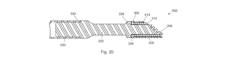
Figures 17 through 21 depict a distal assembly 500 for an ablator formed in
accordance
with the principles of this invention. Floating electrode 400 is coaxially
assembled to insulator
300 and affixed thereto by a brazed joint formed between proximal surface 408
of electrode 400
and distal surface 309 of flange 306 of insulator 300. Insulator 300 and
electrode 400 affixed
thereto are coaxially assembled to distal end element (active electrode) 200
and affixed thereto
by a brazed joint formed between proximal face 308 of flange 306 of insulator
300 and distal
surface 228 of flange 226 of active electrode 200. The proximal end 202 of
element 200 is
23/33
CA 02797967 2012-10-30
WO 2011/143200 PCT/US2011/035902
assembled to distal end 552 of tube 550. Surface 206 of active electrode 200
protrudes beyond
surface 314 of insulator 300 distance 502 and is parallel thereto. Surface 314
of insulator 300
protrudes beyond surface 414 of floating electrode 400 distance 504 and is
parallel thereto. The
electrode assembly 500 shown in Figure 17 has a circular or cylindrical
geometry. However, as
noted above, those skilled in the art will understand that the choice of the
geometry is a design
preference and that other geometries may be used to optimize performance for
specific surgical
procedures.
The operation of an ablator having a floating electrode is described in detail
in U.S.
Patent No. 7,563,261 to Carmel et al. Ablator 1 with brazed distal assembly
500 differs in
operation from that in the Carmel patent only in its ability to withstand
higher operating
temperatures without thermal failure. The benefit is equally great when second
(floating)
electrode 400 is electrically connected to the electrosurgical generator so as
to function as a
return electrode, or when second (floating) electrode 400 is eliminated so as
to form a
conventional (active electrode only) ablator. In all cases the device may be
operated at higher
temperatures without failure due to the high temperature capability of the
brazed joints, and the
higher temperature capability of the heat-shrink polymeric insulation used to
insulate the distal
portion of the device compared to the powder-coat insulations currently in
use. If
thermal/electrical failure of the polymeric insulation occurs the insulator
and/or second electrode
will not be ejected into the patient because the brazed joint will not fail.
Figures 22 through 30 depict an alternate embodiment of the invention herein
disclosed
configured for use with a resectoscope. Probe 600 has an elongated tubular
member 602 with a
proximal end 604 having an electrical connector 606 suitable for connecting
via an electrical
cable to an electrosurgical generator, and a distal end 608. Members 610 have
proximal ends
612 mounted to distal end 608 of elongated tubular member 602, and distal ends
614 to which
are mounted electrode assembly 616. Electrode stabilizer 618 for stabilizing
the distal end of
probe 600 is proximate to a distal region of a telescope mounted in a
resectoscope working
element. Conductive member 620 covered by insulation 622 extends from
electrical connector
606 to proximal end 624 of insulated conductive member 626.
Referring now to Figures 28 and 29 depicting the distal-most portion of probe
600,
electrode assembly 616 includes active electrode 630, insulator 632 and
floating electrode 634.
Active electrode 630 has a plurality of grooves 636 of width 638 and depth
640, width 638 and
24/33
CA 02797967 2012-10-30
WO 2011/143200 PCT/US2011/035902
depth 640 being selected to trap bubbles in the grooves. Active electrode 630
is formed from a
suitable metal material, such as stainless steel, nickel, titanium or
tungsten. Insulator 632 is
formed from a suitable dielectric material such as alumina or zirconia.
Floating electrode 634 is
formed from a suitable metal material, such as stainless steel, nickel,
titanium or tungsten.
Active electrode 630 protrudes beyond insulator 632 distance 642. Insulator
632 protrudes
beyond floating electrode 634 distance 644. Insulated conductive member 626
has a conductive
portion 646 coated with dielectric material 648. Distal end 650 portion 646 is
connected to
active electrode 630. Active electrode 632 has surface 652 segmented by
grooves 636. Surface
636 forms an acute angle 654 with the axis of tubular member 602.
Surface 660 of active electrode 630 is affixed to surface 662 of insulator 632
by a brazed
joint formed by braze alloy 664 placed therebetween as depicted in Figure 30.
Surface 668 of
insulator 632 is affixed to surface 670 of electrode 634 by a brazed joint
formed by braze alloy
672 placed therebetween as depicted in Figure 30. Braze alloy 664 is depicted
as a single round
deposition. However, as noted above, those skilled in the art will readily
recognize that the
quantity, size and shape of the braze alloy depositions or "braze spots" may
be routinely varied
in accordance with desired design constraints. As with the previous
embodiment, second
electrode 634 is depicted and described as a floating electrode unconnected to
the power supply.
However, the benefits of this invention are the equally realized when second
electrode 634 is
connected to the power supply so as to serve as a return electrode, or when
electrode 634 is
eliminated so as to make ablator 600 a conventional monopolar ablator.
Figures 31 through 34 depict an active electrode 740 for an alternate
embodiment of the
invention herein disclosed. Electrode 740 has a hemispherical portion 742 of
radius 748 with a
planar upper surface 744 from which protrude cylindrical portions 746. In a
preferred
embodiment portions 746 are integral with hemispherical portion 742. In other
embodiments
portions 746 are cylindrical elements inserted into holes in hemispherical
portion 742 and
welded in place.
Figures 35 through 38 depict an insulator 760 for use with the alternate
embodiment
active electrode 740. Insulator 760 has a cylindrical portion 762 of radius
764 equal to radius
748 of active electrode 740, and height 766. Cylindrical portion 762 has a top
surface 768 and a
bottom surface 770. Protruding from top surface 768 tubular portions 772 have
lumens 774
25/33
CA 02797967 2012-10-30
WO 2011/143200 PCT/US2011/035902
which extend through cylindrical portion 762, the size and spacing of lumens
774 being
configured to receive cylindrical portions 746 of active electrode 740 as
shown in Figure 39.
Referring to Figure 39, electrode 740 and insulator 760 when assembled to form
assembly 780 (Figures 40 through 43) have braze material element 782
positioned between top
bottom surface 770 of insulator 760 and upper surface 744 of insulator 740.
Braze material 782,
when assembly 780 is heated in a furnace through a predetermined thermal
cycle, adheres to
surface 770 of insulator 760 and surface 744 of electrode 740 so as to form a
brazed joint
between insulator 760 and electrode 740. Braze material element 782 has a
predetermined size
which produces a brazed joint of sufficient strength without exceeding the
maximum shear
strength of the brazed joint components. As with Figure 30, although the braze
alloy 782 is
depicted as a single round deposition, other quantities, sizes and shapes are
contemplated.
Preferred distribution and sizing is as discussed above.
Figures 44 through 47 depict the distal portion 800 of an ablator electrode
designed for
use with a resectoscope. Electrode assembly 780 is affixed to elbows 784 which
are affixed to
the distal ends of wires 786 by laser welding. Wires 786, elbows 784, exposed
regions of
protrusions 746 of electrode 740, and tubular portions 772 of insulator 760
are covered by a
dielectric coating. In a preferred embodiment the dielectric coating is a heat
shrink tubing
formed from polyolefin, PTFE, PET, or another suitable polymeric material.
Industrial Applicability:
The present invention replaces mechanical fastening means, epoxies and other
high-
temperature adhesives for connecting electrode(s) to insulator(s) with brazed
joints to yield
electrosurgical devices capable of safely and reliably operating at high power
densities and
elevated temperatures without thermal failure of the bonds between the
electrode and the
insulator. The use of brazed joints further permits the construction of
miniaturized, compact
electrosurgical devices, of both monopolar or bipolar configurations, having
utility in a number
of divergent fields, from arthroscopy to otolaryngology to oncology, and
applicable to both
laparoscopic and open surgery techniques. Thus, active electrodes and
electrosurgical devices of
the present invention maximize efficiency, safety and reliability while
minimizing manufacturing
cost and device profile.
26/33
CA 02797967 2012-10-30
WO 2011/143200 PCT/US2011/035902
All patents and publications mentioned herein are incorporated by reference in
their
entirety. Nothing herein is to be construed as an admission that the invention
is not entitled to
antedate such disclosure by virtue of prior invention.
While the invention has been described in detail and with reference to
specific
embodiments thereof, it is to be understood that the foregoing description is
exemplary and
explanatory in nature and is intended to illustrate the invention and its
preferred embodiments.
Through routine experimentation, one skilled in the art will readily recognize
that various
changes and modifications can be made therein without departing from the
spirit and scope of
the invention.
Other advantages and features will become apparent from the claims filed
hereafter, with
the scope of such claims to be determined by their reasonable equivalents, as
would be
understood by those skilled in the art. Thus, the invention is intended to be
defined not by the
above description, but by the following claims and their equivalents.
27/33