Note: Descriptions are shown in the official language in which they were submitted.
CA 02797976 2012-10-30
WO 2012/024004 PCT/US2011/034880
Devices and Methods for Tissue Engineering
Field of the Invention
The present invention relates generally to the field of porous fibrous medical
implants. More
specifically, the invention relates to a bioactive fibrous implant having
osteostimulative properties in
applications of in vivo environments.
Background of the Invention
Prosthetic devices are often required for repairing defects in bone tissue in
surgical and
orthopedic procedures. Prostheses are increasingly required for the
replacement or repair of diseased
or deteriorated bone tissue in an aging population and to enhance the body's
own mechanism to
produce rapid healing of musculoskeletal injuries resulting from severe trauma
or degenerative
disease.
Autografting and allografting procedures have been developed for the repair of
bone defects.
In autografting procedures, bone grafts are harvested from a donor site in the
patient, for example
from the iliac crest, to implant at the repair site, in order to promote
regeneration of bone tissue.
However, autografting procedures are particularly invasive, causing risk of
infection and unnecessary
pain and discomfort at the harvest site. In allografting procedures, bone
grafts are used from a donor
of the same species but the use of these materials can raise the risk of
infection, disease transmission,
and immune reactions, as well as religious objections. Accordingly, synthetic
materials and methods
for implanting synthetic materials have been sought as an alternative to
autografting and allografting.
Synthetic prosthetic devices for the repair of defects in bone tissue have
been developed in an
attempt to provide a material with the mechanical properties of natural bone
materials, while
promoting bone tissue growth to provide a durable and permanent repair.
Knowledge of the structure
and bio-mechanical properties of bone, and an understanding of the bone
healing process provides
guidance on desired properties and characteristics of an ideal synthetic
prosthetic device for bone
repair. These characteristics include, but are not limited to:
bioresorbability so that the device
effectively dissolves in the body without harmful side effects;
osteostimulation and/or
osteoconductivity to promote bone tissue in-growth into the device as the
wound heals; and load
bearing or weight sharing to support the repair site yet exercise the tissue
as the wound heals to
promote a durable repair.
Materials developed to date have been successful in attaining at least some of
the desired
characteristics, but nearly all materials compromise at least some aspect of
the bio-mechanical
requirements of an ideal hard tissue scaffold.
1
CA 02797976 2012-10-30
WO 2012/024004 PCT/US2011/034880
Brief Summary of the Invention
The present invention meets the objectives of an effective synthetic bone
prosthetic for the
repair of bone defects by providing a material that is bioresorbable,
osteostimulative, and load
bearing. The present invention provides a bioresorbable (i.e., resorbable)
tissue scaffold of bioactive
glass fiber with a bioactive glass bonding at least a portion of the fiber to
form a rigid three
dimensional porous matrix. The porous matrix has interconnected pore space
with a pore size
distribution in the range of about 10 pm to about 500 pm with porosity between
40% and 85% to
provide osteoconductivity once implanted in bone tissue. Embodiments of the
present invention
include pore space having a bi-modal pore size distribution.
Methods of fabricating a synthetic bone prosthesis according to the present
invention are also
provided that include mixing a glass fiber with a bonding agent, a pore
former, and a liquid to provide
a plastically formable batch material. In this method, the composition of the
glass fiber and the
bonding agent are each precursors to a bioactive composition. The formable
batch is mixed and
kneaded to evenly distribute the glass fiber with the bonding agent, pore
former, and binder, and
formed into a desired shape. The formed shape is then dried to remove the
liquid, and the pore former
is removed. The formed shape is then heated to react the glass fiber with the
bonding agent to form a
porous fiber scaffold having the bioactive composition.
Alternative methods of fabricating a synthetic bone prosthesis according to
the present
invention are also provided that include the application of a precursor
material to a porous fiber
scaffold that is then reaction-formed into a bioactive composition.
The method of the present invention generally involves a reaction-formation of
a bioactive
composition using raw materials that are precursors to the bioactive
composition that include fiber
precursors, while generally maintaining the form and relative position of the
fiber precursors.
These and other features of the present invention will become apparent from a
reading of the
following descriptions and may be realized by means of the instrumentalities
and combinations
particularly pointed out in the appended claims.
Brief Description of the Several Views of the Drawing
The foregoing and other objects, features, and advantages of the invention
will be apparent
from the following detailed description of the several embodiments of the
invention, as illustrated in
the accompanying drawings in which like reference characters refer to the same
parts throughout the
different views. The drawings are not necessarily to scale, with emphasis
instead being placed upon
illustrating the principles of the invention.
FIG. 1 is a ternary phase diagram of soda-lime glass according to the
background art.
2
CA 02797976 2012-10-30
WO 2012/024004 PCT/US2011/034880
FIG. 2 is a scanning electron micrograph at approximately 100X magnification
showing an
embodiment of a bioactive tissue scaffold according to the present invention.
FIG. 3 is a flowchart of an embodiment of a method of the present invention
for forming the
bioactive tissue scaffold of FIG. 1.
FIG. 4 is a flowchart of an embodiment of a curing step according to the
method of FIG. 3.
FIG. 5 is a schematic representation of an embodiment of an object fabricated
according to a
method of the present invention.
FIG. 6 is a schematic representation of the object of FIG. 5 upon completion
of a volatile
component removal step of the method of the present invention.
FIG. 7 is a schematic representation of the object of FIG 6 upon completion of
a reaction
formation step of the method of the present invention.
FIG. 8 is a flowchart of an alternate embodiment of the present invention for
forming the
bioactive tissue scaffold of FIG. 1.
FIG. 9 is a side elevation view of a bioactive tissue scaffold according to
the present invention
manufactured into a spinal implant.
FIG. 10 is a side perspective view of a spine having the spinal implant of
FIG. 9 implanted in
the intervertebral space.
FIG. 11 is a schematic drawing showing an isometric view of a bioactive tissue
scaffold
according to the present invention manufactured into an osteotomy wedge.
FIG. 12 is a schematic drawing showing an exploded view of the osteotomy wedge
of FIG. 11
operable to be inserted into an osteotomy opening in a bone.
While the above-identified drawings set forth presently disclosed embodiments,
other
embodiments are also contemplated, as noted in the discussion. This disclosure
presents illustrative
embodiments by way of representation and not limitation. Numerous other
modifications and
embodiments can be devised by those skilled in the art which fall within the
scope and spirit of the
principles of the presently disclosed embodiments.
Detailed Description of the Invention
The present invention provides a synthetic prosthetic tissue scaffold for the
repair of tissue
defects. As used herein, the terms "synthetic prosthetic tissue scaffold" and
"bone tissue scaffold" and
"tissue scaffold" and "synthetic bone prosthetic" in various forms may be used
interchangeably
throughout. In an embodiment, the synthetic prosthetic tissue scaffold is
bioresorbable once
implanted in living tissue. In an embodiment, the synthetic prosthetic tissue
scaffold is
3
CA 02797976 2012-10-30
WO 2012/024004 PCT/US2011/034880
osteoconductive once implanted in living tissue. In an embodiment, the
synthetic prosthetic tissue
scaffold is osteostimulative once implanted in living tissue. In an
embodiment, the synthetic
prosthetic tissue scaffold is load bearing once implanted in living tissue.
Various types of synthetic implants have been developed for tissue engineering
applications in
an attempt to provide a synthetic prosthetic device that mimics the properties
of natural bone tissue
and promotes healing and repair of tissue. Metallic and bio-persistent
structures have been developed
to provide high strength in a porous structure that promotes the growth of new
tissue. These materials
however, are not bioresorbable and must either be removed in subsequent
surgical procedures or left
inside the body for the life of the patient. A disadvantage of bio-persistent
metallic and biocompatible
implants is that the high load bearing capability does not transfer to
regenerated tissue surrounding the
implant. When hard tissue is formed, stress loading results in a stronger
tissue but the metallic implant
shields the newly formed bone from receiving this stress. Stress shielding of
bone tissue therefore
results in weak bone tissue which can actually be resorbed by the body, which
is an initiator of
prosthesis loosening.
Implants into living tissue evoke a biological response dependent upon a
number of factors,
such as the composition of the implant. Biologically inactive materials are
commonly encapsulated
with fibrous tissue to isolate the implant from the host. Metals and most
polymers produce this
interfacial response, as do nearly inert ceramics, such as alumina or
zirconia. Biologically active
materials or bioactive materials, elicit a biological response that can
produce an interfacial bond
securing the implant material to the living tissue, much like the interface
that is formed when natural
tissue repairs itself. This interfacial bonding can lead to an interface that
stabilizes the scaffold or
implant in the bony bed and provide stress transfer from the scaffold across
the bonded interface into
the bone tissue. When loads are applied to the repair, the bone tissue
including the regenerated bone
tissue is stressed, thus limiting bone tissue resorption due to stress
shielding. Bioactive materials can
exhibit a range of bioactivity: low levels of bioactivity exhibit a slow rate
of bonding to living tissue;
and high levels of bioactivity exhibit relatively fast rates of bonding to
living tissue. A bioresorbable
material can elicit the same response as a bioactive material, but can also
exhibit complete chemical
degradation by body fluid.
The challenge in developing a resorbable tissue scaffold using biologically
active and
resorbable materials is to attain load bearing strength with porosity
sufficient to promote the growth of
bone tissue. Conventional bioactive bioglass and bioceramic materials in a
porous form are not known
to be inherently strong enough to provide load-bearing strength as a synthetic
prosthesis or implant.
Conventional bioactive materials prepared into a tissue scaffold with
sufficient porosity to be
osteostimulative have not exhibited load bearing strength. Similarly,
conventional bioactive materials
4
CA 02797976 2012-10-30
WO 2012/024004 PCT/US2011/034880
in a form that provides sufficient strength do not exhibit a pore structure
that can be considered to be
osteostimulative.
Fiber-based structures are generally known to provide inherently higher
strength to weight
ratios, given that the strength of an individual fiber can be significantly
greater than powder-based or
particle-based materials of the same composition. A fiber can be produced with
relatively few
discontinuities that contribute to the formation of stress concentrations for
failure propagation. By
contrast, a powder-based or particle-based material requires the formation of
bonds between each of
the adjoining particles, with each bond interface potentially creating a
stress concentration.
Furthermore, a fiber-based structure provides for stress relief and thus,
greater strength, when the
fiber-based structure is subjected to strain in that the failure of any one
individual fiber does not
propagate through adjacent fibers. Accordingly, a fiber-based structure
exhibits superior mechanical
strength properties over an equivalent size and porosity than a powder-based
material of the same
composition.
Examples of bioactive glass materials include materials composed of Si02 ,
Na20 , CaO, and
P2O5 in various ranges of compositions. Other compositions, including B203 and
small amounts of
A1203 and others can be included, with the compositional makeup determining
the level of bioactivity
and the rate of absorption in vivo. FIG. 1 is a ternary phase diagram for soda
lime glass 10 indicating
regions for which compositions of Si02-CaO-Na2O have been shown to exhibit
bioactivity according
to the background art. In FIG. 1, the bioactive region A 11 is a compositional
range in which materials
have exhibited various degrees of bone bonding and/or resorption indicating
bioactivity. The bio-
compatible region B 12 is a compositional range in which materials are
compatible as an implant in
living tissue, but bioactivity has not been observed. Materials within the
compositional range of the
biocompatible region B 12 are readily formed into a fiber form due to the high
silica content. By
contrast, the bio-compatible region C 13 is a compositional range that can be
compatible as an
implant in living tissue, though without exhibiting bioactivity, but these
materials are not readily
provided in a fiber form. Materials in the bioactive region A 12 can be formed
into a fiber if the
compositional range is on the high side for the silica component, and the
materials cannot be readily
formed into a fiber for compositional ranges with lower quantities of silica.
In multi-component systems, such as Si02-NaO2-CaO-P205-B203-A1203 the
compositional
makeup to bioactivity relationship cannot be expressed in a two-dimensional
diagram, such as FIG. 1.
Furthermore, the addition of various components, to enhance bioactivity can
prevent the ability to
readily provide the material in a fiber form. Conversely, the addition of
components to enhance the
ability to form the material into a fiber, such as, for example, alumina, can
reduce the level of
bioactivity. Accordingly, the components and constituents of the materials
that result in bioactivity can
create difficulties in conventional fiber-forming processes and methods.
CA 02797976 2012-10-30
WO 2012/024004 PCT/US2011/034880
The present invention provides a fiber-based material for tissue engineering
applications that
is bioresorbable, with load bearing capability, and osteostimulative with a
pore structure that can be
controlled and optimized to promote the in-growth of bone, that can be formed
from readily obtained
fibrous raw materials. A fiber material that is a precursor to a bioactive
composition, but not
necessarily bioactive in the raw fiber material form, is used to create a
fiber-based material that
exhibits bioactivity.
FIG. 2 is an optical micrograph at approximately 100X magnification showing an
embodiment of a bioactive tissue scaffold 100 of the present invention. The
bioactive tissue scaffold
100 is a rigid three-dimensional matrix 110 forming a structure that mimics
bone structure in strength
and pore morphology. As used herein, the term "rigid" means the structure does
not significantly yield
upon the application of stress until it fractured in the same way that natural
bone would be considered
to be a rigid structure. The scaffold 100 is a porous material having a
network of pores 120 that are
generally interconnected. In an embodiment, the interconnected network of
pores 120 provide
osteoconductivity. As used herein, the term osteoconductive means that the
material can facilitate the
in-growth of bone tissue. Cancellous bone of a typical human has a compressive
crush strength
ranging between about 4 to about 12 MPa with an elastic modulus ranging
between about 0.1 to
about 0.5 GPa. As will be shown herein below, the bioactive tissue scaffold
100 of the present
invention can provide a porous osteostimulative structure in a bioactive
material with porosity greater
than 50% and compressive crush strength greater than 4 MPa, up to, and
exceeding 22 MPa.
In an embodiment, the three dimensional matrix 110 is formed from fibers that
are bonded
and fused into a rigid structure, with a composition that exhibits
bioresorbability. The use of fibers as
a raw material for creating the three dimensional matrix 110 provides a
distinct advantage over the
use of conventional bioactive or bioresorbable powder-based raw materials. In
an embodiment, the
fiber-based raw material provides a structure that has more strength at a
given porosity than a powder-
based structure. In an embodiment, the use of fibers as the primary raw
material results in a bioactive
material that exhibits more uniform and controlled dissolution rates in body
fluid.
In an embodiment, the fiber-based material of the three-dimensional matrix 110
exhibits
superior bioresorbability characteristics over the same compositions in a
powder-based or particle-
based system. For example, dissolution rates are increasingly variable and
thus, unpredictable, when
the material exhibits grain boundaries, such as a powder-based material form,
or when the material is
in a crystalline phase. Particle-based materials have been shown to exhibit
rapid decrease in strength
when dissolved by body fluids, exhibiting failures due to fatigue from crack
propagation at the
particle grain boundaries. Since bioactive glass or ceramic materials in fiber
form are generally
amorphous, and the heat treatment processes of the methods of the present
invention can better
6
CA 02797976 2012-10-30
WO 2012/024004 PCT/US2011/034880
control the amount and degree of ordered structure and crystallinity, the
tissue scaffold 100 of the
present invention can exhibit more controlled dissolution rates, with higher
strength.
The bioactive tissue scaffold 100 of the present invention provides desired
mechanical and
chemical characteristics, combined with pore morphology to promote
osteoconductivity. The network
of pores 120 is the natural interconnected porosity resulting from the space
between intertangled,
nonwoven fiber material in a structure that mimics the structure of natural
bone. Furthermore, using
methods described herein, the pore size can be controlled, and optimized, to
enhance the flow of
blood and body fluid within of the scaffold 100 and regenerated bone. For
example, pore size and pore
size distribution can be controlled through the selection of pore formers and
organic binders that are
volatilized during the formation of the scaffold 100. Pore size and pore size
distribution can be
determined by the particle size and particle size distribution of the pore
former including a single
mode of pore sizes, a bi-modal pore size distribution, and/or a multi-modal
pore size distribution. The
porosity of the scaffold 100 can be in the range of about 40% to about 85%. In
an embodiment, this
range promotes the process of osteoinduction of the regenerating tissue once
implanted in living tissue
while exhibiting load bearing strength.
The scaffold 100 according to the present invention is fabricated using fibers
as a raw material
that create a bioactive composition. The fibers can be composed of a material
that is a precursor to a
bioactive material. The term "fiber" as used herein is meant to describe a
filament or elongated
member in a continuous or discontinuous form having an aspect ratio greater
than one, and formed
from a fiber-forming process such as drawn, spun, blown, or other similar
process typically used in
the formation of fibrous materials or high aspect-ratio materials.
Bioactive materials, such as silica- or phosphate-based glass materials with
certain
compositional modifiers that result in bioactivity, including but not limited
to modifiers such as oxides
of magnesium, sodium, potassium, calcium, phosphorus, and boron exhibit a
narrow working range
because the modifiers effectively reduce the devitrification temperature of
the bioactive material. The
working range of a glass material is typically known to be the range of
temperatures at which the
material softens such that it can be readily formed. In a glass fiber forming
process, the glass material
in a billet or frit form is typically heated to a temperature in the working
range upon which the glass
material is molten and can be drawn or blown into a continuous or
discontinuous fiber. The working
range of bioactive glass materials is inherently narrow since the
devitrification temperature of the
glass material is either extremely close or within the working range of the
material. In other words, in
a typical process for the formation of fiber-based bioactive glass
compositions, the temperature at
which a fiber can be drawn, blown, or otherwise formed, is close to the
devitrification temperature of
the bioactive glass composition. When certain bioactive glass materials are
drawn or blown into a
7
CA 02797976 2012-10-30
WO 2012/024004 PCT/US2011/034880
fiber form at or near the devitrification temperature, the molten or softened
glass undergoes a phase
change through crystallization that inhibits the continuous formation of
fiber.
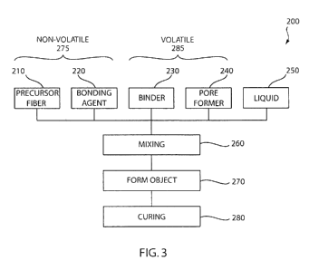
Referring to Fig. 3, an embodiment of a method 200 of forming the bioactive
tissue scaffold
100 is shown. As will be described in greater detail below, the method 200
provides for the
fabrication of a bioactive tissue scaffold using raw materials including a
precursor fiber 210 that are
precursors to a bioactive composition that react to form the three-dimensional
matrix 110 in a
bioactive composition. Generally, bulk precursor fibers 210 are mixed with a
bonding agent 220, a
binder 230, and a liquid 250 to form a plastically moldable material, which is
then cured to form the
bioactive tissue scaffold 100. The curing step 280 selectively removes the
volatile elements of the
mixture, leaving the pore space 120 open and interconnected, and effectively
fuses and bonds the
fibers 210 into the rigid three-dimensional matrix 110 in a bioactive
composition.
The bulk fibers 210 can be provided in bulk form, or as chopped fibers in a
composition that
is a precursor to a bioactive material. A fiber 210 that is precursor to a
bioactive material includes a
fiber having a composition that is at least one component of the desired
bioactive composition. For
example, the fiber 210 can be a silica fiber, or it can be a phosphate fiber,
or a combination of any of
the compositions used to form the desired bioactive composition. The diameter
of the fiber 210 can
range from about 1 to about 200 pm and typically between about 5 to about 100
pm. Fibers 210 of
this type are can be produced with a relatively narrow and controlled
distribution of fiber diameters or
depending upon the method used to fabricate the fiber, a relatively broad
distribution of fiber
diameters can be produced. Bulk fibers 210 of a given diameter may be used, or
a mixture of fibers
having a range of fiber diameters can be used. The diameter of the fibers 210
will influence the
resulting pore size, pore size distribution, strength, and elastic modulus of
the porous structure, as
well as the size and thickness of the three-dimensional matrix 110, which will
influence not only the
osteoconductivity of the scaffold 100, but also the rate at which the scaffold
100 is dissolved by body
fluids when implanted in living tissue and the resulting strength
characteristics, including compressive
strength and elastic modulus.
The fibers 210 used according to the present invention as herein described are
typically
continuous and/or chopped glass fiber. As described herein above certain
bioactive glass compositions
are difficult to form as a fiber because the working range of the material is
extremely narrow. Silica
glass in various compositions can be readily drawn into continuous or
discontinuous fiber but the
addition of calcium oxide and/or phosphate compounds necessary to create a
silica-based bioactive
composition are the very compounds that result in the reduction of the working
range of the silica-
based glass. The use of a fiber 210 that has a composition that is a precursor
to the desired bioactive
composition provides for a readily-obtained and easily formed fiber material
to form a porous fiber-
8
CA 02797976 2012-10-30
WO 2012/024004 PCT/US2011/034880
based structure that is converted into the desired bioactive composition
during the formation of the
tissue scaffold.
Examples of fiber 210 that can be used according to the present invention
include silica glass
or quartz glass fiber. Silica-based materials having a calcium oxide content
less than 30% by weight
can be typically drawn or blown into fiber form. Silica-based glass materials
are generally required to
have an alumina content less than 2% by weight since any amount of alumina in
excess of that
amount will reduce the bioactive characteristics of the resulting structure.
Phosphate glasses are
precursors to bioactive compositions and can be readily provided in fiber
form. These precursor
materials that exhibit a sufficient working range can be made into a fiber
form through melting in any
one of various methods. An exemplary method involves a combination of
centrifugal spinning and
gaseous attenuation. A glass stream of the appropriate viscosity flows
continuously from a furnace
onto a spinner plate rotating at thousands of revolutions per minute.
Centrifugal forces project the
glass outward to the spinner walls containing thousands of holes. Glass passes
through the holes,
again driven by centrifugal force, and is attenuated by a blast of heated gas
before being collected. In
another exemplary method, glass in a molten state is heated in a vessel
perforated by one or more
holes of a given diameter. The molten glass flows and is drawn through these
holes, forming
individual fibers. The fibers are merged into strands and collected on a
mandrel.
Alternative methods for producing materials that are precursors to bioactive
compositions in
fiber form can be performed at temperatures less than the melting temperature
of the precursor
materials. For example, a sol-gel fiber drawing method pulls or extrudes a sol-
gel solution of the
precursor with the appropriate viscosity into a fiber strand that is
subsequently heat treated to bind the
material into a cohesive fiber. The sol-gel fiber can be formed from a
precursor material or a
combination of one or more precursor materials that react with each other
and/or the bonding agent
220 to create the desired bioactive composition at the reaction formation 330
step, as described in
further detail below. Yet other alternative methods can be used to provide a
precursor fiber 210. For
example, a fiber can be drawn from one precursor composition, such as silica
quartz glass, that can be
co-drawn into a composite composition of a coated fiber, such as silica quartz
glass coated with a
magnesia-silicate glass, or a calcium-silicate glass. The co-drawn fiber would
provide silica and
magnesia or silica and calcium oxide as precursors to a bioactive composition,
such as 13-93 glass to
form a bioactive composition at the reaction formation 330 step with
additional bonding agent 220
including precursors of oxides of magnesium, sodium, potassium, calcium, and
phosphorus.
The binder 230 and the liquid 250, when mixed with the fiber 210, create a
plastically
formable batch mixture that enables the fibers 210 to be evenly distributed
throughout the batch, while
providing green strength to permit the batch material to be formed into the
desired shape in the
subsequent forming step 270. An organic binder material can be used as the
binder 230, such as
9
CA 02797976 2012-10-30
WO 2012/024004 PCT/US2011/034880
methylcellulose, hydroxypropyl methylcellulose (HPMC), ethylcellulose and
combinations thereof.
The binder 230 can include materials such as polyethylene, polypropylene,
polybutene, polystyrene,
polyvinyl acetate, polyester, isotactic polypropylene, atactic polypropylene,
polysulphone, polyacetal
polymers, polymethyl methacrylate, fumaron-indane copolymer, ethylene vinyl
acetate copolymer,
styrene-butadiene copolymer, acryl rubber, polyvinyl butyral, inomer resin,
epoxy resin, nylon, phenol
formaldehyde, phenol furfural, paraffin wax, wax emulsions, microcrystalline
wax, celluloses,
dextrines, chlorinated hydrocarbons, refined alginates, starches, gelatins,
lignins, rubbers, acrylics,
bitumens, casein, gums, albumins, proteins, glycols, hydroxyethyl cellulose,
sodium carboxymethyl
cellulose, polyvinyl alcohol, polyvinyl pyrrolidone, polyethylene oxide,
polyacrylamides,
polyethyterimine, agar, agarose, molasses, dextrines, starch, lignosulfonates,
lignin liquor, sodium
alginate, gum arabic, xanthan gum, gum tragacanth, gum karaya, locust bean
gum, irish moss,
scleroglucan, acrylics, and cationic galactomanan, or combinations thereof.
Although several binders
230 are listed above, it will be appreciated that other binders may be used.
The binder 230 provides
the desired rheology and cohesive strength of the plastic batch material in
order to form a desired
object and maintaining the relative position of the fibers 210 in the mixture
while the object is formed,
while remaining inert with respect to the bioactive materials. The physical
properties of the binder
230 will influence the pore size and pore size distribution of the pore space
120 of the scaffold 100.
Preferably, the binder 230 is capable of thermal disintegration, or selective
dissolution, without
impacting the chemical composition of the bioactive components, including the
fiber 210.
The fluid 250 is added as needed to attain a desired rheology in the plastic
batch material
suitable for forming the plastic batch material into the desired object in the
subsequent forming step
270. Water is typically used, though solvents of various types can be
utilized. Rheological
measurements can be made during the mixing step 260 to evaluate the plasticity
and cohesive strength
of the mixture prior to the forming step 270.
Pore formers 240 can be included in the mixture to enhance the pore space 120
of the
bioactive scaffold 100. Pore formers are non-reactive materials that occupy
volume in the plastic
batch material during the mixing step 260 and the forming step 270. When used,
the particle size and
size distribution of the pore former 240 will influence the resulting pore
size and pore size distribution
of the pore space 120 of the scaffold 100. Particle sizes can typically range
between about 25 pm or
less to about 450 pm or more, or alternatively, the particle size for the pore
former can be a function
of the fibers 210 diameter ranging from about 0.1 to about 100 times the
diameter of the fibers 210.
The pore former 240 must be readily removable during the curing step 280
without significantly
disrupting the relative position of the surrounding fibers 210. In an
embodiment of the invention, the
pore former 240 can be removed via pyrolysis or thermal degradation, or
volatilization at elevated
temperatures during the curing step 280. For example, microwax emulsions,
phenolic resin particles,
CA 02797976 2012-10-30
WO 2012/024004 PCT/US2011/034880
flour, starch, or carbon particles can be included in the mixture as the pore
former 240. Other pore
formers 240 can include carbon black, activated carbon, graphite flakes,
synthetic graphite, wood
flour, modified starch, celluloses, coconut shell husks, latex spheres, bird
seeds, saw dust, pyrolyzable
polymers, poly (alkyl methacrylate), polymethyl methacrylate, polyethyl
methacrylate, poly n-butyl
methacrylate, polyethers, poly tetrahydrofuran, poly (1, 3-dioxolane), poly
(alkalene oxides),
polyethylene oxide, polypropylene oxide, methacrylate copolymers,
polyisobutylene,
polytrimethylene carbonate, polyethylene oxalate, polybeta-propiolactone,
polydelta-valerolactone,
polyethylene carbonate, polypropylene carbonate, vinyl toluene/alpha-
methylstyrene copolymer,
styrene/alpha-methyl styrene copolymers, and olefin-sulfur dioxide copolymers.
Pore formers 240
may be generally defined as organic or inorganic, with the organic typically
burning off at a lower
temperature than the inorganic. Although several pore formers 240 are listed
above, it will be
appreciated that other pore formers 240 may be used. Pore formers 240 can be,
though need not be,
fully biocompatible since they are removed from the scaffold 100 during
processing.
Additional precursors to the desired bioactive material can be provided as a
bonding agent
220 to combine with the composition of the fiber 210 to form the bioactive
composition of the three-
dimensional matrix 110 and to promote strength and performance of the
resulting bioactive scaffold
100. The bonding agent 220 can include powder-based material of the same
composition as the bulk
fiber 210, or it can include powder-based material of a different composition.
In an embodiment of the
invention the bonding agent 220 can be coated on the fibers 210 as a sizing or
coating. In this
embodiment, additional precursors to the bioactive composition are added to
the fiber, for example, as
a sizing or coating. In an alternate embodiment, the bonding agent 220 is a
sizing or coating that is
added to the fiber during or prior to the mixing step 260. The bonding agent
220 can be solids
dissolved in a solvent or liquid that are deposited on the fiber and/or other
bonding agent 220
precursors when the liquid or solvent is removed. As will be explained in
further detail below, the
bonding agent 220 based additives enhance the bonding strength of the
intertangled fibers 210
forming the three-dimensional matrix 110 through the formation of bonds
between adjacent and
intersecting fibers 210 when the bonding agent 220 reacts with the fiber 210
to form the desired
bioactive composition. The relative quantities of the fiber 210 and the
bonding agent 220 generally
determine the resulting composition of the three-dimensional matrix 110.
The relative quantities of the respective materials, including the bulk fiber
210, the binder
230, and the liquid 250 depend on the overall porosity desired in the
bioactive tissue scaffold 100. For
example, to provide a scaffold 100 having approximately 60% porosity, the
nonvolatile components
275, such as the fiber 210, would amount to approximately 40% of the mixture
by volume. The
relative quantity of volatile components 285, such as the binder 230 and the
liquid 250 would amount
to approximately 60% of the mixture by volume, with the relative quantity of
binder to liquid
11
CA 02797976 2012-10-30
WO 2012/024004 PCT/US2011/034880
determined by the desired rheology of the mixture. Furthermore, to produce a
scaffold 100 having
porosity enhance by the pore former 240, the amount of the volatile components
285 is adjusted to
include the volatile pore former 240. Similarly, to produce a scaffold 100
having strength enhanced by
the bonding agent 220, the amount of the nonvolatile components 275 would be
adjusted to include
the nonvolatile bonding agent 220. It can be appreciated that the relative
quantities of the nonvolatile
components 275 and volatile components 285 and the resulting porosity of the
scaffold 100 will vary
as the material density may vary due to the reaction of the components during
the curing step 280.
Specific examples are provided herein below.
In the mixing step 260, the fiber 210, the binder 230, the liquid 250, the
pore former 240 and/
or the bonding agent 220, if included, are mixed into a homogeneous mass of a
plastically deformable
and uniform mixture. The mixing step 260 may include dry mixing, wet mixing,
shear mixing, and
kneading, which can be necessary to evenly distribute the material into a
homogeneous mass while
imparting the requisite shear forces to break up and distribute or de-
agglomerate the fibers 210 with
the non-fiber materials. The amount of mixing, shearing, and kneading, and
duration of such mixing
processes depends on the selection of fibers 210 and non-fiber materials,
along with the selection of
the type of mixing equipment used during the mixing step 260, in order to
obtain a uniform and
consistent distribution of the materials within the mixture, with the desired
rheological properties for
forming the object in the subsequent forming step 270. Mixing can be performed
using industrial
mixing equipment, such as batch mixers, shear mixers, and/or kneaders. The
kneading element of the
mixing step 260 distributes the fiber 210 with the bonding agent 220 and the
binder 230 to provide a
formable batch of a homogeneous mass with the fiber being arranged in an
overlapping and
intertangled relationship with the remaining fiber in the batch.
The forming step 270 forms the mixture from the mixing step 260 into the
object that will
become the bioactive tissue scaffold 100. The forming step 270 can include
extrusion, rolling,
pressure casting, or shaping into nearly any desired form in order to provide
a roughly shaped object
that can be cured in the curing step 280 to provide the scaffold 100. It can
be appreciated that the final
dimensions of the scaffold 100 may be different than the formed object at the
forming step 270, due to
expected shrinkage of the object during the curing step 280, and further
machining and final shaping
may be necessary to meet specified dimensional requirements. In an exemplary
embodiment to
provide samples for mechanical and in vitro and in vivo testing, the forming
step 270 extrudes the
mixture into a cylindrical rod using a piston extruder forcing the mixture
through a round die.
The object is then cured into the bioactive tissue scaffold 100 in the curing
step 280, as
further described in reference to FIG. 4. In the embodiment shown in FIG, 4,
the curing step 280 can
be performed as the sequence of three phases: a drying step 310; a volatile
component removal step
320; and a reaction formation step 330. In the first phase, drying 310, the
formed object is dried by
12
CA 02797976 2012-10-30
WO 2012/024004 PCT/US2011/034880
removing the liquid using slightly elevated temperature heat with or without
forced convection to
gradually remove the liquid. Various methods of heating the object can be
used, including, but not
limited to, heated air convection heating, vacuum freeze drying, solvent
extraction, microwave or
electromagnetic/radio frequency (RF) drying methods. The liquid within the
formed object is
preferably not removed too rapidly to avoid drying cracks due to shrinkage.
Typically, for aqueous
based systems, the formed object can be dried when exposed to temperatures
between about 90 C and
about 150 C for a period of about one hour, though the actual drying time may
vary due to the size
and shape of the object, with larger, more massive objects taking longer to
dry. In the case of
microwave or RF energy drying, the liquid itself, and/or other components of
the object, adsorb the
radiated energy to more evenly generate heat throughout the material. During
the drying step 310,
depending on the selection of materials used as the volatile components, the
binder 230 can congeal
or gel to provide greater green strength to provide rigidity and strength in
the object for subsequent
handling.
Once the object is dried, or substantially free of the liquid component 250 by
the drying step
310, the next phase of the curing step 280 proceeds to the volatile component
removal step 320. This
phase removes the volatile components (e.g., the binder 230 and the pore
former 240) from the object
leaving only the non-volatile components that form the three-dimensional
matrix 110 of the tissue
scaffold 100. The volatile components can be removed, for example, through
pyrolysis or by thermal
degradation, or solvent extraction. The volatile component removal step 320
can be further parsed
into a sequence of component removal steps, such as a binder burnout step 340
followed by a pore
former removal step 350, when the volatile components 285 are selected such
that the volatile
component removal step 320 can sequentially remove the components. For
example, HPMC used as a
binder 230 will thermally decompose at approximately 300 C. A graphite pore
former 220 will
oxidize into carbon dioxide when heated to approximately 600 C in the
presence of oxygen.
Similarly, flour or starch, when used as a pore former 220, will thermally
decompose at temperatures
between about 300 C and about 600 C. Accordingly, the formed object composed
of a binder 230 of
HPMC and a pore former 220 of graphite particles, can be processed in the
volatile component
removal step 320 by subjecting the object to a two-step firing schedule to
remove the binder 230 and
then the pore former 220. In this example, the binder burnout step 340 can be
performed at a
temperature of at least about 300 C but less than about 600 C for a period
of time. The pore former
removal step 350 can then be performed by increasing the temperature to at
least about 600 C with
the inclusion of oxygen into the heating chamber. This thermally-sequenced
volatile component
removal step 320 provides for a controlled removal of the volatile components
285 while maintaining
the relative position of the non-volatile components 275 in the formed object.
13
CA 02797976 2012-10-30
WO 2012/024004 PCT/US2011/034880
FIG. 5 depicts a schematic representation of the various components of the
formed object
prior to the volatile component removal step 320. The fibers 210 are
intertangled within a mixture of
the bonding agent 220, binder 230 and the pore former 240. FIG. 6 depicts a
schematic representation
of the formed object upon completion of the volatile component removal step
320. The fibers 210 and
bonding agent 220 maintain their relative position as determined from the
mixture of the fibers 210
with the volatile components 285 as the volatile components 285 are removed.
Upon completion of
the removal of the volatile components 285, the mechanical strength of the
object may be somewhat
fragile, and handling of the object in this state should be performed with
care. In an embodiment, each
phase of the curing step 280 is performed in the same oven or kiln. In an
embodiment, a handling tray
is provided upon which the object can be processed to minimize handling
damage.
FIG. 7 depicts a schematic representation of the formed object upon completion
of the last
step of the curing step 280, reaction formation 330. Pore space 120 is created
between the bonded and
intertangled fibers where the binder 230 and the pore former 240 were removed,
and the fibers 210
and bonding agent 220 are fused and bonded into the three dimensional matrix
110. The
characteristics of the volatile components 285, including the size of the pore
former 240 and/or the
distribution of particle sizes of the pore former 240 and/or the relative
quantity of the binder 230,
together cooperate to predetermine the resulting pore size, pore size
distribution, and pore
interconnectivity of the resulting tissue scaffold 100. The bonding agent 220
and the glass bonds that
form at overlapping nodes 610 and adjacent nodes 620 of the three dimensional
matrix 110 provide
for structural integrity of the resulting three-dimensional matrix 110 having
a bioactive composition.
Referring back to FIG. 4, the reaction formation step 330 converts the
nonvolatile
components 275, including the bulk fiber 210, into the rigid three-dimensional
matrix 110 having a
bioactive composition as the tissue scaffold 100 while maintaining the pore
space 120 created by the
removal of the volatile components 275 and maintaining the relative
positioning of the fiber 210. The
reaction formation step 330 heats the non-volatile components 275 to a
temperature upon which the
bulk fibers 210 react with the bonding agent 220 to form the bioactive
composition and bond to
adjacent and overlapping fibers 210, and for a duration sufficient for the
reaction to occur and to form
the bonds, without melting the fibers 210 or otherwise destroying the relative
positioning of the non-
volatile components 275. The reaction and bond formation temperature and
duration depends on the
chemical composition of the non-volatile components 275, including the bulk
fiber 210. A bioactive
glass fiber or powder of a particular composition exhibits softening and a
capability for plastic
deformation without fracture at a glass transition temperature. Glass
materials typically have a
devitrification temperature upon which the amorphous glass structure
crystallizes. In an embodiment
of the invention, the reaction and bond formation temperature in the reaction
formation step 330 is in
the working range between the glass transition temperature and the
devitrification temperature of the
14
CA 02797976 2012-10-30
WO 2012/024004 PCT/US2011/034880
precursors to the bioactive material. For example where precursors to the 13-
93 bioactive glass
composition are used to form the 13-93 bioactive composition, the reaction
temperature can be above
the glass transition temperature of about 606 C and less than the
devitrification temperature of about
1,140 C.
In the reaction formation step 330, the formed object is heated to the
reaction and bond
formation temperature resulting in the formation of glass bonds at overlapping
nodes 610 and adjacent
nodes 620 of the fiber structure. The bonds are formed at overlapping nodes
610 and adjacent nodes
620 of the fiber structure through a reaction of the bonding agent 220 that
flows around the fibers 210,
reacting with the fibers 210 to form the bioactive composition including a
glass coating and/or glass
bonds. In the reaction formation step 330, the material of the fibers 210
participates in a chemical
reaction with the bonding agent 220. Further still, the bulk fibers 210 may be
a mixture of fiber
compositions, with a portion, or all of the fibers 210 participating in a
reaction forming bonds to
create the three-dimensional matrix 110 in a bioactive composition.
The duration of the reaction formation step 330 depends on the temperature
profile during the
reaction formation step 330, in that the time at the reaction and bond
formation temperature of the
fibers 210 is limited to a relatively short duration so that the relative
position of the non-volatile
components 275, including the bulk fibers 210, does not significantly change.
The pore size, pore size
distribution, and interconnectivity between the pores in the formed object are
determined by the
relative position of the bulk fibers 210 by the volatile components 285. While
the volatile components
285 are likely burned out of the formed object by the time the bond formation
temperature is attained,
the relative positioning of the fibers 210 and non-volatile components 275 are
not significantly
altered. The formed object will likely undergo slight or minor densification
during the reaction
formation step 330, but the control of pore size and distribution of pore
sizes can be maintained, and
therefore predetermined by selecting a particle size for the pore former 240
that is slightly oversize or
adjusting the relative quantity of the volatile components 285 to account for
the expected
densification.
In an embodiment of the invention, the bonding agent 220 is a precursor to a
bioactive
material in a fine powder or nano-particle (e.g., 1 - 100 nanometers) form. In
this embodiment, the
small particle sizes react more quickly with the fiber 210 in the reaction
formation step 330. In an
embodiment of the invention, the reaction between the bonding agent 220 and
the fiber 210 also forms
a glass that covers and bonds the overlapping nodes 610 and adjacent nodes 620
of the fiber structure
before the fiber material is appreciably affected by the exposure to the
reaction temperature at or near
its glass transition temperature. In this embodiment, for the bonding agent
220 to be more reactive
than the bulk fibers 210, the particle size can be in the range of 1 to 1000
times smaller than the
diameter of the fibers 210, for example, in the range of 10 microns to 10
nanometers when using 10
CA 02797976 2012-10-30
WO 2012/024004 PCT/US2011/034880
micron diameter bulk fibers 210. Nanoparticle sized powder can be produced by
milling bioactive
glass material in a milling or comminution process, such as impact milling or
attrition milling in a ball
mill or media mill.
The temperature profile of the reaction formation step 330 can be controlled
to control the
amount of crystallization and/or minimize the devitrification of the resulting
three-dimensional matrix
110. As described above, bioactive glass and bioresorbable glass compounds
exhibit more controlled
and predictable dissolution rates in living tissue when the amount of
accessible grain boundaries of
the materials is minimized. These bioactive and bioresorbable materials
exhibit superior performance
as a bioactive device due to the amorphous structure of the material when
fabricated into fibers 210,
and the controlled degree of crystallinity that occurs during the heat
treatment processing during the
bond formation step 330. Therefore, in an embodiment of the method of the
present invention, the
temperature profile of the reaction formation step 330 is adapted to form the
bioactive composition
and bond the fiber structure without increasing grain boundaries in the non-
volatile materials 275.
In an embodiment of the method of the present invention, the reaction and bond
formation
temperature exceeds the devitrification temperature of the bulk fibers 210
during the bond formation
step 330. Resulting compositions of bioactive glass from the precursors can
exhibit a narrow working
range between its glass transition temperature and the crystallization
temperature. In this embodiment,
the crystallization of the resulting structure may not be avoided in order to
promote the formation of
the bioactive composition and the formation of bonds between overlapping and
adjacent nodes of the
fibers 210 in the structure. For example, bioactive glass in the 45S5
composition has an initial glass
transition temperature of about 550 C and a devitrification temperature of
about 580 C with
crystallization temperatures of various phases at temperatures at about 610,
about 800, and about 850
C. With such a narrow working range, the formation of the 45S5 composition may
be difficult to
perform, and as such, the reaction and bond formation temperature may require
temperatures in
excess of about 900 C to form the structure. In an alternative embodiment,
the reaction and bond
formation temperature can exceed the crystallization temperature of at least a
portion of the precursors
to the bioactive composition, yet still fall within the working range of the
remaining precursor
materials. In this embodiment, the fibers 210 of a first precursor composition
may crystallize, with
glass bonds of a second precursor composition forming at overlapping and
adjacent nodes of the fiber
structure during the formation of the bioactive composition. For example a 13-
93 composition in a
powder form as a bonding agent 220 can be used with bioactive glass fibers in
a 45S5 composition, to
form a glass bond above the glass transition temperature of the 13-93
composition but less than the
devitrification temperature of the 13-93 composition but exceeds the
devitrification temperature of the
45S5 glass fiber composition to form a composite formed object.
16
CA 02797976 2012-10-30
WO 2012/024004 PCT/US2011/034880
In an embodiment of the invention, the temperature profile of the reaction
formation step 330
is configured to reach a reaction and bond formation temperature quickly and
briefly, with rapid
cooling to avoid devitrification of the resulting bioactive material. Various
heating methods can be
utilized to provide this heating profile, such as forced convection in a kiln,
heating the object directly
in a flame, laser, or other focused heating methods. In this embodiment, the
focused heating method is
a secondary heating method that supplements a primary heating method, such as
a kiln or oven
heating apparatus. The secondary heating method provides the brief thermal
excursion to the bond
formation temperature, with a fast recovery to a temperature less than the
glass transition temperature
in order to avoid devitrification of the resulting three-dimensional matrix
110.
In an embodiment of the invention, combustion of the pore former 240 can be
used to provide
rapid and uniform heating throughout the object during the bond formation step
330. In this
embodiment, the pore former removal step 350 generally occurs during the
reaction formation step
330. The pore former 240 is a combustible material, such as carbon or
graphite, starch, organics or
polymers, such as polymethyl methacrylate, or other material that
exothermically oxidizes at elevated
temperatures less than or equal to the devitrification temperature of the
bioactive glass fiber material
210. Generally, the pore former 240 is selected based on the temperature at
which the material
initiates combustion, as can be determined by thermal analysis, such as
Thermogravimetric Analysis
(TGA) or Differential Thermal Analysis (DTA), or a combination of TGA and DTA,
such as a
simultaneous DTA/TGA which detects both mass loss and thermal response. For
example, Table 1
shows the results of a DTA/TGA analysis of various materials to determined the
exothermic
combustion point of the material.
Table 1
Pore Former Combustion Temperature
Activated Carbon 621 C
Graphite Flakes 603 C
HPMC 375 C
PMMA 346 C
Wood Flour 317 C
Corn Starch 292 C
During the curing step 280, adapted so the pore former removal step 350
generally occurs
during the reaction formation step 330, the pore former combustion increases
the temperature of the
formed object substantially uniformly and at an increased rate throughout the
object. In this way the
desired bond formation temperature can be attained reasonably quickly. Once
the pore former is fully
17
CA 02797976 2012-10-30
WO 2012/024004 PCT/US2011/034880
combusted, the internal temperature of the formed article will decrease
because of the thermal
gradient between the internal temperature of the formed object resulting from
the pore former
combustion and the temperature of the surrounding environment in the kiln or
oven. The result is that
the thermal profile of the curing process 280 can include a sharp and brief
thermal excursion at or near
the devitrification temperature of the resulting bioactive composition of the
three-dimensional matrix
110.
Additional control over the curing step 280 can be provided by controlling the
environment of
the kiln. For example, inert or stagnant air in the kiln or oven environment
can delay the point at
which the volatile components 285 are removed or control the rate at which the
volatile components
are removed. Furthermore, the pore former removal step 340 can be further
controlled by the
environment by purging with an inert gas, such as nitrogen, until the
temperature is greater than the
combustion temperature of the pore former, and nearly that of the desired
reaction and bond formation
temperature. Oxygen can be introduced at the high temperature, so that when
the pore former
oxidizes, the temperature of the non-volatile materials can be locally
increased at or above the glass
transition temperature of the precursors, or at or above the reaction and bond
formation temperature,
until the pore former is fully combusted. At that point, the temperature can
be reduced to avoid
devitrification and/or the growth of grain boundaries of and within the
resulting structure.
Referring now to FIG. 8, an alternate embodiment of the present invention is
shown. In this
embodiment, an alternative method 360 provides a fiber-based tissue scaffold
formed from precursor
fiber 210. As shown in FIG. 8, the precursor fiber 210 is used to form a glass
fiber scaffold at step
370, where the precursor is then applied at step 375, which is then reaction
formed into a bioactive
composition at step 380.
In this alternative method 360, the forming step 370 can be similar to the
method described
above with reference to FIG. 3 and FIG. 4 wherein the resulting scaffold is
not fully converted into a
bioactive composition or converted into a bioactive composition that has a low
level of bioactivity. In
other words, at forming step 370 the precursor fiber 210 and any additives
that may be utilized to
form the glass fiber scaffold does not fully convert into a bioactive
scaffold. The post-processing of
application step 375 applies the precursor materials that can fully convert
the scaffold material into a
bioactive composition, or increase the bioactivity of the scaffold material,
at the reaction step 380.
Alternatively, the forming step 370 can be sintered bulk precursor fiber 210
to form a scaffold
material, though this method would not provide control of pore size
distribution and other
characteristics that can be provided by the method described above with
reference to FIG. 3 and FIG.
4.
18
CA 02797976 2012-10-30
WO 2012/024004 PCT/US2011/034880
The apply precursor step 375 can be performed in any number of methods to
introduce a
precursor to the glass fiber scaffold produced at step 370. For example, the
precursor can be in a
colloidal solution that can be immersion applied to the scaffold, or vacuum
drawn into the porous
matrix of the fiber scaffold. Alternatively, the precursor can be in liquid
form or dissolved in a solvent
that can be applied by immersion followed by drying. Still more examples
include chemical vapor
deposition of the precursor or other gas phase deposition of precursor
compositions.
The reaction step 380 can be heating the precursor glass fiber with applied
precursors in a kiln
or furnace to a reaction formation temperature for a duration of time
sufficient for the applied
precursors to react with the precursor fiber to form the desired bioactive
composition. In this reaction
step 380, the precursors applied at step 375 react with the precursor fiber
210 to form the bioactive
composition.
In an example of the alternative method 360, a calcium-silica glass fiber
having
approximately 27.4% calcium and 72.6% silica is the precursor fiber 210 that
can be readily
fabricated in a continuous fiber form. The calcium-silica glass fiber is used
to form a three-
dimensional porous matrix by sintering the calcium-silica fiber in chopped
form to approximately 655
C for about 30 minutes and cooled to form a glass fiber scaffold. A colloidal
solution of precursors of
oxides of sodium (22% Na2O), magnesium (19% MgO), phosphorus (14.8% P2O5), and
potassium
(44.4% K2O) are applied to load approximately 27% solids of the precursors to
the calcium-silica
glass fiber scaffold and dried. The scaffold with the precursors applied are
fired in a stagnant air kiln
at 800 C for approximately 60 minutes for the precursors to react with the
calcium-silica glass fiber
to form a bioactive composition having a uniform composition of 53% Si02, 5%
MgO, 6% Na2O,
12% K20,20% CaO, and 4% P2O5 (by weight).
In an embodiment of the present invention, the strength and durability of the
tissue scaffold
100 can be enhanced by annealing the formed object subsequent to or during the
curing step 280.
During the reaction formation step 330 when the non-volatile components 275
are heated to the
reaction and bond formation temperature and subsequently cooled, thermal
gradients within the
materials may occur during a subsequent cooling phase. Thermal gradients in
the material during
cooling may induce internal stress that pre-loads the structure with stress
that effectively reduces the
amount of external stress the object can endure before mechanical failure.
Annealing the tissue
scaffold 100 involves heating the object to a temperature that is the stress
relief point of the material,
i.e., a temperature at which the glass material is still hard enough to
maintain its shape and form, but
enough for any internal stress to be relieved. The annealing temperature is
determined by the
composition of the resulting structure (i.e., the temperature at which the
viscosity of the material
softens to stress relief point), and the duration of the annealing process is
determined by the relative
size and thickness of the internal structure (i.e. the time at which the
temperature reaches steady state
19
CA 02797976 2012-10-30
WO 2012/024004 PCT/US2011/034880
throughout the object). The annealing process cools slowly at a rate that is
limited by the heat
capacity, thermal conductivity, and thermal expansion coefficient of the
material. In an exemplary
embodiment of the present invention, a fourteen millimeter diameter extruded
cylinder of a porous
bioactive tissue scaffold having a 13-93 composition can be annealed by
heating the object in a kiln or
oven at 500 C for six hours and cooled to room temperature over approximately
four hours.
The bioactive tissue scaffolds of the present invention can be used in
procedures such as an
osteotomy (for example in the hip, knee, hand and jaw), a repair of a
structural failure of a spine (for
example, an intervertebral prosthesis, lamina prosthesis, sacrum prosthesis,
vertebral body prosthesis
and facet prosthesis), a bone defect filler, fracture revision surgery, tumor
resection surgery, hip and
knee prostheses, bone augmentation, dental extractions, long bone arthrodesis,
ankle and foot
arthrodesis, including subtalar implants, and fixation screws pins. The
bioactive tissue scaffolds of the
present invention can be used in the long bones, including, but not limited
to, the ribs, the clavicle, the
femur, tibia, and fibula of the leg, the humerus, radius, and ulna of the arm,
metacarpals and
metatarsals of the hands and feet, and the phalanges of the fingers and toes.
The bioactive tissue
scaffolds of the present invention can be used in the short bones, including,
but not limited to, the
carpals and tarsals, the patella, together with the other sesamoid bones. The
bioactive tissue scaffolds
of the present invention can be used in the other bones, including, but not
limited to, the cranium,
mandible, sternum, the vertebrae and the sacrum. In an embodiment, the tissue
scaffolds of the
present invention have high load bearing capabilities compared to conventional
devices. In an
embodiment, the tissue scaffolds of the present invention require less
implanted material compared to
conventional devices. Furthermore, the use of the tissue scaffold of the
present invention requires less
ancillary fixation due to the strength of the material. In this way, the
surgical procedures for
implanting the device are less invasive, more easily performed, and do not
require subsequent surgical
procedures to remove instruments and ancillary fixations.
In one specific application, a tissue scaffold of the present invention,
fabricated as described
above, can be used as a spinal implant 800 as depicted in FIG. 9 and FIG. 10.
Referring to FIG. 9 and
FIG. 10, the spinal implant 800 includes a body 810 having a wall 820 sized
for engagement within a
space S between adjacent vertebrae V to maintain the space S. The device 800
is formed from
bioactive glass fibers that can be formed into the desired shape using
extrusion methods to form a
cylindrical shape that can be cut or machined into the desired size. The wall
820 has a height h that
corresponds to the height H of the space S. In one embodiment, the height h of
the wall 820 is slightly
larger than the height H of the intervertebral space S. The wall 820 is
adjacent to and between a
superior engaging surface 840 and an inferior engaging surface 850 that are
configured for engaging
the adjacent vertebrae V as shown in FIG. 10.
CA 02797976 2012-10-30
WO 2012/024004 PCT/US2011/034880
In another specific application, a tissue scaffold of the present invention,
fabricated as
described above, can be used as an osteotomy wedge implant 1000 as depicted in
FIG. 11 and FIG.
12. Referring to FIG. 11 and FIG. 12, the osteotomy implant 1000 may be
generally described as a
wedge designed to conform to an anatomical cross section of, for example, a
tibia, thereby providing
mechanical support to a substantial portion of a tibial surface. The osteotomy
implant is formed from
bioactive glass fibers bonded and fused into a porous material that can be
formed from an extruded
rectangular block, and cut or machined into the contoured wedge shape in the
desired size. The
proximal aspect 1010 of the implant 1000 is characterized by a curvilinear
contour. The distal aspect
1020 conforms to the shape of a tibial bone in its implanted location. The
thickness of the implant
1000 may vary from about five millimeters to about twenty millimeters
depending on the patient size
and degree of deformity. Degree of angulation between the superior and
inferior surfaces of the wedge
may also be varied.
FIG. 12 illustrates one method for the use of the osteotomy wedge implant 1000
for
realigning an abnormally angulated knee. A transverse incision is made into a
medial aspect of a tibia
while leaving a lateral portion of the tibia intact and aligning the upper
portion 1050 and the lower
portion 1040 of the tibia at a predetermined angle to create a space 1030. The
substantially wedge-
shaped implant 1000 is inserted in the space 1030 to stabilize portions of the
tibia as it heals into the
desired position with the implant 1000 dissolving into the body as herein
described. Fixation pins may
be applied as necessary to stabilize the tibia as the bone regenerates and
heals the site of the implant.
Generally, the use of a resorbable bone tissue scaffold of the present
invention as a bone graft
involves surgical procedures that are similar to the use of autograft or
allograft bone grafts. The bone
graft can often be performed as a single procedure if enough material is used
to fill and stabilize the
implant site. In an embodiment, fixation pins can be inserted into the
surrounding natural bone, and/or
into and through the resorbable bone tissue scaffold. The resorbable bone
tissue scaffold is inserted
into the site and fixed into position. The area is then closed up and after a
certain healing and maturing
period, the bone will regenerate and become solidly fused.
The use of a resorbable bone tissue scaffold of the present invention as a
bone defect filler
involves surgical procedures that can be performed as a single procedure, or
multiple procedures in
stages or phases of repair. In an embodiment, a resorbable tissue scaffold of
the present invention is
placed at the bone defect site, and attached to the bone using fixation pins
or screws. Alternatively, the
resorbable tissue scaffold can be externally secured into place using braces.
The area is then closed up
and after a certain healing and maturing period, the bone will regenerate to
repair the defect.
A method of filling a defect in a bone includes filling a space in the bone
with a resorbable
tissue scaffold comprising bioactive fibers bonded into a porous matrix, the
porous matrix having a
21
CA 02797976 2012-10-30
WO 2012/024004 PCT/US2011/034880
pore size distribution to facilitate in-growth of bone tissue; and attaching
the resorbable tissue
scaffold to the bone.
A method of treating an osteotomy includes filling a space in the bone with a
resorbable tissue
scaffold comprising bioactive fibers bonded into a porous matrix, the porous
matrix having a pore size
distribution to facilitate in-growth of bone tissue; and attaching the
resorbable tissue scaffold to the
bone.
A method of treating a structural failure of a vertebrae includes filling a
space in the bone
with a resorbable tissue scaffold comprising bioactive fibers bonded into a
porous matrix, the porous
matrix having a pore size distribution to facilitate in-growth of bone tissue;
and attaching the
resorbable tissue scaffold to the bone.
A method of fabricating a synthetic bone prosthesis includes mixing bioactive
fiber with a
binder, a pore former and a liquid to provide a plastically formable batch;
kneading the formable
batch to distribute the bioactive fiber with the pore former and the binder,
the formable batch a
homogeneous mass of intertangled and overlapping bioactive fiber; forming the
formable batch into a
desired shape to provide a shaped form; drying the shaped form to remove the
liquid; heating the
shaped form to remove the binder and the pore former; and heating the shaped
form to a bond
formation temperature using a primary heat source and a secondary heat source
to form bonds
between the intertangled and overlapping bioactive glass fiber.
In an embodiment, the present invention discloses the use of precursors to
form a porous
matrix having a bioactive composition through a chemical reaction that leads
to the transformation of
one set of chemical substances (the precursors) to another chemical substance
(the bioactive
composition). The reaction forms at elevated temperatures that is sustained
over a period of time.
In an embodiment, the present invention discloses use of fibers bonded into a
porous matrix
having a bioactive composition, the porous matrix having a pore size
distribution to facilitate in-
growth of bone tissue for the treatment of a bone defect.
In an embodiment, the present invention discloses use of fibers bonded into a
porous matrix
having a bioactive composition, the porous matrix having a pore size
distribution to facilitate in-
growth of bone tissue for the treatment of an osteotomy.
In an embodiment, the present invention discloses use of fibers bonded into a
porous matrix
having a bioactive composition, the porous matrix having a pore size
distribution to facilitate in-
growth of bone tissue for the treatment of a structural failure of various
parts of a spinal column.
22
CA 02797976 2012-10-30
WO 2012/024004 PCT/US2011/034880
The present invention has been herein described in detail with respect to
certain illustrative
and specific embodiments thereof, and it should not be considered limited to
such, as numerous
modifications are possible without departing from the spirit and scope of the
appended claims.
23