Note: Descriptions are shown in the official language in which they were submitted.
CA 2790932 201.7-05-29
1 -
Title: THE N-DOMAIN OF CARCINOEMBRYONIC ANTIGEN AND
COMPOSITIONS, METHODS AND USES THEREOF
Related Application
[0001]
Field of the disclosure
[0002] The disclosure relates to the N-domain of carcinoembryonic
antigen (CEA) and methods and uses thereof. In particular, the disclosure
relates to compositions, methods and uses of the N-domain of CEA for
treating cancer.
Background of the disclosure
[0003] The human carcinoembryonic antigen (CEA, CEACAM5,
CD66e) is a GPI-linked glycoprotein that was originally described as a
gastrointestinal oncofetal antigen [Gold and Freedman, 1965]. This cell
surface antigen is frequently over-expressed on epithelial carcinomas of the
intestinal and respiratory tracts, as well as cancers of the breast, pancreas,
stomach, and ovary [Goldenberg et al., 1976; Shively et al., 1985; Thompson
et al., 1991; Gold and Goldenberg, 1997; Hammarstrom, 1999]. From a
clinical perspective, high preoperative levels of CEA in the blood of cancer
patients negatively correlate with disease free survival. Intercellular
adhesion
events involving CEA have been linked to cancer invasion and metastasis
[Jessup and Thomas, 1998; Yoshioka et al., 1998; Thomas et al., 1995]. As
such, strategies interfering with CEA-specific functions and CEA-dependent
cellular interactions may block or delay the establishment of metastatic
tumour foci in vivo.
[0004] Structurally, CEA is composed of seven extracellular lg-like
domains (N, A1, Bi, A2, B2, A3 and B3) and self-associates (defined as
homotypic binding and homophilic cellular interactions) mainly through
interactions involving its N and A3B3 Ig-like modules [Zhou et al., 1993].
Experimentally, the addition of monoclonal antibodies (mAbs) directed at
CA 02798932 2012-11-08
WO 2011/140634 PCT/CA2011/000540
- 2 -
epitopes found in the N domain of CEA [Jessup et al., 1993; Yamanka et al.,
1996] as well as cyclic peptides derived from sequences within the N domain
of CEA [Taheri et at., 2000] have been shown to inhibit CEA-specific cellular
adhesion events in vitro.
Similarly, administration of Fab' recognizing
epitopes located in the N and adjacent AiBi domains of CEA have been
shown to increase the survival of nude mice harbouring CEA-expressing lung
micrometastases [Blumenthal et al., 2005]. These findings suggest that an
immune response specifically focused at blocking interactions involving the N
domain of CEA may halt or limit the formation of tumour metastases in
patients.
[0005] Previous
attempts at developing CEA-based anti-tumor vaccines
have centered on vaccine formulations based either on dendritic cells
preloaded with predicted T-cell epitopes or recombinant viruses delivering the
full length molecule [Curigliano et al., 2006; Berinstein, 2002; Zimmer and
Thomas, 2001; Crosti et al., 2006; Shen et al., 2004; Kobayashi et al. 2002;
Matsuda et al., 2004]. The majority of putative T-cell epitopes have been to
short sequences located in the central region of this molecule [Curigliano et
at., 2006; Berinstein, 2002; Zimmer and Thomas, 2001; Crosti et al., 2006;
Shen et al., 2004; Kobayashi et al. 2002; Matsuda et al., 2004]. In another
instance, predicted T cell (CTL) epitopes were altered to include a Val
residue
as the last residue, as an attempt to improve the peptide binding to HLA-A2
and therefore mount CEA-specific CTL responses [W02009002418].
Unfortunately, the lack of immunogenicity of these epitopes coupled with the
presence of immuno-suppressive regulatory T (Treg) cells in tumour
microenvironments were shown to compromise the efficacy of anti-tumour
CEA-based vaccines [Morse et al., 2008; Bos et al., 2008]. Overcoming these
limitations has been attempted either through the depletion of immuno-
suppressive Treg cells [Morse et al., 2008; Bos et at., 2008] or by co-
administering TAA in combination with co-stimulatory molecules [Gulley et al.,
2008; Dai et al., 2008].
CA 02798932 2012-11-08
WO 2011/140634 PCT/CA2011/000540
- 3 -
[0006] A therapeutic vaccine aimed at blocking CEA-dependent
adhesion events and the establishment of tumour foci may represent a more
appropriate and achievable objective. Importantly, the role of CEA in
metastasis is linked to its over-expression and associations which correlates
with the early inactivation of caspase-9 and activation of the P13-K/Akt
survival
pathway as well as the inactivation of caspase-8 [Camacho-Leal and
Stanners, 2008] presumably by directly binding TRAIL-R2 (DR5) through its
PELPK motif (residues 108-112 of the N domain of CEA) [Samara et al.,
2007]. This peptide motif is responsible for mediating the lodging of
metastasizing cells to the hepatic parenchyma leading to the development of
metastatic foci by promoting intercellular aggregations through homophilic
cell
interactions involving the IgV-like N- and the IgC-like A3 domains
[Berinstein,
2002; Benchimol et al., 1989; Taheri et al., 2000; Zimmer and Thomas, 2001].
Summary of the disclosure
[0007] The present inventors have demonstrated that administration of
both wild type and a deglycosylated mutant form of the N-domain of
carcinoembryonic antigen (CEA) with adjuvant was able to overcome
immunological tolerance and to raise an immune response capable of
significantly interfering with tumor growth in transgenic mice expressing
human CEA.
[0008] Accordingly, the present disclosure provides an immunogenic
composition comprising an N-domain of carcinoembryonic antigen (CEA) or a
nucleic acid encoding the N-domain. In one embodiment, the immunogenic
composition further comprises an adjuvant, such as poly I:C, and/or a
pharmaceutically acceptable carrier.
[0009] In one embodiment, the N-domain of CEA comprises the amino
acid sequence as shown in SEQ ID NO:1, 2 or 7. In another embodiment, the
N-domain of CEA consists of the amino acid sequence as shown in SEQ ID
NO:1, 2 or 7. In yet another embodiment, the nucleic acid molecule comprises
the nucleic acid sequence as shown in SEQ ID NO:3, 4 or 14.
CA 02798932 2012-11-08
WO 2011/140634 PCT/CA2011/000540
- 4 -
[0010] In another embodiment, the immunogenic composition further
comprises a second CEA domain. In one embodiment, the second CEA
domain is an A3B3 domain. In a further embodiment, the immunogenic
composition further comprises an adjuvant. In one embodiment, the adjuvant
is poly I:C.
[0011] Also provided herein is a method of inducing or enhancing an
immune response against carcinoembryonic antigen (CEA) comprising
administering an effective amount of an N-domain of CEA or a nucleic acid
molecule encoding the N-domain to an animal or cell in need thereof. Also
provided herein is a method of inducing or enhancing an immune response
against CEA comprising administering an effective amount of CEA N-domain
specific sera or CEA N-domain specific antibodies to an animal or cell in need
thereof.
[0012] Further provided is a method of inhibiting the growth of a
carcinoembryonic antigen (CEA)-expressing tumour cell comprising
administering an effective amount of an N-domain of CEA or a nucleic acid
molecule encoding the N-domain to an animal or cell in need thereof. Also
provided herein is a method of inhibiting growth of a CEA-expressing tumour
cell comprising administering an effective amount of CEA N-domain specific
sera or CEA N-domain specific antibodies to an animal or cell in need thereof.
[0013] Even further provided is a method of treating a subject with
carcinoembryonic antigen (CEA)-associated cancer or an increased risk of
said cancer comprising administering an effective amount of an N-domain of
CEA or a nucleic acid molecule encoding the N-domain. Also provided herein
is a method of treating a subject with CEA-associated cancer or an increased
risk of said cancer comprising administering an effective amount of CEA N-
domain specific sera or CEA N-domain specific antibodies to an animal or cell
in need thereof. In one embodiment, the cancer is an epithelial cancer, such
as cancer of the gastrointestinal tract, breast, lung or pancreas.
[0014] In another embodiment, the methods of the disclosure further
comprise administration of a second CEA domain. In one embodiment, the
CA 02798932 2012-11-08
WO 2011/140634 PCT/CA2011/000540
- 5 -
second CEA domain is an A3B3 domain. In a further embodiment, the
methods of the disclosure further comprise administering an adjuvant. In one
embodiment, the adjuvant is poly I:C.
[0015] Also provided herein is an in vitro screening assay for
identifying
inhibitors of CEA-mediated homophilic interactions comprising incubating
MC38.CEALuc cells with a monolayer of non-luminescent MC38.CEA cells in
the presence of a test compound; and assessing cell adherence by
quantifying bioluminescence signal emitted by adhered MC38.CEALuc cells,
wherein a decrease in bioluminescence compared to a control indicates that
the test compound is an inhibitor of CEA-mediated homophilic interactions.
[0016] Other features and advantages of the present disclosure will
become apparent from the following detailed description. It should be
understood, however, that the detailed description and the specific examples
while indicating embodiments of the disclosure are given by way of
illustration
only, since various changes and modifications within the spirit and scope of
the disclosure will become apparent to those skilled in the art from this
detailed description.
Brief description of the drawings
[0017] The disclosure will now be described in relation to the
drawings
in which:
[0018] Figure 1 shows a schematic representation of human
carcinoembryonic antigen (CEA) and its expressed recombinant modules. A.
Schematic representation of the domain structure of human CEA [Modified
from Conaghan et al. 2008]. B. Schematic depiction of the generated rCEA
domains as well as their expected molecular weights in kiloDaltons (KDa). C.
Coomassie stained SDS-PAGE depicting the purity and molecular weights of
the purified rCEA constructs.
[0019] Figure 2 shows purified human recombinant CEA retain their cell
adhesive properties. A. Demonstration of homotypic rCEA interactions using
an ELISA-based protein binding assay. Ninety six wells polystyrene plates
CA 02798932 2012-11-08
WO 2011/140634 PCT/CA2011/000540
- 6 -
were coated with untagged rCEA N-domain (1 mg per well) and incubated
with either His-tagged rCEA modules or non-specific proteins (BSA and TNF-
a). The presence of bound His-tagged protein was determined using HRPO-
coupled anti-His mAb (Hisl; 1: 5000 dilution). B. Pull-down using magnetic Ni-
NTA beads showing the specific interaction of untagged WT rCEA N domain
with His-tagged rCEA modules. TNF-a was used as a control protein which
did not pull down untagged rCEA N domain. C. Reversal of CEA-mediated
cell aggregation kinetics. CEA expressing cells human colorectal
adenocarcinoma- (HT-29) as well as CEA-expressing murine colorectal and
gastric adenocarcinoma cell lines (MC38.CEA and mGC4.CEA, respectively)
were detached from their substratum and incubated in suspension with either
the rCEA modules (N, A3/B3 or A3) or an irrelevant protein (TNF-a) at a ratio
of
1 mg protein per 106 cells per mL.
[0020] Figure 3 shows engineering and immunoreactivity of WT and
mutant rCEA N domains. A. Primary sequence of the CEA N-domain (WT:
SEQ ID NO:1 and mutant: SEQ ID NO:2), known immuno-dominant epitopes
(underlined), the sequences responsible for adhesion and metastasis
(italicized), and engineered 0-glycosylation sites (bolded). B. Purity and
immunoreactivity of expressed rCEA N domain modules. Coomassie-stained
SDS-PAGE depicting the purity and molecular weights of the purified WT and
mutant rCEA constructs. C. Western blot analysis of the immunoreactivity of
the expressed rCEA wild type (WI) and mutant (MUT) N domains with a
panel of antibodies specific to either the affinity tag (PentaHis mAb;
Qiagen),
the wild type CEA N domain (Coll mAb; Invitrogen) or a polyclonal specific to
various CEA epitopes (pan CEA P20; Santa-Cruz Biotechnology).
[0021] Figure 4 shows control of the growth of an aggressive tumor
following the administration of endotoxin-free rCEA WT or mutant N domains
as therapeutic vaccines. A. Tumor growth in CEA.Tg mice (8 per group)
following the administration of rCEA N domain. Each ascending line
represents a single mouse. Eight to twelve weeks-old CEA.Tg mice were
subcutaneously implanted with 5 X 10⁵ MC38.CEA cells in the right hind leg.
CA 02798932 2012-11-08
WO 2011/140634 PCT/CA2011/000540
- 7 -
Treated mice received an IP injection of 100 mg rCEA admixed with 100 mg
poly I:C (Sigma-Aldrich) at day 6 post xenograft implantation. Animals were
boosted a week later with 100 mg rCEA admixed with 100 mg poly I:C at day
13. B. Kaplan-Meier curve depicting the survival rates of CEA.Tg mice from
different groups. Despite the aggressive growth of the MC38.CEA tumor
xenografts, it did not kill the animals. However, animals displaying
ulceration
at the site of tumor growth and/or reaching a tumor diameter > 15 mm were
euthanized as per institutional animal care ethics guidelines.
[0022] Figure 5 shows prophylactic administration of endotoxin-free
rCEA WT or Mut N domains results in the retardation of tumor growth in
immunized CEA.Tg mice. A. Outline of experimental schedule. Eight to twelve
weeks-old CEA.Tg mice were primed by intraperitoneal injection of 100 mg
rCEA admixed with 100 mg poly I:C (Sigma-Aldrich) and boosted IP on days 3
and 10 with 50 mg rCEA admixed with 100 mg poly I:C. Four days following
the last immunization, the mice were challenged with 5 X 10⁵ MC38.CEA
subcutaneously implanted as a xenograft in the right hind leg. B. Tumor
growth in CEA.Tg mice (8 per group) following the prophylactic administration
of endotoxin-free rCEA N domain. Each ascending line represents a single
mouse. C. Kaplan-Meier curve depicting the survival rates of CEA.Tg mice
from different groups. Despite the aggressive growth of the MC38.CEA tumor
xenografts, it did not kill the animals. However, animals displaying
ulceration
at the site of tumor growth and/or reaching a tumor size > 15 mm were
euthanized as per institutional animal care ethics guidelines.
[0023] Figure 6 shows stimulation of cytokine production following
vaccination. Enumeration of CEA-specific IL-4 (A), IL-10 (B) and IFN-y (C)
spot forming units (SFUs) in the spleens of CEA-immunized and control mice.
Spleen leukocytes were collected 4 days following the last immunization and
stimulated in vitro using either Concanavalin A (Con A; 2.5 ug per mL; Sigma-
Aldrich), the full-length tumor glycoform of human CEA (FL-CEA; 1 ug per mL;
Sigma-Aldrich) or the rCEA VVT N-domain (WI N-domain; 1 ug per mL). The
number of Ag-specific cytokine secreting SFUs was counted using an
CA 02798932 2012-11-08
WO 2011/140634 PCT/CA2011/000540
- 8 -
automated ELISPOT plate counter (Cellular Technologies Inc). Data is
presented as the difference between the number of spots observed in Ag- /
ConA-stimulated wells and that of unstimulated wells. The data represents the
mean DSFU from individual animals + SD.
[0024] Figure 7
shows stimulation of IgG1 and IgG2a production
following the intraperitoneal administration of rCEA N domain. Serum was
collected from CEA-immunized and control mice 4 days following the last
immunization and tested for the presence of CEA N domain-specific IgG,
IgG1 and IgG2a antibodies by indirect ELISA. The results represent the
observed optical density of anti-CEA WT N domain pooled serum samples at
a 1:1000 dilution. Significant when compared to non-immunized, *P 0.01;
Student-t-test.
[0025] Figure 8
shows complement-dependent lysis of tumor cells.
MC38.CEA cells were suspended at a density of 1 X 10⁵ cells per mL in either
PBS alone or PBS supplemented with rabbit complement (1:100 final dilution;
Cedarlane labs) and treated with sera from immunized or control mice (1:100
final dilution). Cell suspensions were incubated for one hour at 37 C and the
percentage of lysed cells was assessed by Trypan blue dye exclusion. NS;
statistically insignificant when compared to untreated cells. Significant when
compared to cells treated with complement and sera from naïve CEA.Tg
mice, *P 5_ 0.01; Student-t-test.
[0026] Figure 9
shows the analysis of the folding and homotypic
binding activity of the generated rCEA N modules. A. Co-immunoprecipitation
of recombinant CEA N and A363 domains. Magnetic protein A beads pre-
coated with either mAb Coll (recognizing the N domain of CEA; lane 1) or an
isotype control mAb (lane 2) were added to a suspension containing 1pM of
each recombinant protein. Recovered protein complexes were resolved by
SDS-PAGE and protein bands visualized by Coomassie staining. B. Relative
binding affinities of the rCEA N domain to either the full-length tumor
glycoform of CEA (=), rCEA N domain (i) or the rCEA A3B3 domain (m) as
CA 02798932 2012-11-08
WO 2011/140634 PCT/CA2011/000540
- 9 -
defined by ELISA. Each data point represents the average absorbance value
( SEM) from experiments performed in triplicates.
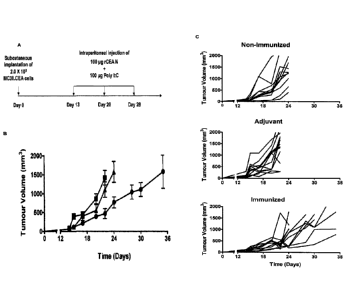
[0027] Figure 10 shows vaccination of CEA.Tg mice i.p. with the rCEA
N domain as an immunogen results in the stabilization of tumour growth in
immunized mice. A. Experimental design and immunization schedule. B.
Tumour growth kinetics of an established CEA-expressing, murine colonic
carcinoma MC38.CEA implanted s.c. in the hind leg of non-immunized
CEA.Tg mice (A; n=12), mice who received the adjuvant poly I:C only (II;
n=12) or mice immunized with rCEA N domain and adjuvant (.; n=12). C.
Collection of individual tumour growth curves observed for every CEA.Tg mice
within each experimental treatment group. Each line represents a single
mouse.
[0028] Figure 11 shows vaccination of CEA.Tg mice (i.p.) with the rCEA
N domain as an immunogen prevents the development of pulmonary tumor
nodules. A. Experimental design and immunization schedule. B. CEA-
expressing murine colonic carcinoma MC38. CEA cells were injected i.v. (tail
vein) into CEA.Tg mice at day 28 post-vaccination. Photographs highlight
tumour masses (black arrows) present in lung tissues isolated from
immunized and control CEA.Tg mice at day 60 post tumor injection. C.
Haematoxylin and eosin (H&E) stained sections of whole mouse lungs
displaying large tumour nodes in the case of non-vaccinated or adjuvant
alone-treated animals (dark stained areas). The histological features of lung
tissues from immunized mice were similar to that of a normal mouse lung. D.
Enumeration of tumor foci in H&E stained lung specimens (n=6, whole lungs
from each treatment group). E. Total volume of lung tissues (including tumour
masses; n=12) at day 60 post tumour implantation. Statistical significance
was determined using Student-t-test.
[0029] Figure 12 shows vaccination of CEA.Tg mice i.p. with the rCEA
N domain as an immunogen prevents the development of peritoneal tumour
nodules. A. Experimental design and immunization schedule. Stably-
transformed MC38.CEALuc cells expressing luciferase were injected i.p. into
CA 02798932 2012-11-08
WO 2011/140634 PCT/CA2011/000540
- 10 -
non-immunized, adjuvant-treated or immunized CEA.Tg mice B. In situ
monitoring of MC38.CEALuc cell growth and expansion at day 1, 3 and 8 post-
implantation. Recorded luminescence signals in animals (Xenogen IVIS; i.p.
injection of luciferin) after tumour implantation demonstrate a drop in signal
with time for implanted MC38.CEALuc cells in vaccinated animals. C.
Photographs highlighting the absence and presence of tumour nodules in the
viscera of immunized and control mice at day 35 post-tumour injection. The
tumour nodules are indicated by green arrows. D. Number of tumour nodules
present in the peritoneal cavity of immunized and control mice (n=5).
Statistical significance was determined using Student-t-test.
[0030] Figure 13 shows CEA-specific TH cytokine expression profiles
for vaccinated, adjuvant-treated and non-vaccinated CEA.Tg mice. A.
Experimental design and immunization schedule. B. Enumeration of rCEA-
specific IFN-y, IL-10 and IL-4 spot forming units (ASFUs) from immunized and
control mice as measured by ELISPOT assays. Histogram bars represent
averaged ASFU values measured from two independent immunization trials
(n = 3 per group). The number of Ag-specific cytokine secreting lymphocytes
(ASFUs) was calculated by subtracting background values (from wells
containing unstimulated cells) from measured values in treated groups.
Asterisk denotes statistical significance (P 5 0.05; Student-t-test) when
compared to the frequency of CEA-specific cytokine secreting cells derived
from non-immunized CEA.Tg mice.
[0031] Figure 14 shows vaccination of CEA.Tg mice with the rCEA N
domain and poly I:C results in the production of N domain-specific serum
IgGs. A. Sera of non-immunized, adjuvant treated or immunized CEA.Tg mice
were analyzed by ELISA for the presence of circulating N domain-specific
IgG, IgG1 and IgG2a antibody titers. The results represent the mean
observed optical density (+ SEM) at 450 nm of pooled serum samples (n = 12;
at a 1:1000 dilution). B. Comparison of individual mice CEA N domain-specific
IgG1 and IgG2a titers as determined by ELISA at a serum dilution of 1:1000.
CA 02798932 2012-11-08
WO 2011/140634 PCT/CA2011/000540
- 11 -
[0032] Figure
15 shows serum from CEA.Tg mice vaccinated with the
rCEA N domain display ADCC and CDC cytotoxicity functions towards CEA-
expressing cells as well as CEA-specific anti-adhesive properties. Only serum
(1:250 dilution) derived from vaccinated CEA.Tg mice can kill CEA-expressing
MC38. CEA tumor cells by A. Ab-dependent cellular cytotoxicity(ADCC) and
B. complement dependent cytotoxicity (CDC). C. Addition of anti-CEA anti-
serum (1:250 dilution) from immunized CEA.Tg mice inhibits CEA-dependent
adhesion of MC38.CEALuc cells to a MC38.CEA monolayer. The pre-
incubation of 1 pM of rCEA N domain with the serum of immunized mice
reverses the inhibition of CEA-specific cell adhesion of M038.CEA cells by
serum antibodies. NS; not
statistically significant when compared to
untreated cells. D. Specific inhibition of homotypic binding between
recombinant pure rCEA N and A3B3 domains by the addition of serum (1:1000
dilution) derived from mice immunized with the rCEA N domain. Asterisk
denotes statistical significance (P 5. 0.001) when compared to samples treated
with sera from non-immunized CEA.Tg mice, Student-t-test. Experiments
were conducted using pooled serum samples (n = 8).
[0033] Figure
16 shows adoptive transfer of CEA N-domain specific
antibodies or B lymphocytes derived from vaccinated CEA.Tg mice into naive
CEA.Tg recipients prevents the development of peritoneal tumour nodules. A.
In situ monitoring of MC38.CEALuc cell growth and expansion at day 1, 3 and
8 post-implantation. Recorded luminescence signals in animals (Xenogen
IVIS) after tumour implantation demonstrate a drop in signal with time for
implanted MC38.CEALuc cells into naïve CEA.Tg animals pre-treated either
with purified B cells (i.v.) or serum (i.p.) derived from vaccinated CEA.Tg
mice. B. Cumulative tumour volumes at day 21 post-MC38.CEA"c
implantation into the peritoneal cavity of naive CEA.Tg mice.
[0034] Figure
17 shows sensitivity of CEA-expressing human
adenocarcinoma cell lines to complement-dependent lysis. CEA + (MC38.CEA,
HT-29, MCF-7 and BxPC3) and CEA- (MC38, HeLa) cells were suspended at
a density of lx 106 cells per mL in a medium supplemented with rabbit
CA 02798932 2012-11-08
WO 2011/140634 PCT/CA2011/000540
- 12 -
complement (1:250 final dilution) and treated with sera derived from
immunized or control mice (1:250 final dilution). The percentage of cell lysis
was calculated from the surviving cell fraction measured by trypan blue dye
exclusion. Each bar represents the average % cytotoxicity ( SEM) calculated
from experiments performed in quadruplicates. Asterisk denotes statistical
significance (P 5 0.05; Student-t-test) when compared to cells treated with
complement and sera from non-immunized CEA.Tg mice.
[0035] Figure 18 shows yeast-2-hybrid experiments confirming the
binding of rCEA N domain to itself and to the A3B3 domain. A plasmid vector
expressing the IgV-like N domain of CEA as a C-terminal fusion to the GAL4
DNA-binding domain (Bait vector), was co-transformed in the yeast strain
AH109 with a vector expressing either the CEA IgV-like N- or the IgC-like
A3B3 domains fused to the C terminus of the GAL4 activation domain (Prey
vectors) [McCluskey et al., 2008]. The resulting yeast colonies were grown
overnight and spotted (5 pl) as tenfold serial dilutions onto either SD medium
lacking Trp to select for the presence of the Bait plasmid in yeast cells; SD
medium lacking both Trp and Leu to select for the presence of both Bait and
Prey plasmids in yeast cells or by spotting onto SD lacking Trp, Leu, and His
to select for Bait and Prey plasmids expressing the N-Gal and A3B3-Gal fusion
proteins that interact with each other leading to colony growth. The yeast
growth results (last panel to the right) suggests that the CEA N domain Bait
fusion protein interacts with its CEA N domain Prey fusion counterpart or with
the CEA A363 domain Prey fusion construct.
[0036] Figure 19 shows lack of lymphoproliferation in response to
stimulation with the rCEA N domain or full length CEA. Cells were isolated
from the spleens of immunized or control mice (n = 4 mice per group) and
cultured for 72 h in the presence of either full length glycosylated CEA (FL-
CEA) or the rCEA N domain. Splenocytes from immunized and control mice
were stimulated in vitro with either concanavalin A (ConA; 5 pg per mL), the
full length tumour glycoform of human CEA (1 pg per mL), rCEA WT N
domain (1 pg per mL) or left as unstimulated controls. Cells were then grown
CA 02798932 2012-11-08
WO 2011/140634 PCT/CA2011/000540
- 13 -
for 48 hours (37 C, 5% CO2) followed by pulsing with 3H-thymidine for 24
hours. The amount of incorporated thymidine was measured in harvested
cells using a scintillation counter. Results
of lymphoproliferation are
represented as a stimulation index (cpm of Ag stimulated cells/cpm of
unstimulated cells). A stimulation index greater than 1.5 is considered an
indicator of significant Ag-specific lymphoproliferation. Stimulation of
lymphocytes (from any of the test groups) with ConA yielded a stimulation
index greater than 10 (data not shown). No statistically significant
proliferations were observed from splenocytes from immunized or control
mice when stimulated with CEA constructs. Data sets were analyzed by
ANOVA.
[0037] Figure
20 shows expression of human CEA by adenocarcinoma
cell lines used. lmmunoblot analyses of the relative CEA-expression profile by
(A) murine colonic carcinoma MC38 cell lines (70 kDa band) or (B) human
adenocarconma cell lines BxPC3, HT29, MCF-7 and HeLa. Cell lysates from
6 x 10⁴ cells were resolved on a 7.5% discontinuous Laemmli SDS-PAGE gel
and transferred onto nitrocellulose membranes. The presence of CEA was
confirmed by Western blot using the CEA N domain-specific mAb Coll
(1:1,000 dilution) followed by an HRP-coupled anti-mouse secondary antibody
conjugate (1:1,000 dilution).
Detailed description of the disclosure
[0038] The
present inventors have shown that intraperitoneal
administration of the N-domain of carcinoembryonic antigen (CEA) with
adjuvant leads to the production of CEA-specific IFN-y and IL-4 responses as
well as high levels of circulating IgG1 and IgG2a antibodies capable of
mediating antibody-dependent tumor lysis. The present inventors further
demonstrated that tumor growth was retarded in CEA transgenic (CEA.Tg)
mice harboring hCEA-expressing murine tumors leading to improved survival
times for these animals. The present inventors also demonstrated that sera
and B lymphocytes collected from mice immunized with the N-domain of CEA
protected against tumour implantation.
CA 02798932 2012-11-08
WO 2011/140634 PCT/CA2011/000540
- 14 -
Compositions
[0039] Accordingly, the present disclosure provides an immunogenic
composition comprising an N-domain of carcinoembryonic antigen (CEA) or a
nucleic acid encoding the N-domain and optionally, a pharmaceutically
acceptable carrier.
[0040] The term "immunogenic composition" as used herein refers to a
composition that is able to elicit an immune response, including without
limitation, production of antibodies or cell mediated immune responses,
against an antigen present in the composition.
[0041] .. In one embodiment, the immunogenic composition further
comprises an adjuvant. The term "adjuvant" as used herein refers to a
substance that is able to enhance the innmunostimulatory effects of the N-
domains described herein but does not have any specific antigenic effect
itself. Typical adjuvants include, without limitation, Freund's adjuvant,
aluminium salts, squalene, poly I:C, poly I:C LC (also known as HiltonolTM
from Oncovir), archaeosomes, virosomes, microsomes, bacterial outer
membrane or membrane proteins preparations (OMP), Titer Max adjuvant
formulation, lmmunostimulatory complexes (ISCOMs), GM-CSF, SB-AS2,
Ribi adjuvant system, Gerbu adjuvant, CpG and monophosphoryl Lipid A. In
one embodiment, the adjuvant is poly I:C. In another embodiment, the
adjuvant is poly I:C LC.
[0042] In one embodiment, the immunogenic composition is a vaccine.
The term "vaccine" as used herein refers to an immunogenic composition that
is capable of eliciting a prophylactic and/or therapeutic response that
prevents, cures or ameliorates disease.
[0043] The term "carcinoembryonic antigen" or "CEA" as used herein
refers to CEA from any species or source and refers to the 180-kD GPI
anchored immunoglobulin (Ig) like glycoprotein. CEA is also known as
CEACAM5 or CD66e. The human CEA consists of 651 amino acids and
seven distinct Ig domains: the variable N-domain and six constant-domain-like
CA 02798932 2012-11-08
WO 2011/140634 PCT/CA2011/000540
- 15 -IgC regions (Al, B1, A2, B2, A3 and B3). In one embodiment, the CEA is
human CEA. Human
CEA has the Genbank accession number
NM 004363.2.
[0044] The term
"N-domain" as used herein refers to an isolated protein
that has the immunoglobulin variable-like N-terminal region of the mature CEA
protein (i.e. lacking the signal peptide), structurally comprising the IgV-
like
globular module and, optionally, a spacer sequence that separates the N and
Al IgC-like domains of CEA and minimally containing the immunodominant
epitopes of the N-domain and the sequences responsible for adhesion and
metastasis, for example, as shown in Figure 3 and has a sugar structure
different from the wild type CEA protein. In one embodiment, the N domain is
non-glycosylated. In another embodiment, the N domain has altered
glycosylation. In one embodiment, the N-domain comprises the wild type
sequence as shown in SEQ ID NO:1 or a tagged wild type sequence as
shown in SEQ ID NO:7 or a homolog or analog thereof, or is encoded by the
nucleotide sequence as shown in SEQ ID NO:3 or 14 or a homolog or analog
thereof. In another embodiment, the N-domain consists of the amino acid
sequence of SEQ ID NO:1 or 7. In yet another embodiment, the N-domain
comprises a deglycosylated mutant sequence as shown in SEQ ID NO:2 or a
homolog or analog thereof, or is encoded by the nucleotide sequence as
shown in SEQ ID NO:4 or a homolog or analog thereof. In a further
embodiment, the N-domain consists of the amino acid sequence as shown in
SEQ ID NO:2. (See Table 1).
[0045] The term
"homolog" means those amino acid or nucleic acid
sequences which have slight or inconsequential sequence variations from the
sequences in SEQ ID NOs:1-4, 7 or 14, i.e., the sequences function in
substantially the same manner. The variations may be attributable to local
mutations or structural modifications. Sequences having substantial homology
include nucleic acid sequences having at least 65%, at least 85%, or 90-95%
identity with the sequences as shown in SEQ ID NOs:1-4, 7 or 14. Sequence
identity can be calculated according to methods known in the art. For
- 16 -
example, nucleic acid sequence identity is readily assessed by the algorithm
of BLAST version 2.1 advanced search,
References to BLAST
searches are: Altschul, S.F., Gish, W., Miller, W., Myers, E.W. & Lipman, D.J.
(1990) "Basic local alignment search tool." J. Mol. Biol. 215:403410; Gish, W.
& States, D.J. (1993) "Identification of protein coding regions by database
similarity search." Nature Genet. 3:266272; Madden, T.L., Tatusov, R.L. &
Zhang, J. (1996) "Applications of network BLAST server" Meth. Enzymol.
266:131_141; Altschul, S.F., Madden, T.L., Schaffer, A.A., Zhang, J., Zhang,
Z., Miller, W. & Lipman, D.J. (1997) "Gapped BLAST and PSI_BLAST: a new
generation of protein database search programs." Nucleic Acids Res.
25:33893402; Zhang, J. & Madden, T.L. (1997) "PowerBLAST: A new
network BLAST application for interactive or automated sequence analysis
and annotation." Genome Res. 7:649656. In addition, homologs of the N-
domain of CEA include, without limitation, all CEACAMs that have
homologous N-domains in terms of their sequence. Such CEACAMs are
typically involved in bacterial infections (adhesion) and bacterial
colonization.
[0046] The term
"analog" means an amino acid or nucleic acid
sequence which has been modified as compared to the sequence of SEQ ID
NOs:1-4, 7 or 14 wherein the modification does not alter the utility of the
sequence (e.g. as an immune activator) as described herein. The modified
sequence or analog may have improved properties over the sequences
shown in SEQ ID NOs:1-4, 7 or 14. One example of a nucleic acid
modification to prepare an analog is to replace one of the naturally occurring
bases (i.e. adenine, guanine, cytosine or thymidine) of the sequence with a
modified base such as xanthine, hypoxanthine, 2-aminoadenine, 6-methyl, 2-
propyl and other alkyl adenines, 5-halo uracil, 5-halo cytosine, 6-aza uracil,
6-
aza cytosine and 6-aza thymine, pseudo uracil, 4-thiouracil, 8-halo adenine,
8-aminoadenine, 8-thiol adenine, 8-thiolalkyl adenines, 8-hydroxyl adenine
CA 2798932 2019-01-10
CA 02798932 2012-11-08
WO 2011/140634 PCT/CA2011/000540
- 17 -
and other 8-substituted adenines, 8-halo guanines, 8 amino guanine, 8-thiol
guanine, 8-thiolalkyl guanines, 8-hydroxyl guanine and other 8-substituted
guanines, other aza and deaza uracils, thymidines, cytosines, adenines, or
guanines, 5-trifluoromethyl uracil and 5-trifluoro cytosine.
[0047] Another example of a modification is to include modified
phosphorous or oxygen heteroatoms in the phosphate backbone, short chain
alkyl or cycloalkyl intersugar linkages or short chain heteroatomic or
heterocyclic intersugar linkages in the nucleic acid molecules shown in SEQ
ID NO:3, 4 or 14. For example, the nucleic acid sequences may contain
phosphorothioates, phosphotriesters, methyl phosphonates, and
phosphorodithioates.
[0048] A further example of an analog of a nucleic acid molecule of the
disclosure is a peptide nucleic acid (PNA) wherein the deoxyribose (or ribose)
phosphate backbone in the DNA (or RNA), is replaced with a polyamide
backbone which is similar to that found in peptides (P.E. Nielsen, et al
Science 1991, 254, 1497). PNA analogs have been shown to be resistant to
degradation by enzymes and to have extended lives in vivo and in vitro.
PNAs also bind stronger to a complimentary DNA sequence due to the lack of
charge repulsion between the PNA strand and the DNA strand. Other nucleic
acid analogs may contain nucleotides containing polymer backbones, cyclic
backbones, or acyclic backbones. For example, the nucleotides may have
morpholino backbone structures (U.S. Pat. No. 5,034,506). The analogs may
also contain groups such as reporter groups, a group for improving the
pharmacokinetic or pharmacodynamic properties of nucleic acid sequence.
[0049] The disclosure also includes sequences that hybridize to the
sequences shown in SEQ ID NO:3, 4 or 14 or a fragment thereof and
maintain the property of encoding a protein that activates the immune
response. The term "sequence that hybridizes" means a nucleic acid
sequence that can hybridize to a sequence of SEQ ID NO:3, 4 or 14 under
stringent hybridization conditions. Appropriate "stringent hybridization
conditions" which promote DNA hybridization are known to those skilled in the
CA 02798932 2012-11-08
WO 2011/140634 PCT/CA2011/000540
- 18 -
art, or may be found in Current Protocols in Molecular Biology, John Wiley &
Sons, N.Y. (1989), 6.3.1-6,3.6. The term "stringent hybridization conditions"
as used herein means that conditions are selected which promote selective
hybridization between two complementary nucleic acid molecules in solution.
Hybridization may occur to all or a portion of a nucleic acid sequence
molecule. The hybridizing portion is at least 50% the length with respect to
one of the polynucleotide sequences encoding a polypeptide. In this regard,
the stability of a nucleic acid duplex, or hybrids, is determined by the Tm,
which in sodium containing buffers is a function of the sodium ion
concentration, G/C content of labeled nucleic acid, length of nucleic acid
probe (I), and temperature (Tm = 81.5 C ¨ 16.6 (Log10 [Na+]) + 0.41(%(G+C)
¨ 600/1). Accordingly, the parameters in the wash conditions that determine
hybrid stability are sodium ion concentration and temperature. In order to
identify molecules that are similar, but not identical, to a known nucleic
acid
molecule a 1% mismatch may be assumed to result in about a 1 C decrease
in Tm, for example if nucleic acid molecules are sought that have a greater
than 95% identity, the final wash will be reduced by 5 C. Based on these
considerations stringent hybridization conditions shall be defined as:
hybridization at 5 x sodium chloride/sodium citrate (SSC)/5 x Denhardt's
solution/1.0% SDS at Tm (based on the above equation) - 5 C, followed by a
wash of 0.2 x SSC/0.1% SDS at 60 C.
[0050] The N-
domains described herein may be modified to contain
amino acid substitutions, insertions and/or deletions that do not alter the
property of activating the immune response. Conserved
amino acid
substitutions involve replacing one or more amino acids of the protein with
amino acids of similar charge, size, and/or hydrophobicity characteristics.
When only conserved substitutions are made the resulting analog should be
functionally equivalent to the protein. Non-conserved substitutions involve
replacing one or more amino acids of the protein with one or more amino
acids which possess dissimilar charge, size, and/or hydrophobicity
characteristics.
CA 02798932 2012-11-08
WO 2011/140634 PCT/CA2011/000540
- 19 -
[0051] The N-domains described herein may be modified to make it
more therapeutically effective or suitable. For example, the protein may be
converted into pharmaceutical salts by reacting with inorganic acids including
hydrochloric acid, sulphuric acid, hydrobromic acid, phosphoric acid, etc., or
organic acids including formic acid, acetic acid, propionic acid, glycolic
acid,
lactic acid, pyruvic acid, oxalic acid, succinic acid, malic acid, tartaric
acid,
citric acid, benzoic acid, salicylic acid, benzenesulphonic acid, and
tolunesulphonic acids.
[0052] The disclosure also includes expression vectors comprising a
nucleic acid sequence disclosed herein. Possible expression vectors include
but are not limited to cosmids, plasmids, artificial chromosomes, viral
vectors
or modified viruses (e.g. replication defective retroviruses, adenoviruses and
adeno-associated viruses), so long as the vector is compatible with the host
cell used. The expression vectors are "suitable for transformation of a host
cell", which means that the expression vectors contain a nucleic acid
molecule of the disclosure and regulatory sequences selected on the basis of
the host cells to be used for expression, which is operatively linked to the
nucleic acid molecule. Operatively linked is intended to mean that the nucleic
acid is linked to regulatory sequences in a manner which allows expression of
the nucleic acid,
[0053] The disclosure therefore contemplates a composition comprising
. a recombinant expression vector of the disclosure containing a nucleic
acid
molecule of the disclosure, or a fragment thereof, and the necessary
regulatory sequences for the transcription and translation of the inserted
protein-sequence.
[0054] Suitable regulatory sequences may be derived from a variety of
sources, including plant, algal, bacterial, fungal, viral, mammalian, or
insect
genes (for example, see the regulatory sequences described in Goeddel,
Gene Expression Technology: Methods in Enzymology 185, Academic Press,
San Diego, CA (1990)). Selection of appropriate regulatory sequences is
dependent on the host cell chosen as discussed below, and may be readily
CA 02798932 2012-11-08
WO 2011/140634 PCT/CA2011/000540
- 20 -
accomplished by one of ordinary skill in the art. Examples of such regulatory
sequences include: a transcriptional promoter and enhancer or RNA
polymerase binding sequence, a ribosomal binding sequence, including a
translation initiation signal. Additionally, depending on the host cell chosen
and the vector employed, other sequences, such as an origin of replication,
additional DNA restriction sites, enhancers, and sequences conferring
inducibility of transcription may be incorporated into the expression vector.
It
will also be appreciated that the necessary regulatory sequences may be
supplied by the CEA sequences and/or their flanking regions.
[0055] The recombinant expression vectors of the disclosure may also
contain a selectable marker gene which facilitates the selection of host cells
transformed or transfected with a recombinant molecule of the disclosure.
Examples of selectable marker genes are genes encoding a protein such as
G418 and hygromycin which confer resistance to certain drugs, (3-
galactosidase, chloramphenicol acetyltransferase, firefly luciferase, or an
immunoglobulin or portion thereof such as the Fc portion of an
immunoglobulin optionally IgG. Transcription of the selectable marker gene is
monitored by changes in the concentration of the selectable marker protein
such as 13-galactosidase, chloramphenicol acetyltransferase, or firefly
luciferase. If the selectable marker gene encodes a protein conferring
antibiotic resistance such as neomycin resistance transformant cells can be
selected with G418. Cells that have incorporated the selectable marker gene
will survive, while the other cells die. This makes it possible to visualize
and
assay for expression of recombinant expression vectors of the disclosure and
in particular to determine the effect of a mutation on expression and
phenotype. It will be appreciated that selectable markers can be introduced
on a separate vector from the nucleic acid of interest.
[0056] The recombinant expression vectors may also contain genes
which encode a moiety which provides increased expression of the
recombinant protein; increased solubility of the recombinant protein; and aid
in the purification of the target recombinant protein by acting as a ligand in
CA 02798932 2012-11-08
WO 2011/140634 PCT/CA2011/000540
- 21 -
affinity purification. For example, a proteolytic cleavage site may be added
to
the target recombinant protein to allow separation of the recombinant protein
from the fusion moiety subsequent to purification of the fusion protein.
Typical
fusion expression vectors include pGEX (Amrad Corp., Melbourne, Australia),
pMal (New England Biolabs, Beverly, MA) and pRIT5 (Pharmacia,
Piscataway, NJ) which fuse glutathione S-transferase (GST), maltose E
binding protein, or protein A, respectively, to the recombinant protein.
[0057]
Recombinant expression vectors can be introduced into host
cells to produce a transformed host cell. The term "transformed host cell" is
intended to include cells that are capable of being transformed or transfected
with a recombinant expression vector of the disclosure. The terms
"transduced", "transformed with", "transfected with", "transformation" and
"transfection" are intended to encompass introduction of nucleic acid (e.g. a
vector or naked RNA or DNA) into a cell by one of many possible techniques
known in the art. Prokaryotic cells can be transformed with nucleic acid by,
for example, electroporation or calcium-chloride mediated transformation. For
example, nucleic acid can be introduced into mammalian cells via
conventional techniques such as calcium phosphate or calcium chloride co-
precipitation, DEAE-dextran mediated transfection, lipofectin,
electroporation,
microinjection, RNA transfer, DNA transfer, artificial chromosomes, viral
vectors and any emerging gene transfer technologies. Suitable methods for
transforming and transfecting host cells can be found in Sambrook et al.
(Molecular Cloning: A Laboratory Manual, 2nd Edition, Cold Spring Harbor
Laboratory press (1989)), and other laboratory textbooks.
[0058] The N domain
of CEA may be generated using a variety of
systems to yield a non-glycosylated polypeptide. These would include:
chemical synthesis of the entire polypeptide; expression of the CEA N domain
in mutant Chinese hamster ovary (CHO) cells which are deficient in N-linked
glycosylation; expression using cell free expression systems (both prokaryotic
and eukaryotic); or expressing the protein in any eukaryotic expression
system followed by deglycosylation in vitro.
CA 02798932 2012-11-08
WO 2011/140634 PCT/CA2011/000540
- 22 -
[0059] Suitable host cells include a wide variety of eukaryotic host
cells
and prokaryotic cells. For example, the proteins of the disclosure may be
expressed in algal cells, yeast cells, insect cells, transgenic plant cells,
eukaryotic or prokaryotic cell-free expression systems, or mammalian cells.
Other suitable host cells can be found in Goeddel, Gene Expression
Technology: Methods in Enzymology 185, Academic Press, San Diego, CA
(1991). In addition, the proteins of the disclosure may be expressed in
prokaryotic cells, such as Escherichia coil (Zhang et al., Science 303(5656):
371-3 (2004)) or in prokaryotic expression platforms such as Gram positive
and lactic acid bacteria, including without limitation, Streptococcus
gordonii,
Lactococcus lactis and Lactobacillus spp.
[0060] Mammalian cells suitable for carrying out the present
disclosure
include, among others: 293T cells, COS (e.g., ATCC No. CRL 1650 or 1651),
BHK (e.g. ATCC No. CRL 6281), CHO (ATCC No. CCL 61), HeLa (e.g.,
ATCC No. CCL 2), 293 (ATCC No. 1573) and NS-1 cells.
[0061] The mammalian cells can also be derived from a human or
animal and include stem cells (including hematopoietic stem cells), somatic
cells, progenitor cells (including endothelial progenitor cells), fibroblasts,
lymphocytes, and mesenchymal stem cells (MSCs) that have been genetically
engineered to express the proteins described herein.
[0062] Suitable expression vectors for directing expression in
mammalian cells generally include a promoter (e.g., derived from viral
material such as polyoma, Adenovirus 2, cytomegalovirus and Simian Virus
40), as well as other transcriptional and translational control sequences.
Examples of mammalian expression vectors include pCDM8 (Seed, B.,
Nature 329:840 (1987)), pMT2PC (Kaufman et al., EMBO J. 6:187-195
(1987)) and pCMV (Clontech, California, U.S.A.). pCDNA and vectors derived
thereof (Gateway series; Invitrogen) may also be used.
[0063] In another embodiment, the immunogenic compositions
described herein further comprise a second CEA domain. In one embodiment,
the immunogenic compositions described herein further comprise an A3B3
CA 02798932 2012-11-08
WO 2011/140634 PCT/CA2011/000540
- 23 -IgC-like domain or truncations thereof. The term "A3B3 IgC-like domain"
as
used herein refers to the 3rd tandem immunoglobulin constant-like region of
the human CEA molecule, or homologues thereof. In one embodiment, the
A3B3 IgC-like domain is human and comprises the amino acid sequence of
SEQ ID NO:5 or is encoded by the nucleic acid sequence of SEQ ID NO:6.
(see Table 2).
Methods and Uses
[0064] The present disclosure also provides methods and uses of the
immunogenic compositions described herein for inducing or enhancing an
immune response, for inhibiting the growth of a CEA-expressing tumour cell,
and/or for treating cancer.
[0065] Accordingly, the present disclosure provides a method of
inducing or an enhancing an immune response against carcinoembryonic
antigen (CEA) comprising administering an effective amount of an N-domain
of CEA or a nucleic acid molecule encoding the N-domain to an animal or cell
in need thereof. The present disclosure also provides a use of an effective
amount of an N-domain of CEA or a nucleic acid molecule encoding the N-
domain for inducing or enhancing an immune response against CEA in an
animal or cell in need thereof. Also provided is a use of an effective amount
of
an N-domain of CEA or a nucleic acid molecule encoding the N-domain in the
preparation of a medicament for inducing or enhancing an immune response
against CEA in an animal or cell in need thereof. Further provided is an
effective amount of an N-domain of CEA or a nucleic acid molecule encoding
the N-domain for use in inducing or enhancing an immune response against
CEA in an animal or cell in need thereof.
[0066] The present disclosure also provides a method of inducing or
enhancing an immune response against CEA comprising administering an
effective amount of CEA N-domain specific sera or CEA N-domain specific
antibodies to an animal or cell in need thereof. The present disclosure
further
provides a use of an effective amount of CEA N-domain specific sera or CEA
N-domain specific antibodies for inducing or enhancing an immune response
CA 02798932 2012-11-08
WO 2011/140634 PCT/CA2011/000540
- 24 -
against CEA in an animal or cell in need thereof. Also provided is a use of an
effective amount of CEA N-domain specific sera or CEA N-domain specific
antibodies in the preparation of a medicament for inducing or enhancing an
immune response against CEA in an animal or cell in need thereof. Further
provided is an effective amount of CEA N-domain specific sera or CEA N-
domain specific antibodies for use in inducing or enhancing an immune
response against CEA in an animal or cell in need thereof.
[0067] The term "inducing an immune response" as used herein refers
to activating the immune response. The term "enhancing an immune
response" as used herein refers to augmenting an existing immune response.
[0068] In one embodiment, the immune response comprises a TH2
response, such as the production of IL-4 and IL-10. In another embodiment,
the immune response comprises production of circulating IgG1 and/or IgG2a
antibodies.
[0069] The term "CEA N-domain specific sera" as used herein refers to
sera containing polyclonal antibodies isolated from animals previously
immunized with an immunogenic composition disclosed herein.
[0070] The term "CEA N-domain specific antibodies" as used herein
refers to antibodies or fragments thereof isolated from animals previously
immunized with an immunogenic composition disclosed herein.
[0071] The term "antibody" as used herein is intended to include
monoclonal antibodies, polyclonal antibodies, and chimeric antibodies. The
antibody may be from recombinant sources and/or produced in transgenic
animals. The term "antibody fragment" as used herein is intended to include
without limitations Fab, Fab', F(ab')2, scFv, dsFv, ds-scFv, dimers,
minibodies, diabodies, and multimers thereof, multispecific antibody
fragments and domain antibodies. Antibodies can be fragmented using
conventional techniques. For example, F(ab')2 fragments can be generated
by treating the antibody with pepsin. The resulting F(ab')2 fragment can be
treated to reduce disulfide bridges to produce Fab' fragments. Papain
CA 02798932 2012-11-08
WO 2011/140634 PCT/CA2011/000540
- 25 -
digestion can lead to the formation of Fab fragments. Fab, Fab' and F(ab')2,
scFv, dsFv, ds-scFv, dinners, minibodies, diabodies, bispecific antibody
fragments and other fragments can also be synthesized by recombinant
techniques.
[0072] Conventional methods can be used to prepare antibodies. For
example, by using a N-domain of CEA, polyclonal antisera or monoclonal
antibodies can be made using standard methods. A mammal, (e.g., a mouse,
hamster, or rabbit) can be immunized with the N-domain which elicits an
antibody response in the mammal. Techniques for conferring immunogenicity
include conjugation to carriers or other techniques well known in the art. For
example, the N-domain can be administered in the presence of adjuvant. The
progress of immunization can be monitored by detection of antibody titers in
plasma or serum. Standard ELISA or other immunoassay procedures can be
used with the immunogen as antigen to assess the levels of antibodies.
Following immunization, antisera can be obtained and, if desired, polyclonal
antibodies isolated from the sera.
[0073] To produce monoclonal antibodies, antibody producing cells
(lymphocytes) can be harvested from an immunized animal and fused with
myeloma cells by standard somatic cell fusion procedures thus immortalizing
these cells and yielding hybridoma cells. Such techniques are well known in
the art, (e.g., the hybridoma technique originally developed by Kohler and
Milstein (Nature 256:495-497, 1975) as well as other techniques such as the
human B-cell hybridoma technique (Kozbor and Roder, Immunology Today
4:3, 72-79, 1983), the EBV-hybridoma technique to produce human
monoclonal antibodies (Cole et al., "The EBV-Hybridoma Technique and its
Application to Human Lung Cancer" in "Monoclonal Antibodies in Cancer
Therapy", Allen R. Bliss, Inc. (1985), pages 77-96) and screening of
combinatorial antibody libraries (Huse et al. Science 246:4935, 1275-1282,
1989). Hybridoma cells can be screened immunochemically for production of
antibodies specifically reactive with the N-domain and the monoclonal
antibodies can be isolated. Therefore, the disclosure also contemplates
CA 02798932 2012-11-08
WO 2011/140634 PCT/CA2011/000540
- 26 -
hybridoma cells secreting monoclonal antibodies with specificity for N-domain.
[0074] Chimeric
antibody derivatives, i.e., antibody molecules that
combine a non-human animal variable region and a human constant region
are also contemplated. Chimeric antibody molecules can include, for
example, the antigen binding domain from an antibody of a mouse, rat, or
other species, with human constant regions. Conventional methods may be
used to make chimeric antibodies containing the immunoglobulin variable
region which recognizes the N-domain (See, for example, Morrison et al.
(PNAS 81:21, 6851-6855, 1984), and Takeda et al. (Nature 314:452-454),
and the patents of Cabilly et al., U.S. Patent No. 4,816,567; Boss et at.,
U.S.
Patent No. 4,816,397; Tanaguchi et al., European Patent Publication No.
EP171496; European Patent Publication No. 0173494, United Kingdom
patent GB 2177096B).
[0075]
Monoclonal or chimeric antibodies specifically reactive with an
N-domain of CEA as described herein can be further humanized by producing
human constant region chimeras, in which parts of the variable regions,
particularly the conserved framework regions of the antigen-binding domain,
are of human origin and only the hypervariable regions are of non-human
origin. Such immunoglobulin molecules may be made by techniques known
in the art, (e.g., Teng et al. (1983) Proc. Natl. Acad. Sci. 80:12, 7308-
7312),
Kozbor and Roder (1983) Immunology Today 4:3, 72-79; Olsson et at. (1982)
Methods in Enzymol. 92, 3-16, PCT Patent Application Publication No.
W092/06193 and EP Patent Application Publication No. 0 239 400).
Humanized antibodies can also be commercially produced (Scotgen Limited,
2 Holly Road, Twickenham, Middlesex, Great Britain.)
[0076] Specific
antibodies, or antibody fragments, reactive against an
N-domain of CEA may also be generated by screening expression libraries
encoding immunoglobulin genes, or portions thereof, expressed in bacteria
with peptides produced from the nucleic acid molecules encoding an N-
domain of CEA. For example, complete Fab fragments, VH regions and FV
regions can be expressed in bacteria using phage expression libraries (See
CA 02798932 2012-11-08
WO 2011/140634 PCT/CA2011/000540
- 27 -
for example Ward et al. (1989) Nature 348:544-546, Huse et al. (1989)
Science 246:4935, 1275-1282, and McCafferty et al. (1989) Nature 348, 552-
555).
[0077] Antibodies may also be prepared using DNA immunization. For
example, an expression vector containing a nucleic acid encoding an N-
domain of CEA may be injected into a suitable animal such as mouse. The
protein will therefore be expressed in vivo and antibodies will be induced.
The
antibodies can be isolated and prepared as described above for protein
immunization.
[0078] The present disclosure also provides a method of inhibiting the
growth of a carcinoembryonic antigen (CEA)-expressing tumour cell
comprising administering an effective amount of an N-domain of CEA or a
nucleic acid molecule encoding the N-domain to an animal or cell in need
thereof. Also provided is a use of an effective amount of an N-domain of CEA
or a nucleic acid molecule encoding the N-domain for inhibiting the growth of
a CEA-expressing tumour cell in an animal or cell in need thereof. Further
provided is a use of an effective amount of an N-domain of CEA or a nucleic
acid molecule encoding the N-domain in the preparation of a medicament for
inhibiting the growth of a CEA-expressing tumour cell in an animal or cell in
need thereof. Even further provided is an effective amount of an N-domain of
CEA or a nucleic acid molecule encoding the N-domain for use in inhibiting
the growth of a CEA-expressing tumour cell in an animal or cell in need
thereof.
[0079] Also provided herein is a method of inhibiting growth of a CEA-
expressing tumour cell comprising administering an effective amount of CEA
N-domain specific sera or CEA N-domain specific antibodies to an animal or
cell in need thereof. Also provided is a use of an effective amount of CEA N-
domain specific sera or CEA N-domain specific antibodies for inhibiting the
growth of a CEA-expressing tumour cell in an animal or cell in need thereof.
Further provided is a use of an effective amount of CEA N-domain specific
sera or CEA N-domain specific antibodies in the preparation of a medicament
CA 02798932 2012-11-08
WO 2011/140634 PCT/CA2011/000540
- 28 -
for inhibiting the growth of a CEA-expressing tumour cell in an animal or cell
in need thereof. Even further provided is an effective amount of CEA N-
domain specific sera or CEA N-domain specific antibodies for use in inhibiting
the growth of a CEA-expressing tumour cell in an animal or cell in need
thereof.
[0080] The phrase "inhibiting the growth of a CEA-expressing tumour"
as used herein refers to slowing down the growth of the tumour cells and/or
killing the tumour cell. In one embodiment, the tumor cells are killed by
complement-mediated lysis or by antibody-depedent cytotoxicity (ADCC).
[0081] Also provided herein is a method of treating a subject with
carcinoembryonic antigen (CEA)-associated cancer or an increased risk of
said cancer comprising administering an effective amount of an N-domain of
CEA or a nucleic acid molecule encoding the N-domain. The disclosure
further provides a use of an effective amount of an N-domain of CEA or a
nucleic acid molecule encoding the N-domain for treating a subject with
carcinoembryonic antigen (CEA)-associated cancer or an increased risk of
said cancer. Also provided is a use of an effective amount of an N-domain of
CEA or a nucleic acid molecule encoding the N-domain in the preparation of a
medicament for treating a subject with carcinoembryonic antigen (CEA)-
associated cancer or an increased risk of said cancer. Further provided is an
effective amount of an N-domain of CEA or a nucleic acid molecule encoding
the N-domain for use in treating a subject with carcinoembryonic antigen
(CEA)-associated cancer or an increased risk of said cancer.
[0082] Also provided herein is a method of treating a subject with CEA-
associated cancer or an increased risk of said cancer comprising
administering an effective amount of CEA N-domain specific sera or CEA N-
domain specific antibodies to an animal or cell in need thereof. The
disclosure
further provides a use of an effective amount of CEA N-domain specific sera
or CEA N-domain specific antibodies for treating a subject with
carcinoembryonic antigen (CEA)-associated cancer or an increased risk of
said cancer. Also provided is a use of an effective amount of CEA N-domain
CA 02798932 2012-11-08
WO 2011/140634 PCT/CA2011/000540
- 29 -
specific sera or CEA N-domain specific antibodies in the preparation of a
medicament for treating a subject with carcinoembryonic antigen (CEA)-
associated cancer or an increased risk of said cancer. Further provided is an
effective amount of CEA N-domain specific sera or CEA N-domain specific
antibodies for use in treating a subject with carcinoembryonic antigen (CEA)-
associated cancer or an increased risk of said cancer.
[0083] The term "administering an N-domain" includes both the
administration of the protein as well as the administration of a nucleic acid
sequence encoding the protein to an animal or to a cell in vitro or in vivo.
The
term "administering" also includes the administration of a cell that expresses
the protein.
[0084] The N-domains described herein may be administered in vivo or
ex vivo to a cell which is then administered. For example, cells may be
transformed or transduced with the nucleic acid encoding the protein
described herein and then the cells are administered in vivo.
[0085] The term "treating" or "treatment" as used herein means
administering to a subject a therapeutically effective amount of the
compositions of the present disclosure and may consist of a single
administration, or alternatively comprise a series of applications.
[0086] As used herein, and as well understood in the art, "treatment" or
"treating" is also an approach for obtaining beneficial or desired results,
including clinical results. Beneficial or desired clinical results can
include, but
are not limited to, alleviation or amelioration of one or more symptoms or
conditions, diminishment of extent of disease, stabilized (i.e. not worsening)
state of disease, preventing spread of disease, delay or slowing of disease
progression, amelioration or palliation of the disease state, and remission
(whether partial or total), whether detectable or undetectable. "Treatment"
can
also mean prolonging survival as compared to expected survival if not
receiving treatment. Further any of the treatment methods or uses described
herein can be formulated alone or for contemporaneous administration with
CA 02798932 2012-11-08
WO 2011/140634 PCT/CA2011/000540
- 30 -
other agents or therapies. "Treatment" or "treating" can also include
preventing the onset of disease.
[0087] The term "subject" or "animal" as used herein includes all
members of the animal kingdom including mammals, suitably humans
including patients.
[0088] The term "increased risk of cancer" as used herein means a
subject that has a higher risk of developing a particular cancer than the
average risk of the population. A subject may have a higher risk due to
previously having had said particular cancer and or having a genetic risk
factor for said particular cancer.
[0089] In another embodiment, the CEA-associated cancer is a cancer
caused by CEA-expressing tumor cells. In an embodiment, the cancer is of
the gastrointestinal tract, breast, lung, colorectal, pancreas, female
reproductive tract, such as cervical, ovarian or uterine, neuroblastoma,
Hodgkin's disease, non-Hodgkin's lymphoma, sarcoma, cutaneous
malignancies or medullary thyroid carcinoma.
[0090] In another embodiment, the methods and uses described herein
further comprise coadministration of a second CEA domain. In one
embodiment, the methods and uses described herein further comprise
coadministration or use of the A3B3 IgC-like domain or truncations thereof. In
one embodiment, the A3B3 IgC-like domain is human and comprises the
amino acid sequence of SEQ ID NO:5 or is encoded by the nucleic acid
sequence of SEQ ID NO:6. (see Table 2).
[0091] In yet another embodiment, the methods and uses described
herein further comprise using or administering an adjuvant. As described
herein, typical adjuvants include, without limitation, Freund's adjuvant,
aluminium salts, squalene, poly I:C, poly I:C LC, archaeosomes, virosomes,
microsomes, OMP preparations, Titer Max adjuvant formulation, ISCOMs,
GM-CSF, SB-AS2, Ribi adjuvant system, Gerbu adjuvant, CpG and
CA 02798932 2012-11-08
WO 2011/140634 PCT/CA2011/000540
- 31 -
monophosphoryl Lipid A. In one embodiment, the adjuvant is poly I:C. In
another embodiment the adjuvant is poly I:C LC.
[0092] In all of the above therapeutic applications, the protein can
be
administered as a protein or as a nucleic acid molecule encoding the protein.
In one embodiment, as noted above, expression of the protein occurs as a
result of the administration of nucleic acid encoding the protein to an
organism. Thus, the protein will be produced endogenously in the organism,
rather than administered in a protein form. The therapy may be done at an
embryonic stage of the organism, such that the germ cells of the organism
contain the protein nucleic acid, resulting in a transgenic organism, or at a
later stage of development to specific somatic cells, such that only a
particular
tissue or portion of a tissue contains the protein nucleic acid. Techniques
for
nucleic acid therapy are well known in the art, as are the techniques for the
creation of transgenic organisms (Carl A. Pinkert. Transgenic Animal
Technology: A Laboratory Handbook. Academic Press; 1st edition (1994)).
[0093] It is to be understood that the administration of the protein
nucleic acid in gene therapy may take several forms, all of which are included
in the scope of the present disclosure. The nucleic acid encoding the protein
may be administered in such a manner as to add the protein nucleic acid to
the genome of the cell or the organism. For example, administering a nucleic
acid encoding the protein, under the control of a promoter which results in an
increase in expression of the protein, results in the incorporation of the
nucleic
acid into the genome of the cell or the organism, such that increased levels
of
the protein are made.
[0094] Construction of appropriate expression vehicles and vectors for
therapeutic applications will depend on the organism to be treated and the
purpose of the gene therapy. The selection of appropriate promoters and
other regulatory DNA will proceed according to known principles, based on a
variety of known gene therapy techniques. For example, retroviral mediated
gene transfer is a very effective method for therapy, as systems utilizing
packaging defective viruses allow the production of recombinants which are
CA 02798932 2012-11-08
WO 2011/140634 PCT/CA2011/000540
- 32 -
infectious only once, thus avoiding the introduction of wild-type virus into
an
organism. Alternative methodologies for therapy include non-viral transfer
methods, such as calcium phosphate co-precipitation, mechanical techniques,
for example microinjection, membrane fusion-mediated transfer via
liposomes, as well as direct DNA uptake and receptor-mediated DNA transfer.
ASSAYS
[0095] The present
inventors have utilized a modified MC38.CEA cell
line that expresses firefly Luciferase and GFP previously described in
Tiscornia et al. 2006 to measure CEA homophilic interactions. Accordingly,
the present disclosure further provides an in vitro screening assay for
identifying inhibitors of CEA-mediated homophilic interactions comprising
incubating MC38.CEALuc cells with a monolayer of non-luminescent M38.CEA
cells in the presence of a test compound; and assessing cell adherence by
quantifying bioluminescence signal emitted by adhered MC38.CEALuc cells,
wherein a decrease in bioluminescence compared to a control indicates that
the test compound is an inhibitor of CEA-mediated homophilic interactions.
[0096] The term "control"
as used herein refers to cells in the absence
of test compound. The control can also be a predetermined standard or
reference range of values.
[0097] The above disclosure
generally describes the present
disclosure. A more complete understanding can be obtained by reference to
the following specific examples. These examples are described solely for the
purpose of illustration and are not intended to limit the scope of the
disclosure.
Changes in form and substitution of equivalents are contemplated as
circumstances might suggest or render expedient. Although specific terms
have been employed herein, such terms are intended in a descriptive sense
and not for purposes of limitation.
[0098] The following non-
limiting examples are illustrative of the
present disclosure:
Examples
CA 02798932 2012-11-08
WO 2011/140634 PCT/CA2011/000540
- 33 -
EXAMPLE 1:
Generation of folded rCEA modules:
[0099] Figure 1 panel B and C shows the different rCEA constructs
expressed and purified to date from E. co/i. By optimizing the expression and
purification protocols, high yields of purified proteins were achieved,
typically
mg of soluble recombinant protein per 100 ml culture from a single poly-
histidine affinity chromatography step using Ni-NTA agarose beads.
[00100] CEA domains were purified to homogeneity under denaturing
conditions. Interactions between the N domain to itself and to the A3B3
10 domain were initially tested to confirm proper folding. The remaining CEA
modules (Figure 1 panels B and C) were also expressed and their lack of
binding to the N domain was confirmed using an ELISA-based binding assay.
Figure 2 A shows the specific binding of the N domain either to itself, to
A3B3
or the A3 domain, but not to other CEA domains or TNF-a (irrelevant protein
control). The observed domain interactions were confirmed by pull-down
assay, whereby the untagged N domain is released with 8 M urea from
magnetic Ni-NTA beads coated with either His-tagged rCEA N, A3 or A3B3
modules; but not from beads coated with His-tagged TNF-a (Figure 2 B).
[00101] Lastly, CEA-mediated intercellular aggregation assay was used
[Benchimol et al. 1989, Zhou et al. 1993] and it was tested if soluble rCEA
domains could disaggregate HT-29 human colorectal adenocarcinoma cells in
suspension. Figure 2C shows the inhibition of CEA-mediated interactions
following the addition of the N, A3B3 or the A3 domains but not by TNF-a.
[00102] Together, these observations validated that the generated rCEA
N module was properly folded and capable of mediating CEA-CEA homotypic
interactions.
Culture of CEA-expressing tumor xenografts:
[00103] Two CEA expressing murine cell lines (as well as their CEA
null
background cells that are compatible with the CEA.Tg mouse strain) were
acquired. The murine gastric cancer cell line mGC4.CEA, as well as its CEA
CA 02798932 2012-11-08
WO 2011/140634 PCT/CA2011/000540
- 34 -
negative cell line (mGC8) was acquired from Dr. Wolfgang Zimmerman
(Tumor Immunology Laboratory, LIFE-Center, Klinikum Grosshadern, Ludwig-
Maximilians-University; Germany). The MC38.CEA and its CEA null
background (MC38) was acquired from Dr. Jeffrey Schlom (Laboratory of
Tumor Immunology and Biology, National Cancer Institute, NIH; Bethesda,
Maryland). Analysis of CEA expression profile showed that the mGC4.CEA
was a low expresser, whereas MC38.CEA was a high expresser. Moreover,
both cell lines were found to be susceptible to inhibition of CEA-mediated
intercellular aggregation following the addition of the N, A3, and A3B3
modules
in a manner similar to HT-29 cells (Figure 2C).
Establishment of a CEA.Tg mouse colony:
[00104] CEA.Tg mice were acquired from Dr. Wolfgang Zimmerman
(Tumor Immunology Laboratory, LIFE-Center, Klinikum Grosshadern, Ludwig-
Maximilians-University; Germany). Generation of CEA positive litters was
done by backcrossing CEA positive animals with C57BL/6 mice. However,
challenges were experienced in generating ample numbers of this mouse
strain caused as a result of a lower reproductive rate and the paucity of CEA
positive offsprings. Nevertheless, a stable breeding program was successfully
maintained, where 25-33% of the progeny being CEA positive litters, with
litter
sizes ranging from 4-6 pups (compared to 1-3).
Engineering a mutant rCEA N domain to test as a candidate vaccinogen:
[00105] Originally, injecting rCEA modules 0-glycosylated with GaINAc
was considered. For that purpose, a mutant rCEA N-domain (Figure 3) was
engineered that retained all the putative immuno-dominant epitopes as well as
the sequences responsible for mediating its homotypic adhesive properties.
[00106] Following extensive testing, it was found that human CEA
incorporated 1 (+2) GaINAc groups. Nonetheless, the usefulness of the
mutant CEA N domain, lacking GaINAc groups, was tested in surmounting
immunological anergy towards CEA in CEA.Tg mice.
Testing the therapeutic potential of rCEA:
CA 02798932 2012-11-08
WO 2011/140634 PCT/CA2011/000540
- 35 -
[00107] Having satisfied the two main priorities (i.e. generating
folded
rCEA modules and setting up a CEA.Tg mouse model), an immunization trial
was conducted to determine the feasibility of surmounting immunological
tolerance to CEA in CEA.Tg mice. Furthermore, it was needed to determine
whether immunization with the N domain can interfere with the growth of
implanted MC38.CEA tumor xenografts.
[00108] To that end, twenty four 12 weeks-old CEA.Tg mice were
subdivided between three groups of eight animals each. One group was left
as an untreated control group, one group of animals received the WT CEA N
domain whereas the last group received the mutant CEA N domain. Half a
million (5 X10⁵) MC38.CEA cells were subcutaneously implanted into each
animal. On days six and thirteen following xenograft implantation, the mice
received an intraperitoneal injection of 100 lug of rCEA N domain admixed
with 100 !Ag poly I:C. Poly I:C was used as adjuvant because of its capacity
to
stimulate both B cell activation [Scher et al. 1973] as well as type 1
responses
through TLR-3/7 signaling [Barchet 2008], a combination of immune
responses that have been shown to positively influence the development of
protective anti-tumor immune responses in mice and in clinical trials [Barchet
2008]. The inherent antigenicity of purified human CEA has not been reported
in the past. Thus the intraperitoneal route of injection was used, since the
immunogenicity of a given antigen is traditionally determined following its
administration via intraperitoneal injections [Garvey et al. 1983].
[00109] Figure 4 A shows the progression of tumor volume in immunized
and control CEA.Tg mice. The development of tumor volumes that are >750
mm3 occurred within 7 to 14 days in 100% of control mice implanted with 5 X
105 MC38.CEA cells. Nonetheless, significant retardation of tumor growth was
observed following the intraperitoneal injection of endotoxin-free WT and
mutant rCEA N domains (Figure 4 A). The effects of this vaccine formulation
(N domain + poly I:C) were pronounced in 25% of animals receiving either Ag,
in that significant drops in tumor volumes were observed following injections
(Figure 4 A). Furthermore, injection of this formulation did not exert overt
CA 02798932 2012-11-08
WO 2011/140634 PCT/CA2011/000540
- 36 -
signs of toxicity/pathology. Finally, the intraperitoneal administration of
rCEA
N domain significantly improved survival rate (Figure 4B), It is important to
outline that despite the aggressive growth of the MC38.CEA tumor xenografts,
implantation of this cell line did not kill the animals. However, animals
displaying ulceration at the site of tumor growth and/or a tumor diameter > 15
mm were euthanized as per institutional animal care ethics guidelines.
[00110] Taken together, these findings suggest that the
intraperitoneal
injection of endotoxin-free WT or mutant rCEA N modules with poly I:C
overcame immunological tolerance to CEA and was efficient in significantly
reducing tumor growth.
Testing the prophylactic potential of rCEA:
[00111] CEA-based vaccines are best discussed in the context of
interfering with the process of tumor metastasis. Having observed the effect
of
CEA-based immunization on retarding tumor growth, it was also of interest in
determining if the vaccine carried the prophylactic potential of preventing
the
establishment of CEA-expressing tumor cells. As such, 24 twelve weeks old
CEA.Tg mice were subdivided into three groups, each containing eight mice.
Two groups received endotoxin-free rCEA WT or mutant N domains, or were
left as untreated controls (Figure 5). Four days following the last
immunization
step, 5 X 10⁵ MC38.CEA cells were subcutaneously implanted as xenografts
and the growth of tumors were measured with calipers. Although the
immunization did not prevent the implanted tumor xenografts from
establishing, it did nonetheless significantly retard their. growth (Figure 5
B)
and significantly prolong survival (Figure 5 C) in a manner reminiscent of
that
observed with the therapeutic administration of the CEA vaccine formulation
(Figure 4).
Characterization of the engendered CEA-specific immune responses:
[00112] In light of the observed tumor retardation, the underlying
immunological mechanisms were analyzed. To that effect, twelve weeks old
mice were subdivided into four groups: two groups of four CEA.Tg mice
immunized with either endotoxin-free VVT or mutant rCEA N domain using the
CA 02798932 2012-11-08
WO 2011/140634 PCT/CA2011/000540
- 37 -
schedule outlined in figure 5A; one group of two C57BL/6 mice injected with
the WT rCEA N domain using the schedule outlined in figure 5A, and lastly an
unimmunized control group containing four CEA.Tg mice.
[00113] Four days following the last immunization, the animals were
sacrificed and their blood and spleens collected to analyze correlates of
immune responses.
Cellular responses:
[00114] Spleen leukocytes from immunized and control mice were
collected, suspended to a final density of 106 cells per mL and stimulated ex
vivo with either concanavalin A (ConA; 5 [kg per mL, Sigma-Aldrich), the full
length tumor glycoform of human CEA (1 g per mL; Sigma-Aldrich), rCEA
WT N domain (1 lAg per mL) or left as unstimulated controls. Cell viability
following harvest was > 95%.
[00115] Cytokine ELISPOT was performed using the splenocytes from
immunized and control mice to monitor the frequency of CEA-specific IFN-y,
IL-10 and IL-4 spot-forming units (SFU) in order to determine the vaccine
induced TH polarity. Cells derived from unimmunized CEA.Tg mice (referred
to as naïve) did not produce any of the above cytokines in response to
antigenic stimulation with CEA, but did so following mitogenic stimulation
with
ConA (Figure 6). Conversely, CEA-immunized C57BL/6 mice produced all
three cytokines in response to antigenic stimulation (Figure 6), albeit at
lower
levels, which comes in agreement with the observations of Woo et al. [Woo et
al. 2008]. CEA.Tg mice immunized with the WT rCEA N domain produced
equal levels of IL-4, IL-10 and IFN-y SFUs following stimulation with either
rCEA N domain or FL-CEA (Figure 6), whereas CEA.Tg mice immunized with
the mutant rCEA N domain produced IL-4 and IL-10, but not IFN-y (Figure 6).
This disparity in Ag-specific cytokine production was observed in all animals
comprising these experimental cohorts and with both forms of CEA
(recombinant and tumor glycoform). Consistent with these observations, little
or no proliferation of splenocytes derived from immunized and control mice
CA 02798932 2012-11-08
WO 2011/140634 PCT/CA2011/000540
- 38 -
was seen in response to antigenic stimulation thereby suggesting a minor
stimulation of T cells by this immunization strategy.
H u moral responses:
[00116] Since the observed CEA-specific cytokine expression profile
was reminiscent of TH2 immune responses, the serum of immunized and
control animals was analyzed for the presence of CEA-specific antibodies.
Using the WT rCEA N domain as capture antigen in an indirect ELISA, high
levels of rCEA-specific IgG serum antibodies were detected (Figure 7).
Analysis of the CEA-specific IgG subclass revealed the presence of high
levels of CEA-specific IgG1 antibodies and some IgG2a (Figure 7).
Interestingly, the relatively higher levels of CEA-specific IgG2a produced in
CEA.Tg and C57BL/6 mice immunized with WT N domain (Figure 7)
correlated with production of IFN-y by these animals (Figure 6).
[00117] Finally, the stimulation of CEA-specific IgG1 antibodies led the
present inventors to ask the question if they could interfere with CEA-
mediated intercellular adhesion events or mediate the killing of tumor cells
by
either complement-mediated lysis or by antibody-dependent cytotoxicity
(ADCC). Incubation of MC38.CEA cells with sera from vaccinated mice did
not result in a significant reduction of CEA-mediated intercellular
aggregations; rather it further augmented aggregate formation. This outcome
was not unexpected since co-incubation of polyclonal antibodies with their
cognate antigen (presented as particulate Ag) results in an Ag-Ab lattice
formation.
[00118] In order to investigate the efficacy of the vaccine-stimulated
antibodies in mediating complement-dependent lysis, MC38.CEA cells were
incubated with rabbit complement (1:100 dilution) and serum from either
immunized or control animals (1:100 dilution). Significant lysis was observed
when cells were co-incubated with complement and serum from immunized
CEA.Tg or C57BL/6 mice (Figure 8). On the other hand, incubation of
M038.CEA cells with complement and serum from non-immunized (naïve)
mice did not result in cell lysis (Figure 8).
CA 02798932 2012-11-08
WO 2011/140634 PCT/CA2011/000540
- 39 -
[00119] In summary, the protein expression and purification protocol
that
was developed allowed generation of properly folded CEA domains capable
of mediating homotypic interactions as well as interfering with CEA-mediated
intercellular adhesions. Second, the vaccine formulation and route of
injection
were sufficient in overcoming immunological tolerance against CEA in
CEA.Tg mice. Although the engendered immune response appeared to be
mainly a humoral response (TH2), it did nonetheless result in curbing the
growth of a very aggressive tumor xenog raft.
EXAMPLE 2:
Materials and Methods
Cloning, Expression, and purification of rCEA modules
[00120] The human CEA cDNA open reading frame was purchased from
Genecopoeia Inc (GermanTown, MD) and was used as a cloning template.
The CEA N domain (amino acids 1-132) that included an N-terminal His-tag
and TEV cleavage site was amplified using the CEA-N forward primer (5' -
GCGATA CAT ATG CAT CAT CAC CAT CAC CAT GAA AAC CTC TAT TTC
CM MG CTC ACT AU GM TCC ACG CCG TTC MT ¨ 3') (SEQ ID NO:8)
and the CEA-N reverse primer (5"-GTC CTG AGT GGA TCC CTC GAG CTA
GGT GM GG CCA CAGC ¨ 3") (SEQ IS NO:9). FLAG-tagged rCEA N was
generated by fusing a FLAG epitope to the His-tagfTEV cleavage site of a
recombinant CEA domain composed of residues 1-214 using the FLAG-N
forward primer (5'- CCCAT ATG GGC AGC AGC CAT CAT CAT CAT CAT
CAC AGC AGC GGC GAC TAC MG GAC GAC GAT GAC MG MG CTC
ACT ATT GM TCC ACG CCG TTC MT GT - 3') (SEQ ID NO:10) and the
FLAG N reverse primer (5"- GTT CAG ATT TTC CCC CTC GAG CTA AGA
TGT GTT TAG AGG GGA MT GOT GGG GGC ATC CGG -3") (SEQ ID
NO:11). The inclusion of additional CEA residues at the C-terminus was found
to improve the stability of the construct and increase its yield. The CEA A3B3
domain (residues 445-656) included a C-terminal His-tag and was generated
using the A3B3 forward primer (5'- TATACC CATATG GCC MT MC TCA -3')
(SEQ ID NO:12) and the A3B3 reverse primer (5"- CTA TAT CTC GAG TCA
CA 02798932 2012-11-08
WO 2011/140634 PCT/CA2011/000540
- 40 -
ATG GTG ATG GTG ATG GTG ATG GTG GCC GAO ACT GGC CCC AGO
TGA GAG ACC -3") (SEQ ID NO:13). PCR Amplicons were subcloned into
pET30b (Novagen; Gibbstown, NJ) between the Ndel and Xhol restriction
sites and the constructs were transformed into the E. coli strain BL21 DE3
Star (Invitrogen; Ontario, Canada).
[00121] .. Expression of rCEA modules was achieved by growing
transformed cells in Lauria Bertani (LB) broth supplemented with kanamycin
(75pg/mL) at 37 C to an optical density (0D600 nm) of 0.5, followed by the
induction of protein expression with 1mM IPTG over a period of 24 hours at
37 C. Cell pellets were collected by centrifugation (8,000 g, 15 minutes, 4
C),
then resuspended and lysed in 1% Triton X-100, 25 mM Tris (pH 8), 150 mM
NaCI containing lysozyme (100 U per mL; Sigma-Aldrich; Ontario, Canada)
and Benzonase nuclease (1 U per mL; Novagen). Inclusion bodies containing
the expressed rCEA domains were sedimented by centrifugation (25,000 g;
4 C; 20 minutes) and the resulting pellets resuspended in 8 M urea, 25 mM
Tris (pH 8), 250 mM NaCI, and 10 mM 13-mercaptoethanol. The suspensions
were then re-sedimented and the supernatants containing the solubilised His-
tagged rCEA proteins collected and loaded directly onto a Ni-NTA agarose
column (Sigma-Aldrich). The rCEA modules were purified under denaturing
conditions where contaminating proteins were washed away using a buffer
containing 8M urea, 25 mM Tris, pH 8, 250 mM NaCI, 10 mm [3-
mercaptoethanol and 5mM imidazole. The bound recombinant CEA modules
were specifically eluted with 8 M urea, 25 mM Tris, pH 8, 250 mM NaCI, 10
mM 13-mercaptoethanol and 50 mM imidazole.
[00122] Fractions containing purified rCEA protein modules were
concentrated by ultrafiltration against a buffer containing 50 mM Tris, pH
8.0,
150 mM NaCI and 10 mM (3-mercaptoethanol. The removal of the His-tag
affinity arm from the purified CEA modules was performed by incubating 15
mg of purified rCEA N domain in 50 mM Tris (pH 8), 150 mM NaCl, 1mM OTT
and 1mM EDTA supplemented with 250 pg of recombinant Tobacco etch
virus (rTEV) protease for 20 hours at room temperature. Separation of the
CA 02798932 2012-11-08
WO 2011/140634 PCT/CA2011/000540
- 41 -
cleaved rCEA N domain (residues 1-132) from the released affinity tag, rTEV
and uncleaved His-tagged proteins was achieved by passing the cleaved
protein solution through a Ni-NTA agarose column. The extent of cleavage
and the purity of the final recombinant products were monitored by SDS
PAGE.
[00123] For vaccination purposes, endotoxin contamination was
removed from rCEA N domain preparations by passing solutions of the
purified protein through Detoxigel columns (Pierce, Thermo Scientific,
Ontario,
Canada). The final products were stored at 4 C until further use.
Immunoprecipitation assay
[00124] Co-immunoprecipitations of rCEA N and A3B3 complexes were
performed by mixing magnetic protein A beads (New England Biolabs;
Pickering, Ontario, Canada) coated with 1 pg of either mAb Coll (mouse IgG1
mAb specific to the CEA N domain; Invitrogen) or an isotype control antibody.
The coated beads were then mixed with a 1 mL solution containing 1 pM of
each module. The bound complexes were resolved by SDS-PAGE and
visualized by Coomassie staining.
Measurement of CEA homotypic interactions by ELISA
[00125] The binding of a FLAG-tagged rCEA N to rCEA N, rCEA A3B3 or
the full length tumour glycoform of CEA was assessed using an enzyme-
linked immunosorbent assay (ELISA) [Madrid et al., 20041. Briefly, 96-well
flat-
bottomed Falcon microtiter plates (Becton¨Dickinson Biosciences; Franklin
Lakes, NJ) were coated with 2 pg of purified CEA modules or full length CEA
per well. After blocking with PBS containing Tween (0.05 % v/v) and bovine
serum albumin (1 % w/v), the plates were then incubated for one hour at room
temperature with increasing concentrations of the FLAG-tagged rCEA N
diluted in PBS-Tween (0.05%; 100 pL). The presence of bound FLAG-tagged
rCEA was detected by incubating the plates for 1 hour at room temperature
with horseradish peroxidase (HRP) coupled anti-FLAG monoclonal antibody
M2 (1:5,000 dilution; Sigma-Aldrich). The chromogenic HRP substrate
CA 02798932 2012-11-08
WO 2011/140634 PCT/CA2011/000540
- 42 -3,3',5,5'-tetramethylbenzidine (TMB; Sigma) was used to measure at 450
nm
the amount of bound FLAG-tagged N domain.
Cell lines and growth conditions
[00126] CEA-expressing human cancer cell lines BxPC-3 (ATCC No.
CRL-1687, human pancreatic adenocarcinoma), HT-29 (ATCC No. HTB-38;
human colorectal adenocarcinoma) and MCF-7 (ATCC No. HTB22; human
breast adenocarcinoma) were used to monitor their sensitivity to complement-
dependent cytotoxicity (CDC) in the presence of serum derived from
vaccinated mice. The murine colonic carcinoma MC38.CEA and MC38 cells
were kindly provided by Dr. Jeffrey Schlom (National Cancer Institute;
Bethesda, Maryland). The human cervical adenocarcinoma cell line HeLa
(ATCC No. CCL-2) provided CEA- cells in the CDC studies. All cells were
cultured at 37 C, 5.0% CO2 in Dulbecco's modified Eagle's medium
supplemented with 10% fetal bovine serum, penicillin (100 U/mL), and
dihydrostreptomycin (100 pg/mL).
[00127] A bi-cistronic lentiviral vector co-expressing the firefly
Luciferase
and green fluorescent protein was used to generate stably transfected,
luciferase producing MC38.CEA (MC38.CEALuc) cells as previously described
[Tiscornia et al., 2006]. Briefly, HEK-283T cells were used to produce
LUC/GFP encoding (lue gfp+) lentiviruses by co-transfecting them with
plasmid pHR2 coding for packaging and envelope pCMV and plasmid VSV-G.
Supernatants from producer cells were used to transduce MC38.CEA cells in
the presence of polybrene (10 pg/mL; Sigma) and protamine sulfate (10
pg/mL; Sigma). Following transfection, the cells were sorted twice for high
GFP expression. Surviving colonies were amplified and screened for both
bioluminescence using a Xenogen IVIS spectrum (Caliper Life Sciences;
Hopkinton, MA) as well as CEA expression by immunoblotting.
Animals
[00128] Breeder pairs of mice expressing human CEA as a transgene
(CEA.Tg) were a gift from Dr. Wolfgang Zimmerman (Tumour Immunology
Laboratory, LIFE-Center, Klinikum Grosshadern, Ludwig-Maximilians:
CA 02798932 2012-11-08
WO 2011/140634 PCT/CA2011/000540
- 43 -
University; Germany). CEA-positive litters were generated by backcrossing
CEA-positive animals with parental C57BL/6 mice [Nikkei et al., 2006]. The
genotype of CEA.Tg mice was confirmed by PCR [Mickel et al., 2006].
Transgenic animals as well as C57BL/6 mice were bred and kept under
standard pathogen-free conditions at the Ontario Cancer Institute animal
facility. All experiments were performed under the approval of the local
animal
welfare committee and in accordance with the rules and regulations of the
Canadian Council for Animal Care.
Immunizations and tumour challenge
[00129] For immunizations following tumour implantation, 12-16 weeks-
old CEA.Tg mice received 2.0 x10⁵ MC38.CEA cells subcutaneously (s.c.) in
their hind leg. All animals were then randomly subdivided into three groups.
One group of CEA.Tg mice was left untreated (non-immunized group),
whereas the second group received an intraperitoneal dose (i.p.) of 100 pg
poly I:C alone (referred to thereafter as adjuvant). The last group of animals
was given i.p. 100 pg of endotoxin-free rCEA N domain mixed with 100 pg
poly I:C (referred to thereafter as immunized). Animals were primed on day 13
and boosted on days 20 and 28 post tumour implantation.
[00130] For immunizations preceding tumour implantation, CEA.Tg mice
were primed on day 1 by i.p. injection with 100 pg of endotoxin-free rCEA N
domain mixed with 100 pg poly I:C followed by two i.p. boosts composed of
50 pg endotoxin-free rCEA N domain and 100 pg poly I:C on days 3 and 10
post-injection. The sera from CEA.Tg mice were screened for anti-CEA IgG
antibodies and only responders (80-90% of immunized mice) were included in
subsequent tumour challenge experiments. MC38.CEALuc tumour cells were
implanted i.p. on day 28 for peritoneal invasion studies or were injected iv.
(tail vein) for lung colonization studies.
Monitoring of tumour growth and tumour burden
[00131] The length and width of s.c. implanted tumours were measured
with calipers. Tumour volumes were calculated using the following formula:
Volume in mm3 = ((x2 X y) / 2). To enumerate pulmonary tumour nodules,
CA 02798932 2012-11-08
WO 2011/140634 PCT/CA2011/000540
-44 -
formalin-fixed lung specimens were embedded in paraffin, sectioned at three
different depths, and 4 pm sections were stained with hematoxylin and eosin
(H&E). Images of stained slides were recorded and analyzed for tumour foci
using an Aperio slides scanner and ImageScope software (Aperio
Technologies Inc; Vista, CA). The growth and expansion of MC38.CEALuc
tumour cells implanted in the peritoneal cavity was monitored by injecting
luciferin (100 pL; 100 mM) i.p. and recording the luminescence signal emitted
from the peritoneal region of CEA.Tg mice using a Xenogen IVIS spectrum
(Caliper Life Sciences; Hopkinton, MA) on days 1, 3 and 8. At day 35 post-
tumour implantation, the animals were euthanized, dissected and their tumour
burden assessed by measuring the number and size of established tumour
nodules.
Preparation and cultivation of leukocytes
[00132] Spleens were aseptically removed from vaccinated CEA.Tg
mice following euthanasia. Splenic leukocytes were then collected by gently
forcing the organs through a 100 pm cell strainer (Falcon). The resulting cell
suspensions were subsequently washed three times with cold RPM!
supplemented with penicillin (100 U/mL), streptomycin (100 pg/mL) and 1%
FBS. Cell viability following harvest was typically > 95%, as determined using
a Trypan blue dye exclusion assay. These leukocytes were suspended at a
density of 1 x 10⁶ cells per mL in RPMI-1640 supplemented with penicillin
(100 U/mL), streptomycin (100 pg/mL), 2 mM I-glutamine, 1 mM HEPES, 0.05
mM 6-mercaptoethanol and 10 % FBS and maintained at 37 C in a
humidified 5.0% CO2 atmosphere.
Analysis of CEA-specific cellular immunity
[00133] Splenocytes from immunized and control mice were stimulated
ex vivo with either concanavalin A (ConA; 5 pg per mL; Sigma-Aldrich), the
full length tumour glycofornn of human CEA (1 pg per mL; Sigma-Aldrich),
rCEA WT N domain (1 pg per mL) or left as unstimulated controls.
Quantification of CEA-specific cytokine secreting cells was performed using
IFN-y, IL-10 and IL-4 ELISPOT assay kits (R&D Systems; Minneapolis, MN,
CA 02798932 2012-11-08
WO 2011/140634 PCT/CA2011/000540
- 45 -
USA). The spots were enumerated using an automated ELISPOT plate
counter (Cellular Technologies Inc; Shaker Heights, OH). Frequencies of
CEA-specific cytokine secreting cells were calculated by subtracting
background values (calculated from wells containing unstimulated cells) from
measured values derived from tested conditions as previously described
[Abdul-Wahid and Faubert, 2007].
Detection of CEA-specific antibodies by ELISA
[00134] Antibody responses raised in CEA.Tg mice and directed at the N
domain of CEA were analyzed by ELISA as previously described [Abdul-
Wahid and Faubert, 2007]. Briefly, 96-well microtiter ELISA plates (Falcon)
were coated with 1 pg per well of rCEA N domain. Sera derived from
immunized or control mice were serially diluted in 1% BSA¨PBS¨EDTA (25
mM) and incubated at room temperature with gentle shaking for 1 hour. After
washing the plates with PBS-Tween (0.05%), wells were exposed to solutions
of either HRP-coupled anti-mouse IgG, IgG1 or IgG2a secondary antibodies
(diluted in 0.5% BSA¨PBS¨EDTA; 1:5,000; Bethyl Laboratories; Montgomery,
TX) for 1 hour at room temperature. The plates were then washed and
developed using TMB as a substrate. The chromogenic reactions were
stopped using half volume of 0.5 M H2SO4 and absorbance readings
measured at 450 nm.
Generation of lymphokine-activated killer (LAK) cells
[00135] Lymphokine-activated killer (LAK) cells were generated as
previously described [Nishimura et al., 2008], by stimulating splenic
leukocytes derived from CEA.Tg mice with recombinant murine IFN-y (1000 U
per mL; Pepprotech Inc; Rocky Hill, NJ) and IL-2 (250 U per mL; Pepprotech
Inc) for 48 hours followed by IL-2 treatment every 3 days until day 10 when
the cells were harvested for ADCC assays.
Analysis of antibody-dependent cytotoxicity
[00136] Analyses of antibody-mediated killing of tumour cells by
either
complement-dependent cytotoxicity (CDC) or Ab-dependent cellular
cytotoxicity (ADCC) were performed by incubating MC38.CEALuc cells with
CA 02798932 2012-11-08
WO 2011/140634 PCT/CA2011/000540
-46 -
sera obtained from either immunized, adjuvant-treated or non-immunized
mice (1:250 dilution) in the presence of either LAK cells (3:1 effector to
target
ratio) or exogenous complement (1:250 dilution; Cedarlane laboratories;
Burlington, Ontario, Canada). Following a three-hour incubation period at
37 C, cell viability was assessed by adding luciferin to MC38.CEALuc target
cells and recording the relative luminescence signal using a Xenogen IVIS
imaging system (Caliper Life Sciences Inc.; Hopkinton, MA).
Inhibition of CEA-mediated cell adhesion
[00137] MC38.CEALuc cells were mixed with sera (1:250 dilution) from
either immunized or control CEA.Tg mice for 15 minutes at 37 C. The cell
suspensions were then added to multi-well plates seeded with confluent
MC38.CEA monolayers and incubated for 2 hours at 37 C. Unbound cells
were washed away and the presence of bound MC38.CEALuc cells was
determined by measuring their luminescence using a Xenogen IVIS spectrum
imaging system (Caliper Life Sciences; Hopkinton, MA) as previously
described. The specificity of the homophilic intercellular interaction for the
CEA N domain was determined by mixing the sera from immunized CEA.Tg
mice with 1 pM rCEA N domain prior to treating MC38.CEALuc cells with
serum and performing the cell adhesion assay.
Adoptive transfer of lymphocytes
[00138] To assign the role of lymphocytes in protecting mice from
developing tumour nodules in the peritoneal cavity, total spleen lymphocytes
or purified B cells derived from immunized CEA.Tg mice were injected into the
tail vein of immunologically naïve recipient CEA.Tg mice. B lymphocytes
were purified by negative selection (EasySep mouse B cell enrichment kit;
StemCell Technologies, British Colombia, Canada) from single cell
suspensions of total spleen leukocytes collected from immunized CEA.Tg
mice (n = 6). Specifically, B cells were separated from cells of other
hematopoietic origin defined by the surface antigens CD4, CD8, CD11b,
CD43, CD49b, Ly-6G (GR-1) and TER119. Recipient naïve mice received 2 x
106 B cells per mouse (corresponding to a mouse equivalent) or 4.1 x 10⁶
CA 02798932 2012-11-08
WO 2011/140634 PCT/CA2011/000540
- 47 -
spleen lymphocytes. Three days later, 2.0 x 10⁵ MC38.CEALuc cells were
implanted in the peritoneal cavity of treated mice. The proliferation of
MC38.CEALuc cells and the development of tumour nodules were monitored
as described previously for the first 21 days post-tumour implantation.
Passive immunization with hyper-immune sera
[00139] Sera from immunized CEA.Tg mice (n = 6) were collected one
day following the last booster injection (day 11 post immunization), pooled
and diluted with PBS (1:10), filter sterilized and stored at -20 C until use.
The
presence of CEA N domain-specific serum antibodies was verified by ELISA
as previously described. Serum samples (200 pL) were injected i.p. into
immunologically naïve CEA.Tg mice (n = 5) on days -5 to 3 and days 10 to
17. On day 0, 2.0 x 10⁵ MC38.CEALuc cells were implanted in the peritoneal
cavity and the development of tumours was monitored as described above.
Statistics and data analysis
[00140] Collected data sets were analyzed for significance by ANOVA
and individual groups were compared using Student-t-test. All statistical
analyses and graphs were done using PRISM (version 5.01; Graph Pad
Software for Science, San Diego, CA). P values < 0.05 were considered
significant.
Results
Expression of recombinant CEA domains involved in homotypic
association
[00141] Generating an immune response able to block the cell adhesion
properties of CEA required production of individual lg-like domains of CEA
involved in such interactions. The N and A3B3 domains of CEA have been
shown in the past to interact with each other resulting in the homotypic
association of CEA molecules [Zhou et al., 1993]. In the present example,
these domains were separately expressed in E. coil, purified and their folded
state confirmed in three distinct assays measuring their homotypic
association. Specifically, individual N and A3B3 domains were expressed as
fusion constructs with Gal promoter complementary domains in a yeast 2-
CA 02798932 2012-11-08
WO 2011/140634 PCT/CA2011/000540
- 48 -
hybrid assay with their interaction resulting in yeast survival (Figure 18)
[McCluskey et al., 2008]. Secondly, protein A magnetic beads coated with the
mAb Coll (specific for the CEA N domain) were used to specifically pull-down
the rCEA N domain either alone or with rCEA A3B3 thereby confirming that
both domains did form complexes in solution (Fig. 9A). Finally, the rCEA N
domain was shown by ELISA [Madrid et al., 2004] to form comparably tight
complexes with either the rCEA A3B3 fragment or the wild type full length CEA
derived from tumour cells (Fig. 9B). A weaker interacting pair involving the N
domain binding to itself was also observed by ELISA (Fig. 9B).
[00142] In theory, both the IgV-like N- and the IgC-like A3B3 fragments
would represent proper immunogens in a vaccination strategy aimed at
countering CEA-mediated cell aggregation events. However, the N domain of
CEA offers two additional advantages over all other IgC-like domains within
CEA. It incorporates the PELPK motif (residues 108-112) [Samara et al.,
2007] shown to mediate the lodging of CEA-positive tumour cells into the
liver.
An immune response engendered to this domain may block the presentation
of this motif and thus its role in tumour implantation at distal organs.
Secondly, the IgV-like N domain lacks cysteine residues and its protein fold
does not require the presence of a disulfide bridge as in the case of all
other
IgC-like domains of CEA, a useful feature in designing a stable, properly
folded immunogen.
[00143] A key challenge in producing a useful immune response to the
carcinoembryonic antigen is the fact that CEA is a self antigen and as such,
host immune responses to this antigen are typically dampened through
central and peripheral tolerance mechanisms [Morse et al., 2008; Bos et al.,
2008]. CEA (as is the case with other related CEACAM molecules) is heavily
N-glycosylated when presented on cancer cells. It was postulated that by
expressing a single domain of CEA (residues 1-132) in bacteria [rCEA N], this
resulting N domain would lack N-glycans and present a non-natural C-
terminus leading to the display of a distinct set of determinants/epitopes in
relation to the full length antigen. These unique structural features of the
CA 02798932 2012-11-08
WO 2011/140634 PCT/CA2011/000540
- 49 -
rCEA N domain would skew the host immune system into perceiving this
immunogen as an altered form of a self- antigen.
Therapeutic immunization with the CEA N domain retards the growth of
subcutaneously implanted CEA-expressing murine tumours in a CEA
transgenic mouse model.
[00144] A vaccination protocol using the recombinant CEA N domain as
an immunogen was evaluated to assess if an immune response to CEA in
CEA.Tg mice could interfere with the growth of implanted murine colonic
MC38.CEA tumour cells. Specifically, 2 x 10⁵ MC38.CEA cells were implanted
into the hind leg of CEA.Tg mice. The animals were subsequently subdivided
into three groups. The first group was not vaccinated with a formulation of
endotoxin-free rCEA N mixed with the adjuvant poly I:C (non-immunized) and
served as a control group for tumour growth. The second group of animals
received only the adjuvant administered directly into their intraperitoneal
cavity (i.p) (poly I:C; group referred to thereafter as the adjuvant group).
The
final group received endotoxin-free rCEA N mixed with poly I:C given in a
similar i.p. route (immunized group).
[00145] As presented in Figure 10A, the subcutaneous implantation of 2
x105 M038.CEA cells resulted in the formation of palpable tumor nodules in
the majority of mice within twelve days post implantation. On day 13 post
tumor implantation, the mice were vaccinated by injecting (i.p.) 100 pg
endotoxin-free rCEA N domain mixed with 100 pg poly I:C and boosted on
days 20 and 28. Poly I:C was chosen as the adjuvant in view of its capacity to
stimulate both B cell activation [Scher et al., 1973] as well as TH1 responses
through TLR-3/7 signalling [Barchet 2008], a combination of immune
responses that have been shown to positively influence the development of
protective anti-tumor immune responses in mice and in clinical trials [Barchet
2008]. The inherent immunogenicity of purified human CEA has not been
reported in the past. It was thus decided to use an intraperitoneal route of
administration, since the immunogenicity of an antigen is traditionally
evaluated using this immunization route [Garvey et al., 1983]. As highlighted
CA 02798932 2012-11-08
WO 2011/140634 PCT/CA2011/000540
- 50 -
in Figure 10B and 10C, vaccination with the rCEA N domain delayed the rapid
growth of established hind leg tumours in relation to tumour cells implanted
in
non-immunized and adjuvant only-treated CEA.Tg mice.
Colonization and formation of pulmonary tumour nodules in CEA
transgenic mice are blocked by pre-vaccination with the rCEA N domain.
[00146] A rapidly expending localized tumour mass, as in the case of the
s.c. implantation of murine MC38.CEA in the hind leg, represents a tumour
burden that is typically treated by local surgery and radiation therapy
[Berinstein, 2002; von Mehren, 2005]. An appropriate use of CEA-based anti-
cancer vaccines would preferably be in the context of adjuvant therapies
targeting metastasizing cells rather than eradicating primary tumours as the
deregulated overexpression of CEA appears to be directly linked to the
process of tumour metastasis [Samara et al., 2007; Zimmer and Thomas,
2001; Bast et al., 2001; Molina et al., 2005; Duffy, 2006].
[00147] Tumour metastases in distal organs were not observed following
the subcutaneous implantation of MC38.CEA cells in the hind leg. To address
this limitation of the hind leg implantation model, mice were first vaccinated
i.p. with endotoxin-free rCEA N domain and poly I:C and subsequently
challenged with 2 x 10⁵ MC38.CEA tumour cells administered intravenously
as outlined in Fig. 11A. Sixty days post tumour injection, the animals were
euthanized and dissected to determine the distribution of tumour nodules in
organs.
[00148] A visual inspection of dissected organs indicated that the
majority of animals having received MC.38.CEA cells intravenously developed
large tumour masses in the lungs within 60 days (Fig. 11B and 11C). Tumour
nodules were also observed in the liver in a limited subset of animals (less
than 5 percent of untreated mice). Histological examination of lung tissues
(H&E stained lung sections; n=18; 6 randomly chosen lungs from each group)
derived from control animals (non-immunized and adjuvant treated groups)
confirmed that lungs were significantly enlarged as a consequence of the
number and size of tumour foci in contrast to lungs taken from either normal
CA 02798932 2012-11-08
WO 2011/140634 PCT/CA2011/000540
- 51 -
age-matched or immunized animals (Fig. 11, panels B to E). These results
suggest that the administration of the rCEA N domain as an immunogen was
effective in preventing the development of pulmonary tumour nodules.
Pre-vaccination with the rCEA N domain prevents tumour colonization
and the formation of tumour nodules in the peritoneal cavity.
[00149] CEA-expressing adenocarcinomas, particularly in the case of
patients with gastric cancer, have been reported to metastasize to the
abdominal cavity [Asao et al., 1989]. Pre-vaccinating CEA.Tg mice with the
rCEA N domain could prevent the establishment of tumour foci within the
peritoneal cavity. Briefly, animals were pre-vaccinated i.p. as described
previously (Fig.12A), 18 days prior to the intraperitoneal implantation of 2 x
105 MC38.CEALuc cells. The MC38.CEALuc cell line was created to allow for
the visualization and expansion of these cells in vivo post-implantation (as a
measure of luciferase production). As indicated in Fig. 12B, pre-vaccination
of
CEA.Tg mice resulted in the loss of luminescence signal in their peritoneal
cavities of these animals. By day 8 post-implantation, no luminescence could
be detected (Fig. 12B) in immunized mice, while significant luminescence
signals were recorded in the abdominal cavity of non-immunized and adjuvant
only-treated animals (Fig. 12B). At day 35 post-tumour implantation, the
animals were euthanized, their organs dissected and examined for the
presence of tumour nodules. No tumour masses were detected outside of the
peritoneal cavity (site of implantation). Non-immunized and adjuvant only-
treated animals had developed large tumour nodules while vaccinated
animals displaying an immune response to CEA remained tumour-free (Fig.
12C and 12D).
The intraperitoneal administration of rCEA N domain with poly I:C
produces a strong CEA-specific humoral response
[00150] The vaccination protocol used in this example led to positive
outcomes in the context of three distinct in vivo tumour implantation models.
To define the immunological mechanisms responsible for protection, age-
matched CEA.Tg mice were subdivided into three groups (Fig. 5A); either
CA 02798932 2012-11-08
WO 2011/140634 PCT/CA2011/000540
- 52 -
non-immunized, treated (i.p.) with poly I:C alone or with the rCEA N domain
mixed with poly I:C. Four days following the last immunization step, the
animals were sacrificed and their blood and spleens collected to analyze
correlates of immune responses.
[00151] The presence of CEA-specific cellular responses was first
analysed following the isolation of spleen leukocytes derived from CEA.Tg
mice from all three groups. The leukocytes were stimulated in vitro with
either
rCEA N domain or the full length tumour glycoform of CEA (FL-CEA).
Irrespective of the antigen used for stimulation, little or no proliferation
of
leukocytes was observed (Figure 19). This observation suggested that the
immunization protocol yielded a modest level of T cell stimulation. The
development of CEA-specific, TH-cell responses was subsequently assessed
by measuring the number of antigen-specific cytokine (IL-4, IL-10 and IFN-y)
secreting cells by ELISPOT assays [Berinstein, 2002; Hodge et al., 2009;
Woo et al., 2008]. Non-immunized CEA.Tg mice as well as mice given the
adjuvant alone did not stimulate the production of CEA-specific cellular
immune responses, since leukocytes derived from these animals did not
secrete cytokines in response to antigenic stimulation with either rCEA N
domain or the full length tumour glycoform of CEA (Fig. 13B). In contrast,
stimulation of lymphocytes (derived from immunized CEA.Tg animals) with
either the rCEA N domain or the full length CEA tumour glycoform yielded a
balanced cytokine production profile, as suggested by the equal numbers of
recorded antigen-specific IL-4, IL-10 and IFN-y secreting cells (as spot
forming units from ELISPOT assays; Fig. 13B).
[00152] The presence of circulating anti-CEA antibodies was
subsequently analyzed by ELISA. High titres of circulating anti-CEA IgG
antibodies were observed only in sera derived from immunized CEA.Tg mice
(Fig. 14A). Isotype analysis revealed high titres of CEA-specific IgG1 and
IgG2a (Fig. 14A). These high IgG1 and IgG2a titres were consistently
observed in >90% of individual vaccinated animals derived from independent
immunization trials (Fig. 14B) and correlated with the observed balanced
CA 02798932 2012-11-08
WO 2011/140634 PCT/CA2011/000540
- 53 -
CEA-specific cytokine response (Fig 13B). Moreover, the vaccination protocol
yielded anti-CEA antibodies that specifically reacted with MC38.CEA cell
lysates, implying that the presence of N-linked sugars had no consequence
on the recognition of epitopes by rCEA N-domain-specific serum antibodies.
Together, these observations suggest that the immunization strategy yielded
a strong humoral immune response supported by a modest TH cell response.
N domain-specific antibodies can mediate Ab-dependent killing of
tumour cells as well as block CEA-dependent intercellular adhesion
events
[00153] The ability of N domain-specific serum antibodies in mediating
antibody-dependent cell killing was first assessed in ADCC or complement-
dependent cell killing assays (Fig. 15). Specifically, MC38.CEALuc cells were
incubated with sera collected from either vaccinated, adjuvant-treated and
untreated CEA.Tg mice (1:250 dilution) in the presence of either [AK cells
(3:1 effector to target ratio) or exogenous complement (1:250 dilution).
Following a three-hour incubation period at 37 C, cell viability was assessed
by quantifying the bioluminescence signal emitted by surviving MC38.CEALuc
cells. Under these conditions, significant killing of MC38.CEALuc cells only
occurred when incubated in the presence of sera derived from CEA-
immunized mice (Fig. 15A,B).
[00154] .. The capacity of CEA-specific serum antibodies in interfering with
CEA-mediated homophilic interactions was assessed using two methods.
First, the ability of the vaccine-elicited immune sera in interfering with
homophilic cellular interactions was investigated. MC38.CEALuc cells were
pre-mixed with serum from immunized or control mice (1:250 dilution) and the
resulting suspensions were incubated with a monolayer of non-luminescent
MC38.CEA cells. Residual cell adherence was calculated from the
measurement of relative luminescence signal emitted by MC38.CEALuc cells
still bound to the cell monolayer in the presence of interfering antibodies.
Pre-treating the MC38.CEALuc cell suspension with sera from immunized
mice significantly reduced CEA-mediated cell adhesion, but not the sera
CA 02798932 2012-11-08
WO 2011/140634 PCT/CA2011/000540
- 54 -
derived from non-vaccinated or adjuvant only-treated mice (Fig. 15C). The
loss of CEA-mediated cell adhesion due to serum antibodies from vaccinated
animals was completely reversed by adding 1 pM soluble rCEA N-domain to
the serum dilution prior to performing the cell adhesion assay (Fig. 15C).
[00155] The ability of the CEA-specific antibodies in inhibiting CEA
homotypic interactions at the protein level was investigated following the
results observed with CEA-expressing cell lines. Specifically, an ELISA-based
protein binding assay was used to compare the inhibition of interaction
between soluble FLAG-tagged rCEA N domain and immobilized rCEA A3B3
due to the addition of sera from immunized or control mice (Fig. 15D). Using
the ELISA signal recorded for the rCEA N domain interacting with the rCEA
A3B3 domain as a positive control signal for maximal binding, it was found
that
the addition of sera from control mice had no effect on blocking the homotypic
binding between the N and A3B3 domain (Fig. 15D). In contrast, the addition
of sera from immunized mice reduced homotypic binding by ¨60% (Fig. 15D).
In summary, these findings demonstrate that the production of antibodies
recognizing the rCEA N domain possess both cytotoxic and homophilic
adhesion blocking properties.
Passive immunization experiments support the importance of the
vaccine-engendered anti-N-domain antibodies as the key effector
mechanism against tumour colonization.
[00156] Adoptive transfer studies were carried out to define the effector
mechanism responsible for conferring the protection observed in vaccinated
CEA.Tg. Specifically, sera and B lymphocytes were collected from immunized
CEA.Tg mice and adoptively transferred to naïve CEA.Tg recipient mice.
Following the adoptive transfer step, animals were challenged with an i.p.
infusion of 2 x 10⁵ MC38.CEALuc cells.
[00157] Recorded luminescence images of MC38.CEALuc cells within
the peritoneal cavity of naïve CEA.Tg mice indicated an expansion of tumour
cells on days 1, 4 and 8 post tumor implantation (Fig. 16A). In contrast,
spleen-derived leukocytes or purified B cells transferred by i.v. injection 3
CA 2798932 2017-05-29
- 55 -
days earlier into naïve CEA.Tg mice as well as animals passively immunized
with immune serum displayed a time-dependent regression in
bioluminescence signals within the peritoneal cavity (Fig. 16A). The loss of
signal observed in all three animal groups subjected to an adoptive transfer
step paralleled the reduction in signal observed for CEA.Tg mice pre-
vaccinated with the rCEA N domain/poly I:C formulation within the 8-day time
follow-up period (Fig. 16A). Mice were sacrificed at day 21 post-
mc38.,cEALuc implantation and their peritoneal cavity examined for the early
occurrence of tumour nodules. Small tumour masses were readily observed in
all untreated naïve mice while only one animal displayed tumour nodules (in
either the serum-treated or the B cell treated groups) within all actively and
passively immunized naïve CEA. Tg groups (Fig. 16B). These results support
the view that the iv. expansion of B cells committed to the production of CEA
N domain-specific antibodies is responsible for the observed protection
against tumour implantation in the peritoneal cavity of immunized mice.
CEA-expressing human adenocarcinoma cell lines are sensitive to
complement-dependent cytotoxicity
[00158] To validate the observed broad cytocidal property of serum
antibodies specifically raised against the rCEA N domain, the capacity of sera
derived from each of the 3 CEA.Tg mice groups was tested to kill a panel of
CEA-expressing human tumour cells in a complement-dependent cell lysis
assay. CEA + (MC38.CEA, HT-29, MCF-7 and BxPC3) and CEA- (MC38,
HeLa) human cancer cell lines were treated with complement and sera
derived from either immunized or control mice and the number of non-
surviving cells quantified by Trypan blue dye exclusion. As depicted in Fig.
17,
complement-dependent killing was only observed for CEA* MC38.CEA,
BxPC-3, HT-29, and MCF-7 cells in the presence of serum derived from
vaccinated animals, but not for CEA- HeLa or MC38 cells. The intensities of
the complement-dependent killing qualitatively correlated with the degree of
CEA expression on cell lines (Fig. 17: Figure 20).
Discussion
CA 02798932 2012-11-08
WO 2011/140634 PCT/CA2011/000540
- 56 -
[00159] The aberrant over-expression of the carcinoembryonic antigen
(CEA) is associated with cancer progression and tumour metastasis
[Berinstein, 2002; Samara et at., 2007; Benchimol et al., 1989; Taheri et al.,
2000]. Consequently, this antigen serves as a useful clinical biomarker for
monitoring recurrence and the management of metastatic cancers [Molina et
at., 1998; Curigliano et al., 2006; Berinstein, 2002; Bast et at., 2001 ;
Molina et
al., 2005; Duffy, 2006]. One known function of CEA is its role in both
homotypic and heterotypic interactions [Taheri, 2000; Singer et al., 2010;
Zhou et at., 1993] which strongly correlates with the establishment and growth
of tumour metastases in tissues such as the liver, lung and the peritoneal
cavity [Berinstein, 2002; Samara et at., 2007; Zimmer and Thomas, 2001;
Zhou et at., 1993]. The IgV-like N domain of CEA represents the common
denominator in all CEA-dependent interactions. Mechanistically, CEA over-
expression and its self-association correlate with the early inactivation of
caspase-9, the activation of the P13-K/Akt survival pathway as well as the
inactivation of caspase-8 [Camacho-Leal P and Stanners, 2008] presumably
by directly binding TRAIL-R2 (DR5) via its pentapeptide PELPK motif
(residues 108-112 found in its N domain) [Samara et at., 2007]. In the case of
colorectal cancer, the PELPK sequence was found to be required for the
lodging of metastatic CEA-expressing cells onto the liver parenchyma
[Samara et at., 2007; Zimmer and Thomas, 2001; Hostetter et at., 1990]. From
a structural perspective, the IgV-like N domain of CEA strongly interacts with
its IgC-like A3B3 domain, allowing adjacent CEA molecules to homotypically
adhere to each other. Such homotypic adhesion events on CEA-positive cells
yield networks of homophilic intercellular interactions that further
contribute to
lodging additional cells within the context of expanding nascent metastatic
foci
[Samara et at., 2007; Taheri et at., 2000; Zimmer and Thomas, 2001; Zhou et
at., 1993; Hostetter et al., 19901. It was hypothesized that an immune
response focused on the CEA N domain would yield a polyclonal antibody
response able to block homophilic cell adhesion events between CEA-positive
cells that lead to tumour implantation and growth as well as producing
antibodies capable of destroying CEA-bearing tumour cells through antibody-
CA 02798932 2012-11-08
WO 2011/140634 PCT/CA2011/000540
- 57 -
dependent cell-mediated cytotoxicity (ADCC) and complement-dependent
cytotoxicity (CDC). Accordingly, a recombinant form of the N domain of CEA
(residues 1-132) was expressed in E. cofi and purified to serve as an
immunogen in a formulation aimed at vaccinating CEA.Tg mice. The
recombinant IgV-like N domain of CEA (rCEA N) is a bacterially produced
protein that represents an altered form of a self-antigen by virtue of the
fact
that it lacks naturally-occurring N-linked glycans and displays an unnatural C-
terminus. This feature addresses the key issue that the human immune
system is normally tolerant of self-antigens such as CEA [Morse et al., 2008;
Bos et al., 2008]. The generated rCEA N domain also includes the PELPK
motif (residues 108-112) associated with the lodging of CEA-expressing
metastasizing tumour cells into the liver. Finally, the use of a single domain
of
CEA as the immunogen will narrow the immune response to a focused and
distinct set of determinants in relation to the full length antigen.
[00160]
Experimentally, the expressed rCEA N domain retained its
known binding properties to itself, the rCEA A3B3 module as well as the full
length, tumour-derived glycosylated CEA (Fig. 9). The folded endotoxin-free
rCEA N domain was then mixed with the adjuvant poly I:C and the mixture
was administered i.p. into CEA.Tg mice with a view of eliciting a protective
immune response against CEA + murine colonic M038.CEA tumour cells. Poly
I:C was chosen as the adjuvant in view of its capacity to stimulate both B
cell
activation [Scher et al., 1973] as well as type 1 responses through TLR-3/7
signalling [Barchet et al., 20081, a combination of immune responses that
have been shown to positively influence the development of protective anti-
tumor immune responses in both mice and patients [Barchet et al., 2008].
Additionally, the rCEA N domain/poly I:C formulation was administered i.p to
gauge the immunogenicity of this altered-self antigen [Garvey et al., 1983].
[00161] MC38.CEA cells were
implanted into CEA.Tg mice using 3
distinct approaches. As a first implantation model, MC38.CEA cells were
introduced subcutaneously into the hind leg of transgenic mice; an approach
that led to the very rapid establishment and growth of localized large tumour
CA 02798932 2012-11-08
WO 2011/140634 PCT/CA2011/000540
- 58 -
masses. Vaccination of animals following tumour establishment provided a
significant delay in tumour growth (Fig. 10). However, it was projected that a
mounted immune response to the rCEA N domain would serve a more
appropriate role in blocking the establishment of new tumour foci rather than
in arresting the uncontrolled localized growth of a solid tumour mass. CEA.Tg
mice were thus given an intravenous bolus of M38.CEA cells which led to the
development of large tumour foci in the lungs within 60 days post-injection.
Immunizing CEA.Tg mice with the N domain prior to the i.v. injection of
tumour cells protected all vaccinated animals from developing pulmonary
tumour nodules (Fig. 11 A-E). In contrast, all non-immunized and adjuvant
only-treated mice displayed numerous pulmonary tumour nodules within the
same time period (Fig. 11 A-E), suggesting that vaccination prevented the
lodging and establishment of tumour foci in the lungs.
[00162] CEA+ tumour metastases are also observed in the peritoneal
cavity [Asao et al., 1989]. MC38.CEA cells were thus directly implanted into
the peritoneal cavity of CEA.Tg mice to observe the development of tumour
nodules. In this implantation model, the luminescence signal generated by
MC38.CEALuc cells was used to monitor the rapid dissemination and
expansion of these cells within the first 8 days post-implantation. The
outcome of immunizing CEA.Tg mice with the N domain prior to the i.p.
injection of tumour cells was identical to the i.v. implantation route, with
the
luminescent MC38.CEALuc cells vanishing from the peritoneal cavity (Fig.
12B) resulting in the absence of tumour nodules being observed in the
intraperitoneal space (and elsewhere) when animals were sacrificed at day 35
(Fig. 12C and 12D). A different outcome occurred in all non-immunized and
adjuvant-treated mice where MC38.CEA cells expended quickly post
implantation resulting in the occurrence of numerous tumour foci by day 35
(Fig. 12 B-D). As predicted, pre-vaccinating CEA.Tg mice prevented the
growth of M038.CEA cells injected i.p. and the establishment of tumour
masses in the peritoneal cavity.
CA 02798932 2012-11-08
WO 2011/140634 PCT/CA2011/000540
- 59 -
[00163] .. The use of a recombinant rCEA N protein domain as an
immunogen within the context of a simple vaccination procedure limits
concerns for safety [Woo et al., 2008]. This vaccine is also appealing over
previously published vaccination strategies since the engendered response
will target a narrower range of potentially relevant epitopes, bypassing
antigenic competition from irrelevant epitopes [Crosti et al., 2006; Shen et
al.,
2004; Kobayashi et al., 2002; Matsuda et al., 2004; Dai et at., 2008] present
in full length CEA. Only one report describes the use of CEA-based subunit
vaccine. Specifically, the rCEA A3B3 domain was mixed with CpG
oligonucleotides and subcutaneously injected into C57BL/6 mice [Woo et at.,
2008]. The authors reported that this strategy produced a weak CEA-specific
immune response that failed to protect C57BL/6 mice against a lethal tumour
implant (when compared to a TAT-fused construct) [Woo et al., 2008]. In
contrast, the present study uses the CEA N domain mixed with poly I:C to
produce an effective CEA-specific immune response in CEA.Tg mice against
tumour implantation to the lungs and peritoneal cavity (Fig. 13-15).
[00164] The engendered CEA-specific immune response in CEA.Tg
mice is dominated by the production of IgG1 and IgG2a antibodies directed at
the N domain of CEA (Fig. 13,14). In contrast, no proliferation of leukocytes
was observed in vaccinated transgenic mice irrespective of the antigen used
for stimulation (Figure 19), a finding that suggests a modest level of T cell
stimulation as a component of the immune response to rCEA N domain. The
detection of antigen-specific cytokine (IL-4, IL-10 and IFN-y) secreting cells
by
ELISPOT however indicated the presence of a balanced CEA-specific, TH-cell
responses (Fig. 13B) . The overall response to the CEA N domain is thus
distinct from most cancer vaccine strategies aimed at producing an Ag-
specific cellular immune response [Berinstein, 2002]. The CEA N domain-
specific IgG1 and IgG2a antibody response however has proven beneficial in
modulating or blocking the growth of implanted tumours (Fig. 10-14), in
inducing Ab-dependent tumour lysis (both by ADCC and CDC) and in
interfering with CEA-mediated cellular adhesion (Fig. 15). The importance of
B lymphocyte populations in vaccinated mice was confirmed by adoptively
CA 2798932 2017-05-29
- 60 -
transferring CEA-specific B cells or sera from vaccinated animals into naïve
CEA.Tg mice; components of the immune response that rescue naïve
recipient mice from developing tumour nodules in their peritoneal cavity (Fig.
16). This outcome parallels the results reported in the study of Park and
colleagues [Park et al., 20081, who showed that mounting a polyclonal
antibody response targeting the extracellular portion of HER-2/neu led to the
cure of large established subcutaneous as well as pulmonary ErbB-2¨
expressing tumours in mice, presumably by disrupting its biological functions
[Park et al., 2008].
[00165] In summary, the simple i.p. injection of an altered-self form of
the CEA N domain (residues 1-132) elicited CEA N domain-specific immune
response in transgenic mice that express this antigen. The engendered
antibody response prevented tumour colonization and the development of
tumour nodules in either the lungs or the peritoneal cavity of CEA.Tg mice.
This antibody-dominated (IgG1 and IgG2a) response led to the specific killing
of CEA-expressing cells by Ab-dependent cell lysis mechanisms (ADCC and
CDC) in addition to impeding CEA-dependent intercellular adhesion. Since
high circulating levels of CEA in the serum of cancer patients frequently
correlate with a higher incidence of metastatic relapse, a cancer vaccine
formulation using this rCEA N domain as an immunogen may represent a safe
and simple adjuvant therapy for cancer patients displaying elevated serum
CEA levels prior to surgery.
[00166] While the present disclosure has been described with reference
to what are presently considered to be the preferred examples, it is to be
understood that the disclosure is not limited to the disclosed examples. To
the
contrary, the disclosure is intended to cover various modifications and
equivalent arrangements included within the spirit and scope of the appended
claims.
CA 2798932 2017-05-29
- 61 -
Table 1:
1) CEA WT N domain:
A. DNA:
MG CTC ACT ATT GM TCC ACG CCG TTC MT GTC GCA GAG GGG
MG GAG GTG CTT CTA CTT GTC CAC MT CTG CCC CAG CAT CTT
UT GGC TAC AGC TGG TAC W GGT GM AGA GTG GAT GGC MC
CGT CM ATT ATA GGA TAT GTA ATA GGA ACT CM CM GOT ACC
CCA GGG CCC GCA TAC ACT GGT CGA GAG ATA ATA TAC CCC MT
GCA TCC CTG CTG ATC CAG MC ATC ATC CAG MT GAC ACA GGA
TTC TAC ACC CTA CAC GTC ATA MG TCA GAT CTT GTG AAT GM GM
GCA ACT GGC CAG TTC CGG GTA TAC CCG GAG CTG CCC MG CCC
TCC ATC TCC AGC MC MC TCC MA CCC GTG GAG GAC MG GAT
GOT GTG GCC TTC ACC (SEQ ID NO:3)
B. Protein:
KLTIESTPFNVAEGKEVLLLVHNLPQHLF C YSWYKGERVDGNRQIIGYVIGTQ
QATPG PAYSGREI IYP NASLLI QN I IQNDTGFYTLHVIKSDLVNEEATGQFRVY
PELPKPSISSNNSKPVEDKDAVAFT (SEQ ID NO:1)
2) Tagged rCEA Mutant N domain nucleotide sequence:
A. DNA:
GCGATA catatg CAT CAT CAC CAT CAC CAT GAA AAC CTC TAT TTC CAA
AAG CTC ACT AGC ACT TCC ACG CCG TTC AAT GTC GCA GAG GGG
AAG GAG GTG CTT CTA CTT GTC CAC AAT CTG CCC CAG CAT CTT
TTT GGC TAC AGC TOG TAC AAA GOT GAA AGA GTG GAT GGC AAC
CGT CAA ATT ATA GGA TAT GTA ATA GGA ACT CAA CAA OCT ACC
CCA GGG CCC GCA TAC AGT GGT CGA GAG ATA ATA TAC CCC AAT
GCA TCC CTG CTG ATC CAG AAC ATC ATC CAG AAT GAC ACA GGA
TTC TAC ACC CTA CAC GTC ATA AAG TCA GAT CTT GTG AAT GAA
GAA GCA ACT GGC CAG TTC COG GTA TAC CCG GAG CTG CCC AAG
CCC TCC ACC TCC AGC ACG ACT TCC MA CCC GTG GAG GAC AAG
GAT OCT GTG GCC TTC ACC TAG CTCGAG GGA TCC ACT CAG GAC
(SEQ ID NO:4)
B. Cleaved Mutant rCEA N Protein:
KLTSTSTPFNVAEGKEVLLLVHNLPQHLFG YSWYKGERVDGNRQUGYVIGT
QQATPGPAYSGREI IYPNASLL IQ N II QND TGFYTLHVIKSDLVNEEATGQFRV
YPELPKPSTSSTTSKPVEDKDAVAFT (SEQ ID NO:2)
CA 2798932 2017-05-29
- 62 -
3) His-tamed rCEA N: Used as immunogen in Example 2
(Residues 1-132)
Tagged rCEA N domain nucleotide sequence:
ATG CAT CAT CAC CAT CAC CAT GM MC CTC TAT TTC CM MG CTC
ACT AU GM TCC ACG CCC TTC MT GTC GCA GAG COG MG GAG
GTG CTT CIA CU GTC CAC MT CTG CCC CAG CAT CU ITT GGC
TAC AGC TGG TAC AM GOT GM AGA GTG GAT GGC MC COT CM
ATT ATA GGA TAT GTA ATA GGA ACT CM CM GCT ACC CCA GGG
CCC GCA TAC AGT GOT CGA GAG ATA ATA TAC CCC MT GCA TCC
CTG CTG ATC CAG MC ATC ATC CAG MT GAC ACA GGA TTC TAC
ACC CTA CAC GTC ATA MG TCA GAT CTT GTG MT GM GM GCA
ACT GGC CAG TTC CGG GTA TAC CCG GAG CTG CCC MG CCC TCC
ATC TCC AGC MC MC TCC AM CCC GTG GAG GAC MG GAT GCT
GTG GCC TTC ACC TAG (SEQ ID NO:14)
Taqqed polypeptide:
MHHHHHHHHENLYFOKLTIESTPFN VAEGKEVLLLVHN LPQ HLFGYSVVY KG
E RVDGNRQ I IGYVIGTQQATPGPAYSGR E IlYP NASLLIQ N II QNDTGFYTLHVI
KSDLVNEEATGQFRVYPELPKPSISSNNSKPVEDKDAVAFT (SEQ ID NO:7)
Cleaved rCEA N:
KLTIESTPFNVAEGKEVLLLVHNLPQHLFGYSWYKG ERVDGNRQ I IGYVIGTQ
QATPGPAYSGR El IYPNASLLI QNI I QNDTGFYILHVI KSDLVNEEATGQ F RVY
PELPKPSISSNNSKPVEDKDAVAFT (SEQ ID NO:1)
Table 2:
CEA A3I33:
A. DNA sequence (as cloned in pET30):
tatacc CATATG
GCC MT MC TCA GCC AGT GGC CAC AGC AGG ACT ACA GTC MG
ACA ATC ACA GTC TCT GCG GAG CTG CCC MG CCC TCC ATC TCC
AGC MC MC TCC MA CCC GTG GAG GAC MG GAT GCT GTG GCC
TTC ACC TGT GM OCT GAG OCT CAG MC ACA ACC TAC CTG TGG
TGG GTA MT GGT CAG AGC CTC CCA GTC AGT CCC AGG CTG CAG
CTG TCC MT GGC MC AGG ACC CTC ACT CIA TTC MT GTC ACA
CA 2798932 2017-05-29
- 63 -
AGA MT GAG GCA AGA GCC TAT GTA TGT GGA ATC GAG MC TCA
GIG AGT GCA AAC CGC AGT GAC CCA GTC ACC CTG GAT GTC CTC
TAT GGG CCG GAG ACC CCC ATC AU TCC CCC CCA GAG TCG TCT
TAG OTT TCG GGA GCG MC CTC MC CTC TCC TGC CAC TCG GCC
TCT MC CCA TCC CCG CAG TAT TCT TGG CGT ATC MT GGG ATA
CCG GAG CM CAC ACA CM GTT CTC UT ATC GCC MA ATC ACG
CCA MT MT MC GGG ACC TAT GCC TGT ITT GTC TCT MC TTG GCT
ACT GGC CGC MT MT TCC ATA GTC MG AGC ATC ACA GTC TCT
GCA TCT GGA ACT TCT CCT GGT CTC TCA GCT GGG GCC ACT GTC
GGC CAC CAT CAC CAT CAC CAT CAC CAT TGA CTCGAG atatag (SEQ
ID NO:5)
B. Protein sequence (as expressed from pET30):
MANNSASGHSRTTVKTITVSAELPKPSISSNNSKPVEDKDAVAFTCEPEAONTT
YLVVVVVNGQSLPVSPRLQLSNGNRTLTLFNVTRNDARAYVCGIQNSVSANRSD
PVTLDVLYG PDT PI ISPPDSSYLSGANLNLSCHSASNPSPQYSVVRINGI PQQHTQ
VLFIAKITPNNNGTYACFVSNLATGRNNSIVKSITVSASGTSPGLSAGATVGHHH
HHHHH (SEQ ID NO:6)
CA 2798932 2017-05-29
- 64 -
References:
Abdul-Wahid A, Faubert G. Mucosal delivery of a transmission-blocking DNA
vaccine encoding Giardia lamblia CWP2 by Salmonella typhimurium
bactofection vehicle. Vaccine. 2007. 25(50):8372-83.
Asao T, Fukuda T, Yazawa S, Nagamachi Y. CEA levels in peritoneal washings
from gastric cancer patients as a prognostic guide. Cancer Lett. 1989, 47(1-
2):79-
81.
Barchet W, Wimmenauer V, Schlee M, Hartmann G. Accessing the therapeutic
potential of immunostimulatory nucleic acids. Cliff Opin Immunol. 2008.
20(4):389-95.
Bast RC Jr, Ravdin P, Hayes DF, Bates S, Fritsche H Jr, Jessup JM, Kemeny N,
Locker GY, Mennel RG, Somerfield MR; American Society of Clinical Oncology
Tumor Markers Expert Panel. 2000 update of recommendations for the use of
tumor markers in breast and colorectal cancer: clinical practice guidelines of
the
American Society of Clinical Oncology. J Clin Oncol. 2001. 19(6):1865-78.
Benchimol S, Fuks A, Jothy S, Beauchemin N, Shirota K, Stanners CP.
Carcinoembryonic antigen, a human tumor marker, functions as an intercellular
adhesion molecule. Cell. 1989. 57(2):327-34.
Berinstein NL. Carcinoembryonic antigen as a target for therapeutic anticancer
vaccines: a review. J Clin Oncol. 2002. 20(8):2197-207.
Bhattacharya-Chatterjee M, Saha A, Foon KA, Chatterjee SK. Carcinoembryonic
antigen transgenic mouse models for immunotherapy and development of cancer
vaccines. Curr Protoc Immunol. 2008. Chapter 20:Unit 20.8,
Blumenthal RD, Hansen Hi, Goldenberg DM. Inhibition of adhesion, invasion,
and metastasis by antibodies targeting CEACAM6 (NCA-90) and CEACAM5
(Carcinoembryonic Antigen). Cancer Res. 2005. 65(19):8809-17.
Bos R, van Duikeren S. Morreau H, Frank.en K, Schumacher TN, Haanen J13, van
der Burg SH, Melief CJ, Offringa R. Balancing between antitumor efficacy and
autoimmune pathology in T-cell-mediated targeting of carcinoembryonic antigen.
Cancer Res. 2008. 68(20):8446-55.
Camacho-Leal P, Stanrters CP. The human carcinoembryonic antigen (CEA) GPI
anchor mediates anoikis inhibition by inactivation of the intrinsic death
pathway.
Oncogene. 2008. 27(11):1545-53.
Conaghan, P. J. et al. 2008. Brit .1 Cancer. 98: 1217-1225.
Crosti M, Longhi R, Consogno G, Melloni G, Zannini P. Protti MP.
Identification
of novel subdominant epitopes on the carcinoembryonic antigen recognized by
CD4+ T cells of lung cancer patients. J Immunol. 2006. 176(8):5093-9.
CA 2798932 2017-05-29
- 65 -
Curigliano G, Spitaleri G, Pietri E, Rescigno M, de Braud F, Cardillo A,
Munzone
E, Rocca A, Bonizzi G, Brichard V, Orlando L, Goldhirsch A, Breast cancer
vaccines: a clinical reality or fairy tale? Ann Oncol. 2006, 17(5):750-62.
Dai S, Wei D, Wu Z, Zhou X, Wei X, Huang H, Li G. Phase I clinical trial of
autologous ascites-derived exosomes combined with GM-CSF for colorectal
cancer. Mol Ther. 2008. 16(4):782-90.
Duffy MJ. Serum tumor markers in breast cancer: are they of clinical value?
Clin
Chem. 2006. 52(3):345-51.
Garvey JS, Cremer NE, Sussdorf DH. Methods in immunology, A laboratory text
-- for instruction and research; 3rd ed. Benjamin Cummings Publishing Co.
1983.
189-193.
Gold P, Freedman SO. Specific carcinoembryonic antigens of the human digestive
system. J Exp Med. 1965;122:467-481.
Gold P, Goldenberg NA. The carcinoembryonic antigen (CEA): past present, and
-- future. McGill J Med. 1997;3:46-66.
Goldenberg DM, Sharkey RM, Primus FJ. Carcinoembryonic antigen in
histopathology: immunoperoxidase staining of conventional tissue sections. J
Nat!
Cancer Inst. 1976;57:11-22.
Gulley JL, Arlen PM, Tsang KY, Yokokawa J, Palena C, Poole DJ, Remondo C,
-- Cereda V, Jones JL, Pazdur MP, Higgins JP, Hodge JW, Steinberg SM, Kotz H,
Dahut WL, Schlom J. Pilot study of vaccination with recombinant CEA-MUC-1-
TRICOM poxviral-based vaccines in patients with metastatic carcinoma. Clin
Cancer Res. 2008. 14(10):3060-9.
Hammarstrom S. The carcinoembryonic antigen (CEA) family: structures,
-- suggested functions and expression in normal and malignant tissues. Semin
Cancer Biol. 1999;9:67-81.
Hodge JW. Carcinoembryonic antigen as a target for cancer vaccines. Cancer
Immunol Immunother. 1996 Nov;43(3):127-34.
Hodge JW, Higgins J, Schlom J. Harnessing the unique local immunostimulatory
-- properties of modified vaccinia Ankara (MVA) virus to generate superior
tumor-
specific immune responses and antitumor activity in a diversified prime and
boost
vaccine regimen. Vaccine. 2009. 27(33):4475-82.
Hostetter RB, Augustus LB, Mankarious R, Chi KF, Fan D, Toth C, Thomas P,
Jessup JM. Carcinoembryonic antigen as a selective enhancer of colorectal
cancer
-- metastasis. J Nat! Cancer Inst. 1990. 82(5):380-5.
Jessup JM, Kim JC, Thomas P, Ishii S, Ford R, Shively JE, Durbin II, Stanners
CP, Fuks A, Zhou H, Hansen HJ, Goldenberg DM, Steele Jr G. Adhesion to
carcinoembryonic antigen by human colorectal carcinoma cells involves at least
two epitopes. Int I Cancer. 1993. 55(2):262-8.
-- Jessup JM, Thomas P. CEA and metastasis: a facilitator of site-specific
metastasis. In: Stanners CP, editor. Cell Adhesion and Communication by the
CA 2798932 2017-05-29
- 66 -
CEA Family. Vol. 5. Amsterdam, Harwood Academic Publishers; 1998. pp. 195-
222.
Kobayashi H, Omiya R, Ruiz M, Huarte E, Sarobe P, Lasarte JJ, Herraiz M,
Sangro B, Prieto J, Borras-Cuesta F, Celis E. Identification of an antigenic
epitope
for helper T lymphocytes from carcinoembryonic antigen. Clin Cancer Res. 2002.
8(10):3219-25.
Madrid KP, De Crescenzo G, Wang S. Jardim A. Modulation of the Leishinania
donovani peroxin 5 quaternary structure by peroxisomal targeting signal 1
ligands.
Mol Cell Biol. 2004. 24(17):7331-44.
Matsuda K, Tsunoda T, Tanaka H, Umano Y, Tanimura H, Nukaya I, Takesako
K, Yamaue H. Enhancement of cytotoxic T-lymphocyte responses in patients with
gastrointestinal malignancies following vaccination with CEA peptide-pulsed
dendritic cells. Cancer Immunol Immunother. 2004. 53(7):609-16.
McCluskey AJ, Peon GM, Bolewska-Pedyczak E, Srikumar T, Jeram SM, Raught
B, Gariepy 1 The catalytic subunit of shiga-like toxin 1 interacts with
ribosomal
stalk proteins and is inhibited by their conserved C-terminal domain. J Mel
Biol.
2008. 378(2):375-86.
Molina R, Jo J,Ila X, Zanon G, Pahisa J, Mu noz M, Farms B, Latre ML,
Escriche C, Estape J, Ballesta AM. c-erbB-2 oncoprotein, CEA, and CA 15.3 in
patients with breast cancer: prognostic value. Breast Cancer Res Treat. 1998.
51(2):109-19.
Molina R, Barak V, van Dalen A, Duffy MJ, Einarsson R, Gion M, Goike H,
Lamerz R, Nap M, Sbletormos G, Stieber P. Tumor markers in breast cancer-
European Group on Tumor Markers recommendations. Tumour Biol. 2005.
26(6):281-93.
Morse MA, Hobeika AC, Osada T. Serra D, Niedzwiecki D, Lyerly IIK, Clay
TM. Depletion of human regulatory T cells specifically enhances antigen-
specific
immune responses to cancer vaccines. Blood. 2008. I12(3):610-8.
Nishimura R, Baker J, Beilhack A, Zeiser R, Olson JA, Sega El, Karimi M,
Negrin RS. In vivo trafficking and survival of cytokine-induced killer cells
resulting in minimal GVIID with retention of antitumor activity. Blood. 2008.
112(0:2563-74.
Nikkei J, van den Engel NK, Winter II, Hatz RA, Zimmermann W, Kammerer R.
Characterization of gastric adenocarcinoma cell lines established from
CEA424/SV40 T antigen-transgenic mice with or without a human CEA
transgenc. BMC Cancer, 2006. 6:57.
Park JM, Terabe M, Steel JC, Forni G, Sakai Y, Morris JC, Berzofsky JA.
Therapy of advanced established murine breast cancer with a recombinant
adenoviral ErbB-2/neu vaccine. Cancer Res. 2008. 68(6):1979-87.
Samara RN, Laguinge LM, Jessup JM. Carcinoembryonic antigen inhibits anoikis
in colorectal carcinoma cells by interfering with TRAIL-R2 (DR5) signaling.
Cancer Res. 2007. 67(10):4774-82.
CA 2798932 2017-05-29
- 67 -
Scher I, Strong DM, Ahmed A, Knudsen RC, Sell KW. Specific murine B-cell
activation by synthetic single-and double-stranded polynucleotides. J Exp Med.
1973. 138(6):1545-63.
Shen L, Schroers R, Hammer J, Huang XF, Chen SY. Identification of a MIIC
class-II restricted epitope in carcinoembryonic antigen. Cancer Immunol
Immunother. 2004. 53(5):391-403.
Shively JE, Beatty JD, CEA-related antigens: molecular biology and clinical
significance. Crit Rev Oncol Hematol. 1985;2:355-399.
Singer BB, Scheffralm I, Kammerer R, Suttorp N, Ergun 8, Slevogt
Deregulation of the CEACAM expression pattern causes undifferentiated cell
growth in human lung adenocarcinoma cells. PLoS One. 2010. 5(1):e8747.
Taheri M, Saragovi U, Fuks A, Makkerh J, Mort J, Stanners CP. Self recognition
in the Ig superfamily. Identification of precise subdomains in
carcinoembryonic
antigen required for intercellular adhesion. J Biol Chem. 2000. 275(35):26935-
43.
' Thomas P, Gangopadhyay A, Steele GJ, 4drews C, Nakazoto H, Oikawa S. '
Jessup JM. The effect of transfection of the CEA gene on the metastatic
behavior .
of the human colorectal cancer cell line MIP-101. Cancer Lett. 1995;92:59-66.
Thompson JA, Grunert F, Zimmermann W. Carcinoembryonic antigen gene
family: molecular biology and clinical perspectives. J Clin Lab Anal.
1991;5:344-
366.
Tiscornia G, Singer 0, Verma IM, Production and purification of lentiviral
vectors. Nat Protoc. 2006. 1(1):241-5.
von Mehren M. Colorectal cancer vaccines: what we know and what we don't yet
know. Semin Oncol. 2005. 32(1):76-84.
Woo SJ, Kim CH, Park MY, Kim HS, Sohn HJ, Park JS, Kim HJ, Oh ST, Kim
TG. Co-administration of carcinoembryonic antigen and HIV TAT fusion protein
with CpG-oligodeoxynucleotide induces potent antitumor immunity. Cancer Sci.
2008. 99(5):1034-9.
Yarnanka T, Kuroki M, Matsuo Y, Matsuoka Y. Analysis of heterophilic cell
adhesion mediated by CD66b and CD66c using their soluble recombinant
proteins. Biochent Biophys Res Commun, 1996. 219(3).842-7.
Yoshioka T, Masuko T, Kotanagi H, Aizawa 0, Saito Y, Nakazato II, Koyama K,
Hashimoto Y. Homotypic adhesion through carcinoembryonic antigen plays a role
in hepatic metastasis development. Jpn 3 Cancer Res. 1998;89:177-185.
Zhou H, Fuks A, Alcaraz G, Bolling TJ, Stanners CP. Homophilic adhesion
between lg superfamily carcinoembryonic antigen molecules involves double
reciprocal bonds. J Cell Biol, 1993. 122(4):951-60.
Zimmer R, Thomas P. Mutations in the carcinoembryonic antigen gene in
colorectal cancer patients: implications on liver metastasis. Cancer Res.
2001.
61(7):2822-6.