Note: Descriptions are shown in the official language in which they were submitted.
CA 02799095 2012-11-09
WO 2011/141820 PCT/IB2011/001406
1
TITLE OF THE INVENTION
MEGANUCLEASE VARIANTS CLEAVING A DNA TARGET SEQUENCE FROM
THE DYSTROPHIN GENE AND USES THEREOF
BACKGROUND OF THE INVENTION
Field of the Invention
The invention relates to meganuclease variants which cleave a DNA target
sequence
from the human Dystrophin gene (DMD) to vectors encoding such variants, to a
cell, an
animal or a plant modified by such vectors and to the use of these
meganuclease variants and
products derived therefrom for genome therapy, ex vivo (gene cell therapy) and
genome
engineering including therapeutic applications and cell line engineering.
Discussion of the Background Art
Duchenne Muscular Dystrophy is one of the most prevalent types of muscular
dystrophy occurring for about 1/3500 boys worldwide. Duchenne Muscular
Dystrophy is an
X-linked recessive disorder caused by mutations in the dystrophin gene. The
dystrophin gene
is the largest known gene spanning -2.2Mb at Xp21.1-21.2 encoding a major 14-
kb mRNA
transcript processed from 79 exons. The coding sequence amounts for less then
1% of the
locus, the rest being the introns with the average size of 27kb (the smallest
is intron 14 which
is only 107bp and the largest is intron 44, spanning 248,401 bp). Duchenne
muscular
dystrophy is caused by a deficiency of a full-length 3685 amino acids (427kD)
dystrophin
protein. The full length dystrophin expressed in skeletal muscle fibres,
cardiomyocytes and
smooth muscle cells contains 79 exons. Most of the mutations result in the
absence of protein
in the whole skeletal musculature and the cardiac muscle leading to a severe
Duchenne
phenotype characterized by a rapid progression of muscle degeneration.
There are currently several therapeutic avenues being pursued for Duchenne
Muscular
Dystrophy. (1) In vivo gene therapy with adeno-associated virus (AAV) vectors
(Ohshima S
et al, Liu M et al, Lai Y et al, Wang Z et al, Odom GL et al) using a -
dystrophin to protect
the muscle fibers (Harper SQ et al). The main drawbacks are that the .t-
dystrophin gene may
not fully replace the full length dystrophin in humans, the potential immune
response against
CA 02799095 2012-11-09
WO 2011/141820 PCT/IB2011/001406
2
the AAV capsids and risks of random integration. (2) Transplantation of muscle
precursor
cells to introduce in muscle fibers normal nuclei containing the normal
dystrophin gene
(Peault B et al, Deasy BM et al, Ikemoto M et al, Sampaolesi M et al,). We
have
demonstrated that this restored the expression of dystrophin in up to 34% of
the muscle fibers
(Skuk D et al, 2006; Skuk D et al, 2007). This strategy requires multiple
injections due to
inefficient migration of myoblasts and immunosuppression to prevent rejection.
(3)
Pharmacologic rescue of a nonsense dystrophin mutation using PTC 124, a
potential approach
for 13-15% of DMD patients, would require a life long administration of the
drug (Welch EM
et al, Wilton S et al). (4) Exon skipping aims to restore the translation of
carboxy-terminal
expression in patients with an out of frame deletion or a nonsense mutation by
bypassing one
or several exons (Williams JH el al, Jearawiriyapaisarn N et al, Yokota T et
al). This will
convert DMD patients into Becker-type patients. Its drawbacks are the
requirement for a life-
long administration of the exon skipping oligos and the potential long-term
toxicity of these
non-degradable oligonucleotides. Thus, there is still a need today for methods
to address
Duchenne Muscular Dystrophy.
The successful treatment of several X-SCID patients by gene therapy nearly 10
years
ago was one of the most significant milestones in the field of gene therapy
(Gaspar, H. B. et
al Cavazzana-Calvo, M. et al.). This tremendous achievement was followed by
significant
success in other clinical trials addressing different diseases, including
another form of SCID
(Aiuti, A. et al.), Epidermolysis Bullosa (De Luca, M. et al) and Leber
Amaurosis
(Bainbridge, J. W. et al., Maguire, A. M. et al.). However, these initial
successes have long
been overshadowed by a series of severe adverse events (SAES), i.e., the
appearance of
leukemia in X-SCID treated patients (Hacein-Bey-Abina, S. et al. 2003, Hacein-
Bey-Abina,
S. et al. 2008, Howe, S. J. et al.). All cases of leukemia, except one, could
eventually be
treated by chemiotherapy and the approach appears globally as a success, but
these SAEs
highlighted the major risks of current gene therapy approaches.
Indeed, most of the gene therapy protocols that are being developed these days
for the
treatment of inherited diseases are based on the complementation of a mutant
allele by an
additional and functional copy of the disease-causing gene. In non-dividing
tissues, such as
retina, this copy can be borne by a non integrative vector, derived for
example, from an
Adeno Associated Virus (AAV) (Bainbridge, J. W. et al., Maguire, A. M. et
al.). However,
when targeting stem cells, such as hematopoietic stem cells (HSCs), whose fate
is to
proliferate, persistent expression becomes an issue, and there is a need for
integrative vectors.
CA 02799095 2012-11-09
WO 2011/141820 PCT/IB2011/001406
3
Gamma-retroviral and lentiviral vectors, which integrate in the genome and
replicate with the
hosts' chromosomes, have proved efficient for this purpose (Chang, A. H. et
al), but the
random nature of their insertion has raised various concerns, all linked with
gene expression.
The cases of leukemia observed in the X-SCID trials were clearly linked to the
activation of
proto-oncogenes in the vicinity of the integration sites (Hacein-Bey-Abina, S.
et al. 2003,
Hacein-Bey-Abina, S. et al. 2008, Howe, S. J. et al.). In addition,
inappropriate expression of
the transgene could result in metabolic or immunological problems. Finally,
insertion could
result in the knock-out of endogenous genes. Gene expression concerns are also
related to
efficacy. For example, achieving a therapeutic level of expression of a beta-
globin transgene
proved to be a nightmare for a generation of researchers (May, C. et al.,
Sadelain, M. et al.).
Furthermore, even highly expressed transgenes can be silenced over time, and
gene
extinction remains a significant problem in the field (Ellis, J. et al.).
Therefore, there is a need in the art for a tool allowing the targeted
insertion of
transgenes into loci of the genome that can be considered as "safe harbors"
for gene addition.
In addition, it would be extremely advantageous if this tool could be used for
inserting
transgenes irrespective to their sequences, thereby allowing the treatment of
numerous
diseases by gene therapy using a same tool. Moreover, it would be extremely
advantageous if
this this tool allowed inserting transgenes with a high efficacity.
Several strategies have been developed to address these different issues. For
example,
new generations of safer viral vectors, like the Self Inactivating (SIN) gamma-
retroviral and
lentiviral vectors, should alleviate the activation of nearby potential
oncogenes by the viral
LTRs (Wilton S et al, Williams JH et al, Jearawiriyapaisarn N et al). In
addition, vectors with
restricted tropism or gene expression (Ellis, J. et al., Yu, S. F. et al.,
Yee, J. K. et al.) should
help in avoiding inappropriate expression. However, several recent
developments have
highlighted the potential of other strategies, with the aim to achieve better
control of the
genomic events themselves. The use of meganuclease to induce high-frequency
gene
targeting is one of these methods.
Meganucleases can induce double-strand breaks (DSB) at specific unique sites
in
living cells, thereby enhancing gene targeting by 1000-fold or more in the
vicinity of the
cleavage site (Puchta et al., Nucleic Acids Res., 1993, 21, 5034-5040 ; Rouet
et al., Mol.
Cell. Biol., 1994, 14, 8096-8106 ; Choulika et al., Mol. Cell. Biol., 1995,
15, 1968-1973;
Puchta et al., Proc. Natl. Acad. Sci. U.S.A., 1996, 93, 5055-5060 ; Sargent et
al., Mol. Cell.
Biol., 1997, 17, 267-277; Cohen-Tannoudji et al., Mol. Cell. Biol., 1998, 18,
1444-1448 ;
CA 02799095 2012-11-09
WO 2011/141820 PCT/IB2011/001406
4
Donoho, et al., Mol. Cell. Biol., 1998, 18, 4070-4078; Elliott et al., Mol.
Cell. Biol., 1998,
18, 93-101).
Although several hundred natural meganucleases, also referred to as "homing
endonucleases" have been identified (Chevalier, B.S. and B.L. Stoddard,
Nucleic Acids Res.,
2001, 29, 3757-3774), the repertoire of cleavable target sequences is too
limited to allow the
specific cleavage of a target site in a gene of interest as there is usually
no cleavable site in a
chosen gene of interest.
Theoretically, the making of artificial sequence-specific endonucleases with
chosen
specificities could alleviate this limit. To overcome this limitation, an
approach adopted by a
number of workers in this field is the fusion of Zinc-Finger Proteins (ZFPs)
with the catalytic
domain of Fokl, a class IIS restriction endonuclease, so as to make functional
sequence-
specific endonucleases (Smith et al., Nucleic Acids Res., 1999, 27, 674-681;
Bibikova et al.,
Mol. Cell. Biol., 2001, 21, 289-297; Bibikova et al., Genetics, 2002, 161,
1169-1175;
Bibikova et al., Science, 2003, 300, 764; Porteus, M.H. and D. Baltimore,
Science, 2003,
300, 763-; Alwin et al., Mol. Ther., 2005, 12, 610-617; Urnov et al., Nature,
2005, 435, 646-
651; Porteus, M.H., Mol. Ther., 2006, 13, 438-446). Such ZFP nucleases have
been used for
the engineering of the IL2RG gene in human lymphoid cells (Urnov et al.,
Nature, 2005, 435,
646-651).
The binding specificity of Cys2-His2 type Zinc-Finger Proteins, is easy to
manipulate
because specificity is driven by essentially four residues per zinc finger
(Pabo et al., Annu.
Rev. Biochem., 2001, 70, 313-340; Jamieson et al., Nat. Rev. Drug Discov.,
2003, 2, 361-
368). Studies from the Pabo laboratories have resulted in a large repertoire
of novel artificial
ZFPs, able to bind most G/ANNG/ANNG/ANN sequences (Rebar, E.J. and C.O. Pabo,
Science, 1994, 263, 671-673; Kim, J.S. and C.O. Pabo, Proc. Natl. Acad. Sci. U
S A, 1998,
95, 2812-2817), Klug (Choo, Y. and A. Klug, Proc. Natl. Acad. Sci. USA, 1994,
91, 11163-
11167; Isalan M. and A. Klug, Nat. Biotechnol., 2001, 19, 656-660) and Barbas
(Choo, Y.
and A. Klug, Proc. Natl. Acad. Sci. USA, 1994, 91, 11163-11167; Isalan M. and
A. Klug,
Nat. Biotechnol., 2001, 19, 656-660).
Nevertheless, ZFPs have serious limitations, especially for applications
requiring a
very high level of specificity, such as therapeutic applications. It was shown
that FokI
nuclease activity in ZFP fusion proteins can act with either one recognition
site or with two
sites separated by variable distances via a DNA loop (Catto et al., Nucleic
Acids Res., 2006,
34, 1711-1720). Thus, the specificities of these ZFP nucleases are degenerate,
as illustrated
CA 02799095 2012-11-09
WO 2011/141820 PCT/IB2011/001406
by high levels of toxicity in mammalian cells and Drosophila (Bibikova et al.,
Genetics,
2002, 161, 1169-1175; Bibikova et al., Science, 2003, 300, 764-.).
To bypass these problems heretofore existing in the art, the inventors have
adopted a
different approach using engineered meganucleases.
In the wild, meganucleases are essentially represented by homing
endonucleases.
Homing Endonucleases (HEs) are a widespread family of natural meganucleases
including
hundreds of proteins families (Chevalier, B.S. and B.L. Stoddard, Nucleic
Acids Res., 2001,
29, 3757-3774). These proteins are encoded by mobile genetic elements which
propagate by
a process called "homing": the endonuclease cleaves a cognate allele from
which the mobile
element is absent, thereby stimulating a homologous recombination event that
duplicates the
mobile DNA into the recipient locus. Given their exceptional cleavage
properties in terms of
efficacy and specificity, they could represent ideal scaffold to derive novel,
highly specific
endonucleases.
HEs belong to four major families. The LAGLIDADG family, named after a
conserved peptidic motif involved in the catalytic center, is the most
widespread and the best
characterized group. Seven structures are now available. Whereas most proteins
from this
family are monomeric and display two LAGLIDADG motifs, a few have only one
motif, but
dimerize to cleave palindromic or pseudo-palindromic target sequences.
Although the LAGLIDADG peptide is the only conserved region among members of
the family, these proteins share a very similar architecture (Figure 2A). The
catalytic core is
flanked by two DNA-binding domains with a perfect two-fold symmetry for
homodimers
such as I-Crel (Chevalier, et al., Nat. Struct. Biol., 2001, 8, 312-316) and I-
MsoI (Chevalier
et al., J. Mol. Biol., 2003, 329, 253-269) and with a pseudo symmetry for
monomers such as
I-SceI (Moure et al., J. Mol. Biol., 2003, 334, 685-69, I-Dmol (Silva et al.,
J. Mol. Biol.,
1999, 286, 1123-1136) or I-Anil (Bolduc et al., Genes Dev., 2003, 17, 2875-
2888). Both
monomers or both domains of monomeric proteins contribute to the catalytic
core, organized
around divalent cations. Just above the catalytic core, the two LAGLIDADG
peptides play
also an essential role in the dimerization interface. DNA binding depends on
two typical
saddle-shaped aRRa3Ra folds, sitting on the DNA major groove. Other domains
can be
found, for example in inteins such as PI-PfuI (Ichiyanagi et al., J. Mol.
Biol., 2000, 300, 889-
901) and PI-SceI (Moure et al., Nat. Struct. Biol., 2002, 9, 764-770), which
protein splicing
domain is also involved in DNA binding.
CA 02799095 2012-11-09
WO 2011/141820 PCT/IB2011/001406
6
The making of functional chimeric meganucleases, by fusing the N-terminal I-
DmoI
domain with an I-CreI monomer (Chevalier et al., Mol. Cell., 2002, 10, 895-
905; Epinat et
al., Nucleic Acids Res, 2003, 31, 2952-62; International PCT Applications WO
03/078619
and WO 2004/031346) have demonstrasted the plasticity of meganucleases.
Different groups have used a semi-rational approach to locally alter the
specificity of
I-CreI (Seligman et al., Genetics, 1997, 147, 1653-1664; Sussman et al., J.
Mol. Biol., 2004,
342, 31-41; International PCT Applications WO 2006/097784 and WO 2006/097853;
Arnould et al., J. Mol. Biol., 2006, 355, 443-458; Rosen et al., Nucleic Acids
Res., 2006, 34,
4791-4800 ; Smith et al., Nucleic Acids Res., 2006, 34, e149), I-Scel (Doyon
et al., J. Am.
Chem. Soc., 2006, 128, 2477-2484), PI-Scel (Gimble et al., J. Mol. Biol.,
2003, 334, 993-
1008 ) and I-Msol (Ashworth et al., Nature, 2006, 441, 656-659).
In addition, hundreds of I-CreI derivatives with locally altered specificity
were
engineered by combining the semi-rational approach and High Throughput
Screening:
- Residues Q44, R68 and R70 or Q44, R68, D75 and 177 of I-CreI were
mutagenized
and a collection of variants with altered specificity at positions 3 to 5 of
the DNA target
(5NNN DNA target) were identified by screening (International PCT Applications
WO
2006/097784 and WO 2006/097853; Arnould et al., J. Mol. Biol., 2006, 355, 443-
458; Smith
et al., Nucleic Acids Res., 2006, 34, e149).
- Residues K28, N30 and Q38 or N30, Y33, and Q38 or K28, Y33, Q38 and S40 of I-
CreI were mutagenized and a collection of variants with altered specificity at
positions 8 to
of the DNA target (IONNN DNA target) were identified by screening (Smith et
al.,
Nucleic Acids Res., 2006, 34, e149; International PCT Applications WO
2007/060495 and
WO 2007/049156).
Two different variants were combined and assembled in a functional
heterodimeric
endonuclease able to cleave a chimeric target resulting from the fusion of a
different half of
each variant DNA target sequence (Arnould et al., precited; International PCT
Applications
WO 2006/097854 and WO 2007/034262), as illustrated on figure 2B.
Interestingly, the novel
proteins had kept proper folding and stability, high activity, and a narrow
specificity.
Furthermore, residues 28 to 40 and 44 to 77 of I-CreI were shown to form two
separable functional subdomains, able to bind distinct parts of a homing
endonuclease half-
site (Smith et al. Nucleic Acids Res., 2006, 34, e149; International PCT
Applications WO
2007/049095 and WO 2007/057781).
CA 02799095 2012-11-09
WO 2011/141820 PCT/IB2011/001406
7
The combination of mutations from the two subdomains of I-Crel within the same
monomer allowed the design of novel chimeric molecules (homodimers) able to
cleave a
palindromic combined DNA target sequence comprising the nucleotides at
positions 3 to 5
and 8 to 10 which are bound by each subdomain (Smith et al., Nucleic Acids
Res., 2006,
34, e149; International PCT Applications WO 2007/060495 and WO 2007/049156),
as
illustrated on figure 2C.
The combination of the two former steps allows a larger combinatorial
approach,
involving four different subdomains. The different subdomains can be modified
separately
and combined to obtain an entirely redesigned meganuclease variant
(heterodimer or single-
chain molecule) with chosen specificity, as illustrated on figure 2D. In a
first step, couples of
novel meganucleases are combined in new molecules ("half-meganucleases")
cleaving
palindromic targets derived from the target one wants to cleave. Then, the
combination of
such "half-meganuclease" can result in a heterodimeric species cleaving the
target of interest.
The assembly of four sets of mutations into heterodimeric endonucleases
cleaving a model
target sequence or a sequence from different genes has been described in the
following patent
applications: XPC gene (W02007093918), RAG gene (W02008010093), HPRT gene
(W02008059382), beta-2 microglobulin gene (W02008102274), Rosa26 gene
(W02008152523), Human hemoglobin beta gene (W02009013622) and Human
Interleukin-
2 receptor gamma chain (W02009019614).
These variants can be used to cleave genuine chromosomal sequences and have
paved
the way for novel perspectives in several fields, including gene therapy.
However, even though the base-pairs 1 and 2 do not display any contact with
the
protein, it has been shown that these positions are not devoid of content
information
(Chevalier et al., J. Mol. Biol., 2003, 329, 253-269), especially for the base-
pair 1 and could
be a source of additional substrate specificity (Argast et al., J. Mol. Biol.,
1998, 280, 345-
353; Jurica et al., Mol. Cell., 1998, 2, 469-476; Chevalier, B.S. and B.L.
Stoddard, Nucleic
Acids Res., 2001, 29, 3757-3774). In vitro selection of cleavable I-Crel
target (Argast et al.,
precited) randomly mutagenized, revealed the importance of these four base-
pairs on protein
binding and cleavage activity. It has been suggested that the network of
ordered water
molecules found in the active site was important for positioning the DNA
target (Chevalier et
al., Biochemistry, 2004, 43, 14015-14026). In addition, the extensive
conformational changes
that appear in this region upon I-Crel binding suggest that the four central
nucleotides could
contribute to the substrate specificity, possibly by sequence dependent
conformational
CA 02799095 2012-11-09
WO 2011/141820 PCT/IB2011/001406
8
preferences (Chevalier et al., 2003, precited). As shown by Arnould et al.
(Arnould et al. J
Mol Biol 2007 371 49-65) in the XPC gene the inventors have now found active
new
endonucleases cleaving targets within the DMD gene containing changes in these
four central
nucleotides, which are G_2T_1A+iC+2 in the wildtype palindromic I-CreI target
C1221 (SEQ
ID NO: 2); these meganuclease variants and products derived therefrom could be
used for
genome therapy, ex vivo (gene cell therapy) and genome engineering including
therapeutic
applications and cell line engineering.
SUMMARY OF THE INVENTION
Three different strategies can be envisioned with meganucleases, in order to
correct a
genetic defect.
First approach is the correction of the mutated gene itself. This gene
correction
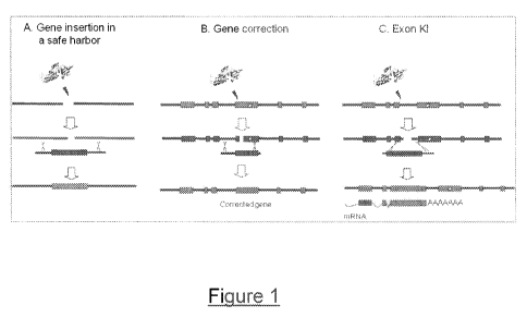
strategy requires very precise genome editing at the targeted locus (Figure 1-
B). The
advantage being, that it directly addresses the cause of the disease: instead
of compensating
the effect of the mutation by a second genome alteration (such as an insertion
in a safe
harbor), the true reversion of the disease-causing mutation is the least
invasive event one can
imagine. However, this precision comes with an inherent drawback: the
correction of the
mutation, usually based on homologous gene repair, is a very local event, and
one needs a
different meganuclease for each disease, and in most cases, for each mutation
or at least each
mutation hotspot related to the disease. This kind of approach can be
envisioned as a
treatment for monogenic diseases in which a prevalent mutation is responsible
for the
majority of the cases, such as Sickle Cell Anemia (SCA), in which a single
mutation (E6V) is
present in 100% of the patients (Sadelain, M. et al) and Cystic Fibrosis FTR,
where almost
70% of the patients carry a deletion of a Phenylalanine in position 508
(Rosenecker, J. et al)
of the CFTR gene. However, it is much more difficult to envision for a large
gene such as
DMD, with the mutations scattered along a 2 Mb regions.
Another approach involves use of an intermediate approach between targeted
gene
correction and gene addition, named here "exon knock-in" (Figure 1-C). In this
approach, a
complete or partial cDNA of the affected gene would be integrated in the very
endogenous
targeted locus. This genomic insertion would be less invasive to the cellular
genome, since
the locus itself would act as a kind of safe harbor for the specific disease.
However, this does
not alleviate all the possible risks: the resulting gene could lack sequences
involved in gene
regulation if they are found in the missing introns. Additionally, the genomic
locus would be
CA 02799095 2012-11-09
WO 2011/141820 PCT/IB2011/001406
9
significantly modified, with potential consequences at the transcriptional
level. In a more
refined form, gene replacement could be used to replace a whole region of the
locus.
A promising alternative to random integration of viral vectors is a site-
specific
integration in a safe locus (Figure. 1-A). The major challenge is the
availability of a region in
the genome that could be considered as a "safe harbor" for gene addition. This
locus should
be chosen in a way that the probability of insertional mutagenesis would be
minimized,
retaining a long-term and high level of expression of the transgene.
Given the large size of the DMD gene and the large diversity of mutations
resulting in
Duchenne's Muscular Distrophy, among which, a variety of deletions and
duplications, the
exon KI strategy is the most adapted to correct this gene in a large number of
cases.
Therefore, a first main aspect of the present invention concerns endonucleases
variants that
could be used in this approach to induce a double strand break in the DMD gene
and for
genome therapy of DMD disease and also allowing further experimental study of
this
important disease in cellular or other types of model systems.
The "exon knock-in" approach has the advantage of allowing the use of a same
reagent
to correct many different mutations, and treat many different patients.
Eventually, targeting a
"safe harbor" would allow to treat different diseases using a same reagent
(although one
would also have to use different inserts). It has therefore several advantages
over the other
approaches. However, its feasibility depends on the identification of a good
"safe harbor"
locus, which should display the following properties (i) it should allow for
stable and
sufficient expression of the inserted transgene, in order to insure efficacy
of the treatment (ii)
insertion in this locus should have no impact on the expression of other
genes.
Given the very large size of the DMD locus, it is unlikely that targeted
insertion into
this locus could result into cis-activation of other genes. However, it could
disrupt the DMD
gene itself. Therefore, one can consider the DMD locus as a safe harbor:
(i) in cells that do not normally express DMD, provided the insert can be
expressed
from this locus.
(ii) in cells that do normally express DMD, provided the insertion does not
affect
the expression of DMD, or provided there remain a functional allele in the
cell.
For example, insertion in introns can be made with no or minor modification of
the expression pattern.
Therefore, in a second main aspect of the present invention, the inventors
have found
that endonucleases variants targeting DMD gene can be used for inserting
therapeutic
CA 02799095 2012-11-09
WO 2011/141820 PCT/IB2011/001406
transgenes other than DMD at the dystrophin gene locus, using this locus as a
safe harbor
locus.
In a third main aspect of the present invention, the inventors have found that
the
dystrophin locus could be used as a landing pad to insert and express genes of
interest
(GOIs).
The above objects highlight certain aspects of the invention. Additional
objects,
aspects and embodiments of the invention are found in the following detailed
description of
the invention.
BRIEF DESCRIPTION OF THE FIGURES
In addition to the preceding features, the invention further comprises other
features
which will emerge from the description which follows, which refers to examples
illustrating
the I-Crel meganuclease variants and their uses according to the invention, as
well as to the
appended drawings. A more complete appreciation of the invention and many of
the attendant
advantages thereof will be readily obtained as the same becomes better
understood by
reference to the following Figures in conjunction with the detailed
description below.
- Figure 1: Illustration of three different strategies for correcting a
genetic defect with
meganuclease-induced recombination. A. Site-specific integration in a safe
locus; the major
challenge is the availability of such a region in the genome that could be
considered as a
"safe harbor" for gene addition. This locus should be chosen in a way that the
probability of
insertional mutagenesis would be minimized, retaining a long-term and high
level of
expression of the transgene. B. Gene correction. A mutation occurs within the
dystrophin
gene. Upon cleavage by a meganuclease and recombination with a repair matrix
the
deleterious mutation is corrected. C. Exonic sequences knock-in. A mutation
occurs within
the dystrophin gene. The mutated mRNA transcript is featured below the gene.
In the repair
matrix, all exons necessary to reconstitute a complete cDNA are fused in
frame, with a
polyadenylation site to stop transcription in 3'. Introns and exons sequences
can be used as
homologous regions. Exonic sequences knock-in results into an engineered gene,
transcribed
into a mRNA able to code for a functional dystrophin protein.
- Figure 2: Modular structure of homing endonucleases and the combinatorial
approach for custom meganucleases design. A. Tridimensional structure of the I-
CreI homing
CA 02799095 2012-11-09
WO 2011/141820 PCT/IB2011/001406
11
endonuclease bound to its DNA target. The catalytic core is surrounded by two
a(3(3a(3(3a folds forming a saddle-shaped interaction interface above the DNA
major groove.
B. Different binding sequences derived from the I-Crel target sequence (top
right and bottom
left) to obtain heterodimers or single chain fusion molecules cleaving non
palindromic
chimeric targets (bottom right). C. The identification of smaller independent
subunit, i. e.,
subunit within a single monomer or a(3Pc 3(3a fold (top right and bottom left)
would allow
for the design of novel chimeric molecules (bottom right), by combination of
mutations
within a same monomer. Such molecules would cleave palindromic chimeric
targets (bottom
right). D. The combination of the two former steps would allow a larger
combinatorial
approach, involving four different subdomains. In a first step, couples of
novel
meganucleases could be combined in new molecules ("half-meganucleases")
cleaving
palindromic targets derived from the target one wants to cleave. Then, the
combination of
such "half-meganuclease" can result in an heterodimeric species cleaving the
target of
interest. Thus, the identification of a small number of new cleavers for each
subdomain
would allow for the design of a very large number of novel endonucleases.
- Figure 3: Exon Knock in strategies by insertion (A) or by replacement (B)
for the
dystrophin gene.
- Figure 4: DMD21 and DMD21-derived targets. The DMD21 target sequence (SEQ
ID NO: 4) and its derivatives IOAAC_P (SEQ ID NO: 5), IOTAC_P (SEQ ID NO: 7),
5CAA_P (SEQ ID NO: 6) and 5TTG_P (SEQ ID NO: 8), P stands for Palindromic) are
derivatives of C1221, found to be cleaved by previously obtained I-CreI
mutants. C1221
(SEQ ID NO: 2), IOAAC_P (SEQ ID NO: 5), 10TAC_P (SEQ ID NO: 7), 5CAA_P (SEQ ID
NO: 6) and 5TTG_P (SEQ ID NO: 8) were first described as 24 bp sequences, but
structural
data suggest that only the 22 bp are relevant for protein/DNA interaction.
DMD21 (SEQ ID
NO: 4) is the DNA sequence located in the human dystrophin gene at position
993350-
993373. DMD21.3 (SEQ ID NO: 9) is the palindromic sequence derived from the
left part of
DMD21, and DMD21.4 (SEQ ID NO: 10) is the palindromic sequence derived from
the right
part of DMD21.
- Figure 5: Activity cleavage in CHO cells of single chain heterodimer SCOH-
DMD21: pCLS2872, pCLS2873, pCLS2874, pCLS2875, pCLS3385, pCLS3387 and
pCLS3388 compared to ISceI (pCLS 1090) and SCOH-RAG-CLS (pCLS2222)
CA 02799095 2012-11-09
WO 2011/141820 PCT/IB2011/001406
12
meganucleases as positive controls. The empty vector control (pCLS 1069) has
also been
tested on each target. Plasmid pCLS1728 contains control RAG1.10.1 target
sequence.
- Figure 5 bis: Activity cleavage in CHO cells of single chain heterodimer
SCOH-
DMD21: pCLS2874, pCLS5353, pCLS5354, pCLS5355 and pCLS5356 compared to IScel
and SCOH-RAG meganucleases as positive controls.
- Figure 6: DMD24 and DMD24-derived targets. The DMD24 target sequence (SEQ
ID NO: 11) and its derivatives I OTAC_P (SEQ ID NO: 12), l OTAT_P (SEQ ID NO:
14),
5ATT_P (SEQ ID NO: 13) and 5GAC_P ((SEQ ID NO: 15), P stands for Palindromic)
are
derivatives of C1221, found to be cleaved by previously obtained I-CreI
mutants. C1221
(SEQ ID NO: 2), IOTAC_P (SEQ ID NO: 12), 10TAT_P (SEQ ID NO: 14), 5ATT_P (SEQ
ID NO: 13) and 5GAC_P ((SEQ ID NO: 15) were first described as 24 bp
sequences, but
structural data suggest that only the 22 bp are relevant for protein/DNA
interaction. DMD24
(SEQ ID NO: 11) is the DNA sequence located in the human dystrophin gene at
position
995930-995953. DMD24.2 (SEQ ID NO: 16) differs from DMD24 at positions -2;-
1;+1;+2
where I-CreI cleavage site (GTAC) substitutes the corresponding DMD24
sequence.
DMD24.3 (SEQ ID NO: 17) is the palindromic sequence derived from the left part
of
DMD24.2, and DMD24.4 (SEQ ID NO: 18) is the palindromic sequence derived from
the
right part of DMD24.2. DMD24.5 (SEQ ID NO: 19) is the palindromic sequence
derived
from the left part of DMD24, and DMD24.6 (SEQ ID NO: 20) is the palindromic
sequence
derived from the right part of DMD24.
- Figure 7: Activity cleavage in CHO cells of single chain heterodimer SCOH-
DMD24 pCLS3402 compared to IScel (pCLS 1090) and SCOH-RAG-CLS (pCLS2222)
meganucleases as positive controls. The empty vector control (pCLS 1069) has
also been
tested on each target. Plasmid pCLS 1728 contains control RAG1.10.1 target
sequence.
- Figure 8: DMD31 and DMD31-derived targets. The DMD31 target sequence (SEQ
ID NO: 21) and its derivatives I OTGT_P (SEQ ID NO: 22), l OAAC_P (SEQ ID NO:
24),
5GAT_P (SEQ ID NO: 23) and 5ATT_P (SEQ ID NO: 25), (P stands for Palindromic)
are
derivatives of C1221, found to be cleaved by previously obtained I-CreI
mutants. C1221
(SEQ ID NO: 2), 10TGT-P (SEQ ID NO: 22), 10AAC_P (SEQ ID NO: 24), 5GAT_P (SEQ
ID NO: 23) and 5ATT_P (SEQ ID NO: 25) were first described as 24 bp sequences,
but
structural data suggest that only the 22 bp are relevant for protein/DNA
interaction. DMD31
CA 02799095 2012-11-09
WO 2011/141820 PCT/IB2011/001406
13
(SEQ ID NO: 21) is the DNA sequence located in the human dystrophin gene at
position
1125314-1125337. DMD31.2 (SEQ ID NO: 26) differs from DMD31 at positions -2;-
1;+1;+2
where I-Crel cleavage site (GTAC) substitutes the corresponding DMD31
sequence.
DMD31.3 (SEQ ID NO: 27) is the palindromic sequence derived from the left part
of
DMD31.2, and DMD31.4 (SEQ ID NO: 28) is the palindromic sequence derived from
the
right part of DMD31.2. DMD31.5 (SEQ ID NO: 29) is the palindromic sequence
derived
from the left part of DMD31, and DMD31.6 (SEQ ID NO: 30) is the palindromic
sequence
derived from the right part of DMD3 1.
- Figure 9: Activity cleavage in CHO cells of single chain heterodimer SCOH-
DMD31: pCLS3631-SCOH-DD3lbl2-B and pCLS3633-SCOH-DD31bl2-D compared to
IScel (pCLS1090) and SCOH-RAG-CLS (pCLS2222) meganucleases as positive
controls.
The empty vector control (pCLS1069) has also been tested on each target.
Plasmid
pCLS 1728 contains control RAG 1.10.1 target sequence.
- Figure 10: DMD33 and DMD33-derived targets. The DMD33 target sequence (SEQ
ID NO: 31) and its derivatives 1 OATC_P (SEQ ID NO: 32), 10GAG_P (SEQ ID NO:
34),
5GCC_P (SEQ ID NO: 33) and 5ACT_P (SEQ ID NO: 35), (P stands for Palindromic)
are
derivatives of C1221, found to be cleaved by previously obtained I-Crel
mutants. C1221
(SEQ ID NO: 2), 10ATC_P (SEQ ID NO: 32), 10GAG_P (SEQ ID NO: 34), 5GCC_P (SEQ
ID NO: 33) and 5ACT_P (SEQ ID NO: 35) were first described as 24 bp sequences,
but
structural data suggest that only the 22 bp are relevant for protein/DNA
interaction. DMD33
(SEQ ID NO: 31) is the DNA sequence located in the human dystrophin gene at
position
1031834-1031857. DMD33.2 (SEQ ID NO: 36) differs from DMD33 at positions -2;-
1;+1;+2
where I-Crel cleavage site (GTAC) substitutes the corresponding DMD33
sequence.
DMD33.3 (SEQ ID NO: 37) is the palindromic sequence derived from the left part
of
DMD33.2, and DMD33.4 (SEQ ID NO: 38) is the palindromic sequence derived from
the
right part of DMD33.2. DMD33.5 (SEQ ID NO: 39) is the palindromic sequence
derived
from the left part of DMD33, and DMD33.6 (SEQ ID NO: 40) is the palindromic
sequence
derived from the right part of DMD33.
- Figure 11: Activity cleavage in CHO cells of single chain heterodimer SCOH-
DMD33 pCLS3326 and pCLS3333 compared to IScel (pCLS1090) and SCOH-RAG-CLS
(pCLS2222) meganucleases as positive controls. The empty vector control
(pCLS1069) has
CA 02799095 2012-11-09
WO 2011/141820 PCT/IB2011/001406
14
also been tested on each target. Plasmid pCLS1728 contains control RAG1.10.1
target
sequence.
- Figure 12: DMD35 and DMD35-derived targets. The DMD35 target sequence (SEQ
ID NO: 41) and its derivatives 1 OTTT_P (SEQ ID NO: 42), 10AAT_P (SEQ ID NO:
44),
5GTT_P (SEQ ID NO: 43) and 5ACT_P (SEQ ID NO: 45), (P stands for Palindromic)
are
derivatives of C1221, found to be cleaved by previously obtained I-CreI
mutants. C1221
(SEQ ID NO: 2), 1OTTT_P (SEQ ID NO: 42), 1OAAT_P (SEQ ID NO: 44), 5GTT_P (SEQ
ID NO: 43) and 5ACT_P (SEQ ID NO: 45) were first described as 24 bp sequences,
but
structural data suggest that only the 22 bp are relevant for protein/DNA
interaction. DMD35
(SEQ ID NO: 41) is the DNA sequence located in the human dystrophin gene at
position
1561221-1561244. DMD35.2 (SEQ ID NO: 46) differs from DMD35 at positions -2;-
1;+1;+2
where I-CreI cleavage site (GTAC) substitutes the corresponding DMD35
sequence.
DMD35.3 (SEQ ID NO: 47) is the palindromic sequence derived from the left part
of
DMD35.2, and DMD35.4 (SEQ ID NO: 48) is the palindromic sequence derived from
the
right part of DMD35.2. DMD35.5 (SEQ ID NO: 49) is the palindromic sequence
derived
from the left part of DMD35, and DMD35.6 (SEQ ID NO: 50) is the palindromic
sequence
derived from the right part of DMD35.
- Figure 13: DMD37 and DMD37-derived targets. The DMD37 target sequence (SEQ
ID NO: 51) and its derivatives 1 OATC_P (SEQ ID NO: 52), 10AGG_P (SEQ ID NO:
54),
5GTT_P (SEQ ID NO: 53) and 5GAT_P (SEQ ID NO: 55), (P stands for Palindromic)
are
derivatives of C1221, found to be cleaved by previously obtained I-CreI
mutants. C1221
(SEQ ID NO: 2), 1OATC_P (SEQ ID NO: 52), 1OAGG_P (SEQ ID NO: 54), 5GTT_P (SEQ
ID NO: 53) and 5GAT_P (SEQ ID NO: 55) were first described as 24 bp sequences,
but
structural data suggest that only the 22 bp are relevant for protein/DNA
interaction. DMD37
(SEQ ID NO: 51) is the DNA sequence located in the human dystrophin gene at
position
1659873-1659896. DMD37.2 (SEQ ID NO: 56) differs from DMD37 at positions -2;-
1;+1;+2
where I-CreI cleavage site (GTAC) substitutes the corresponding DMD37
sequence.
DMD37.3 (SEQ ID NO: 57) is the palindromic sequence derived from the left part
of
DMD37.2, and DMD37.4 (SEQ ID NO: 58) is the palindromic sequence derived from
the
right part of DMD37.2. DMD37.5 (SEQ ID NO: 59) is the palindromic sequence
derived
from the left part of DMD37, and DMD37.6 (SEQ ID NO: 60) is the palindromic
sequence
derived from the right part of DMD37.
CA 02799095 2012-11-09
WO 2011/141820 PCT/IB2011/001406
- Figure 14: Activity cleavage in CHO cells of single chain heterodimer SCOH-
DMD37 pCLS4606, pCLS4607-SCOH-DMD37b11-B, pCLS4608-SCOH-DMD37b11-C,
pCLS4609, pCLS4610, pCLS461 1, pCLS4612, pCLS4613 and pCLS4614 compared to
IScel
(pCLS 1090) and SCOH-RAG-CLS (pCLS2222) meganucleases as positive controls.
The
empty vector control (pCLS 1069) has also been tested on each target. Plasmid
pCLS1728
contains control RAG 1.10.1 target sequence.
- Figure 14 bis: Activity cleavage in CHO cells of single chain heterodimer
SCOH-
DMD37 pCLS4607-SCOH-DMD37b11-B, pCLS4608-SCOH-DMD37b11-C, pCLS4613
and pCLS4614, pCLS6602, pCLS6603, pCLS7389, pCLS7390, pCLS7391 and pCLS7392
compared to IScel and SCOH-RAG-CLS meganucleases as positive controls. The
empty
vector control (pCLS 1069) has also been tested on each target. Plasmid pCLS
1728 contains
control RAG 1.10.1 target sequence (not shown).
- Figure 15: Vector Map of pCLS 1072
- Figure 16: Vector Map of pCLS1090
- Figure 17: Vector Map of pCLS2222
- Figure 18: Vector Map of pCLS1853
- Figure 19: Vector Map of pCLS 1107
- Figure 20: Vector Map of pCLS0002
- Figure 21: Vector Map of pCLS1069
- Figure 22: Vector Map of pCLS 1058
- Figure 23: Vector Map of pCLS1728
- Figure 24: Vector Maps of pIM-DMD-Luc and pIM-DMD-MCS
- Figure 25: Description of universal integration matrices. Schematic
representation of
the different genetic elements introduced in universal integration matrices.
First, positive and
selection marker genes are added in two different places: the former inserted
in and the latter
inserted out of the recombinogenic element. Second, different restriction
sites have been
introduced: 8bp cutting sites for the cloning of left and right homology arms
for any type of
CA 02799095 2012-11-09
WO 2011/141820 PCT/IB2011/001406
16
integration locus, a multiple cloning site (MCS) for the integration of any
GOI and other
restriction sites in the case of additional element cloning (i.e.enhancers,
silencers).
- Figure 26: Location of PCR primers F_HS2_PCRsc and R_HS2-PCRsc on pIM-
DMD-Luc integration matrix.
- Figure 27: Southern blot analysis of human DMD targeted clones. Panel A:
Hybridization of the neo probe on gDNA digested with EcoRV restriction enzyme
from
NeoRPCR+ HEK293 clones; C-: Control lane (gDNA from native HEK293). Panel B:
Hybridization of the neo probe on gDNA digested with EcoRV restriction enzyme
from
NeoRPCR+ U 2-OS clones. Right arrows indicate the 4.8kb expected band,
demonstrating the
correct targeted integration at the DMD locus.
- Figure 28: Luciferase reporter gene expression under the control of six
different
promoters in human DMD-targeted HEK293 clones.
- Figure 29: Activity cleavage in CHO cells of single chain heterodimer SCOH-
DMD35 pCLS4901, pCLS4902, pCLS4903 and pCLS4904 compared to IScel and SCOH-
RAG-CLS meganucleases as positive controls. The empty vector control (pCLS
1069) has
also been tested on each target. Plasmid pCLS1728 contains control RAG1.10.1
target
sequence (not shown).
- Figure 30: Activity cleavage in CHO cells of single chain heterodimer SCOH-
DMD35 pCLS4902, pCLS4904 and pCLS6601 compared to IScel and SCOH-RAG-CLS
meganucleases as positive controls. The empty vector control (pCLS 1069) has
also been
tested on each target. Plasmid pCLS1728 contains control RAG 1.10.1 target
sequence (not
shown).
DETAILED DESCRIPTION OF THE INVENTION
Unless specifically defined herein below, all technical and scientific terms
used herein
have the same meaning as commonly understood by a skilled artisan in the
fields of gene
therapy, biochemistry, genetics, and molecular biology.
CA 02799095 2012-11-09
WO 2011/141820 PCT/IB2011/001406
17
All methods and materials similar or equivalent to those described herein can
be used
in the practice or testing of the present invention, with suitable methods and
materials being
described herein. All publications, patent applications, patents, and other
references
mentioned herein are incorporated by reference in their entirety. In case of
conflict, the
present specification, including definitions, will control. Further, the
materials, methods, and
examples are illustrative only and are not intended to be limiting, unless
otherwise specified.
According to a first aspect of the present invention is an I-CreI variant,
which has two
I-CreI monomers and at least one of the two I-CreI monomers has at least two
substitutions,
where there is at least one mutation in each of the two functional subdomains
of the
LAGLIDADG core domain situated from positions 26 to 40 and 44 to 77 of I-CreI,
respectively, and said variant cleaves a DNA target sequence from the DMD
gene. Within
this embodiment, the I-CreI variant is obtained by a method comprising at
least the steps of:
(a) constructing a first series of I-CreI variants having at least one
substitution in a
first functional subdomain of the LAGLIDADG core domain situated from
positions 26 to 40
of I-CreI,
(b) constructing a second series of I-CreI variants having at least one
substitution
in a second functional subdomain of the LAGLIDADG core domain situated from
positions
44 to 77 of I-CreI,
(c) selecting and/or screening the variants from the first series of step (a)
which
are able to cleave a mutant I-CreI site wherein at least one of (i) the
nucleotide triplet in
positions -10 to -8 of the I-CreI site has been replaced with the nucleotide
triplet which is
present in positions -10 to -8 of said DNA target sequence from DMD gene and
(ii) the
nucleotide triplet in positions +8 to +10 has been replaced with the reverse
complementary
sequence of the nucleotide triplet which is present in position -10 to -8 of
said DNA target
sequence from DMD gene,
(d) selecting and/or screening the variants from the second series of step (b)
which
are able to cleave a mutant I-CreI site wherein at least one of (i) the
nucleotide triplet in
positions -5 to -3 of the I-CreI site has been replaced with the nucleotide
triplet which is
present in positions -5 to -3 of said DNA target sequence from DMD gene and
(ii) the
nucleotide triplet in positions +3 to +5 has been replaced with the reverse
complementary
sequence of the nucleotide triplet which is present in position -5 to -3 of
said DNA target
sequence from DMD gene,
CA 02799095 2012-11-09
WO 2011/141820 PCT/IB2011/001406
18
(e) selecting and/or screening the variants from the first series of step (a)
which
are able to cleave a mutant I-CreI site wherein at least one of (i) the
nucleotide triplet in
positions +8 to +10 of the I-CreI site has been replaced with the nucleotide
triplet which is
present in positions +8 to +10 of said DNA target sequence from DMD gene and
(ii) the
nucleotide triplet in positions -10 to -8 has been replaced with the reverse
complementary
sequence of the nucleotide triplet which is present in position +8 to +10 of
said DNA target
sequence from DMD gene,
(f) selecting and/or screening the variants from the second series of step (b)
which
are able to cleave a mutant I-CreI site wherein at least one of (i) the
nucleotide triplet in
positions +3 to +5 of the I-CreI site has been replaced with the nucleotide
triplet which is
present in positions +3 to +5 of said DNA target sequence from DMD gene and
(ii) the
nucleotide triplet in positions -5 to -3 has been replaced with the reverse
complementary
sequence of the nucleotide triplet which is present in position +3 to +5 of
said DNA target
sequence from DMD gene,
(g) combining in a single variant, the mutation(s) in positions 26 to 40 and
44 to
77 of two variants from step (c) and step (d), to obtain a novel homodimeric I-
CreI variant
which cleaves a sequence wherein (i) the nucleotide triplet in positions -10
to -8 is identical
to the nucleotide triplet which is present in positions -10 to -8 of said DNA
target sequence
from DMD gene, (ii) the nucleotide triplet in positions +8 to +10 is identical
to the reverse
complementary sequence of the nucleotide triplet which is present in positions
-10 to -8 of
said DNA target sequence from DMD gene, (iii) the nucleotide triplet in
positions -5 to -3 is
identical to the nucleotide triplet which is present in positions -5 to -3 of
said DNA target
sequence from DMD gene and (iv) the nucleotide triplet in positions +3 to +5
is identical to
the reverse complementary sequence of the nucleotide triplet which is present
in positions -5
to -3 of said DNA target sequence from DMD gene, and/or
(h) combining in a single variant, the mutation(s) in positions 26 to 40 and
44 to
77 of two variants from step (e) and step (f), to obtain a novel homodimeric I-
CreI variant
which cleaves a sequence wherein (i) the nucleotide triplet in positions +8 to
+10 of the I-
CreI site has been replaced with the nucleotide triplet which is present in
positions +8 to +10
of said DNA target sequence from DMD gene and (ii) the nucleotide triplet in
positions -10
to -8 is identical to the reverse complementary sequence of the nucleotide
triplet in positions
+8 to +10 of said DNA target sequence from DMD gene, (iii) the nucleotide
triplet in
positions +3 to +5 is identical to the nucleotide triplet which is present in
positions +3 to +5
CA 02799095 2012-11-09
WO 2011/141820 PCT/IB2011/001406
19
of said DNA target sequence from DMD gene, (iv) the nucleotide triplet in
positions -5 to -3
is identical to the reverse complementary sequence of the nucleotide triplet
which is present
in positions +3 to +5 of said DNA target sequence from DMD gene,
(i) combining the variants obtained in steps (g) and (h) to form heterodimers,
and (j)
selecting and/or screening the heterodimers from step (i) which cleave said
DNA target
sequence from DMD gene.
In the present Patent Application the terms meganuclease (s) and variant (s)
and
variant meganuclease (s) will be used interchangeably herein.
One of the step(s) (c), (d), (e), (f), (g), (h) or (i) may be omitted. For
example, if step
(c) is omitted, step (d) is performed with a mutant I-CreI target wherein both
nucleotide
triplets at positions -10 to -8 and -5 to -3 have been replaced with the
nucleotide triplets
which are present at positions -10 to -8 and -5 to -3, respectively of said
genomic target, and
the nucleotide triplets at positions +3 to +5 and +8 to +10 have been replaced
with the reverse
complementary sequence of the nucleotide triplets which are present at
positions -5 to -3 and
-10 to -8, respectively of said genomic target.
The (intramolecular) combination of mutations in steps (g) and (h) may be
performed
by amplifying overlapping fragments comprising each of the two subdomains,
according to
well-known overlapping PCR techniques.
The (intermolecular) combination of the variants in step (i) is performed by
co-
expressing one variant from step (g) with one variant from step (h), so as to
allow the
formation of heterodimers. For example, host cells may be modified by one or
two
recombinant expression vector(s) encoding said variant(s). The cells are then
cultured under
conditions allowing the expression of the variant(s), so that heterodimers are
formed in the
host cells, as described previously in the International PCT Application WO
2006/097854
and Arnould et al., J. Mol. Biol., 2006, 355, 443-458.
The selection and/or screening in steps (c), (d), (e), (f), and/or (j) may be
performed
by measuring the cleavage activity of the variant according to the invention
by any well-
known, in vitro or in vivo cleavage assay, such as those described in the
International PCT
Application WO 2004/067736; Epinat et al., Nucleic Acids Res., 2003, 31, 2952-
2962;
Chames et al., Nucleic Acids Res., 2005, 33, e178; Arnould et al., J. Mol.
Biol., 2006, 355,
CA 02799095 2012-11-09
WO 2011/141820 PCT/IB2011/001406
443-458, and Arnould et al., J. Mol. Biol., 2007, 371, 49-65. For example, the
cleavage
activity of the variant of the invention may be measured by a direct repeat
recombination
assay, in yeast or mammalian cells, using a reporter vector. The reporter
vector comprises
two truncated, non-functional copies of a reporter gene (direct repeats) and
the genomic (non-
palindromic) DNA target sequence within the intervening sequence, cloned in
yeast or in a
mammalian expression vector. Usually, the genomic DNA target sequence
comprises one
different half of each (palindromic or pseudo-palindromic) parent homodimeric
I-CreI
meganuclease target sequence. Expression of the heterodimeric variant results
in a functional
endonuclease which is able to cleave the genomic DNA target sequence. This
cleavage
induces homologous recombination between the direct repeats, resulting in a
functional
reporter gene, whose expression can be monitored by an appropriate assay. The
cleavage
activity of the variant against the genomic DNA target may be compared to wild
type I-CreI
or I-Scel activity against their natural target.
According to another advantageous embodiment of said method, steps (c), (d),
(e), (f)
and/or (j) are performed in vivo, under conditions where the double-strand
break in the
mutated DNA target sequence which is generated by said variant leads to the
activation of a
positive selection marker or a reporter gene, or the inactivation of a
negative selection marker
or a reporter gene, by recombination-mediated repair of said DNA double-strand
break.
Furthermore, the homodimeric combined variants obtained in step (g) or (h) are
advantageously submitted to a selection/screening step to identify those which
are able to
cleave a pseudo-palindromic sequence wherein at least the nucleotides at
positions -11 to -3
(combined variant of step (g)) or +3 to +11 (combined variant of step (h)) are
identical to the
nucleotides which are present at positions -I1 to -3 (combined variant of step
(g)) or +3 to
+11 (combined variant of step (h)) of said genomic target, and the nucleotides
at positions +3
to +11 (combined variant of step (g)) or -11 to -3 (combined variant of step
(h)) are identical
to the reverse complementary sequence of the nucleotides which are present at
positions -11
to -3 (combined variant of step (g)) or +3 to +11 (combined variant of step
(h)) of said
genomic target.
Preferably, the set of combined variants of step (g) or step (h) (or both
sets) undergoes
an additional selection/screening step to identify the variants which are able
to cleave a
pseudo-palindromic sequence wherein :
CA 02799095 2012-11-09
WO 2011/141820 PCT/IB2011/001406
21
(1) the nucleotides at positions -11 to -3 (combined variant of step (g)) or
+3 to +11
(combined variant of step (h)) are identical to the nucleotides which are
present at positions -
11 to -3 (combined variant of step (g)) or +3 to +11 (combined variant of step
h)) of said
genomic target, and
(2) the nucleotides at positions +3 to +11 (combined variant of step (g)) or -
11 to -3
(combined variant of step (h)) are identical to the reverse complementary
sequence of the
nucleotides which are present at positions -11 to -3 (combined variant of step
(g)) or +3 to
+11 (combined variant of step (h)) of said genomic target.
This additional screening step increases the probability of isolating
heterodimers
which are able to cleave the genomic target of interest (step (k)).
Steps (a), (b), (g), (h) and (i) may further comprise the introduction of
additional
mutations at other positions contacting the DNA target sequence or interacting
directly or
indirectly with said DNA target, at positions which improve the binding and/or
cleavage
properties of the variants, or at positions which either prevent or impair the
formation of
functional homodimers or favor the formation of the heterodimer, as defined
above.
The additional mutations may be introduced by site-directed mutagenesis and/or
random mutagenesis on a variant or on a pool of variants, according to
standard mutagenesis
methods which are well-known in the art, for example by using PCR.
In particular, random mutations may be introduced into the whole variant or in
a part
of the variant to improve the binding and/or cleavage properties of the
variants towards the
DNA target from the gene of interest.
Site-directed mutagenesis at positions which improve the binding and/or
cleavage
properties of the variants, for example at positions 19, 54, 66, 80, 87, 105
and /or 132, may
also be combined with random-mutagenesis. The mutagenesis may be performed by
generating random/site-directed mutagenesis libraries on a pool of variants,
according to
standard mutagenesis methods which are well-known in the art. Site-directed
mutagenesis
may be advantageously performed by amplifying overlapping fragments comprising
the
mutated position(s), as defined above, according to well-known overlapping PCR
techniques.
In addition, multiple site-directed mutagenesis, may advantageously be
performed on a
variant or on a pool of variants.
CA 02799095 2012-11-09
WO 2011/141820 PCT/IB2011/001406
22
Preferably, the mutagenesis is performed on one monomer of the heterodimer
formed
in step (i) or step (j), advantageously on a pool of monomers, preferably on
both monomers
of the heterodimer of step (i) or (j).
Possibly or not, at least two rounds of selection/screening are performed
according to
the process illustrated Arnould et al., J. Mol. Biol., 2007, 371, 49-65. In
the first round, one
of the monomers of the heterodimer is mutagenised, co-expressed with the other
monomer to
form heterodimers, and the improved monomers Y+ are selected against the
target from the
gene of interest. In the second round, the other monomer (monomer X) is
mutagenised, co-
expressed with the improved monomers Y+ to form heterodimers, and selected
against the
target from the gene of interest to obtain meganucleases (X+ Y) with improved
activity. The
mutagenesis may be random-mutagenesis or site-directed mutagenesis on a
monomer or on a
pool of monomers, as indicated above. Both types of mutagenesis are
advantageously
combined. Additional rounds of selection/screening on one or both monomers may
be
performed to improve the cleavage activity of the variant.
Preferably the variant may be obtained by a method comprising the additional
steps
of:
(k) selecting heterodimers from step (j) and constructing a third series of
variants
having at least one substitution in at least one of the monomers in said
selected heterodimers,
(1) combining said third series variants of step (k) and screening the
resulting
heterodimers for altered cleavage activity against said DNA target from DMD
gene.
Preferably in step (k) at least one substitution is introduced by site
directed
mutagenesis in a DNA molecule encoding said third series of variants, and/or
by random
mutagenesis in a DNA molecule encoding said third series of variants.
Preferably steps (k) and (1) are repeated at least two times and wherein the
heterodimers selected in step (k) of each further iteration are selected from
heterodimers
screened in step (1) of the previous iteration which showed altered cleavage
activity against
said DNA target from DMD gene.
Given the large size of the DMD gene and the large diversity of mutations
resulting in
Duchenne's Muscular Dystrophy, among which, a variety of deletions and
duplications, the
CA 02799095 2012-11-09
WO 2011/141820 PCT/IB2011/001406
23
exon KI strategy is the most adapted to correct this gene in a large number of
cases.
However, even with this strategy, limitations linked to the maximal size of
the sequences that
can be inserted into existing vectors have to be envisioned.
The inventors envision two different sub-types of exon KI strategies: in a
first one,
one would insert at a "starting point" a partial cDNA, providing all the exons
downstream of
this insertion point. This starting point has been placed in exon 44, or in
the exons just
upstream (Figure 3-A). This strategy would address up to 60% of the existing
mutations. It
would require the insertion of a 4.8kb sequence, corresponding to the
downstream exons. The
repair matrix would in addition have to include I kb of homology on each side
(in the
flanking introns), resulting in a fragment of about 7 kbs. This size remains
compatible with
the use of lentiviral vectors, and to a certain extent, with the use of AAV
vectors for research
purpose (although inserts up to 7 kb have been reported in such AAV vector,
such long
inserts should dramatically reduce the yield of large scale productions).
As a consequence, a cleavage 3' of exon 44 can induce a gene targeting event
with one
breakpoint in the exon just 5' of the break, i. e., in exon 44, and another
one in the part of the
intron just 3' of the break. The resulting recombination event is described in
Figure 3-A.
Importantly, recombination should occur between large homology regions, in
intronic
sequences (from intron 43 and 44). The presence of shorter stretches of
homology between
the exons of the cDNA to be knocked in and the endogenous exons should not
interfere with
the process, given the small size of the exons. In a similar approach,
meganucleases targeting
sequences in 3' of former exons could be used to induce gene targeting events
in exons 5' of
exon 44.
Thus, cleavage in the DMD21, DMD24, DMD31, DMD33, DMD35 and DMD37
sequences described in Table 1 could be used to induce gene targeting events
with junctions
in exons 38, 39, 42, 44, 51 and 53 respectively . The repair matrix would have
to be in the
range of 6.8 to 7.9 kb (i. e., about 5.9 kbs for exons 38-79, or 4.8 kbs for
exons 44-79, with in
addition 1 kb of homologous sequence on each side).
A second sub-type of exon knock-in strategy consists in the replacement of a
very
large region with a cDNA, requiring a second break in the chromosome, 5'of a
downstream
exon that would represent the second breakpoint or junction of the
recombination event
(Figure 3-B). This second breakpoint has been placed after exon 50. This
strategy would
address up to 30-40% of the existing mutations, and would require the
insertion of a 1,2kb
sequence for exons 44 to 51 (3,2kb repair matrix) and up to 2,5kb for exons 38
to 53 (4.5kb
CA 02799095 2012-11-09
WO 2011/141820 PCT/IB2011/001406
24
repair matrix). The replacement strategy is more "elegant" than the insertion,
for it avoids
duplications within the genome that could result in expression issues
(repeated sequences
may trigger gene inactivation). In addition, it would allow for the use of a
smaller repair
matrix. This size of the insert used here is also compatible with the use of
lentiviral vectors,
and with the use of meganuclease-induced recombination. The major unknown
factor is
actually the efficiency of recombination involving two chromosomal breakpoints
placed
several hundreds of Kb away. It has been demonstrated before that two I-SceI
breaks located
a few kbs away could induce efficient recombination in a process mimicking the
one
described in Figure 17A (refs 30-31). Moreover, recombination involving
rejoining of two I-
SceI induced DSBs separated by 200 kb of sequences have been described (ref
32) , and even
breaks placed on different chromosomes have been shown to interact very
efficiently (refs
33-34). For DMD, the target cells could be mesoangioblasts, which can be
grafted by
systemic injection. Another option is the targeting of myoblasts, although
these cells need to
be grafted locally.
I-Crel variants to these targets were created using a combinatorial approach,
to
entirely redesign the DNA binding domain of the I-CreI protein and thereby
engineer novel
meganucleases with fully engineered specificity for the desired DMD gene
target. Some of
the DNA targets identified by the inventors to validate their invention are
given in the table I
below. Derivatives of these DNA targets are given in Figures 4, 6, 8, 10, 12
and 14.
mega position targeted sequence Target for KI
DMD21 993350 - 993373 GA-AAC-CT-CAA-GTAC-CAA-AT-GTA-AA 3' of exon 38
Intron 38
DMD24 995930 - 995953 TT-TAC-CT-ATT-TTAA-GTC-AG-ATA-CA 3' of exon 39
Intron 39
DMD33 1031834 - 1031857 AA-ATC-CT-GCC-TTAA-AGT-AT-CTC-AT 3' of exon 42
Intron 42
DMD31 1125314 - 1125337 AA-TGT-CT-GAT-GTTC-AAT-GT-GTT-GA 3' of exon 44
Intron 44
--- - ---------
DMD35 1 561 221 - 1561244 TC-TTT-AT-GTT-TTAA-AGT-AT-ATT-CC 5' of exon 51
Intron 50
DMD37 1 659 873 - 1659896 GA-ATC-CT-GTT-GTTC-ATC-AT-CCT-AG 5' of exon 53
Intron 52
CA 02799095 2012-11-09
WO 2011/141820 PCT/IB2011/001406
Table I : sequences and location of the targeted sites in the DMD gene
The combinatorial approach, as illustrated in Figure 2D was used to entirely
redesign
the DNA binding domain of the I-CreI protein and thereby engineer novel
meganucleases
with fully engineered specificity.
In particular the heterodimer of step (i) may comprise monomers obtained in
steps (g)
and (h), with the same DNA target recognition and cleavage activity
properties.
Alternatively the heterodimer of step (i) may comprise monomers obtained in
steps
(g) and (h), with different DNA target recognition and cleavage activity
properties.
In particular the first series of I-CreI variants of step (a) are derived from
a first parent
meganuclease.
In particular the second series of variants of step (b) are derived from a
second parent
meganuclease.
In particular the first and second parent meganucleases are identical.
Alternatively the first and second parent meganucleases are different.
In particular the variant may be obtained by a method comprising the
additional steps
of:
(k) selecting heterodimers from step (j) and constructing a third series of
variants
having at least one substitution in at least one of the monomers of said
selected heterodimers,
(1) combining said third series variants of step (k) and screening the
resulting
heterodimers for enhanced cleavage activity against said DNA target from DMD
gene.
In a preferred embodiment of said variant, said substitution(s) in the
subdomain
situated from positions 44 to 77 of I-CreI are at positions 44, 68, 70, 75
and/or 77.
In another preferred embodiment of said variant, said substitution(s) in the
subdomain
situated from positions 28 to 40 of I-CreI are at positions 28, 30, 32, 33, 38
and/or 40.
In another preferred embodiment of said variant, it comprises one or more
mutations
in I-CreI monomer(s) at positions of other amino acid residues that contact
the DNA target
sequence or interact with the DNA backbone or with the nucleotide bases,
directly or via a
CA 02799095 2012-11-09
WO 2011/141820 PCT/IB2011/001406
26
water molecule; these residues are well-known in the art (Jurica et al.,
Molecular Cell., 1998,
2, 469-476; Chevalier et al., J. Mol. Biol., 2003, 329, 253-269). In
particular, additional
substitutions may be introduced at positions contacting the phosphate
backbone, for example
in the final C-terminal loop (positions 137 to 143; Prieto et al., Nucleic
Acids Res., Epub 22
April 2007).
Preferably said residues are involved in binding and cleavage of said DNA
cleavage
site.
More preferably, said residues are at positions 138, 139, 142 or 143 of I-
CreI. Two
residues may be mutated in one variant provided that each mutation is in a
different pair of
residues chosen from the pair of residues at positions 138 and 139 and the
pair of residues at
positions 142 and 143. The mutations which are introduced modify the
interaction(s) of said
amino acid(s) of the final C-terminal loop with the phosphate backbone of the
I-CreI site.
Preferably, the residue at position 138 or 139 is substituted by a hydrophobic
amino acid to
avoid the formation of hydrogen bonds with the phosphate backbone of the DNA
cleavage
site. For example, the residue at position 138 is substituted by an alanine or
the residue at
position 139 is substituted by a methionine. The residue at position 142 or
143 is
advantageously substituted by a small amino acid, for example a glycine, to
decrease the size
of the side chains of these amino acid residues.
More preferably, said substitution in the final C-terminal loop modify the
specificity
of the variant towards the nucleotide at positions 1 to 2, 6 to 7 and/or
11 to 12 of the I-
CreI site.
In another preferred embodiment of said variant, it comprises one or more
additional
mutations that improve the binding and/or the cleavage properties of the
variant towards the
DNA target sequence from the DMD gene. The additional residues which are
mutated may
be on the entire I-CreI sequence, and in particular in the C-terminal half of
I-CreI (positions
80 to 163). Both I-CreI monomers are advantageously mutated; the mutation(s)
in each
monomer may be identical or different. For example, the variant comprises one
or more
additional substitutions at positions: 2, 19, 43, 80 and 81. Said
substitutions are
advantageously selected from the group consisting of: N2S, G19S, F43L, E80K
and 181T.
More preferably, the variant comprises at least one substitution selected from
the group
consisting of: N2S, G19S, F43L, E80K and 181T. The variant may also comprise
additional
CA 02799095 2012-11-09
WO 2011/141820 PCT/IB2011/001406
27
residues at the C-terminus. For example a glycine (G) and/or a proline (P)
residue may be
inserted at positions 164 and 165 of I-CreI, respectively.
According to a preferred embodiment, said additional mutation in said variant
further
impairs the formation of a functional homodimer. More preferably, said
mutation is the G 19S
mutation. The G19S mutation is advantageously introduced in one of the two
monomers of a
heterodimeric I-CreI variant, so as to obtain a meganuclease having enhanced
cleavage
activity and enhanced cleavage specificity. In addition, to enhance the
cleavage specificity
further, the other monomer may carry a distinct mutation that impairs the
formation of a
functional homodimer or favors the formation of the heterodimer.
In another preferred embodiment of said variant, said substitutions are
replacement of
the initial amino acids with amino acids selected from the group consisting
of. A, D, E, G, H,
K, N, P, Q, R, S, T, Y, C, V, L, M, F, I and W.
In particular the variant is selected from the group consisting of SEQ ID NO:
40 to
65.
The variant of the invention may be derived from the wild-type I-CreI (SEQ ID
NO:
1) or an I-CreI scaffold protein having at least 85 % identity, preferably at
least 90 %
identity, more preferably at least 95 % identity with SEQ ID NO: 1, such as
the scaffold
called I-CreI N75 (167 amino acids; SEQ ID NO: 3) having the insertion of an
alanine at
position 2, and the insertion of AAD at the C-terminus (positions 164 to 166)
of the I-CreI
sequence. In the present Patent Application all the I-CreI variants described
comprise an
additional Alanine after the first Methionine of the wild type I-CreI sequence
(SEQ ID NO:
1). These variants also comprise two additional Alanine residues and an
Aspartic Acid
residue after the final Proline of the wild type I-CreI sequence. These
additional residues do
not affect the properties of the enzyme and to avoid confusion these
additional residues do
not affect the numeration of the residues in 1-CreI or a variant referred in
the present Patent
Application, as these references exclusively refer to residues of the wild
type I-CreI enzyme
(SEQ ID NO: 1) as present in the variant, so for instance residue 2 of I-CreI
is in fact residue
3 of a variant which comprises an additional Alanine after the first
Methionine.
In addition, the variants of the invention may include one or more residues
inserted at
the NH2 terminus and/or COOH terminus of the sequence. For example, a tag
(epitope or
CA 02799095 2012-11-09
WO 2011/141820 PCT/IB2011/001406
28
polyhistidine sequence) is introduced at the NH2 terminus and/or COOH
terminus; said tag is
useful for the detection and/or the purification of said variant. The variant
may also comprise
a nuclear localization signal (NLS); said NLS is useful for the importation of
said variant into
the cell nucleus. The NLS may be inserted just after the first methionine of
the variant or just
after an N-terminal tag.
The variant according to the present invention may be a homodimer which is
able to
cleave a palindromic or pseudo-palindromic DNA target sequence.
Alternatively, said variant is a heterodimer, resulting from the association
of a first
and a second monomer having different substitutions at positions 28 to 40 and
44 to 77 of I-
Crel, said heterodimer being able to cleave a non-palindromic DNA target
sequence from the
DMD gene.
In particular said heterodimer variant is composed by one of the possible
associations
between variants constituting N-terminal and C-terminal monomers of single
chain molecules
from the group consisting of SEQ ID NO: 62 to SEQ ID NO: 105, SEQ ID NO: 116
to SEQ
ID NO: 119, SEQ ID NO: 121 and SEQ ID NO: 122 to SEQ ID NO: 130.
The DNA target sequences are situated in the DMD Open Reading Frame (ORF) and
these sequences cover all the DMD ORF. In particular, said DNA target
sequences for the
variant of the present invention and derivatives are selected from the group
consisting of the
SEQ ID NO: 4 to SEQ ID NO: 60, as shown in figures 4, 6, 8, 10, 12 and 14 and
Table I.
The sequence of each I-CreI variant is defined by the mutated residues at the
indicated positions. The positions are indicated by reference to I-CreI
sequence (SEQ ID NO:
1) ; I-CreI has N, S, Y, Q, S, Q, R, R, D, I and E at positions 30, 32, 33,
38, 40, 44, 68, 70,
75, 77 and 80 respectively.
Each monomer (first monomer and second monomer) of the heterodimeric variant
according to the present invention may also be named with a letter code, after
the eleven
residues at positions 28, 30, 32, 33, 38, 40, 44, 68 and 70, 75 and 77 and the
additional
residues which are mutated, as indicated above. For example, the mutations
7E30R40E44T46G68T70S73M75A77R80K96EI32V154N in the N-terminal monomer
constituting a single chain molecule targeting the DMD21 target of the present
invention
(SEQ ID NO: 64).
CA 02799095 2012-11-09
WO 2011/141820 PCT/IB2011/001406
29
In the present invention, for a given DNA target, ".2" derivative target
sequence
differs from the initial genomic target at positions -2, -1, +1, +2, where I-
CreI cleavage site
(GTAC) substitutes the corresponding sequence at these positions of said
initial genomic
target. ".3" derivative target sequence is the palindromic sequence derived
from the left part
of said ".2" derivative target sequence. ".4" derivative target sequence is
the palindromic
sequence derived from the right part of said ".2" derivative target sequence.
".5" derivative
target sequence is the palindromic sequence derived from the left part of the
initial genomic
target. ".6" derivative is the palindromic sequence derived from the left part
of the initial
genomic target. As an illustrative example, for DMD 24 (figure 4), "DMD24.2"
derivative
target sequence differs from the initial genomic target (DMD24) at positions -
2, -1, +1, +2,
where I-CreI cleavage site (GTAC) substitutes the corresponding sequence at
these positions
of said initial genomic target (DMD24). "DMD24.3" derivative target sequence
is the
palindromic sequence derived from the left part of said "DMD24.2" derivative
target
sequence. "DMD24.4" derivative target sequence is the palindromic sequence
derived from
the right part of said "DMD24.2" derivative target sequence. "DMD24.5"
derivative target
sequence is the palindromic sequence derived from the left part of the initial
genomic target
(DMD24). "DMD24.6" derivative is the palindromic sequence derived from the
right part of
the initial genomic target (DMD24).
In the present invention, a "N-terminal monomer" constituting one of the
monomers
of a single chain molecule, refers to a variant able to cleave ".3" or ".5"
palindromic
sequence. In the present invention, a "C-terminal monomer" constituting one of
the
monomers of a single chain molecule, refers to a variant able to cleave ".4"
or ".6"
palindromic sequence.
The heterodimeric variant as defined above may have only the amino acid
substitutions as indicated above. In this case, the positions which are not
indicated are not
mutated and thus correspond to the wild-type I-CreI (SEQ ID NO: 1).
The invention encompasses I-CreI variants having at least 85 % identity,
preferably at
least 90 % identity, more preferably at least 95 % (96 %, 97 %, 98 %, 99 %)
identity with the
sequences as defined above, said variant being able to cleave a DNA target
from the DMD
gene.
CA 02799095 2012-11-09
WO 2011/141820 PCT/IB2011/001406
The heterodimeric variant is advantageously an obligate heterodimer variant
having at
least one pair of mutations corresponding to residues of the first and the
second monomers
which make an intermolecular interaction between the two I-CreI monomers,
wherein the
first mutation of said pair(s) is in the first monomer and the second mutation
of said pair(s) is
in the second monomer and said pair(s) of mutations prevent the formation of
functional
homodimers from each monomer and allow the formation of a functional
heterodimer, able to
cleave the genomic DNA target from the DMD gene.
To form an obligate heterodimer, the monomers have advantageously at least one
of
the following pairs of mutations, respectively for the first monomer and the
second monomer:
a) the substitution of the glutamic acid at position 8 with a basic amino
acid,
preferably an arginine (first monomer) and the substitution of the lysine at
position 7 with an
acidic amino acid, preferably a glutamic acid (second monomer); the first
monomer may
further comprise the substitution of at least one of the lysine residues at
positions 7 and 96, by
an arginine,
b) the substitution of the glutamic acid at position 61 with a basic amino
acid,
preferably an arginine (first monomer) and the substitution of the lysine at
position 96 with
an acidic amino acid, preferably a glutamic acid (second monomer); the first
monomer may
further comprise the substitution of at least one of the lysine residues at
positions 7 and 96, by
an arginine,
c) the substitution of the leucine at position 97 with an aromatic amino acid,
preferably a phenylalanine (first monomer) and the substitution of the
phenylalanine at
position 54 with a small amino acid, preferably a glycine (second monomer);
the first
monomer may further comprise the substitution of the phenylalanine at position
54 by a
tryptophane and the second monomer may further comprise the substitution of
the leucine at
position 58 or lysine at position 57, by a methionine, and
d) the substitution of the aspartic acid at position 137 with a basic amino
acid,
preferably an arginine (first monomer) and the substitution of the arginine at
position 51 with
an acidic amino acid, preferably a glutamic acid (second monomer).
For example, the first monomer may have the mutation D137R and the second
monomer, the mutation R51 D. The obligate heterodimer meganuclease comprises
CA 02799095 2012-11-09
WO 2011/141820 PCT/IB2011/001406
31
advantageously, at least two pairs of mutations as defined in a), b), c) or
d), above; one of the
pairs of mutation is advantageously as defined in c) or d). Preferably, one
monomer
comprises the substitution of the lysine residues at positions 7 and 96 by an
acidic amino acid
(aspartic acid (D) or glutamic acid (E)), preferably a glutamic acid (K7E and
K96E) and the
other monomer comprises the substitution of the glutamic acid residues at
positions 8 and 61
by a basic amino acid (arginine (R) or lysine (K); for example, E8K and E61
R). More
preferably, the obligate heterodimer meganuclease, comprises three pairs of
mutations as
defined in a), b) and c), above.
The obligate heterodimer meganuclease consists advantageously of a first
monomer
(A) having at least the mutations (i) E8R, E8K or E8H, E61 R, E61 K or E61 H
and L97F,
L97W or L97Y; (ii) K7R, E8R, E61R, K96R and L97F, or (iii) K7R, E8R, F54W,
E61R,
K96R and L97F and a second monomer (B) having at least the mutations (iv) K7E
or K7D,
F54G or F54A and K96D or K96E; (v) K7E, F54G, L58M and K96E, or (vi) K7E,
F54G,
K57M and K96E. For example, the first monomer may have the mutations K7R, E8R
or
E8K, E61 R, K96R and L97F or K7R, E8R or E8K, F54W, E61 R, K96R and L97F and
the
second monomer, the mutations K7E, F54G, L58M and K96E or K7E, F54G, K57M and
K96E. The obligate heterodimer may comprise at least one NLS and/or one tag as
defined
above; said NLS and/or tag may be in the first and/or the second monomer.
The subject-matter of the present invention is also a single-chain chimeric
meganuclease (fusion protein) derived from an I-CreI variant as defined above.
The single-
chain meganuclease may comprise two I-CreI monomers, two I-CreI core domains
(positions
6 to 94 of I-CreI) or a combination of both. Preferably, the two monomers
/core domains or
the combination of both, are connected by a peptidic linker. Said peptidic
linker can be RM2
linker (SEQ ID NO: 61) or BQY linker (SEQ ID NO: 120) or another suitable
linker.
More preferably the single-chain chimeric meganuclease is composed by one of
the
possible associations between variants from the group consisting of N-terminal
monomers
and C-terminal monomers, given in Tables II to VII, respectively for a given
DNA target,
DMD21, DMD24, DMD31, DMD33, DMD35 and DMD37, said monomer variants being
connected by a linker. More preferably the single-chain chimeric meganuclease
according to
the present invention is one from the group consisting of SEQ ID NO: 62 to SEQ
ID NO:
105, SEQ ID NO: 116 to SEQ ID NO: 119, SEQ ID NO: 121 and SEQ ID NO: 122 to
SEQ
ID NO: 130. Regarding DMD21 target, the single-chain chimeric meganuclease
according to
CA 02799095 2012-11-09
WO 2011/141820 PCT/IB2011/001406
32
the present invention is one from the group consisting of SEQ ID NO: 62 to SEQ
ID NO: 68
and SEQ ID NO: 116 to SEQ ID NO: 119. Regarding DMD24 target, the single-chain
chimeric meganuclease according to the present invention is one from the group
consisting of
SEQ ID NO: 69 to SEQ ID NO: 77. Regarding DMD31 target, the single-chain
chimeric
meganuclease according to the present invention is one from the group
consisting of SEQ ID
NO: 78 to SEQ ID NO: 84. Regarding DMD33 target, the single-chain chimeric
meganuclease according to the present invention is one from the group
consisting of SEQ ID
NO: 85 to SEQ ID NO: 95. Regarding DMD35 target, the single-chain chimeric
meganuclease according to the present invention is one from the group
consisting of SEQ ID
NO: 96 to SEQ ID NO: 99 and SEQ ID NO: 121. Regarding DMD37 target, the single-
chain
chimeric meganuclease according to the present invention is one from the group
consisting of
SEQ ID NO: 100 to SEQ ID NO: 105 and SEQ ID NO: 122 to SEQ ID NO: 130.
It is understood that the scope of the present invention also encompasses the
I-Cre1
variants per se, including heterodimers, obligate heterodimers, single chain
meganucleases as
non limiting examples, able to cleave one of the sequence targets in DMD gene.
The subject-matter of the present invention is also a polynucleotide fragment
encoding a variant or a single-chain chimeric meganuclease as defined above;
said
polynucleotide may encode one monomer of a homodimeric or heterodimeric
variant, or two
domains/monomers of a single-chain chimeric meganuclease. It is understood
that the
subject-matter of the present invention is also a polynucleotide fragment
encoding one of the
variant species as defined above, obtained by any method well-known in the
art.
The subject-matter of the present invention is also a recombinant vector for
the
expression of a variant or a single-chain meganuclease according to the
invention. The
recombinant vector comprises at least one polynucleotide fragment encoding a
variant or a
single-chain meganuclease, as defined above. In a preferred embodiment, said
vector
comprises two different polynucleotide fragments, each encoding one of the
monomers of a
heterodimeric variant.
A vector which can be used in the present invention includes, but is not
limited to, a
viral vector, a plasmid, a RNA vector or a linear or circular DNA or RNA
molecule which
may consists of a chromosomal, non chromosomal, semi-synthetic or synthetic
nucleic acids.
Preferred vectors are those capable of autonomous replication (episomal
vector) and/or
expression of nucleic acids to which they are linked (expression vectors).
Large numbers of
suitable vectors are known to those skilled in the art and commercially
available.
CA 02799095 2012-11-09
WO 2011/141820 PCT/IB2011/001406
33
Viral vectors include retrovirus, adenovirus, parvovirus (e. g. adeno-
associated
viruses), coronavirus, negative strand RNA viruses such as orthomyxovirus (e.
g., influenza
virus), rhabdovirus (e. g., rabies and vesicular stomatitis virus),
paramyxovirus (e. g. measles
and Sendai), positive strand RNA viruses such as picornavirus and alphavirus,
and double-
stranded DNA viruses including adenovirus, herpesvirus (e. g., Herpes Simplex
virus types I
and 2, Epstein-Barr virus, cytomegalovirus), and poxvirus (e. g., vaccinia,
fowlpox and
canarypox). Other viruses include Norwalk virus, togavirus, flavivirus,
reoviruses,
papovavirus, hepadnavirus, and hepatitis virus, for example. Examples of
retroviruses
include: avian leukosis-sarcoma, mammalian C-type, B-type viruses, D type
viruses, HTLV-
BLV group, lentivirus (particularly self inactivacting lentiviral vectors),
spumavirus (Coffin,
J. M., Retroviridae: The viruses and their replication, In Fundamental
Virology, Third
Edition, B. N. Fields, et al., Eds., Lippincott-Raven Publishers,
Philadelphia, 1996).
Vectors can comprise selectable markers, for example: neomycin
phosphotransferase,
histidinol dehydrogenase, dihydrofolate reductase, hygromycin
phosphotransferase, herpes
simplex virus thymidine kinase, adenosine deaminase, Glutamine Synthetase, and
hypoxanthine-guanine phosphoribosyl transferase for eukaryotic cell culture;
TRP1, URA3
and LEU2 for S. cerevisiae; tetracycline, rifampicin or ampicillin resistance
in E. coli.
Preferably said vectors are expression vectors, wherein the sequence(s)
encoding the
variant/single-chain meganuclease of the invention is placed under control of
appropriate
transcriptional and translational control elements to permit production or
synthesis of said
variant. Therefore, said polynucleotide is comprised in an expression
cassette. More
particularly, the vector comprises a replication origin, a promoter
operatively linked to said
polynucleotide, a ribosome-binding site, an RNA-splicing site (when genomic
DNA is used),
a polyadenylation site and a transcription termination site. It also can
comprise an enhancer.
Selection of the promoter will depend upon the cell in which the polypeptide
is expressed.
Preferably, when said variant is a heterodimer, the two polynucleotides
encoding each of the
monomers are included in one vector which is able to drive the expression of
both
polynucleotides, simultaneously. Suitable promoters include tissue specific
and/or inducible
promoters. Examples of inducible promoters are: eukaryotic metallothionine
promoter which
is induced by increased levels of heavy metals, prokaryotic lacZ promoter
which is induced
in response to isopropyl-(3-D-thiogalacto-pyranoside (IPTG) and eukaryotic
heat shock
promoter which is induced by increased temperature. Examples of tissue
specific promoters
CA 02799095 2012-11-09
WO 2011/141820 PCT/IB2011/001406
34
are skeletal muscle creatine kinase, prostate-specific antigen (PSA), (x-
antitrypsin protease,
human surfactant (SP) A and B proteins, (3-casein and acidic whey protein
genes.
According to another advantageous embodiment of said vector, it includes a
targeting
construct comprising sequences sharing homologies with the region surrounding
the genomic
DNA cleavage site as defined above.
For instance, said sequence sharing homologies with the regions surrounding
the
genomic DNA cleavage site of the variant is a fragment of the DMD gene.
Alternatively, the
vector coding for an I-Crel variant/single-chain meganuclease and the vector
comprising the
targeting construct are different vectors.
More preferably, the targeting DNA construct comprises:
a) sequences sharing homologies with the region surrounding the genomic DNA
cleavage site as defined above, and
b) a sequence to be introduced flanked by sequences as in a) or included in
sequences
as in a).
Preferably, homologous sequences of at least 50 bp, preferably more than 100
bp and
more preferably more than 200 bp are used. Therefore, the targeting DNA
construct is
preferably from 200 bp to 6000 bp, more preferably from 1000 bp to 2000 bp.
Indeed, shared
DNA homologies are located in regions flanking upstream and downstream the
site of the
break and the DNA sequence to be introduced should be located between the two
arms. The
sequence to be introduced may be any sequence used to alter the chromosomal
DNA in some
specific way including a sequence used to repair a mutation in the DMD gene,
restore a
functional DMD gene in place of a mutated one, modify a specific sequence in
the DMD
gene, to attenuate or activate the DMD gene, to inactivate or delete the DMD
gene or part
thereof, to introduce a mutation into a site of interest or to introduce an
exogenous gene or
part thereof. Such chromosomal DNA alterations are used for genome engineering
(animal
models/recombinant cell lines) or genome therapy (gene correction or recovery
of a
functional gene). The targeting construct comprises advantageously a positive
selection
marker between the two homology arms and eventually a negative selection
marker upstream
of the first homology arm or downstream of the second homology arm. The
marker(s)
CA 02799095 2012-11-09
WO 2011/141820 PCT/IB2011/001406
allow(s) the selection of cells having inserted the sequence of interest by
homologous
recombination at the target site.
The sequence to be introduced is a sequence which repairs a mutation in the
DMD
gene (gene correction or recovery of a functional gene), for the purpose of
genome therapy
(figure 1 B and 1 Q. For correcting the DMD gene, cleavage of the gene occurs
in the vicinity
of the mutation, preferably, within 500 bp of the mutation (Figure 113). The
targeting
construct comprises a DMD gene fragment which has at least 200 bp of
homologous
sequence flanking the target site (minimal repair matrix) for repairing the
cleavage, and
includes a sequence encoding a portion of wild-type DMD gene corresponding to
the region
of the mutation for repairing the mutation (Figure 1 B). Consequently, the
targeting construct
for gene correction comprises or consists of the minimal repair matrix; it is
preferably from
200 bp to 6000 bp, more preferably from 1000 bp to 2000 bp. Preferably, when
the cleavage
site of the variant overlaps with the mutation the repair matrix includes a
modified cleavage
site that is not cleaved by the variant which is used to induce said cleavage
in the DMD gene
and a sequence encoding wild-type DMD gene that does not change the open
reading frame
of the DMD gene.
Alternatively, for the generation of knock-in cells/animals, the targeting DNA
construct may comprise flanking regions corresponding to DMD gene fragments
which has at
least 200 bp of homologous sequence flanking the target site of the I-CreI
variant for
repairing the cleavage, an exogenous gene of interest within an expression
cassette and
eventually a selection marker such as the neomycin resistance gene.
For the insertion of a sequence, DNA homologies are generally located in
regions
directly upstream and downstream to the site of the break (sequences
immediately adjacent to
the break; minimal repair matrix). However, when the insertion is associated
with a deletion
of ORF sequences flanking the cleavage site, shared DNA homologies are located
in regions
upstream and downstream the region of the deletion.
Alternatively, for restoring a functional gene (Figures 1 B et 1 C), cleavage
of the gene
occurs in the vicinity or upstream of a mutation. Preferably said mutation is
the first known
mutation in the sequence of the gene, so that all the downstream mutations of
the gene can be
corrected simultaneously. The targeting construct comprises the exons
downstream of the
cleavage site fused in frame (as in the cDNA) and with a polyadenylation site
to stop
CA 02799095 2012-11-09
WO 2011/141820 PCT/IB2011/001406
36
transcription in 3'. The sequence to be introduced (exon knock-in construct)
is flanked by
introns or exons sequences surrounding the cleavage site, so as to allow the
transcription of
the engineered gene (exon knock-in gene) into a mRNA able to code for a
functional protein
(Figure 1 C). For example, the exon knock-in construct is flanked by sequences
upstream and
downstream of the cleavage site, from a minimal repair matrix as defined
above.
The subject matter of the present invention is also a targeting DNA construct
as
defined above.
The subject-matter of the present invention is also a composition
characterized in that
it comprises at least one meganuclease as defined above (variant or single-
chain chimeric
meganuclease) and/or at least one expression vector encoding said
meganuclease, as defined
above.
In a preferred embodiment of said composition, it comprises a targeting DNA
construct, as defined above.
Preferably, said targeting DNA construct is either included in a recombinant
vector or
it is included in an expression vector comprising the polynucleotide(s)
encoding the
meganuclease according to the invention.
The subject-matter of the present invention is further the use of a
meganuclease as
defined above, one or two polynucleotide(s), preferably included in expression
vector(s), for
reparing mutations of the dystrophin gene.
The subject-matter of the present invention is also further a method of
treatment of a
genetic disease caused by a mutation in DMD gene comprising administering to a
subject in
need thereof an effective amount of at least one variant encompassed in the
present invention.
According to an advantageous embodiment of said use, it is for inducing a
double-
strand break in a site of interest of the DMD gene comprising a genomic DNA
target
sequence, thereby inducing a DNA recombination event, a DNA loss or cell
death.
According to the invention, said double-strand break is for: repairing a
specific
sequence in the DMD gene, modifying a specific sequence in the DMD gene,
restoring a
functional DMD gene in place of a mutated one, attenuating or activating the
DMD gene,
introducing a mutation into a site of interest of the DMD gene, introducing an
exogenous
CA 02799095 2012-11-09
WO 2011/141820 PCT/IB2011/001406
37
gene or a part thereof, inactivating or deleting the DMD gene or a part
thereof, translocating a
chromosomal arm, or leaving the DNA unrepaired and degraded.
Given the very large size of the DMD locus, it is unlikely that targeted
insertion into
this locus could result into cis-activation of other genes. However, it could
disrupt the DMD
gene itself. Therefore, one can consider the DMD locus as a safe harbor
(iii) In cells that do not normally express DMD, provided the insert can be
expressed
from this locus.
(iv) In cells that do normally express DMD, provided the insertion does not
affect
the expression of DMD, or provided there remain a functional allele in the
cell.
For example insertion in introns can be made with no or minor modification of
the expression pattern.
Therefore, in a second main aspect of the present invention, the inventors
have found
that endonucleases variants targeting DMD gene can be used for inserting
therapeutic
transgenes other than DMD at the dystrophin gene locus, using this locus as a
safe harbor
locus. In other terms, the invention relates to a mutant endonuclease capable
of cleaving a
target sequence in DMD gene locus, for use in safely inserting a transgene,
wherein said
disruption or deletion of said locus does not modify expression of genes
located outside of
said locus, and/or the cellular proliferation and/or the growth rate of the
cell, tissue or
individual.
The subject-matter of the present invention is also further a method of
treatment of a
genetic disease caused by a mutation in a gene other than DMD gene comprising
administering to a subject in need thereof an effective amount of at least one
variant
encompassed in the present invention.
Those skilled in the art can easily verify whether disruption or deletion of a
locus
modifies expression of genes located outside of said locus using proteomic
tools. Many
protein expression profiling arrays suitable for such an analysis are
commercially available.
In particular, disruption or deletion of the DMD gene locus does not modify
expression of
neighboring genes, i.e., of genes located at the vicinity of the DMD gene
locus. By
"neighboring genes" is meant the 1, 2, 5, 10, 20 or 30 genes that are located
at each end of the
DMD gene locus.
CA 02799095 2012-11-09
WO 2011/141820 PCT/IB2011/001406
38
In a derived third main aspect of the present invention, the inventors have
found that
the dystrophin locus could be used as a landing pad to insert and express
genes of interest
(GOIs) other than therapeutics. In this aspect, inventors have found that
genetic constructs
containing a GOI could be integrated into the genome at the DMD gene locus via
meganuclease-induced recombination by specific meganuclease variants targeting
DMD gene
locus according to the first aspect of the invention.
The subject-matter of the present invention is also further a method for
inserting a
transgene into the genomic DMD locus of a cell, tissue or non-human animal
wherein at least
one variant of claim 1 is introduced in said cell, tissue or non-human animal.
In a preferred embodiment, the DMD locus further allows stable expression of
the
transgene. In another preferred embodiment, the target sequence inside the DMD
locus is
only present once within the genome of said cell, tissue or individual.
In another preferred embodiment meganuclease variants according to the present
invention
can be part of a kit to introduce a sequence encoding a GOI into at least one
cell. In a more
preferred embodiment, the at least one cell is selected form the group
comprising: CHO-KI
cells; HEK293 cells; Caco2 cells; U2-OS cells; NIH 3T3 cells; NSO cells; SP2
cells; CHO-S
cells; DG44 cells; K-562 cells, U-937 cells; MRCS cells; IMR90 cells; Jurkat
cells; HepG2
cells; HeLa cells; HT-1080 cells; HCT-116 cells; Hu-h7 cells; Huvec cells;
Molt 4 cells.
The subject-matter of the present invention is also a method for making a DMD
gene
knock-out or knock-in recombinant cell, comprising at least the step of:
(a) introducing into a cell, a meganuclease as defined above (I-Crel variant
or single-
chain derivative), so as to induce a double stranded cleavage at a site of
interest of the DMD
gene comprising a DNA recognition and cleavage site for said meganuclease,
simultaneously
or consecutively,
(b) introducing into the cell of step (a), a targeting DNA, wherein said
targeting DNA
comprises (1) DNA sharing homologies to the region surrounding the cleavage
site and (2)
DNA which repairs the site of interest upon recombination between the
targeting DNA and
the chromosomal DNA, so as to generate a recombinant cell having repaired the
site of
interest by homologous recombination,
CA 02799095 2012-11-09
WO 2011/141820 PCT/IB2011/001406
39
(c) isolating the recombinant cell of step (b), by any appropriate means.
The subject-matter of the present invention is also a method for making a DMD
gene
knock-out or knock-in animal, comprising at least the step of.
(a) introducing into a pluripotent precursor cell or an embryo of an animal, a
meganuclease as defined above, so as to induce a double stranded cleavage at a
site of
interest of the DMD gene comprising a DNA recognition and cleavage site for
said
meganuclease, simultaneously or consecutively,
(b) introducing into the animal precursor cell or embryo of step (a) a
targeting DNA,
wherein said targeting DNA comprises (1) DNA sharing homologies to the region
surrounding the cleavage site and (2) DNA which repairs the site of interest
upon
recombination between the targeting DNA and the chromosomal DNA, so as to
generate a
genetically modified animal precursor cell or embryo having repaired the site
of interest by
homologous recombination,
(c) developing the genetically modified animal precursor cell or embryo of
step (b)
into a chimeric animal, and
(d) deriving a transgenic animal from the chimeric animal of step (c).
Preferably, step (c) comprises the introduction of the genetically modified
precursor
cell generated in step (b) into blastocysts so as to generate chimeric
animals.
The targeting DNA is introduced into the cell under conditions appropriate for
introduction of the targeting DNA into the site of interest.
For making knock-out cells/animals, the DNA which repairs the site of interest
comprises sequences that inactivate the DMD gene.
For making knock-in cells/animals, the DNA which repairs the site of interest
comprises the sequence of an exogenous gene of interest, and eventually a
selection marker,
such as the neomycin resistance gene.
In a preferred embodiment, said targeting DNA construct is inserted in a
vector.
The subject-matter of the present invention is also a method for making a
dystrophin-
deficient cell, comprising at least the step of:
CA 02799095 2012-11-09
WO 2011/141820 PCT/IB2011/001406
(a) introducing into a cell, a meganuclease as defined above, so as to induce
a double
stranded cleavage at a site of interest of the DMD gene comprising a DNA
recognition and
cleavage site of said meganuclease, and thereby generate genetically modified
DMD gene-
deficient cell having repaired the double-strands break, by non-homologous end
joining, and
(b) isolating the genetically modified DMD gene-deficient cell of step (a), by
any
appropriate mean.
The subject-matter of the present invention is also a method for making a DMD
gene
knock-out animal, comprising at least the step of:
(a) introducing into a pluripotent precursor cell or an embryo of an animal, a
meganuclease, as defined above, so as to induce a double stranded cleavage at
a site of
interest of the DMD gene comprising a DNA recognition and cleavage site of
said
meganuclease, and thereby generate genetically modified precursor cell or
embryo having
repaired the double-strands break by non-homologous end joining,
(b) developing the genetically modified animal precursor cell or embryo of
step (a)
into a chimeric animal, and
(c) deriving a transgenic animal from a chimeric animal of step (b).
Preferably, step (b) comprises the introduction of the genetically modified
precursor
cell obtained in step (a), into blastocysts, so as to generate chimeric
animals.
The cells which are modified may be any cells of interest as long as they
contain the
specific target site. For making knock-in/transgenic mice, the cells are
pluripotent precursor
cells such as embryo-derived stem (ES) cells, which are well-known in the art.
For making
recombinant human cell lines, the cells may advantageously be PerC6 (Fallaux
et al., Hum.
Gene Ther. 9, 1909-1917, 1998) or HEK293 (ATCC # CRL-1573) cells.
The animal is preferably a mammal, more preferably a laboratory rodent (mice,
rat,
guinea-pig), or a rabbit, a cow, pig, horse or goat.
Said meganuclease can be provided directly to the cell or through an
expression
vector comprising the polynucleotide sequence encoding said meganuclease and
suitable for
its expression in the used cell.
CA 02799095 2012-11-09
WO 2011/141820 PCT/IB2011/001406
41
For making recombinant cell lines expressing an heterologous protein of
interest, the
targeting DNA comprises a sequence encoding the product of interest (protein
or RNA), and
eventually a marker gene, flanked by sequences upstream and downstream the
cleavage site,
as defined above, so as to generate genetically modified cells having
integrated the
exogenous sequence of interest in the DMD gene, by homologous recombination.
The sequence of interest may be any gene coding for a certain protein/peptide
of
interest, included but not limited to: reporter genes, receptors, signaling
molecules,
transcription factors, pharmaceutically active proteins and peptides, disease
causing gene
products and toxins. The sequence may also encode a RNA molecule of interest
including for
example an interfering RNA such as ShRNA, miRNA or siRNA, well-known in the
art.
The expression of the exogenous sequence may be driven, either by the
endogenous
DMD gene promoter or by a heterologous promoter, preferably a ubiquitous or
tissue specific
promoter, either constitutive or inducible, as defined above. In addition, the
expression of the
sequence of interest may be conditional; the expression may be induced by a
site-specific
recombinase such as Cre or FLP (Akagi K, Sandig V, Vooijs M, Van der Valk M,
Giovannini
M, Strauss M, Berns A (May 1997). " Nucleic Acids Res. 25 (9): 1766-73.; Zhu
XD,
Sadowski PD (1995). JBiol Chem 270).
Thus, the sequence of interest is inserted in an appropriate cassette that may
comprise
an heterologous promoter operatively linked to said gene of interest and one
or more
functional sequences including but not limited to (selectable) marker genes,
recombinase
recognition sites, polyadenylation signals, splice acceptor sequences,
introns, tag for protein
detection and enhancers.
The subject matter of the present invention is also a kit for making DMD gene
knock-
out or knock-in cells/animals comprising at least a meganuclease and/or one
expression
vector, as defined above. Preferably, the kit further comprises a targeting
DNA comprising a
sequence that inactivates the DMD gene flanked by sequences sharing homologies
with the
region of the DMD gene surrounding the DNA cleavage site of said meganuclease.
In
addition, for making knock-in cells/animals, the kit includes also a vector
comprising a
sequence of interest to be introduced in the genome of said cells/animals and
eventually a
selectable marker gene, as defined above.
The subject-matter of the present invention is also the use of at least one
meganuclease and/or one expression vector, as defined above, for the
preparation of a
CA 02799095 2012-11-09
WO 2011/141820 PCT/IB2011/001406
42
medicament for preventing, improving or curing a pathological condition caused
by a
mutation in the DMD gene as defined above, in an individual in need thereof.
The use of the meganuclease may comprise at least the step of (a) inducing in
somatic
tissue(s) of the donor/ individual a double stranded cleavage at a site of
interest of the DMD
gene comprising at least one recognition and cleavage site of said
meganuclease by
contacting said cleavage site with said meganuclease, and (b) introducing into
said somatic
tissue(s) a targeting DNA, wherein said targeting DNA comprises (1) DNA
sharing
homologies to the region surrounding the cleavage site and (2) DNA which
repairs the DMD
gene upon recombination between the targeting DNA and the chromosomal DNA, as
defined
above. The targeting DNA is introduced into the somatic tissues(s) under
conditions
appropriate for introduction of the targeting DNA into the site of interest.
According to the present invention, said double-stranded cleavage may be
induced, ex
vivo by introduction of said meganuclease into somatic cells from the diseased
individual and
then transplantation of the modified cells back into the diseased individual.
The subject-matter of the present invention is also a method for preventing,
improving
or curing a pathological condition caused by a mutation in the DMD gene, in an
individual in
need thereof, said method comprising at least the step of administering to
said individual a
composition as defined above, by any means. The meganuclease can be used
either as a
polypeptide or as a polynucleotide construct encoding said polypeptide. It is
introduced into
mouse cells, by any convenient means well-known to those in the art, which are
appropriate
for the particular cell type, alone or in association with either at least an
appropriate vehicle
or carrier and/or with the targeting DNA.
According to an advantageous embodiment of the uses according to the
invention, the
meganuclease (polypeptide) is associated with:
- liposomes, polyethyleneimine (PEI); in such a case said association is
administered
and therefore introduced into somatic target cells.
- membrane translocating peptides (Bonetta, The Scientist, 2002, 16, 38; Ford
et al.,
Gene Ther., 2001, 8, 1-4 ; Wadia and Dowdy, Curr. Opin. Biotechnol., 2002, 13,
52-56); in
such a case, the sequence of the variant/single-chain meganuclease is fused
with the sequence
of a membrane translocating peptide (fusion protein).
CA 02799095 2012-11-09
WO 2011/141820 PCT/IB2011/001406
43
According to another advantageous embodiment of the uses according to the
invention, the meganuclease (polynucleotide encoding said meganuclease) and/or
the
targeting DNA is inserted in a vector. Vectors comprising targeting DNA and/or
nucleic acid
encoding a meganuclease can be introduced into a cell by a variety of methods
(e.g.,
injection, direct uptake, projectile bombardment, liposomes, electroporation).
Meganucleases
can be stably or transiently expressed into cells using expression vectors.
Techniques of
expression in eukaryotic cells are well known to those in the art. (See
Current Protocols in
Human Genetics: Chapter 12 "Vectors For Gene Therapy" & Chapter 13 "Delivery
Systems
for Gene Therapy"). Optionally, it may be preferable to incorporate a nuclear
localization
signal into the recombinant protein to be sure that it is expressed within the
nucleus.
Once in a cell, the meganuclease and if present, the vector comprising
targeting DNA
and/or nucleic acid encoding a meganuclease are imported or translocated by
the cell from
the cytoplasm to the site of action in the nucleus.
Since meganucleases recognize a specific DNA sequence, any meganuclease
developed in the context of human dystrophin gene therapy could be used in
other contexts
(other organisms, other loci, use in the context of a landing pad containing
the site) unrelated
with gene therapy of DMD in human as long as the site is present.
For purposes of therapy, the meganucleases and a pharmaceutically acceptable
excipient are administered in a therapeutically effective amount. Such a
combination is said
to be administered in a "therapeutically effective amount" if the amount
administered is
physiologically significant. An agent is physiologically significant if its
presence results in a
detectable change in the physiology of the recipient. In the present context,
an agent is
physiologically significant if its presence results in a decrease in the
severity of one or more
symptoms of the targeted disease and in a genome correction of the lesion or
abnormality.
Vectors comprising targeting DNA and/or nucleic acid encoding a meganuclease
can be
introduced into a cell by a variety of methods (e.g., injection, direct
uptake, projectile
bombardment, liposomes, electroporation). Meganucleases can be stably or
transiently
expressed into cells using expression vectors. Techniques of expression in
eukaryotic cells
are well known to those in the art. (See Current Protocols in Human Genetics:
Chapter 12
"Vectors For Gene Therapy" & Chapter 13 "Delivery Systems for Gene Therapy").
In one embodiment of the uses according to the present invention, the
meganuclease
is substantially non-immunogenic, i.e., engender little or no adverse
immunological response.
A variety of methods for ameliorating or eliminating deleterious immunological
reactions of
CA 02799095 2012-11-09
WO 2011/141820 PCT/IB2011/001406
44
this sort can be used in accordance with the invention. In a preferred
embodiment, the
meganuclease is substantially free of N-formyl methionine. Another way to
avoid unwanted
immunological reactions is to conjugate meganucleases to polyethylene glycol
("PEG") or
polypropylene glycol ("PPG") (preferably of 500 to 20,000 daltons average
molecular weight
(MW)). Conjugation with PEG or PPG, as described by Davis et al. (US
4,179,337) for
example, can provide non-immunogenic, physiologically active, water soluble
endonuclease
conjugates with anti-viral activity. Similar methods also using a polyethylene-
-polypropylene
glycol copolymer are described in Saifer et al. (US 5,006,333).
The invention also concerns a prokaryotic or eukaryotic host cell which is
modified
by a polynucleotide or a vector as defined above, preferably an expression
vector.
The invention also concerns a non-human transgenic animal or a transgenic
plant,
characterized in that all or a part of their cells are modified by a
polynucleotide or a vector as
defined above.
As used herein, a cell refers to a prokaryotic cell, such as a bacterial cell,
or an
eukaryotic cell, such as an animal, plant or yeast cell.
The subject-matter of the present invention is also the use of at least one
meganuclease variant, as defined above, as a scaffold for making other
meganucleases. For
example, further rounds of mutagenesis and selection/screening can be
performed on said
variants, for the purpose of making novel meganucleases.
The different uses of the meganuclease and the methods of using said
meganuclease
according to the present invention include the use of the I-Crel variant, the
single-chain
chimeric meganuclease derived from said variant, the polynucleotide(s),
vector, cell,
transgenic plant or non-human transgenic mammal encoding said variant or
single-chain
chimeric meganuclease, as defined above.
The subject matter of the present invention is also an I-Crel variant having
mutations
at positions 28 to 40 and/or 44 to 77 of I-Crel that is useful for engineering
the variants able
to cleave a DNA target from the DMD gene, according to the present invention.
In particular,
the invention encompasses the I-Crel variants as defined in step (c) to (f) of
the method for
engineering I-CreI variants, as defined above, including the variants at
positions 28, 30, 32,
33, 38 and 40, or 44, 68, 70, 75 and 77. The invention encompasses also the I-
Crel variants as
defined in step (g), (h), (i), (j), (k) and (1) of the method for engineering
I-Crel variants, as
defined above including the variants monomers constituting the single chain
molecules of
Table II to Table VII .
CA 02799095 2012-11-09
WO 2011/141820 PCT/IB2011/001406
Single-chain chimeric meganucleases able to cleave a DNA target from the gene
of
interest are derived from the variants according to the invention by methods
well-known in
the art (Epinat et al., Nucleic Acids Res., 2003, 31, 2952-62; Chevalier et
al., Mol. Cell.,
2002, 10, 895-905; Steuer et al., Chembiochem., 2004, 5, 206-13; International
PCT
Applications WO 03/078619, WO 2004/031346 and WO 2009/095793). Any of such
methods, may be applied for constructing single-chain chimeric meganucleases
derived from
the variants as defined in the present invention. In particular, the invention
encompasses also
the I-Crel variants defined in the tables II and IV.
The polynucleotide sequence(s) encoding the variant as defined in the present
invention may be prepared by any method known by the man skilled in the art.
For example,
they are amplified from a cDNA template, by polymerase chain reaction with
specific
primers. Preferably the codons of said cDNA are chosen to favour the
expression of said
protein in the desired expression system.
The recombinant vector comprising said polynucleotides may be obtained and
introduced in a host cell by the well-known recombinant DNA and genetic
engineering
techniques.
The I-CreI variant or single-chain derivative as defined in the present
invention are
produced by expressing the polypeptide(s) as defined above; preferably said
polypeptide(s)
are expressed or co-expressed (in the case of the variant only) in a host cell
or a transgenic
animal/plant modified by one expression vector or two expression vectors (in
the case of the
variant only), under conditions suitable for the expression or co-expression
of the
polypeptide(s), and the variant or single-chain derivative is recovered from
the host cell
culture or from the transgenic animal/plant.
The practice of the present invention will employ, unless otherwise indicated,
conventional techniques of cell biology, cell culture, molecular biology,
transgenic biology,
microbiology, recombinant DNA, and immunology, which are within the skill of
the art.
Such techniques are explained fully in the literature. See, for example,
Current Protocols in
Molecular Biology (Frederick M. AUSUBEL, 2000, Wiley and son Inc, Library of
Congress,
USA); Molecular Cloning: A Laboratory Manual, Third Edition, (Sambrook et al,
2001, Cold
Spring Harbor, New York: Cold Spring Harbor Laboratory Press); Oligonucleotide
Synthesis
(M. J. Gait ed., 1984); Mullis et al. U.S. Pat. No. 4,683,195; Nucleic Acid
Hybridization (B.
D. Harries & S. J. Higgins eds. 1984); Transcription And Translation (B. D.
Hames & S. J.
Higgins eds. 1984); Culture Of Animal Cells (R. I. Freshney, Alan R. Liss,
Inc., 1987);
CA 02799095 2012-11-09
WO 2011/141820 PCT/IB2011/001406
46
Immobilized Cells And Enzymes (IRL Press, 1986); B. Perbal, A Practical Guide
To
Molecular Cloning (1984); the series, Methods In ENZYMOLOGY (J. Abelson and M.
Simon, eds.-in-chief, Academic Press, Inc., New York), specifically, Vols.154
and 155 (Wu
et al. eds.) and Vol. 185, "Gene Expression Technology" (D. Goeddel, ed.);
Gene Transfer
Vectors For Mammalian Cells (J. H. Miller and M. P. Calos eds., 1987, Cold
Spring Harbor
Laboratory); Immunochemical Methods In Cell And Molecular Biology (Mayer and
Walker,
eds., Academic Press, London, 1987); Handbook Of Experimental Immunology,
Volumes I-
IV (D. M. Weir and C. C. Blackwell, eds., 1986); and Manipulating the Mouse
Embryo,
(Cold Spring Harbor Laboratory Press, Cold Spring Harbor, N.Y., 1986).
Definitions
- Amino acid residues in a polypeptide sequence are designated herein
according to
the one-letter code, in which, for example, Q means Gln or Glutamine residue,
R means Arg
or Arginine residue and D means Asp or Aspartic acid residue.
- Amino acid substitution means the replacement of one amino acid residue with
another, for instance the replacement of an Arginine residue with a Glutamine
residue in a
peptide sequence is an amino acid substitution.
- Altered/enhanced/increased cleavage activity, refers to an increase in the
detected
level of meganuclease cleavage activity, see below, against a target DNA
sequence by a
second meganuclease in comparison to the activity of a first meganuclease
against the target
DNA sequence. Normally the second meganuclease is a variant of the first and
comprise one
or more substituted amino acid residues in comparison to the first
meganuclease.
- Nucleotides are designated as follows: one-letter code is used for
designating the
base of a nucleoside: a is adenine, t is thymine, c is cytosine, and g is
guanine. For the
degenerated nucleotides, r represents g or a (purine nucleotides), k
represents g or t, s
represents g or c, w represents a or t, m represents a or c, y represents t or
c (pyrimidine
nucleotides), d represents g, a or t, v represents g, a or c, b represents g,
t or c, h represents a,
t or c, and n represents g, a, t or c.
- by "meganuclease", is intended an endonuclease having a double-stranded DNA
target sequence of 12 to 45 bp. Said meganuclease is either a dimeric enzyme,
wherein each
domain is on a monomer or a monomeric enzyme comprising the two domains on a
single
polypeptide.
- by "meganuclease domain" is intended the region which interacts with one
half of
the DNA target of a meganuclease and is able to associate with the other
domain of the same
CA 02799095 2012-11-09
WO 2011/141820 PCT/IB2011/001406
47
meganuclease which interacts with the other half of the DNA target to form a
functional
meganuclease able to cleave said DNA target.
- by "meganuclease variant" or "variant" it is intended a meganuclease
obtained by
replacement of at least one residue in the amino acid sequence of the parent
meganuclease
with a different amino acid.
- by "peptide linker" it is intended to mean a peptide sequence of at least 10
and
preferably at least 17 amino acids which links the C-terminal amino acid
residue of the first
monomer to the N-terminal residue of the second monomer and which allows the
two variant
monomers to adopt the correct conformation for activity and which does not
alter the
specificity of either of the monomers for their targets.
- by "subdomain" it is intended the region of a LAGLIDADG homing endonuclease
core domain which interacts with a distinct part of a homing endonuclease DNA
target half-
site.
- by "targeting DNA construct/minimal repair matrix/repair matrix" it is
intended to
mean a DNA construct comprising a first and second portions which are
homologous to
regions 5' and 3' of the DNA target in situ. The DNA construct also comprises
a third
portion positioned between the first and second portion which comprise some
homology with
the corresponding DNA sequence in situ or alternatively comprise no homology
with the
regions 5' and 3' of the DNA target in situ. Following cleavage of the DNA
target, a
homologous recombination event is stimulated between the genome containing the
dystrophin gene or part of the dystrophin gene and the repair matrix, wherein
the genomic
sequence containing the DNA target is replaced by the third portion of the
repair matrix and a
variable part of the first and second portions of the repair matrix.
- by "functional variant" is intended a variant which is able to cleave a DNA
target
sequence, preferably said target is a new target which is not cleaved by the
parent
meganuclease. For example, such variants have amino acid variation at
positions contacting
the DNA target sequence or interacting directly or indirectly with said DNA
target.
- by "selection or selecting" it is intended to mean the isolation of one or
more
meganuclease variants based upon an observed specified phenotype, for instance
altered
cleavage activity. This selection can be of the variant in a peptide form upon
which the
observation is made or alternatively the selection can be of a nucleotide
coding for selected
meganuclease variant.
CA 02799095 2012-11-09
WO 2011/141820 PCT/IB2011/001406
48
- by "screening" it is intended to mean the sequential or simultaneous
selection of one
or more meganuclease variant (s) which exhibits a specified phenotype such as
altered
cleavage activity.
- by "derived from" it is intended to mean a meganuclease variant which is
created
from a parent meganuclease and hence the peptide sequence of the meganuclease
variant is
related to (primary sequence level) but derived from (mutations) the sequence
peptide
sequence of the parent meganuclease.
- by "I-CreI" is intended the wild-type I-CreI having the sequence of pdb
accession
code 1 g9y, corresponding to the sequence SEQ ID NO: I in the sequence
listing.
- by "I-CreI variant with novel specificity" is intended a variant having a
pattern of
cleaved targets different from that of the parent meganuclease. The terms
"novel specificity",
"modified specificity", "novel cleavage specificity", "novel substrate
specificity" which are
equivalent and used indifferently, refer to the specificity of the variant
towards the
nucleotides of the DNA target sequence. In the present Patent Application all
the I-CreI
variants described comprise an additional Alanine after the first Methionine
of the wild type
1-CreI sequence (SEQ ID NO: 1). These variants also comprise two additional
Alanine
residues and an Aspartic Acid residue after the final Proline of the wild type
I-CreI sequence.
These additional residues do not affect the properties of the enzyme and to
avoid confusion
these additional residues do not affect the numeration of the residues in I-
CreI or a variant
referred in the present Patent Application, as these references exclusively
refer to residues of
the wild type I-CreI enzyme (SEQ ID NO: 1) as present in the variant, so for
instance residue
2 of I-CreI is in fact residue 3 of a variant which comprises an additional
Alanine after the
first Methionine.
- by "I-CreI site" is intended a 22 to 24 bp double-stranded DNA sequence
which is
cleaved by I-CreI. I-CreI sites include the wild-type non-palindromic I-CreI
homing site and
the derived palindromic sequences such as the sequence 5'-
t_12c_11a_10a_9a_8a_7c_6g_st4c_3g_2t_
la+lc+2g+3a+4c+sg+6t+7t+8t+9t+log+l 1a+12 (SEQ ID NO: 2), also called C1221
(Figures 4, 6, 8, 10,
12 and 14).
- by "domain" or "core domain" is intended the "LAGLIDADG homing endonuclease
core domain" which is the characteristic al (31(32a2(33(34a3 fold of the
homing endonucleases of
the LAGLIDADG family, corresponding to a sequence of about one hundred amino
acid
residues. Said domain comprises four beta-strands ((31(32133P4) folded in an
anti-parallel beta-
CA 02799095 2012-11-09
WO 2011/141820 PCT/IB2011/001406
49
sheet which interacts with one half of the DNA target. This domain is able to
associate with
another LAGLIDADG homing endonuclease core domain which interacts with the
other half
of the DNA target to form a functional endonuclease able to cleave said DNA
target. For
example, in the case of the dimeric homing endonuclease I-Crel (163 amino
acids), the
LAGLIDADG homing endonuclease core domain corresponds to the residues 6 to 94.
- by "subdomain" is intended the region of a LAGLIDADG homing endonuclease
core domain which interacts with a distinct part of a homing endonuclease DNA
target half-
site.
- by "chimeric DNA target" or "hybrid DNA target" it is intended the fusion of
a
different half of two parent meganuclease target sequences. In addition at
least one half of
said target may comprise the combination of nucleotides which are bound by at
least two
separate subdomains (combined DNA target).
- by "beta-hairpin" is intended two consecutive beta-strands of the
antiparallel beta-
sheet of a LAGLIDADG homing endonuclease core domain (P1P2 or,(33(34) which
are
connected by a loop or a turn,
- by "single-chain meganuclease", "single-chain chimeric meganuclease",
"single-
chain meganuclease derivative", "single-chain chimeric meganuclease
derivative" or "single-
chain derivative" is intended a meganuclease comprising two LAGLIDADG homing
endonuclease domains or core domains linked by a peptidic spacer. The single-
chain
meganuclease is able to cleave a chimeric DNA target sequence comprising one
different half
of each parent meganuclease target sequence.
- by "DNA target", "DNA target sequence", "target sequence" , "target-site",
"target" , "site", "site of interest", "recognition site", "recognition
sequence", "homing
recognition site", "homing site", "cleavage site" is intended a 20 to 24 bp
double-stranded
palindromic, partially palindromic (pseudo-palindromic) or non-palindromic
polynucleotide
sequence that is recognized and cleaved by a LAGLIDADG homing endonuclease
such as I-
CreI, or a variant, or a single-chain chimeric meganuclease derived from I-
Crel. These terms
refer to a distinct DNA location, preferably a genomic location, at which a
double stranded
break (cleavage) is to be induced by the meganuclease. The DNA target is
defined by the 5'
to 3' sequence of one strand of the double-stranded polynucleotide, as
indicate above for
C1221. Cleavage of the DNA target occurs at the nucleotides at positions +2
and -2,
respectively for the sense and the antisense strand. Unless otherwise
indicated, the position at
CA 02799095 2012-11-09
WO 2011/141820 PCT/IB2011/001406
which cleavage of the DNA target by an I-Cre I meganuclease variant occurs,
corresponds to
the cleavage site on the sense strand of the DNA target.
- by "DNA target half-site", "half cleavage site" or half-site" is intended
the portion of
the DNA target which is bound by each LAGLIDADG homing endonuclease core
domain.
- by "chimeric DNA target" or "hybrid DNA target" is intended the fusion of
different
halves of two parent meganuclease target sequences. In addition at least one
half of said
target may comprise the combination of nucleotides which are bound by at least
two separate
subdomains (combined DNA target).
- by "DMD gene" is intended a dystrophin gene (DMD), preferably the DMD gene
of
a vertebrate, more preferably the DMD gene of a mammal such as human. DMD gene
sequences are available in sequence databases, such as the NCBI/GenBank
database. This
gene has been described in databanks as human dystrophin gene (DMD) NCBI
NC_000023.
- by "DNA target sequence from the DMD gene", "genomic DNA target sequence", "
genomic DNA cleavage site", "genomic DNA target" or "genomic target" is
intended a 22 to
24 bp sequence of the DMD gene as defined above, which is recognized and
cleaved by a
meganuclease variant or a single-chain chimeric meganuclease derivative.
- by "parent meganuclease" it is intended to mean a wild type meganuclease or
a
variant of such a wild type meganuclease with identical properties or
alternatively a
meganuclease with some altered characteristic in comparison to a wild type
version of the
same meganuclease. In the present invention the parent meganuclease can refer
to the initial
meganuclease from which the first series of variants are derived in step (a)
or the
meganuclease from which the second series of variants are derived in step (b),
or the
meganuclease from which the third series of variants are derived in step (k).
- by "vector" is intended a nucleic acid molecule capable of transporting
another
nucleic acid to which it has been linked.
- by "homologous" is intended a sequence with enough identity to another one
to lead
to homologous recombination between sequences, more particularly having at
least 95 %
identity, preferably 97 % identity and more preferably 99 %.
- "identity" refers to sequence identity between two nucleic acid molecules or
polypeptides. Identity can be determined by comparing a position in each
sequence which
may be aligned for purposes of comparison. When a position in the compared
sequence is
occupied by the same base, then the molecules are identical at that position.
A degree of
similarity or identity between nucleic acid or amino acid sequences is a
function of the
CA 02799095 2012-11-09
WO 2011/141820 PCT/IB2011/001406
51
number of identical or matching nucleotides at positions shared by the nucleic
acid
sequences. Various alignment algorithms and/or programs may be used to
calculate the
identity between two sequences, including FASTA, or BLAST which are available
as a part
of the GCG sequence analysis package (University of Wisconsin, Madison, Wis.),
and can be
used with, e.g., default setting.
- by "mutation" is intended the substitution, deletion, insertion of one or
more
nucleotides/amino acids in a polynucleotide (cDNA, gene) or a polypeptide
sequence. Said
mutation can affect the coding sequence of a gene or its regulatory sequence.
It may also
affect the structure of the genomic sequence or the structure/stability of the
encoded mRNA.
- "gene of interest" or "GOl" refers to any nucleotide sequence encoding a
known or
putative gene product.
- As used herein, the term "locus" is the specific physical location of a DNA
sequence
(e.g. of a gene) on a chromosome. The term "locus" usually refers to the
specific physical
location of an endonuclease's target sequence on a chromosome. Such a locus,
which
comprises a target sequence that is recognized and cleaved by an endonuclease
according to
the invention, is referred to as "locus according to the invention".
- by "safe harbor" locus of the genome of a cell, tissue or individual, is
intended a
gene locus wherein a transgene could be safely inserted, the disruption or
deletion of said
locus consecutively to the insertion not modifying expression of genes located
outside of said
locus, and/or the cellular proliferation and/or the growth rate of the cell,
tissue or individual.
- As used herein, the term "transgene" refers to a sequence encoding a
polypeptide.
Preferably, the polypeptide encoded by the transgene is either not expressed,
or
expressed but not biologically active, in the cell, tissue or individual in
which the transgene
is inserted. Most preferably, the transgene encodes a therapeutic polypeptide
useful for
the treatment of an individual.
The above written description of the invention provides a manner and process
of
making and using it such that any person skilled in this art is enabled to
make and use the
same, this enablement being provided in particular for the subject matter of
the appended
claims, which make up a part of the original description.
As used above, the phrases "selected from the group consisting of," "chosen
from,"
and the like include mixtures of the specified materials.
CA 02799095 2012-11-09
WO 2011/141820 PCT/IB2011/001406
52
Where a numerical limit or range is stated herein, the endpoints are included.
Also,
all values and subranges within a numerical limit or range are specifically
included as if
explicitly written out.
The above description is presented to enable a person skilled in the art to
make and
use the invention, and is provided in the context of a particular application
and its
requirements. Various modifications to the preferred embodiments will be
readily apparent
to those skilled in the art, and the generic principles defined herein may be
applied to other
embodiments and applications without departing from the spirit and scope of
the invention.
Thus, this invention is not intended to be limited to the embodiments shown,
but is to be
accorded the widest scope consistent with the principles and features
disclosed herein.
Having generally described this invention, a further understanding can be
obtained by
reference to certain specific examples, which are provided herein for purposes
of illustration
only, and are not intended to be limiting unless otherwise specified.
EXAMPLES
Example 1: Engineering meganucleases targeting the DMD21 locus
a) Construction of variants targeting the DMD21 locus
DMD21 is an example of a target for which meganuclease variants have been
generated. The DMD21 target sequence (GA-AAC-CT-CAA-GTAC-CAA-AT-GTA-AA,
SEQ ID NO: 4) is located at positions 993350 - 993373 in 3' of exon 38 of DMD
gene,
within intron 38.
The DMD21 sequence is partially a combination of the IOAAC _P (SEQ ID NO: 5),
5CAA _P (SEQ ID NO: 6), 10TAC_P (SEQ ID NO: 7) and 5TTG_P (SEQ ID NO: 8)
target
sequences which are shown on Figure 4. These sequences are cleaved by mega-
nucleases
obtained as described in International PCT applications WO 2006/097784 and WO
2006/097853, Arnould et al. (J. Mol. Biol., 2006, 355, 443-458) and Smith et
al. (Nucleic
Acids Res., 2006).
Two palindromic targets, DMD21.3 and DMD21.4, were derived from DMD21
(Figure 4). Since DMD21.3 and DMD21.4 are palindromic, they are be cleaved by
homodimeric proteins. Therefore, homodimeric I-CreI variants cleaving either
the DMD21.3
palindromic target sequence of SEQ ID NO: 9 or the DMD21.4 palindromic target
sequence
of SEQ ID NO: 10 were constructed using methods derived from those described
in Chames
et al. (Nucleic Acids Res., 2005, 33, e178), Arnould et al. (J. Mol. Biol.,
2006, 355, 443-
CA 02799095 2012-11-09
WO 2011/141820 PCT/IB2011/001406
53
458), Smith et al. (Nucleic Acids Res., 2006, 34, e149) and Arnould et al.
(Arnould et al. J
Mol Biol. 2007 371:49-65).
Single chain obligate heterodimer constructs were generated for the I-CreI
variants
able to cleave the DMD21 target sequences when forming heterodimers. These
single chain
constructs were engineered using the linker RM2
(AAGGSDKYNQALSKYNQALSKYNQALSGGGGS) (SEQ ID NO: 61). During this
design step, mutations K7E, K96E were introduced into the mutant cleaving
DMD21.3
(monomer 1) and mutations E8K, G19S,E61R into the mutant cleaving DMD21.4
(monomer
2) to create the single chain molecules: monomerl(K7E K96E)-RM2-monomer2(E8K
G19S
E61 R) that is called SCOH-DMD21 (Table II).
Four additional amino-acid substitutions have been found in previous studies
to
enhance the activity of I-CreI derivatives: these mutations correspond to the
replacement of
Phenylalanine 54 with Leucine (F54L), Glutamic acid 80 with Lysine (E80K),
Valine 105
with Alanine (V 105A) and Isoleucine 132 with Valine (1132V). Some
combinations were
introduced into the coding sequence of N-terminal and C-terminal protein
fragment, and
some of the resulting proteins were assayed for their ability to induce
cleavage of the DMD21
target.
pCLS SCOH Nterminal mutations in Single Cterminal mutations in Single Cleavage
SEQ ID
DMD21 Chains (SC) Chains (SC) in CHO NO:
7E30R40E44T46G68T70S73 M7 8K19S28R33A38Y40Q61R70S
pCLS2872 + 62
5A77R80K96E154N 77K
7E30R40E44T46G68170573 M 7 8K19S28R33A38Y40Q61R70S
pCLS2873 + 63
5A77R80K96E132V154N 77K
7E30R40E44T46G68T70573 M7 8K19S28R33A38Y40Q61R70S
pCLS2874 + 64
5A77R80K96E132V154N 77K132V
7E30R40E44T46G68T70S73 M7 8K19S28R33A38Y40Q61R70S
pCLS2875 + 65
5A77R80K96E154N 77K132V
7E30R40E44T46G68T70S73 M 7 8K19S28R33A38Y40Q61R70S
pCLS3385 + 66
5A77R80K96E105A132V154N 77K132V
7E30R40E44T46G68T70573 M 7 8K19S28R33A38Y40Q61R66H
pCLS3387 + 67
5A77R80K96E132V154N 70S77K132V
pCLS3388 7E30R40E44T46G68T70573 M7 8K19S28R33A38Y40Q61R70S + 68
CA 02799095 2012-11-09
WO 2011/141820 PCT/IB2011/001406
54
5A77R80K96E105A132V154N 77K80K105A132V
7E30R40E44T46G68T70S73 M 8K19S28R33A38Y40Q44R61R
+ 116
pCLS5353 75A77R80K96E132V154N 68Y70S132V
7E19S30R40E44T46G68T705 8K28R33A38Y40Q44R61R68Y
+ 117
pCLS5354 73 M 75A77R80K96E132V154N 70S132V
7E30R40E44T46G50R68T70S 8K19S28R33A38Y40Q44R61R
+ 118
pCLS5355 73 M 75A77R96E132V 68Y70S132V
7E 19S30R40E44T46G50R68T 8K28R33A38Y40Q44R61R68Y
+ 119
pCLS5356 70573 M 75A77R96E132V 70S132V
Table II : example of SCOH-DMD21 useful for DMD21 targeting
b) Validation of some SCOH-DMD21 variants in a mammalian cells
Extrachromosomal
assay.
The activity of the single chain molecules against the DMD21 target was
monitored
using the described CHO assay along with our internal control SCOH-RAG and I-
Sce I
meganucleases. All comparisons were done from 0.78 to 25ng transfected variant
DNA
(Figure 5 and Figure 5 bis). All the single molecules displayed DMD21 target
cleavage
activity in CHO assay as listed in Table II. Variants shared specific
behaviour upon assayed
dose depending on the mutation profile they bear (Figure 5). For example,
pCLS2874 has a
similar profile than our standard control SCOH-RAG (pCLS2222). Its activity
reaches the
maxima at low DNA quantity transfected. All of the variants described are
strongly active
and can be used for targeting genes into the DMD21 locus.
Example 2: Engineering meganucleases targeting the DMD24 locus
a) Construction of variants targeting the DMD241ocus
DMD24 is an example of a target for which meganuclease variants have been
generated. The DMD24 target sequence (TT-TAC-CT-ATT-TTAA-GTC-AG-ATA-CA, SEQ
ID NO: 11) is located at positions 995930 - 995953 in 3' of exon 39 of DMD
gene, within
intron 39.
The DMD24 sequence is partially a combination of the l OTAC_P (SEQ ID NO: 12),
5ATT_P (SEQ ID NO: 13), IOTAT_P (SEQ ID NO: 14) and 5GAC_P (SEQ ID NO: 15)
target sequences which are shown on Figure 6. These sequences are cleaved by
mega-
CA 02799095 2012-11-09
WO 2011/141820 PCT/IB2011/001406
nucleases obtained as described in International PCT applications WO
2006/097784 and WO
2006/097853, Arnould et al. (J. Mol. Biol., 2006, 355, 443-458) and Smith et
al. (Nucleic
Acids Res., 2006).
Two palindromic targets, DMD24.3 and DMD24.4, and two pseudo palindromic
targets, DMD24.5 and DMD24.6, were derived from DMD24 and DMD24.2 (Figure 6).
Since DMD24.3 and DMD24.4 are palindromic, they are be cleaved by homodimeric
proteins. Therefore, homodimeric I-CreI variants cleaving either the DMD24.3
palindromic
target sequence of SEQ ID NO: 17 or the DMD24.4 palindromic target sequence of
SEQ ID
NO: 18 were constructed using methods derived from those described in Chames
et al.
(Nucleic Acids Res., 2005, 33, e178), Arnould et al. (J. Mol. Biol., 2006,
355, 443-458),
Smith et al. (Nucleic Acids Res., 2006, 34, e149) and Arnould et al. (Arnould
el al. J Mol
Biol. 2007 371:49-65).
Single chain obligate heterodimer constructs were generated for the I-CreI
variants
able to cleave the DMD24 target sequences when forming heterodimers. These
single chain
constructs were engineered using the linker RM2
(AAGGSDKYNQALSKYNQALSKYNQALSGGGGS) (SEQ ID NO: 61). During this
design step, mutations K7E, K96E were introduced into the mutant cleaving
DMD24.3
(monomer 1) and mutations E8K, G 19S,E61 R into the mutant cleaving DMD24.4
(monomer
2) to create the single chain molecules: monomerl(K7E K96E)-RM2-monomer2(E8K G
19S
E61 R) that is called SCOH-DMD24 (Table III).
Four additional amino-acid substitutions have been found in previous studies
to
enhance the activity of I-CreI derivatives: these mutations correspond to the
replacement of
Phenylalanine 54 with Leucine (F54L), Glutamic acid 80 with Lysine (E80K),
Valine 105
with Alanine (V 105A) and Isoleucine 132 with Valine (1132V). Some
combinations were
introduced into the coding sequence of N-terminal and C-terminal protein
fragment, and
some of the resulting proteins were assayed for their ability to induce
cleavage of the DMD24
target.
CA 02799095 2012-11-09
WO 2011/141820 PCT/IB2011/001406
56
SCOH- Nterminal mutations in Single Cterminal mutations in Single Cleavage SEQ
ID
DMD24 Chains (SC) Chains (SC) in CHO NO:
7E24V32G33C68A70S75N77R79G 8K19S32H33C40A44Y61R68Y70
pCLS3397 + 69
96E132V153G80K S75R77V105A132V
7E24V32G33C68A70S75N77R79G 8K19S32H33C40A44Y61R68Y70
pCLS3399 + 70
96E132V153G80K105A S75R77V132V
7E24V32G33C40C68A70S75N77R 8K19S32H33C40A44Y61R68Y70
pCLS3400 + 71
96E S75R77V
7E24V32G33C40C68A70S75N77R 8K19S32H33C40A44Y61R68Y70
pCLS3401 + 72
96E132V S75R77V132V
7E24V32G33C40C68A70S75N77R 8K19S32H33C40A44Y61R68Y70
pCLS3402 + 73
80K96E132V S75R77V105A132V
7E24V32G33C40C68A70S75N77R 8K19S32H33C40A44Y61R68Y77
pCLS4713 + 74
80K96E132V V105A132V
7E24V32G33C40C68A70S75N77R 8K19S32H33C40A44Y61R68Y70
pCLS3403 + 75
80K96E105A132V S75R77V80K105A132V
7E24V32G33C40C68A70S75N77R 8K19S32H33C40A44Y61R68Y70
pCLS3404 + 76
80K96E105A132V S75R77V132V
7E24V32G33C40C68A70S75N77R 8K19S32H33C40A44Y61R68Y77
pCLS4327 + 77
80K96E105A132V V132V
Table III : example of SCOH-DMD24 useful for DMD24 targeting
b) Validation of some SCOH-DMD24 variants in a mammalian cells
Extrachromosomal
assay.
The activity of the single chain molecules against the DMD24 target was
monitored
using the described CHO assay along with our internal control SCOH-RAG and I-
Sce I
meganucleases. All comparisons were done from 0.78 to 25ng transfected variant
DNA
(Figure 7). All the single molecules displayed DMD24 target cleavage activity
in CHO assay
as listed in Table III. Variants shared specific behaviour upon assayed dose
depending on the
mutation profile they bear (Figure 7). For example, pCLS3402 has a similar
profile than our
standard control SCOH-RAG (pCLS2222) at low doses, reaches and maxima and
decrease
with increasing DNA doses. All of the variants described are strongly active
and can be used
for targeting genes into the DMD24 locus.
CA 02799095 2012-11-09
WO 2011/141820 PCT/IB2011/001406
57
Example 3: Engineering meganucleases targeting the DMD31 locus
a) Construction of variants targeting the DMD3 I locus
DMD31 is an example of a target for which meganuclease variants have been
generated. The DMD31 target sequence (AA-TGT-CT-GAT-GTTC-AAT-GT-GTT-GA,
SEQ ID NO: 21) is located at positions 1125314 - 1125337 in 3' of exon 44 of
DMD gene,
within intron 44.
The DMD31 sequence is partially a combination of the 10 TGT _P (SEQ ID NO:
22),
GAT _P (SEQ ID NO: 23), 10 AAC _P (SEQ ID NO: 24) and 5 ATT _P (SEQ ID NO: 25)
target sequences which are shown on Figure 8. These sequences are cleaved by
mega-
nucleases obtained as described in International PCT applications WO
2006/097784 and WO
2006/097853, Arnould et al. (J. Mol. Biol., 2006, 355, 443-458) and Smith et
al. (Nucleic
Acids Res., 2006).
Two palindromic targets, DMD31.3 and DMD31.4, and two pseudo palindromic
targets, DMD31.5 and DMD31.6, were derived from DMD31 and DMD31.2 (Figure 8).
Since DMD31.3 and DMD31.4 are palindromic, they are be cleaved by homodimeric
proteins. Therefore, homodimeric I-CreI variants cleaving either the DMD31.3
palindromic
target sequence of SEQ ID NO: 27 or the DMD31.4 palindromic target sequence of
SEQ ID
NO: 28 were constructed using methods derived from those described in Chames
et al.
(Nucleic Acids Res., 2005, 33, e178), Arnould et al. (J. Mol. Biol., 2006,
355, 443-458),
Smith et al. (Nucleic Acids Res., 2006, 34, e149) and Arnould et al. (Arnould
et al. J Mol
Biol. 2007 371:49-65).
Single chain obligate heterodimer constructs were generated for the I-CreI
variants
able to cleave the DMD31 target sequences when forming heterodimers. These
single chain
constructs were engineered using the linker RM2
(AAGGSDKYNQALSKYNQALSKYNQALSGGGGS) (SEQ ID NO: 61). During this
design step, mutations K7E, K96E were introduced into the mutant cleaving
DMD31.3
(monomer 1) and mutations E8K, G 19S,E61 R into the mutant cleaving DMD31.4
(monomer
2) to create the single chain molecules: monomerl(K7E K96E)-RM2-monomer2(E8K
G19S
E61R) that is called SCOH-DMD31 (Table IV). Four additional amino-acid
substitutions
have been found in previous studies to enhance the activity of I-CreI
derivatives: these
mutations correspond to the replacement of Phenylalanine 54 with Leucine
(F54L), Glutamic
CA 02799095 2012-11-09
WO 2011/141820 PCT/IB2011/001406
58
acid 80 with Lysine (E80K), Valine 105 with Alanine (V 105A) and Isoleucine
132 with
Valine (1132V). Some combinations were introduced into the coding sequence of
N-terminal
and C-terminal protein fragment, and some of the resulting proteins were
assayed for their
ability to induce cleavage of the DMD31 target.
SCOH- Nterminal mutations in Cterminal mutations in SEQ ID
Cleavage in CHO
DMD31 Single Chains (SC) Single Chains (SC) NO:
7E30H32T33C38R44A50R70 8K19S24V33R40E44160G61R
pCLS3627 + 78
S75Y77T96E132V 70S75N77R129A132V156G
8K19S24V33R40E44160G61R
7E30H32T33C38R44A50R70
pCLS3628 70S75N77R105A129A132V1 + 79
S75Y77T80K96E132V
56G
8K19S24V33R40E44160G61R
7E30H32T33C38R44A50R70
pCLS3629 70S75N77R80K105A129A13 + 80
S75Y77T80K96E105A132V
2V156G
7E30H32T33C38R44A70S75 8K19S33R40E44L59A61R62
pCLS3630 + 81
Y77T80K96E V70A75N77R80K129A156G
8K19S33R40E44L59A61R62
7E30H32T33C38R44A70S75
pCLS3631 V70A75N77R80K129A132V1 + 82
Y77T80K96E132V
56G
8K19S33R40E44L59A61R62
7E30H32T33C38R44A70575
pCLS3632 V70A75N77R80K105A129A + 83
Y77T80K96E105A132V
132V156G
8K19S33R40E44L59A61R62
7E30H32T33C38R44A70S75
pCLS3633 V70A75N77R80K129A132V1 + 84
Y77T80K96E105A132V
56G
Table IV : example of SCOH-DMD31 useful for DMD31 targeting
b) Validation of some SCOH-DMD31 variants in a mammalian cells
Extrachromosomal
assay.
The activity of the single chain molecules against the DMD31 target was
monitored
using the described CHO assay along with our internal control SCOH-RAG and I-
Sce I
CA 02799095 2012-11-09
WO 2011/141820 PCT/IB2011/001406
59
meganucleases. All comparisons were done from 0.78 to 25ng transfected variant
DNA
(Figure 9). All the single molecules displayed DMD31 target cleavage activity
in CHO assay
as listed in Table IV. Variants shared specific behaviour upon assayed dose
depending on the
mutation profile they bear (Figure 9). For example, pCLS3631 and pCLS3633 have
a similar
profile , even higher activity, than our standard control SCOH-RAG (pCLS2222).
They
reache a maxima at very low DNA concentration. All of the variants described
are strongly
active and can be used for targeting genes into the DMD31 locus.
Example 4: Engineering meganucleases targeting the DMD33 locus
a) Construction of variants targeting the DMD33locus
DMD33 is an example of a target for which meganuclease variants have been
generated. The DMD33 target sequence (AA-ATC-CT-GCC-TTAA-AGT-AT-CTC-AT,
SEQ ID NO: 31) is located at positions 1031834 - 1031857 in 3' of exon 42 of
DMD gene,
within intron 42.
The DMD33 sequence is partially a combination of the 10 ATC _P (SEQ ID NO:
32),
GCC _P (SEQ ID NO: 33), 10 GAG _P (SEQ ID NO: 34) 5 ACT _P (SEQ ID NO: 35),
target sequences which are shown on Figure. These sequences are cleaved by
mega-nucleases
obtained as described in International PCT applications WO 2006/097784 and WO
2006/097853, Arnould et al. (J. Mol. Biol., 2006, 355, 443-458) and Smith et
al. (Nucleic
Acids Res., 2006).
Two palindromic targets, DMD33.3 and DMD33.4, and two pseudo palindromic
targets, DMD33.5 and DMD33.6, were derived from DMD33 and DMD33.2 (Figure 10).
Since DMD33.3 and DMD33.4 are palindromic, they are be cleaved by homodimeric
proteins. Therefore, homodimeric I-Crel variants cleaving either the DMD33.3
palindromic
target sequence of SEQ ID NO: 37 or the DMD33.4 palindromic target sequence of
SEQ ID
NO: 38 were constructed using methods derived from those described in Chames
et al.
(Nucleic Acids Res., 2005, 33, e178), Arnould et al. (J. Mol. Biol., 2006,
355, 443-458),
Smith et al. (Nucleic Acids Res., 2006, 34, e149) and Arnould et al. (Arnould
et al. J Mol
Biol. 2007 371:49-65).
Single chain obligate heterodimer constructs were generated for the I-CreI
variants
able to cleave the DMD33 target sequences when forming heterodimers. These
single chain
constructs were engineered using the linker RM2
CA 02799095 2012-11-09
WO 2011/141820 PCT/IB2011/001406
(AAGGSDKYNQALSKYNQALSKYNQALSGGGGS) (SEQ ID NO: 61). During this
design step, mutations K7E, K96E were introduced into the mutant cleaving
DMD33.3
(monomer 1) and mutations E8K, G 19S,E61 R into the mutant cleaving DMD33.4
(monomer
2) to create the single chain molecules: monomerl(K7E K96E)-RM2-monomer2(E8K G
19S
E61R) that is called SCOH-DMD33 (Table V).
Four additional amino-acid substitutions have been found in previous studies
to
enhance the activity of I-Crel derivatives: these mutations correspond to the
replacement of
Phenylalanine 54 with Leucine (F54L), Glutamic acid 80 with Lysine (E80K),
Valine 105
with Alanine (V 105A) and Isoleucine 132 with Valine (1132V). Some
combinations were
introduced into the coding sequence of N-terminal and C-terminal protein
fragment, and
some of the resulting proteins were assayed for their ability to induce
cleavage of the DMD33
target.
CA 02799095 2012-11-09
WO 2011/141820 PCT/IB2011/001406
61
SCOH- Nterminal mutations in Single Cterminal mutations in Cleavage in SEQ ID
DMD33 Chains (SC) Single Chains (SC) CHO NO:
7E30R32A33N40E44T68Y70S75 8K19S32G33H44K61R68Y70
pCLS3326 + 85
K77Q80K96E132V S75Y77N132V
7E30R32A33N40E44T68Y70575 8K19S32G33H44K61R68Y70
pCLS3327 + 86
K77Q80K96E S75Y77N132V
7E30R32A33N40E44T68Y70575 8K19S32G33H44K61R68Y70
pCLS3328 + 87
K77Q80K96E132V S75Y77N105A132V
7E19S30R32A33N40E70S75R77 8K32G33H44K61R68Y70S77
pCLS3329 + 88
T80K96E K132V
7E19S30R32A33N40E70S75R77 8K32G33H44K61R68Y70S77
pCLS3330 + 89
T80K96E132V K132V
7E 19S30R32A33N40E70S75R77 8K32G33H44K61R68Y70S77
pCLS3331 + 90
T80K96E132V K132V105A
7E19S30R32A33N40E70S75R77 8K32G33H44K61R68Y70S77
pCLS3332 + 91
T80K96E105A132V K132V80K105A
7E30R32A33N40E70S75R77T80 8K19S32G33H44K61R68Y70
pCLS3333 + 92
K96E S77K132V
7E17A19S30R32A33N40E70S75 8K32G33H44K57E61R68Y70
pCLS3335 + 93
R77T80K96E S75Y77N132V
7E17A19S30R32A33N40E70S75 8K32G33H44K57E61R68Y70
pCLS3336 + 94
R77T80K96E132V S75Y77N132V
8K
7E17A30R32A33N40E70S75R7
pCLS3340 19S32G33H44K57E61R68Y7 + 95
7T80K96E132V
0S75Y77N132V
Table V : example of SCOH-DMD33 useful for DMD33 targeting
b) Validation of some SCOH-DMD33 variants in a mammalian cells
Extrachromosomal
assay.
The activity of the single chain molecules against the DMD33 target was
monitored
using the described CHO assay along with our internal control SCOH-RAG and I-
Sce I
meganucleases. All comparisons were done from 0.78 to 25ng transfected variant
DNA
(Figure 11). All the single molecules displayed DMD33 target cleavage activity
in CHO
CA 02799095 2012-11-09
WO 2011/141820 PCT/IB2011/001406
62
assay as listed in Table V. Variants shared specific behaviour upon assayed
dose depending
on the mutation profile they bear (Figure 11). For example, pCLS3326 and
pCLS3333 have a
similar profile than our standard control SCOH-RAG (pCLS2222). They reach a
maxima at
very low DNA concentration. All of the variants described are strongly active
and can be
used for targeting genes into the DMD33 locus.
Example 5: Engineering meganucleases targeting the DMD35 locus
a) Construction of variants targeting the DMD35 locus
DMD35 is an example of a target for which meganuclease variants have been
generated. The DMD35 target sequence (TC-TTT-AT-GTT-TTAA-AGT-AT-ATT-CC, SEQ
ID NO: 41) is located at positions 1 561 221 - 1561244 in 5' of exon 51 of DMD
gene,
within intron 50.
The DMD35 sequence is partially a combination of the 10 TTT _P (SEQ ID NO:
42),
GTT _P (SEQ ID NO: 43), 10 AAT _P (SEQ ID NO: 44) 5 ACT _P (SEQ ID NO: 45),
target sequences which are shown on Figure. These sequences are cleaved by
mega-nucleases
obtained as described in International PCT applications WO 2006/097784 and WO
2006/097853, Arnould et al. (J. Mol. Biol., 2006, 355, 443-458) and Smith et
al. (Nucleic
Acids Res., 2006).
Two palindromic targets, DMD35.3 and DMD35.4, and two pseudo palindromic
targets, DMD35.5 and DMD35.6, were derived from DMD35 and DMD35.2 (Figure 12).
Since DMD35.3 and DMD35.4 are palindromic, they are be cleaved by homodimeric
proteins. Therefore, homodimeric I-CreI variants cleaving either the DMD35.3
palindromic
target sequence of SEQ ID NO: 47 or the DMD35.4 palindromic target sequence of
SEQ ID
NO: 48 were constructed using methods derived from those described in Chames
el al.
(Nucleic Acids Res., 2005, 33, e178), Arnould et al. (J. Mol. Biol., 2006,
355, 443-458),
Smith et al. (Nucleic Acids Res., 2006, 34, e149) and Arnould et al. (Arnould
et al. J Mol
Biol. 2007 371:49-65).
Single chain obligate heterodimer constructs were generated for the I-CreI
variants
able to cleave the DMD35 target sequences when forming heterodimers. These
single chain
constructs were engineered using either the linker RM2
(AAGGSDKYNQALSKYNQALSKYNQALSGGGGS) (SEQ ID NO: 61) for pCLS4901
(SEQ ID NO: 96), pCLS4902 (SEQ ID NO: 97), pCLS4903 (SEQ ID NO: 98) and
pCLS4904 (SEQ ID NO: 99), either the linker BQY (GDSSVSNSEHIAPLSLPSSPPSVGS)
CA 02799095 2012-11-09
WO 2011/141820 PCT/IB2011/001406
63
(SEQ ID NO: 120) for pCLS6601 (SEQ ID NO: 121). During this design step,
mutations
K7E, K96E were introduced into the mutant cleaving DMD35.3 (monomer 1) and
mutations
E8K, G19S,E61R into the mutant cleaving DMD35.4 (monomer 2) to create the
single chain
molecules: monomerl(K7E K96E)-RM2-monomer2(E8K G19S E61R) that is called SCOH-
DMD35 (Table VI).
Four additional amino-acid substitutions have been found in previous studies
to
enhance the activity of I-Crel derivatives: these mutations correspond to the
replacement of
Phenylalanine 54 with Leucine (F54L), Glutamic acid 80 with Lysine (E80K),
Valine 105
with Alanine (V 105A) and Isoleucine 132 with Valine (1132V). Some
combinations were
introduced into the coding sequence of N-terminal and C-terminal protein
fragment, and
some of the resulting proteins were assayed for their ability to induce
cleavage of the DMD35
target.
SCOH- Nterminal mutations in Cterminal mutations in Single Cleavage in SEQ ID
DMD35 Single Chains (SC) Chains (SC) CHO NO:
7E33C38A54L70S75H77Y96 8K19S24V32K44R46A61R66H6
pCLS4901 + 96
E132V 8Y70S75Y77N115T132V
8K19S24V32K44R46A54L61R66
7E33C38A54L70S75H77Y80
pCLS4902 H68Y70S75Y77N111R121R132 + 97
K96E 132V
V139R
7E33C38A54L70S75H77Y80 8K19S24V32K44R46A61R66H6
pCLS4903 + 98
K96E 132V 8Y70S75Y77N81V132V
7E33C38A54L70S75H77Y80 8K19S24V32K44R46A61R66H6
pCLS4904 + 99
K96E 132V 8Y70S75Y77N1158K T132V
7E19S33C38A54L70S75H 8K24V32K44R60E61R64A66H
pCLS6601 + 121
77Y80K96E132V 68Y70F75Y77N79C109T132V
Table VI : example of SCOH-DMD 35 useful for DMD35 targeting
b) Validation of some SCOH-DMD35 variants in a mammalian cells
Extrachromosomal
assay.
The activity of the single chain molecules against the DMD35 target was
monitored
using the described CHO assay along with our internal control SCOH-RAG and I-
Sce I
meganucleases. All comparisons were done from 0.78 to 25ng transfected variant
DNA
CA 02799095 2012-11-09
WO 2011/141820 PCT/IB2011/001406
64
(Figure 29 and Figure 30). All the single molecules displayed DMD35 target
cleavage
activity in CHO assay as listed in Table VI.
Example 6: Engineering meganucleases targeting the DMD37 locus
a) Construction of variants targeting the DMD37 locus
DMD37 is an example of a target for which meganuclease variants have been
generated. The DMD37 target sequence (GA-ATC-CT-GTT-GTTC-ATC-AT-CCT-AG, SEQ
ID NO: 51) is located at positions 1 659 873 - 1659896 in 5' of exon 53 of DMD
gene,
within intron 52.
The DMD37 sequence is partially a combination of the 10 ATC _P (SEQ ID NO:
52),
GTT _P (SEQ ID NO: 53), 10 AGG _P (SEQ ID NO: 54) 5 GAT _P (SEQ ID NO: 55),
target sequences which are shown on Figure. These sequences are cleaved by
mega-nucleases
obtained as described in International PCT applications WO 2006/097784 and WO
2006/097853, Arnould et al. (J. Mol. Biol., 2006, 355, 443-458) and Smith et
al. (Nucleic
Acids Res., 2006).
Two palindromic targets, DMD37.3 and DMD37.4, and two pseudo palindromic
targets, DMD37.5 and DMD37.6, were derived from DMD37 and DMD37.2 (Figure 13).
Since DMD37.3 and DMD37.4 are palindromic, they are be cleaved by homodimeric
proteins. Therefore, homodimeric I-Crel variants cleaving either the DMD37.3
palindromic
target sequence of SEQ ID NO: 57 or the DMD37.4 palindromic target sequence of
SEQ ID
NO: 58 were constructed using methods derived from those described in Chames
et al.
(Nucleic Acids Res., 2005, 33, e178), Arnould et al. (J. Mol. Biol., 2006,
355, 443-458),
Smith et al. (Nucleic Acids Res., 2006, 34, e149) and Arnould et al. (Arnould
et al. J Mol
Biol. 2007 371:49-65).
Single chain obligate heterodimer constructs were generated for the I-CreI
variants
able to cleave the DMD37 target sequences when forming heterodimers. These
single chain
constructs were engineered using either the linker RM2
(AAGGSDKYNQALSKYNQALSKYNQALSGGGGS) (SEQ ID NO: 61) for pCLS4612
(SEQ ID NO: 122), pCLS4613 (SEQ ID NO: 123), pCLS4614 (SEQ ID NO: 124),
pCLS7389 (SEQ ID NO: 127), pCLS7390 (SEQ ID NO: 128), pCLS7391 (SEQ ID NO:
129)
and pCLS7392 (SEQ ID NO: 130), either the linker BQY
(GDSSVSNSEHIAPLSLPSSPPSVGS) (SEQ ID NO: 120) for pCLS6602 (SEQ ID NO: 125)
and pCLS6603 (SEQ ID NO: 126). During this design step, mutations K7E, K96E
were
CA 02799095 2012-11-09
WO 2011/141820 PCT/IB2011/001406
introduced into the mutant cleaving DMD37.3 (monomer 1) and mutations E8K, G
19S,E61 R
into the mutant cleaving DMD37.4 (monomer 2) to create the single chain
molecules:
monomerl(K7E K96E)-RM2-monomer2(E8K G19S E61R) that is called SCOH-DMD37
(Table VII).
Four additional amino-acid substitutions have been found in previous studies
to
enhance the activity of I-CreI derivatives: these mutations correspond to the
replacement of
Phenylalanine 54 with Leucine (F54L), Glutamic acid 80 with Lysine (E80K),
Valine 105
with Alanine (V 105A) and Isoleucine 132 with Valine (1132V). Some
combinations were
introduced into the coding sequence of N-terminal and C-terminal protein
fragment, and
some of the resulting proteins were assayed for their ability to induce
cleavage of the DMD37
target.
SCOH- Nterminal mutations in Cterminal mutations in Cleavage in SEQ ID
DMD37 Single Chains (SC) Single Chains (SC) CHO NO:
7E16L30T40R54L70S75H77Y 8K19S30G38R44T61R70Q
pCLS4606 + 100
96E 75Y96R
7E16L30T40R54L70S75H77Y 8K19S30G38R44T61R70Q
pCLS4607 + 101
96E132V 75Y96R132V
7E16L30T40R54L70S75H77Y 8K19S30G38R44T61R70Q
pCLS4608 + 102
80K96E132V 75Y96R132V
8K30G38R39144A61R70Q7
7E 19S16L30T40R54L70S75H
pCLS4609 5N92H103D120G140K147 + 103
77Y96E
S
8K30G38R39144A61R7007
7E 19S16L30T40R54L70S75H
pCLS4610 5N92H103D120G132V140 + 104
77Y96E
K147S
8K30G38R39144A61R7007
7E 19S16L30T40R54L70S75H
pCLS4611 5N80K92H103D120G132V + 105
77Y96E
140K147S
7E19S30T40R70S75H77Y 8K30G38R44T61R70Q75Y
+ 122
pCLS4612 96E105A 96R
7E19S30T40R70S75H77Y 8K30G38R44T61R70Q75Y
+ 123
pCLS4613 96E105A132V 96R132V
CA 02799095 2012-11-09
WO 2011/141820 PCT/IB2011/001406
66
7E19S30T40R70S75H77Y 8K30G38R44T61R70Q75Y
+ 124
pCLS4614 96E105A132V 80K96R132V
7E16L30T40R54L70S75H77Y 8K19S30G38R44T61R70Q
+ 125
pCLS6602 96E132V 75Y132V
7E30133T68A70S75N77R 8K19S30G38R44T61R700
+ 126
pCLS6603 80K96E132V 75Y96R132V
7E16L30T40R54L70S75H77Y 8K19S30G38R44T61R70Q
+ 127
pCLS7389 96E105A132V 75Y96R132V
7E16L30T40R54L70S75H77Y 8K19S30G38R44T61R70Q
+ 128
pCLS7390 80K96E105A132V 75Y96R132V
7E30T40R70S75H77Y96E 8K19S30G38R44T61R70Q
+ 129
pCLS7391 105A132V 75Y96R132V
7E30T40R70S75H77Y96E 8K19S30G38R44T61R70Q
+ 130
pCLS7392 105A132V 75Y80K96R132V
Table VII: example of SCOH-DMD37 useful for DMD37 targeting
b) Validation of some SCOH-DMD37 variants in a mammalian cells
Extrachromosomal
assay.
The activity of the single chain molecules against the DMD37 target was
monitored
using the described CHO assay along with our internal control SCOH-RAG and I-
Sce I
meganucleases. All comparisons were done from 0.78 to 25ng transfected variant
DNA
(Figure 14). All the single molecules displayed DMD37 target cleavage activity
in CHO
assay as listed in Table VII. Variants shared specific behaviour upon assayed
dose depending
on the mutation profile they bear (Figure 14 and Figure 14 bis). For example,
pCLS4607 and
pCLS4608 have a similar profile, even higher, than our standard control SCOH-
RAG
(pCLS2222). They reach a maxima at very low DNA concentration. All of the
variants
described are strongly active and can be used for targeting genes into the
DMD37 locus.
Example 7: Cloning and extrachromosomal assay in mammalian cells.
a) Cloning of DMD21, DMD24, DMD31, DMD33, DMD35, DMD37 targets in a vector
for CHO screen
CA 02799095 2012-11-09
WO 2011/141820 PCT/IB2011/001406
67
The targets were cloned as follows using oligonucleotide corresponding to the
target
sequence flanked by gateway cloning sequence; the following oligonucleotides
were ordered
from PROLIGO.
These oligonucleotides have the following sequences:
DMD21:
5'- TGGCATACAAGTTTGAAACCTCAAGTACCAAATGTAAACAATCGTCTGTCA -
3' (SEQ ID NO: 106),
DMD24:
5'- TGGCATACAAGTTTTTTACCTATTTTAAGTCAGATACACAATCGTCTGTCA -3'
(SEQ ID NO: 107),
DMD31:
5'- TGGCATACAAGTTTAATGTCTGATGTTCAATGTGTTGACAATCGTCTGTCA -3'
(SEQ ID NO: 108),
DMD33:
5'- TGGCATACAAGTTTAAATCCTGCCTTAAAGTATCTCATCAATCGTCTGTCA -3'
(SEQ ID NO: 109),
DMD35:
5'- TGGCATACAAGTTTTCTTTATGTTTTAAAGTATATTCCCAATCGTCTGTCA -3'
(SEQ ID NO: 110),
DMD37:
5'- TGGCATACAAGTTTGAATCCTGTTGTTCATCATCCTAGCAATCGTCTGTCA -3'
(SEQ ID NO: 111)
Double-stranded target DNA, generated by PCR amplification of the single
stranded
oligonucleotide, was cloned using the Gateway protocol (INVITROGEN) into CHO
reporter
vector (pCLS 1058). Target was cloned and verified by sequencing (MILLEGEN).
b) Cloning of the single chain molecules
A series of synthetic gene assembly was ordered to TopGene Technologies, MWG-
EUROFINS. Synthetic genes coding for the different single chain variants
targeting DMD
were cloned in pCLS1853 using AscI and XhoI restriction sites.
c) Extrachromosomal assay in mammalian cells
CA 02799095 2012-11-09
WO 2011/141820 PCT/IB2011/001406
68
CHO KI cells were transfected as described in example 1.2. 72 hours after
transfection,
culture medium was removed and 150 l of lysis/revelation buffer for b -
galactosidase liquid
assay was added. After incubation at 37 C, OD was measured at 420 nm. The
entire process
is performed on an automated Velocityl I BioCel platform. Per assay, 150 ng of
target vector
was cotransfected with an increasing quantity of variant DNA from 0.8 to 25
ng. The total
amount of transfected DNA was completed to 175ng (target DNA, variant DNA,
carrier
DNA) using an empty vector (pCLS0002).
Numerous modifications and variations on the present invention are possible in
light
of the above teachings. It is, therefore, to be understood that within the
scope of the
accompanying claims, the invention may be practiced otherwise than as
specifically
described herein.
Example 8: Meganuclease activity at the DMD21 and DMD37 loci: example of
mutagenesis
and homologous recombination.
a) Meganuclease-induced mutagenesis assay
The efficiency of the dedicated meganucleases to promote mutagenesis at their
endogenous recognition site was evaluated by sequencing the DNA surrounding
the
meganuclease cleavage site after transfection of human 293H cells with,
respectively,expression vectors bearing SCOH-DMD21 or SCOH-DMD37 genes without
DNA repair matrix. Following the conditions described below, genomic DNA was
extracted
and DNA fragments bearing the targeted locus was amplified by PCR and
submitted to 454
sequencing. The background was calculated using the sample conditions but an
empty
expression vector. PCR fragments carrying mutations were quantified and
compared with the
initial sequence. The percentage of PCR fragments carrying insertion or
deletion at the
meganuclease cleavage site was related to the mutagenesis induced by the
meganuclease
through NHEJ pathway in a cell population,
Materials and methods
The human 293H cells (ATCC) were plated at a density of 1.2 x 10⁶ cells per 10
cm
dish in complete medium (DMEM supplemented with 2 mM L-glutamine, penicillin
(100
IU/ml), streptomycin (100 g/ml), amphotericin B (Fongizone: 0.25 g/ml,
Invitrogen-Life
Science) and 10% FBS. For this assay, 293H cells were co-transfected the
following day with
CA 02799095 2012-11-09
WO 2011/141820 PCT/IB2011/001406
69
3 g of DMD21 or DMD37 meganuclease expressing vector using Lipofectamine 2000
transfection reagent (Invitrogen) according to the manufacturer's protocol.
Seven days post-transfection, genomic DNA was extracted. 200ng of genomic DNA
were used to amplify (PCR amplification) the endogenous locus surrounding the
meganuclease cleavage site. PCR amplification is performed to obtain a
fragment flanked by
specific adaptor sequences [adaptor A: 5'-CCATCTCATCCCTGCGTGTCTCCGACTCAG-
NNNN-3' (SEQ ID NO: 131) and adaptor B, 5'-
CCTATCCCCTGTGTGCCTTGGCAGTCTCAG-3' (SEQ ID NO: 132)] provided by the
company offering sequencing service (GATC Biotech AG, Germany) on the 454
sequencing
system (454 Life Sciences). The primers sequences used for PCR amplification
were
DMD21 F: 5'- CCATCTCATCCCTGCGTGTCTCCGACTCAG-NNNN-
AATTTCTAGAACTACACTAAAAAAGC -3' (SEQ ID NO: 133) and DMD21_R: 5'-
CCTATCCCCTGTGTGCCTTGGCAGTCTCAGAAACAACAAGTACAGTCTTCATTTT
GG-3' (SEQ ID NO: 134) and DMD37_F: 5'-
CCATCTCATCCCTGCGTGTCTCCGACTCAG-NNNN-
TCAACTGTTGCCTCCGGTTCTG -3' (SEQ ID NO: 135) and DMD37_R: 5'-
CCTATCCCCTGTGTGCCTTGGCAGTCTCAG-TGATGGGTGCTGAAGTGGCA -
3'(SEQ ID NO: 136). Sequences specific to the locus are underlined. The
sequence NNNN in
primer F1 is a Barcode sequence (Tag) needed to link the sequence with a PCR
product. The
percentage of PCR fragments carrying insertion or deletion at the meganuclease
cleavage site
is related to the mutagenesis induced by the meganuclease through NHEJ pathway
in a cell
population, and therefore correlates with the meganuclease activity at its
endogenous
recognition site. 5000 to 10000 sequences were analyzed per conditions.
Results
Designed meganucleases targeting the DMD21 or the DMD37 sequences are able to
promote InDel (Insertion/deletion) mutations in 1,4% and 1,0% (background
0.05% and
0.08%) (Table VIII).
InDel
InDel RH frequencies corrected
Meganuclease Plasmid (%) background a
(%) by plating efficiency (%)
DMD21 pCLS2874 (SEQ ID NO: 64) 1,4 0,05 8,7
CA 02799095 2012-11-09
WO 2011/141820 PCT/IB2011/001406
DMD37 pCLS4607 (SEQ ID NO: 101) 1,0 0,08 4,0
Table VIII: Examples of InDel events and homologous recombination efficiencies
at the
DMD21 and DMD37 loci using specific DMD21 and DMD37 meganucleases.
b) Meganuclease-induced gene targeting assay
Meganucleases were then evaluated for gene targeting at the endogenous locus
DMD21 or DMD37. Following the method described below, expression vectors
bearing the
meganuclease gene and a DNA repair matrix were co-tranfected in 293H cells. A
specific
matrix was designed for DMD21 or DMD37 locus. After treatment, genomic DNA was
extracted and targeted DNA matrix integration was monitored by specific PCR
amplification.
Materials and methods
The human 293H cells (ATCC) were plated at a density of 1.2 x 10⁶ cells per 10
cm
dish in complete medium (DMEM supplemented with 2 mM L-glutamine, penicillin
(100
IU/ml), streptomycin (100 .ig/ml), amphotericin B (Fongizone: 0.25 g/ml,
Invitrogen-Life
Science) and 10% FBS. For this assay, 293H cells were co-transfected the
following day with
3 g of DMD21 or DMD37 meganuclease expressing vector and 2 g of respective
DNA
repair matrix using Lipofectamine 2000 transfection reagent (Invitrogen)
according to the
manufacturer's protocol.
The DNA repair matrix consists of a left and right arms corresponding to
isogenic
sequences of l kb located on both sides of the meganuclease recognition site.
These two
homology arms are separated by a heterologous fragment of 29 bp (sequence:
AATTGCGGCCGCGGTCCGGCGCGCCTTAA, SEQ ID NO: 137). Two days post-
transfection, cells were replated in 96 wells plate at a density of 10 cells
per well. Two weeks
later, DNA extraction was performed with the ZR-96 genomic DNA kit (Zymo
research)
according to the supplier's protocol. The detection of targeted DNA matrix
integration at
DMD21 locus was performed by specific PCR amplification using the primers
DMD2I KI F: 5'- AGGCCTCCATTCCTTTGAAGGAATTGG -3' (SEQ ID NO: 138) and
DMD21 KI R: 5'- CCGGCGCGCCTTAAACTTGAGG -3' (SEQ ID NO: 139);
DMD21_KI_F is located outside the left homology arm of the integration matrix
and
DMD21_KI_R is located inside the heterologous fragment of said integration
matrix. The
detection of targeted DNA matrix integration at DMD37 locus was performed by
specific
CA 02799095 2012-11-09
WO 2011/141820 PCT/IB2011/001406
71
PCR amplification using the primers DMD37_KI_F: 5'-
TTAAGGCGCGCCGGACCGCGGC -3' (SEQ ID NO: 140) and DMD37_KI_R: 5'-
GCATCAGTTGCCTGGTATGTCTAGC -3'(SEQ ID NO: 141); DMD37_KI_F is located
inside the heterologous fragment of the integration matrix and DMD37_KI_R is
located
outside the right homology arm of said integration matrix.
Results
Some results are shown in previous table VIII. The frequencies of targeted
integrations using meganucleases designed for DMD21 and DMD37 sequences can
respectively reach 8.7% and 4.0% with no selection pressure.
These results demonstrate that the meganucleases tailored for DMD21 or DMD37
sequences
are active at their endogenous locus and can promote efficient targeted
integration without
selection pressure in a human cell line.Example 9: Application: GOI targeted
integrations in
DMD gene of different human cell lines using specific meganucleases.
Example 9.1) Transfection and Selection
In this example, the technical process leading to the identification of gene
of interest
(GOI) targeted integration using a meganuclease specific for a target located
in the DMD
human gene is presented. Plasmid maps related to DMD-specific integration
matrices that
have been used for the demonstrations given here below [pIM-DMD-MCS (SEQ ID
NO:
112) pIM-DMD-Luc (SEQ ID NO: 113)] are depicted in Figure 24. Since the
engineered
meganuclease can recognize and cut within the human DMD gene, targeted
integration can
be obtained in virtually all human cell lines. Depending of the capacity of
cells to adhere to
plastic, transfection and selection procedures are different but both lead to
the efficient
identification of targeted clones.
Universal plasmid backbones have been designed and constructed in order to
allow
meganuclease driven homologous recombination in any cell type (Figure 25).
Certain genetic
elements which are cloned in the integration matrix are mandatory such as the
homology
arms, the selection cassette and the GOI expression cassette. The homology
arms are
necessary to achieve specific gene targeting. They are produced by PCR
amplification using
specific primers for i) the genomic region upstream of the meganuclease target
site (left
homology arm) and ii) the genomic region downstream of the meganuclease target
site (right
CA 02799095 2012-11-09
WO 2011/141820 PCT/IB2011/001406
72
homology arm). The positive selection cassette is composed of a resistance
gene controlled
by a promoter region and a terminator sequence, which is also the case for the
counter
(negative) selection cassette. In pIM-DMD plasmids (Figure 24, SEQ ID NO: 112
and 113),
positive and negative selection marker genes are respectively neomycin and HSV
TK. The
expression cassette is composed of a multiple cloning site (MCS) where the GOI
is cloned
using classical molecular biology techniques. The MCS is flanked by promoter
(upstream)
and terminator (downstream) sequences. In pIM-DMD plasmids, the promoter is
pCMV and
the terminator sequence is bovine growth hormone polyadenylation signal BGH pA
as
described in Figure 24.
Integration matrix and meganuclease expression vector are transfected into
cells using
known techniques. There are various methods of introducing foreign DNA into a
eukaryotic
cell and many materials have been used as carriers for transfection, which can
be divided into
three kinds: (cationic) polymers, liposomes and nanoparticles. Other methods
of transfection
include nucleofection, electroporation, heat shock, magnetofection and
proprietary
transfection reagents such as Lipofectamine, Dojindo Hilymax, Fugene, JetPEI,
Effectene,
DreamFect, PolyFect, Nucleofector, Lyovec, Attractene, Transfast, Optifect.
a) Transfection and selection of adherent HEK-293 cells
As an example, the procedure used for the transfection of HEK-293 (human
adherent
cell line) with Lipofectamine is described below.
Materials and methods
One day prior to transfection, HEK-293 cells were seeded in a 10cm tissue
culture
dish (106 cells per dish). On transfection day (D), Human DMD meganuclease
expression
plasmid and integration matrix (pIM-DMD-MCS and its derived GOI-containing
plasmid
with the GOI in place of the MCS, or pIM-DMD-Luc as positive control) were
diluted in
300 l of serum-free medium. On the other hand, 10 l of Lipofectamine reagent
was diluted
in 290 l of serum-free medium. Both mixes were incubated 5 minutes at room
temperature.
Then, the diluted DNA was added to the diluted Lipofectamine reagent (and
never the way
around). The mix was gently homogenized by tube inversion and was incubated 20
minutes
at room temperature. The transfection mix was then dispensed over plated cells
and
transfected cells were incubated in a 37 C, 5% CO2 humidified incubator. The
next day,
transfection medium was replaced with fresh complete medium.
CA 02799095 2012-11-09
WO 2011/141820 PCT/IB2011/001406
73
Three days after transfection, cells were harvested and counted. Cells were
then
seeded in 10cm tissue culture dishes at the density of 200 cells/ml in a total
volume of l Oml
of complete medium. 10cm tissue culture dishes were incubated at 37 C, 5% CO2
for a total
period of 7 days. At the end of the 7 days period, single colonies of cells
were visible.
Ten days after transfection (or seven days after plating), culture medium was
replaced
with fresh medium supplemented with selection agent (i.e. corresponding to the
resistance
gene present on the integration matrix). In this example, the integration
matrix contains a full
neomycin resistance gene. Therefore, selection of clones was done with G418
sulfate at the
concentration of 0.4 mg/ml. The medium replacement was done every two or three
days for a
total period of seven days. At the end of this selection phase, resistant
cells were either
isolated in a 96-well plates or were maintained in the 10cm dish (adherent
cells) or re-arrayed
in new 96-well plates (suspension cells) for counter selection.
Since the HSV TK counter selection marker is present on the integration
matrix,
resistant cells or colonies can be cultivated in the presence of 10 M of
ganciclovir (GCV) to
eliminate unwanted integration events such as random integration. After 5 days
of culture in
the presence of GCV, double resistant (G418R-GCVR) cell colonies can be
isolated for further
characterization.
At the end of this selection phase, resistant (G418R-GCVR) cell colonies can
be
isolated for molecular screening by PCR (see example 9.2 below).
b) Transfection and selection of adherent U-2 OS cells
As another example, the procedure used for the transfection of U-2 OS (human
adherent cell line) with the Amaxa Cell Line Nucleofector Kit V reagents
commercialized
by Lonza is described below.
Materials and methods
On transfection day (D), cells were not more than 80% confluent. Cells were
harvested from their sub-culturing vessel (T162 Tissue Culture Flask) by
trypsinization and
were collected in a 15ml conical tube. Harvested cells were counted. 106 cells
were needed
per transfection point. Cells were centrifuged at 300g for 5 min and were
resuspended in Cell
Line Nucleofector Solution V at the concentration of 106cells/100 l. Amaxa
electroporation
cuvette was prepared by adding i) the integration matrix (pIM-DMD-MCS and its
derived
GOI-containing plasmid with the GOI in place of the MCS, or pIM-DMD-Luc as
positive
CA 02799095 2012-11-09
WO 2011/141820 PCT/IB2011/001406
74
control) and the hsDMD Meganuclease Plasmid ((Endofree quality preparation),
ii) 100 l of
cell suspension (106 cells). Cells and DNA were gently mixed and
electroporated using
Amaxa program X-001. Immediately after electroporation, pre-warmed complete
medium
was added to cells and cells suspension was split into two 10cm dishes (5ml
per dish)
containing 5ml of 37 C pre-warmed complete medium. 10 cm dishes were then
incubated in
a 37 C, 5% CO2 humidified incubator.
Two days after transfection (D+2) the complete culture medium was replaced
with
fresh complete medium supplemented with 0.4mg/ml of G418. This step was
repeated every
2 or 3 days for a total period of 7 days. At D+9, the complete culture medium
supplemented
with 0.4mg/ml G418 was replaced with fresh complete medium supplemented with
0.4mg/ml
of G418 and 50 M Ganciclovir. This step was repeated every 2 or 3 days for a
total period
of 5 days. At D+14, G418 and GCV resistant clones were picked in a 96-well
plate. At this
step cells were maintained in complete medium supplemented with 0.4mg/ml of
G418 only.
At the end of this selection phase, resistant (G418R-GCVR) cell colonies were
isolated
for molecular screening by PCR (see example 9.2 below).
Transfection and selection of adherent HCT116 cells
As another example, the procedure used for the transfection of HCT 116 (human
adherent cell line) with FuGENE HD is described below.
Materials and methods
One day prior to transfection, HCT 116 cells were seeded in a 10cm tissue
culture
dish (5x10⁵ cells per dish). On transfection day (D), Human DMD meganuclease
expression
plasmid and integration matrix (pIM-DMD-MCS and its derived GOI-containing
plasmid
with the GOI in place of the MCS, or pIM-DMD-Luc as positive control) were
diluted in
500 l of serum-free medium. Then, 15pl of FuGENE HD reagent was diluted in
the DNA
mix. The mix was gently homogenized by tube inversion and incubated 15 minutes
at room
temperature. The transfection mix was then dispensed over plated cells and
transfected cells
were incubated in a 37 C, 5% CO2 humidified incubator.
The day after transfection (D+1), the complete culture medium was replaced
with
fresh complete medium supplemented with 0.4mg/ml of G418. This step was
repeated every
2 or 3 days for a total period of 7 days. At D+9, the complete culture medium
supplemented
CA 02799095 2012-11-09
WO 2011/141820 PCT/IB2011/001406
with 0.4mg/ml G418 was replaced with fresh complete medium supplemented with
0.4mg/ml
of G418 and 50 M Ganciclovir. This step was repeated every 2 or 3 days for a
total period
of 5 days. At D+14, G418 and GCV resistant clones were picked in a 96-well
plate. At this
step, cells were maintained in complete medium supplemented with 0.4mg/ml of
G418 only.
At the end of this selection phase, resistant (G418R-GCVR) cell colonies were
isolated
for molecular screening by PCR (see example 9.2 below).
Example 9.2) PCR screening
Once the selection and optionally counter selection was achieved, resistant
colonies or
clones were re-arrayed in 96-well plates and maintained in the 96-well format.
Replicas of
plates were done in order to generate genomic DNA from resistant cells. PCR
were then
performed to identify targeted integration.
Materials and methods
Genomic DNA preparation: genomic DNAs (gDNAs) from double resistant cell
clones were prepared with the ZR-96 Genomic DNA Kit TM (Zymo Research)
according to
the manufacturer's recommendations.
PCR primer design: In the present example (human DMD locus), PCR primers were
chosen according to the following rules and as represented in Figure 26. The
forward primer
is located in the heterologous sequence (i.e. between the homology arms). For
instance the
forward PCR primer is situated in the BGH polyA sequence (F_HS2_PCRsc:
CCTTCCTTGACCCTGGAAGGTGCCACTCCC; SEQ ID NO: 114), terminating the
transcription of the GOI. The reverse PCR primer is located within the DMD
locus but
outside the right homology arm (R_HS2_PCRsc:
TTAAACACTGCTATTCAGTAGGACACACACC; SEQ ID NO: 115). Therefore, PCR
amplification was possible only when a specific targeted integration occurs.
Moreover, this
combination of primers can be used for the screening of targeted events,
independently to the
GOI to be integrated.
PCR conditions: PCR reactions were carried out on 5p1 of gDNA in 25 l final
volume with 0.25 M of each primers, 10 M of dNTP and 0.5 1 of Herculase II
FusionDNA
polymerase (Stratagene).
CA 02799095 2012-11-09
WO 2011/141820 PCT/IB2011/001406
76
PCR program:
Temperature Time Cycle
( C) (minutes) number
95 2 1
95 0.5
68 0.5 30
72 1.5
72 5 1
Results
According to this molecular screening by PCR, results of targeted
integration into the hsDMD locus of the different human cell lines, for which
a specific
protocol has been developed (see a) to c)) are summarized in Table IX. The
level of specific
targeted integration was comprised between 7% and 44%, demonstrating the
efficacy of the
cGPS custom system. It also demonstrate that the system could be applied to
any kind of cell
lines (adherent, suspension, primary cell lines), providing that an adapted
protocol is
optimized.
Targeted clones (%) Single copy integrants (%)
Adherent HEK-293 41 41.5
cell line U-2 OS 14 54
HCT 116 4 31
CA 02799095 2012-11-09
WO 2011/141820 PCT/IB2011/001406
77
Table IX: Summary of targeted integration in the different cell lines.
In order to further characterize these positive clones, cells from
corresponding wells
were maintained in culture and individually amplified from the 96-well plate
format to a
10cm dish culture format.
Example 9.3) Molecular characterization (Southern blot)
A correct targeted integration in double resistant clones can be easily
identified at the
molecular level by Southern blot analysis (Figure 27).
Materials and methods
gDNA from targeted clones was purified from 107 cells (about a nearly
confluent 10
cm dish) using the Blood and Cell culture DNA midi kit (Qiagen). 5 to 10 g of
gDNA was
digested with a 10-fold excess of restriction enzyme by overnight incubation
(here EcoRV
restriction enzymes). Digested gDNA was separated on a 0.8% agarose gel and
transfer on
nylon membrane. Nylon membranes were then probed with a 32P DNA probe specific
for the
neomycin gene. After appropriate washes, the specific hybridization of the
probe was
revealed by autoradiography (panel A: HEK-293 targeted clones; panel B: U 2-OS
targeted
clones).
Results
In the example presented here, the hybridization pattern of 15 HEK 293
targeted
clones (panel A: figure 27) and 13 U 2-OS targeted clones (panel B: figure 27)
were
analyzed. According to the chosen restriction enzyme and the specific neo
probe, a band at
4.8 kb in targeted clones was expected in contrast to the negative control
(native untargeted
cell line). It has been shown that the 4.8 kb band was present in 14 out of 15
HEK293 clones
while this band was present in all U 2-OS targeted clones. These results
demonstrate the
efficiency of the DMD meganuclease driven targeted integration of exogenous
DNA
sequence, provided an efficient transfection and selection process.
Example 10: Luciferase expression
In this example, the level of expression of luciferase under the control of 6
different
promoters in HEK293 targeted clones was monitored.
CA 02799095 2012-11-09
WO 2011/141820 PCT/IB2011/001406
78
The firefly luciferase reporter gene was cloned in different pIM-DMD-MCS
vectors.
The resulting vectors were transfected in HEK293 cells according to the
protocol described in
example 9.1, section a). Targeted cell clones surviving the selection and
counter selection
processes as described in example 9.1, section a) are isolated and
characterized according to
examples 9.2 and 9.3.
Materials and methods
Luciferase expression: Cells from targeted clones were washed twice in PBS
then
incubated with 5 ml of trypsin-EDTA solution. After 5 min. incubation at 37 C,
cells were
collected in a 15 ml conical tube and counted.
Cells were then resuspended in complete DMEM medium at the density of 50,000
cells/ml. 100 l (5,000 cells) were aliquoted in triplicate in a white 96-well
plate (Perkin-
Elmer). 100 l of One-Glo reagent (Promega) was added per well and after a
short incubation
the plate was read on a microplate luminometer (Viktor, Perkin-Elmer).
Results
Corresponding data are presented in Figure 28. For each promoter, the mean
level of
luciferase expression for 3 clones is shown. These data indicates that
expression of the
luciferase reporter gene is directly dependant on the strength of the chosen
promoter,
allowing the modulation of expression of a gene of interest.
CA 02799095 2012-11-09
WO 2011/141820 PCT/IB2011/001406
79
List of references cited in the description
1. Ohshima S, Shin J-H, Yuasa K, Nishiyama A, Kira J, Okada T et al. The
journal of the
American Society of Gene Therapy 2009; 17: 73-80.
2. Liu M, Yue Y, Harper SQ, Grange RW, Chamberlain JS, Duan D. Mol Ther 2005;
11:
245-256.
3. Lai Y, Yue Y, Liu M, Duan D. Hum Gene Ther 2006; 17: 1036-1042.
4. Wang Z, Kuhr CS, Allen JM, Blankinship M, Gregorevic P, Chamberlain JS et
al. Mol
Ther 2007; 15: 1160-1166.
5. Odom GL, Gregorevic P, Allen JM, Finn E, Chamberlain JS. Mol Ther 2008; 16:
1539-
1545.
6. Harper SQ, Hauser MA, DelloRusso C, Duan D, Crawford RW, Phelps SF et al.
Nat Med
2002; 8: 253-261.
7. Peault B, Rudnicki M, Torrente Y, Cossu G, Tremblay JP, Partridge T et al.
Mol Ther
2007; 15: 867-877.
8. Deasy BM, Jankowski RJ, Huard J. Blood Cells Mol Dis 2001; 27: 924-933.
9. Ikemoto M, Fukada S-I, Uezumi A, Masuda S, Miyoshi H, Yamamoto H et al.
Molecular
therapy : the journal of the American Society of Gene Therapy 2007; 15: 2178-
2185.
10. Sampaolesi M, Blot S, D'Antona G, Granger N, Tonlorenzi R, Innocenzi A et
al. Nature
2006; 444: 574-579.
11. Skuk D, Goulet M, Roy B, Chapdelaine P, Bouchard J-P, Roy R et al. Journal
of
neuropathology and experimental neurology 2006; 65: 371-386.
12. Skuk D, Goulet M, Roy B, Piette V, Cote CH, Chapdelaine P et al.
Neuromuscul Disord
2007; 17: 38-46.
13. Welch EM, Barton ER, Zhuo J, Tomizawa Y, Friesen WJ, Trifillis P et al.
Nature 2007;
447: 87-91.
14. Wilton S. PTC124, Neuromuscul Disord 2007; 17: 719-720.
15. Williams JH, Schray RC, Sirsi SR, Lutz GJ. BMC Biotechnol 2008; 8: 35.
16. Jearawiriyapaisarn N, Moulton HM, Buckley B, Roberts J, Sazani P,
Fucharoen S et al.
Mol Ther 2008; 16: 1624-1629.
17. Yokota T, Duddy W, Partridge T. Acta Myol 2007; 26: 179-184.
18. Gaspar, H. B. et al. Lancet 364, 2181-7 (2004).
CA 02799095 2012-11-09
WO 2011/141820 PCT/IB2011/001406
19. Cavazzana-Calvo, M. et al. Science 288, 669-72 (2000).
20 Aiuti, A. et al. Science 296, 2410-3 (2002).
21. De Luca, M., Pellegrini, G. & Mavilio, F. Br J Dermatol (2009).
22. Bainbridge, J. W. et al. N Engl J Med 358, 2231-9 (2008).
23. Maguire, A. M. et al. N Engl J Med 358, 2240-8 (2008).
24. Hacein-Bey-Abina, S. et al. J Clin Invest 118, 3132-42 (2008).
25. Hacein-Bey-Abina, S. et al. Science 302, 415-9 (2003).
26. Howe, S. J. et al. J Clin Invest 118, 3143-50 (2008).
27. Chang, A. H. & Sadelain, M. Mol Ther 15, 445-56 (2007).
28. May, C. et al. Nature 406, 82-6 (2000).
29. Sadelain, M. Curr Opin Hematol 13, 142-8 (2006).
30. Ellis, J. Hum Gene Ther 16, 1241-6 (2005).
31. Yu, S. F. et al. Proc Natl Acad Sci U S A 83, 3194-8 (1986).
32. Yee, J. K. et al. Proc Natl Acad Sci U S A 84, 5197-201 (1987).
33. Puchta et al. Nucleic Acids Res 1993 21 : 5034-5040
34. Rouet et al. Mol Cell Biol 1994 14: 8096-8106
35. Choulika et al. Mol Cell Biol 1995 15 : 1968-1973
36. Puchta et al. Proc Natl Acad Sci U.S.A 1996 93 : 5055-5060
37. Sargent et al. Mol Cell Biol 1997 17: 267-277
38. Cohen-Tannoudji et al. Mol Cell Biol 1998 18: 1444-1448
39. Donoho et al. Mol Cell Biol 1998 18 : 4070-4078
40. Elliott et al. Mol Cell Biol 1998 18 : 93-101
41. Chevalier and Stoddard Nucleic Acids Res 2001 29 : 3757-3774
42. Smith et al. Nucleic Acids Res 1999 27: 674-681
43. Bibikova et al. Mol Cell Biol 2001 21: 289-297
44. Bibikova et al. Genetics 2002 161: 1169-1175
45. Bibikova et al. Science 2003 300 : 764
46. Porteus and Baltimore Science 2003 300 : 763
47. Alwin et al. Mol Ther 2005 12 : 610-617
48. Urnov et al. Nature 2005 435 : 646-651
49. Porteus M.H. Mol Ther 2006 13: 438-446
50. Pabo et al. Annu Rev Biochem 2001 70 : 313-340
51. Jamieson et al. Nat Rev Drug Discov 2003 2 : 361-368
CA 02799095 2012-11-09
WO 2011/141820 PCT/IB2011/001406
81
52. Rebar and Pabo Science 1994 263 : 671-673
53. Kim and Pabo Proc Natl Acad Sci U S A 1998 95 : 2812-2817
54. Klug et al. Proc Natl Acad Sci USA 1994 91 : 11163-11167
55. Isalan and Klug Nat Biotechnol 2001 19 : 656-660
56. Catto et al. Nucleic Acids Res 2006 34 : 1711-1720
57. Chevalier et al. Nat Struct Biol 2001 8 : 312-316
58. Chevalier et al. J Mol Biol 2003 329 : 253-269
59. Moure et al. J Mol Biol 2003 334 : 685-693,
60. Silva et al. J Mol Biol 1999 286: 1123-1136
61. Bolduc et al. Genes Dev 2003 17 : 2875-2888
62. Ichiyanagi et al. J Mol Biol 2000 300 : 889-901
63. Moure et al. Nat Struct Biol 2002 9 : 764-770
64. Chevalier et al. Mol Cell 2002 10 : 895-905
65. Epinat et al. Nucleic Acids Res 2003 31 : 2952-62
66. Seligman et al.Genetics 1997 147: 1653-1664
67. Sussman et al. J Mol Biol 2004 342 31-41
68. Arnould et al. J Mol Biol 2006 355 443-458
69. Rosen et al. Nucleic Acids Res 2006 34 : 4791-4800
70. Smith et al. Nucleic Acids Res 2006 34 e149
71. Doyon et al. J Am Chem Soc 2006 128 : 2477-2484
72. Gimble et al. J Mol Biol 2003 334: 993-1008
73. Ashworth et al. Nature 2006 441 656-659
74. Argast et al. J Mol Biol 1998 280 345-353
75. Rosenecker, J., Huth, S. & Rudolph, C. Curr Opin Mol Ther 8, 439-45
(2006).