Note: Descriptions are shown in the official language in which they were submitted.
CA 02799266 2012-11-13
WO 2011/140663 PCT/CA2011/050302
METHOD OF PROCESSING AND DISPLAYING ORAL HEALTH DIAGNOSTIC
DATA
CROSS-REFERENCE TO RELATED APPLICATION
This application claims priority to U.S. Provisional Application No.
61/334,415 titled "Method of Processing and Displaying Oral Health Diagnostic
Data" and filed on May 13th, 2010, the entire contents of which are
incorporated
herein by reference.
BACKGROUND
The present disclosure relates to diagnostic methods in dentistry and oral
health care, and more particularly, the present disclosure relates to methods
of
processing and presenting results from oral diagnostic procedures.
With the widespread use of fluoride, the prevalence of dental caries has
been considerably reduced. Nonetheless, the development of a non-invasive,
non-contact technique that can detect and monitor early demineralization and
or
carious lesions on or beneath the enamel, dentin, root surface or dental
restorations, is essential for the clinical management of this problem. A
number
of different diagnostic devices and methods have been developed to meet this
need, including laser-induced fluorescence of enamel or to the fluorescence
caused by porphyrins present in carious tissue [R. Hibst, K. Konig, "Device
for
Detecting Dental Caries", U.S. Pat. No. 5,306,144 (1994)] and photothermal
radiometry [A. Mandelis, L. Nicolaides, C. Feng, and S. H. Abrams, "Novel
Dental
1
CA 02799266 2012-11-13
WO 2011/140663 PCT/CA2011/050302
Depth Profilometric Imaging Using Simultaneous Frequency-Domain Infrared
Photothermal Radiometry and Laser Luminescence", Biomedical Optoacoustics .
Proc SPIE, A. Oraevsky (ed), 3916, 130-137 (2000), L. Nicolaides, A. Mandelis,
and S.H. Abrams, "Novel Dental Dynamic Depth Profilometric Imaging Using
Simultaneous Frequency-Domain Infrared Photothermal Radiometry and Laser
Luminescence", J Biomed Opt, 5, 31-39 (2000), and R. J. Jeon C. Han A.
Mandelis V. Sanchez S. H. Abrams "Diagnosis of Pit and Fissure Caries using
Frequency Domain Infrared Photothermal Radiometry and Modulated Laser
Luminescence" Caries Research 38,497-513 (2004)] smooth surface and
interproximal lesion detection].
While these oral health diagnostic devices succeed in providing
quantitative measures of existing and anticipated oral health decay, their
results
are often not directly amenable to clinical practice. Firstly, the recording
of
numerical data based from a diagnostic device presents a workflow challenge to
an oral health provider, and the manual recording of results is susceptible to
transcription errors that could result in costly or inappropriate treatment.
Secondly, describing and transcribing the status of oral tissues including
exact
colour, shape and position of a pathological condition is most challenging and
may lead to inaccuracies and inability to track changes in the tissues over
time.
Furthermore, merely sharing a numerical value provided by a diagnostic device
with a patient offers little insight to the patient in terms of the severity
of a
problem. Such raw and direct results do not assist in providing a path that
the
2
CA 02799266 2012-11-13
WO 2011/140663 PCT/CA2011/050302
patient and provider can take together to manage a given condition and/or
mitigate risks of developing an oral health problem in the future.
SUMMARY
Embodiments provided herein disclose a method for calculating,
monitoring, tracking and displaying an oral health status of a tooth, section
of a
tooth or an entire dentition, based on a measurement with a diagnostic device.
Diagnostic data pertaining to a selected tooth, tooth surface, section of
tooth
surface, or numbers of teeth in a mouth, is recorded from an oral health
diagnostic device, optionally along with an image of the particular tooth or
tooth
surface examined. The diagnostic data is processed and compared with
reference data to determine an oral health status of the tooth. The oral
health
status of the tooth is then displayed on an odontogram or other report shown
in a
user interface. The user interface may also provide reports comparing changes
in
the measured data and/or images along with the therapies used, thereby
enabling the measurement and tracking of outcomes from various therapies over
time.
Accordingly, in one aspect, there is provided a computer implemented
method of displaying oral health status information of a patient, the method
comprising the steps of receiving, from an oral health diagnostic device,
diagnostic data pertaining to two or more surfaces of a selected tooth;
processing the diagnostic data for each surface of the one or more surfaces to
determine an oral health status of the selected tooth; and displaying, a user
3
CA 02799266 2012-11-13
WO 2011/140663 PCT/CA2011/050302
interface, an odontogram comprising an indication of the oral health status of
the
selected tooth.
In another aspect, there is provided a system for displaying an oral health
status of a selected tooth or tooth surface, the system comprising: an
interface
for receiving diagnostic data pertaining to the selected tooth or tooth
surface from
an oral health diagnostic device; a processor programmed to: compare the
diagnostic data to reference data and infer an oral health status of the
selected
tooth or tooth surface; display a user interface comprising an odontogram; and
displaying an indication of the oral health status of the selected tooth
(based
upon the analysis of all data from one or more surfaces of the tooth) on the
odontogram; and a display for displaying the user interface. The system may
include the oral health diagnostic device.
A further understanding of the functional and advantageous aspects of the
disclosure can be realized by reference to the following detailed description
and
drawings.
BRIEF DESCRIPTION OF THE DRAWINGS
Embodiments will now be described, by way of example only, with
reference to the drawings, in which:
Figure 1 provides a flowchart illustrating a method of displaying an
odontogram comprising results from a diagnostic oral health device;
Figure 2 illustrates an odontogram with integrated results from a
photothermal radiometric and luminescence (PTR-LUM) device on several
scanned teeth;
4
CA 02799266 2012-11-13
WO 2011/140663 PCT/CA2011/050302
Figure 3 is a screenshot from a user interface displaying an odontogram
adjacent to a treatment note;
Figure 4 is a block diagram illustrating various screens accessible via the
user interface;
Figure 5(a) illustrates a touchscreen user interface for displaying the oral
health status of selected teeth;
Figure 5(b) illustrates a touchscreen user interface displaying the results
of the scanning examination.
Figure 6 provides screenshots showing (a) the scanning of a selected
tooth, with the selected tooth identified on the odontogram adjacent to an
image
of the tooth, (b) the scanning of a particular region on the surface of a
tooth
pertaining to an element of a grid and (c) shows a report from examining a
tooth
surface;
Figure 7 is a photograph illustrating locations for performing interproximal
areas;
Figure 8 provides a flowchart illustrating a method of acquiring an image
of a tooth and subsequently performing a scan of the imaged tooth;
Figure 9 is an example screenshot illustrating the selection and generation
of a report;
Figure 10 provides an example of a report generated by the system;
Figure 11 is a schematic of a local computing system for receiving,
processing and displaying diagnostic data on an odontogram;
CA 02799266 2012-11-13
WO 2011/140663 PCT/CA2011/050302
Figure 12 is a schematic of a network comprising a computing system for
receiving, processing and displaying diagnostic data on an odontogram on a
remote workstation;
Figure 13 is a schematic block diagram of an embodiment of the dental
diagnostic device forming part of the dental data management system;
Figure 14 provides (a) a schematic block diagram of an embodiment of the
hand piece forming part of the dental diagnostic device and dental data
management system, and (b) a schematic showing the internal components of a
handpiece with an integrated camera; and
Figure 15 shows (a) a graph plotting the time dependence of the Canary
Number measured over four separate visits, and (b) images of the tooth
indicating the surface area scanned.
DETAILED DESCRIPTION
Various embodiments and aspects of the disclosure will be described with
reference to details discussed below. The following description and drawings
are
illustrative of the disclosure and are not to be construed as limiting the
disclosure.
Numerous specific details are described to provide a thorough understanding of
various embodiments of the present disclosure. However, in certain instances,
well-known or conventional details are not described in order to provide a
concise discussion of embodiments of the present disclosure.
As used herein, the terms, "comprises" and "comprising" are to be
construed as being inclusive and open ended, and not exclusive. Specifically,
when used in the specification and claims, the terms, "comprises" and
6
CA 02799266 2012-11-13
WO 2011/140663 PCT/CA2011/050302
"comprising" and variations thereof mean the specified features, steps or
components are included. These terms are not to be interpreted to exclude the
presence of other features, steps or components.
As used herein, the term "exemplary" means "serving as an example,
instance, or illustration," and should not be construed as preferred or
advantageous over other configurations disclosed herein.
As used herein, the term "diagnostic data" means data that relates to a
measurement that is performed by an oral health detection or evaluation device
or an oral health tool.
As used herein, the terms "about" and "approximately", when used in
conjunction with ranges of dimensions of particles, compositions of mixtures
or
other physical properties or characteristics, are meant to cover slight
variations
that may exist in the upper and lower limits of the ranges of dimensions so as
to
not exclude embodiments where on average most of the dimensions are satisfied
but where statistically dimensions may exist outside this region. It is not
the
intention to exclude embodiments such as these from the present disclosure.
In one embodiment, a method is provided for the processing and display
of data relating to a measurement made by an oral health diagnostic/detection
device.
The oral health diagnostic device is used for capturing of data indicative of
the health or disease present in a tooth, section of tooth and supporting
structure
(hard and soft tissues in the oral cavity) including information on dental
caries,
cracks, erosion lesions, restorations, integrity of restorations, periodontal
disease
7
CA 02799266 2012-11-13
WO 2011/140663 PCT/CA2011/050302
and other diseases of the hard and soft tissues. In one embodiment, the data
is
obtained by scanning a tooth surface or section of a tooth surface using the
dental diagnostic device for the detection and monitoring of dental caries,
erosion, secondary caries or caries around the margins of restorations and
capturing of this data and other relevant information used in the dental
diagnostic
device. This data is then stored in a device in association with identifying
information such as patient ID, demographic data, tooth and/or site examined.
Several non-limiting examples of oral health detection devices are provided
below.
The oral health diagnostic data may include quantitative data correlated
with the presence or absence of one or more oral health conditions. Example
non-limiting conditions include of demineralization of teeth, remineralization
of
teeth, presence of dental caries on enamel surfaces, presence of dental caries
on root surfaces, erosion, defects in restorations, defects and caries along
the
margins of restorations, cracks, periodontal disease, diseases of the hard and
soft tissues, and oral cancer. Additionally, the device may detect changes
associated with the health of a tooth, such as demineralization or caries on
the
enamel surface, demineralization or caries on the root surface,
remineralization
of the root surface, remineralization of the enamel surface, and restoration
in or
on the tooth or its surrounding tissue. Those skilled in the art will
appreciate that
a wide variety of oral health detection devices are compatible with
embodiments
of the disclosure.
8
CA 02799266 2012-11-13
WO 2011/140663 PCT/CA2011/050302
The oral health diagnostic device may employ an optical signal for the
measurement of a dental health condition. Such optical signals include, but
are
not limited to, luminescence, fluorescence, and/or thermal emission. Such
optical
signals may be at various frequencies. Many biological objects containing
fluorescing components (fluorophores) exhibit intrinsic fluorescence (or
autofluorescence). In dentistry, the aim of recent scientific research has
been the
use of laser fluorescence for detection of tooth demineralization or caries on
enamel and/or root surfaces, dental deposits, and dental calculus and
quantitative analysis of lesion depth and size, as well as the mineral
composition
of the enamel [M. L. Sinyaeva, Ad. A. Mamedov, S. Yu. Vasilchenko, A. I.
Volkova , and V. B. Loschenov, 2003, "Fluorescence Diagnostics in Dentistry",
Laser Physics, 14, No. 8, 2004, pp. 1132-1140].
UV radiation (488 nm) has been used to examine dental enamel [Susan
M. Higham, Neil Pender, Elbert de Josselin de Jong, and Philip W. Smith, 2009.
Journal of Applied Physics 105, 102048, R. Hibst and R. Paulus, Proc. SPIE
3593, 141 (1999)]. The studies showed that autofluorescence of healthy enamel
were peaked at a wavelength of 533 nm, whereas the autofluorescence of
carious tissue was red-shifted by 40 nm. It was also demonstrated that the
autofluorescence intensity of carious zones was an order-of-magnitude lower
than the autofluorescence intensity of a healthy tooth in spite of the fact
that the
absorbance of the carious zone at the excitation wavelength was significantly
higher.
9
CA 02799266 2012-11-13
WO 2011/140663 PCT/CA2011/050302
The reduction in fluorescence when enamel demineralizes or develops a
carious lesion has been attributed to the increase in porosity of carious
lesions
when compared with sound enamel. There is an associated uptake of water and
decrease in the refractive index of the lesion resulting in increased
scattering and
a decrease in light-path length, absorption, and autofluorescence [H.
Bjelkhagan,
F. Sundstrom, B. Angmar-Mansson, and H. Ryder, Swed Dent. J. 6, 1982].
At long wavelengths excitation, the autofluorescence intensity of a carious
cavity can be higher than the autofluorescence intensity of healthy tissue [R.
Hibst et al.]. For excitation wavelengths of 640 or 655 nm, the integral (at
wavelengths greater than 680 nm) autofluorescence intensity of a carious
cavity
could be approximately one order-of-magnitude greater than the corresponding
integral autofluorescence intensity of healthy enamel. There is some
indication
that the induced fluorescence with these wavelengths results from the
excitation
of fluorescent fluorophores from bacterial metabolites. These fluorophores are
thought to originate from porphyrins found in some bacterial species [S. M.
Higham et al.] but not the primary bacterial species (Strep Mutans and
Lactobaccli) that are the causative organisms in dental caries.
Accordingly, in one embodiment, the diagnostic data may be provided by
an oral health detection device such as, but not limited to, commercial dental
diagnostic systems such as those offered by QLFTM and DIAGNOdentTM More
recently, a new system has been developed based on the
combination of laser induced fluorescence and photothermal radiometry. The
system, commercially available as The Canary SystemTM, which examines
CA 02799266 2012-11-13
WO 2011/140663 PCT/CA2011/050302
luminescence and photothermal effect (PTR-LUM) of laser light on a tooth, as
described in US Patent Application No. 2007/0021670, titled "Method and
Apparatus Using Infrared Photothermal Radiometry (PTR) and Modulated Laser
Luminescence (LUM) for Diagnostics of Defects in Teeth", filed July 18, 2006,
which is herein incorporated by reference in its entirety. The laser is non-
invasive
and can detect tooth decay a fraction of a millimeter in depth and up to five
millimeters below a tooth's surface.
When pulses of laser light are focused on a tooth, the tooth glows and
releases heat. By analyzing the emitted light and heat signatures from the
tooth,
very accurate information about the tooth's condition can be obtained
including
signs of early demineralization (carious lesions) of enamel or root surface
[Nicolaides, L, Mandelis, A., Abrams, S. H., "Novel Dental Dynamic Depth
Profilometric Imaging using Simultaneous Frequency Domain Infrared
Photothermal Radiometry and Laser Luminescence", Journal of Biomedical
Optics, 2000, January, Volume 5, # 1, pages 31 - 39, Jeon, R. J., Han, C.,
Mandelis, A., Sanchez, V., Abrams, S. H., "Non-intrusive, Non-contacting
Frequency-Domain Photothermal Radiometry and Luminescence Depth
Profilometry of Carious and Artificial Sub-surface Lesions in Human Teeth,"
Journal of Biomedical Optics 2004, July - August ,9, # 4, 809 - 81, Jeon R.
J.,
Hellen A., Matvienko A., Mandelis A., Abrams S. H., Amaechi B. T., In vitro
Detection and Quantification of Enamel and Root Caries Using Infrared
Photothermal Radiometry and Modulated Luminescence. Journal of Biomedical
Optics 13(3), 048803, 2008]. As a lesion grows, there is a corresponding
change
11
CA 02799266 2012-11-13
WO 2011/140663 PCT/CA2011/050302
in the signal. As remineralization progresses, a signal reversal indicates an
improvement in the condition of the tooth. By changing the frequency of the
signal one can probe up to 5 mm below the tooth surface. Low frequency signals
can penetrate the defects and lesions beneath the tooth surface.
One example implementation of a diagnostic device is a hybrid PTR-LUM
system, which may be a phase-sensitive detection system that performs four
measurements per location and/or per frequency at each location:
1. PTR Amplitude: the strength of the emitted blackbody IR signal
2. LUM Amplitude: the strength of the luminescence signal
3. PTR Phase: the shift in phase of the emitted blackbody IR signal
4. LUM Phase: the shift in phase of the luminescence signal
These four measurements, when combined, provide information on the status of
the tooth surface and changes in the carious lesion. Example 1 below provides
an example implementation for combining the PTR and LUM data. Alternatively,
a subset of the above measurements may be combined for use with the above
method, for example, combining PTR amplitude data and LUM amplitude data.
Referring now to Figure 1, a measurement is performed on a selected
tooth at step 100 using an oral health diagnostic device. The device may be
any
diagnostic device that provides a measure related to the oral health status of
a
specific tooth, such as the example provided above. In step 105, the measured
data is provided to a processor, such as a computer or data acquisition card.
The
data may comprise any type of measured data, including raw data and pre-
processed data. For example, the data may be normalized or otherwise
12
CA 02799266 2012-11-13
WO 2011/140663 PCT/CA2011/050302
processed by an additional processor or computing system located within the
diagnostic device, or within a subsystem of the diagnostic device, prior to
being
provided to the processor in step 105.
The data is subsequently processed by the processor in step 110 to
provide an indication of the oral health status of the tooth based on the
measured
data. This step involves the comparison of the measured data, for each
measured surface, with a reference value to determine whether the measured
data corresponds to a healthy tooth or if the measured data is characteristic
of an
oral health condition or a risk of developing an oral health condition. The
reference data may comprise a multitude of forms, including, but not limited
to,
reference data provided by the device manufacturer indicative of healthy and
unhealthy oral health status of a tooth, reference data based on measurement
of
reference samples, reference data based on analysis of a patient population,
and
reference data obtained based on published studies.
In step 115, an odontogram is displayed in a graphical user interface for
the provider and/or patient to observe, assess or review the oral health
status of
the patient's teeth, and a visual indication of the oral health status of the
measured tooth is provided. Figure 2 illustrates an example odontogram 200
that
includes a chart showing the upper and lower jaw with teeth 210 of a patient.
The
odontogram may be a basic odontogram as shown in Figure 2, or may comprise
additional oral health information such as existing problems, restorations
including fillings implants and or crowns, pocket depths and recession
readings.
The odontogram may display the permanent dentition, primary dentition,
13
CA 02799266 2012-11-13
WO 2011/140663 PCT/CA2011/050302
supernumerary dentition or mixed dentition. Clicking or touching a tooth may
activate the primary tooth replacement (if in the permanent dentition) or the
permanent tooth replacement if in the primary dentition.
It is to be understood that the odontogram shown in Figure 2 is merely one
example of an odontogram, and that many other representations a patient's
teeth
are possible and intended to lie within the scope of the present disclosure.
Odontograms may take various forms and levels of abstraction for showing a
visual representation of a patient's teeth. Another example odontogram
provides
a representation in the form of parallel rows of teeth, instead of the oval
shaped
representation shown in Figure 2.
In one embodiment, two or more surfaces of a selected tooth are
measured (e.g. scanned), and the multiple readings may be combined when
determining the oral health status for the tooth. For example, the highest
value of
the scanned values obtained for the given tooth may be employed when
determining the oral health status (for example, the highest value obtained
may
be compared with a reference value). In another example, the values may be
averaged when determining the oral health status. In yet another example, the
multiple readings may be processed such that an integrated diagnostic value is
provided that is related to the number of readings above a certain threshold.
In
another example, multiple ranges, defined by two or more thresholds, may be
employed in the calculation of the integrated diagnostic value, and where the
integrated diagnostic value is determined by calculating the weighted sum of
the
14
CA 02799266 2012-11-13
WO 2011/140663 PCT/CA2011/050302
number of measurements within each range (the resulting sum may be
normalized to a suitable value).
In the example embodiment shown in Figure 2, the oral health status
determination is made by binning the diagnostic measurement into one of
several
categories indicative of various degrees of oral health. Figure 2 shows a
particular and non-limiting example in which the various categories shown in
legend 230 are classified by the Canary number, which is a unified measure of
PTR-LUM data measurements as described above. The oral health is classified
by the magnitude of the Canary number, where Canary values are binned as
follows:
0-10: Healthy
- 30: Possible Oral Health Problem
> 30: Carious Lesion
As shown in legend 230, known problems based on the patient's oral health
history may also be optionally shown in odontogram 200.
Odontogram 200 shows results of four scanned teeth, with various results
based on the magnitude of the Canary reading. As shown, the Canary readings,
or more generally, the measured values from the diagnostic device, may be
shown on the odontogram to further assist in the interpretation of the
results.
Teeth 240 and 245 have low associated Canary numbers of 3 and 4,
respectively, suggestive of good oral health. Tooth 250 has a high Canary
number of 33, which is above the threshold for indicating poor oral health,
such
as a carious lesion. Tooth 255, which has a Canary number of 15, is suggestive
CA 02799266 2012-11-13
WO 2011/140663 PCT/CA2011/050302
of a possible oral health problem, or a risk of developing a future oral
health
condition such as a carious lesion. However, according to odontogram 200,
tooth
255 also has an associated known problem history, therefore meriting clinical
concern and attention. Tooth 260, like teeth 240 and 245, appears to be
healthy
based on its low Canary reading.
While the example shown in Figure 2 illustrates the visual display of
various oral health conditions of measured teeth based on colouring or
patterning
the teeth as displayed, it should be understood that a wide variety of visual
indicators are contemplated by embodiments of the present disclosure. In
several
non-limiting examples, the oral health status of a measured tooth may be
indicated by textual markings adjacent to the tooth, the fill or outline
colour of a
tooth, the fill pattern of a tooth, the intensity of the displayed tooth
outline, and
the intensity of the fill of a tooth.
While the example embodiment shown in Figure 2 provides a binned
categorization of the oral health status, other embodiments may be provided in
which the oral health status is displayed using a continuous range. Such a
continuous range may be displayed, in non-limiting examples, by shades of a
colour or different continuous levels of intensity of a displayed tooth, tooth
outline, or adjacent textual label.
In one embodiment, the oral health status is determined by a combination
of the measured diagnostic data, and patient risk factor data, as taught in co-
pending US Patent Application No. 12/718,746, titled "Method of Assessing Oral
Health Risk" and filed on March 5, 2010, which is herein incorporated by
16
CA 02799266 2012-11-13
WO 2011/140663 PCT/CA2011/050302
reference in its entirety. The patient risk factor data may be provided as
additional input that is separate from the diagnostic device data (although
the
data can be input by an input device connected to the diagnostic device, such
as
a keyboard and mouse, voice activated software and data input systems). The
risk factor data thus provides information regarding additional tertiary
factors
(such as subjective risk factors) that can impact the risk of developing an
oral
health condition.
In one example implementation, the patient risk factors may be
categorized into two or more groups, such as pathological risk factors,
protective
risk factors, historical factors, behavioural and/or extrinsic factors. The
pathological risk factors may include, but are not limited to, a plaque index,
quantity of existing tooth decay, size of existing tooth decay, distribution
of
existing tooth decay, presence of acidogenic or pathologic bacteria, reduced
salivary flow, bleeding of gums when brushed or flossed, number of decayed,
missing or filled teeth, numbers of decayed missing and filled tooth surfaces,
crowding or mal-alignment of the teeth and frequency of carbohydrate
ingestion.
The historical risk factors may include, but are not limited to, an integrity
of a
tooth surface, a status of oral tissues, a history of grinding or clenching or
bruxing
of the teeth, exposed root surfaces, number of years living in a fluoridated
community, and a number within a prescribed period of fillings, root canals,
crowns, bridges, partial dentures, tooth extractions, oral and periodontal
surgical
procedures and implants.
17
CA 02799266 2012-11-13
WO 2011/140663 PCT/CA2011/050302
The protective risk factors may include, but are not limited to, use of
remineralization agents, an amount of salivary flow, the presence of salivary
components comprising one or more of proteins, calcium, phosphate, fluoride,
immunoglobins, and antibacterials in saliva. Behavioral risk factors may
include,
but are not limited to, chewing gums and consumption of dairy products,
consumption of carbohydrates and tendency to grind teeth.
Self-care risk factors may include, but are not limited to, frequency of tooth
brushing, timing of oral health maintenance including brushing or flossing,
frequency of tooth flossing, manual dexterity and ability to properly use
various
oral health aids properly including a tooth brush, use of a fluoridated
toothpaste,
use of other oral health home care aids, and use of selected mouth rinses.
Furthermore, the extrinsic risk factors may include, but are not limited to,
diet, sufficiency of home care, access to oral care, gender, age, geographic
location, socio-economic status and one or more demographic factors.
As described in co-pending US Patent Application No. 12/718,746, patient
risk measures related to the patient risk factor data may be determined by the
processor carrying out a series of computational steps. Patient risk factor
data
may be compared to pre-determined risk-associated risk factor values to obtain
the patient risk measures. In one example implementation, the patient risk
measures may be obtained by comparing the risk factor data to pre-determined
risk factor values and obtaining a risk score based on the comparison.
The patient risk measures and the diagnostic data obtained from the
diagnostic device for a specific tooth or portion of a tooth surface may then
be
18
CA 02799266 2012-11-13
WO 2011/140663 PCT/CA2011/050302
processed to obtain a single integrated risk measure for the specific tooth.
In one
non-limiting example implementation, a numerical value is attributed to each
patient data risk measure, and the values for each risk measure and the
diagnostic results are weighted and averaged to obtain the integrated risk
measure. The risk measures may be weighted prior to being processed in order
to obtain a clinically significant integrated risk measure.
Similar to the aforementioned case where more than one location of a
tooth surface is scanned and where multiple diagnostic measurements are
obtained, the multiple readings may be combined when determining the
integrated risk measure for the tooth. For example, the highest value of the
scanned values obtained for the given tooth may be employed when determining
the integrated risk measure. In another example, the values may be averaged
when determining the integrated risk measure. In yet another example, the
multiple readings may be processed such that an integrated diagnostic value is
provided that is related to the number of readings above a certain threshold,
and
the integrated diagnostic value is employed when determining the integrated
risk
measure. In another example, multiple ranges, defined by two or more
thresholds, may be employed in the calculation of the integrated diagnostic
value, and where the integrated diagnostic value is determined by calculating
the
weighted sum of the number of measurements within each range (the resulting
sum may be normalized to a suitable value).
Having obtained the integrated risk measure made up of both the
measured diagnostic data and the patient risk factor data, a visual indication
of
19
CA 02799266 2012-11-13
WO 2011/140663 PCT/CA2011/050302
the value of the integrated risk measure may be shown on the odontogram for
the measured tooth. The visual indication may be provided in a multitude of
forms, as described above. Example visual indications include textual markings
adjacent to the tooth, the fill or outline colour of a tooth, the fill pattern
of a tooth,
the intensity of the displayed tooth outline, and the intensity of the fill of
a tooth.
As described above, the integrated risk measure may be binned into
various categorizations. In a non-limiting example, the categories may include
"healthy", "low risk of developing carious lesions", "high risk of developing
carious
lesions", and "carious lesions detected". Alternatively, integrated risk
measure
results may be shown separately, with the odontogram only showing the
diagnostic results, as in the aforementioned embodiment. In one example, the
user may control the display of data on the odontogram, such that one or both
of
the diagnostic data and the integrated risk measure data are displayed. The
integrated risk measure may be obtained by assessing the diagnostic
measurements for a collection of the patient's teeth, and determining an
overall
diagnostic risk measure that is combined with the patient data risk measure to
obtain an overall integrated risk factor, as disclosed in US Patent
Application No.
12/718,746.
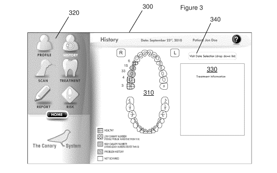
Figure 3 shows a non-limiting example of a user interface for displaying
the odontogram and the oral health status of various scanned teeth. The user
interface window 300 includes the odontogram 310, a series of menu labels 320,
and an additional window region 330 showing a treatment note relating to the
patient. In the embodiment shown in the Figure, the "History" menu label has
CA 02799266 2012-11-13
WO 2011/140663 PCT/CA2011/050302
been selected for reviewing historical results from past patient visits. The
"History" screen shown in the figure provides a drop down menu 340 for
selecting
a specific date or past appointment.
Figure 4 shows a hierarchical diagram 350 of an exemplary yet non-
limiting embodiment of the user interface where the user may navigate among a
variety of screens. The user interface comprises a series of welcome and login
screens 355, which direct the user to a home screen 360, where the user may
select between a series of setting screens 365 and patient/clinical screens
370.
For example, the "History" screen shown in Figure 3 is accessed by selecting
the
"History" screen 375 from the patient/clinical screens 365. Additional patient
screens include the patient profile for entering an managing patient
information, a
scanning screen for scanning individual teeth using the diagnostic device, a
treatment screen for entering treatment notes, a reporting screen for
generating a
patient encounter report, and a risk screen for entering patient risk factors
and/or
reviewing a risk assessment generated as described above. The user interface
may be provided as a touchscreen interface for rapid and convenient navigation
among the various screens, as shown in Figures 5(a) and 5(b).
An embodiment of a scanning user interface screen is shown in Figure
6(a), where the user interface displays an odontogram 400 and scan selection
buttons 405. The user interface displays the selected tooth 410 on the
odontogram, for example, by colouring or shading the selected tooth as shown
in
the Figure. Additionally, further information identifying the scanned tooth
may be
provided, for example by displaying the tooth number 415 using a standard
21
CA 02799266 2012-11-13
WO 2011/140663 PCT/CA2011/050302
system such as the FDI system. In one embodiment, the display may also
identify the scanned surface by an appropriate identifier, such as letter M
shown
at 420, identifying the scanned surface as a mesial surface. In another
embodiment, the user interface may allow the operator to select either primary
or
a permanent tooth, for example, by touching or activating a touch screen on
the
diagnostic device console or a separate display.
After having obtained the scan results, the results of the scan may be
shown in the user interface. In the embodiment shown in the Figure, the Canary
number is shown at 425, which displays the highest measured number obtained
over the series of measurements included in the scan. Alternatively, the
diagnostic values obtained while scanning may be plotted graphically, showing
the spatial profile of the measured values (see Figure 15).
In one embodiment, the user interface also displays an image of the
scanned tooth as shown at 430 in Figure 6(a), so that an image of the scanned
tooth may be displayed adjacent to the odontogram. This allows the provider to
clearly identify the region of the tooth that was scanned to obtain the
result, and
facilitates dialogue and treatment discussion between the provider and the
patient. The image may be obtained, for example, from the diagnostic device,
provided that it is equipped with a camera. In cases where more than one
surface of a tooth is measured, one or more images per tooth surface measured
may be recorded and optionally displayed.
In general, scanning a tooth surface involves examining an area of the
tooth surface to determine whether or not any pathology is present. An
objective
22
CA 02799266 2012-11-13
WO 2011/140663 PCT/CA2011/050302
of scanning is to identify those areas of the tooth surface that require
treatment
either remineralization or operative intervention (placement of a filling).
Considering the specific example of the Canary PTR-LUM system, the beam
diameter is approximately 150 microns, but information is gathered from the
surrounding tooth structure. The operator may collect data by slowly moving
the
diagnostic device handpiece around the tooth surface gathering information.
Clinical trials have identified an area that needs study usually by its visual
characteristics such as:
= Brown or white spot on a smooth tooth surface
= Stained grooves on the occlusal or biting surface of the tooth
= Shadows in the interproximal or contact areas of the tooth
= Shadows on the occlusal, buccal or lingual surface beneath the pits
and fissure
= Contact areas between teeth in general.
In one embodiment, scanning involves first capturing an image of the area
to be examined. The operator places the handpiece on the tooth surface and
begins the scan. For scanning involving the Canary PTR-LUM system, each
scan requires approximately five seconds and is followed by a musical tone and
voice prompt with the Canary Number or a request to repeat the scan. Provided
that the scan need not be repeated, the probe may be moved to an adjacent site
and the scan will begin again automatically. After the operator has finished
examining the tooth, the operator may provide input to the user interface that
the
scan is complete (e.g. selecting, via a touchscreen display, the "done" button
of
23
CA 02799266 2012-11-13
WO 2011/140663 PCT/CA2011/050302
bottoms 405 in Figure 6(a)) and the scan will end. The operator may conclude
the scan with the handpiece measuring the area of tooth that is of concern or
has
the large Canary number. The operator may then move on to the next surface or
next tooth.
In one embodiment, the surface of the tooth may be divided into two or
more image elements for scanning. For example, image elements may include
the mesial occlusal and disto-occlusal areas. This may be particularly useful
for
scanning molar teeth. To further facilitate such scanning over multiple areas,
a
grid system can be overlaid on the tooth image allowing the operator to scan
multiple sections of the tooth surface. This allows for multiple measurements
to
be taken and stored of one tooth surface under examination.
Figure 6(b) provides an example implementation where a grid 500 is
overlaid on the image 505 of the tooth. Grid 500 identifies multiple surface
elements that may be optionally scanned, such that a scan within a particular
grid
element is correlated with the spatial location of the grid element. For
example, a
user may select a particular grid element to scan, and then scan an area
within
the grid element as shown by grid 500. This example implementation is shown in
Figure 6(b), where a particular grid element 510 has been selected for
scanning.
The operator scans the surface of the tooth corresponding to the area shown in
element 510 of image 500, and the scan results for the grid element are
recorded
by the system.
For example, for grid element 510 in Figure 6(b), a Canary number of 26
is measured and stored in association with grid element 510 (this number may
be
24
CA 02799266 2012-11-13
WO 2011/140663 PCT/CA2011/050302
the maximum Canary number obtained while scanning within the region
corresponding to element 510). A provider may subsequently select the tooth
515 in the odontogram and display the Canary readings obtained for any of the
grid elements for which measurements were obtained. In one example,
diagnostic readings for two or more grid elements may be combined when
determining an oral health status of a selected tooth. For example, the user
interface may display the highest diagnostic reading (e.g. Canary number in
the
present case) obtained for all elements, which corresponds to the surface
region
with the greater pathological problem or risk. This reading may be compared
with
reference data to determine the oral health status of the selected tooth.
In the above example, up to 9 distinct areas of the tooth surface can be
examined and diagnostic measurements can be saved for each grid element.
However, it is to be understood that the grid shown in Figure 6(b) is merely
an
example of a grid arrangement, and the grid may contain more or less elements
than as shown.
In one example involving the scanning of interproximal areas, scanning
typically involves capturing data from adjacent teeth at their respective
contact
points. As shown in Figure 7, an operator examines the teeth from three sides,
occlusal or biting surface, buccal or outside surface and lingual or tongue or
roof
of the mouth surface. Scanning involves moving the device around the contact
area or ridge on the biting surface and also along the sides of the teeth.
These
scans may be classified as interproximal scans or lesions in the contact area
or
contact point. Detection of a lesion at the contact point may involve scanning
CA 02799266 2012-11-13
WO 2011/140663 PCT/CA2011/050302
three surfaces, as shown at 435, 440, and 445 in Figure 7, and associating
these
surfaces with a tooth. Accordingly, an interproximal scan may involve
occlusal,
buccal and lingual scanning at the contact point. In one embodiment, an
interproximal scan may be performed by measuring within one or more elements
of a grid, as described above.
Figure 8 provides a flow chart illustrating the process of acquiring an
image of a selected tooth and performing a diagnostic measurement of scan on
the selected tooth. The user is presented with a scanning screen in step 450,
and
a specific tooth may be selected or skipped in step 455. An image of the
selected
tooth is then obtained in step 460, followed by a diagnostic scan that is
recorded
in step 470 provided that the image is accepted in step 465. The flow chart
further provides additional steps for confirming or redoing various steps in
the
process, as shown in Figure 8 at 472, 474, 476, 478 and 480.
In yet another embodiment, the user interface may include drawing tools,
such as lines, arrows, and freeform tools, for annotating an image on a tooth.
These tools may be used to illustrate, on the image, the location where the
scan
is performed. Additional features, such as areas of visible oral health issues
such
as decay, recession, dental restorations or past dental procedures, may also
be
annotated.
In one example shown in Figure 6(c), the user may view a report showing
both the results of the scanning as well as the recommended treatments for a
particular examination day. This type of report provides the operator/user of
the
device with information on past examinations and therapeutic interventions.
26
CA 02799266 2012-11-13
WO 2011/140663 PCT/CA2011/050302
In one example implementation, the user may instruct the system to
prepare a report, as illustrated in the example screen shot provided in Figure
9.
Show in Figure 9 is the selection of a report icon, which generates example
report 530 that is shown in full in Figure 10.
Referring to Figure 10, the example report includes odontogram 535,
patient information 540, and treatment recommendation information 545.
Odontogram 535 may be displayed with diagnostic data that is provided on a per
tooth basis, as shown in the example where per-tooth Canary numbers 550 are
provided for each scanned tooth. It is noted that the present report provides
Canary Numbers that are normalized to a scale ranging from zero to 100.
As indicated by legend 555, the results from the diagnostic measurements
may be additionally (or alternatively) displayed on a qualitative or semi-
quantitative basis. In the example shown in Figure 10, legend 555 shows that
teeth having a low Canary number between 0 and 20 are shown as
"Healthy/Sound Tooth Structure" and displayed in odontogram 535 according to
a first fill type shown at 560. Teeth having a moderate Canary number between
21 and 70 are shown as "Early Decay" and displayed in odontogram 535
according to a second fill type shown at 565. Teeth having a high Canary
number
between 71 and 100 are shown as "Advanced Decay" and displayed in
odontogram 535 according to a third fill type shown at 570. Additional fill
types
may be provided to show other types of measurements, such as at tooth 575 for
which the only data obtained is in the form of a camera image.
27
CA 02799266 2012-11-13
WO 2011/140663 PCT/CA2011/050302
The fill types of the displayed teeth may be provided according to a wide
variety of colours, textures, shades, and other features that are visually
distinguishable. In one example, the fill types may be different colours that
are
associated with hazards or risk. For example, the first fill type, which is
associated with healthy teeth, may be shown as a shade of green, the second
fill
type, which is associated with early decay, may be shown as a shade of yellow
or orange, and the third fill type, which is associated with advanced decay,
may
be shown as a shade of red. In another example, the intensity of the colour is
correlated with the diagnostic measurement. For example, a very low Canary
number of 2 may be shown in bright green, while a Canary number of 18 may be
shown in a lighter green colour. It will be understood that there are many
different
ways of visually conveying risk, inferred oral health problems, or known oral
health problems.
Figure 10 provides one example implementation in which the visual
display of the odontogram includes per-tooth oral health status information
and
diagnostic data. Additionally or alternatively, risk factor data may be
displayed on
a per tooth basis, either quantitatively, qualitatively, or both, as described
above.
For example, the report may include per-tooth, or per-tooth surface,
integrated
risk factor measures that are based on risk factor data and measurements made
with a diagnostic device, as described above.
In another example implementation, the user interface may include one or
more review screens for reviewing oral health data. The review screens may be
useful as a tool for a patient to discuss, for example, a patient's diagnostic
28
CA 02799266 2012-11-13
WO 2011/140663 PCT/CA2011/050302
results, risk factors, oral health history, treatment history, and planned or
suggested treatments and/or interventions. This review process may be
effective
in improving patient awareness of his or her oral health, and for engaging the
patient to take a more active role in maintaining or improving his or her oral
health.
According to one example, the review screen may include a graph of time-
dependent (e.g. historical) diagnostic and/or risk measure data, which
supports
tracking of oral health status and the effectiveness of treatments. The review
screen may take the form of a screen that is similar to the screen displayed
in
Figures 2, 3, 6(a), or 6(b), where selecting a given tooth or tooth surface
(such as
via a touchscreen interface) causes the system to display historical data for
the
given tooth or tooth surface. In one example, the historical data may be
provided
in a tabulated form, where in another example, the historical data may be
displayed as a function of time. In another example, selecting a single tooth
generates a time dependent plot showing data from all individually scanned
tooth
surfaces, such as those pertaining to different areas of a grid. Images of the
tooth
or tooth surface obtained over multiple time points may also be provided. Such
an example is described in detail in Example 3 below, where Figure 15
illustrates
the ability of the system to track and store data on the oral health status of
a
section of tooth over time.
In one embodiment, the time-dependent diagnostic and/or risk measure
data may be displayed with information indicating the timing and optionally
the
nature of therapeutic treatments or interventions. By combining the display of
the
29
CA 02799266 2012-11-13
WO 2011/140663 PCT/CA2011/050302
diagnostic data and/or risk measure data with treatment information, the
operator, provider and/or patient may readily assess and/or observe the
relationship between treatments and oral health.
In another example implementation, the review screen may be provided in
the form of an interactive review screen that displays integrated risk measure
information for one or more teeth or tooth surfaces, and also accepts input
allowing the operator to vary one or more risk factors. The integrated risk
measure information then varies according to the change in the risk factors.
Accordingly, a tool may be provided for presenting and communicating a
sensitivity analysis, where the sensitivity of a patient's specific per-tooth
or per-
tooth surface integrated risk measures to changes in one or more risk factors
is
shown. This may be useful in motivating a patient to make one or more changes
relating to the risk factors, such as a change in diet or oral hygiene habits,
in
order to achieve an improved clinical outcome in a subsequent visit.
The user interface may also include one or more screens whereupon an
operator may provide input to record treatment recommendations for a
pathological condition identified during a particular patient visit. Using the
aforementioned review and tracking screens. As a result, the user interface
facilitates the interpretation of the effectiveness of the treatment
recommendations to influence or change the numerical value associated with a
particular tooth, tooth surface or section of tooth surface, which may be
documented in the form of a report showing the outcomes of the treatment
recommendations on diagnostic measurements.
CA 02799266 2012-11-13
WO 2011/140663 PCT/CA2011/050302
Figures 11 and 12 provide non-limiting embodiments showing various
computing systems for carrying out the aforementioned embodiments. Figure 11
provides a schematic of a local system 600 in which the diagnostic device 605
(and optionally input device 610) is connected to a local computing system 615
such as a personal computer, workstation, or local server through a data
interface. The interface may be a wired connection, such as a USB connection
or
a wireless connection, such as through a local WiFiTM network.
Computing system 615 may also be connected to an input device 610 for
providing patient risk factor data, as discussed above. In a non-limiting
example,
the input device may be a keyboard connected to a computer housing processor
615, or may alternatively be an external input device such as a second
computer,
patient kiosk, or workstation connected to processor 615.
A display 620 is connected to computing system 615 for displaying the
oral health status of teeth scanned with the diagnostic device 605, as
described
in the embodiments listed above. The display may include a monitor directly
connected to computing system 615, or may alternatively be provided as an
external display device, such as a laptop, netbook, electronic document
reader,
tablet, smart phone, or other portable media device or built directly into the
computing system. Display 620 may enable a user to view the oral health status
of selected teeth, and optionally, to control or otherwise interface with the
diagnostic device, through a user interface as described above. In one
embodiment, the system further comprises a data input device such as a
touchscreen or a keyboard. The display may be a touchscreen display. Any or
all
31
CA 02799266 2012-11-13
WO 2011/140663 PCT/CA2011/050302
of system components 610, 615 and 620 may be integrated with diagnostic
device 605. For example, diagnostic device 605 may include a display.
In one embodiment, system 600 includes a general purpose computer or
any other hardware equivalents. Thus, the system may include at least one
processor (CPU/microprocessor), a memory, which may include random access
memory (RAM), one or more storage devices (e.g., a tape drive, a floppy drive,
a
hard disk drive or a compact disk drive), and/or read only memory (ROM), and
various input/output devices (e.g., a receiver, a transmitter, a speaker, a
display,
an imaging sensor, such as those used in a digital still camera or digital
video
camera, a clock, an output port, a user input device, such as a keyboard, a
keypad, a mouse, a position tracked stylus, a position tracked probe, a foot
switch, 6-degree input device based on the position tracking of a handheld
device, and the like, and/or a microphone for capturing speech commands,
etc.).
While some embodiments have been described in the context of fully
functioning computers and computer systems, those skilled in the art will
appreciate that various embodiments are capable of being distributed as a
program product in a variety of forms and are capable of being applied
regardless of the particular type of machine or computer readable media used
to
actually effect the distribution.
Examples of computer-readable media include but are not limited to
recordable and non-recordable type media such as volatile and non-volatile
memory devices, read only memory (ROM), random access memory (RAM),
flash memory devices, floppy and other removable disks, magnetic disk storage
32
CA 02799266 2012-11-13
WO 2011/140663 PCT/CA2011/050302
media, optical storage media (e.g., Compact Disk Read-Only Memory (CD
ROMS), Digital Versatile Disks, (DVDs), etc.), among others. The instructions
can be embodied in digital and analog communication links for electrical,
optical,
acoustical or other forms of propagated signals, such as carrier waves,
infrared
signals, digital signals, etc.
A machine readable medium can be used to store software and data
which when executed by a data processing system causes the system to perform
various methods. The executable software and data can be stored in various
places including for example ROM, volatile RAM, non-volatile memory and/or
cache. Portions of this software and/or data can be stored in any one of these
storage devices. In general, a machine readable medium includes any
mechanism that provides (i.e., stores and/or transmits) information in a form
accessible by a machine (e.g., a computer, network device, personal digital
assistant, manufacturing tool, any device with a set of one or more
processors,
etc.).
Some aspects of the present disclosure can be embodied, at least in part,
in software. That is, the techniques can be carried out in a computer system
or
other data processing system in response to its processor, such as a
microprocessor, executing sequences of instructions contained in a memory,
such as ROM, volatile RAM, non-volatile memory, cache, magnetic and optical
disks, or a remote storage device. Further, the instructions can be downloaded
into a computing device over a data network in a form of compiled and linked
version.
33
CA 02799266 2012-11-13
WO 2011/140663 PCT/CA2011/050302
Alternatively, the logic to perform the processes as discussed above could
be implemented in additional computer and/or machine readable media, such as
discrete hardware components as large-scale integrated circuits (LSI's),
application-specific integrated circuits (ASIC's), or firmware such as
electrically
erasable programmable read-only memory (EEPROM's).
A networked computing environment 800 is schematically shown in Figure
12, where oral health diagnostic device 705 (and optionally input device 710)
is
shown connected to computing system 715. Computing system 715 interfaces
with first network 720 and is connected to server 725 for storing, archiving
and
accessing patient records. A display or workstation 735 may be connected to
computing system 715 via first network 720 for displaying the oral health
status
of teeth scanned with the diagnostic device 705, for example, through a user
interface controlled by server 720. Additionally or alternatively, a display
with an
optional data input means may be directly connected to processor 715, as shown
in Figure 11. Alternatively, computing system 715 may reside beyond first
network 720 and may be directly interfaced with or reside within server 725
Server 725 may communicate with one or more workstations 735 through
second network 730 for displaying oral health status of scanned teeth on
workstations 735, where the display is may be provided via a user interface.
In
one embodiment, first network 720 comprises a local network, such as a network
within a clinical setting, and second network 730 comprises a remote network
such as the internet. By providing access to a user interface located on a
remote
workstation, patients, providers, insurers, and researchers may access
relevant
34
CA 02799266 2012-11-13
WO 2011/140663 PCT/CA2011/050302
oral health status information relating to the scanned teeth of a given
patient, for
example, provided that suitable credentials are established. In one
embodiment,
patients may be granted access to their oral health records, and may view the
oral health status of their scanned teeth from a remote computing environment.
In one embodiment, the user interface accessed by users at workstations 735
and 740 are provided in a web-based or hosted configuration. It is to be
understood that workstations 735 and 740 may comprise any computing system
adaptable for the display the user interface, including, but not limited to, a
laptop,
netbook, electronic document reader, tablet, smart phone, or other portable
media device.
In one example, the oral health data associated with the system may be
stored on a cloud-based server. Such a server, having patient and provider
privacy restrictions, would maintain a repository of the data and allow the
provider and patient access to the reports once they have provided the
appropriate identification and authentication. The oral health provider may
therefore have access to not only single patient reports, but reports on all
patients in the practice for which diagnostic data has been stored. The
reports
may provide analysis by one of more measures of interest, such as age,
geographic location, teeth with Canary Numbers at certain ranges, outcomes of
various preventive and remineralization therapies. In addition the oral health
provider may optionally access to billing and utilization reports. The patient
may
be provided with access to their own personal report containing, for example,
historical readings, information on various therapies and the overall
outcomes. In
CA 02799266 2012-11-13
WO 2011/140663 PCT/CA2011/050302
addition, access to this data may be granted after having removed patient
and/or
provider identifiers to support analysis of demographic data, caries disease
rate
data and outcomes from various therapies on a patient population basis.
The following examples are presented to enable those skilled in the art to
understand and to practice embodiments of the present disclosure. They should
not be considered as a limitation on the scope of the present embodiments, but
merely as being illustrative and representative thereof.
EXAMPLES
Example 1: Utility of PTR-LUM Diagnostic Data
In a PTR or PTR-LUM system, such as The Canary Dental Caries
Detection SystemTM, a beam of energy (typically a laser) intensity-modulated
at a
certain frequency is focused onto the sample surface. The resulting periodic
heat
flow due to the absorbed optical energy in the material is a diffusive
process,
producing a periodic temperature rise (distribution) which is called a
"thermal
wave". This temperature distribution in turn causes a modulated thermal
infrared
(black-body or Planck radiation) emission which is used to monitor the
material
under examination. PTR has the ability to penetrate, and yield information
about,
an opaque medium well beyond the range of optical imaging. Specifically, the
frequency dependence of the penetration depth of thermal waves makes it
possible to perform depth profiling of materials.
In PTR applications involving turbid media, such as hard dental tissue,
depth information is obtained following optical-to-thermal energy conversion
and
transport of the incident laser power in two distinct modes: conductively,
from a
36
CA 02799266 2012-11-13
WO 2011/140663 PCT/CA2011/050302
near-surface distance controlled by the thermal diffusivity of enamel (50-500
m)
[Brown WS, Dewey WA, Jacobs HR: Thermal properties of teeth. J Dent Res
1970; 49: 752-754] and radiatively, through blackbody emissions from
considerably deeper regions commensurate with the optical penetration of the
diffusely scattered laser-induced optical field (several mm). For example,
deeper
subsurface lesions are possible by using a longer wavelength (830-nm) laser
source than a 659-nm probe [Jeon, R. J., Han, C., Mandelis, A., Sanchez, V.,
Abrams, S. H., "Non-intrusive, Non-contacting Frequency-Domain Photothermal
Radiometry and Luminescence Depth Profilometry of Carious and Artificial Sub-
surface Lesions in Human Teeth," Journal of Biomedical Optics 2004, July -
August ,9, # 4, 809 - 819].
PTR measurements of artificially induced caries on extracted human teeth
have shown that the PTR amplitude increases gradually with increasing
demineralization time and decreases after remineralisation. The PTR phase also
shows gradual and consistent changes with demineralization and
demineralization treatment. This behaviour has been attributed to the higher
scatter of the diffuse photon field and to thermal-wave confinement in the
form of
standing waves in the treated region, accompanied by decreased thermophysical
properties (thermal diffusivity and thermal conductivity).
Good correlation of PTR-LUM results with the mineral loss or the lesion
depth measured with TMR results has indicated that PTR-LUM is capable of
monitoring artificially created carious lesions, their evolution during
demineralization, and the reversal of the lesions under the growth of a
37
CA 02799266 2012-11-13
WO 2011/140663 PCT/CA2011/050302
remineralized surface layer [Jeon R. J., Hellen A., Matvienko A., Mandelis A.,
Abrams S. H., Amaechi B. T., In vitro Detection and Quantification of Enamel
and
Root Caries Using Infrared Photothermal Radiometry and Modulated
Luminescence. Journal of Biomedical Optics 13(3), 048803, 2008]. The PTR-
LUM methodology for dental applications has been extensively studied.
Literature reports include applications in depth profiling, early lesion
evaluation,
caries detection in smooth, occlusal, root and interproximal areas, and
theoretical
modeling.
One of the main advantages of PTR-LUM is the ability to perform depth
profiling through scanning of the excitation source modulation frequency. By
selecting a fixed modulation frequency, radiometric measurements at different
depths in the enamel can be obtained. The first attempt to apply the depth
profilometric capability of PTR-LUM toward the inspection of dental defects
was
reported by Mandelis et al.[ Jeon, R. J., Mandelis, A., Abrams, S. H., "Depth
profilometric case studies in caries diagnostics of human teeth using
modulated
laser radiometry and luminescence", Review of Scientific Instruments, 2003,
January, Volume 74 # 1, pages 380 - 383]. In these studies a laser of 488 nm
was used as the excitation source. This work showed that the photothermal
radiometric signals were anti-correlated with the luminescence signals, as a
result of the nature of the two physical signal generation processes. While
the
PTR amplitude increased for carious lesions the LUM amplitude decreased. The
LUM signal results were consistent with previous reports [R. Hibst et al.]. In
addition, these studies showed that the radiometric amplitude exhibited much
38
CA 02799266 2012-11-13
WO 2011/140663 PCT/CA2011/050302
superior dynamic (2 orders of magnitude signal resolution) range to
luminescence (a factor of 2 only) in distinguishing between intact and cracked
sub-surface structures in the enamel. Furthermore, the radiometric signal
(amplitude and phase) produced dental images with much better defect
localization, delineation, and resolution than those obtained with modulated
luminescence.
Further experimental studies [Jeon, R. J., Han, C., Mandelis, A., Sanchez,
V., Abrams, S. H., "Non-intrusive, Non-contacting Frequency-Domain
Photothermal Radiometry and Luminescence Depth Profilometry of Carious and
Artificial Sub-surface Lesions in Human Teeth," Journal of Biomedical Optics
2004, July - August ,9, # 4, 809 - 819] used excitation sources of 659 and 830
nm to assess the feasibility of PTR-LUM to detect deep lesions. PTR frequency
scans over the surface of an occlusal fissure into demineralized enamel and
dentin showed higher amplitude than those for healthy teeth, as well as a
pronounced curvature in both the amplitude and phase signal channels. These
can be excellent markers for the diagnosis of subsurface carious lesions. The
results showed that PTR-LUM is able to detect artificial subsurface defects
with
sharp boundaries at depths greater than 5 mm. In addition PTR exhibited
superior sensitivity to the presence of sharp boundaries, as well as to
changes in
natural demineralized regions of the tooth. These results suggested the
possibility to detect carious lesions on both occlusal surfaces and the
interproximal area of the tooth [Jeon et al.].
39
CA 02799266 2012-11-13
WO 2011/140663 PCT/CA2011/050302
In experimental studies, it was found that PTR Amplitude had a very
strong correlation with lesion size and shape. LUM phase provided limited
information. PTR Phase provided an indication of operator movement if there
was a strong shift in the phase number from the norm. If this occurred, the
operator was instructed to re-measure the area.
In an embodiment providing a single unified quantitative indication of oral
health from a measurement at a given location, the data from each location is
stored as four separate signals; PTR amplitude and phase and LUM amplitude
and phase. A unified diagnostic measure is obtained according to the following
weighting formula:
^ PTR Amplitude weighted at 45% of the total value
^ PTR Phase weighted at 15% of the total value
^ LUM Phase weighted at 10% of the total value
^ LUM Amplitude weighted at 30% of the total value
The four readings are compared to the readings one finds from the healthy
enamel surface and/or from a standardized piece of hydroxyapatite. The
measured signal number is compared to healthy enamel surface as well. Results
from the comparison step may be provided on a fixed scale for each reading,
for
example, on a scale of 1 to 100 (the scales need not be equal for each reading
type), indicating a severity of a condition. The four fixed-scale results are
then
weighted as described above, providing the operator a ranking or range (for
example, on a scale from 1 - 100) indicating the health of the area examined.
The utility of multiple readings in diagnostic assessment with a PTR and LUM
CA 02799266 2012-11-13
WO 2011/140663 PCT/CA2011/050302
detection device was illustrated in Jeon [Jeon et al., "Diagnosis of Pit and
Fissure
Caries Using Frequency-Domain Infrared Photothermal Radiometry and
Modulated Laser Luminescence", Caries. Res. 38, 497-513, 2004].
In another embodiment, the reading from a single frequency is combined
in the following manner: (PTR amplitude x PTR Phase) / (LUM Amplitude x LUM
Phase) to create one single reading. Error checking is done by combining the
standard deviation from each reading into one number as follows:
LUM amplitude x LUM Phase x PTR Amplitude x PTR Phase. The ratio of single
reading / combined standard deviation is examined and if the ratio increases
dramatically this indicates an error in the reading and this is conveyed to
the
operator. The single reading is then conveyed to the operator along with its
difference from the single reading derived from examining health enamel and
healthy teeth.
Example 2: Photothermal Radiometric and Luminescence System
Figure 13 illustrates non-limiting example of a diagnostic dental device
according to one embodiment involving a hybrid PTR-LUM device 300, shown
with its main components. Details of the oral health detection device 300 are
disclosed in United States Patent Publication No. US20070021670 published on
January 25, 2007, which is incorporated herein in its entirety by reference.
United
States Patent No. 6,584,341 issued to Mandelis et al. entitled "Method and
apparatus for detection of defects in teeth", which is incorporated herein in
its
entirety by reference, discloses a similar system. In one embodiment, the
system
includes an optical imaging system, such as, but not limited to, a CCD camera
for
41
CA 02799266 2012-11-13
WO 2011/140663 PCT/CA2011/050302
imaging capture of dental tissue. Other imaging devices may include an infra-
red
imaging device.
The PTR-LUM system 800 as disclosed in these two US Patent
publications is used for scanning and data capture of dental tissue. The
device is
designed for locating and monitoring small early carious lesions, areas of
erosion
and caries around restorations in a non-invasive fashion. The core technology
in
device is photothermal radiometry (PTR) and ac luminescence (LUM) as
described in other the previously referenced United States
patents/applications
incorporated by reference. By using PTR and LUM and applying comparison to
normal healthy enamel or other mineralized tissue, one can then assess the
health of the tooth and monitor ongoing changes. The device can monitor
ongoing demineralization (break down of the enamel crystal); early stages of
dental caries and remineralization as well as erosion of the tooth surface or
caries around dental restorations.
As shown in Figure 13, the system includes a laser light source 810 for
irradiating a portion of a dental surface 820 with an effective wavelength, in
which
modulated photothermal radiometric signals and modulated luminescence
signals are responsively emitted from the dental surface. A first detector 830
detects the emitted modulated luminescence signals, and a second detector 840
detects the emitted modulated photothermal signals. The laser light is emitted
from a hand held probe head 850, and a flexible optical fiber bundle 860
having a
distal end connected is to the hand held probe head.
42
CA 02799266 2012-11-13
WO 2011/140663 PCT/CA2011/050302
The optical fiber bundle includes a first optical fiber 870 having a proximal
end in optical communication with the light source and a distal end terminated
at
the hand held probe head for transmitting light from the light source to a
patient's
dental tissue by a clinician handling the hand held probe head. The optical
fiber
bundle additionally includes a plurality of multi-mode optical fibers having
distal
ends 880 terminated at the hand held probe head and proximal ends optically
coupled to the two detectors. A first pre-selected number of the multi-mode
optical fibers 880 are near-infrared-transmitting optical fibers for
transmitting the
modulated luminescence signals to the first detector, and a second pre-
selected
number of the multi-mode optical fibers 890 are mid-infrared-transmitting
optical
fibers for transmitting the photothermal radiometry signals to the second
detector.
Device 800 includes a demodulator for demodulating the modulated
photothermal signals into photothermal phase and amplitude components and
the modulated luminescence signals into luminescence phase and amplitude
signals. Device 800 further includes a computer processor and or data
acquisition and processing card (such as a card offered by National
Instruments,
Inc., for comparing the photothermal phase and amplitude signals to
photothermal phase and amplitude signals of a reference sample and comparing
the luminescence phase and amplitude signals to luminescence phase and
amplitude signals of a reference sample to obtain differences, if any, between
the
portion of the dental tissue and the reference sample and correlating any
differences with defects in the dental tissue. In Figure 13 both the
demodulator
and the computer processor are shown generally as processing unit 875. A
43
CA 02799266 2012-11-13
WO 2011/140663 PCT/CA2011/050302
computer with touch screen 810, keyboard, and mouse input interface and
included and a CCD camera 895 may be included for capturing images of the
examined surface of the dental tissue of the patient.
Prior to initiating a scan, information relating to the identification of the
patient and the oral tissue (teeth or gum or any other dental tissue and its
location) are input through an input device 885 such as a touch screen. The
resulting optical signal from sample 820 recorded using the device are
collected
by the hand piece 850 and optical fiber bundle 860 and sent to detectors 830
and
840. An image of the examined surface is obtained with the CCD camera 895
and sent to the processing unit 875. In an alternative embodiment, an imaging
device (such as a CCD camera) may be integrated in the scanning hand piece
850. While a dental technician is operating device 800, the data is captured
by
scanning the tooth surfaces with an optical probe in hand piece 850.
An example schematic of the internal optical configuration for the hand
piece 850 is shown in Figure 14(a). A housing 950 contains the optical fiber
bundle 960, mirror 970, lens 980, and spacer 990. The optical fiber bundle 960
delivers the laser light (shown at 995) and collects the scanning results
through
mirror 970 and lens 980.
Figure 14(b) illustrates another example of a handpiece for performing
measurements based on photothermal radiometry, where the handpiece does
not utilize fiber optic beam delivery, as disclosed in co-pending US
Provisional
Patent Application No. 61/334,436, titled "Handpiece with Integrated Optical
44
CA 02799266 2012-11-13
WO 2011/140663 PCT/CA2011/050302
System for Photothermal Radiometry and Luminescence Measurements", and
filed on May 13, 2010, which is incorporated herein by reference in its
entirety.
A detailed view of the optical apparatus housed within the body portion
1110 and tip portion 1120 is shown in Figure 14(b). The optical bench 1200
supports semiconductor laser 1205 and lens 1210, which collimates emitted
laser
light that is subsequently redirected by mirror 1215 towards dichroic window
1220. Semiconductor laser 1205 may be a laser diode having a wavelength of
approximately 660 nm for the simultaneous generation of luminescence and
photothermal radiation from a tooth surface.
The collimated laser beam is redirected by dichroic window 1220, which
may comprises an optical coating having that generate high reflectivity of the
incident laser beam while passing thermal radiation. The laser beam propagates
in a substantially axial direction into tip portion 1120 along beam path 1225,
bypassing pick-off prism 1230 and encountering mirror 1235 at the distal end
of
tip portion 1120. Mirror 1235 reflects the collimated laser beam towards
focusing
element 1240, which focuses the laser beam as it emerges from tip portion
1120.
Focusing element 1240 is transparent in the visible light to mid-infrared
and has a proper focal length (8.6mm from the lens surface) for focusing the
laser to a spot size of approximately 50 micrometers in average. Focusing
element provides the additional role of collecting and substantially
collimating
both luminescent and photothermal radiation emitted from a tooth surface in
response to laser irradiation. While focusing element 1240 is shown as a
transmissive optical component, it will be apparent to those skilled in the
art that
CA 02799266 2012-11-13
WO 2011/140663 PCT/CA2011/050302
focusing element 1240 and mirror 1235 could be replaced with a single curved
off-axis parabolic mirror.
Collected luminescence is directed by mirror 1235 along an axis of tip
portion 1120, and a portion of the collected luminescence beam encounters pick-
off prism 1230 and is directed towards optical filter 1245 and photodetector
1250.
Optical filter 1245 removes unwanted reflected and scattered laser light, and
photodetector 1250 is selected to have an optical bandwidth for the detection
of
the collected luminescence. Photodetector 1250 may be a silicon photodiode,
and optical filter 1245 may be an inexpensive color glass filter having a
bandwidth and optical density matched to the laser wavelength and power (such
as RG 715 Longpass color filter).
As noted above, focusing element 1240 also collects and collimates
emitted photothermal radiation, which is reflected by mirror 1235 and directed
towards dichroic window 1220. Dichroic window 1220 passes thermal radiation
and reflects or absorbs any light which wavelength is below 1.85 micrometers
(lower bandwidth of germanium window) including reflected or scattered laser
light and luminescence. A suitable material for the absorptive substrate is
germanium.
Accordingly, dichroic window with proper coating to enhance transmissivity
of mid-infrared and reflectivity of the laser light 1220 passes the collected
photothermal radiation, which is subsequently focused by lens 1255 onto
infrared
detector 1260. Infrared detector may be a sensitive mid-infrared detector,
such
as a photovoltaic HgCdZnTe detector, with a sensitive spectral region spanning
46
CA 02799266 2012-11-13
WO 2011/140663 PCT/CA2011/050302
approximately 2 to 5 m. The infrared detector may be mounted on a thermo-
electric cooler for enhanced performance and sensitivity.
As shown in Figure 14(b), the optical components described above are
mounted on optical bench 1200, which may be formed in a lightweight and
thermally conductive material such as aluminum. As shown in the Figure 14(b),
the optical bench, while housed within body portion 1110, may be attached to
tip
portion 1120, thereby enabling rapid and efficient heat sinking into tip
portion
1120. Accordingly, tip portion 1120 may also be made from a lightweight and
thermally conductive material, such as aluminum. Such an embodiment enables
tip portion to act as an efficient air-cooled heat sink for optical components
(primarily the laser diode and optionally a TE (thermo-electric) cooler
attached to
detector 1260) mounted on optical bench 1200. This feature is especially
important to optimize the performance of infrared detector 1260, which has a
noise floor that is strongly dependent on temperature. Furthermore by
attaching
the optical bench directly to the rigid tip portion 1120, superior mechanical
isolation is obtained relative to attaching the optical components to the body
portion 1110.
While the system shown in Figures 11 and 12, and their application, have
been described and illustrated within the context of an exemplary embodiment,
it
is to be understood that that numerous embodiments of device system may be
made without departing from the scope of the disclosure.
To obtain an output indicative of the oral health of a patient, the oral
health
detection device, such as the PTR-LUM device discussed above, or a processor
47
CA 02799266 2012-11-13
WO 2011/140663 PCT/CA2011/050302
or computing system interfaced with the detection device, analyzes the raw
data
captured from a patient and compares it to a normalized signal for that
particular
hard tissue. The norm could be either an internally generated function or the
signal from a healthy section of hard tissue or a signal generated by
hydroxyapatite or other mineralized tissue or commercially produced samples.
Scanning a tooth surface with the device can involve a single frequency,
two or more selected frequencies or a frequency scan from 1 Hz to 1000 Hz. In
one embodiment, the device is scanned with the option of either 1 or 4
frequencies. The single frequency is used to examine a particular section of
tooth surface such as a stained groove. Scanning a tooth with multiple
frequencies would create a depth profile picture of the lesion. In one
embodiment, scans at 2 Hz, 5 Hz, 20 Hz and 100 Hz provide information on a
lesion from near surface to a depth of approximately 4 mm.
Example 3: Plotting and Reviewing Historical Data
The present example illustrates the utility of plotting time dependent data
associated with a tooth, as described in the aforementioned embodiments. The
present example involves a patient having a history of oral health problems,
for
which remineralization therapy was recommended by a provider.
Prior to initiating remineralization therapy, a first measurement was
performed with the CanaryTM System, a photothermal diagnostic device as
described above. An initial value of the Canary Number for the tooth was
determined at two different frequencies (2 Hz and 5 Hz), as shown in Figure
15(a) (it is noted that the Canary Number shown in Figure 15(a) are provided
on
48
CA 02799266 2012-11-13
WO 2011/140663 PCT/CA2011/050302
a different scale than the Canary numbers described elsewhere in the present
disclosure).
Following the initialization of remineralization treatment, the Canary
Number was measured for the tooth during three separate visits at both
frequencies. In addition to the diagnostic data, images of the tooth surface
were
also obtained for each diagnostic measurement, as shown in Figure 15(b).
Having obtained and recorded historical data associated with the tooth,
the provider may review the recorded data and images in order to determine the
effectiveness of the therapy. As described above, this may be achieved by
selecting the tooth in the odontogram that is displayed in the user interface,
which then optionally allows the provider to obtain the plot shown in Figure
15(a).
The corresponding images shown in Figure 15(b) may also be shown, optionally
with an indication of the region scanned, such as rectangle 1000, which may be
an image element of a grid (as described above). The plot clearly shows, for
the
2 Hz measurements, that the mineralization therapy is effective and improving
the oral health associated with the tooth.
In another example, the system may store and display various
remineralization therapies employed over time, and display information
relating to
these therapies in addition to the image and Canary Number data shown in
Figure 15. In addition, as noted above, risk measures may also be tracked and
plotted as a function of time for assessing the impact of the prescribed
therapy
on oral health risk and outcomes.
49
CA 02799266 2012-11-13
WO 2011/140663 PCT/CA2011/050302
This improvement in the oral health status of the selected tooth is also
reflected in the display of the tooth in the odontogram, as described above.
For
example, due to the reduction of the Canary number, the displayed tooth in the
odontogram may change from red to green in colour over the four visits.
The specific embodiments described above have been shown by way of
example, and it should be understood that these embodiments may be susceptible
to
various modifications and alternative forms. It should be further understood
that the
claims are not intended to be limited to the particular forms disclosed, but
rather to
cover all modifications, equivalents, and alternatives falling within the
spirit and scope of
this disclosure.