Note: Descriptions are shown in the official language in which they were submitted.
CA 02799737 2012-11-16
WO 2011/146969 PCT/AU2011/000609
METHOD OF DIAGNOSIS AND LOCATION OF A SOFT TISSUE INJURY
FIELD OF THE INVENTION:
The present invention relates to a method of medical diagnosis and more
particularly to a method of
diagnosis and location of a soft tissue injury.
BACKGROUND:
Soft tissue injuries are identified as a major source of pain and disability
and occur across a wide
section of the community.
Soft tissue injuries arise generally as a result of damage to muscles, nerves,
connective
tissues, fascia, joint capsules, periosteum etc as a result of excessive
force/stress in a given moment, or
repetitive strain placed upon these tissues over an extended period of time.
As such, soft tissue injuries
are very common in the workplace. Additionally, soft tissue injuries that
occur as a result of trauma
may not be immediately obvious, to the individual at the time of the trauma
but may become apparent
at some point in the future.
A soft tissue injury can be considered to be a fracture because it is the
local separation of a body
into two, or more pieces under the action of stress. Hence damage to soft
tissue can be referred to by
either of the terms soft tissue stress fracture or soft tissue injury and can
be used interchangeably.
A method to identify soft tissue damage is with the use of Magnetic Resonance
Imaging (MRI).
Such equipment requires detailed understanding of the symptoms of the injured
person, his/her case
history, and then, based on that information, very precise and localised use
of the equipment to
observe a microscopic injury. The equipment used for this form of imagery is
very expensive and
therefore cannot be used day-to-day by general practitioners and as such MRI
is not considered to be a
useful tool for general diagnosis of soft tissue injuries.
Inflammation of soft tissue is a result of a complex cascade of events that
includes changes to
concentration of various chemical components within the body, such as
histamines, prostaglandins,
cytokines etc along with inflammatory cells such as leukocytes, fibroblasts
and macrophages. The
inflammatory response results, physiologically, in an increase in inflammatory
hormones and/or
nerve chemicals at the site of injury, swelling, hypersensitivity, neuritis,
fasciculation, involuntary
muscle contraction, heat, reduced blood flow, and critically, a reduced
ability of the lymphatic system
to drain interstitial fluid (lymphoedema). All of this causes a vicious cycle
of pain for the individual.
It is a commonly-held belief that infrared thermography detects differences in
heat, and
therefore inflammation. However, infrared imaging is actually detecting a
selected range of
infrared wavelengths (photons with wavelengths in the range of 700 nm to 2000
nm, or in the
range of 810 nm to 820 nm, as two possible examples). The hotter a body of
matter is, the more
infrared intensity it emits in this infrared spectrum range. Therefore, within
a specified
wavelength range, the amount of heat corresponds to the infrared intensity
within that
CA 02799737 2012-11-16
WO 2011/146969 PCT/AU2011/000609
2
wavelength range. The narrower the wavelength range, the smaller the range of
temperatures that the
thermograph can detect. Recently, digital infrared thermographs that
correspond to the narrow
temperature range of metabolic heat have been demonstrated, enabling highly
accurate thermographs of
the surface temperature of the human body.
These methods, however, simply rely on a change of the surface temperature and
are insufficient
to provide a reliable diagnosis. The surface temperature is only an indirect
indication of the temperature at
the site of the injury, which is located inside the body of the soft tissue.
There is no current method for
accurately detecting the precise location and the amount of inflammation
around the site a soft tissue injury.
There exists, therefore, a need for an accurate, non-invasive, rapid and
inexpensive method of detecting
inflammation below the surface of the body. `
SUMMARY OF THE INVENTION:
According to the present invention, although this should not be seen as
limiting the invention in
any way, there is provided a method of diagnosing, and determining the
position of, a microscopic or
a macroscopic soft tissue injury or soft tissue stress fracture in a patient,
including the steps of:
determining on the skin of the patient a pain area;
applying electromagnetic energy or radiation, in a selected portion or range
of the visible or
infrared spectrums, to parts of the body's surface that correspond with the
pain area;
obtaining feedback from the patient to determine the sensations that the
patient experiences as a
result of visible or infrared energy being applied to the tissue at a specific
region of pain area; and
establishing the site of the microscopic or macroscopic soft tissue injury at
the specific region
where the sensations are greatest.
In this way, a primary soft tissue injury site may be determined as a result
of tingling, aching,
heat, or 'pins and needles' sensations travelling along a nerve of the patient
to a site distal from the
visible or infrared laser probe.
The explanation which the inventor believes explains the observed reaction to
the application of
visible or infrared energy, but to which the inventor does not necessarily
wish to be restricted, is that the
visible or infrared energy interacts with cells and proteins which are
accumulated at a site of
microscopic or macroscopic soft tissue injury in a body and thereby provide
the observed sensations.
The inventor has observed that the energy from the visible or infrared
spectrums is not absorbed
at sites where there is no inflammation but it is absorbed at sites where
there is inflammation. This has
been observed from both patient feedback of sensations at the site of
inflammation and also indicative
data from monitoring the change in the digital infrared thermographs of the
surface of the body, near the
site of the soft tissue injury.
It is believed that the photons from the visible or infrared spectrums
interact with the
inflammatory cells and proteins along a nerve fibre connected to the soft
tissue injury thereby providing
the observed sensations.
In one embodiment of the invention the step of determining on the skin of the
patient a pain area
CA 02799737 2012-11-16
WO 2011/146969 PCT/AU2011/000609
3
comprises the step of observing surface temperature on the skin of the patient
with the highest surface
temperature indicating a pain area.
In an alternative embodiment of the invention the step of determining on the
skin of the patient a
pain area comprises the steps of:
obtaining a thermographic image of the pain area of the patient to enable
visualization of
variation in surface temperature of the pain area,
reviewing the thermographic image to determine the point or points of greatest
surface
temperature; and
applying the electromagnetic energy or radiation to the point or points of
greatest surface
temperature.
Preferably the application of the visible or infrared energy at any one point
is for no longer than
two or three minutes.
Preferably the visible or infrared energy is applied at a selected wavelength
or a set of
wavelengths in the visible, near-infrared and infra-red wavelength spectrums.
Preferably the visible or infrared energy is applied using a laser probe and
which is applied via
direct contact of the laser probe with the patient's skin, delivered via a
fibre optic delivery system from
the laser probe to the patient's skin, or delivered by pointing the beam from
the laser probe through the
air to the patients skin. Alternatively the visible or infrared energy is
being applied using a probe or
optical emitter device other than a laser device, such as a Light Emitting
Diode (LED), a light bulb or
similar optical emitter.
Preferably the probe is operated at a selected wavelength or a set of
wavelengths in the range of
400 run to 10,000 nm, which corresponds to wavelengths in the visible, near-
infrared and infra-red
wavelength spectrums.
The step of obtaining feedback from the patient can comprise establishing
dialogue with the
patient to understand the sensations that they experience as a result of
visible or infrared energy being
applied to the tissue at the point or points.
Alternatively the step of obtaining feedback from the patient comprises using
a feedback device,
such as a switch, a lever or a rotating variable knob as examples, that the
patient can input the presence
or not of sensation and the amount of sensation, in real time.
Preferably the invention can further including an initial step of obtaining a
case-history of the
patient to determine possible areas of injury and pain areas. This additional
step may or may not be
beneficial to the diagnostic process, depending on the accuracy .and success
of previous diagnoses
and treatments.
The method of the present invention can be used to diagnoses soft-tissue
injuries that result in
symptoms such as lower back pain, neck pain, migraines, Type 2 diabetes,
sciatica, tinnitus, carpal
tunnel syndrome, chronic pain syndrome, fibromyalgia or be used to diagnoses
soft-tissue injuries that
result in other symptoms that are either not known at this time or not
described above. Hence the
method of the present invention may be used to diagnose and determine the site
of an injury which is
CA 02799737 2012-11-16
WO 2011/146969 PCT/AU2011/000609
4
not apparent as observed by other techniques and which can be the original
source of their pain.
Once a diagnosis has been obtained by determining the site of the microscopic
or macroscopic
soft tissue injury treatment can be applied using a visible or infrared laser
probe.
In preference, for treatment of a soft tissue injury there may be 2 x 300 mW
830 rim infrared
lasers used for periods greater than five minutes and less than 60 minutes per
treatment. In preference,
the time of application of visible or infrared energy to an injury site is
greater than five minutes and less
than eight minutes per treatment but no more than two to three minutes at any
one time.
BRIEF DESCRIPTION OF THE DRAWINGS:
0 By way of example, an employment of the invention is described more fully
hereinafter with reference to the accompanying drawings, in which:
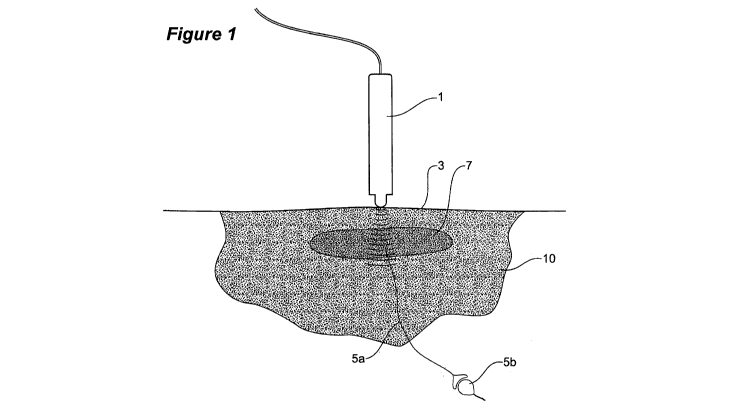
Figure 1 shows a schematic overview of the present invention.
DETAILED DESCRIPTION OF THE INVENTION:
5 In a preferred embodiment of the invention as a first step an infrared
thermographic image
(thermogram) is taken to observe the patient's dermatomal neurophysiology of
pain areas.
An interpretation of the thermogram can be given to the patient regarding
their general problem area
but then further diagnosis is necessary according to the present invention to
refine in more detail the site
of the injury. This is done as discussed above by application of visible or
infrared energy using a laser
0 probe and obtaining feedback as to sensations felt by a patient.
Usually, but not always, the laser will take five to eight minutes at the
start of a diagnosis
session to generate enough energy to produce any perceivable sensation in the
patient.
Visible or infrared laser energy is applied to the site of pain and/or
hotspots indicated on the
thermogram for a period of no less than one minute and no more than three
minutes.'
5 The patient is advised that it is very important to communicate the
sensations that they feel
in their body during therapy. Sensations of heat and pain are best
communicated using a
feedback arrangement based upon a scale of zero to 10 with zero being no
pain/heat (cold)' and 10
being 'too painful/hot, please move the probe'. Other sensations, such as
tingling, `pins and
needles', dull aches, bubbling, numbness, "ants crawling under my skin" and
many more may be
0 communicated without a scale.
When heat or other sensation is experienced by the patient, at the location of
the laser,
inflammation is being detected.
When radiating sensations are experienced by the patient to distal parts of
the body, such as
heat, tingling, aching, pins and needles etc, soft tissue stress fracture have
been detected.
5 According to the interpretation of the inventor, the patient is experiencing
sensations of
the inflamed neuron(s) within ruptured collagen fibres. Applying an amount of
infrared `energy to the
site of the injury stimulates or excites nerve chemicals and/or inflammatory
proteins, such as
histamines, prostaglandins, substance P, kinins, bradykinins etc along the
neuron localised from the
CA 02799737 2012-11-16
WO 2011/146969 PCT/AU2011/000609
injury site. The injury is a source, or cause, of inflammation being present
in the region.
Figure 1 shows an overview of this process in which visible or infrared energy
from a probe
source I is applied to an inflammatory site 10 of an area of the patient's
body 3. This site 10 is
identified by obtaining a obtaining a thermographic image of a pain area of
the patient to
5 enable visualization of variation in surface temperature of the pain area.
The visible or
infrared energy, at the frequencies used, is able to penetrate the body and
can come into contact
with a soft tissue stress fracture 7 and an associated nerve fibre 5a
connecting to nerve fibre 5b to a
distal location of the body. The visible or infrared energy travels along
nerve fibre 5a exciting the
inflammation proteins within the nerve itself. As infrared energy travels
along the length of the nerve
0 fibre 5a to a neuron 5b more of the inflammatory proteins are excited
causing referred sensations as
discussed above, enabling a diagnosis to be made as to the site where the
laser energy meets the
soft tissue injury (stress fracture) 7.
When the patient experiences referred sensation (i.e. sensations at a location
distant from
the probe caused by visible or infrared energy), the laser energy is
travelling along the neuron(s) and is
5 having a far-reaching effect on the patient.
The visible or infrared laser may be moved every one to three minutes to a
nearby location.
The probe is required to stay in one location for at least one minute to
assess whether any
perceivable sensations are occurring (as some sensations build over time) and
not more than three
minutes to avoid bioinhibition of healing.
0 The nearby location may be right next to the previous spot or in a
completely new area
depending on what areas of the body have been treated already as well as the
case-history of the
patient and the results of the thermographic image.
After moving the laser from an area of the patient's body that created
significant sensations, the
therapist will move to a new location for a period of time to avoid bio-
inhibition of the injury before
5 coming back to the area of significance.
Avoiding bioinhibition can be a fine line, but if the therapist sticks to the
general guideline of
not treating an injury for greater than eight minutes per therapy session, the
results will be positive.
This is imperative to avoid the possible anti-therapeutic effects of
electromagnetic radiation.
Various modifications may be made in details of design and construction and
process steps,
0 parameters of operation etc without departing from the scope and ambit of
the invention.