Note: Descriptions are shown in the official language in which they were submitted.
CA 2799757 2017-04-25
=
WO 2011/143779 PCT/CA2011/050315
METHODS FOR THE ASSESSMENT OF COLORECTAL
CANCER AND COLORECTAL POLYPS BY MEASUREMENT
OF METABOLITES IN URINE
FIELD OF THE INVENTION
[0001] This invention relates to the assessment of colorectal
cancer and colorectal
polyps by measurement of metabolites in urine.
[0002]
BACKGROUND
[0003] Colorectal Cancer (CRC) is among the leading causes of
morbidity. The
chance of surviving CRC is closely related to the stage of the disease at
diagnosis; the
earlier the diagnosis, the greater the likelihood of survival. In many
instances CRC is
preceded by colorectal polyps, particularly adenomatous colorectal polyps. If
identified
early at the colorectal polyp or precancerous lesion stage, CRC is more likely
to be
curable. Therefore, subjects with CRC and/or colorectal polyps would greatly
benefit
from early diagnosis.
[0004] Current CRC screening methods consist of one or a
combination of the
followings: fecal occult blood testing (FOBT), flexible sigmoidoscopy, air-
contrast barium
enema, computerized tomography colonography (CTC) and/or colonoscopy. These
current screening methods all have limitations or potential risks that limit
their application.
[0005] Colonoscopy is currently the standard test for the
presence or absence of
CRC or colorectal polyps. However, colonoscopy is invasive and can impose
unnecessary hazards and risks caused by sedation or the procedure itself. A
known
non-invasive CRC diagnostic method is FOBT. FOBT, however, has very low
sensitivity
in detection of CRC and is unattractive as the handling of fecal matter is
required. CTC
is a recent non-invasive technique for imaging the colon. However, its
performance
varies due primarily to technological differences in the subject preparation
and the
hardware and software used for the analysis. Several new screening methods
based on
DNA analysis are now available. These are typically PCR-based assays used to
identify
mutations known to occur in the
1
CA 02799757 2012-11-16
WO 2011/143779
PCT/CA2011/050315
adenoma-to-carcinoma sequence, or in familial CRC. However, whether genomics-
based
tests will result in high diagnostic accuracy for sporadic CRC remains to be
seen.
[0006] Accordingly, there is a need to develop improved methods of
assessing CRC
and colorectal polyps in a subject.
SUMMARY
[0007] Methods for the diagnosis of CRC, colorectal polyps in general
and
adenomatous polyps in particular by measurement of metabolites in urine are
described. In
some embodiments, certain metabolites are identified as being elevated or
reduced in
concentration or quantity in subjects with CRC and/or colorectal polyps as
compared with
subjects without CRC or colorectal polyps. The measurement of these
metabolites in urine
can indicate the presence of CRC or colorectal polyps in general or
adanomatous polyps in
particular in a subject.
[0008] In one aspect, the invention provides a method for assessing
whether a
subject has or is predisposed to developing CRC and/or colorectal polyps, said
method
comprising:
(a) providing a urine sample from said subject;
(b) obtaining a metabolite profile from said urine sample;
(c) comparing said metabolite profile with a reference metabolite profile;
and
(d) assessing, based on said comparison in step (c), whether said subject
has or
is predisposed to developing CRC and/or colorectal polyps.
[0009] A further aspect of the invention relates to a method for
identifying urine
metabolites indicative of the presence or absence of CRC and/or colorectal
polyps, said
method comprising:
(a) providing a urine sample from a subject;
(b) obtaining a metabolite profile from said urine sample;
(c) comparing said metabolite profile with a reference metabolite
profile; and
2
CA 02799757 2012-11-16
WO 2011/143779 PCT/CA2011/050315
(d) identifying, based on said comparison in step (c), one or more
metabolites in
said metabolite profile that are indicative of the presence of or
predisposition to in said
subject of CRC and/or colorectal polyps.
[0010] A further aspect of the invention relates to a use of a urine
metabolite profile
comprising one or more of metabolites selected from the group consisting of:
1,6-Anhydro-13-D-glucose, 1-Methylnicotinamide, 2-Hydroxyisobutyrate,
2-0xoglutarate, 3-Aminoisobutyrate, 3-Hydroxybutyrate, 3-Hydroxyisovalerate,
3-Hydroxymandelate, 3-Hydroxyphenylacetate, 3-Indoxylsulfate,
4-Hydroxyphenylacetate, Acetate, Acetone, Adipate, Alanine, Ascorbate,
Asparagine, Benzoate, Betaine, Butyrate, Carnitine, Citrate, Creatine,
Creatinine, Dimethylamine, Ethanol, Formate, Galactose, Glucose, Glutamine,
Glycerol, Glycine, Glycolate, Guanidoacetate, Hippurate, Histidine,
Hypoxanthine, Isoleucine, Lactate, Leucine, Lysine, Mannitol, Methanol,
Methylguanidine, N,N-Dimethylglycine, O-Acetylcarnitine, Pantothenate,
Propylene glycol, Pyroglutamate, Pyruvate, Serine, Succinate, Sucrose,
Tartrate, Taurine, Threonine, Trigonelline, Trimethylamine, Trimethylamine
N-oxide, Tyrosine, Uracil, Urea, Valine, Xylose, cis-Aconitate,I3-Alanine,
TT-Methylhistidine, T-Methylhistidine and trans-Aconitate,
for assessing whether a subject has or is predisposed to developing CRC and/or
colorectal
polyps.
[0011] A further aspect of the invention relates to a kit for
assessing whether a
subject has or is predisposed to developing CRC and/or colorectal polyps, said
kit comprising
one or more reagents for detecting the presence and/or concentration and/or
amount of one
or more metabolites in a urine sample of a subject, and instructions for use
of said kit for
assessing whether a subject has or is predisposed to developing CRC and/or
colorectal
polyps.
[0012] A further aspect of the invention relates to a system
comprising:
(a) a CRC- and/or colorectal polyps-assessing apparatus including a
control unit
and a memory unit to assess a CRC state in a subject; and
3
CA 02799757 2012-11-16
WO 2011/143779 PCT/CA2011/050315
(b) an information communication terminal apparatus that provides
data on the
presence and/or concentration and/or amount of metabolites in a urine sample
from the
subject connected to each other communicatively,
wherein the information communication terminal apparatus includes:
(a) a data sending unit that transmits the data on the presence and/or
concentration and/or amount of metabolites in the sample to the CRC- and/or
colorectal
polyps-assessing apparatus; and
(b) an assessment result-receiving unit that receives the
assessment result of the
CRC and/or colorectal polyps state of the subject transmitted from the CRC-
and/or colorectal
polyps-assessing apparatus,
wherein the control unit of the CRC- and/or colorectal polyps-assessing
apparatus includes:
(a) a data-receiving unit that receives the data on the metabolite
concentration
and/or amount of the sample transmitted from the information communication
terminal
apparatus;
(b) a discriminant value-calculating unit that calculates a discriminant
value that is
a value of multivariate discriminant, based on both the concentration and/or
amount value of
the metabolite in the sample received by the data-receiving unit and a
multivariate
discriminant with the concentration and/or amount of the metabolite as
explanatory variable
stored in the memory unit;
(c) a discriminant value criterion-assessing unit that assesses the CRC or
colorectal polyps state in the subject, based on the discriminant value
calculated by the
discriminant value-calculating unit; and
(d) an assessment result-sending unit that transmits the assessment
result of the
subject obtained by the discriminant value criterion-assessing unit to the
information
communication terminal apparatus.
[0013] A further aspect of the invention relates to a method for
identifying and
evaluating effectiveness of pharmaceutical agents and/or surgical treatments
and/or physical
treatments against CRC and/or colorectal polyps, said method comprising:
4
CA 02799757 2012-11-16
WO 2011/143779 PCT/CA2011/050315
(a) providing a first urine sample from a subject having CRC or colorectal
polyps;
(b) obtaining a metabolite profile from said first urine sample;
(c) administering one or more pharmaceutical candidates and/or performing
one
or more physical or surgical treatments to or on said subject;
(d) providing a second urine sample from said subject in step (c);
(e) obtaining a metabolite profile from said second urine sample;
(f) comparing said metabolite profile obtained in steps (b) and (e) with a
reference metabolite profile; and
(g) assessing, based on said comparison in step (f), whether the one or
more
pharmaceutical candidates and/or treatments is effective against CRC and/or
colorectal
polyps.
BRIEF DESCRIPTION OF THE DRAWINGS
[0014] In the drawings, which illustrate embodiments of the invention
by way of
example only:
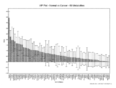
[0015] Figure 1 is a variable importance in the projection (VIP) plot of
analyzed
metabolites in order of their contribution to the separation between data from
urine samples
obtained from subjects having CRC and that from subjects without CRC and/or
colorectal
polyps for 69 metabolites;
[0016] Figure 2 is a VIP plot of analyzed metabolites in order of
their contribution to
the separation between data from urine samples obtained from subjects having
CRC and that
from subjects without CRC and/or colorectal polyps for 20 metabolites with a
VIP value
higher than 1;
[0017] Figure 3 is a 2-dimensional orthogonal partial least square
(OPLS) scatter plot
of the data from urine samples obtained from subjects without CRC and/or
colorectal polyps
(grey squares) compared to that from subjects having CRC (black dots)
constructed from
69 metabolites;
5
CA 02799757 2012-11-16
WO 2011/143779 PCT/CA2011/050315
[0018] Figure 4 is a 2-dimensional OPLS scatter plot of the data from
urine samples
obtained from subjects without CRC and/or colorectal polyps (grey squares)
compared to that
from subjects having CRC (black dots) constructed from 20 metabolites with a
VIP value
higher than 1;
[0019] Figure 5 is a 2-dimensional partial least square discriminant
analysis
(PLS-DA) scatter plot of the data from urine samples obtained from subjects
without CRC
and/or colorectal polyps (grey squares) compared to that from subjects having
CRC (black
dots) constructed from 69 metabolites;
[0020] Figure 6 is a 2-dimensional PLS-DA scatter plot of the data
from urine
samples obtained from subjects without CRC and/or colorectal polyps (grey
squares)
compared to that from subjects having CRC (black dots) constructed from 20
metabolites
with a VIP value higher than 1;
[0021] Figure 7 is an observed versus predicted plot of the OPLS model
of Figure 3.
Data from urine sample obtained from subjects without CRC and/or colorectal
polyps is
displayed as grey squares and that from subjects having CRC is displayed as
black dots;
[0022] Figure 8 is an observed versus predicted plot of the OPLS model
of Figure 4.
Data from urine sample obtained from subjects without CRC and/or colorectal
polyps is
displayed as grey squares and that from subjects having CRC is displayed as
black dots;
[0023] Figure 9 is a receiver operating characteristics (ROC) curve of
the OPLS
model of Figure 3;
[0024] Figure 10 is a ROC curve of the OPLS model of Figure 4;
[0025] Figure 11 is a VIP plot of analyzed metabolites in order of
their contribution to
the separation between the data from urine samples obtained from subjects
without CRC
and/or colorectal polyps and that from subjects having colorectal polyps for
69 metabolites;
[0026] Figure 12 is a VIP plot of analyzed metabolites in order of their
contribution to
the separation between the data from urine samples obtained from subjects
without CRC
and/or colorectal polyps and that from subjects having colorectal polyps for
26 metabolites
with a VIP value higher than 1;
6
CA 02799757 2012-11-16
WO 2011/143779 PCT/CA2011/050315
[0027] Figure 13 is a 2-dimensional OPLS plot of the data from urine
samples
obtained from subject without CRC and/or colorectal polyps (grey squares)
compared to that
from subjects having colorectal polyps (black diamonds) constructed from 69
metabolites;
[0028] Figure 14 is a 2-dimensional OPLS plot of the data from urine
samples
obtained from subject without CRC and/or colorectal polyps (grey squares)
compared to that
from subjects having colorectal polyps (black diamonds) constructed from 26
metabolites
with a VIP value higher than 1;
[0029] Figure 15 is a 2-dimensional PLS-DA scatter plot of the data
from urine
samples obtained from subjects without CRC and/or colorectal polyps (grey
squares)
compared to that from subject having colorectal polyps (black diamonds)
constructed from
69 metabolites;
[0030] Figure 16 is a 2-dimensional PLS-DA scatter plot the data from
urine samples
obtained from subjects without CRC and/or colorectal polyps (grey squares)
compared to that
from subject having colorectal polyps (black diamonds) constructed from 26
metabolites with
a VIP value higher than 1;
[0031] Figure 17 is an observed versus predicted plot of the OPLS
model of
Figure 13. Data from urine samples obtained from subjects without CRC and/or
colorectal
polyps are displayed as grey squares and that from subjects having colorectal
polyps are
displayed as black diamonds;
[0032] Figure 18 is an observed versus predicted plot of the OPLS model of
Figure 14. Data from urine samples obtained from subjects without CRC and/or
colorectal
polyps are displayed as grey squares and that from subjects having colorectal
polyps are
displayed as black diamonds;
[0033] Figure 19 is a ROC curve of the OPLS model of Figure 13;
[0034] Figure 20 is a ROC curve of the OPLS model of Figure 14;
[0035] Figure 21 is a VIP plot of analyzed metabolites in order of
their contribution to
the separation between the data from urine samples obtained from subjects
without CRC
and/or colorectal polyps and that from the group of subjects having
adenomatous polyps for
69 metabolites;
7
CA 02799757 2012-11-16
WO 2011/143779 PCT/CA2011/050315
[0036] Figure 22 is a VIP plot of analyzed metabolites in order of
their contribution to
the separation between the data from urine samples obtained from subjects
without CRC
and/or colorectal polyps and that from subjects having adenomatous polyps for
17 metabolites with a VIP value higher than 1;
[0037] Figure 23 is a 2-dimensional OPLS plot of the data from urine
samples
obtained from subjects without CRC and/or colorectal polyps (grey squares)
compared to that
from subjects having adenomatous polyps (black diamonds) constructed from
69 metabolites;
[0038] Figure 24 is an observed versus predicted plot of the OPLS
model of
Figure 23. Data from urine samples obtained from subjects without CRC and/or
colorectal
polyps are displayed as grey squares and that from subjects having adenomatous
polyps are
displayed as black diamonds;
[0039] Figure 25 is an observed versus predicted plot of the OPLS
model of the
2-dimensional OPLS plot with 17 metabolites with a VIP value higher than 1.
The
2-dimensional OPLS plot was prepared based on the data from urine samples
obtained from
subjects without CRC and/or colorectal polyps compared to that from subjects
having
adenomatous polyps. Data from urine samples obtained from subjects without CRC
and/or
colorectal polyps are displayed as grey squares and that from subjects having
adenomatous
polyps are displayed as black diamonds;
[0040] Figure 26 is a ROC curve of the OPLS model of Figure 23;
[0041] Figure 27 is a ROC curve of the OPLS model of the 2-dimensional
OPLS plot
with 17 metabolites with a VIP value higher than 1. The 2-dimensional OPLS
plot was
prepared based on the data from urine samples obtained from subjects without
CRC and/or
colorectal polyps compared to that from subjects having adenomatous polyps;
[0042] Figure 28 is a 2-dimensional OPLS plot based on the data from urine
samples
obtained from subjects without CRC and/or colorectal polyps (triangles)
compared to that
from subjects having adenomatous polyps (diamonds), superimposed with that
from subjects
having hyperplastic polyps (squares), constructed from 69 metabolites;
8
CA 02799757 2012-11-16
WO 2011/143779
PCT/CA2011/050315
[0043] Figure 29 is a diagram of the invention that provides a system
for assessing
whether a subject has or is predisposed to developing CRC and/or colorectal
polyps; and
[0044] Figure 30 is an overview of the 02PLS model relating two data
tables to each
other.
DETAILED DESCRIPTION
CRC and Colorectal Polyps
[0045] CRC is among the leading causes of morbidity. CRC is the third
most common
malignancy in the world, and represents approximately ten per cent of the
world's total
cancer incidence. CRC appears not only in humans but also in animal species,
and in both
sexes. Among human beings, more than 9 out of 10 people diagnosed with CRC are
over
the age of 50. However, younger individuals can develop CRC.
[0046] The chance of surviving CRC is closely related to the stage of
the disease at
diagnosis. The likelihood of survival is greater if the diagnosis is made
earlier, permitting
earlier treatment. Adenomatous and some other types of colorectal polyps may
progress to
malignant carcinomas and may thus be indicative that a subject is at risk of
developing CRC.
Thus, not only is it beneficial to be able to detect CRC itself, it is useful
to be able to detect
also the presence of precancerous lesions such as colorectal polyps.
[0047] There are a number of types of colorectal polyps. Adenomatous
polyps are
known to be a precursor to full-blown CRC. Other types of polyps may not
themselves have
malignant potential. Nevertheless, they may be useful indicators that a
subject is at risk of
developing CRC. For instance, unlike adenomatous polyps, hyperplastic polyps
have been
historically recognized as benign growths of the colon that have no malignant
potential i.e.
they were thought to be innocent bystanders. However, hyperplastic polyps have
been noted
to be more prevalent in populations with a higher incidence of cancer.
Moreover,
.. hyperplastic polyps may represent a heterogenous group of polyps, some of
which have
significant risk for malignant potential. These potentially malignant lesions
are known as
sessile serrated adenoma and have been linked to the microsatellite
instability cancer
pathway and thus are potential precursors of sporadic microsatellite unstable
CRC.
9
CA 02799757 2012-11-16
WO 2011/143779 PCT/CA2011/050315
[0048] Currently, the risk factors for CRC are not well understood and
few specific
risk factors other than diet have been established for the disease. As such,
CRC is typically
diagnosed from a complete subject history and physical examination, followed
by endoscopic
and/or radiological imaging. The diagnosis is confirmed with histopathological
examination of
biopsies or surgically removed specimens.
[0049] Current CRC screening methods consist of one or a combination
of the
followings: FOBT, flexible sigmoidoscopy, air-contrast barium enema, CTC and
colonoscopy.
These current screening methods all have limitations or potential risks that
limit their
application.
[0050] Colonoscopy is currently the standard test for assessing the
presence or
absence of CRC and/or colorectal polyps. However, colonoscopy is invasive and
can impose
unnecessary hazards and risks to an individual caused by sedation or the
procedure itself,
and complications with colonoscopy can include perforation, hemorrhage,
respiratory
depression, arrhythmias, and infection. In addition, it requires considerable
physical
resources and skilled personnel.
[0051] A known non-invasive CRC diagnostic method is FOBT. FOBT,
however, has
very low sensitivity in detection of CRC. FOBT is based on the assumption that
cancers will
bleed, therefore, can be detected in the stool using chemical or immunological
assays, and
involves a crude test for the peroxidase-like activity of heme in hemoglobin.
However, the
sensitivity of the test is only approximately 50%, with a 20% sensitivity for
adenomas, due to
the fact that not all adenomas and CRCs bleed. In addition, it is an
unattractive test for
subjects as the handling of fecal matter is required.
[0052] CTC, or virtual colonoscopy, is a recent non-invasive technique
for imaging
the colon. However, its performance varies due primarily to technological
differences in the
subject preparation and the hardware and software used for the analysis. Other
limitations of
CTC include high false positives (FP) readings, inability to detect flat
adenomas, no capacity
to remove polyps, repetitive and cumulative radiation doses, and cost.
[0053] With advances in the CRC related molecular pathology, several
new
screening methods based on DNA analysis from stool samples became available.
These are
typically PCR-based assays used to identify mutations known to occur in the
adenoma-to-
carcinoma sequence, or in familial CRC. Commonly screened gene mutations
include
CA 02799757 2012-11-16
WO 2011/143779 PCT/CA2011/050315
KRAS, TP53, APC, as well as assays for micro satellite instability and
hypermethylated DNA.
However, whether genomics-based tests will result in high diagnostic accuracy
for sporadic
CRC remains to be seen.
Metabolomics and Diagnosis of CRC or Colorectal Polyps
[0054] Metabolomics is an emerging field of research downstream from
genomics,
proteomics and transcriptomics. A metabolome is a quantitative collection of
low molecular
weight compounds, such as metabolic substrates and products, lipids, small
peptides,
vitamins, and other protein cofactors, generated by metabolism. A metabolome
is
downstream from a transcriptome and a proteome and thus any changes from a
normal state
are amplified and are numerically more tractable. Metabolomics can be a
precise, consistent,
and quantitative tool to examine and describe cellular growth, maintenance,
and function.
[0055] Metabolomics can be performed on urine, serum, tissue, and even
on saliva
and amniotic fluid. Generally, urine metabolomics represents a much less
invasive method
of testing compared to tissue or serum metabolomics.
[0056] The present invention uses urine metabolomics to identify subjects
having or
at risk of developing CRC and/or colorectal polyps. This is beneficial in the
management of
the risk of CRC and/or colorectal polyps, both in prevention and treatment.
The use of urine
metabolomics in the present invention has a number of potential benefits.
Obtaining a urine
sample and its analysis are relatively simple, non-invasive, and cost
efficient compared to the
existing methods for assessing presence or absence of CRC or colorectal
polyps. The
invention also permits monitoring of individual susceptibility to CRC prior to
resorting to, or in
combination with, conventional screening methods, and provides for population-
based
monitoring of CRC and/or colorectal polyps.
[0057] A wide range of analytical techniques to assay and quantitate
components of
a metabolome and to extract useful metabolite profiles from the data are
available, including
e.g. liquid and gas chromatography coupled with mass spectrometry (LCMS or
GCMS),
nuclear magnetic resonance (NMR) spectroscopy, high performance liquid
chromatography
(HPLC), thin layer chromatography (TLC), electrochemical analysis, refractive
index
spectroscopy, ultra-violet spectroscopy, fluorescent analysis, radiochemical
analysis,
near-infrared spectroscopy and light scattering analysis. The outputs from
such analytical
techniques can be further analyzed using multivariate analysis such as
principal component
11
CA 02799757 2012-11-16
WO 2011/143779 PCT/CA2011/050315
analysis (PCA), partial least squares discriminant analysis (PLS-DA) and
orthogonal partial
least squares (OPLS).
[0058] One or more metabolite profiles obtained from the previously
described
analysis based on a reference population of known CRC and/or colorectal polyp
status can
be used as a reference to assess the presence or absence of CRC or colorectal
polyps in a
subject. For example, a reference population may be composed of healthy
subjects
(i.e. subjects known or assessed by other means to be free of CRC and/or
colorectal polyps),
or alternatively may be composed of subjects already identified to have or to
be predisposed
to developing CRC or colorectal polyps. This assessment can be performed by:
(a) providing
a urine sample from a subject that is suspected to have or be predisposed to
developing
CRC and/or colorectal polyps; (b) obtaining a metabolite profile from said
urine sample;
(c) comparing said metabolite profile with a reference metabolite profile; and
(d) assessing,
based on said comparison in step (c), whether said subject has or is
predisposed to
developing CRC and/or colorectal polyps.
Providing and Processing Urine Samples
[0059] Urine samples can be collected from subjects that are known or
suspected to
have CRC or colorectal polyps, and from subjects without CRC or colorectal
polyps, by
known protocols. The subjects of this invention include both sexes of animal
species that are
susceptible to CRC and/or colorectal polyps, including humans.
[0060] In addition to providing a urine sample, subjects can take a FOBT,
fecal
immune testing (FIT), and/or a colonoscopy, the results of which can be used
to determine
classification of subjects into one of the groups of: subjects without CRC
and/or colorectal
polyps (normal group); subjects having colorectal polyps in general (polyp
group); or subjects
having adenomatous polyps specifically (adenomatous group). Pathology of
resected
surgical specimens can be used as the standard to classify subjects into a
group where
subjects have CRC (CRC group). Relevant clinical information such as age,
gender, family
history, comorbidities, medications etc. can be obtained from study
questionnaires and
subjects' medical charts, which could also be used to determine classification
of subjects.
Such testing can be used in the development of reference urine metabolite
profiles and can
also be used as an adjunct to screening test subjects by the methods of the
invention to
confirm or further refine a diagnosis of CRC and/or colorectal polyps.
12
CA 02799757 2012-11-16
WO 2011/143779
PCT/CA2011/050315
[0061] Urine samples can be collected from subjects any time, e.g.
during routine
screening or in connection with a regular check-up or visit to a physician, or
prior to or
together with administration of treatment, such as the administration of a
medicine or
performance of surgery. Urine samples can be collected one or more times for a
separate or
combined analysis, e.g. 15-700 ml each time. Urine sample collection
containers can vary in
size and shape, but ideally can accommodate e.g. 20-1,000 ml of urine sample.
Typically,
the container is sterile. If desired, sample containers can be pre-filled or
treated with agents
for preventing contamination of the sample by microorganisms such as bacteria
and fungi
while a sample is waiting to be stored, or such agents can be added after
sample collection.
Metabolomic analysis of the collected urine samples may occur immediately or
the samples
may be processed for storage and later analysis. For example, the whole or
part of the
sample could be stored in a freezer at -5-10 C within 0-48 hours of
collection, or could be
frozen at -120-- -10 C within 0-48 hours of collection, or could be processed
with chemicals
for future analysis or use before being stored. If samples have been stored
frozen, they may
be thawed (e.g. at room temperature for 12-48 hours), prior to analysis.
Obtaining a Metabolite Profile from the Sample
[0062] The analytical techniques that make it possible to obtain
metabolite profiles
from the urine samples can include one or a combination of, but not limited
to, mass
spectrometry (MS) coupled with gas chromatography (GCMS) or liquid
chromatography
(LCMS), HPLC, NMR spectroscopy, TLC, electrochemical analysis, refractive
index
spectroscopy, ultra-violet spectroscopy, fluorescent analysis, radiochemical
analysis,
near-infrared spectroscopy and light scattering analysis. The outputs obtained
from such
analyses can be further analyzed using multivariate statistical analysis to
aid in the
characterization of differences of metabolite profile between samples related
to CRC or
colorectal polyps. Such analytical tools include, but are not limited to,
principal component
analysis (PCA), partial least squares discriminant analysis (PLS-DA) and
orthogonal partial
least squares (OPLS). Though HPLC or technologies involving MS can be used for
measuring metabolite concentrations in the sub-molar range, they are often
laborious and
time consuming as they require that chromatography (liquid or gas) to separate
the
metabolites be done first, and also require multiple internal standards.
13
CA 02799757 2012-11-16
WO 2011/143779 PCT/CA2011/050315
[0063] NMR spectroscopy is an ideal tool for metabolonomics study
because it can
quantify a large number of metabolites simultaneously, requires only one
standard, and is
generally faster to yield statistical analysis results such as PCA and/or OPLS
plots.
[0064] In some embodiments, urine samples may be processed prior to
analysis. For
example, for non-automated (manual) NMR acquisition, about 100-1,000 [tL urine
sample
can be taken from the collected and/or stored sample, then diluted with an
internal standard
at a ratio of e.g. 1:1-1:20 (v/v). The internal standard can include e.g.1-20
mM of sodium
2,2-dimethy1-2-silapentane-5-sulfonate (DSS) or its salt form, 4,4-Dimethy1-4-
silapentane-1-
ammonium trifluoroacetate (DSA), or Trimethylsilyl propionate (TSP). Agents
for preventing
microbial contamination can also be added. Such additions can include e.g. 10-
200 mM
imidazole, or 0.1-0.5 % or 0.5-51.IM of sodium azide. The total volume can be
e.g.
100-1,300 4. The sample for NMR analysis can be stored in a freezer at e.g. 1-
6 C. The
same process applies to the automated (robotic) NMR acquisition. On the day of
NMR
acquisition, the pH of each sample is measured. Various concentrations of
acids and bases,
for example, but not limited to, HCI and NaOH, can be added to the samples to
achieve a pH
between e.g. 6.7 and 6.8 to minimize chemical exchange as the chemical shift
can change
with pH. An aliquot of e.g. 100-1,000 IA_ of the samples can be placed in NMR
tubes and
capped for the samples for both non-automated and the automated NMR.
[0065] One-dimensional NMR spectra can be acquired. After the spectra
are
obtained, the pH of each sample can be rechecked to ensure that the pH has not
shifted a
significant amount. This data can be recorded to be referenced if a particular
sample would
produce an unexpected spectrum. Samples can be frozen and stored again at a
sub-zero
temperature.
Identification of Metabolites for a Reference Metabolite Profile
[0066] The present invention involves the discovery that metabolite
profiles in the
urine of subjects having or predisposed to developing CRC and/or colorectal
polyps can be
reliably distinguished from metabolite profiles in the urine of healthy
subjects (i.e. those
without CRC and/or colorectal polyps) such that this distinction can be used
to assess
whether a particular subject has or is predisposed to developing CRC and/or
colorectal
polyps. One or more reference profiles concerning metabolites present in the
urine of a
reference population known either to be free of CRC and/or colorectal polyps
or to have or be
14
CA 02799757 2012-11-16
WO 2011/143779 PCT/CA2011/050315
predisposed to developing CRC or colorectal polyps is developed, which can
then be used
for comparison against a corresponding metabolite profile generated from the
urine of a test
subject. By analyzing the metabolite content of urine of subjects of known CRC
or colorectal
polyp status, it is then possible to compare this to the content of the same
metabolites in
subjects of a different CRC or colorectal polyp status, thus identifying
metabolites which
correlate significantly with the CRC or colorectal polyp status of an
individual. In the
illustrative examples herein, 240 metabolites were considered and 69 found to
be of
particular significance. However, urine contains thousands of metabolites, and
the
techniques described can be employed to assess whether other urine metabolites
are
similarly diagnostic of CRC and/or colorectal polyps.
[0067] Thus, in one aspect, the invention provides a method for
identifying urine
metabolites indicative of the presence or absence of CRC and/or colorectal
polyps, the
method comprising: (a) providing a urine sample from a subject; (b) obtaining
a metabolite
profile from the urine sample; (c) comparing the metabolite profile with a
reference metabolite
profile; and (d) identifying, based on the comparison in step (c), one or more
metabolites in
the metabolite profile that are indicative of the presence of or
predisposition to in said subject
of colorectal cancer and/or colorectal polyps.
[0068] Quantification of metabolites, e.g. by concentration or in
absolute amount, can
be done once the analysis data is available from, for example, but not limited
to, GCMS,
LCMS, HPLC, NMR spectroscopy, TLC, electrochemical analysis, refractive index
spectroscopy, ultra-violet spectroscopy, fluorescent analysis, radiochemical
analysis,
near-infrared spectroscopy and light scattering analysis. The quantification
data can be used
to identify and to set a standard to determine a reference metabolite profile
based on urine
samples obtained from subjects known to be free of CRC and/or colorectal
polyps.
[0069] For example, once the spectra are acquired from NMR spectroscopy,
quantification of metabolites can be done using tools that compare the
integral of a known
reference signal, such as DSS, DSA or TSP, with signals derived from a library
of
compounds to determine concentration relative to the reference signal. The
tools can include
softwares such as Chenomx NMRSuite v4.6 software. The quantification process
can be
done by more than one individual for reading and verification to optimize
accuracy.
CA 02799757 2012-11-16
WO 2011/143779 PCT/CA2011/050315
[0070] Levels of the specific metabolites over or below a determined
critical value,
either in concentration or in amount, can indicate the presence of CRC or
colorectal polyps in
general or adenomatous polyps in particular. The concentrations or the amount
of the
metabolites can be interpreted independently using an individual cut-off for
each metabolite
or they can be interpreted collectively. Metabolite concentrations or amounts
obtained can
be used as they are (i.e. as the raw data) or be normalized. For example, the
concentration
or amount of a metabolite can be log-transformed to normalize the
concentrations or
amounts to the concentration or the amount of other metabolites. The
metabolites can also
be normalized to the concentration of all metabolites minus the concentration
of selected
compounds such as e.g. urea to obtain similar results.
[0071] Those metabolites which are not products of normal metabolism
of a subject
(e.g. xenobiotics such as ibuprofen and salicylurate) or internal standards
(e.g. DSS) can be
excluded in the analysis.
[0072] Multivariate statistical analysis can be applied to the
collected data or complex
spectral data to identify differences arising between the groups of data sets
obtained from the
urine sample. The metabolite measurements in samples from subject having CRC
or
colorectal polyps in general or adenomatous polyps specifically can be
compared to
metabolite measurements in samples from subjects without CRC or colorectal
polyps to
identify metabolites that significantly contribute to the separation of
different groups. Data
comparison can be performed using any appropriate tools that fulfill the
purpose. The tools
include PCA, PLS-DA, OPLS and support vector machines (SVM), and softwares
that can
perform one or more of such analyses, e.g., Simca-P+, can be used. These are
statistical
methods of compressing multi-dimensional data down to two or three main
components.
PLS-DA and OPLS are supervised, that is, they take into account the class
assignments,
while RCA is unsupervised and can be influenced by many factors such as
gender,
comorbidities etc.
[0073] An optimized multivariate cut-off for the underlying
combination of metabolites
can be used to discriminate a cancerous or pre-cancerous state from a healthy
state. Upon
determination of which specific metabolites are the significant contributors
to the data
.. separation between the CRC group and the normal group samples or the polyp
group and
the normal group samples or the adenoma group and the normal group samples,
one or
more profiles of these specific metabolites can be established. One or more
metabolite
16
CA 02799757 2012-11-16
WO 2011/143779
PCT/CA2011/050315
profiles or its combination can be used as a reference metabolite profile to
assess CRC or
colorectal polyps in general or adenomatous polyps in particular in a subject.
[0074] In some embodiments, metabolites that were significant in
separating normal
group from CRC group, normal group from polyp group, and normal group from
adenoma
group were identified as: 1,6-Anhydro-I3-D-glucose; 1-Methylnicotinamide;
2-Hydroxyisobutyrate; 2-0xoglutarate; 3-Aminoisobutyrate; 3-Hydroxybutyrate;
3-Hydroxyisovalerate; 3-Hydroxymandelate; 3-Hydroxyphenylacetate; 3-
Indoxylsulfate;
4-Hydroxyphenylacetate; Acetate; Acetone; Adipate; Alanine; Ascorbate;
Asparagine;
Benzoate; Betaine; Butyrate; Carnitine; Citrate; Creatine; Creatinine;
Dimethylamine;
Ethanol; Formate; Galactose; Glucose; Glutamine; Glycerol; Glycine; Glycolate;
Guanidoacetate; Hippurate; Histidine; Hypoxanthine; Isoleucine; Lactate;
Leucine; Lysine;
Mannitol; Methanol; Methylguanidine; N,N-Dimethylglycine; O-Acetylcarnitine;
Pantothenate;
Propylene glycol; Pyroglutamate; Pyruvate; Serine; Succinate; Sucrose;
Tartrate; Taurine;
Threonine; Trigonelline; Trimethylamine; Trimethylamine N-oxide; Tyrosine;
Uracil; Urea;
Valine; Xylose; cis-Aconitate;13-Alanine; II-Methylhistidine; T-
Methylhistidine; and
trans-Aconitate.
[0075] However, not all features of the metabolite analysis results
are always
required for a proper diagnosis of CRC, colorectal polyps in general or
adenomatous polyps
specifically. Since there would be an incremental cost to obtaining more
information about a
subject's urine metabolite profile, it may be beneficial to use the minimal
number of
metabolites possible. In order to determine which specific metabolites are the
strongest
contributors to the data separation between the CRC group and the normal group
samples or
the polyp group and the normal group samples or the adenoma group and the
normal group
samples, further data analysis can be performed. This further data analysis
could be made
by an appropriate analytical method such as, but not limited to, a VIP plot.
[0076] The VIP plot allows identification of metabolites that have a
greater impact on
driving the separation between groups in models. Each metabolite used to
construct models
can be assigned a VIP score. This score is assigned through a statistical
formula that is
used to calculate the influence of each metabolite. The higher the VIP score,
the greater the
influence of the metabolite with the score on separating different groups. The
VIP plot also
allows for the comparison of the influence of one metabolite to another's. In
VIP plot
analysis, factors with a large VIP, usually greater than 1, are said to be the
most relevant.
17
CA 02799757 2012-11-16
WO 2011/143779 PCT/CA2011/050315
Metabolites with a VIP value higher than 1 can be the strongest contributors,
and all or part of
them can constitute a reference metabolite profile once its capability of
assessing CRC or
colorectal polyps is successfully demonstrated thorough a comparison with the
reference
metabolite profile consisting of all the metabolites found significant in the
separation of
different groups.
[0077] There are many ways to evaluate a selected metabolite profile
to assess
whether a subject has or is predisposed to developing CRC and/or colorectal
polyps. The
values measured for metabolites can be mathematically combined and the
combined value
can be correlated to the underlying diagnostic question. Metabolite values may
be combined
by any appropriate mathematical method. Mathematical methods for correlating a
metabolite
combination to a disease can employ methods such as, but not limited to,
discriminant
analysis (DA) (i.e. linear-, quadratic-, regularized-DA), Kernel Methods (i.e.
SVM),
Nonparametric Methods (i.e. k-Nearest-Neighbor Classifiers), PLS (Partial
Least Squares),
Tree-Based Methods (i.e. Logic Regression, CART, Random Forest Methods,
Boosting/Bagging Methods), Generalized Linear Models (i.e. Logistic
Regression), Principal
Components based Methods (i.e. SIMCA), Generalized Additive Models, Fuzzy
Logic based
Methods, Neural Networks and Genetic Algorithms based Methods. For the SVM
model, the
linear coefficients of each feature in an SVM classifier can be used to select
the most
important features. Those features that had the largest absolute value can be
selected, and
the SVM model can be re-calculated using only the selected features and the
training set if
necessary.
[0078] When comparing test results from two different populations, for
example, one
with a disease and the other without the disease, a perfect separation between
the two
groups is rarely observed. Indeed, the distribution of the test results will
overlap. Therefore,
when a cut-off point or criterion value to discriminate between the two
populations is selected
and applied, there will be some cases with the disease correctly classified as
positive (True
Positive fraction), but some cases with the disease will be classified
negative (False Negative
fraction). On the other hand, some cases without the disease will be correctly
classified as
negative (True Negative fraction), but some cases without the disease will be
classified as
positive (False Positive fraction).
18
CA 02799757 2012-11-16
WO 2011/143779 PCT/CA2011/050315
[0079] The diagnostic performance of such a test, or the accuracy of a
test to
discriminate diseased groups from healthy groups, can be evaluated using tools
such as
ROC curve analysis. The ROC curve is a graphical representation of the
spectrum of
sensitivities and specificities generated using the various cut-offs, using
the sensitivity as the
y-axis and 1-specificity as the x-axis. In an ROC curve the true positive rate
(Sensitivity) is
plotted in function of the FP rate (100-Specificity) for different cut-off
points. Each point on
the ROC curve represents a sensitivity/specificity pair corresponding to a
particular decision
threshold. A test with perfect discrimination (no overlap in the two
distributions) has a ROC
curve that passes through the upper left corner (100% sensitivity, 100%
specificity).
Therefore, qualitatively, the closer the plot is to the upper left corner, the
higher the overall
accuracy of the test. Area under the ROC curve (AUC) reflects the accuracy of
the test and
is displayed on the left lower corner of the plot. An AUC of 0.9 to 1
represents an excellent
diagnostic test whereas an AUC of 0.8-0.9 represents a good test and an AUC of
0.7 to 0.8
represents a fair test.
Development of Reference Metabolite Profiles
[0080] Generally, the more metabolites that are assessed, the more
accurate will be
the assessment of CRC and/or colorectal polyps. In exemplary embodiments, more
than
240 metabolites were considered, and 69 metabolites were used to assess
whether a subject
has or is predisposed to developing CRC or colorectal polyps. Indeed, other,
or additional
urine metabolites beyond these metabolites identified can be included in the
metabolite
profile. However, as noted above, this involves greater effort and cost. In
many instances, a
less accurate, specific, or detailed assessment may be sufficient,
particularly if the
assessment is only preliminary in nature, or is to be conducted together with
or followed by
another diagnostic test, such as colonoscopy. Further, a test involving the
assessment of
fewer metabolites may be more readily reduced to a simplified kit or test that
can be used by
a subject at home, or by a medical practitioner at the point of care, without
need for sending a
urine sample to a laboratory for analysis.
[0081] As explained above, VIP values greater than 1 are considered to
reflect
metabolites with the greatest potential for discriminating between healthy and
diseased
subjects. For the assessment of CRC per se, as distinct from colorectal polyps
of any kind,
as detailed in Table 1, the following metabolites, have been shown to exhibit
VIP values
greater than 1.0, presented from highest to lowest VIP value: adipate; 3-
hydroxybutyrate;
19
CA 02799757 2012-11-16
WO 2011/143779 PCT/CA2011/050315
creatine; guanidoacetate; dimethylamine; hypoxanthine; benzoate; 0-
acetylcarnitine;
pyruvate; methanol; lactate; creatinine; xylose; 3-indoxylsulfate;
trigonelline; taurine;
threonine; p-methylhistidine; glucose; and 4-hydroxyphenylacetate.
[0082] In an embodiment, the reference metabolic profile is directed
to assessing
whether a subject has or is predisposed to developing CRC, and includes
measurements of
concentrations in a urine sample of at least any 1, 2, 3, 4, 5, 6, 7, 8, 9,
10, 11, 12, 13, 14, 15,
16 ,17, 18, 19 or 20 metabolites selected from the group consisting of:
adipate;
3-hydroxybutyrate; creatine; guanidoacetate; dimethylamine; hypoxanthine;
benzoate;
0-acetylcarnitine; pyruvate; methanol; lactate; creatinine; xylose; 3-
indoxylsulfate;
trigonelline; taurine; threonine; p-methylhistidine; glucose; and 4-
hydroxyphenylacetate.
[0083] Generally, if fewer than all 20 of these metabolites are to be
used in the
reference metabolite profile, preference will be given to those with the
highest VIP values. As
described in Table 2, a profile containing only the top five metabolites was
demonstrated to
have acceptable sensitivity and specificity, and fewer may be used to develop
an acceptable
profile. Thus, in various embodiments, the reference profile for detecting CRC
includes one
or more metabolites in a set of metabolites selected from the group consisting
of:
(i) adipate; 3-hydroxybutyrate; creatine; guanidoacetate; dimethylamine;
hypoxanthine; benzoate; 0-acetylcarnitine; pyruvate; methanol; lactate;
creatinine; xylose;
3-indoxylsulfate; trigonelline; taurine; threonine; p-methylhistidine;
glucose; and
4-hydroxyphenylacetate;
(ii) adipate; 3-hydroxybutyrate; creatine; guanidoacetate; dimethylamine;
hypoxanthine; benzoate; 0-acetylcarnitine; pyruvate; methanol; lactate;
creatinine; xylose;
3-indoxylsulfate; trigonelline; taurine; threonine; p-methylhistidine; and
glucose;
(iii) adipate; 3-hydroxybutyrate; creatine; guanidoacetate; dimethylamine;
hypoxanthine; benzoate; 0-acetylcarnitine; pyruvate; methanol; lactate;
creatinine; xylose;
3-indoxylsulfate; trigonelline; taurine; threonine; and p-methylhistidine;
(iv) adipate; 3-hydroxybutyrate; creatine; guanidoacetate; dimethylamine;
hypoxanthine; benzoate; 0-acetylcarnitine; pyruvate; methanol; lactate;
creatinine; xylose;
3-indoxylsulfate; trigonelline; taurine; and threonine
CA 02799757 2012-11-16
WO 2011/143779
PCT/CA2011/050315
(v) adipate; 3-hydroxybutyrate; creatine; guanidoacetate; dimethylamine;
hypoxanthine; benzoate; 0-acetylcarnitine; pyruvate; methanol; lactate;
creatinine; xylose;
3-indoxylsulfate; trigonelline; and taurine;
(vi) adipate; 3-hydroxybutyrate; creatine; guanidoacetate; dimethylamine;
hypoxanthine; benzoate; 0-acetylcarnitine; pyruvate; methanol; lactate;
creatinine; xylose;
3-indoxylsulfate; and trigonelline;
(vii) adipate; 3-hydroxybutyrate; creatine; guanidoacetate; dimethylamine;
hypoxanthine; benzoate; 0-acetylcarnitine; pyruvate; methanol; lactate;
creatinine; xylose;
and 3-indoxylsulfate;
(viii) adipate; 3-hydroxybutyrate; creatine; guanidoacetate; dimethylamine;
hypoxanthine; benzoate; 0-acetylcarnitine; pyruvate; methanol; lactate;
creatinine; and
xylose;
(vix) adipate; 3-hydroxybutyrate; creatine; guanidoacetate;
dimethylamine;
hypoxanthine; benzoate; 0-acetylcarnitine; pyruvate; methanol; lactate; and
creatinine;
(x) adipate; 3-hydroxybutyrate; creatine; guanidoacetate; dimethylamine;
hypoxanthine; benzoate; 0-acetylcarnitine; pyruvate; methanol; and lactate;
(xi) adipate; 3-hydroxybutyrate; creatine; guanidoacetate; dimethylamine;
hypoxanthine; benzoate; 0-acetylcarnitine; pyruvate; and methanol;
(xii) adipate; 3-hydroxybutyrate; creatine; guanidoacetate; dimethylamine;
hypoxanthine; benzoate; 0-acetylcarnitine; and pyruvate;
(xiii) adipate; 3-hydroxybutyrate; creatine; guanidoacetate; dimethylamine;
hypoxanthine; benzoate; and 0-acetylcarnitine;
(xiv) adipate; 3-hydroxybutyrate; creatine; guanidoacetate; dimethylamine;
hypoxanthine; and benzoate;
(xv) adipate; 3-hydroxybutyrate; creatine; guanidoacetate; dimethylamine;
and
hypoxanthine;
(xvi) adipate; 3-hydroxybutyrate; creatine; guanidoacetate; and
dimethylamine;
21
CA 02799757 2012-11-16
WO 2011/143779
PCT/CA2011/050315
(xvii) adipate; 3-hydroxybutyrate; creatine; and guanidoacetate;
(xviii) adipate; 3-hydroxybutyrate; and creatine;
(xix) adipate and 3-hydroxybutyrate; and
(xx) adipate.
[0084] In some embodiments of the invention, it is the concentration (e.g.
measured
in of
the urine metabolites that is measured, and a higher or lower concentration of
the
metabolite in the urine of a test subject relative to that in reference
metabolite profile (based
either on raw or normalized concentrations) is indicative of CRC.
[0085] In some embodiments, an elevated concentration of any one or
more
metabolites selected from the group consisting of adipate; 3-hydroxybutyrate;
creatine;
guanidoacetate; dimethylamine; benzoate; 0-acetylcarnitine; lactate; xylose; 3-
indoxylsulfate;
trigonelline; taurine; threonine; p-methylhistidine and 4-hydroxyphenylacetate
is indicative
that the subject has or is predisposed to developing CRC.
[0086] It will be understood that by "elevated" it is meant that the
concentration of a
metabolite in the urine of a subject that has or is predisposed to developing
CRC is higher
than in the urine of subjects that do not have or are not predisposed to CRC.
For instance,
referring to Table 1, it will be seen that the mean concentration of adipate
in the urine of
individuals with CRC was 218.1 jiM, much higher than the mean concentration of
adipate in
the urine of "normal" subjects without CRC, which was found to be 1.3 0. Thus,
on a
comparative basis relative to healthy subjects, subjects with CRC had elevated
adipate
concentrations in their urine.
[0087] In some embodiments, a reduced concentration of any one or more
metabolites selected from the group consisting of hypoxanthine; pyruvate;
methanol;
creatinine and glucose is indicative that the subject has or is predisposed to
developing CRC.
[0088] It will be understood that by "reduced" it is meant that the
concentration of a
metabolite in the urine of a subject that has or is predisposed to developing
CRC is lower
than in the urine of subjects that do not have or are not predisposed to CRC.
For instance,
referring to Table 1, it will be seen that the mean concentration of
hypoxanthine in the urine
22
CA 02799757 2012-11-16
WO 2011/143779
PCT/CA2011/050315
of subjects with CRC was 188.4 uM, lower than the mean concentration of
hypoxanthine in
the urine of "normal" subjects without CRC, which was found to be 208.4 0.
Thus, on a
comparative basis relative to healthy subjects, subjects with CRC had reduced
hypoxanthine
concentrations in their urine.
[0089] A reference metabolite profile that is diagnostic of colorectal
polyps may be
different than a reference metabolite profile for CRC per se. That is, the
reference diagnostic
profile may be made up of a different set of relevant metabolites, and
different relative
concentrations of these metabolites may be relevant.
[0090] In certain embodiments, the reference metabolite profile is for
adenomatous
polyps and includes concentrations of at least any 1, 2, 3, 4, 5, 6, 7, 8, 9,
10, 11, 12, 13, 14,
15, 16 or 17 metabolites selected from the group consisting of: butyrate;
serine; methanol;
13-alanine; p-methylhistidine; 3-hydroxybutyrate; asparagine; trigonelline;
3-hydroxyphenylacetate; histidine; acetone; 2-oxoglutarate; ethanol; adipate;
3-hydroxymandelate; tyrosine and benzoate.
[0091] As above, these are metabolites found to have VIP scores of 1.0 or
above and
are listed in descending order in Table 5. As above, acceptable specificity
and sensitivity
was demonstrated with a profile based on only the top five metabolites (Table
6) and fewer
may be used. Thus, if fewer than all of the metabolites are included in the
reference
metabolite profile, the profile may include one or more metabolites in a set
of metabolites
selected from the group consisting of:
(i) butyrate; serine; methanol; 13-alanine; p-methylhistidine; 3-
hydroxybutyrate;
asparagine; trigonelline; 3-hydroxyphenylacetate; histidine; acetone; 2-
oxoglutarate; ethanol;
adipate; 3-hydroxymandelate; tyrosine and benzoate;
(ii) butyrate; serine; methanol; 13-alanine; p-methylhistidine; 3-
hydroxybutyrate;
asparagine; trigonelline; 3-hydroxyphenylacetate; histidine; acetone; 2-
oxoglutarate; ethanol;
adipate; 3-hydroxymandelate and tyrosine;
(iii) butyrate; serine; methanol; 13-alanine; p-methylhistidine; 3-
hydroxybutyrate;
asparagine; trigonelline; 3-hydroxyphenylacetate; histidine; acetone; 2-
oxoglutarate; ethanol;
adipate and 3-hydroxymandelate;
23
CA 02799757 2012-11-16
WO 2011/143779
PCT/CA2011/050315
(iv) butyrate; serine; methanol; p-alanine; p-methylhistidine; 3-
hydroxybutyrate;
asparagine; trigonelline; 3-hydroxyphenylacetate; histidine; acetone; 2-
oxoglutarate; ethanol
and adipate;
(v) butyrate; serine; methanol; 13-alanine; p-methylhistidine; 3-
hydroxybutyrate;
asparagine; trigonelline; 3-hydroxyphenylacetate; histidine; acetone; 2-
oxoglutarateand
ethanol;
(vi) butyrate; serine; methanol; 13-alanine; p-methylhistidine; 3-
hydroxybutyrate;
asparagine; trigonelline; 3-hydroxyphenylacetate; histidine; acetoneand 2-
oxoglutarate;
(vii) butyrate; serine; methanol; p-alanine; p-methylhistidine; 3-
hydroxybutyrate;
asparagine; trigonelline; 3-hydroxyphenylacetate; histidine; and acetone;
(viii) butyrate; serine; methanol; P-alanine; p-methylhistidine; 3-
hydroxybutyrate;
asparagine; trigonelline; 3-hydroxyphenylacetateand histidine;
(ix) butyrate; serine; methanol; P-alanine; p-methylhistidine; 3-
hydroxybutyrate;
asparagine; trigonelline and 3-hydroxyphenylacetate;
(x) butyrate; serine; methanol; p-alanine; p-methylhistidine; 3-
hydroxybutyrate;
asparagine and trigonelline;
(xi) butyrate; serine; methanol; p-alanine; p-methylhistidine; 3-
hydroxybutyrate
and asparagine;
(xii) butyrate; serine; methanol; p-alanine; p-methylhistidine; and
3-hydroxybutyrate;
(xiii) butyrate; serine; methanol; P-alanine and p-methylhistidine;
(xiv) butyrate; serine; methanol and p-alanine;
(xv) butyrate; serine and methanol;
(xvi) butyrate and serine; and
(xvii) butyrate.
24
CA 02799757 2012-11-16
WO 2011/143779 PCT/CA2011/050315
[0092] In some embodiments, an elevated concentration of any one or
more
metabolites selected from the group consisting of p-methylhistidine; 3-
hydroxybutyrate;
asparagine; trigonelline; 3-hydroxyphenylacetate; histidine; acetone; adipate;
3-hydroxymandelate; tyrosine and benzoate is indicative that the subject has
or is
predisposed to developing adenomatous polyps. As above, "elevated" is relative
to a
corresponding urine metabolite concentration of healthy subjects.
[0093] In some embodiments, a reduced concentration of any one or more
metabolites selected from the group consisting of butyrate; serine; methanol;
P-alanine;
2-oxoglutarate and ethanol is indicative that the subject has or is
predisposed to developing
adenomatous polyps. As above, "reduced" is relative to a corresponding urine
metabolite
concentration of healthy subjects.
[0094] Elevated and reduced urine metabolite concentrations for
subjects having
adenomatous polyps are shown in Table 5.
[0095] In some embodiments, the reference metabolite profile is
designed to identify
subjects having or predisposed to colorectal polyps, but not necessarily to
distinguish one
type of polyp from another. For instance, the polyp may be adenomatous or
hyperplastic, but
the reference diagnostic profile does not necessarily distinguish between the
two.
[0096] In certain embodiments, the reference metabolite profile is for
colorectal
polyps that are either adenomatous polyps or hyperplastic polyps and includes
urine
concentrations of at least any 1, 2, 3, 4, 5, 6, 7, 8, 9, 10, 11, 12, 13, 14,
15, 16, 17, 18, 19,
20, 21, 22, 23, 24, 25 or 26 metabolites selected from the group consisting
of: butyrate;
serine; asparagine; p-methylhistidine; 3-hydroxybutyrate; methanol; 3-
hydroxymandelate;
tyrosine; trigonelline; 13-alanine; histidine; dimethylamine; urea; 1,6-
anhydro-3-D-glucose;
glucose; ethanol; benzoate; acetone; threonine; 2-hydroxyisobutyrate;
creatinine;
3-hydroxyphenylacetate; 3-indoxylsulfate; hippurate; ascorbate; and 4-
hydroxyphenylacetate.
[0097] As above, these are metabolites found to have VIP scores of 1.0
or above and
are listed in descending order (Table 3). As above, acceptable specificity and
sensitivity was
demonstrated with a profile based on only the top five metabolites (Table 4)
and fewer may
be used. Thus, if fewer than all of the metabolites are included in the
reference metabolite
profile, the profile may include one or more metabolites in a set of
metabolites selected from
the group consisting of:
CA 02799757 2012-11-16
WO 2011/143779
PCT/CA2011/050315
(i) butyrate; serine; asparagine; p-methylhistidine; 3-hydroxybutyrate;
methanol;
3-hydroxymandelate; tyrosine; trigonelline;13-alanine; histidine;
dimethylamine; urea;
1,6-anhydro-13-D-glucose; glucose; ethanol; benzoate; acetone; threonine;
2-hydroxyisobutyrate; creatinine; 3-hydroxyphenylacetate; 3-indoxylsulfate;
hippurate;
ascorbate; and 4-hydroxyphenylacetate;
(ii) butyrate; serine; asparagine; p-methylhistidine; 3-hydroxybutyrate;
methanol;
3-hydroxymandelate; tyrosine; trigonelline;13-alanine; histidine;
dimethylamine; urea;
1,6-anhydro-I3-D-glucose; glucose; ethanol; benzoate; acetone; threonine;
2-hydroxyisobutyrate; creatinine; 3-hydroxyphenylacetate; 3-indoxylsulfate;
hippurate and
ascorbate;
(iii) butyrate; serine; asparagine; p-methylhistidine; 3-hydroxybutyrate;
methanol;
3-hydroxymandelate; tyrosine; trigonelline; 13-alanine; histidine;
dimethylamine; urea;
1,6-anhydro-13-D-glucose; glucose; ethanol; benzoate; acetone; threonine;
2-hydroxyisobutyrate; creatinine; 3-hydroxyphenylacetate; 3-indoxylsulfate and
hippurate;
(iv) butyrate;
serine; asparagine; p-methylhistidine; 3-hydroxybutyrate; methanol;
3-hydroxymandelate; tyrosine; trigonelline; P-alanine; histidine;
dimethylamine; urea;
1,6-anhydro-13-D-glucose; glucose; ethanol; benzoate; acetone; threonine;
2-hydroxyisobutyrate; creatinine; 3-hydroxyphenylacetate and 3-indoxylsulfate;
(v) butyrate; serine; asparagine; p-methylhistidine; 3-hydroxybutyrate;
methanol;
3-hydroxymandelate; tyrosine; trigonelline; 13-alanine; histidine;
dimethylamine; urea;
1,6-anhydro-3-D-glucose; glucose; ethanol; benzoate; acetone; threonine;
2-hydroxyisobutyrate; creatinine and 3-hydroxyphenylacetate;
(vi) butyrate; serine; asparagine; p-methylhistidine; 3-hydroxybutyrate;
methanol;
3-hydroxymandelate; tyrosine; trigonelline; 13-alanine; histidine;
dimethylamine; urea;
1,6-anhydro-13-D-glucose; glucose; ethanol; benzoate; acetone; threonine;
2-hydroxyisobutyrate and creatinine;
(vii) butyrate; serine; asparagine; p-methylhistidine; 3-hydroxybutyrate;
methanol;
3-hydroxymandelate; tyrosine; trigonelline; 13-alanine; histidine;
dimethylamine; urea;
1,6-anhydro-3-D-glucose; glucose; ethanol; benzoate; acetone; threonine and
2-hydroxyisobutyrate;
26
CA 02799757 2012-11-16
WO 2011/143779
PCT/CA2011/050315
(viii)
butyrate; serine; asparagine; p-methylhistidine; 3-hydroxybutyrate; methanol;
3-hydroxymandelate; tyrosine; trigonelline; 13-alanine; histidine;
dimethylamine; urea;
1 ,6-anhydro-I3-D-glucose; glucose; ethanol; benzoate; acetone and threonine;
(vix)
butyrate; serine; asparagine; p-methylhistidine; 3-hydroxybutyrate; methanol;
3-hydroxymandelate; tyrosine; trigonelline; P-alanine; histidine;
dimethylamine; urea;
1 ,6-anhydro-I3-D-glucose; glucose; ethanol; benzoate and acetone;
(x)
butyrate; serine; asparagine; p-methylhistidine; 3-hydroxybutyrate; methanol;
3-hydroxymandelate; tyrosine; trigonelline; 13-alanine; histidine;
dimethylamine; urea;
1 ,6-anhydro-13-D-glucose; glucose; ethanol andbenzoate;
(xi) butyrate; serine; asparagine; p-methylhistidine; 3-hydroxybutyrate;
methanol;
3-hydroxymandelate; tyrosine; trigonelline; 13-alanine; histidine;
dimethylamine; urea;
1 ,6-anhydro-13-D-glucose; glucose and ethanol;
(xii) butyrate; serine; asparagine; p-methylhistidine; 3-hydroxybutyrate;
methanol;
3-hydroxymandelate; tyrosine; trigonelline; 13-alanine; histidine;
dimethylamine; urea;
1 ,6-anhydro-3-D-glucoseand glucose;
(xiii) butyrate; serine; asparagine; p-methylhistidine; 3-hydroxybutyrate;
methanol;
3-hydroxymandelate; tyrosine; trigonelline; 13-alanine; histidine;
dimethylamine; urea and
1 ,6-anhydro-3-D-glucose;
(xiv) butyrate; serine; asparagine; p-methylhistidine; 3-hydroxybutyrate;
methanol;
3-hydroxymandelate; tyrosine; trigonelline; 13-alanine; histidine;
dimethylamine and urea;
(xv) butyrate; serine; asparagine; p-methylhistidine; 3-hydroxybutyrate;
methanol;
3-hydroxymandelate; tyrosine; trigonelline; 13-alanine; histidine and
dimethylamine;
(xvi) butyrate; serine; asparagine; p-methylhistidine; 3-hydroxybutyrate;
methanol;
3-hydroxymandelate; tyrosine; trigonelline; 13-alanine and histidine;
(xvii) butyrate; serine; asparagine; p-methylhistidine; 3-hydroxybutyrate;
methanol;
3-hydroxymandelate; tyrosine; trigonelline and 13-alanine;
27
CA 02799757 2012-11-16
WO 2011/143779 PCT/CA2011/050315
(xviii) butyrate; serine; asparagine; p-methylhistidine; 3-hydroxybutyrate;
methanol;
3-hydroxymandelate; tyrosine and trigonelline;
(xix) butyrate; serine; asparagine; p-methylhistidine; 3-hydroxybutyrate;
methanol;
3-hydroxymandelateand tyrosine;
(xx) butyrate; serine; asparagine; p-methylhistidine; 3-hydroxybutyrate;
methanol
and 3-hydroxymandelate;
(xxi) butyrate; serine; asparagine; p-methylhistidine; 3-hydroxybutyrate
and methanol;
(xxii) butyrate; serine; asparagine; p-methylhistidine and 3-
hydroxybutyrate;
(xxiii) butyrate; serine; asparagine and p-methylhistidine;
(xxiv) butyrate; serine and asparagine;
(xxv) butyrate and serine; and
(xxvi) butyrate.
[0098] In some embodiments, an elevated concentration of any one or
more
metabolites selected from the group consisting of asparagine; p-
methylhistidine;
3-hydroxybutyrate; 3-hydroxymandelate; tyrosine; trigonelline; histidine;
dimethylamine; urea;
1,6-anhydro-P-D glucose; glucose; benzoate; acetone; threonine; 2-
hydroxyisobutyrate;
creatinine; 3-hydroxyphenylacetate; 3-indoxylsulfate; hippurate; and 4-
hydroxyphenylacetate
is indicative that the subject has or is predisposed to developing colorectal
polyps which are
either adenomatous polyps or hyperplastic polyps. As above, "elevated" is
relative to a
.. corresponding urine metabolite concentration of healthy individuals.
[0099] In some embodiments, a reduced concentration of any one or more
metabolites selected from the group consisting of butyrate; serine; methanol;
p-alanine;
ethanol and ascorbate is indicative that the subject has or is predisposed to
developing
colorectal polyps which are either adenomatous polyps or hyperplastic polyps.
As above,
"reduced" is relative to a corresponding urine metabolite concentration of
healthy subjects.
[0100] Elevated and reduced urine metabolite concentrations for
subjects having
polyps that are either adenomatous or hyperplastic are shown in Table 3.
28
CA 02799757 2012-11-16
WO 2011/143779 PCT/CA2011/050315
Assessing whether a subject has or is predisposed to developing CRC and/or
colorectal polyps
[0101] The invention provides methods for assessing whether a subject
has or is
predisposed to developing CRC and/or colorectal polyps, the method comprising:
(a) providing a urine sample from said subject; (b) obtaining a metabolite
profile from said
urine sample; (c) comparing said metabolite profile with a reference
metabolite profile; and
(d) assessing, based on said comparison in step (c), whether said subject has
or is
predisposed to developing CRC and/or colorectal polyps.
[0102] Urine samples can be obtained as described above. The
metabolite profile
from the subject contains the corresponding information concerning the
subject's urine
sample as contained in the selected reference metabolite profile, as described
above.
Comparison of the metabolite profile from the subject to the reference
metabolite profile
allows for assessment of whether the subject has or is predisposed to
developing CRC
and/or colorectal polyps.
[0103] Merely by way of an illustrative example, the method might be a
method for
assessing whether a subject has or is predisposed to developing CRC. A urine
sample could
be taken and concentrations of the following metabolites measured: adipate;
3-hydroxybutyrate; creatine; guanidoacetate; dimethylamine; hypoxanthine;
benzoate;
0-acetylcarnitine; pyruvate; methanol; lactate; creatinine; xylose; 3-
indoxylsulfate; trigonelline;
taurine; threonine; p-methylhistidine; glucose; and 4-hydroxyphenylacetate.
The concentration
of each of these metabolites in the subject's urine is then compared to the
concentrations of
the corresponding metabolites in the reference metabolite profile. Detection
of a higher
concentration of any one or more of adipate, 3-hydroxybutyrate, creatine,
guanidoacetate,
dimethylamine, benzoate, 0-acetylcarnitine, lactate, xylose, 3-indoxylsulfate,
trigonelline,
taurine, threonine, p-methylhistidine and 4-hydroxyphenylacetate in the
subject's metabolite
profile than in the reference metabolite profile may indicate that the subject
has or is
predisposed to developing CRC. Similarly, a lower concentration of any one or
more of
hypoxanthine, pyruvate, methanol, creatinine, and glucose in the subject's
metabolite profile
than in the reference metabolite profile may indicate that the subject has or
is predisposed to
developing CRC.
29
CA 02799757 2012-11-16
WO 2011/143779
PCT/CA2011/050315
Diagnostic kits
[0104] The invention also provides kits for assessing whether a
subject has or is
predisposed to developing CRC and/or colorectal polyps. Such kits may comprise
one or
more reagents for detecting the presence and/or concentration of one or more
metabolites in
a urine sample of a subject, and may include instructions for use of the kit
for assessing
whether a subject has or is predisposed to developing CRC and/or colorectal
polyps.
[0105] The most reliable results are likely obtained when urine
samples are
processed, e.g. by NMR spectroscopy, in a laboratory setting. For instance, a
urine sample
might be obtained from a subject in the office of a medical practitioner and
then sent to a
hospital or commercial medical laboratory for further testing. However, in
many instances, it
may be desirable to provide immediate results in a clinician's office or to
permit a subject to
conduct testing at home. The need for a test that is portable, pre-packaged,
disposable,
usable by a subject without assistance or direction, etc. may in some
instances be of more
importance than a high degree of accuracy. In many instances, particularly
where there will
be follow-up with a medical practitioner, a preliminary test, even one with
reduced sensitivity
and/or specificity may be sufficient. Thus, an assay presented in kit form may
involve
detection and measurement of a relatively small number of metabolites, to
reduce the
complexity and cost of the assay.
[0106] Any form of urine assay capable of detecting urine metabolites
as described
herein may be used. Typically, the assay will quantitate the urine metabolites
to some extent
e.g. whether they are higher or lower in concentration or in amount than a
predetermined
threshold value. Such kits may take the form of a test strip, dip stick,
cassette, cartridge,
chip-based or bead-based array, multi-well plate, or series of containers, or
the like. One or
more reagents are provided to detect the presence and/or concentration and/or
amount of
selected urine metabolites. The subject's urine may be dispensed directly onto
the assay or
indirectly from a stored sample. The presence or absence of a metabolite above
or below a
pre-determined threshold may be displayed e.g. by a chromogenic, fluorogenic,
electrochemiluminescent or other output, e.g. as in an enzyme immunoassay
(EIA) such as
an enzyme-linked immunoassay (ELISA).
CA 02799757 2012-11-16
WO 2011/143779 PCT/CA2011/050315
[0107] In an embodiment, a kit may comprise a solid substrate, such as
e.g. a chip,
slide, array, etc., with reagents capable of detecting and/or quantitating one
or more urine
metabolites immobilized at predetermined locations on the substrate. By way of
an
illustrative example, a chip can be provided with reagents immobilized at
discrete,
predetermined locations for detecting and quantitating in a urine sample the
concentration of
adipate; 3-hydroxybutyrate; creatine; guanidoacetate and dimethylamine. As
discussed
above, elevated levels of these metabolites were found in the urine of
subjects with CRC.
The chip may be configured such that a detectable output (e.g. colour change)
is provided
only if the concentration of one or more of these metabolites is over a
threshold value, the
.. threshold value being selected to distinguish between a metabolite
concentration indicative of
healthy subjects and those having or predisposed to developing CRC. Thus, the
presence of
a detectable output such as a colour change provides an immediate indication
that the urine
sample contains significantly elevated levels of one or more relevant urine
metabolites,
indicating that the subject has or is predisposed to developing CRC.
Systems for Performing the Assessment of CRC or Colorectal Polyps
[0108] In an embodiment, the invention provides a system for assessing
whether a
subject has or is predisposed to developing CRC and/or colorectal polyps. As
shown in
Figure 29, such a system may comprise:
(a) a CRC- and/or colorectal polyps-assessing apparatus including a control
unit
and a memory unit to assess a CRC state in a subject; and
(b) an information communication terminal apparatus that provides data on
the
presence and/or concentration and/or amount of metabolites in a urine sample
from the
subject connected to each other communicatively,
wherein the information communication terminal apparatus includes:
(a) a data sending unit that transmits the data on the presence and/or
concentration and/or amount of metabolites in the sample to the CRC- and/or
colorectal
polyps-assessing apparatus; and
31
CA 02799757 2012-11-16
WO 2011/143779 PCT/CA2011/050315
(b) an assessment result-receiving unit that receives the
assessment result of the
CRC and/or colorectal polyps state of the subject transmitted from the CRC-
and/or colorectal
polyps-assessing apparatus,
wherein the control unit of the CRC- and/or colorectal polyps-assessing
apparatus includes:
(a) a data-receiving unit that receives the data on the metabolite
concentration
and/or amount of the sample transmitted from the information communication
terminal
apparatus;
(b) a discriminant value-calculating unit that calculates a discriminant
value that is
a value of multivariate discriminant, based on both the concentration and/or
amount value of
.. the metabolite in the sample received by the data-receiving unit and a
multivariate
discriminant with the concentration and/or amount of the metabolite as
explanatory variable
stored in the memory unit;
(c) a discriminant value criterion-assessing unit that assesses the CRC or
colorectal polyps state in the subject, based on the discriminant value
calculated by the
discriminant value-calculating unit; and
(d) an assessment result-sending unit that transmits the assessment result
of the
subject obtained by the discriminant value criterion-assessing unit to the
information
communication terminal apparatus.
Evaluation of Efficacy of Pharmaceutical Agents and/or Physical Treatments
and/or
Surgical Treatment
[0109] Metabolomic analysis is ideal for identification of and
evaluation of the effects
of potential pharmaceutical agents and/or new physical and/or surgical
treatments against
CRC, colorectal polyps and/or adenomatous polyps. Urine samples can be taken
one or
more times, by methods described previously herein, from a subject before and
after
treatment. The treatment can include administration of one or more
pharmaceutical agents
at one or more doses, and/or carrying out one or more physical and/or surgical
treatments, to
or on a subject. The administration of pharmaceutical agents can be made in
many different
ways including, but not limited to, injection, oral administration, patch or
ointment application.
32
CA 02799757 2012-11-16
WO 2011/143779 PCT/CA2011/050315
[0110] The metabolite profiles obtained from the samples can be
compared with each
other and/or with the metabolite profile from subjects without CRC and/or
colorectal polyps.
The comparison can indicate the efficacy of the pharmaceutical agents and/or
the physical
treatment and/or surgical treatment through changes of the metabolite profile
in urine
samples of the subject. Also, comorbidities and medications of a subject can
be studied in
subsequent analyses to determine their effects on the metabolomic test results
and
specifically whether they contribute to discordant results. In addition, the
metabolite profiles
of the CRC samples can be correlated with operative and histological findings
to determine
whether CRC location or stage can change a metabolite profile.
[0111] This invention is further illustrated by the following non-limiting
examples.
Example 1. Assessment of CRC Group versus Normal Group
[0112] Subjects for the normal group were recruited from a population
based study
of 1,200 asymptomatic subjects who were supposed to be exposed to an average
or high risk
of CRC, based on family history of colorectal cancer or personal history of
colorectal polyps.
Subjects for the CRC group were all newly diagnosed with CRC.
[0113] Four hundred forty four subjects without CRC and/or colorectal
polyps were
selected and classified as the normal group. Seventy seven CRC subjects were
classified as
the CRC group. Clinical information was obtained from study questionnaires,
and subjects
completed a medical questionnaire, had a FOBT, FIT, and a colonoscopy for
determination of
classification.
[0114] Urine samples were collected from subjects of the two groups.
The urine
samples were frozen at -80 C within 24 hours of collection. Urine sample
collection
containers were pre-filled with sodium azide powder to stop any bacterial
growth in the urine
while it is waiting to be frozen at -80 C.
[0115] Urine samples were thawed at room temperature in the biohood 24
hours prior
to NMR acquisition. For the non-automated (manual) NMR acquisition, 5854 of
each
sample was diluted with 654 of internal standard consisting of 5 mM sodium 2,2-
dimethy1-2-
silapentane-5-sulfonate (DSS), 100 mM imidazole and 0.2% sodium azide in 99%
D20
(Chenomx Inc., Edmonton, AB) to achieve a total volume of 6504 and stored at 4
C. For
33
CA 02799757 2012-11-16
WO 2011/143779 PCT/CA2011/050315
the automated (robotic) NMR acquisition, 6754 of each sample was diluted with
754 of the
same Chenomx internal standard to achieve a total volume of 7504 and stored at
4 C. On
the day of NMR acquisition, the pH of each sample was measured. Various
concentrations
of HCI and NaOH were added to the samples to achieve a pH between 6.7 and 6.8
to
minimize chemical exchange as the chemical shift would change with pH. For the
samples
for the non-automated NMR, an aliquot of 6004 of the samples was placed in 5
mm NMR
tubes and capped; for the samples for the automated NMR, 7004 was used.
[0116] Manual/Non-automated Mode: One-dimensional NMR spectra were
acquired
using an Oxford 600Hz NMR spectrometer with a Varian VNMRS two channel console
and
running VNMRJ software version 2.20 on a RHEL 4 host computer in the Canadian
National
High Field NMR Centre (NANUC). Samples (600 [tL) were set to a depth of 66 mm
in the
depth gage and then inserted into the spectrometer. All samples were run at a
sweep width
(sw) of 7225.43 Hz. The saturation frequency (sfrq), transmitter offset (tof)
and pulse width
(pw) were all individually calibrated at the start of each set of sample runs.
The tof ranged
from (-213 to -215 Hz) and the pw ranged from 6 to 8 microseconds. Shims were
optimized
until an acceptable line width value was obtained at relative peaks heights
of: 50% (< 1.0 Hz),
0.55% (< 12.0 Hz), and 0.11% (< 20.0 Hz). During post-processing of the
sample, zero filling
was used to increase the actual acquired data points to the next largest
factor of 2. No
weighting functions were applied. The first increment of a 2D-1H,1H-NOESY
pulse sequence
was utilized for the acquisition of 1H-NMR (Hydrogen-1 nuclear magnetic
resonance)data and
for suppressing the solvent signal. Experiments contained a 100 ms mixing time
along with
a 990 ms pre-saturation time (-80 Hz gammaB1). Spectra were collected at 25 C
through a
total of 32 scans over a period of 3.5 minutes; a total recycle delay of 5
seconds was also
used.
[0117] Automated Mode: Automated runs followed exactly the same
experimental
parameters used in the manual mode with the exception of i) use of 700
sample and ii) an
additional 30s of equilibration time in the NMR to allow the sample to
equilibrate to 25 C. All
sample handling was done with a Varian 768 AS sample handling robot. The first
sample of
the batch was manually shimmed to satisfactory line width values and
subsequent samples
were automatically shimmed. Any spectra that did not meet acceptable line
height values
were discarded and the sample was re-run.
34
CA 02799757 2012-11-16
WO 2011/143779
PCT/CA2011/050315
[0118] After the spectra were obtained, samples from both manual and
automated
mode were removed from NMR tubes with glass Pasteur pipettes and transferred
into
eppendorf tubes. The pH of each sample was then rechecked to ensure that the
pH had not
shifted a significant amount. Samples were re-stored in the -80 C freezer.
[0119] Once the spectra were acquired, quantification of metabolites was
done using
Chenomx NMRSuite v4.6 software (Chenomx, Inc. Edmonton, Canada), which
compared the
integral of a known reference signal (in this case DSS) with signals derived
from a library of
compounds to determine concentration relative to the reference signal. The
quantification
was done by one individual and verified by a second individual to optimize
accuracy.
[0120] Over 240 metabolites were considered, and 72 were found to be
significant,
that is, the spectral peaks of 72 metabolites in the compound library were
identified in the
spectra of the samples: 1,6-Anhydro-13-D-glucose, 1-Methylnicotinamide,
2-Hydroxyisobutyrate, 2-0xoglutarate, 3-Aminoisobutyrate, 3-Hydroxybutyrate,
3-Hydroxyisovalerate, 3-Hydroxymandelate, 3-Hydroxyphenylacetate, 3-
Indoxylsulfate,
4-Hydroxyphenylacetate, Acetate, Acetone, Adipate, Alanine, Ascorbate,
Asparagine,
Benzoate, Betaine, Butyrate, Carnitine, Citrate, Creatine, Creatinin, DSS
(Chemical Shape
Indicator), Dimethylamine, Ethanol, Formate, Galactose, Glucose, Glutamine,
Glycerol,
Glycine, Glycolate, Guanidoacetate, Hippurate, Histidine, Hypoxanthine,
Ibuprofen,
Isoleucine, Lactate, Leucine, Lysine, Mannitol, Methanol, Methylguanidine,
N,N-Dimethylglycine, 0-Acetylcamitine, Pantothenate, Propylene glycol,
Pyroglutamate,
Pyruvate Salicylurate, Serine, Succinate, Sucrose, Tartrate, Taurine,
Threonine,
Trigonelline, Trimethylamine, Trimethylamine N-oxide, Tyrosine, Uracil, Urea,
Valine, Xylose,
cis-Aconitate, trans-Aconitate, f3-Alanine, II-Methylhistidine, T-
Methylhistidine.
[0121] Metabolite concentrations were log transformed to normalize the
concentrations. Those metabolites that were not products of normal human
metabolism, i.e.
xenobiotics, such as ibuprofen and salicylurate, were excluded. The internal
standard DSS
was also excluded in the analysis, and 69 metabolites were obtained as a
reference
metabolite profile.
[0122] The metabolite measurements in samples from the CRC group were
compared to metabolite measurements in samples from the normal group. Simca-P+
v12Ø1
software (Umetrics, Umea, Sweden) was used to perform the multivariate
statistical analyses
CA 02799757 2012-11-16
WO 2011/143779 PCT/CA2011/050315
to identify differences arising between the groups of data sets. These
analyses included
PLS-DA, and OPLS.
[0123] Further data analysis was preformed in order to determine which
specific
metabolites were the strongest contributors to the data separation between the
CRC group
and the normal group samples by a VIP plot. The metabolites identified with a
VIP score of
greater than 1 were Adipate, 3-Hydroxybutyrate, Creatine, Guanidoacetate,
Dimethylamine,
Hypoxanthine, Benzoate, 0-Acetylcarnitine, Pyruvate, Methanol, Lactate,
Creatinine, Xylose,
3-Indoxylsulfate, Trigonelline, Taurine, Threonine, IT-Methylhistidine,
Glucose,
4-Hydroxyphenylacetate. The result is summarized in Table 1 together with the
list of
69 metabolites.
[0124] The following assessments were performed with two different
metabolite
profiles, one with all the 69 metabolites found to be significant for the
separation of the CRC
group and the normal group, and the other with 20 metabolites with a VIP value
higher than 1.
[0125] The VIP plots were generated using SIMCA-P+ to illustrate which
metabolites
contribute the most to the separation between the normal and CRC groups
(Figures 1 and 2).
[0126] Using two-component separation, the OPLS scatter plots shown in
Figures 3
and 4, implemented in SIMCA-P+12, illustrated the normal group as grey squares
and the
CRC group as black dots. Notwithstanding a degree of overlap, the two groups
generally
appeared on the different (right and left) sides of the plot.
[0127] The 2-dimensional scatter plots of the PLS model were shown in
Figures 5
and 6. In each plot, the normal group were in grey squares and the CRC group
were in black
dots. A similar separation to the OPLS scatter plots of the normal group and
the CRC group
could be seen. Even though there was an overlap between the CRC group and the
normal
group, the two groups appear on the different (top and bottom) sides of each
plot.
[0128] To generate sensitivity and specificity data, the observed versus
predicted
data plots were generated for the OPLS models (Figures 7 and 8) and arbitrary
cut-off points
for the predicted value (YPred) were chosen where the two groups overlapped
(Figures 7
and 8). The grey squares, indicating the normal group, to the left of the cut-
off were TN and
those that are to the right of the cut-off were the FP. T he black dots,
indicating CRC group,
36
CA 02799757 2012-11-16
WO 2011/143779 PCT/CA2011/050315
to the left of the cut-off were false negatives (FN), while those to the right
were the true
positives (TP). Sensitivity and specificity data were summarized in Table 2.
[0129] In Table 2, the model column indicated which metabolites were
used to
construct the model. The term "main model" referred to the model containing
all
69 metabolites. The cut-off column corresponded to certain cut-off points on
the ROC curve.
Sensitivity and Specificity are measures of how accurate and precise the test
is. The ROC
Curve is a measure of how robust the models are. R2Y and Q2 are measures of
the quality of
the models constructed; which means, the higher the numbers the better the
model. A
negative number means that the model is unusable.
[0130] From Table 2, it could be seen that with a cut-off point ranging
from
0.114184 - 0.302331, results in a sensitivity range of 87.18-25.64% and
specificity range of
54.03-98.10% would be achieved when using the metabolites with a VIP score
greater
than 1. Similarly, different cut-off points can be used for numerous subsets
of the
metabolites, which can also be observed in Table 2 with the different subsets
of metabolites.
.. For example, when using the top 15 metabolites and a cut-off range from
0.120717-0.326168
a sensitivity range of 79.49-20.51% and a specificity range of 34.12-99.53%
can be achieved.
[0131] With the data from Table 2, the ROC curves of sensitivity
versus 1-specificity
were plotted (Figures 9 and 10) using STATA10.0 (College Station, Texas). The
ROC curves
in the Figures had AUG scores of 0.9178 and 0.8465, respectively. This result
showed that
the metabolite profile consisting of 20 metabolites out of 69, with higher VIP
value than 1, can
also be used to assess whether a subject has or is predisposed to developing
CRC, though
the metabolite profile consisting of 69 metabolites might provide more
accurate assessment.
Table 2 also demonstrates that even five metabolites with highest VIP values
could be used
to assess whether a subject has or is predisposed to developing CRC.
Example 2. Assessment of Polyp Group versus Normal Group
[0132] Subjects for the normal group and the polyp group were
recruited from a
population based study of 1,200 asymptomatic subjects who were supposed to be
exposed
to an average or high risk of CRC, based on family history of CRC or personal
history of
colorectal polyps. All subjects completed a medical questionnaire, had a FOBT,
FIT, and a
colonoscopy to determine classification of the subjects. One subject in the
polyp group was
found to be with CRC, and excluded from the test.
37
CA 02799757 2012-11-16
WO 2011/143779 PCT/CA2011/050315
[0133] Four hundred forty four subjects without CRC and/or colorectal
polyps were
selected and classified as the normal group. The polyp group consisted of two
hundred thirty
six with tubular, tubulovillous, villous adenomas and hyperplastic polyps.
[0134] The process as described previously in "Assessment of Cancer
Group versus
Normal Group" was followed for urine sample collection, treatment of the
sample, NMR
acquisition, and analysis of the data obtained.
[0135] Over 240 metabolites were considered, and 72 were found to be
significant,
that is, the spectral peaks of 72 metabolites in the compound library were
identified in the
spectra of the study samples: 1,6-Anhydro-13-D-glucose, 1-Methylnicotinamide,
2-Hydroxyisobutyrate, 2-0xoglutarate, 3-Aminoisobutyrate, 3-Hydroxybutyrate,
3-Hydroxyisovalerate, 3-Hydroxymandelate, 3-Hydroxyphenylacetate, 3-
Indoxylsulfate,
4-Hydroxyphenylacetate, Acetate, Acetone, Adipate, Alanine, Ascorbate,
Asparagine,
Benzoate, Betaine, Butyrate, Carnitine, Citrate, Creatine, Creatinin, DSS
(Chemical Shape
Indicator), Dimethylamine, Ethanol, Formate, Galactose, Glucose, Glutamine,
Glycerol,
Glycine, Glycolate, Guanidoacetate, Hippurate, Histidine, Hypoxanthine,
Ibuprofen,
Isoleucine, Lactate, Leucine, Lysine, Mannitol, Methanol, Methylguanidine,
N,N-Dimethylglycine, 0-Acetylcarnitine, Pantothenate, Propylene glycol,
Pyroglutamate,
Pyruvate Salicylurate, Serine, Succinate, Sucrose, Tartrate, Taurine,
Threonine,
Trigonelline, Trimethylamine, Trimethylamine N-oxide, Tyrosine, Uracil, Urea,
Valine, Xylose,
cis-Aconitate, trans-Aconitate,I3-Alanine, ft-Methylhistidine, T-
Methylhistidine.
[0136] Metabolite concentrations were log transformed to normalize the
concentrations. Those metabolites that were not products of normal human
metabolism, i.e.
xenobiotics, such as ibuprofen and salicylurate, were excluded. The internal
standard DSS
was also excluded in the analysis, and 69 metabolites were obtained as a
reference
metabolite profile.
[0137] The metabolites identified with a VIP score of greater than 1
are Butyrate; Serine;
Asparagine; p-Methylhistidine; 3-Hydroxybutyrate; Methanol; 3-
Hydroxymandelate; Tyrosine;
Trigonelline; 13-Alanine; Histidine; Dimethylamine; Urea; 1,6-Anhydro-13-D-
glucose; Glucose;
Ethanol; Benzoate; Acetone; Threonine; 2-Hydroxyisobutyrate; Creatinine;
.. 3-Hydroxyphenylacetate; 3-Indoxylsulfate; Hippurate; Ascorbate; and 4-
Hydroxyphenylacetate.
The result was summarized in Table 3 together with the list of 69 metabolites.
38
CA 02799757 2012-11-16
WO 2011/143779 PCT/CA2011/050315
[0138] The following analysis was performed with two different
metabolite profiles,
one with all the 69 metabolites found to be significant for the separation of
the polyp group
and the normal group, and the other with 26 metabolites with a VIP value
higher than 1.
[0139] The VIP plots were generated to illustrate which metabolites
contribute the
most to the separation between the normal and polyp groups (Figures 11 and
12). The
resulting OPLS (Figures 13 and 14), PLS-DA 2-dimensional scatter plots
(Figures 15
and 16), observed verses predicted plots (Figures 17 and 18), ROC curves
(Figures 19
and 20) and sensitivity & specificity data (Table 4) were produced.
[0140] In the OPLS scatter plot, the normal group was in grey squares
and the polyp
group was in black diamonds. Figures 13 and 14 showed, even though there was
an overlap
between the two groups, that the polyp group clustered together and the normal
group also
clustered together, and they appeared on the different (right and left) sides
of each plot.
[0141] In the PLS-DA scatter plot, the normal group was in grey
squares and the
polyp group was in black diamonds. Figures 15 and 16 showed, even though there
was an
overlap between the two groups, similarly to the OPLS scatter plot, the polyp
group clustered
together on the top of the plot and the normal group clustered together on the
bottom.
[0142] From Table 4, a sensitivity range of 94-57% and specificity
range of 40-78%
would be achieved with a cut-off range of 0.25-0.45. In the setting of a
screening test, a low
FN rate is more important than a low FP rate, hence higher sensitivity could
be achieved at
the expense of a lower specificity. In this case, a cut-off of 0.3 could be
used to achieve a
sensitivity of 88% and a specificity of 51%. In contrast, preliminary analysis
of fifty-two
subjects of the normal and the polyp group samples that showed FOBT had a
sensitivity
of 9% and specificity of 100%.
[0143] The ROC curves in Figures 19 and 20 had the AUC scores of
0.7673
and 0.7015, respectively. This result showed that the metabolite profile
consisting of
26 metabolites out of 69, with higher VIP value than 1, can also be used to
assess whether a
subject has colorectal polyps, though the metabolite profile consisting of 69
metabolites might
provide more accurate assessment. Table 4 also demonstrates that even five
metabolites
with highest VIP values could be used to assess whether a subject has or is
predisposed to
developing colorectal polyps.
39
CA 02799757 2012-11-16
WO 2011/143779
PCT/CA2011/050315
Example 3. Assessment of Adenoma Polyp Group versus Normal Group
[0144] Subjects for the normal group and the adenoma group were
recruited from a
population based study of 1,200 asymptomatic subjects who were supposed to be
exposed
to an average or high risk of CRC, based on family history of CRC or personal
history of
colorectal polyps.
[0145] Four hundred forty four healthy subjects without CRC and/or
colorectal polyps
were selected and classified as the normal group. The adenoma group consisted
of one
hundred sixty two subjects with adenomatous polyp. Clinical information was
obtained from
study questionnaires, and subjects completed a medical questionnaire, had a
FOBT, FIT,
and a colonoscopy for determination of classification.
[0146] The process as described previously in "Assessment of Cancer
Group versus
Normal Group" was followed for urine sample collection, treatment of the
sample, NMR
acquisition, and analysis of the data obtained.
[0147] Over 240 metabolites were considered, and 72 were found to be
significant,
that is, the spectral peaks of 72 metabolites in the compound library were
identified in the
spectra of the study samples: 1,6-Anhydro-p-D-glucose, 1-Methylnicotinamide,
2-Hydroxyisobutyrate, 2-0xoglutarate, 3-Aminoisobutyrate, 3-Hydroxybutyrate,
3-Hydroxyisovalerate, 3-Hydroxymandelate, 3-Hydroxyphenylacetate, 3-
Indoxylsulfate,
4-Hydroxyphenylacetate, Acetate, Acetone, Adipate, Alanine, Ascorbate,
Asparagine,
Benzoate, Betaine, Butyrate, Carnitine, Citrate, Creatine, Creatinin, DSS
(Chemical Shape
Indicator), Dimethylamine, Ethanol, Formate, Galactose, Glucose, Glutamine,
Glycerol,
Glycine, Glycolate, Guanidoacetate, Hippurate, Histidine, Hypoxanthine,
Ibuprofen,
Isoleucine, Lactate, Leucine, Lysine, Mannitol, Methanol, Methylguanidine,
N,N-Dimethylglycine, O-Acetylcarnitine, Pantothenate, Propylene glycol,
Pyroglutamate,
Pyruvate Salicylurate, Serine, Succinate, Sucrose, Tartrate, Taurine,
Threonine,
Trigonelline, Trimethylamine, Trimethylamine N-oxide, Tyrosine, Uracil, Urea,
Valine, Xylose,
cis-Aconitate, trans-Aconitate,I3-Alanine, II-Methylhistidine, T-
Methylhistidine.
[0148] Metabolite concentrations were log transformed to normalize the
concentrations. Those metabolites that were not products of normal human
metabolism, i.e.
xenobiotics, such as ibuprofen and salicylurate, were excluded. The internal
standard DSS
CA 02799757 2012-11-16
WO 2011/143779
PCT/CA2011/050315
was also excluded in the analysis, and 69 metabolites were obtained as a
reference
metabolite profile.
[0149] The metabolites identified with a VIP score of greater than one
are Butyrate,
Serine, Asparagine,11-Methylhistidine, 3-Hydroxybutyrate, Methanol, 3-
Hydroxymandelate,
Tyrosine, Trigonelline,13-Alanine, Histidine, Dimethylamine, Urea, 1-6-Anhydro-
I3-D-glucose,
Glucose, Ethanol, Benzoate, Acetone, Threonine, 2-Hydroxyisobutyrate,
Creatinine,
3-Hydroxyphenylacetate, 3-Indoxylsulfate, hippu rate, Ascorbate, 4-
Hydroxyphenylacetate.
The result was summarized in Table 5 together with the list of 69 metabolites.
[0150] The following analysis was performed with two different
metabolite profiles,
one with all the 69 important metabolites, and the other with 17 metabolites
with a VIP value
higher than 1.
[0151] The VIP plots were generated to illustrate which metabolites
contribute the
most to the separation between the normal and adenoma groups (Figures 21 and
22). The
resulting OPLS (Figures 23), PLS scatter plots (not shown), observed vs.
predicted plots
(Figures 24 and 25), ROC curve (Figures 26 and 27), and sensitivity &
specificity data
(Table 6) were produced.
[0152] In the OPLS scatter plot, the normal group is in grey squares
and the polyp
group is in black diamonds. Figures 23 shows, even though there is an overlap
between the
two groups, that the polyp group clusters together and the normal group also
clusters
together, and they appear on the different (right and left) sides of the plot.
The OPLS scatter
plot for 17 metabolites with a VIP value higher than 1 is not shown.
[0153] In the PLS-DA scatter plot, the normal group is in grey squares
and the polyp
group is in black diamonds. Even though there is an overlap between the two
groups,
similarly to the OPLS scatter plot, the polyp group clusters together on the
top of the plot and
the normal group clusters together on the bottom.
[0154] From Table 6, a sensitivity range of 65.55-5.88% and
specificity range
of 50.71-98.58% would be achieved with a cut-off range of 0.329791-0.577397
for the
metabolites with a VIP score greater than 1. In the setting of a screening
test, a low FN rate
is more important than a low FP rate, hence higher sensitivity could be
achieved at the
expense of a lower specificity. In this case, a cut-off of 0.329791 could be
used to achieve a
41
CA 02799757 2012-11-16
WO 2011/143779 PCT/CA2011/050315
sensitivity of 65.55% and a specificity of 50.71%. In contrast, preliminary
analysis of fifty-two
normal and colorectal polyps samples that showed FOBT had a sensitivity of 9%
and
specificity of 100%.
[0155] The ROC curves in Figures 26 and 27 had the AUC scores of
0.7524
and 0.6937, respectively. This result showed that the metabolite profile
consisting of
17 metabolites out of 69, with higher VIP value than 1, can also be used to
assess whether a
subject has adenomatous polyps, though the metabolite profile consisting of 69
metabolites
might provide more accurate assessment. Table 6 also demonstrates that even
five
metabolites with highest VIP values could be used to assess whether a subject
has or is
.. predisposed to developing adenomatous polyps.
Example 4. Assessment of Adenoma Polyp Group versus Hyperplastic Polyp Group
[0156] A total of 110 urine samples from subjects with hyperplastic
polyps were
introduced blindly to the Normal versus Adenoma model discussed in Example 3.
The
analysis result showed that the metabolite profile of hyperplastic polyps was
more alike with
the adenomatous group's than the normal group's (Figure 29). This was further
confirmed by
an attempt to establish an OPLS model between hyperplastic polyps and
adenomatous
polyps. A meaningful model to separate the two groups could not be constructed
(R2Y = 0.126, Q2 = -0.0771).
[0157] This result suggests that some of the hyperplastic polyps might
be
pre-cancerous like the adenomatous polyps and thus display a precancerous
metabolomic
fingerprint.
Example 5. Analytical Methods and their Application
Analytical Methods
[0158] PLS (Conventional): Conventional PLS applies to the two-block
(X/Y)
regression problem. It uses X to construct a model of Y, where the objective
is to predict the
latter from the former for new samples in the prediction set. In that sense,
PLS is
unidirectional, i.e., X4Y, but not vice versa.
42
CA 02799757 2012-11-16
WO 2011/143779 PCT/CA2011/050315
[0159] When X is composed of e.g. spectroscopic data, process readings
or
measurements from bioanalytical platforms, there is a risk that systematic
variation may
reside in X which is not linearly correlated with Y. Such variability in X is
usually called
Y-orthogonal variation. Although Y-orthogonal variation in X does not affect
the predictive
power of a PLS model, it may negatively affect model interpretation. The score-
loading
correspondence is perturbed by the presence of Y-orthogonal variation in X.
[0160] OPLS: The OPLS method is a recent modification of the PLS
method, which is
designed to handle variation in X that is orthogonal to Y. It is an extension
to the supervised
PLS regression method with an integrated Orthogonal signal correction (OSC)
filter, which
removes the uncorrelated signals resulting in information of the within-class
variation. OPLS
separates the systematic variation in X into two parts, one that is linearly
related (and
therefore predictive) to Y and one that is orthogonal to Y. The predictive
variation of Y in X is
modeled by the predictive components. The variation in X which is orthogonal
to Y is
modeled by the orthogonal components. This partitioning of the X-data provides
improved
model transparency and interpretability, but does not change the predictive
power. Similarly
to PLS, OPLS is a unidirectional method, where the scope is the relation X4Y.
[0161] OPLS Scatter Plot: The scatter plot is of the OPLS model.
[0162] 02PLS: 02PLS is a generalization of OPLS. In contrast to PLS
and OPLS,
02PLS is bidirectional, i.e. X4Y, and therefore X can be used to predict Y,
and Y can be
used to predict X. Additionally, with 02PLS it is possible to partition the
systematic variability
in X and Y into three parts, (i) the X/Y joint predictive variation, (ii) the
Y-orthogonal variation
in X, and (iii) the X-unrelated variation in Y.
[0163] Figure 30 is an overview of the 02PLS model relating two data
tables to each
other. The Y-orthogonal variation in X (left-hand side of the Figure)
represents the variation of
the observations in X that is varying orthogonally to the corresponding
structure in Y. This
variation is unique to X. The X/Y joint predictive variation (middle part of
the Figure) describes
the predictive variation between X and Y, the information overlap. The X-
unrelated variation in
Y (right-hand side of the Figure) corresponds to the variation of the
observations in Y that is
varying orthogonally to the corresponding structure in X. This variation is
unique to Y.
43
CA 02799757 2012-11-16
WO 2011/143779 PCT/CA2011/050315
[0164] The ability to interpret the X/Y joint predictive variation
separated from the
non-correlated variation implies that the model interpretation is refined and
simplified.
Furthermore, it should be noted that for the single-y case the OPLS and 02PLS
methods are
identical. For such a model there can only be one predictive component
expressing the joint
X/Y predictive variation.
[0165] PLS-DA Scatter Plot: This scatter plot is of the partial least
squares
discriminant analysis (PLS-DA) model. Conventional PLS is used where a
quantitative
relationship exists between two data tables X & Y; it uses X to construct a
model of Y, where
the objective is to predict Y from the X for new samples in the prediction
set. It is another
statistical method used to compress multidimensional and complex data sets
into a more
manageable dataset.
[0166] Observed vs. Predicted plot: The observed vs. predicted plot
displays the
observed values vs. the fitted or predicted values for the selected response.
The observed
vs. predicted plot is a scatter plot of the Y variables (which are normal vs.
cancer, adenoma,
or polyps) verses the predicted values. The observed vs. predicted plot
provides with
Y predicted values, as assigned by the model, for each sample along with their
observed
(normal or cancer/adenoma/polyp) value. Then, these values are taken to
generate a ROC
curve. The observed vs. predicted plot also allows to determine the true
positives, false
positives, true negatives, and false negatives, to calculate sensitivity and
specificity with a
range of cut-offs, and to view the overlap present between two groups.
[0167] ROC Curve: The receiver operating characteristic (ROC) Curve is
a graphical
representation of the spectrum of sensitivities and specificities generated
using the various
cut-offs, using the sensitivity as the y-axis and 1-specificity as the x-axis.
Area under the
ROC curve (AUC) reflects the accuracy of the test and is displayed on the left
lower corner of
the plot. An AUC of 0.9 to 1 represents an excellent diagnostic test whereas
an AUC of
0.8-0.9 represents a good test and an AUC of 0.7 to 0.8 represents a fair
test.
[0168] VIP Plot: Variable Importance in the Projection (VIP) plot
allows to identify
which metabolites have a greater impact on driving the separation between
groups in
models. Each metabolite used to construct models is assigned a VIP score; this
score is
assigned through a statistical formula that is used to calculate the influence
of each model.
The higher the VIP score, the greater the influence of the metabolite with the
score on
44
CA 02799757 2012-11-16
WO 2011/143779
PCT/CA2011/050315
shaping the model. VIP also allows for the comparison of one metabolite to
another. Terms
with a large VIP (greater then 1) are said to be the most relevant for
explaining 'Y'.
Analytical Methods Used
[0169] OPLS and PLS-DA Scatter Plot: The orthogonal PLS named 02PLS
has been
implemented in SIMCA-P+ 12. 02PLS is bidirectional, i.e. X Y, and therefore
X can be
used to predict Y, and Y can be used to predict X. For the single-y case the
OPLS and
02PLS methods are identical.
[0170] The 02PLS model can be written as:
(1) X = TpP'p + T0F0 + E (for model of X)
(2) Y = Upap + U0a0+ F (for model of Y)
where a linear relationship exists between Tp and Up. Here, the score vectors
in Tp and To are mutually orthogonal. The number of components in the
respective set of
components is determined using cross validation.
[0171] For any part of the OPLS/02PLS model, the percentages explained
and
predicted variances can be obtained from plots and lists in the software. The
vectors listed in
Table 7 are unique for OPLS/02PLS. These vectors in addition to the ones
listed for PLS
are computed for each component.
[0172] ROC Curve (Receiver Operating Characteristic): ROC curves were
generated
using STATA 10.0 (College Station, Texas), along with the ROC curves a
complete
sensitivity and specificity report was also generated. The ROC curve is a
fundamental tool
for diagnostic test evaluation. In a ROC curve the true positive rate
(Sensitivity) is plotted in
function of the false positive rate (100-Specificity) for different cut-off
points of a parameter.
Each point on the ROC curve represents a sensitivity/specificity pair
corresponding to a
particular decision threshold. The area under the ROC curve is a measure of
how well a
parameter can distinguish between two diagnostic groups (diseased/normal).
CA 02799757 2012-11-16
WO 2011/143779
PCT/CA2011/050315
[0173] The diagnostic performance of a test, or the accuracy of a test
to discriminate
diseased cases from normal cases is evaluated using Receiver Operating
Characteristic
(ROC) curve analysis. ROC curves can also be used to compare the diagnostic
performance
of two or more laboratory or diagnostic tests.
[0174] A test result comparing two populations, for example, one with a
disease and
the other without the disease, a perfect separation between the two groups is
rarely
observed. Indeed, the distribution of the test results will overlap, as shown
in the following
figure. Therefore, when a cut-off point or criterion value to discriminate
between the two
populations is selected and applied, there will be some cases with the disease
correctly
classified as positive (TP = True Positive fraction), but some cases with the
disease will be
classified negative (FN = False Negative fraction). On the other hand, some
cases without
the disease will be correctly classified as negative (TN = True Negative
fraction), but some
cases without the disease will be classified as positive (FP = False Positive
fraction). In a
Receiver Operating Characteristic (ROC) curve the true positive rate
(Sensitivity) is plotted in
function of the false positive rate (100-Specificity) for different cut-off
points. Each point on
the ROC curve represents a sensitivity/specificity pair corresponding to a
particular decision
threshold. A test with perfect discrimination (no overlap in the two
distributions) has a ROC
curve that passes through the upper left corner (100% sensitivity, 100%
specificity).
Therefore the closer the ROC curve is to the upper left corner, the higher the
overall
accuracy of the test.
[0175] VIP plot: SIMCA-P+ computes the influence on Y of every term
(xk) in the
model, called VIP (variable importance in the projection). VIP is the sum over
all model
dimensions of the contributions VIN (variable influence). For a given PLS
dimension, a,
(VIN)k2 is equal to the squared PLS weight (wak)2 of that term, multiplied by
the explained SS
of that PLS dimension. The accumulated (over all PLS dimensions) value is:
VIPak2= Z(VIN)k2
where the summation is made over a = 1 to A. This value is then divided by
the total explained SS by the PLS model and multiplied by the number of terms
in the model.
The final VIP is the square root of that number. The formula can also be
expressed as:
46
CA 02799757 2012-11-16
WO 2011/143779 PCT/CA2011/050315
1 J ___________________________________________________________
(
PIPA;: = 5 ( 1 L mt.: *(SsY a-i ¨ SS-r .SSa))* . -K
62= ;
[0176] The Sum of squares of all VIP's is equal to the number of terms
in the model
hence the average VIP is equal to 1. One can compare the VIP of one term to
the others.
Terms with large VIP, larger than 1, are the most relevant for explaining Y.
The VIP plot
shows which are the most important variables over the model as a whole.
[0177] The VIP plot carries similar information to the coefficients
plot and in practical
terms the two plots often look very similar. The major difference is that the
VIP plot describes
which X variables characterize the X block well AND which variables correlate
with Y. PLS is
a dual technique which tries to finds directions in X which both characterize
X well and are
related to Y. In extreme cases, it is possible for an X variable to have a
high VIP but not be
related to Y at all.
[0178] The VIP values summarize the overall contribution of each X-
variable to the
PLS model, summed over all components and weighted according to the Y
variation
accounted for by each component, therefore you only ever get one VIP plot per
model.
[0179] Support Vector Machines (SVM): Classifiers were built using Support
Vector
Machines (SVM). SVMs separate the Polyp vs Normal data points in n-dimensional
space
(where n is the number of features) such that the margin of separation is
maximized. We
built a linear SVM, which means that a linear equation is created:
.-'.,; + 1 " + Wu. 6 XII '4)
[0180] The xi values are the individual values of the feature vector for a
subject (as
described in the "Classification" section). The wi values are the weight
values that are found
by the SVM algorithm, along with the b parameter that helps fit the equation
to the data set.
47
CA 02 799757 2012-11-16
WO 2011/143779
PCT/CA2011/050315
Table 1. VIP scores (Normal group vs. CRC group) with concentration analysis
(concentrations in ,LNA)
Normals Cancer Higher
in Higher in
Var ID (Primary) VIP score
Min max mean rredian ryin max
rwan median Normal Cancer
Adipate 3.93 0.0 103.9 1.3 0.0 0.0 8645.5 218.1
0.0 x
3-Hydroxybutyrate 2.25 0.0 498.4 11.7 0.0 0.0 3075.7
157.0 0.0 x
Creatine 1.96 0.0 15102.4 2099.7 1563.5 0.0 13477.6 2280.7
1339.0 x
Guanidoacetate 1.95 0.0 1781.0 204.0
143.0 0.0 2857.9 232.0 148.9 x
Dimethylarrine 1.82 193.0 24617.4 5643.3 4234.9 716.0
35527.5 8175.9 6212.0 x
Hypoxanthine 1.66 0.0 2108.5 208.4
106.5 0.0 1240.1 188.4 144.1 x
Benzoate 1.64 0.0 567.3 3.5 0.0 0.0 1130.9 35.0
[JO x
D-Acetylcamttine 1.60 0.0 131.3 24.7 17.5 0.0 168.2
30.0 21.3 x
Pyruvate 1.56 0.0 539.8 35.4 10.0 0.0 341.2 28.4
8.0 x
Methanol 1.52 0.0 4738.5 224.5 0.0 0.0 2783.6 215.9
0.0 x
Lactate 1.28 0.0 31.5 4.1 0.0 0.0 90.0 8.2
0.0 x
Creatinine 1.26 0.0 10201.1 331.0 58.1 0.0 1245.5 109.8
0.0 x
Xylose 1.22 7.1 8301.4 308.6
163.1 9.9 1554.7 364.0 237.0 x
3-Indoxylsulfate 1.19 0.0
1317.6 120.5 82.5 0.0 1207.0 209.1 135.5 x
Trigonelline 1.13 0.0 474.7 78.5 48.7 0.0 450.6
99.6 60.2 x
Taurine 1.96 0.0 536.6 39.0 22.5 0.0 416.3
43.3 18.7 x
Threonine 1.04 0.0 888.3 45.5 13.4 0.0 715.5
79.1 35.0 x
p-Methylhistidine 1.94 0.0 113.8 20.3 14.2 0.0
164.9 25.7 18.0 x
Glucose 1.03 0.0 614.8 12.8 0.0 0.0 460.6 7.4
0.0 x
4-Hyd-oxyphenylacetate 1.01 0.0 1401.1 72.6 48.8
0.0 856.0 102.7 57.9 x
1,6-Anhydro-1.-0-
glucose 0.98 0.0 2763.6 35.7 13.8 0.0 140.4
27.8 18.1 x
Sucrose 0.91 0.0 982.2 87.7 0.0 0.0 1419.5 96.3
3.0 x
13-Alanine 0.87 6976.6 441697.0
127097.5 111896.6 8573.4 324813.3 115212.8 99853.5 x
Fon-rote 0.86 0.0 27789.2 250.7 0.0 0.0 261.1 9.7
0.0 x
2-Hydroxymandelate 0.81 0.0 2539.3 60.0 0.0 0.0 750.9
37.4 0.0 x
Tii.--ethylarrine N-oxide 0.78 0.0 5028.3 283.2 132.2
0.0 2465.1 450.2 283.1 x
Carnitine 0.76 0.0 907.1 47.8 22.7 0.0 972.1
85.5 36.4 x
Isoleacine 0.76 0.0 1022.5 21.4 9.3 0.0 401.7 55.4
17.8 x
Valine 0.72 0.0 66.4 9.5 6.5 0.0 217.1 12.8
5.4 x
Pantothenate 0.69 0.0 321.3 20.3 11.6 0.0 752.2
57.6 19.5 x
Galactose 0.66 5.1 772.6 121.1 96.0 13.8 3227.7
207.6 105.8 x
3-Hydroxyphenylacetate 0.66 0.0 188.6 11.9 0.0 0.0
249.2 10.7 0.0 x
Succinate 0.62 0.0 282.0 17.9 10.7 0.0 481.1
34.8 19.4 x
Citra:e 0.58 0.0 864.8 140.3 79.4 0.0 1071.5
200.4 108.6 x
Leuc ne 0.57 0.0 2897.0 97.8 52.0 3.8 2710.6
208.9 50.8 x
Trirrethylarrine 0.54 0.0
4024.6 194.7 12.2 0.0 1883.9 119.2 5.7 x
2-0xoglutarate 0.53 0.0 1256.7 56.2 27.0
0.0 409.9 53.9 12.8 x
N,N-Dimethylglycine 0.52 0.0 415.5 29.4 17.3 0.0 188.3
27.7 18.9 x
Gycerol 0.51 0.0 2004.7 149.7 81.6
0.0 1181.5 172.0 98.4 x
Lys:ne 0.47 0.0 112.4 12.6 9.7 0.0 136.9 17.4
12.2 x
1-Met8ylnicotinarride 0.44 0.0 603.3 64.5 26.9 0.0
410.6 36.8 0.0 x
Ethanol 0.42 8.2 1494.3 194.3
135.0 21.7 1317.2 329.7 247.2 x
Acetate 0.41 0.0 12892.6 85.6 33.1 0.0 8769.3 268.5
45.9 x
Ascot bate 0.39 0.0 12949.8 276.2 29.8 0.0 4539.5 237.3
0.0 x
Tyrosine 0.39 0.0 976.3 71.2 48.9 0.0 298.4 64.4
36.4 x
t-Methylnis:idine 0.38 0.0 3078.9 77.0 0.0 0.0
2022.0 162.4 61.0 x
Urea 0.37 0.0 4771.9 246.5
128.6 0.0 1125.0 173.9 90.5 x
Gycolate 0.35 27.8 4976.9 699.3
506.0 0.0 2767.5 736.0 602.5 x
cis-Aconita:e 0.34 0.0 551.8 62.9 42.6 0.0 236.2
68.8 51.5 x
Propylene glycol 0.30 0.0 3745.6 227.6 60.4 0.0 5091.3
190.0 3.5 x
Tar:rate 0.28 0.0 442.9 9.3 0.0 0.0 63.4 1.6 3.5
x
PyrogLitamate 0.26 0.0 536.1 21.7 13.2 0.0 195.9
19.9 3.5 x
Alanine 0.25 0.0 1431.3 169.3
110.5 0.0 786.7 179.6 133.5 x
Acetone 0.25 0.0 115.1 10.0 6.8 0.0 1788.4 57.2
7.7 x
Hippurate 0.23 0.0 1057.1 147.3 98.3
0.0 813.4 95.6 53.5 x
2-Hydroxyisobutyrate 0.22 0.0 643.1 30.9 21.6 0.0
148.5 33.4 24.8 x
Sehre 0.21 0.0 917.4 126.6 92.8 0.0 965.2
148.8 99.5 x
Histidine 0.20 19.0 45262.7 1675.2 862.7 51.2
7274.5 1461.1 849.1 x
3-Arrinoisobutyrate 0.20 0.0
1605.2 83.4 36.2 0.0 3350.0 160.5 57.5 x
Bet aine 0.18 0.0 2675.7 90.9 51.9 0.0 457.9 81.5
51.5 x
Glyche 0.18 0.0 1337.0 7.3 0.0 0.0 0.0 0.0 0.0
x
Butyrate 0.14 0.0 96.4 3.0 0.0 0.0 57.4 3.9
0.0 x
trans-Aconitate 0.97 0.0 530.1 12.3 0.0 0.0 351.2
17.1 0.0 x
3-Hyd-miyisovalerate 0.97 0.0 243.3 31.3 21.9 0.0
140.1 33.3 20.6 x
Met hylguanidine 0.07 0.0 1054.5 57.3 21.7 0.0 490.8
33.5 17.0 x
Uracil 0.96 0.0 456.2 29.1 18.4 0.0 238.1 28.3
19. -= x
Mannitol 0.96 0.0 1877.4 43.5 0.0 0.0 1533.8 65.1
3.5 x
Asparagine 0.02 0.0 670.6 40.2 0.0 0.0 292.0 41.1
3.0 x
Glutamine 0.90 0.0 13433.0 293.2 114.4 0.0 1747.8 293.3
162.8 x
48
CA 02799757 2012-11-16
WO 2011/143779
PCT/CA2011/050315
Table 2. Sensitivity and specificity data (Normal group vs. CRC group)
Training Set Testing Set
Model Cutoff Sensitivity Specificity ROC Curve R.21, Q2
Sensitivity Specificity
0.0885604 97.40% 50.00% 94.87% 56.400%
0.170391 90.91% 73.22% 76.92% 74.41%
Main Model 0.9178 0.408 0.333
0.226651 83.12% 82.94% 64.10% 85.78%
0.412168 50.65% 97.16% 20.51% 98.10%
0.120717 88.31% 57.35% 79.49% 34.12%
0.136656 79.22% 66. 59% 74.36% 82.94%
Top 15 0.8281 0.231 0.195
0.148472 71.43% 71.33% 66.67% 91.47%
0.326168 37.66% 97.16% 20.51% 99.53%
0.126112 88.31% 59.48% 84.62% 63.51%
0.133453 81.82% 64.22% 82.05% 68.72%
Top 14 0.8218 0.226 0.186
0.153027 74.03% 72.75% 69.23% 79.15%
0.306811 44.16% 97.16% 20.15% 98. 580/o
0.129639 85.71% 59.48% 84.62% 63.98%
0.14178 79.22% 66.82% 71.79% 71.56%
Top 13 0.8117 0.213 0.174
0.150067 71.43% 71.33% 69.23% 75.36%
0.305146 38.96% 97.16% 17.95% 99.05%
0.129682 85.71% 58.77% 84.62% 64.93%
0.142588 79.22% 66.82% 71.79% 72.99%
Top 12 0.8069 0209. 0.17
0.153333 72.73% 72.04% 69.23% 78.20%
0.304388 37.66% 97.16% 17.95% 99.53%
0.132533 85.71% 60.19% 82.05% 65.88%
0.144688 79.22% 68.01% 71.79% 75.36%
Top 11 0.8129 0.212 0.173
0.153709 72.73% 72.51% 69.23% 78.20%
0.302045 37.66% 97.16% 17.95% 99.53%
0.133508 85.71% 60.43% 84.62% 67.30%
0.147835 79.22% 69.91% 71.79% 74.88%
Top 10 0.8143 0.212 0.181
0.155504 72.73% 72.51% 69.23% 77.73%
0.298765 36.36% 97.16% 20.51% 98. 580/0
0.131493 85.71% 60.19% 79.49% 65.40%
0.138175 77.92% 64.93% 76.92% 70.14%
Top 9 0.805 0.199 0.166
0.148832 72.73% 72.27% 64.10% 76.30%
0.282037 36.36% 97.16% 17.95% 97.63%
0.131493 85.71% 60.43% 84.62% 63.03%
0.144356 77.92% 69.43% 71.79% 73.93%
Top 8 0.805 0.196 0.162
0.149313 71.43% 72.27% 64.10% 75.83%
0.282037 36.36% 97.16% 17.95% 98.10%
0.127899 85.71% 54.74% 82.05% 61.14%
0.141203 77.92% 68.01% 76.92% 74.41%
Top 7 0.8033 0.196 0.169
0.147994 71.43% 71.33% 69.23% 76.78%
0.282972 36.36% 97.16% 17.95% 98.58%
0.108354 84.42% 50.00% 84.62% 56.40%
0.118968 75.32% 57.11% 82.05% 63.98%
Top 6 0.7653 0.167 0.152
0.138547 67.53% 67.30% 71.79% 72.99%
0.312037 32.47% 97.16% 23.08% 98.10%
0.117829 84.42% 51.18% 89.74% 56.87%
0.134735 76.62% 63.74% 74.36% 70.14%
Top 5 0.7794 0.17 0.164
0.145389 68.83% 68.72% 69.23% 73.93%
0.302779 28.57% 97.16% 20.51% 98.58%
0.114184 92.21% 50.47% 87.18% 54.03%
0.132386 83.12% 64.22% 76.92% 70.14%
VIP > 1 0.8465 0.25 0.211
0.152742 75.32% 75.12% 69.23% 78.20%
0.302331 48.05% 97.16% 25.64% 98.10%
** Model named VIP > 1 contains 20 metabolites
49
CA 0 2 799 757 2 0 12-1 1-1 6
WO 2011/143779
PCT/CA2011/050315
Table 3. VIP scores (Normal group vs. Polyp group) with concentration analysis
(concentrations in uM)
Va r ID (Primary) ry ) VIP score Normals Polyps
Higher in Higher in
cc max meani median mni max mean! mecian
Normal Polyp
Butyrate 2.85 U.0 96.4 3.0 3.0 0.0 15.3 0.1 0.0
x
Benne 2.70 0.0 982.2 87.7 0.0 0.0 851.5 45.7
0.0 x
Asparagine 1.73 0.0 670.6 40.2 0.0 0.0 402.0 47.5
34.5 x
p- Methylhistidine 1.65 0.0 3745.6 227.6 60.4 0.0
2919.3 275.5 118.9 x
3-Hydroxybutyrate 1.65 0.0 498.4 11.7 0.0
0.0 3392.5 19.1 0.0 x
Methanol 1.59 0.0 1054.6 57.3 21.7 0.0 1019.6 39.7
17.4 x
3-Hydroxymandelate 1.57 0.0 2539.3 60.0 0.0 0.0 947.6
82.2 0.7 x
Tyrosine 1.52 0.0 551.8 62.9 42.6 0.0 1196.3 80.6
61.2 x
Trigonelline 1.51 0.0 4771.9 246.5 128.6 0.0 1789.8 287.8
204.6 x
B-Alanine 1.45 0.0 442.9 9.3 0.0 0.0 189.5 3.1 0.0
x
Histidine 1.3S 0.0 2108.5 208.4 106.5 0.0 3400.1
267.0 153 6 x
Dmethylarrine 1.36 8.2 1494.3 194.3 135.0 17.3
1146.5 225.3 177.2 x
Urea 1.28 6976.6 441697.0 127007.5 111896.6
16667.4 376686.7 141754.5 129086.3 x
1,6-Anhydro- D- glucose 1.25 0.0 603.3 64.5 26.9 0.0
1012.1 80.3 40.1 x
ducose 1.22 0.0 13433.0 293.2 114.4 0.0 63614.4
863.9 130.8 x
Ethanol 1.14 0.0 27789.2 250.7 0.0 0.0 6405.3
88.9 0.0 x
Benzoate 1.13 0.0 567.3 3.5 0.0 0.0 6282.2 33.9
0.0 x
Acetone 1.09 0.0 115.1 10.0 6.8 0.0 778.8 11.7
6.6 x
Threonlne 1.06 0.0 976.3 71.2 48.9 0.0 441.7 79.3
60.2 x
2-Hydroxyisobutyrate 1.05 0.0 643.1 30.9 21.6 0.0
162.4 33.9 27.3 x
Creatinine 1.04 193.0 24617.4 5643.3 4234.9 0.0 31595.9
6528.6 5403.3 x
3-Hydroxyphenylacetate 1.04 0.0 188.6 11.9 0.0 0.0
152.3 15.1 0.0 x
3-Indoxylsulfate 1.02 0.0 1317.6 120.5 82.5 0.0 585.8
130.3 100 2 x
Hippurate 1.02 19.0 45262.7 1675.2 862.7 0.0 21449.0
1947.0 1078.2 x
Ascorbate 1.01 0.0 12949.8 276.2 29.8 0.0 10663.2
236.8 0.0 x
4-1-1ydroxypheny acetate 1.01 0.3 1431.1 72.6 48.8 0.0
1354.3 84.8 57.8 x
TCN-Din-pthylglycine 0.95 0.0 131.3 24.7 17.5 0.0
387.6 29.2 19.8 x
Adipate 0.95 0.0 103.9 1.3 0.0 0.0 837.7 6.8 0.0
x
Alanine 0.95 0.0 1431.3 169.3 110.5 0.0
1621.6 185.5 137.9 x
Hypo,ant-unc 0.95 0.0 1022.5 21.4 0.3 0.0 555.6 23.7
14.3 x
2..6mnoi;obuty rate 0.85 0.0 1605.2 83.4 36.2 0.0
2222.4 89.8 34.5 x
cis-Aconitate 0.83 0.0 864.8 140.3 79.4 0.0 1289.9
169.7 105.1 x
Trimethylarnne N-oxide 0.80 7.1 8301.4 308.6 163.1 0.0
5752.9 324.8 215.7 x
3-Hydroxyisovaletate 0.79 0.0 243.3 31.3 21.9 0.0
141.5 32.8 26.9 x
Mannito, 0.79 0.0 4738.5 224.5 0.0 0.0 6932.3 321.5
0.0 x
trans-Aconitate 0.75 0.0 456.2 29.1 18.4 0.0 515.1
33.1 20.8 x
Vaime 0.72 0.0 113.8 20.3 14.2 0.0 151.2 22.8
18.4 x
r ;r1rrine 0.62 0.0 66.4 9.5 6.5 0.0 270.7 11.3
7.5 x
t= M.:thylhislidtne 0.61 0.0 474.7 78.5 48.7 0.0
706.5 92.6 75.1 x
Gy: ine 0.61 27.3 4976.9 699.3 503.0 0.0 8600.5
815.7 504.6 x
La stt3te 0.58 0.0 2897.0 97.8 52.0 0.0 3269.3 109.3
52.7 x
'Tart rate 0.55 0.0 4024.6 104.7 12.2 0.0 2263.2
104.9 16.3 x
0-Acetylcamtine 0.55 0.0 321.3 20.3 11.6 0.0 349.9
19.1 12.2 x
Propylene glycol 0.53 0.0 539.8 35.4 10.0 0.0 594.3
38.5 4.9 x
Citrate 0.52 0.0 15102.4 2099.7 1563.5 0.0 8519.8
2126.3 1566.6 x
Pyruvate 0.51 0.0 282.0 17.9 10.7 0.0 109.7 19.7
13.0 x
Betaile 0.47 0.0 2675.7 90.9 51.9 0.0 740.3 84.9
59.7 x
Taun-re 0.47 0.0 5028.3 283.2 13.2.2 0.0
2142.7 302.2 182.6 x
Pyroglutarnate 0.43 0.0 917.4 126.6 92.8 0.0 587.5
134.4 111.9 x
Creatine 0.38 0.0 10201.1 331.0 58.1 0.0 4663.5
273.1 61.7 x
Camizinc 0.34 0.0 907.1 47.8 22.7 0.0 359.8 48.1
30.6
Methylguanichne 0.34 0.0 415.5 29.4 17.3 0.0 250.0
28.7 20.8 x
Isoleocine 0.32 0.0 31.5 4.1 0.0 0.0 69.6 4.6 0.0
x
Galactose 0.29 0.0 614.8 12.8 0.0 0.0 248.0 6.6
0.0 x
Fut rnate 0.29 5.1 772.6 121.1 96.0 0.0 1424.0 128.3
101.4 x
Uraci 0.28 0.0 530.1 12.3 0.0 0.0 107.0 10.5
0.0 x
Glycerol 0.28 0.0 1337.0 7.3 0.0 0.0 2004.9 11.3
0.0 x
Lysine 0.26 0.0 1877.4 43.5 0.0 0.0 604.4 42.6
0.0 x
leucine 0.24 0.0 112.4 12.6 9.7 0.0 81.7 13.9
11.5 x
Sucrose 0.22 0.0 888.3 45.5 13.4 0.0 5926.7 68.3
14.6 x
Xylose 0.22 0.0 3078.9 77.0 0.0 0.0 1870.0 84.2
0.0 x
Acetate 0.21 0.0 12892.6 85.6 33.1 0.0 6545.0
84.8 36.8 x
Guandoacetate 0.20 0.0 1057.1 147.3 98.3 0.0
1145.0 162.8 118.6 x
Glycolate 0.20 0.0 1781.0 204.0 143.0 0.0
2980.5 228.6 162.6 x
Pantothenate 0.19 0.0 536.1 21.7 13.2 0.0 177.5 17.9
11.6 x
SucD na:e 0.16 0.0 536.6 39.0 22.5 0.0 269.3 33.7
24.2 x
duta-rino 0.13 0.0 2004.7 149.7 81.6 0.0
1121.2 176.9 125.8 x
1-Methylnicotinarride 0.08 0.0 2763.6 35.7 13.8 0.0
381.5 21.7 14.3 x
2-0xoglutarate 0.00 0.0 1256.7 56.2 27.0 0.0 488.8 56.2
30.0 x
CA 02799757 2012-11-16
WO 2011/143779
PCT/CA2011/050315
Table 4. Sensitivity and specificity data (Normal group vs. Polyp group)
Training Set Testing Set
Model Cutoff Sensitivity Spec ific it ROC Curve R2Y
Q2 Sensitivity Specificity
0.297766 88.56% 50.24% 74.79% 51.66%
0.358217 78.39% 62.32% 67.23% 59.24%
main model 0.7673 0.194 0.115
0.398023 68.22% 68.01% 57.98% 68.25%
0.639182 15.68% 97.16% 6.72% 96.21%
0.301863 74.58% 50.00% 64.71% 42.18%
0.319843 68.64% 55.45% 57.98% 52.61%
top 15 0.6763 0.0749 0.0564
0.339898 63.14% 62.80% 51.26% 62.09%
0.621478 8.47% 97.16% 3.36% 98.10%
0.304827 74.58% 50.47% 63.87% 43.13%
top 14 0.320823 68.22% 55.45% 58.82% 52.61%
0.340114 62.71% 62.56% 0.675 0.0716 0.0541
51.26% 62.09%
0.621743 7.63% 97.16% 3.36% 98.58%
0.305219 75.85% 50.47% 67.23% 44.55%
0.323129 68.22% 56.64% 58.82% 54.50%
top 13 0.6802 0.0735 0.0564
0.342031 61.86% 61.85% 51.26% 63.51%
0.59798 8.90% 97.16% 5.04% 98.10%
0.316487 75.85% 51.66% 68.91% 48.82%
0.32912 69.92% 59.48% 62.18% 56.40%
top 12 0.6894 0.0783 0.0595
0.34476 63.14% 62.80% 52.94% 66.35%
0.589455 9.32% 97.16% 4.20% 98.58%
0.326944 77.97% 50.71% 78.15% 48.82%
0.34317 70.34% 60.66% 63.87% 59.24%
top 11 0.6995 0.0813 0.0589
0.352496 64.83% 64.45% 55.46% 65.88%
0.564702 8.90% 97.16% 5.88% 97.16%
0.341829 79.66% 50.24% 82.35% 47.39%
0.351485 73.31% 59.24% 69.75% 58.29%
top 10 0.7036 0.0798 0.0537
0.36358 66.10% 65.88% 55.46% 65.40%
0.521618 10.17% 97.16% 4.20% 95.73%
0.333275 80.08% 50.00% 78.99% 45.02%
0.348153 72.03% 59.72% 62.18% 58.29%
top 9 0.7037 0.0803 0.0538
0.358523 64.83% 64.45% 55.46% 64.93%
0.544911 9.75% 97.16% 4.20% 97.63%
0.347543 78.81% 52.84% 71.43% 50.24%
top 8 0.358489 72.03% 62.56% 59.66% 59.72%
0.364861 66.53% 66.35% 0.7071 0.0799 0.0552
55.46% 64.45%
0.537677 7.20% 97.16% 5.04% 97.63%
0.378897 78.81% 51.66% 73.95% 50.71%
0.382814 71.19% 59.95% 66.39% 58.29%
top 7 0.6997 0.0641 0.0442
0.384873 65.68% 65.40% 59.66% 63.51%
0.471671 11.02% 97.16% 3.36% 97.63%
0.385209 77.97% 50.95% 74.79% 50.71%
0.391155 71.19% 61.14% 60.50% 58.77%
top 6 0.6958 0.0574 0.0413
0.392868 66.95% 66.35% 55.46% 64.45%
0.460474 6.36% 97.16% 4.20% 98.10%
0.379823 71.61% 62.80% 53.78% 59.24%
0.381874 69.07% 65.17% 51.26% 62.09%
top 5 0.6895 0.0552 0.039
0.383028 66.95% 66.82% 50.42% 63.98%
0.472361 5.93% 97.16% 1.68% 96.68%
0.329791 76.69% 51.90% 65.55% 50.71%
0.339597 70.36% 57.82% 58.82% 56.40%
VIP>1 0.7015 0.0976 0.0507
0.352619 64.83% 64.69% 56.30% 64.45%
0.577397 11.44% 97.16% 5.88% 98.58%
** Model named VIP > 1 contains 26 metabolites
51
CA 02 799757 2012-11 -1 6
WO 2011/143779 PCT/CA2011/050315
Table 5. VIP scores (Normal group vs. Adenoma group) with concentration
analysis
(concentrations in uM)
Normals Adenoma Higher in
Higher in
Var ID (Primary) VIP score
min max mean median min max mean median Normal Adenoma
Butyrate 3.41 0.0 96.4 3.0 0.0 0.0 10.1 0.1 0.0
x
Scrinc 2.65 0.0 982.2 87.7 0.0 0.0 664.1 50.5
0.0 x
Methanol 2.29 0.0 1054.6 57.3 21.7 0.0
1019.6 39.8 17.7 x
13-Alanine 2.03 0.0 442.9 9.3 0.0 0.0 142.7 2.0 0.0
x
p-Methylhistidine 1.91 0.0
3745.6 227.6 60.4 0.0 3262.4 271.9 124.6 x
3- Hydroxybutyrate 1.56 0.0 498.4 11.7 0.0 0.0 3392.5
29.4 0.0 x
Asparagine 1.56 0.0 670.6 40.2 0.0 0.0 402.0 45.9
26.9 x
Trigoneillne 1.52 0.0 4771.9 246.5 128.6 0.0
2427.3 295.1 169.6 x
3- Hydroxyphenylacetate 1.39 0.0 188.6 11.9 0.0 0.0
152.3 14.1 0.0 x
Histidine 1.37 0.0 2108.5 208.4
106.5 0.0 3400.1 261.0 155.5 x
Acetone 1.34 0.0 115.1 10.0 6.8 0.0 778.8 14.1
6.0 x
2- Oxoglutarate 1.25 0.0 1256.7 56.2 27.0 0.0 553.6
48.1 12.0 x
Ethanol 1.24 0.0 27789.2 250.7 0.0 0.0 4594.5 53.7
0.0 x
Adipate 1.23 0.0 103.9 1.3 0.0 0.0 837.7 9.0
0.0 x
3- Hydroxymandelate 1.22 0.0 2539.3 60.0 0.0 0.0
1065.4 75.7 0.0 x
Tyrosine 1.20 0.0 551.8 62.9 42.6 0.0 606.8 73.4
53.4 x
Benzoate 1.16 0.0 557.3 3.5 0.0 0.0 6282.2 45.0
0.0 x
F' iTY er'it glycol 0.98 0.0 539.8 35.4 70.17 0.0 451.1
33.5 0.0 x
_- =l,at,odnetidine 0.98 0.0 474.7 78.5 40.7 0.0
7135.5 95.2 75.5 ,
0-Acetylcarnitine 0.97 0.0 321.3 20.3 11.0 0.0 349.9
18.2 10.5 x
Creatine 0.96 0.0 10201.1 331.0 58.1 0.0 5572.1 319.9
72.5 x
1,6- Anhydro-P- D-
glucose 0.94 0.0 633.3 64.5 26.9 0.0 1764.1 87.5
35.9 x
Creatinine 0.90 193.0 24617.4 5643.3 4234.9 465.5
31595.9 6397.5 4691.9 x
Alanhe 0.89 0.0 1431.3 169.3
110.5 6.2 1995.2 182.5 125.7 x
3-Aminoisohutyrate 0.82 0.0 1605.2 83.4
36.2 0.0 2222.4 93.3 33.5 x
Hypoxantrine 0.82 0.0 1022.5 21.4 9.3 0.0 199.9 21.4 12.7
x
Dime: hy la mine 0.76 8.2 1494.3 194.3 135.0 17.9
1146.5 211.5 156.1 x
Uracil 0.74 0.0 530.1 12.3 0.0 0.0 107.0 9.1 0.0
x
Glycet o 0.72 0.0 1337.0 7.3 0.0 0.0 635.5 3.9 0.0
x
4- Hyd-oxypnenylacetate 0.71 0.0 1401.1 72.6 48.8 0.0
1354.3 83.4 55.4 x
2- Hyd-oxyisobut yraze 0.71 0.0 543.1 30.9 21.5 2.2
154.8 31.2 24.0 x
Threonhe 0.66 0.0 976.3 71.2 48.9 0.0 430.8 70.3
53.6 x
Ascorbatc 0.65 0.0 12949.8 276.2 29.8 0.0 10663.2 284.3
0.0 x
3- Indoxylsulfat e 0.62 0.0 1317.6 120.5 82.5 0.0
845.6 121.0 89.9 x
Urea 0.62 6976.6 441697.0
127007.5 111896.6 19467.8 410148.3 130697.5 123744.3 x
IsoleJC ine 0.58 0.0 31.5 4.1 0.0 0.0 69.6 4.0 0.0
x
Pantothenate 0.55 0.0 5-35.1 21.7 13.2 0.0 468.1 21.6
10.8 x
cis-Aconitate 0.52 0.0 854.8 140.3 79.4 0.0 1275.6
120.8 103.9 x
Sucrose 0.51 0.0 888.3 45.5 13.4 0.0 1091.4 48.3
0.0 x
Ci1ra7e 0.50 0.0 15102.4 2099.7 1563.5 0.0 8519.8 1928.5
1180.5 x
Hippuraze 0.46 19.0 45252.7 1675.2 862.7 0.0 10889.5
1802.3 935.4 x
Trimethy amine 0.45 0.0 56.4 9.5 6.5 0.0 270.7
10.9 6.9 x
1 :4e1:-..,tinicotinarnide 0.45 0.0 2763.6 35.7 13.8 0.0
942.8 26.5 13.7 x
Glucose 0.43 0.0 13433.0 293.2 114.4 0.0 39542.0 711.4
119.3 x
3- Hyd-oxyisovalerate 0.40 0.0 243.3 31.3 21.9 0.0
141.5 30.9 24.7 x
Leuc.ne 0.39 0.0 112.4 12.6 9.7 0.0 81.7 13.3
9.8 x
N,N- Dimet hylglycine 0.39 0.0 131.3 24.7 17.5 0.0
387.6 28.2 17.0 x
SLccinate 0.38 0.0 535.6 39.0 22.5 0.0 291.3 34.3
20.5 x
Forrna:e 0.38 5.1 772.6 121.1 96.0 0.0 1424.0 131.4
101.0 x
If des-AL Jnita te 0.35 0.0 456.2 29.1 18.4 0.0 449.7
33.6 18.3 x
Tar:;ate 0.33 0.0 4024.6 104.7 12.2 0.0
2263.2 116.3 11.2 x
Ca ,-it ine 0.33 0.0 907.1 47.8 22.7 0.0 347.3 46.1
29.7 x
Guanideacetate 0.23 0.0 1057.1 147.3
98.3 0.0 1145.0 142.3 95.8 x
Glycolate 0.22 0.0 1781.0 204.0
143.0 0.0 1017.4 208.6 153.6 x
Ta urine 0.21 0.0 5028.3 283.2 132.2 0.0 1715.0
285.6 159.2 x
Pyruv ate 0.20 0.0 282.0 17.9 10.7 0.0 390.5 20.8
11.4 x
Acetate 0.20 0.0 12892.6 85.6 33.1 0.0 6645.0 103.4
36.3 x
Xylose 0.18 0.0 3078.9 77.0 0.0 0.0 1914.4 82.5 0.0
x
Mannitcl 0.15 0.0 4738.5 224.5 0.0 0.0 4200.6 221.9
0.0 x
Methylguanicline 0.15 0.0 415.5 29.4 17.3 0.0 250.0
28.5 20.6 x
Lys.ne 0.14 0.0 1877.4 43.5 0.0 0.0 604.4 40.7 0.0
x
Betaine 0.13 0.0 2575.7 90.9 51.9 0.0 959.5 82.8
52.2 x
Va line 0.13 0.0 113.8 20.3 14.2 0.0 151.2 22.0
16.4 x
Lactate 0.12 0.0 2897.0 97.8 52.0 0.0 5581.3 144.2
50.9 x
Glyc he 0.07 27.8 4976.9 699.3 506.0 42.9 8600.5
768.0 480.4 x
Trimethy amine N- oxide 0.05 7.1 8301.4 308.5 163.1 0.0
1478.1 255.2 194.8 x
PyroglJtamate 0.04 0.0 917.4 126.6 92.8 0.0 587.5
133.2 100.8 x
Galactose 0.03 0.0 614.8 12.8 0.0 0.0 487.3 9.7 0.0
x
Glutamine 0.00 0.0 2004.7 149.7
81.6 0.0 1121.2 169.8 116.0 x
52
CA 02799757 2012-11-16
WO 2011/143779 PCT/CA2011/050315
Table 6. Sensitivity and specificity data (Normal group vs. Adenoma group)
Training Set Validation Set
Model Cutoff Sensitivity Specificib ROC Curve R2Y
Q2 Sensitivity Specificity
0.25947 88.89% 50.24% 82.72% 51.18%
0.294233 77.78% 60.19% 75.31% 59.24%
Main Model 0.7524 0.142 0.0463
0.322602 66.67% 66.59% 67.90% 67.77%
0.527314 11.11% 97.16% 2.47% 97.16%
0.274543 77.78% 50.95% 76.54% 49.76%
0.28085 70.37% 57.82% 69.14% 59.72%
Top 15 0.6966 0.0737 0.0398
0.285875 62.35% 62.32% 59.26% 64.45%
0.405516 12.35% 97.16 /o 2.47 /o 96.68%
0.278997 77.78% 52.37% 75.31% 49.29%
0.284213 70.37% 57.82% 69.14% 56.87%
Top 14 0.6977 0.0738 0.0426
0.288594 62.35% 61.85% 59.26% 63.03%
0.411351 9.88% 96.92% 1.23% 97.63%
0.281655 77.78% 50.95% 74.07% 48.82%
0.287454 70.37% 57.82 /o 67.90% 56.87%
Top 13 . 0 6966 0.0676 0.039
0.29164 62.35% 62.32% 59.26% 62.56%
0.419266 7.41% 97.16 /o 1.23 /o 97.63%
0.279368 78.40% 50.71% 77.78% 48.82%
0.285647 70.37% 58.53% 71.60% 56.87%
Top 12 0.6978 0.0679 0.0366
0.29013 62.35% 62.32% 61.73% 63.03%
0.422674 7.41% 97.16% 1.23 /o 97.63%
0.273899 79.01% 50.24% 81.48 /o 51.18%
0.27869 71.60% 56.64 /o 76.54 /o 54.98%
Top 11 0.6909 0.0657 0.0377
0.286068 63.58% 63.03% 59.26% 63.51%
0.419749 7.41% 97.16% 1.23% 97.63%
0.277371 78.40% 50.47% 79.01% 51.66%
0.28425 70.99% 60.43% 71.60% 57.82%
Top 10 0.6929 0.0655 0.0386
0.289461 63.58% 63.27% 61.73% 62.56%
0.431667 7.41% 97.16% 1.23% 98.10%
0.292693 75.31% 50.95% 74.07% 49.29%
0.295476 68.52% 56.64% 66.67% 53.55%
Top 9 0.681 0.056 0.0311
0.298941 63.58% 62.80% 61.73% 61.61%
0.387558 7.41% 97.16% 1.23 /o 97.63%
0.296909 73.46% 50.24% 74.07% 53.55%
0.300521 66.05% 58.06% 67.90% 58.77%
Top 8 0.6757 0.0534 0.0317
0.302474 62.96% 62.80% 59.26% 61.14%
0.37303 9.26% 97.16% 0.00% 97.63%
0.304543 74.69% 50.24% 71.60% 51.66%
0.308861 68.52% 58.77% 65.43% 57.35%
Top 7 0.6747 0.0465 0.0313
0.310255 63.58% 63.51% 60.49% 60.19%
0.353752 6.79% 97.16 /o 1.23 /o 97.63%
0.30621 73.46% 50.47% 71.60% 52.13%
0.310248 67.28% 54.27 /o 67.90% 55.45%
Top 6 0.6614 0.0404 0.027
0.312955 60.49% 60.43% 60.49% 61.14%
0.342299 4.94% 97.16 /o 2.47 /o 96.21%
0.304164 73.46% 50.00% 72.84 /o 51.66%
0.309997 66.67% 54.50% 69.14 /o 55.45%
Top 5 0.6581 0.0362 0.029
0.312623 61.11% 60.90% 60.49% 60.19%
0.33285 7.41% 97.16 /o 2.47 /o 96.68%
0.258307 77.78% 50.47% 82.72% 49.76%
0.26743 69.14% 56.87% 72.84% 59.24%
VIP > 1 0.6937 0.0801 0.0408
0.274993 61.73% 61.61% 61.73% 64.93%
0.444394 11.73% 97.16 /o 3.70% 97.16%
** Model named VIP > 1 contains 17 metabolites
53
CA 02799757 2012-11-16
WO 2011/143779 PCT/CA2011/050315
Table 7. Vectors Unique for OPLS/02PLS
Vector Description
To Matrix of scores that summarizes the X variation orthogonal to Y.
Tocv Matrix of cross validated orthogonal scores To
Matrix of scores that summarizes the X variation orthogonal to Y for the
ToPS
predictionset.
ToPScv Matrix of cross validated predicted scores ToPS for the
predictionset.
Uo Matrix of scores that summarizes the Y variation orthogonal to X.
Orthogonal loadings of the X-part of the model.
Po Po expresses the importance of the variables in approximating X
variation
orthogonal to Y, in the selected component.
Orthogonal loadings Po, scaled as the correlation coefficient between X and
To,
Po(corr)
in the selected component.
Pocv Orthogonal loadings Po from the X-part of the model, for a
selected model
dimension, computed from the selected cross validation round.
Weights that combine the X variables (first dimension) or the X residuals
Wo (subsequent dimensions) to form the scores To.
These weights are selected so as to minimize the correlation between To and U,
thereby indirectly between To and Y.
Orthogonal weights Wo from the X-part of the model, for a selected model
Wocv
dimension, computed from the selected cross validation round.
Orthogonal loadings of the Y-part of the model.
Qo Qo expresses the importance of the variables in approximating Y
variation
orthogonal to X, in the selected component.
Qocv Orthogonal loadings Qo from the Y-part of the model, for a
selected model
dimension, computed from the selected cross validation round.
Weights that combine the Y variables (first dimension) or the Y residuals
Co (subsequent dimensions) to form the scores Uo.
These weights are selected so as to minimize the correlation between Uo and T,
thereby indirectly between Uo and X.
Cocv Orthogonal weights Co from the Y-part of the model, for a
selected model
dimension, computed from the selected cross validation round.
Loadings of the Y-part of the model.
Q expresses the importance of the variables in approximating Y variation
correlated to X, in the selected component. Y variables with large Q (positive
or
negative) are highly correlated with T (and X).
Qcv Loadings Q from the Y-part of the model, for a selected model
dimension,
computed from the selected cross validation round.
R is the projection of Uo on X.
R contains non-zero entries when the score matrix Uo is not completely
orthogonal to X. The norm of this matrix is usually very small but is used to
enhance the predictions of X.
S is the projection of To on Y.
S contains non-zero entries when the score matrix To is not completely
orthogonal to Y. The norm of this matrix is usually very small but is used to
enhance the predictions of Y.
Display the estimated pure profiles of the underlying constituents in X under
the
Y-Related Profiles assumption of additive Y-variables.
Estimation includes a linear transformation of the Coefficient matrix, Bp(Bp
Bp)"
54
CA 02799757 2012-11-16
WO 2011/143779
PCT/CA2011/050315
1, where Bp is the Coefficient matrix using only the predictive components to
compute the Coefficient matrix (i.e., the components orthogonal to Y are not
included in the computation of Bp).
[0181] The citation of any publication herein is for its disclosure
prior to the filing date
and should not be construed as an admission that the present invention is not
entitled to
antedate such publication by virtue of prior invention.
[0182] Unless defined otherwise, all technical and scientific terms
used herein have
.. the same meaning as commonly understood to one of ordinary skill in the art
to which this
invention belongs. As used in this specification and the appended claims, the
singular forms
"a," "an," and "the" include plural reference unless the context clearly
dictates otherwise.
[0183] Although the foregoing invention has been described in some
detail by way of
illustration and example for purposes of clarity of understanding, it is
readily apparent to
those of ordinary skill in the art in light of the teachings of this invention
that certain changes
and modifications may be made thereto without departing from the spirit or
scope of the
appended claims.