Note: Descriptions are shown in the official language in which they were submitted.
CA 02800565 2012-10-25
WO 2011/134060
PCT/CA2011/000481
Anti-ICAM-1 Single Domain Antibody and Uses Thereof
FIELD OF THE INVENTION
The present invention relates to anti-ICAM-1 single-domain antibodies and uses
thereof. More
specifically, the invention relates to anti-ICAM-1 single-domain antibodies
and their use as
diagnostic tools.
BACKGROUND OF THE INVENTION
Cardiovascular diseases are currently the leading cause of death in developed
countries, and
represent a growing financial burden on health care. Atherosclerosis, the
narrowing of major
arteries by fatty plaques, constitutes the single most important contributor
to this group of
.. diseases. However, in over half of affected individuals, the condition is
left undetected and the
earliest clinical manifestations are myocardial infarction, stroke, or sudden
death. In particular,
carotid artery stenosis (carotid artery disease - CAD), is responsible for
approximately half of
ischemic strokes, and is mostly caused by carotid atherosclerosis.
Landmark clinical trials over the past two decades have demonstrated that
surgical
intervention in cases of symptomatic high-grade stenosis can reduce the risk
of subsequent
stroke (Barnett et al, 1998; Ferguson et al 1999; Gillard, 2003). However, it
has also been
shown that the degree of stenosis is not predictive of risk for stroke; it is
rather the presence of
unstable, inflamed atherosclerotic plaques that is a more accurate predictor
of impending
stroke. Therefore, screening patients diagnosed with CAD for carotid
atherosclerosis is
recommended; however, such screening (MRI or X-ray angiography) might be
costly.
Surgical treatment for CAD is performed via a procedure called endarterectomy,
which
typically comprises surgical removal of plaques from the artery, but
unfortunately carries a high
mortality risk of 2-10%. To justify such a high mortality risk and qualify
patients for high-risk
endarcterectomy, it is necessary to more accurately diagnose CAD caused by
unstable
.. atherosclerotic plaques, which are predictive of stroke.
Most patients with ischemic stroke or transient ischemic attack are screened
for internal carotid
artery stenosis. The current standard of care for detecting carotid stenosis
is based on
conventional imaging techniques such as ultrasound and angiography. These
methods
provide information about the structural consequences of CAD, such as luminal
stenosis, but
yield little to no information about plaque development and plaque
characteristics within the
vessel wall. None of these imaging techniques is able to provide information
on the molecular
1
CA 02800565 2012-10-25
WO 2011/134060 PCT/CA2011/000481
or cellular events within the plaque that predispose it to rupture (i.e., an
unstable plaque), and
hence predict the real risk for stroke.
X-ray angiography remains the current gold standard imaging technique;
however, it has many
limitations. Angiography simply images the lumen of the vessel, and fails to
detect
atherosclerotic lesions that do not protrude into the lumen and provides
little information on
atherosclerotic plaque composition. Thus, it cannot differentiate between
unstable and stable
plaques and, therefore, is unable to predict the risk of plaque rupture.
Consequently, because
it is mostly symptom-driven, its main value is in delineating the causative
lesion in a
symptomatic patient. However, because of positive remodelling, a 'normal'
angiogram cannot
be interpreted as indicating an absence of atherosclerosis. Moreover, MRI and
x-ray
angiography screenings are costly.
Therefore, there remains a need in the art for a cost-effective method of
screening
atherosclerotic plaques to identify unstable plaques and more accurately
predict the risk of
rupture for heart attack and stroke.
SUMMARY OF THE INVENTION
The present invention relates to anti-ICAM-1 single-domain antibodies and uses
thereof. More
specifically, the invention relates to anti-ICAM-1 single-domain antibodies
and their use as
diagnostic tools.
The present invention provides an isolated or purified antibody or fragment
thereof specific to
intercellular adhesion molecule 1 (ICAM-1), comprising
the sequence of complementarity determining region (CDR1) selected from
sequences
LYVMG (SEQ ID NO:1), AFRMG (SEQ ID NO:2), and INDMG (SEQ ID NO:3);
the sequence of CDR2 selected from sequences DITSSGSIYYVDSLKG (SEQ ID
NO:4), VITAGGTTSYIDSVKG (SEQ ID NO:5), and RITRDGSAAYEDSVKG (SEQ ID
NO:6); and
the sequence of CDR3 selected from sequences HVRQDSGSEYLTY (SEQ ID NO:7),
IDYDS (SEQ ID NO:8), and EIITTQTLGRMLGEY (SEQ ID NO:9).
The antibody or fragment thereof may have a CDR1 of sequence LYVMG (SEQ ID
NO:1), a
CDR2 of sequence D1TSSGSIYYVDSLKG (SEQ ID NO:4), and a CDR3 of sequence
HVRQDSGSEYLTY (SEQ ID NO:7). Alternatively, the antibody or fragment thereof
may have
2
CA 02800565 2012-10-25
WO 2011/134060
PCT/CA2011/000481
a CDR1 of sequence AFRMG (SEQ ID NO:2), a CDR2 of sequence VITAGGTTSYIDSVKG
(SEQ ID NO:5), and a CDR3 of sequence IDYDS (SEQ ID NO:8). In yet another
alternative,
the antibody or fragment thereof may have a CDR1 of sequence INDMG (SEQ ID
NO:3), a
CDR2 of sequence RITRDGSAAYEDSVKG (SEQ ID NO:6), and a CDR3 of sequence
EIITTQTLGRMLGEY (SEQ ID NO:9).
The isolated or purified antibody or fragment thereof may be a single-domain
antibody (sdAb);
the sdAb may be of camelid origin. In one specific, non-limiting example, the
isolated or
purified antibody or fragment thereof may comprise the sequence:
QVQLVESGGGLVQPGGSLRLSCAASGSISSLYVMGWYRQAPGKQRELVADITSSGSIYYVDS
LKGRFTISRDNARSTVYLQMNSLE PEDTAVYYCMAHVRQDSGSEYLTYWGQGTQVTVSS
(SEQ ID NO:10),
QVKLEESGGGLVQAGDSLRLSCAASGRTVNAFRMGWYRQAPGKQRERVAVITAGGTTSYID
SVKGRFTISRDNAKNIVYLOMNSLKPEDTAVYYCAAI DYDSRGQGTQVTVSS (SEQ ID
NO:11), or
QVKLEESGGGLVQPGGSLRLSCAASGS IFS INDMGWYRQAPGKQRELVARITRDGSAAYEDS
VKGRFTISRDNAPNTVFLQMNGLKPEDTAVYYCNAEI ITTQTLGRMLGEYWGQGTQVTVSS
(SEQ ID NO:12),
or a sequence substantially identical thereto.
The invention also provides nucleic acid sequences encoding the anti-ICAM-1
antibody or
fragment thereof of the present invention, and vectors comprising the nucleic
acid sequences.
The present invention further provides a targeted therapeutic agent comprising
an antibody or
fragment thereof of the present invention linked to a suitable therapeutic.
The antibody or
fragment thereof may serve to target therapeutic agents to the site of
atherosclerotic plaques,
or may have use as a therapeutic agent itself. In a non-limiting example, the
antibody or
fragment thereof or targeted therapeutic agent may be used for:
therapeutically modifying the
inflammatory component of atherosclerotic disease (e.g., stroke prevention
therapy), or to treat
conditions associated with increased ICAM-1 expression.
The present invention further provides a molecular imaging agent comprising an
antibody or
fragment thereof in accordance with the present invention linked to a
detectable agent. For
example, the anti-ICAM-1 or fragment thereof may be linked to a radioisotope,
a paramagnetic
label, a fluorophore, an echogenic microbubble, an affinity label (for example
biotin, avidin,
3
CA 02800565 2012-10-25
WO 2011/134060
PCT/CA2011/000481
etc), or any other suitable agent that may be detected by diagnostic imaging
methods. In a
specific, non-limiting example, the anti-ICAM-1 or fragment thereof may be
linked to a near
infrared fluorescence (NIRF) imaging dye, for example and not wishing to be
limiting Cy5.5,
Alexa680, Dylight680, or Dylight800 or ICG.
The present invention also provides an ex vivo method of detecting
atherosclerotic plaque
diseases involving inflammation, comprising:
a) providing a tissue sample suspected of inflammation and plaque formation;
b) contacting said sample with an anti-ICAM-1 antibody or fragment thereof of
the
present invention under suitable conditions; and
c) detecting the formation of a protein complex,
wherein the anti-ICAM-1 antibody or fragment thereof binds to the tissue
sample comprising
atherosclerotic plaque formation at a higher rate than that of a control
sample. The tissue
sample may be any suitable tissue sample, for example but not limited to a
vascular tissue
sample or a brain tissue sample. The step of detecting (step c) may be
accomplished by a any
suitable molecular diagnostic imaging method including, but not limited to
optical imaging,
molecular diagnostic imaging or immunohistochemistry, or ELISA.
The present invention also provides an in vivo method of detecting
atherosclerotic plaque
diseases involving inflammation, comprising:
a) administering the molecular imaging agent of the present invention to a
subject; and
b) detecting the binding of the molecular imaging agent,
wherein the molecular imaging agent binds to binds ICAM-1 in vivo at a
detectably higher rate
than the rate of binding to normal vasculature, and wherein the binding of
molecular imaging
agent to the vasculature is indicative of the presence of atherosclerotic
plaques. The step of
detecting (step b) may be accomplished by a non-invasive (molecular)
diagnostic imaging
method including, but not limited to optical imaging, ultrasound, MRI, PET,
and SPECT.
The present invention also provides a method of detecting conditions
characterized with
increased expression of ICAM-1, comprising:
a) administering a molecular imaging agent of the present invention to a
subject of
interest;
b) detecting the molecular imaging agent in vivo,
wherein the molecular imaging agent binds to binds ICAM-1 in vivo at a
detectably higher rate
than the rate of binding to normal tissue. The method may be a non-invasive
(molecular)
diagnostic imaging method including, but not limited to optical imaging,
ultrasound, MRI, PET,
4
CA 02800565 2012-10-25
WO 2011/134060
PCT/CA2011/000481
and SPECT. Conditions associated with increased ICAM-1 expression may include,
but are
not limited to carotid artery disease, stroke, myocardial infarction,
inflammatory bowel disease,
autoimmune diseases, multiple sclerosis, Crohn's disease, and
neovascularization associated
with tumour angiogenesis.
The in vivo detection step in the methods described above may be whole body
imaging for
diagnostic purposes or local imaging at specific sites, such as carotid and
aortic arteries, in a
quantitative manner to assess the progression of disease or host response to a
treatment
regimen.
The methods as described herein may be used to monitor the progression or
regression of
disease over time. The methods described herein may also be used to monitor
the efficacy of
therapy, for example but not limited to drugs such as statins in the treatment
of atheroslerosis.
The present invention further provides a method for diagnosing a clinical
condition associated
with ICAM-1 overexpression in a patient, said method comprising administering
an effective
amount of the molecular imaging agent of the present invention to the patient
and detecting
any ICAM-1 bound to the imaging agent. The clinical condition may be vascular
inflammation,
stroke, cancer, or angiogenesis. The step of detecting may be accomplished by
non-invasive
optical imaging, ultrasound, MRI, PET, or SPEC.
Anti-ICAM-1 single-domain antibodies were obtained by immunization of a llama
with ICAM-1;
three clones in particular were shown to specifically bind ICAM-1. The anti-
ICAM-1 sdAb were
coupled with the near infrared fluorescence (NIRF) imaging dye for
application, which was
advantageous in optical imaging due to the conjugate's high sensitivity and
avoidance of
ionizing radiation. Using this formulation, it was shown that the NIRF-
labelled anti-ICAM-1
sdAb specifically recognized early and developed atherosclerotic plaques in
large vessels in
high-fat diet fed ApoE KO mice; it was additionally shown that this can be
monitored non-
invasively by prospective optical imaging in vivo. The distribution of the
ICAM-1 sdAb in the
plaques was confirmed using microscopic techniques and immunohistochemistry.
The use of sdAb is advantageous as they may be produced easily and
inexpensively in large
quantities, as opposed to antibodies produced from hybridoma cell lines.
Additionally,
hybridoma lines may be unstable and decrease antibody expression levels over
time. sdAb are
also advantageous for molecular imaging applications due to their short plasma
half-life, which
achieves fast contrast-to-noise ratio needed for imaging.
Additional aspects and advantages of the present invention will be apparent in
view of the
following description. The detailed description and examples, while indicating
preferred
5
CA 02800565 2012-10-25
WO 2011/134060
PCT/CA2011/000481
embodiments of the invention, are given by way of illustration only, as
various changes and
modifications within the scope of the invention will become apparent to those
skilled in the art
in light of the teachings of this invention.
BRIEF DESCRIPTION OF THE DRAWINGS
These and other features of the invention will now be described by way of
example, with
reference to the appended drawings, wherein:
FIGURE 1 is a bar graph showing the absorbance readings of ICAM-1 phage ELISA.
Phage
ELISA experiment was performed on individual clones. Phage supernatants from
individual
colonies were added to ICAM-1-coated wells. After washing the wells, bound
phage were
detected with anti-M13-HRP conjugate and addition of KPL peroxidase substrate.
The
absorbance were read at 450 nm.
FIGURE 2 shows the nucleotide (SEQ ID NO:13) and amino acid (SEQ ID NO:14)
sequences
of anti-ICAM sdAb clone 11-4, including c-Myc (underlined) and histidine tags
(bolded).
FIGURE 3 shows the nucleotide (SEQ ID NO:15) and amino acid (SEQ ID NO:16)
sequences
of anti-ICAM sdAb clone 5-5, including c-Myc (underlined) and histidine tags
(bolded).
FIGURE 4 shows the nucleotide (SEQ ID NO:17) and amino acid (SEQ ID NO:18)
sequences
of anti-ICAM sdAb clone 34-1, including c-Myc (underlined) and histidine tags
(bolded).
FIGURE 5 shows a size-exclusion chromatogram of anti-ICAM-1 sdAb clones 11-4,
5-5, and
34-1. All expressed and purified clones were shown to be monomeric.
FIGURE 6 is a graphical representation of ICAM-1 binding of purified anti-ICAM-
1 sdAb clones
5-5, 26-6, 11-4, and 34-1 determined by ELISA. Anti-histidine tag-HRP
antibodies were used
to detect the sdAb bound to recombinant ICAM-1 protein.
FIGURE 7 shows a surface plasmon resonance (SPR) sensorgram depicting the
binding of
llama sdAb clone 11-4 to recombinant human ICAM-1.
FIGURE 8 shows a SPR sensorgram depicting the binding of llama sdAb clone 5-5
to
recombinant human ICAM-1.
FIGURE 9 shows a SPR sensorgram depicting the binding of llama sdAb clone 34-1
to
recombinant human ICAM-1.
6
CA 02800565 2012-10-25
WO 2011/134060
PCT/CA2011/000481
FIGURE 10 shows ICAM-1 immunofluorescence detected with anti-ICAM-1 sdAb clone
11-4
labelled with Cy5.5 or Cy5.5-labelled anti-ICAM-1 IgG mAb (as a reference
antibody) in rat
brain endothelial cells exposed or not to lipopolysacchardies (LPS) to induce
inflammation
FIGURE 11 shows ICAM-1 immunofluorescence detected with anti-ICAM-1 sdAb clone
5-5
labelled with Cy5.5 in rat brain endothelial cells exposed or not to
lipopolysacchardies (LPS) to
induce inflammation
FIGURE 12 shows ICAM-1 immunofluorescence detected with anti-ICAM-1 sdAb clone
34-1
labelled with Cy5.5 in rat brain endothelial cells exposed or not to
lipopolysacchardies (LPS) to
induce inflammation
FIGURE 13 shows images of immunofluorescence of ICAM-1 in aortic sections from
ApoE KO
and C57B Ctrl mice. Data validates the expression of ICAM-1 in ApoE KO mice
after 4 months
of high fat diet. The images show that in the animal models used for in vivo
imaging (i.e.,
ApoE-knockout), ICAM-1 is indeed up-regulated in aorta using immunochemistry
detection
with monoclonal anti-ICAM-1 antibody in isolated aorta.
FIGURE 14 shows images of longitudinal non-invasive in vivo imaging of ICAM-1
using anti-
ICAM-1 sdAb 11-4 in ApoE KO and control mice. The mice were injected with 50
pg anti-
ICAM-1 sdAb 11-4 labelled with Cy5.5, 48 h prior to imaging at indicated time
points after
starting high-fat diet. Data indicates that ApoE KO mice have high intensity
signal in aortic
region compared to control mice from 1 month to 6 months after start of a high
fat diet.
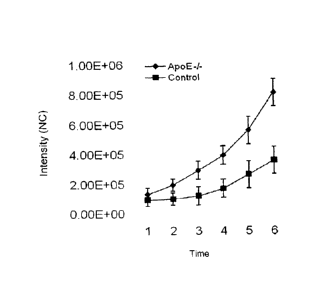
FIGURE 15 is a graph showing quantification of ICAM-1 signal in ApoE KO and
control mice
after longitudinal non-invasive in vivo imaging using anti-ICAM-1 sdAb 11-4.
Each point is
mean 4/- SD of image intensity signal in aortic region (ROI) in four animals
imaged as
described in FIGURE 14.
FIGURE 16 shows results of a three-dimensional analysis of the optical signal
in ApoE KO
mice 6 months after start of a high-fat diet. The mice were injected with 50
pg anti-ICAM-1
sdAb 11-4 labelled with Cy5.5 48 h prior to imaging. The 3D reconstruction
(FIGURE 16B)
confirms that high fluorescence intensity (optical; FIGURE 16A) signal
originates from the
heart and thoracic aorta region characterized with high atherosclerotic
deposits.
FIGURE 17 shows fluorescence Intensity and fluorescence lifetime map images of
ApoE -/-
mice. The image shows different fluorescent lifetime values for the injected
anti-ICAM-1 sdAb-
Cy5.5 in different regions of the body (bladder, liver, and heart). Circles
highlight high
fluorescence intensity in the aorta/heart region.
7
CA 02800565 2012-10-25
WO 2011/134060
PCT/CA2011/000481
FIGURE 18 shows fluorescence intensity and fluorescence lifetime map images of
control
mice. The image shows different fluorescent lifetime values for the injected
anti-ICAM-1 sdAb-
Cy5.5 in different regions of the body. No fluorescence intensity was observed
in the
aorta/heart region (circled).
FIGURE 19 shows gated fluorescence intensity and fluorescence lifetime images
in ApoE -/-
mice. When using time-domain imaging and lifetime gating between (1.05-1.65
ns), most of the
fluorescent signal is in the bladder region, which can be attributed to free
Cy5.5 fluorophore
that has lifetime between 1 and 1.3 ns. The heart/aortic region is circled.
FIGURE 20 shows gated fluorescence intensity and fluorescence lifetime images
of control
mice. When using time-domain and lifetime gating between (1.05-1.65 ns), most
of the
fluorescent signal is in the bladder region and can be attributed to free
Cy5.5 fluorophore,
which has lifetime between 1 and 1.3 ns. The heart/aortic region is circled.
FIGURE 21 shows gated fluorescence intensity and fluorescence lifetime images
in ApoE -/-
mice. When using time-domain optical imaging and lifetime gating between (1.85-
1.95 ns),
most of the fluorescent signal is in the heart/aortic region. This fluorescent
signal is attributed
to anti-ICAM-1 sdAb-Cy5.5 conjugate bound to atherosclerotic plaques. Anti-
ICAM-1 sdAb-
Cy5.5 conjugate has longer fluorescence life time than free Cy5.5 (1-1.3 ns).
The heart/aortic
region is circled.
FIGURE 22 shows gated fluorescence intensity and fluorescence lifetime images
of control
mice. When using time-domain optical imaging and lifetime gating between (1.85-
1.95 ns).
Anti-ICAM-1sdAb-Cy5.5 conjugate has longer fluorescence life time than that of
free Cy5.5 (1-
1.3 ns). The heart/aortic region is circled.
FIGURE 23 is a visualization of atherosclerotic plaques using anti-ICAM 11-4
sdAb (gated
fluorescence lifetime 1.85-1.95 ns), compared between control mice and ApoE -/-
mice. The
heart/aortic region (circled) of ApoE -I- mice has a progressive increase in
fluorescent signal
indicative of increased atheroscleroic plaques. In contrast, control mice have
low fluorescent
signal in the heart/aortic region. This shows that the anti-ICAM-1 sdAb has
the ability to detect
atherosclerotic plaques and that fluorescent lifetime gating can increase
accuracy (specificity)
of detection.
FIGURE 24 shows results of ex vivo imaging (FIGURE 24A) and quantification
(FIGURE 24B)
of the optical signal in isolated heart and thoracic aorta of ApoE KO and
control mice 6 months
after start of high-fat diet. The mice were injected with 50 pg of anti-ICAM-1
sdAb 11-4 labelled
with Cy5.5 and sacrificed 48 h after injection. Hearts and aortas were excised
and imaged ex
8
CA 02800565 2012-10-25
WO 2011/134060
PCT/CA2011/000481
viva The results show significantly higher fluorescence signal in heart/aorta
of APOE KO mice
compared to control mice.
FIGURE 25 shows fluorescently stained frozen sections images of aortas of
control C57BI/6
(upper panels, A) and ApoE KO (lower panels, B) injected with 50 pg anti-ICAM-
1 sdAb 11-4
labelled with Cy5.5 at 6 months of high fat diet. Mice were sacrificed 48 h
after sdAb injection.
Hearts and aortas were excised and sectioned. Fluorescence microscopy data
indicates that
intravenously injected anti-ICAM-1 sdAb 11-4-Cy5.5 (right panel) co-localizes
with
atherosclerotic plaques in frozen sections of aorta in ApoE KO mice. No anti-
ICAM-1 sdAb 11-
4-Cy5.5 signal was observed in aortas of control mice (Arrows point to the
localization of
ICAM-1).
FIGURE 26 is a schematic describing the in vivo optical imaging protocol for
scanning ApoE
KO mice and control mice. It shows the times of scanning and starting of
Atorvastatin (Lipitor)
treatment to reduce atherosclerosis.
FIGURE 27 are graphs representing data obtained while monitoring of
atherosclerotic disease
response to Atorvastatin (Lipitor) using anti-ICAM-1 sdAb 11-4. Quantification
of fluorescence
intensity signal is shown in non-treated and Atorvastatin-treated control
(FIGURE 29A) and
ApoE KO animals (FIGURE 29B). Atorvastatin (Lipitor) was administered at 25
mg/kg/day for 2
months. The mice were injected with 50 pg anti-ICAM-1 sdAb 11-4 labelled with
Cy5.5 48 h
prior to imaging at indicated time points after the start of high-fat diet.
Fluorescence was
quantified in heart/aorta ROI. Atorvastatin-treated ApoE KO animals
demonstrate reduction of
fluorescence signal compared to non-treated ApoE KO animals.
FIGURE 28 shows immunofluorescence images of ICAM-1 expression (right panels)
in brain
vessels (left panels) in control animals (upper panels) and after experimental
stroke (middle
cerebral artery occlusion; MCAO) (bottom panels). Arrows show ICAM-1
expressing brain
vessels after stroke.
FIGURE 29 shows in vivo head imaging after permanent left middle cerebral
artery occlusion
(MCAO) using anti-ICAM-1 11-4 sdAb-Cy5.5. The anti-ICAM-1 sdAb labeled with
Cy5.5 near
infrared fluorophore was injected in the tail vein (50 microgram) in mice that
have undergone
left permanent MCAO for 1 hour. Head region of mice was imaged up to 6 hour
post-injection.
Results show high fluorescence signal in the right side of the head,
contralateral to the side of
permanent MCAO.
FIGURE 30 shows ex vivo brain imaging after permanent MCAO using anti-ICAM-1
11-4
sdAb-Cy5.5. The anti-ICAM-1 sdAb labeled with Cy5.5 near infrared fluorophore
was injected
9
CA 02800565 2012-10-25
WO 2011/134060 PCT/CA2011/000481
in the tail vein (50 microgram) in mice that have undergone left permanent
MCAO for 1 hour.
Ex vivo brain imaging was performed 6 h after injection and shows fluorescent
signal in the
right side of the brain contralateral to permanent MCAO (infarct region
lacking fluorescent
signal).
FIGURE 31 shows multi-modal molecular imaging of vascular activation using
anti-ICAM-1 11-
4 sdAb in permanent MCAO. Animals with left permanent MCAO were injected with
50 pg of
anti-ICAM-1 sdAb labeled with Cy5.5 near infrared fluorophore. Animals were
optically imaged
at 6 hour post-injection. Animals were then perfused with microfill to
visualize the brain
vascular bed using microcomputed tomography. The molecular optical image
indicative of
increased ICAM-1 expression detected using anti-ICAM-1 sdAb was co-registered
with the
brain vessel map generated by microcomputed tomography to obtain a better
anatomical
localization of the molecular signal.
DETAILED DESCRIPTION OF THE INVENTION
The present invention relates to anti-ICAM-1 single-domain antibodies and uses
thereof. More
specifically, the invention relates to anti-ICAM-1 single-domain antibodies
and their use as
diagnostic tools.
The present invention is directed to anti-intercellular adhesion molecule 1
(ICAM-1) antibodies
and molecular imaging agents based on the antibodies. The present invention
also covers
methods and applications for non-invasive molecular imaging of atherosclerotic
disease, which
may provide information on plaque status (stable, active, inflamed, etc) based
on molecular
characteristics or processes within the plaque. The methods as described
herein may be use
to monitor the progression or regression of disease over time, or to monitor
the efficacy of
therapy.
The present invention provides an isolated or purified antibody or fragment
thereof specific to
intercellular adhesion molecule 1 (ICAM-1), comprising
the sequence of complementarity determining region (CDR) 1 selected from
sequences
LYVMG (SEQ ID NO:1), AFRMG (SEQ ID NO:2), and INDMG (SEQ ID NO:3);
the sequence of CDR2 selected from sequences DITSSGSIYYVDSLKG (SEQ ID
NO:4), VITAGGTTSYIDSVKG (SEQ ID NO:5), and RITRDGSAAYEDSVKG (SEQ ID
NO:6); and
CA 02800565 2012-10-25
WO 2011/134060
PCT/CA2011/000481
the sequence of CDR3 selected from sequences HVRQDSGSEYLTY (SEQ ID NO:7),
IDYDS (SEQ ID NO:8), and EIITTQTLGRMLGEY (SEQ ID NO:9).
The term "antibody", also referred to in the art as "immunoglobulin" (Ig),
used herein refers to a
protein constructed from paired heavy and light polypeptide chains; various Ig
isotypes exist,
including IgA, IgD, IgE, IgG, and IgM. When an antibody is correctly folded,
each chain folds
into a number of distinct globular domains joined by more linear polypeptide
sequences. For
example, the immmunoglobulin light chain folds into a variable (Vt.) and a
constant (CO
domain, while the heavy chain folds into a variable (VH) and three constant
(CH, CH21 CH3)
domains. Interaction of the heavy and light chain variable domains (VH and VI)
results in the
formation of an antigen binding region (Fv). Each domain has a well-
established structure
familiar to those of skill in the art.
The light and heavy chain variable regions are responsible for binding the
target antigen and
can therefore show significant sequence diversity between antibodies. The
constant regions
show less sequence diversity, and are responsible for binding a number of
natural proteins to
elicit important biochemical events. The variable region of an antibody
contains the antigen
binding determinants of the molecule, and thus determines the specificity of
an antibody for its
target antigen. The majority of sequence variability occurs in six
hypervariable regions, three
each per variable heavy and light chain; the hypervariable regions combine to
form the
antigen-binding site, and contribute to binding and recognition of an
antigenic determinant.
The specificity and affinity of an antibody for its antigen is determined by
the structure of the
hypervariable regions, as well as their size, shape and chemistry of the
surface they present to
the antigen. Various schemes exist for identification of the regions of
hypervariability, the two
most common being those of Kabat and of Chothia and Lesk. Kabat et al (1991)
define the
"complementarity-determining regions" (CDR) based on sequence variability at
the antigen-
binding regions of the VH and VL domains. Chothia and Lesk (1987) define the
"hypervariable
loops" (H or L) based on the location of the structural loop regions in the VH
and VL domains;
the numbering for the hypervariable loops is defined as H1: 27-35; H2: 52-56;
and H3: 95-102
(equivalent to CDR3 of Kabat numbering) for VHNHH domains (Chothia and Lesk,
1987). As
these individual schemes define CDR and hypervariable loop regions that are
adjacent or
overlapping, those of skill in the antibody art often utilize the terms "CDR"
and "hypervariable
loop" interchangeably, and they may be so used herein.. The CDR amino acids in
VH and VL
regions are defined herein according to the Kabat numbering system (Kabat et
al. 1991).
The region outside of the CDR is referred to as the framework region (FR). The
FR provides
structural integrity to the variable domain and ensure retention of the
immunoglobulin fold.
This characteristic structure of antibodies provides a stable scaffold upon
which substantial
11
CA 02800565 2012-10-25
WO 2011/134060
PCT/CA2011/000481
antigen-binding diversity can be explored by the immune system to obtain
specificity for a
broad array of antigens (Padlan et at, 1994).
An "antibody fragment" as referred to herein may include any suitable antigen-
binding antibody
fragment known in the art. The antibody fragment may be obtained by
manipulation of a
naturally-occurring antibody, or may be obtained using recombinant methods.
For example,
an antibody fragment may include, but is not limited to Fv, single-chain Fv
(scFV; a molecule
consisting VI_ and VH connected with a peptide linker), Fab, Fab', F(abl,
single domain
antibody (sdAb), and multivalent presentations of these.
In a non-limiting example, the antibody fragment may be a single domain
antibody (sdAb)
derived from naturally-occurring sources. Heavy chain antibodies of camelid
origin (Hamers-
Casterman et al, 1993) lack light chains and thus their antigen binding sites
consist of one
domain, termed VHH. sdAb have also been observed in shark and are termed VNARs
(Nuttall
et al, 2003); other sdAb may be engineered based on human heavy or light chain
sequences
(Jespers et at, 2004; To et al, 2005). As used herein, "sdAb" includes those
directly isolated
from VL, VH, VHH or VNAR reservoir of any origin through phage display or
other display
technologies and those generated through further modification of such sdAb by
humanization,
affinity maturation, stabilization, solubilisation (e.g., camelization), or
other methods of antibody
engineering. Also encompassed by the present invention are homologues,
derivatives, or
fragments that retain the antigen-binding function and specificity of the
sdAb.
A person of skill in the art would be well-acquainted with the structure of a
single-domain
antibody (see, for example, 3DWT, 2P42 in Protein Data Bank). A sdAb comprises
a single
immunoglobulin domain that retains the immuglobulin fold;most notably, only
three CDR form
the antigen-binding site. However, not all CDR may be required for binding the
antigen. For
example, and without wishing to be limiting, one, two, or three of the CDR may
contribute to
binding and recognition of the antigen by the sdAb of the present invention.
The CDR of the
sdAb are referred to herein as CDR1, CDR2, and CDR3, and are based on Kabat
numbering
(Kabat et at. 1991).
The terms "antibody" and "antibody fragment" ("fragment thereof') are as
defined above. As
previously stated, the antibody or fragment thereof may be a sdAb. The sdAb
may be of
camelid origin, and thus may be based on camelid framework regions;
alternatively, the CDR
may be grafted onto the framework regions of other antibody domains, for
example but not
limited to VNAR, human VH or human VI framework regions. In yet another
alternative, the
CDR described above may be grafted onto the framework regions of other types
of antibody
fragments (Fv, scFv, Fab). The present embodiment further encompasses an
antibody
12
CA 02800565 2012-10-25
WO 2011/134060
PCT/CA2011/000481
fragment that is "humanized" using any suitable method know in the art, for
example, but not
limited to CDR grafting and veneering. Humanization of an antibody or antibody
fragment
comprises replacing an amino acid in the sequence with its human counterpart,
as found in the
human consensus sequence, without loss of antigen-binding ability or
specificity; this approach
reduces immunogenicity of the antibody or fragment thereof when introduced
into human
subjects. In the process of CDR grafting, one or more than one of the heavy
chain CDR
defined herein may be fused or grafted to a human variable region (VH, or VA
or to other
human antibody fragment framework regions (Fv, scFv, Fab). In such
a case, the
conformation of said one or more than one hypervariable loop is preserved, and
the affinity
and specificity of the sdAb for its target (i.e., ICAM-1) is also preserved.
CDR grafting is
known in the art and is described in at least the following: US Patent No.
6180370, US Patent
No. 5693761, US Patent No. 6054297, US Patent No. 5859205, and European Patent
No.
626390. Veneering, also referred to in the art as "variable region
resurfacing", involves
humanizing solvent-exposed positions of the antibody or fragment; thus, buried
non-
humanized residues, which may be important for CDR conformation, are preserved
while the
potential for immunological reaction against solvent-exposed regions is
minimized. Veneering
is known in the art and is described in at least the following: US Patent No.
5869619, US
Patent No. 5766886, US Patent No. 5821123, and European Patent No. 519596.
Persons of
skill in the art would be amply familiar with methods of preparing such
humanized antibody
fragments.
By "specific to ICAM-1", it is meant that the antibody or fragment thereof of
the present
invention recognizes and binds to intercellular adhesion molecule 1 (ICAM-1),
also refered to
in the art as CD54. ICAM-1 is a cell adhesion molecule in the immunoglobulin
superfamily
expressed on the surface of endothelial cells. ICAM-1 is normally expressed in
low levels in
endothelial cells. However, in inflammatory conditions (e.g., the presence of
inflammatory
cytokines such as TNF-a, interferon-y, interleukin-4 and interleukin-13), the
level of expression
is rapidly increased on the surface of endothelial cells; ICAM-1 plays a role
in inflammatory cell
(leukocytes) adhesion to endothelial cells and their recruitment into inflamed
tissues. ICAM-1
also mediates the firm adhesion of lymphocytes, monocytes and neutrophils to
the sites of
endothelial lesion development. Therefore, the expression of ICAM-1 plays an
important rote in
the amplification of inflammation. ICAM-1 may be an early sign of endothelial
activation and
damage present before the onset of plaque formation (liyama et al, 1999).
The antibody or fragment thereof may have a CDR1 of sequence LYVMG (SEQ ID
NO:1),
AFRMG (SEQ ID NO:2), and INDMG (SEQ ID NO:3); a CDR2 of sequence
DITSSGSIYYVDSLKG (SEQ ID NO:4), VITAGGTTSYIDSVKG (SEQ ID NO:5), and
13
CA 02800565 2012-10-25
WO 2011/134060
PCT/CA2011/000481
RITRDGSAAYEDSVKG (SEQ ID NO:6); and a CDR3 of sequence HVRQDSGSEYLTY (SEQ
ID NO:7), IDYDS (SEQ ID NO:8), and ElITTOTLGRMLGEY (SEQ ID NO:9). The antibody
or
fragment thereof may be a sdAb. The sdAb may be of camelid origin, and thus
may be based
on camelid framework regions; alternatively, the CDR may be grafted onto other
antibody
domains, for example but not limited to VNAR or human VHH framework regions.
In a non-limiting example, the antibody or fragment thereof may have a CDR1 of
sequence
LYVMG (SEQ ID NO:1), a CDR2 of sequence DITSSGSIYYVDSLKG (SEQ ID NO:4), and a
CDR3 of sequence HVRQDSGSEYLTY (SEQ ID NO:7). Alternatively, the antibody or
fragment thereof may have a CDR1 of sequence AFRMG (SEQ ID NO:2), a CDR2 of
sequence VITAGGTTSYIDSVKG (SEQ ID NO:5), and a CDR3 of sequence IDYDS (SEQ ID
NO:8). In yet another alternative, the antibody or fragment thereof may have a
CDR1 of
sequence INDMG (SEQ ID NO:3), a CDR2 of sequence RITRDGSAAYEDSVKG (SEQ ID
NO:6), and a CDR3 of sequence EIITTQTLGRMLGEY (SEQ ID NO:9).
In one specific, non-limiting example, the isolated or purified antibody or
fragment thereof may
comprise the sequence:
QVQLVESGGGLVQPGGSLRLSCAASGSISSLYVMGWYRQAPGKQRELVADITSSGSIYYVDS
LKGRFTIS RDNARSTVYLQMNSLEPE DTAVYYCMAHVRQ DSGSEYLTYWGQGTQVTVSS
(SEQ ID NO:10),
QVKLEESGGGLVQAGDSLRLSCAASGRTVNAFRMGWYRQAPGKQRERVAVITAGGTTSYID
SVKGRFTISRDNAKNTVYLQMNSLKPEDTAVYYCAAIDYDSRGQGTQVTVSS (SEQ ID
NO:11), or
QVKLEESGGGLVQPGGSLRLSCAASGSI FSI NDMGWYRQAPGKQRELVARITRDGSAAYE DS
VKGRFTISRDNAPNTVFLQMNGLKPEDTAVYYCNAEIITTQTLGRMLGEYWGQGTQVTVSS
(SEQ ID NO:12),
or a sequence substantially identical thereto. In a specific, non-limiting
example, the isolated
or purified antibody or fragment thereof may also comprise the sequence of
clone 11-4, 5-5, or
34-1 as shown in Figures 2 to 4, or a sequence substantially identical
thereto.
A substantially identical sequence may comprise one or more conservative amino
acid
mutations. It is known in the art that one or more conservative amino acid
mutations to a
reference sequence may yield a mutant peptide with no substantial change in
physiological,
chemical, or functional properties compared to the reference sequence; in such
a case, the
reference and mutant sequences would be considered "substantially identical"
polypeptides.
14
CA 02800565 2012-10-25
WO 2011/134060
PCT/CA2011/000481
Conservative amino acid mutation may include addition, deletion, or
substitution of an amino
acid; in one non-limiting example, the conservative amino acid mutation is a
conservative
amino acid substitution. A conservative amino acid substitution is defined
herein as the
substitution of an amino acid residue for another amino acid residue with
similar chemical
properties (e.g. size, charge, or polarity).
A conservative amino acid substitution may substitute a basic, neutral,
hydrophobic, or acidic
amino acid for another of the same group. By the term "basic amino acid" it is
meant
hydrophilic amino acids having a side chain pK value of greater than 7, which
are typically
positively charged at physiological pH. Basic amino acids include histidine
(His or H), arginine
(Arg or R), and lysine (Lys or K). By the term "neutral amino acid" (also
"polar amino acid"), it
is meant hydrophilic amino acids having a side chain that is uncharged at
physiological pH, but
which has at least one bond in which the pair of electrons shared in common by
two atoms is
held more closely by one of the atoms. Polar amino acids include serine (Ser
or S), threonine
(Thr or T), cysteine (Cys or C), tyrosine (Tyr or Y), asparagine (Asn or N),
and glutamine (Gin
or Q). The term "hydrophobic amino acid" (also "non-polar amino acid") is
meant to include
amino acids exhibiting a hydrophobicity of greater than zero according to the
normalized
consensus hydrophobicity scale of Eisenberg (1984). Hydrophobic amino acids
include proline
(Pro or P), isoleucine (Ile or l), phenylalanine (Phe or F), valine (Val or
V), leucine (Leu or L),
tryptophan (Trp or W), methionine (Met or M), alanine (Ala or A), and glycine
(Gly or G).
"Acidic amino acid" refers to hydrophilic amino acids having a side chain pK
value of less than
7, which are typically negatively charged at physiological pH. Acidic amino
acids include
glutamate (Glu or E), and aspartate (Asp or D).
Sequence identity is used to evaluate the similarity of two sequences; it is
determined by
calculating the percent of residues that are the same when the two sequences
are aligned for
maximum correspondence between residue positions. Any known method may be used
to
calculate sequence identity; for example, computer software is available to
calculate sequence
identity. Without wishing to be limiting, sequence identity can be calculated
by software such
as NCB! BLAST2 service maintained by the Swiss Institute of Bioinformatics
(and as found at
http://ca.expasy.org/tools/blast/), BLAST-P, Blast-N, or FASTA-N, or any other
appropriate
software that is known in the art.
The substantially identical sequences of the present invention may be at least
70% identical; in
another example, the substantially identical sequences may be at least 70, 71,
72, 73, 74, 75,
80, 85, 90, 91, 92, 93, 94, 95, 96, 97, 98, 99, or 100% identical at the amino
acid level to
sequences described herein. Importantly, the substantially identical sequences
retain the
activity and specificity of the reference sequence. For example, and without
wishing to be
CA 02800565 2012-10-25
WO 2011/134060
PCT/CA2011/000481
limiting, the degree of identity between clones 11-4 and 5-5, clones 5-5 and
34-1 is 70%; the
degree of identity between clones 11-4 and 34-1 is 75%. As would be know to
one of skill in
the art, amino acid residues of an antibody, particularly within the framework
regions may be
mutated (substituted or deleted) without affecting the functional properties
of the antibody
(antigen recognition and binding).
The antibody or fragment thereof of the present invention may also comprise
additional
sequences to aid in expression, detection, or purification of a recombinant
antibody or
fragment thereof. For example, and without wishing to be limiting, the
antibody or fragment
thereof may comprise a targeting or signal sequence (for example, but not
limited to ompA), a
detection tag (for example, but not limited to c-Myc, EQKLISEEDL, SEQ ID
NO:19), a
purification tag (for example, but not limited to a histidine purification
tag, HHHHHH, SEQ ID
NO:20), or any combination thereof.
The antibody or fragment thereof of the present invention may also be in a
multivalent display.
Multimerization may be achieved by any suitable method of know in the art. For
example, and
without wishing to be limiting in any manner, multimerization may be achieved
using self-
assembly molecules (Zhang et al, 2004; Merritt & Hol, 1995), as described in
W02003/046560. The described method produces pentabodies by expressing a
fusion
protein comprising the antibody or fragment thereof of the present invention
and the
pentamerization domain of the B-subunit of an AB5 toxin family (Nielson et al,
2000); the
pentamerization domain assembles into a pentamer, through which a multivalent
display of the
antibody or fragment thereof is formed. Each subunit of the pentamer may be
the same or
different. Additionally, the pentamerization domain may be linked to the
antibody or antibody
fragment using a linker; such a linker should be of sufficient length and
appropriate
composition to provide flexible attachment of the two molecules, but should
not hamper the
antigen-binding properties of the antibody. In one non-limiting example, the
linker may be the
linker GPGGGSGGGGS (SEQ ID NO:21)
Other forms of multivalent display are also encompassed by the present
invention. For
example, and without wishing to be limiting, the antibody or fragment thereof
may be
presented as a dimer, a trimer, or any other suitable oligomer. This may be
achieved by
methods known in the art, for example direct linking connection (Nielsen et
al, 1996), c-jun/Fos
interaction (de Kruif et al, 1996), "Knob into holes" interaction (Ridgway et
al, 1996).
Another method known in the art for multimerization is to dimerize the
antibody or fragment
thereof using a Fc domain. In this approach, a Fc gene in inserted into an
expression vector;
the nucleotide sequence of the antibody or fragment thereof can be amplified
and inserted into
16
the vector such that the C-terminus of the antibody or fragment thereof is
linked to the hinge region of
the Fc without addition of extra residues. The resulting vector can be
transfected to cells and the fusion
protein may be recombinantly expressed, then purified by affinity
chromatography (for example, on a
protein A column). One non-limiting example of such a method of
multimerization is described by Bell et
at, Cancer Lett. 289:81-90 (2010) and lqbal et at, British Journal of
Pharmacology, 160(4): pgs 1016-
1028. Techniques for implementing such dimerization would be known to those of
skill in the art.
The present invention also encompasses nucleic acid sequences encoding the
molecules as
described herein. The nucleic acid sequence may be codon-optimized. The
present invention
also encompasses vectors comprising the nucleic acids as just described.
The present invention further provides a targeted therapeutic agent comprising
an anti-ICAM-1
antibody or fragment thereof of the present invention linked to a suitable
therapeutic. The
antibody or fragment thereof may serve to target therapeutic agents to the
site of
atherosclerotic plaques, or may have use as a therapeutic agent itself. For
example, and
without wishing to be limiting, the antibody or fragment thereof may be used
for therapeutically
modifying the inflammatory component of atherosclerotic disease (e.g., stroke
prevention
therapy). Additionally, the antibody or fragment thereof or the targeted
therapeutic agent may
be used to treat conditions associated with increased ICAM-1 expression; these
may include,
but are not limited to carotid artery disease, stroke, myocardial infarction,
inflammatory bowel
disease, autoimmune diseases, multiple sclerosis, Crohn's disease, and
neovascularization
associated with tumour angiogenesis. For example, and without wishing to be
limiting in any
manner, the therapeutic agent may be an anti-Inflammatory drug, a cholesterol-
lowering drug,
or a plaque-stabilizing drug.
The present invention also encompasses a molecular imaging agent comprising an
anti-ICAM-
1 antibody or fragment thereof in accordance with the present invention linked
to a detectable
agent. For example, the anti-ICAM-1 or fragment thereof may be linked to a
radioisotope, a
paramagnetic label such as gadolinium or iron oxide, a fluorophore, Near Infra-
Red (NIR)
fluorochrome or dye, an echogenic microbubble, an affinity label (for example
biotin, avidin,
etc), enzymes, or any other suitable agent that may be detected by diagnostic
imaging
methods. In a specific, non-limiting example, the anti-ICAM-1 or fragment
thereof may be
linked to a near infrared fluorescence (NIRF) imaging dye, for example and not
wishing to be
limiting Cy5.5, Alexa680, Dylight680, or Dylight800.
An ideal molecular imaging agent for imaging atherosclerotic plaque diseases,
such as carotid
atherosclerosis, should have a high sensitivity and specificity for the
detection of plaques, and
provide information about the probability of adverse outcome in both
symptomatic and
17
CA 2800565 2017-07-24
CA 02800565 2012-10-25
WO 2011/134060
PCT/CA2011/000481
asymptomatic individuals. Because baseline constitutive ICAM-1 expression is
low, it is
expressed on the luminal surface of endothelial cells, and the expression
levels correlate with
the severity of disease (Kitagawa et al, 2002), ICAM-1 is an excellent target
for non-invasive
molecular imaging applications to diagnose pre-symptomatic and/or unstable
carotid artery
disease and other atherosclerotic plaque diseases. To obtain clear images, it
is imperative
that molecular imaging agent has rapid clearance from the circulation and high
target
(atherosclerotic plaque)-to-background (blood pool) ratio. Because of the fast
clearance of
antibody fragments, and single domain antibodies in particular, and their
short half life, they
are superior to whole IgG molecules in achieving a successful image.
Additionally, the
coupling of single domain antibodies against ICAM-1 to a Near Infrared
Fluorescence (NIRF)
imaging dye for optical imaging is advantageous due to high sensitivity and
avoidance of
ionizing radiation.
The therapeutic agent or detectable agent may be linked to the anti-ICAM-1
antibody or
fragment thereof by any method know in the art. By the term "linked", also
referred to herein
as "conjugated", it is meant that the antibody or fragment thereof is linked
directly or indirectly
(e.g., via a linker), covalently or non-covalently (e.g., adsorption, ionic
interaction) to the
therapeutic or detectable agent. A covalent linkage may be achieved through a
chemical
cross-linking reaction, or through fusion using recombinant DNA methodology
combined with
any peptide expression system, such as bacteria, yeast or mammalian cell-based
systems.
Methods for linking an antibody or fragment thereof to a therapeutic agent or
detectable agent
would be well-known to a person of skill in the art.
The antibodies or fragments thereof and/or molecular imaging agents may be
used in methods
and applications for imaging of atherosclerotic disease, which may provide
information on
plaque status (stable, active, inflamed, etc) based on molecular
characteristics/processes
taking place within the plaque. Such information obtained in the realm of
clinical diagnosis and
triage would also present an opportunity to gain insight into the complex
chain of events
underlying atherogenesis, plaque progression, and ultimately atherothrombosis
with
accompanying clinical symptoms.
The present invention provides an ex vivo method of detecting atherosclerotic
plaque diseases
involving inflammation, comprising:
a) providing a tissue sample suspected of inflammation and plaque formation;
b) contacting said sample with an anti-ICAM-1 antibody or fragment thereof of
the
present invention under suitable conditions; and
c) detecting the formation of a protein complex,
18
CA 02800565 2012-10-25
WO 2011/134060
PCT/CA2011/000481
wherein the anti-ICAM-1 antibody or fragment thereof binds to the tissue
sample comprising
atherosclerotic plaque formation at a higher rate than that of a control
sample.
The tissue sample in the method as just described may be any suitable tissue
sample, for
example but not limited to a serum sample, a vascular tissue sample or a brain
tissue sample.
The step of contacting (step b)) is done under suitable conditions, known to
those skilled in the
art, for formation of a complex between the antibody or fragment thereof and
ICAM-1 protein.
The control sample may be a corresponding tissue sample that does not exhibit
increased
ICAM-1 expression.
The step of detecting (step c)) may be accomplished by any suitable method
known in the art,
for example, but not limited to optical imaging, immunohistochemistry or
molecular diagnostic
imaging, ELISA, or other suitable method.
The invention also provides a method for analyzing the ICAM-1 expression in
atherosclerotic
plaques in vivo by means of non-invasive imaging using optical imaging
techniques. The in
vivo method of detecting atherosclerotic plaque diseases involving
inflammation may
comprise:
a) administering the molecular imaging agent of the present invention to a
subject; and
b) detecting the binding of the molecular imaging agent,
wherein the molecular imaging agent binds to binds ICAM-1 in vivo at a
detectably higher rate
than the rate of binding to normal vasculature, and wherein the binding of
molecular imaging
agent to the vasculature is indicative of the presence of atherosclerotic
plaques. The method
as just described may also be used for imaging atherosclerotic plaques
The ability to use non-invasive molecular imaging techniques to detect or
image
atherosclerotic plaques may lead to early diagnosis (asymptomatic) of
atherosclerosis.
Additionally, prospective monitoring via molecular imaging of plaques would
give insight
regarding atherosclerotic disease progression/stability.
The present invention further provides a method of detecting conditions
characterized by
increased expression of ICAM-1, comprising:
a) administering a molecular imaging agent of the present invention to a
subject of
interest;
b) detecting the molecular imaging agent in vivo.
wherein the molecular imaging agent binds to binds ICAM-1 in vivo at a
detectably higher rate
than the rate of binding to normal tissue. As previously described, the level
of expression of
19
CA 02800565 2012-10-25
WO 2011/134060
PCT/CA2011/000481
ICAM-1 is increased on the surface of endothelial cells in inflammatory
conditions (e.g., the
presence of inflammatory cytokines such as TNF-a, interferon-y, interleukin-4
and interleukin-
1 r3). Conditions associated with increased ICAM-1 expression may include, but
are not limited
to carotid artery disease, stroke, myocardial infarction, inflammatory bowel
disease,
autoimmune diseases, multiple sclerosis, Crohn's disease, and
neovascularization associated
with tumour angiogenesis.
The in vivo detection step in the methods described above may be whole body
imaging for
diagnostic purposes or local imaging at specific sites, such as but not
limited to carotid
arteries, in a quantitative manner to assess the progression of disease or
host response to a
treatment regimen. The detection step in the methods as described above may be
immunohistochemistry, or a non-invasive (molecular) diagnostic imaging
techonology
including, but not limited to:
= Optical imaging;
= Positron emission tomography (PET), wherein the detectable agent is an
isotopes such
as 11C, 13N, 150, 18F, 64cu, 62cu, 1241, 76B r, 82
r Rb and 68Ga, with 18F being the most
clinically utilized;
= Single photon emission computed tomography (SPEC), wherein the detectable
agent
is a radiotracer such as 99mTc, 1111n, 1231, 201T., 133
Xe, depending on the specific
application;
= Magnetic resonance imaging (MRI), wherein the detectable agent may be, for
example
and not limited to gadolinium, iron oxide nanoparticles and carbon-coated iron-
cobalt
nanoparticles thereby increasing the sensitivity of MRI for the detection of
plaques.
= Contrast-Enhanced Ultrasonography (CEUS) or ultrasound, wherein the
detectable
agent is at least one acoustically active and gas-filled microbubble.
Ultrasound is a
widespread technology for the screening and early detection of human diseases.
It is
less expensive than MRI or scintigraphy and safer than molecular imaging
modalities
such as radionuclide imaging because it does not involve radiation..
The optimal dose of injection and method of administration (intravenous
(i.v.), intraperitoneal
(i.p), subcutenous (s.c.), oral, or nasal) are generally determined
experimentally.
The methods described herein may be used to diagnose atherosclerosis,
including early
diagnosis (i.e., sub-clinical atherosclerosis), distinguish stable from
unstable plaques, monitor
CA 02800565 2012-10-25
WO 2011/134060 PCT/CA2011/000481
the progression or regression of disease over time, and/or monitor the
efficacy of therapy, for
example but not limited to drugs such as statins in the treatment of
atheroslerosis. To do so,
the methods described herein may be combined with other data, for example, but
not limited to
atherosclerosis staging, atherosclerosis prognosis, and vascular inflammation
levels.
Anti-ICAM-1 single-domain antibodies were obtained by immunization of a llama
with ICAM-1;
three clones in particular were shown to specifically bind ICAM-1. The anti-
ICAM-1 sdAb were
coupled with the near infrared fluorescence (NIRF) imaging dye for
application, which was
advantageous in optical imaging due to the conjugate's high sensitivity and
avoidance of
ionizing radiation. Using this formulation, it was shown that the NIRF-
labelled anti-ICAM-1
sdAb specifically recognized early and developed atherosclerotic plaques in
large vessels in
high-fat diet fed ApoE KO mice; it was additionally shown that this can be
monitored non-
invasively by prospective optical imaging in vivo. The distribution of the
anti-ICAM-1 sdAb in
the plaques was confirmed using microscopic techniques and
immunohistochemistry. Optical
imaging as a means of imaging atherosclerosis in vivo provides a high
sensitivity, allowing for
detection at an earlier stage of the disease, which may potentially lead to
better therapeutic
outcomes.
The use of sdAb is advantageous as they may be produced easily and
inexpensively in large
quantities, as opposed to antibodies produced from hybridoma cell lines.
Additionally,
hybridoma lines may be unstable and decrease antibody expression levels over
time. sdAb are
also advantageous for molecular imaging applications due to their short plasma
half-life, which
achieves fast contrast-to-noise ratio needed for imaging.
The present invention will be further illustrated in the following examples.
However, it is to be
understood that these examples are for illustrative purposes only and should
not be used to
limit the scope of the present invention in any manner.
Example 1: Immunization and PCR amplification
Single-domain antibodies (sdAb) were generated by immunization of a llama with
ICAM-1.
Lymphocytes were collected and DNA corresponding to sdAb was purified.
A llama was immunized with recombinant antigen. For each injection, 100 pg of
recombinant
human ICAM-1 (R&D systems, reconstituted with sterile water according to
manufacturer's
recommendation to prepare a stock solution of 1mg/m1), in a total volume of
0.5 ml was mixed
with an equal volume of incomplete Freund's adjuvant and 0.5 ml was injected,
21
CA 02800565 2012-10-25
WO 2011/134060
PCT/CA2011/000481
subcutaneously. Seven injections were performed at approximately two week
intervals and
blood was collected at each injection.
Total RNA was isolated from approximately 1 X 10⁷ lymphocytes collected from
day 70 of the
immunization protocol with a QIAamp RNA blood mini kit (QIAGEN Sciences,
Mississauga,
ON) and according to the kit instructions. About 5 pg of total RNA was used as
template for
first strand cDNA synthesis with an oligo dT primer using a first-strand cDNA
synthesis kit
(Amersham Biosciences, USA). Based on the camelidae and llama immunoglobulin
databases, three variable domain sense primers (MJ1-3) and two CH2 domain
antisense
primers (CH2 and CH2b3) were designed (Doyle et al. 2008). The first PCR was
performed
with the cDNA as template and the variable regions of both conventional (IgG1)
and heavy
chain antibodies (IgG2 and IgG3) were amplified with combinations of MJ1-3/CH2
and MJ1-
3/CH2b3 primers in two separate PCR reactions. The PCR reaction mixtures
contained the
following components: 2 pl cDNA, 5 pmol of MJ1-3 primer mixture, 5 pmol of
either CH2 or
CH2b3 primer, 5 pt of 10X reaction buffer, 3 pl of 2.5 mM dNTP, 2.5 units of
Taq DNA
polymerase (Roche Applied Science, Indianapolis, IN) and water to a final
volume of 50 pl. The
PCR protocol comprised an initial step at 94 C for 3 min followed by 30 cycles
of 94 C for 30
seconds, 55 C for 30 seconds, 72 C for 1 min and a final extension step at 72
C for 7 min.
The amplified PCR products were run onto a 2% agarose gel and comprised two
major bands
of about 850 bp corresponding to conventional IgG1 and about 600 bp (550-
650bp)
corresponding to heavy chain antibodies. The smaller band was cut out of the
gel, purified with
a QIAquick gel extraction kit (QIAGEN Inc) and re-amplified in a second PCR
reaction
containing 1 pl of the purified DNA template, 5 pmol each of MJ7, a VH sense
primer with a
Sfil restriction site, underlined, (5'- CAT GIG TAG ACT CGC GGC CCA GCC GGC
CAT GGC
C-3'; SEQ ID NO:22) and MJ8, an antisense primer with a Sfil restriction
enzyme site,
underlined, (5'- CAT GTG TAG ATT CCT GGC CGG CCT GGC CTG AGG AGA CGG TGA
CCT GG; SEQ ID NO:23), 5 pl of 10X reaction buffer, 3 pl of 2.5 mM dNTP, 2.5
unit of Taq
DNA polymerase (Roche Applied Science, Indianapolis, IN) and water to a final
volume of 50
pl. The PCR protocol consisted of an initial step at 94 C for 3 min followed
by 30 cycles of
94 C for 30 seconds, 57 C for 30 seconds, 72 C for 1 min and a final extension
step at 72 C
for 7 min. The amplified PCR products (about 400-450bp) that correspond to VHH
fragments
of heavy chain antibodies were purified with a QIAquick PCR purification kit
(QIAGEN Inc.),
digested with Sfil (New England BioLabs ) and re-purified with the same kit.
22
CA 02800565 2012-10-25
WO 2011/134060
PCT/CA2011/000481
Example 2: Phaqe library construction and panning
A phage library containing the DNA isolated in Example 1 was constructed then
panned to
identify anti-ICAM-1 antibodies. Reactivity of the antibodies was tested using
ELISA.
30 pg of pMED1 (Arbabi-Ghahroudi et al., 2009) DNA was digested with Sfil
overnight at 50 C.
To minimize self-ligation, digestion was continued for additional 2 hours at
37 C by adding 20
units of both Xhol and Pstl restriction enzymes. For library construction, 10
pg of phagemid
DNA was ligated with 1.75 pg of VHH fragment DNA (isolated in Example 1) and
incubated for
2 hours at RT using the LigaFast DNA ligation system (Promega, Corp., Madison,
WI),
according to the recommended protocol. The ligated product was precipitated
with n-butanol,
resuspended in sterile water and electroporated into competent E. coil
TG1cells (Stratagene,
Cedar Creek, TX). Transformed bacterial cells were diluted in SOC medium and
incubated for
1 hour at 37 C with slow shaking. The size of library was calculated by
plating aliquots on LB-
Amp. The VHH fragments from 30 colonies were PCR-amplified and sequenced for
diversity
analysis. The library was aliquoted and stored at -80 C.
Panning was performed essentially as described by Arbabi et al. (1997). A 1 ml
aliquot of the
library (5 X 10¹⁰ bacterial cells) was thawed on ice, grown in 300 ml 2 X YT
with 100 pg/ml
ampicillin and 2% glucose for about 2 hours at 37 C (0D600 = 0.4-0.5). The
grown cells were
infected with M13K07 helper phage (New England Biolabs) at a phage to bacteria
ratio of 20:1
for 30 min at 37 C without shaking followed by shaking at 37 C for one hour.
The culture was
then centrifuged at 4 C, the infected cell pellets were re-suspended in 300 ml
of 2X YT with
100 pg/ml ampicillin and 50 pg/ml kanamycin, and the culture was incubated at
37 C overnight
with vigorous shaking (250 rpm). The phage particles in the culture
supernatant were
incubated with 1/5 volume of 20% PEG 6000, 2.5 M NaCl, on ice for 1 hour and
centrifuged at
10,000 rpm for 15 min. The phage pellets were re-suspended in 2 ml of sterile
PBS and titered.
For solid phase panning Maxisorb microtitre plates (Nunc, Roskilde, Denmark)
were coated
overnight at 4 C with 50 pg/well of recombinant human ICAM-1. The wells were
rinsed once
with PBS and blocked with 3% bovine serum albumin (BSA) in PBS for 2 hours at
37 C.
Approximately 101' library phage were added to the blocked wells, including
control wells with
no antigen and incubated for 2 hours at 37 C. After 7X washing with PBS
containing
0.1%Tween 20, bound phage were eluted with 0.1 M triethylamine, then
neutralized and added
to exponentially growing TG1 cells. The eluted phage were titered and the
infected bacterial
cells were super-infected with M13K07 and grown overnight at 37 C. Panning was
continued
for three more rounds following the same procedure except that the amount of
coated ICAM-1
antigen was reduced to 40, 30, and 20 pg for the second, third and fourth
rounds, respectively.
23
CA 02800565 2012-10-25
WO 2011/134060
PCT/CA2011/000481
Colony-PCR was performed on twenty-four individual colonies randomly picked
after the last
round of panning and the sequences of amplified VHH genes were determined.
For phage-ELISA, the positive clones were grown to 0D600= 0.3-0.4 in 2X YT
containing 100
pg/m1 ampicillin and 0.1% glucose, then infected by M13K07 helper phage. The
cultures were
grown at 37 C overnight. Phage supernatants were then collected by
centrifugation and their
reactivity was measured by phage ELISA. Briefly, ELISA wells were coated
overnight at 4 C
with 5 pg/ml of the recombinant ICAM-1 and blocked with 3% BSA for additional
2 hours at
37 C. Phage supernatants were added to the wells and incubated for 2 hours at
37 C. The
presence of phage binding was detected by an anti-M13/HRP conjugate (GE
Healthcare,
Mississauga, ON). After 1 hour at room temperature, KPL peroxidase substrate
(KPL,
Gaithersburg, MD) was added. Color development was stopped by adding 100 ul 1M
phosphoric acid and the plates were read at 450nm. Results are shown in Figure
1.
Example 3: Expression of soluble sdAb
VHH antibodies isolated via phage panning in Example 2 and showing reactivity
to ICAM-1
were expressed and their reactivity confirmed via ELISA.
DNA corresponding to the VHH antibodies identified in Example 2 was inserted
into an
expression vector. Restriction enzyme sites Bbsl and BarnH1 were added to the
5' and 3' ends
of the positive VHH DNA fragments via a PCR using gene-specific sense primer
VHH Bbsl (5'-
TATGAAGACACCAGGCCCAGGTGCAGCTGGTGGAGTCT-3'; SEQ ID NO: 24) and anti-
sense primer VHH-BamHI (5'-CGCGGGATCCTGAGGAGACGGTGACCTGGGT-3'; SEQ ID
NO:25). The amplified DNA was then digested with Bbsl and BamHI restriction
enzymes and
ligated into digested pSJF2 vector using standard techniques (Tanha et al.,
2003). Competent
E. coli TG1 cells were transformed with the vectors and clones expressing anti-
ICAM-1-
specific recombinant VHH were grown in 1-liter cultures of 2xYT medium +
ampicillin (100
mg = mL-1) with 0.1% glucose to an 0D600 of 0.8. Cultures were induced with 1
mM IPTG and
grown overnight on a rotary shaker at 28 C.
After confirmation of expression by SDS-PAGE and Western blotting, recombinant
VHH
proteins were extracted from the bacterial cells by standard lysis methods,
purified by
immobilized metal affinity chromatography (IMAC), and quantified as described
elsewhere
(Tanha et al. 2001). The state of aggregation of the purified protein was
determined by size
exclusion chromatography on Superdex 200 (Amersham Biosciences). The
reactivity of the
individual VHH proteins was confirmed by ELISA, in which rabbit anti-His6
antibody conjugated
to HRP was used for the detection of binding.
24
CA 02800565 2012-10-25
WO 2011/134060 PCT/CA2011/000481
Figures 2 to 4 show nucleotide and amino acid sequences of single-domain
antibody clones
11-4, 5-5 and 34-1, respectively. These clones were reactive with
intracellular adhesion
molecule 1 (ICAM-1).
Size exclusion chromatography employing SuperdexTM 75 was used to assess the
aggregation
state of VHH domains. Non-aggregating VHs should yield chromatograms with a
single,
symmetrical peak in elution volumes expected for a monomeric VH. Briefly, a
SuperdexTM 75
(Superdex 75 10/300, GE Healthcare Cat. No 17-5174-01, ID No 0651148) size
exclusion
column was washed with 50 mL of filtered and degassed ddH20 and subsequently
equilibrated
with 50 mL of running buffer, HBS-EP (10mM HEPES, pH 7.4, 150mM NaCI, 3mM
EDTA,
0.005% surfactant P20), at a pump speed of 0.5 mUmin. 200 pL of purified VH (>
1mg/mL)
was injected and eluted, and obtain a chromatogram was obtained. The monomeric
and
aggregate peaks were integrated to obtain A) monomer. Size-exclusion
chromatography
(Figure 5) of these purified sdAb clones showed them to be monomeric
Additionally, ELISA experiment on individual clones was performed to assess
binding of VHH to
the recombinant human ICAM-1. Briefly, MaxisorpTM microtiter plates (Nunc)
were coated with
100 pl of 5 pg/ml of the recombinant ICAM-1(R&D Systems, Inc., Minneapolis, MN
55413,
USA) in PBS overnight at 4 C. After blocking with 3% bovine serum albumin (300
pl) for 2 h at
RT and subsequent removal of blocking agent, 100 pL His6-tagged VHH at
concentrations of a
few pM were added, followed by incubation for 2 h at 37 C. Wells were washed
5x with PBST,
and 100 pl rabbit anti-His-IgG/horse radish peroxidase (HRP) conjugate (Bethyl
Laboratories,
Inc., Montgomery, TX) was added at a dilution of 1:5000. The wells were then
incubated for 1
h at 37 C. After washing the wells with PBST, 100 pL ABTS substrate (KPL,
Gaithersburg,
MD) was added and the reaction, seen as color development, was stopped after 5
min by
adding 100 pL of 1M phosphoric acid. Absorbance values were measured at a
wavelength of
405 nm using a microtiter plate reader. Assays were performed in duplicates.
The ELISA
assays on individual clones 5-5, 26-6, 11-4, and 34-1 (Figure 6) showed them
to positively
react with ICAM-1.
Example 4: SPR analysis of VHH
Surface plasmon resonance (SPR) assays were conducted on the purified VHH of
Example 4
to determine the binding affinity of individual clones to ICAM-1.
Three clones (5-5, 11-4, and 34-1) obtained in Example 4 were individually
passed through
size exclusion columns, Superdex 75 (GE Healthcare) in 10 mM HEPES, pH 7.4,
containing
150 mM NaCI, 3 mM EDTA; monomeric sdAb fractions were collected and protein
concentrations were determined by measuring A280. SPR analyses were performed
with
CA 02800565 2012-10-25
WO 2011/134060 PCT/CA2011/000481
Biacore 3000 instrument (GE Healthcare). All measurements were carried out at
25 C in 10
mM HEPES, pH 7.4, containing 150 mM NaCl, 3 mM EDTA and 0.005% surfactant P20
(GE
Healthcare). Approximately 700-900 RUs of the recombinant ICAM-1 was captured
on SA
sensor chip (GE Healthcare) at a flow rate of 5 pl/min. Various concentration
of the monomeric
VHH were injected over the ICAM-1 surface, using a SA surface as a reference,
at a flow rate
of 40 pl/min. Surfaces were generated by washing with running buffer. Data
were analysed
with BlAevaluation 4.1 software.
SPR analysis of binding of sdAb clones 11-4, 5-5, and 34-1 to recombinant
human ICAM-1 are
shown in Figures 7, 8, and 9, respectively. Table 1 shows the binding
affinities (Kd, Ka, KD) for
clones 11-4, 5-5 and 34-1. It provides a summary of affinities for the
representative antibodies
of the invention, as determined by surface plasmon resonance (Biacore).
Table 1. Biding affinities of clones 11-4, 5-5, and 34-1.
ICAM-1 binder KD (M) kd (s-1) ka (M-1s-1)
5-5 6 x 10⁻⁵ 4 x 10' 7 x 10⁴
11-4 9 x 10⁻¹ 2 x 10⁻³ 2 x 10⁶
34-1 1 x 10⁻⁸ 2 x 10⁻³ 1 x 10⁵
Example 5: lmmuno fluorescence of anti-ICAM-1 single-domain antibodies in rat
brain
endothelial cells
The purified VHH of Example 4 was tested to validate its activity and to
determining whether
the antibody can detect inflammation.
Rat brain endothelial cells (SV-RBEC) were cultured on coverslips for 3 days
in DMEM and
10% FBS. ICAM-1 expression was induced by adding 5 pg (1mg/mI) of
lipopolysaccharide
(LPS; Sigma) overnight. Cells were then washed 3x with cold PBS, fixed with
3.7%
formaldehyde for 20 min, washed again 3x in PBS at RT, and then blocked with
5% of normal
goat serum/PBS for 1h at RT. 25 pg anti-ICAM-1 IgG or 50 pg anti-ICAM-1 single
domain
antibodies (34-1, 11-4, 5-5) labelled with Cy5.5 (red) were incubated for 1h
at RT then washed
3X in PBS. The washed cells were incubated in 1:500 WGA-FITC/PBS for 1 min on
ice, for
membrane staining (green) then washed again 3X with PBS. Coverslips were then
mounted in
DAKO mounting medium containing 2pg/m1 of Hoechst for nuclear staining (blue).
Results of the ICAM-1 immunofluorescence assay are shown in Figures 10 to 12.
Results
show that the anti-ICAM-1 sdAb can detect ICAM-1 as a marker for LPS-induced
inflammation
in rat brain endothelial cells.
26
Example 6: Mouse model of atherosclerosis
Rodents -do not develop atherosclerosis spontaneously. For this reason the
apolipoprotein E
knockout (apoE KO) mouse model was used to study ability of anti-ICAM-1 sdAb
to detect
atherosclerotic plaques. Apolipoprotein E is a ligand for receptors that clear
remnants of
chylonnicrons and very low density lipoproteins. ApoE KO mice develop
atheromas in large
arteries, including aorta and carotid arteries; these histologically resemble
those found in
humans (Weinreb et al 2007). Although these mice develop atheromas
spontaneously, the
rate of plaque formation is enhanced when they are fed a high fat diet for a
period of 3 to 9.
months. For the present experiments, mice were fed a high fat diet for a
period of 4 months,
starting at 2 months of age.
To confirm that ApoE knockout animals develop atherosclerotic plaques on high-
fat diet and
that these plaques exhibit up-regulated expression of ICAM-1, the animals were
sacrificed
after four moths of high-fat diet and their aortas were dissected, sectioned
and evaluated by
immunofluorescence staining against ICAM-1 using a commercial anti-ICAM
monoclonal
antibody.
Briefly, sample slides of 12 pm saline-perfused, not fixed, frozen aortic
sections of ApoE KO
mice and C57B control (Ctrl) mice were prepared. Samples were fixed with 100%
Me0H 10
min at RT, washed with 3x 1XPBS, then rinsed with MilliQ H20 (to wash off any
OCT tissue tek
embedding medium from the slides) followed, with 3x 1XPBS. Slides were then
blocked with
10% Normal Goat Serum (NGS) (Cat# G6767, Sigma) + 0.01% TritonTmX-100 in 1XPBS
for 1h
at RT. To stain endothelial cells, rat anti-mouse CD31 (Cat# 557355, BD
Pharmingen) was
incubated at a dilution of 1:300 in 5% NGS in 1XPBS for 1h at RT, then rinsed
with 3x 1XPBS.
Secondary antibody Goat anti-rat Alexa568 (Cat# A-11077, Invitrogen) was then
Incubated at
1:500 in 1XPBS for 1 h at RT, and rinsed with 3x 1XPBS. Armenian hamster anti-
ICAM-1
1mg/mL (Cat# 553249, BD Pharmingen) 1:250 in 5 /oNGS in 1XPBS was incubated
for 1h at
RT to detect ICAM-1 expression and then rinsed with 3x 1XPBS. Secondary
antibody goat-
anti-armenian hamster Alexa488 (Cat# sc-2446Santa Cruz) 1:300 in 1XPBS-1h at
RT was
used afterward. Cover slipped in Dako fluorescent mounting medium (Cat# S3023,
Dako)
spiked with Hoechst (1pg/mL) (Cat# H3570, Invitrogen), Images were acquired
using Olympus
IX81 Fluorescent Microscope and InVivo program. Images were corrected for
background
noise using ImagePro v6.2 and AxioVision LE re14.4 software.
Results show up-regulated expression of ICAM-1 in endothelial layer of aortas
from ApoE
knockout animals fed high-fat diet for 4 months compared to control animals
(Figure 13).
=
27
CA 2800565 2017-07-24
CA 02800565 2012-10-25
WO 2011/134060 PCT/CA2011/000481
Plaque formation in this model was further monitored monthly by non-invasive
in vivo imaging
using anti-ICAM-1 single domain antibodies (see Example 7). C57BI/6 mice on a
high fat diet
were used as wild type (WT) controls.
Example 7: In vivo near-infrared fluorescence imaging of APO E knockout mice
The antibodies of Example 4 were labelled with fluorescent agent and were used
for non-
invasive optical imaging of ApoK0 and control mice (of Example 6).
Anti-ICAM-1 antibodies were labelled with Cy5.5 succimidyl ester using methods
recommended by the manufacturer (GE Healthcare). Labelling was optimized such
that each
sdAb had a dye/antibody ratio of two. The presence of plaques in the aorta of
atherosclerotic
mice (Example 6) was visualized monthly via tail vein injection of 50 pg of
anti-ICAM-1 sdAb
using non-invasive in vivo optical imaging. In vivo imaging studies were
performed using a
small-animal time-domain eXplore Optix MX2 pre-clinical imager (Advanced
Research
Technologies, Montreal, QC) at 4, 24, 48, and 72 h after injection. For
imaging, mice were first
anesthetized with isofluorane, and then positioned on an animal stage in a
chamber that allows
for maintenance of gaseous anesthesia. A pre-injection scan was routinely
performed to
determine baseline fluorescence level. At the end of the study, animals were
euthanized and
perfused, organs were removed and imaged. In all imaging experiments, a 670-nm
pulsed
laser diode with a repetition frequency of 80 MHz and a time resolution of 12
Ps light pulse was
used for excitation. The fluorescence emission at 700 nm was collected by a
highly sensitive
time-correlated single photon counting system and detected through a fast
photomultiplier
tube. The data were recorded as temporal point-spread functions (TPSF) and the
images
were reconstructed as fluorescence concentration maps using ART Optix Optiview
analysis
software 2.0 (Advanced Research Technologies, Montreal, QC).
Results are presented in Figures 14 to 25. Figure 14, the longitudinal near-
infrared optical
imaging of atherosclerotic plaques in the aortic artery, shows that the
fluorescent signal is
more intense in the aortic region of the ApoE KO mice compared to control
mice. These
findings were quantified and shown in Figure 15, supporting higher levels and
faster
progression of the ICAM-1 signal in aortic region of interest (ROI) in ApoE-
knockout animals
compared to control animals fed high-fat diet. Figure 16 shows a high optical
signal in the
heart/aortic region of ApoE KO mice and confirms that the fluorescent signal
originated from
the heart and aortic tissues using three-dimensional reconstruction of images.
Figure 17
shows that the fluorescent signal after injecting anti-ICAM-1 sdAb-Cy5.5 was
detected in the
liver, heart, and bladder region of the ApoE -/- mice, compared to control
mice (Figure 18),
where the signal was lacking in the heart region.
28
CA 02800565 2012-10-25
WO 2011/134060 PCT/CA2011/000481
Lifetime gating analyses were performed to analyse differences in Cy5.5 signal
observed in the
heart, liver and bladder. This method selectively displays only the specific
lifetime values.
Briefly, the fluorescence decay was estimated using eXplore Optix OptiView
software. The
software deconvoluted the measured fluorescent intensity-time decay curve
using Levenberg-
Marquardt Algorithm, which applies a nonlinear least-squares minimization
algorithm to
compute the coefficients of a multi-exponential expansion of the fluorescence
decay. Unbound
deactivated Cy5.5 dye has a lifetime value of 1.0 to 1.3 ns, while the
conjugated anti-ICAM-1
sdAb-Cy5.5 shows a value of approximately 1.9 ns. Using the fluorescence
lifetime analyses
and gating for specific fluorescence lifetime spans, the lifetime value of the
signal in each
region of interest was determined. Lifetime gating of 1.05-1.65 ns resulted in
a signal
predominantly in the bladder (Figures 19 and 20); 1.05-1.85 ns showed a signal
predominantly
in the liver region (data not shown), which can be attributed to a mixture of
anti-ICAM-1 sdAb-
Cy5.5 and free Cy5.5 fluorophore due to metabolism; and 1.85 ns-1.95 ns
predominantly
showed anti-ICAM-1 sdAb bound to plaques in the heart/aortic region (Figures
21 and 22).
Thus, gating for fluorescent lifetime (1.85-1.95 ns) showed that the bound
anti-ICAM-1 sdAb-
Cy5.5 is found only in the heart/aortic region in ApoE knockout animals, while
the specific anti-
ICAM-1sdAb-Cy5.5 signal was not detected the same region of control animals
(Figure 23)
Overall, the results confirm the ability of anti-ICAM-1 sdAb to selectively
bind to ICAM-1
receptor in vivo after intravenous injection; they also demonstrate the
ability of anti-ICAM-1
sdAb to differentiate between inflamed plaques in ApoE KO mice and control
mice.
Example 8: Ex-vivo imaging of aortic sections from ApoE KO and C578 control
mice
Hearts and aortas from the mice of Example 7 were dissected and imaged ex vivo
in eXplore
Optix.
Results are shown in Figure 24. Optical analysis of the signal obtained from
ex vivo imaging
(Figure 24) revealed that ICAM-1 signal was localized mainly to the aortic
root as well as to the
aortic arch. Figure 24A shows that the fluorescence intensity signal was
higher in dissected
hearts/aortas from ApoE-knockout animals than in control animals (from Example
6). Figure
24B is a graph showing quantitation of images in Figure 24A. Average signal
intensity from
dissected hearts/aortas from ApoE knockout animals was 3-fold higher than that
from control
animals.
Aortas imaged ex vivo were then frozen, sectioned and processed for
fluorescent microscopy.
Cy5.5 signal of injected anti-ICAM-1 sdAb is visualized under microscope using
appropriate
filters (ex 665/45, em 725/50, beamsplitter FT 695). Data shown in Figure 25
indicates that the
29
CA 02800565 2012-10-25
WO 2011/134060 PCT/CA2011/000481
injected anti-ICAM sdAb-Cy5.5 (red) co-localized with atherosclerotic plaques
in frozen aorta
sections in ApoE KO mice. No ICAM-1 signal (red) was observed in aorta
sections from
control mice injected with anti-ICAM sdAb-Cy5.5.
Example 9: Monitoring therapy of carotid atherosclerosis using anti-ICAM-1
sdAb
The anti-ICAM-1 single domain antibody clone 11-4 (Example 4) was used as a
surrogate
biomarker for monitoring therapy of carotid atherosclerosis. The protocol for
the study is
shown in Figure 26.
ApoE KO and age-matched C57BI/6 WT mice after 4 months of high fat diet, as
described in
Example 6, were divided into two groups; for 2 months, one group was fed their
regular diet
while the other group received 25 mg/Kg/day of the cholesterol-lowering drug
Atorvastatin
(Lipitor) in their food. Animals in each of the 4 groups were then
administered anti-ICAM-1
sdAb (50ug) labelled with Cy5.5 and were imaged non-invasively in eXplore
Optix near-
infrared optical imager.
Results are shown in Figure 27, which indicates that imaging using anti-ICAM-1
sdAb can
demonstrate the reduction of inflammation in atherosclerotic plaques (ICAM-1
expression)
following Atorvastatin treatment.
Example 10: Molecular Imaging of brain vascular/endothelial pro-inflammatory
activation in
stroke using ICAM-1 sdAb
The endothelial adhesion molecule, ICAM-1, is expressed on luminal surface of
brain vessels
and is robustly up-regulated in different brain diseases, including stroke and
multiple sclerosis.
ICAM-1 is associated with leukocyte infiltration into the brain after
ischemia. The clinical need
for assessing and eventually therapeutically modulating endothelial
inflammatory activation
following stroke is still unmet. Developing a molecular imaging agent which
can detect ICAM-1
expression in the brain vessels after stroke can aid in selecting approaches
to manage stroke
(e.g., anti-inflammatory agents to treat brain inflammation).
All procedures using animals were approved by the institutional Animal Care
Committee and
comply with the guidelines established by the Canadian Council on Animal Care.
Male CD-1
mice (23-25 g) were obtained from Charles River and bred locally. Anesthesia
was induced
with 1.5% isoflurane and maintained with 1.0% isoflurane in 69% N20 and 30% 02
using a
vaporizer. Mice were subjected to occlusion of the left middle cerebral artery
(MCA) using an
intraluminal filament. Briefly, an 11-mm silicone-coated nylon thread was
introduced into the
left common carotid artery of an anesthetized mouse and directed into the
internal carotid
CA 02800565 2012-10-25
WO 2011/134060
PCT/CA2011/000481
artery until it obstructed blood flow to the MCA for 1 hour. Sham-operated
mice, which were
subjected to the same brain surgery but no MCAO, were used as controls.
The up-regulation of ICAM-1 expression in brain vessels after stroke was
evaluated using
immunofluorescence approaches. Vessels were detected in brain sections using
fluorescently-
labeled Tomato lectin (green), and ICAM-1 expression was detected using
monoclonal anti-
ICAM-1 antibody followed with secondary antibody. Figure 28 shows ICAM-1
expression in
virtually all brain vessels after stroke compared to occassional vessel
expressing ICAM-1 in
sham-operated animals.
Mice subjected to permanent MCAO for 1 hour or sham-operated animals were
injected in the
tail vein with 50 microgram of anti-ICAM-1 sdAb labeled with Cy5.5 fluorophore
and then
imaged at 3h, 5h, and 6 h post-injection with time-domain optical imager MX2.
After imaging, animals were perfused with heparinized saline and 10% formalin,
and sectioned
using vibrotome into 25 micron thick sections. Sections were histochemically
stained with the
Tomato Lectin-FITC (1:100; 30 min) to identify brain vessels. The anti-ICAM-1
sdAb signal
was assessed by visualizing Cy5.5 in brain using a Zeiss Axiovert 200
fluorescent microscope
(Carl Zeiss, Maple Groove, MN, USA) in a near-infrared mode (a 660- to 680-nm
excitation
filter and a 700-nm longpass emission filter).
Figure 29 shows high and persistent fluorescence signal in the right side of
the head ROI in
animals subjected to permanent MCAO and injected with anti-ICAM-1 sdAb-Cy5.5.
This
indicates a) that the fluorescent probe does not reach left side of the brain
where circulation
was blocked by MCAO, and that it produces high signal in the rest of the brain
indicative of
high ICAM-1 expression in brain vessels after stroke shown in Figure 28.
Figure 30 confirms
the in vivo imaging observations shown in Figure 29, in that in the brains
imaged ex-vivo
fluorescent signal from anti-ICAM-lsdAb-Cy5.5 probe is localized in the brain
regions not
affected by infarct (induced on the left side)).
To confirm the results and provide anatomical information on the ICAM-1 signal
observed by
optical fluorescence imaging, co-registration of optical imaging and
microcomputed
tomography of brain vessels was performed. To visualize the brain vessels and
obtain
anatomical information of the location of the signal originating from anti-
ICAM-1 sdAb injection
after MCAO, Microfil-enhanced X-ray micro-computed tomography was performed.
Briefly,
Micro-CT images were obtained by sacrificing mice with permanent MCAO and
injected with
anti-ICAM-1 sdAb for 6 hours, by intra cardiac perfusion of the blood with
heparinized saline,
followed by infusing a radiopaque silicone polymer as a blood pool contrast
agent (Microfil MV-
122, Flow Tech, Carver, MA), which was left to polymerize over-night, followed
by fixing in
31
WO 2011/134060 PCT/CA2011/000481
10% formalin. In preparation for scanning with micro-CT, the brains were
removed from the
skulls and mounted in 1% agar. Each image was acquired over 900 projection
views through
3600 rotation and three-dimensional CT images were reconstructed with 27 x 27
x 27-pm3
voxels using a GE eXplore Locus Scanner (GE Healthcare Bioaciences, London,
ON) at 27 pm
isotropic resolution. To co-Register injected ICAM-1-cy5.5 (optical imaging)
and microfil
perfused brain (microCT imaging), CT-Fusion volume was generated with the
OptiViewTM CT-
Fusion software module (ART, Advanced Research Technologies Inc.) and exported
in
DICOM format for co-registration using AMIRA , a 3D biomedical visualization
software
analysis tool from Visage Imaging TM (San Diego, CA). The co-registration
technique employed
by OptiViewTM matches the X-ray fiducial markers that appear on microCT images
with
software markers that are inserted at pre-determined positions into the
optical image volume
slices. AMIRA software was then used to co-register and visualize the optical
and CT
images.
Figure 31 shows reconstructed co-registered image of molecular optical signal
(originating
= from injected ICAM-1sdAb-Cy5.5) and the brain vascular bed in the same
animal imaged by
microCT after perfusion With microfilm. The image shows lack of perfusion on
the left side of
the brain (where MCA was occluded) and intact vascular bed on the right side
of the brain. The
optical signal, indicating region of ICAM-1 expression is localized in the
right side of the brain.
The embodiments and examples described herein are illustrative and are not
meant to limit the
scope of the invention as claimed. Variations of the foregoing embodiments,
Including
alternatives, modifications and equivalents, are intended by the inventors to
be encompassed
by the claims. Furthermore, the discussed combination of features might not be
necessary for
the inventive solution.
REFERENCES
Arbabi Ghahroudi M, Desmyter A, Wyns L, Hamers R, Muyldermans S.Selection and
identification of single domain antibody fragments from camel heavy-chain
antibodies. FEBS
Lett. 1997;414;521-6.
Arbabi-Ghahroudi M, MacKenzie R, Tanha J. Selection of non-aggregating VH
binders from
synthetic VH phage-display libraries. Methods Mol Biol, 2009;525:187-216,
32
CA 2800565 2017-07-24
Bell A, Wang ZJ, Arbabi-Ghahroudi M, Chang TA, Durocher Y, Trojahn U,
Baardsnes J,
Jaramillo ML, Li S, Baral TN, O'Connor-McCourt M, Mackenzie R, Zhang J.
Differential tumor-
targeting abilities of three single-domain antibody formats, Cancer Lett.
2010;289:81-90.
Chothia C, Lesk AM. Canonical structures for the hypervariable regions of
immunoglobulins. J
Mol Biol. 1987;196(4):901-17.
de Kruif, J. & Logtenberg, T. Leucine zipper dimerized bivalent and bispecific
scFv antibodies
from a semi-synthetic antibody phage display library. J Biol Chem 271, 7630-
7634 (1996).
Dougherty G.J., Murdoch S., and Hogg N. The function of human intercellular
adhesion
molecule- 1 (ICAM-I) in the generation of an immune response. Eur J Immune!
1988:18, 35-39.
Doyle PJ, Arbabi-Ghahroudi M, Gaudette N, Furzer G, Savard ME, Gleddie S,
McLean MD,
Mackenzie CR, Hall JC. Cloning, expression, and characterization of a single-
domain antibody
fragment with affinity for 15-acetyl-deoxynivalenol. Mol Imnnunol. 2008
;45:3703-13.
Eisenberg, D.; E. Schwarz; M. Komaromy & R. Wall (1984) Analysis of membrane
and surface
protein sequences with the hydrophobic moment plot. J Mol Biol, 179, 125-142
Hamers-Casterman, C. et al. Naturally occurring antibodies devoid of light
chains. Nature 363,
446-448 (1993).
liyama K, Hajra L, liyama M, Li H, DiChiara M, Medoff BD, Cybulsky MI (1999),
Patterns of
vascular cell adhesion molecule-1 and intercellular adhesion molecule-1
expression in rabbit
and mouse atherosclerotic lesions and at sites predisposed to lesion
formation. Circ Res
1999:85:199-207.
lqbal U, Trojahn U, Albaghdadi H, Zhang J, O'Connor M, Stanimirovic D, Tomanek
B,
Sutherland G, Abulrob A (2010) Kinetic analysis of novel mono- and multivalent
VHH-
fragments and their application for molecular targeting of brain tumors.
British Journal of
Pharmacology, 160(4): pgs 1016-1028.
Jaff MR, Goldmakher GV, Lev MH, Romero JM. Imaging of the carotid arteries:
the role of
duplex ultrasonography, magnetic resonance arteriography, and computerized
tomographic
arteriography. Vase Med. 2008 Nov;13(4):281-92.
Jespers, L., Schon, 0., Famm, K. & Winter, G. Aggregation-resistant domain
antibodies
selected on phage by heat denaturation. Nat Biotechnol 22, 1161-1165 (2004).
33
CA 2800565 2017-07-24
CA 02800565 2012-10-25
WO 2011/134060
PCT/CA2011/000481
Kabat EA, Wu U. Identical V region amino acid sequences and segments of
sequences in
antibodies of different specificities. Relative contributions of VH and VL
genes, minigenes, and
complementarity-determining regions to binding of antibody-combining sites. J
lmmunol.
1991147:1709-19.
Kitagawa K, Matsumoto M, Sasaki T, Hashimoto H, Kuwabara K, Ohtsuki T, Hon M
(2002),
Involvement of ICAM-1 in the progression of atherosclerosis in APOE-knockout
mice,
Atherosclerosis 160: 305-310.
Merritt, E.A. & Hol, W.G. AB5 toxins. Current opinion in structural biology 5,
165-171 (1995).
Nakashima Y, Raines EW, Plump AS, Breslow JL, Ross R. Upregulation of VCAM-1
and
ICAM-1 at atherosclerosis-prone sites on the endothelium in the ApoE-deficient
mouse.
Arterioscler Thromb Vasc Biol. 1998:18:842-51.
Nielsen, U.B., Adams, G.P., Weiner, L.M. & Marks, J.D. Targeting of bivalent
anti-ErbB2
diabody antibody fragments to tumor cells is independent of the intrinsic
antibody affinity.
Cancer research 60, 6434-6440 (2000).
Nuttall, S.D. et al. Isolation and characterization of an IgNAR variable
domain specific for the
human mitochondrial translocase receptor Tom70. European journal of
biochemistry / FEBS
270, 3543-3554 (2003).
PadIan, E.A. Anatomy of the antibody molecule. Molecular immunology 31, 169-
217 (1994).
Ridgway, J.B., Presta, L.G. & Carter, P. 'Knobs-into-holes' engineering of
antibody CH3
domains for heavy chain heterodimerization. Protein Eng 9, 617-621 (1996).
Sadat U, Li ZY, Graves MJ, Tang TY, Gillard JH. Noninvasive imaging of
atheromatous carotid
plaques. Nat Clin Pract Cardiovasc Med. 2009 Mar;6(3):200-9.
Tanha J, Muruganandam A, Stanimirovic D. Phage display technology for
identifying specific
antigens on brain endothelial cells. Methods Mol Med. 2003;89:435-49.
Tanha J, Xu P, Chen Z, Ni F, Kaplan H, Narang SA, MacKenzie CR. Optimal design
features
of camelized human single-domain antibody libraries. J Biol Chem. 2001 Jul
6;276(27):24774-
80.
To, R. et al. Isolation of monomeric human V(H)s by a phage selection. J Biol
Chem 280,
41395-41403 (2005).
34
CA 02800565 2012-10-25
WO 2011/134060
PCT/CA2011/000481
Zhang, J. et al. A pentavalent single-domain antibody approach to tumor
antigen discovery and
the development of novel proteomics reagents. J Mol Biol 341, 161-169 (2004).
Zhang, J. et al. Pentamerization of single-domain antibodies from phage
libraries: a novel
strategy for the rapid generation of high-avidity antibody reagents. J Mol
Biol 335, 49-56
(2004).
International PCT Publication No. W02003/046560