Note: Descriptions are shown in the official language in which they were submitted.
CA 02801111 2012-11-29
WO 2011/151318 PCT/EP2011/058931
Device for placement in a hollow organ, in particular for holding open said
hollow organ and method for producing such device
The present invention relates to a device for placement in a hollow organ, in
particular for holding open the hollow organ, and a method for producing such
a device.
In an exemplary application, the device for placement in a hollow organ is a
vascular support for holding open blood vessels, which herein will be
mentioned as an example of a (biological) hollow organ, which is known inter
alia under the term "scent". Vascular prostheses, on the other hand - in
contrast to vascular supports which serve for holding open e.g. a blood vessel
and thus do not replace the blood vessel and respectively the hollow organ -
are medical products provided for replacement of biological tissues.
The invention will be explained hereunder, by way of example, with reference
to a vascular support for a blood vessel embodied as a placement device for
hollow organs, wherein, first, the problematics of presently known vascular
supports will be outlined.
The clinical picture of arteriosclerosis is characterized by a pathological
deposition of various substances - inter alia calcium and fat - on the wall of
a
blood vessel (arteriosclerotic plaque). The resultant narrowing of the inner
vessel diameter causes a reduced passage for the blood flow, which in turn
will
result in an undersupply to the tissue downstream of this site.
The above narrowing of vessels can be clinically reversed by means of a
vascular support (stent), which is effected by mechanically widening the
vessel
wall and keeping it in this state by placement of the stent. However, after
such
an intervention, it frequently happens that a renewed narrowing develops at
CA 02801111 2012-11-29
WO 2011/151318 PCT/EP2011/058931
-2-
the given site because the vessel wall may proliferate into the vessel lumen
through the interspaces of the stent lattice.
In case of an instable plaque, only a small spatial delimitation exists
between
the substances deposited on the vessel wall and the interior of the vessel
(blood flow). In such a case, a plaque rupture may occur so that the deposited
substances will enter the blood flow. In the periphery, these substances can
cause occlusions or initiate thrombotic occlusions. This risk is further
increased
by use of non-coated vascular supports in arteriosclerotic vessels covered by
to instable, inelastic plaque because, at such sites, the dilatation of the
vascular
support will cause an increased mechanical stress acting on the inner wall of
the vessel, which in turn will heighten the risk of rupture.
In a further type of clinical application, vascular supports are used for
treat-
ment of aneurysms in vessels. In this case, use is made of special vascular
supports whose wall is formed by a closed material, normally, a PTFE of little
elasticity. These vascular supports have to be implanted with the aid of a
complex applicator in order to allow them to be converted, within the vessel,
from a folded state to an expanded state and to be placed in position. This
process entails the risk that folds may be generated in cases where it had not
been possible to accomplish an optimal adaptation of the vascular support
diameter to the vessel diameter.
From DE 102 23 399 B4, there is known a vascular support, provided for
supporting a vessel wall, which comprises mutually adjacent support elements
and a film of absorbable material enclosing said support elements, wherein
said film can consist of a nonwoven. Said film or fleece has the function of
keeping the individual support elements at a distance until, over time, they
will have become fixed to the vessel wall. Said film or fleece will be
absorbed
so that, of the overall vascular support, it is only the support elements
which
will ultimately remain in the vessel.
CA 02801111 2012-11-29
WO 2011/151318 PCT/EP2011/058931
-3-
Known from DE 10 2006 028 221 Al is a hose-shaped vascular prosthesis
comprising a wall with an elastic fleece structure which under physiological
conditions is substantially non-absorbable, The vascular prosthesis can be
provided with reinforcement elements. As already mentioned above, the
vascular prosthesis serves for replacement of biological tissue and not for
the
purpose of maintaining it in an open state or for the purpose of supporting
hollow organs. Due to this intended use, it is not possible in vascular
surgery
to use prostheses for maintaining hollow organs in an open state because the
prostheses, according to their purpose, shall replace the hollow organs and
l0 thus, logically, cannot support a hollow organ while the latter remains in
place.
Another prosthesis, namely an end prosthesis, is described in US-A-5 549 663.
This prosthesis comprises a stent component and a graft component capturing
a part of the stent component. Said graft component consists of helically
wound fibers by which the thus tubular graft component is given an axial and
thus anisotropic and respectively one-dimensional preferred stretching
direction.
It is an object of the invention to provide a device for placement in a hollow
organ wherein the risk of the above mentioned complications during and after
implantation is reduced.
According to the present invention, the above object is achieved by a device
for placement in a hollow organ, which comprises
- a placement body having an inner side and an outer side, and
- at least one layer of biostable random-fiber fleece material arranged at
said placement body and at least partially abutting thereon.
The invention further proposes a method for producing said device wherein,
according to said method, there is first provided said placement body and, for
positioning of said layer of biostable random-fiber fleece material, the
placement body is sprayed with fibers by use of a spraying device while the
placement body and the spraying device are moved relative to each other, and
CA 02801111 2012-11-29
WO 2011/151318 PCT/EP2011/058931
-4-
wherein, in dependence on the desired thickness of said layer of biostable
random-fiber fleece material, a plurality of layers of fibers are sprayed on.
In essence, the invention resides in that the placement body for placement in
a hollow organ will be provided with a biostable fleece material comprising
interconnected and, particularly, fine-fibrillated fibers made e.g. of
polyethylene. In this arrangement, the biostable random-fiber fleece material
(hereunder referred to merely as a fleece material) is at least partially in
abutment on the inner side and/or on the outer side of the placement body of
the device.
The following is a description of the invention with reference to its
application
as a vascular support for holding open a hollow organ, wherein the placement
body is referred to as a support body.
Said fleece material, which is biostable, i.e. under physiological conditions
is
substantially not absorbable, preferably comprises a fine-fibrillated fleece
whose fibers are connected to each other. Particularly, this fleece material
can
have such a porosity that the liquid components of the blood or of the
substances taken up by the hollow organ will be substantially allowed to pass
while, however, the cellular components of the blood or of the substances
taken up by the hollow organ and of the vessel wall will be substantially
retained. This feature advantageously allows for a complete exchange of
liquids and chemical elements, particularly of nutrients, metabolites and
other
physiological substances between the inner fluid and the vessel wall, By the
random-fiber fleece material of the invention, the support body can be
stretched in an isotropic manner, which is of advantage for a multi-
dimensional supporting function that is effective in a plurality of spatial
directions.
3o According to a further advantageous embodiment of the invention, it is
provided that the fleece material layer in the area of the inner surface has a
porosity which is smaller than the porosity on the outer surface of the fleece
CA 02801111 2012-11-29
WO 2011/151318 PCT/EP2011/058931
-5-
material layer. When viewed in the thickness direction of the fleece material
layer, the porosity of the fleece material can vary in a continuous, quasi-
continuous or step-wise manner.
According to a further advantageous embodiment of the invention, the bio-
stable fleece material, on its inner surface which comes into contact with the
substances taken up by the hollow organ, is capable of rendering possible, or
facilitating, the adherence and colonization of blood cells, stem cells,
progenitor cells or blood-vessel wall cells or, more generally, of cells of
the
1o substances taken up by the hollow organs. This is achieved particularly by
corresponding selection of the porosity of the fleece material on the surface
thereof which comes into contact with the tissue, and of the porosity at that
site of the fleece material layer which is in connection with the substance
taken up by the hollow organ. The porosity at said above described surface of
the fleece material is selected to the effect that the connective tissue
proliferating from the hollow organ cannot penetrate the fleece material
layer.
Thereby, the risk that the hollow organ might become clogged or overgrown
later on, is minimized.
According to a further advantageous embodiment of the invention, the fleece
material layer comprises an outer surface and an inner structure which
renders possible or facilitates the integration of the connective tissue.
Finally, it is of advantage if the fleece material layer is tightly connected
with
the support body of the vascular support. This is suitable primarily because,
in
this manner, the position of the fleece material layer relative to the support
body will not be changed during the implanting of the vascular support as well
as subsequently, i.e. in situ.
According to a further advantageous embodiment of the invention, it is
provided that the elasticity and the porosity of the fleece material are
adjusted
in such a manner that the desired pore size for promoting a well-aimed cell
migration will be obtained only after a possible intended dilatation of the
CA 02801111 2012-11-29
WO 2011/151318 PCT/EP2011/058931
-6-
vascular support. The inventive biostable fleece material will expand
corresponding to the dilatation of the support body, as far as such a
preferably
permanently expandable support body is used in the inventive vascular sup-
port. In such a vascular support, it is of advantage if, in the dilated state,
the
pore size of the fleece material has the desired value or is in the desired
range
of values. On the basis of the degree of dilatation and the properties of the
fleece material used, it can be determined, by backward calculation, which
pore size the fleece material should have in the not-yet-dilated state of the
support body in order to accomplish a desired pore size or range of pore sizes
in the fleece material and respectively in its surfaces. In any case, the
elasticity of the fleece material has to be provided to the effect that the
dilated
support body cannot be squeezed together again by the widened fleece
material.
The inventive arrangement of a layer of biostable fleece material with at
least
partial abutment on the support body of a vascular support surprisingly leads
to a lower postoperative complication rate after implantation of a vascular
support for the purpose of vessel dilatation. Both the risk of plaque rupture
and the consequences of such a plaque rupture due to the placement of a
vascular support are considerably reduced by using a fleece material layer on
the support body. In this manner, an occlusion of peripheral vessels is
prevented.
According to a further advantageous embodiment of the invention, it is
provided that the biostable fleece material completely covers the Outer side
and/or the inner side of the support body.
A further functional advantage of the invention can be seen in the feature
that
the enclosure does not represent a compactly closed structure but instead
consists of a three-dimensional, microporous, fine-fibrillated fiber
structure.
Thereby, the physiology of the vessel wall is not restricted as much as when
using dense, closed materials. Thus, this material structure allows for an
exchange of substances from and to the vessel wall and also offers the
CA 02801111 2012-11-29
WO 2011/151318 PCT/EP2011/058931
7 ..
possibility of a selective adhesion, migration and proliferation of cells. The
result is the generation of an endothelium-like layer which can be formed
toward the blood.
By the differentiated configuration of the fleece structure, a well-aimed
colonization of cells is achieved. Thereby, the vascular support will become
completely fixed in position by integrative healing, so that the inventive
product can be conceived of as a catalyst for the reestablishment of
physiological conditions on the vessel wall.
The invention described herein is applicable in biological vessel systems,
particularly in coronary vessels, peripheral vessels (arterial and venous
applications) and neurovascular vessels. Apart from these vessels, the hollow
organs which can be kept open by the inventive vascular support also include
lymph vessels, renal ducts, urethrae, the esophagus, nerve cords or uterine
tubes,
In another application, the enclosed placement body as provided according to
the invention can be used for therapy of vessel aneurysms of any type
(fusiform, sacriform, pedunculated and non-pedunculated aneurysms). By the
presence of the rnicroporous fleece structure, there will first occur a stasis
and
a thrombosis formation in the aneurysmal sac. Subsequent wound healing
processes with an absorption of the thrombus and a replacement of connective
tissue will allow the aneurysm to heap Also here, the later formation of a
functional endothelium layer on the inner side of the implant will very
quickly
lead to laminar flow conditions in the area of the aneurysm. This
physiological
replacement for closure of the aneurysmal sac is safer and requires distinctly
less time for the surgical intervention than is possible e.g. through the
conservative method by filling with coils. By implanting the placement body
the aneurysmal sac is bypassed so that blood flows through the lumen of the
placement body, thereby preventing further ingress of blood into the
aneurismal sac. Moreover, this closure of the aneurysmal sac prevents
thrombogenesis in the blood vessel.
CA 02801111 2012-11-29
WO 2011/151318 PCT/EP2011/058931
-8-
According to a still further embodiment, the invention is suited for use also
in
the non-vascular region, e.g. as a gastrointestinal scent. Also in this
application, the promotion of a physiological cell proliferation by the fine-
fibrillated fleece for thus forming a natural vessel-wall layer is of eminent
advantage.
In case of an application for tumor diseases, the tumor tissue can hardly grow
through the areal enclosure into the lumen.
The biostable fleece material of the device of the invention is suitably
formed
from an elastomer, preferably form a thermoplastic elastomer, With
preference, the fleece structure is made of polyurethane, particularly linear
polyurethane. With particular advantage, the polyurethane is an aliphatic
polyurethane, preferably formed of macromolecular and/or low-molecular
aliphatic diols as well as aliphatic diisocyanates. According to the
invention, it
is especially preferred that said macromolecular dials are polycarbonates,
particularly 1.,6--hexanediol polycarbonate. Said low-molecular diols
preferably
are 2,2,4-trimethylhexanediol, 2,4,4-trimethylhexanediol and/or 1,4-
butanediol. Preferably, said aliphatic diisocyanates are 4,4'-
dicyclohexylmethane diisocyanate or 1,4-cyclohexyl diisocyanate. According to
the invention, it can further be preferred that said aliphatic polyurethane is
formed of different diols and/or diisocyanates, wherein preference is given to
the diols and diisocyanates described in this paragraph. Concerning further
details and features of polyurethanes, reference is made to DE-A-36 43 465,
DE-A-33 18 730, DE-A-41 07 284 and to the polymer report "Biocompatible
Polyurethanes for Medical Techniques" of the research institute of Enka AG in
Obernburg, wherein the disclosure of each of said documents is herewith, by
way of reference, incorporated to its full extent into the present
description.
According to the invention, the fleece material layer comprises fibers having
a
diameter from 0.1 pm to 100 pm, preferably from 0.2 pm to 20 pm and more
preferably from 0.3 pm to 1 pm.
CA 02801111 2012-11-29
WO 2011/151318 PCT/EP2011/058931
-9-
According to the arrangement of the invention, the fleece material layer has a
bottom side which is in abutment on the outer side of the support body, and a
top side facing away from the outer side of the support body, Further, the
fleece material layer comprises, on its top side, pores of a size different
from
that of the pores on its bottom side. According to the invention, the pores on
the bottom side of the fleece material layer are smaller than the pores on the
top side of the fleece material layer. The ratio between the pore size on
bottom side and the pore size on top side is 1:50, preferably 2:10 and more
preferably 4:8.
The fleece material layer has a thickness from 10 pm to 3000 pm.
According to a preferred embodiment of the invention, the support body is
expandable, particularly in a permanent manner, with the fleece material layer
being stretched at the same time, wherein, prior to the expansion, the
thickness of the fleece material layer is from 100 pm to 3000 pm, preferably
from 150 pm to 2800 pm and more preferably between 200 pm and 2000 pm.
According to a further preferred embodiment of the invention, the support
body is expandable, particularly in a permanent manner, with the fleece
material layer being stretched at the same time, wherein, after the expansion,
the thickness of the fleece material layer is from 10 pm to 2500 pm,
preferably from 20 pm to 2000 pm and more preferably between 80 pm and
1000 pm.
According to the invention, the vascular support comprises a further layer of
biostable fleece material, wherein the inner side and the outer side of the
support body each comprise respectively one fleece material layer which is
arranged at least partially in abutment on the respective side.
According to the invention, the support body is porous and particularly has a
reticular structure.
CA 02801111 2012-11-29
WO 2011/151318 PCT/EP2011/058931
-10-
In the inventive method for producing the vascular support, there can be
performed e.g. the procedural steps described in the Applicant's not-yet-
published PCT Patent Application PCT/EP2010/066928. According to these
methods, whose features are herewith, by way of reference to the respective
documents, incorporated into the present application, the fleece material is
sprayed in the form of microfibers onto a rotating shaped member. According
to the invention, said shaped member comprises the support body of the
vascular support,
1o Preferably, in said method, there is first provided the support body and,
with
the aid of a spraying device, the support body is sprayed with fibers for thus
applying the layer of biostable fleece material. In the process, the support
body and the spraying device are moved relative to each other. In dependence
on the desired thickness of the layer of biostable fleece material and/or the
desired porosity, a plurality of fiber layers will be spray-deposited,
optionally
with different areal densities.
A full and enabling disclosure of the present invention, including the best
mode thereof, enabling one of ordinary skill in the art to carry out the
invention, is set forth in greater detail in the following description,
including
reference to the accompanying drawing in which
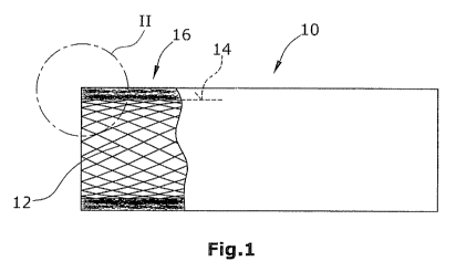
Fig. 1 is a lateral view of a tubular vascular support to be used in a blood
vessel for holding open the blood vessel, wherein a part of the
illustrated vascular support is broken away to visualize the wall
structure of the vascular support, and
Fig, 2 is an enlarged view of the detail II in Fig. 1,
Fig. 1 shows, in lateral view, a tubular vascular support 10 (e.g. a stent)
comprising a reticular or net-shaped support body 12 whose cylindrical outer
side 14 is provided with an enclosure made of a layer 16 of biostable fleece
material. Said fleece material layer 16 comprises a random-laid fleece made of
CA 02801111 2012-11-29
WO 2011/151318 PCT/EP2011/058931
- 11 -
microfibers. Fleece material layer 16 has a larger porosity on its outer side
18
than on its inner side 20. By its outer side 18, fleece material layer 16 is
arranged in abutment on the vessel wall (not shown). As achieved by the
invention, tissue proliferating from the vessel wall will only partially
intrude
into the fleece material layer 16. Such a proliferation of the tissue will be
stopped at the latest in that region of the fleece material layer 16 which is
located near the support body 12, particularly on the inner side 20 of layer
16
whose pore size is selected to the effect that a further proliferation of
tissue
through the fleece material layer 16 will not be possible anymore.