Note: Descriptions are shown in the official language in which they were submitted.
SUTURE DELIVERY TOOLS FOR ENDOSCOPIC AND
ROBOT-ASSISTED SURGERY AND METHODS
FIELD OF INVENTION
[0002] The present invention relates to systems for packaging,
selecting and
delivering sutures to surgical sites within a patient during surgical
procedures including
minimally-invasive surgical procedures.
BACKGROUND OF INVENTION
[0003] Minimally invasive surgery (MIS) procedures avoid open
invasive
surgery in favor of closed or local surgery with less trauma. Minimally
invasive surgical
procedures typically involve remote manipulation of instruments with indirect
observation of the surgical field through an endoscope or similar device, and
are carried
out through a small access port through the skin or through a body cavity or
anatomical
opening. Minimally invasive medical techniques thereby reduce tissue damage
during
diagnostic or surgical procedures, thereby reducing patient recovery time,
discomfort,
and deleterious side effects. Minimally invasive medical techniques
consequently
shorten the average length of a hospital stay for a procedure when compared to
standard
open surgery.
[0004] One form of minimally invasive surgery is endoscopy. Probably
the
most common form of endoscopy is laparoscopy, which is minimally invasive
inspection and surgery inside the abdominal cavity. In standard laparoscopic
surgery, a
patient's abdomen is insufflated with gas, and cannula sleeves are passed
through small
(approximately 1/2 inch) incisions to provide access ports for laparoscopic
surgical
instruments. The laparoscopic surgical instruments generally include an
endoscope for
visualizing the surgical field and specialized surgical instruments which is,
in some
embodiments, passed through the access ports. The instruments can include
clamps,
graspers, scissors, staplers, and needle holders, for example. The surgical
instruments
1
CA 2801271 2017-11-29
CA 02801271 2012-11-29
WO 2011/156733 PCT/US2011/040014
may or may not be similar to those used in conventional (open) surgery;
typically that
the working end of each instrument is separated from its handle by an
elongated shaft
and is sized and configured to fit through the access port. To perform
surgical
procedures, the surgeon passes the surgical instruments through the access
ports to an
internal surgical site and manipulates them from outside the abdomen. The
surgeon
monitors the procedure by means of a monitor that displays an image of the
surgical site
taken from the laparoscope. Similar endoscopic techniques are employed in,
e.g.,
arthroscopy, thoracoscopy, retroperitoneoscopy, pelviscopy, nephroscopy,
cystoscopy,
cisternoscopy, sinoscopy, hysteroscopy, urethroscopy, craniotomy, and natural
orifice
surgery (for example of the airway and gastrointestinal tract).
[0005] There are many disadvantages relating to MIS technology utilizing
hand-
operated instruments. For example, existing MIS instruments deny the surgeon
the
flexibility of instrument placement found in open surgery. Most current
laparoscopic
instruments have rigid shafts, so that it can be difficult to approach the
surgical site
through the small incision. Additionally, the length and construction of many
endoscopic instruments reduces the surgeon's ability to feel forces exerted by
the
instrument on tissues and organs at the surgical site. The lack of dexterity
and
sensitivity of endoscopic instruments is an impediment to the expansion of
minimally
invasive surgery.
[0006] Minimally invasive telesurgery systems have been developed to
increase
a surgeon's dexterity when working within an internal surgical site, as well
as to allow a
surgeon to operate on a patient from a remote location. In a telesurgery
system, the
surgeon is provided with an image of the surgical site as with endoscopy.
However,
rather than manipulating the surgical instruments directly, the surgeon
performs the
surgical procedures on the patient by manipulating master input or control
devices at a
console. The master input and control devices control the motion of surgical
instruments utilizing telemanipulators. Depending on the system, telesurgery
systems
may overcome some but not all of the lack of dexterity and sensitivity of
endoscopic
instruments. Surgical telemanipulator systems are often referred to as robotic
or
robotically-assisted surgery systems.
[0007] Many MIS procedures including MIS telesurgery procedures employ
wound closure devices such as sutures, staples and tacks for closing wounds,
repairing
traumatic injuries or defects, joining tissues together (bringing severed
tissues into
2
CA 02801271 2012-11-29
WO 2011/156733 PCT/US2011/040014
approximation, closing an anatomical space, affixing single or multiple tissue
layers
together, creating an anastomosis between two hollow/luminal structures,
adjoining
tissues, attaching or reattaching tissues to their proper anatomical
location), attaching
foreign elements to tissues (affixing medical implants, devices, prostheses
and other
functional or supportive devices), and for repositioning tissues to new
anatomical
locations (repairs, tissue elevations, tissue grafting and related procedures)
to name but
a few examples. Sutures typically consist of a filamentous suture thread
attached to a
needle with a sharp point. Suture threads can be made from a wide variety of
materials
including bioabsorbable (i.e., that break down completely in the body over
time), or
non-absorbable (permanent; non-degradable) materials. Absorbable sutures have
been
found to be particularly useful in situations where suture removal might
jeopardize the
repair or where the natural healing process renders the support provided by
the suture
material unnecessary after wound healing has been completed; as in, for
example,
completing an uncomplicated skin closure. Non-degradable (non-absorbable)
sutures
are used in wounds where healing is, in some embodiments, expected to be
protracted
or where the suture material is needed to provide physical support to the
wound for long
periods of time; as in, for example, deep tissue repairs, high tension wounds,
many
orthopedic repairs and some types of surgical anastomosis. Also, a wide
variety of
surgical needles are available, and the shape, and size of the needle body and
the
configuration of the needle tip is typically selected based upon the needs of
the
particular application.
[0008] To use an ordinary suture, the suture needle is advanced through
the
desired tissue on one side of the wound and then through the adjacent side of
the
wound. The suture is then formed into a "loop" which is completed by tying a
knot in
the suture to hold the wound closed. Knot tying takes time and causes a range
of
complications, including, but not limited to (i) spitting (a condition where
the suture,
usually a knot) pushes through the skin after a subcutaneous closure), (ii)
infection
(bacteria are often able to attach and grow in the spaces created by a knot),
(iii)
bulk/mass (a significant amount of suture material left in a wound is the
portion that
comprises the knot), (iv) slippage (knots can slip or come untied), and (v)
irritation
(knots serve as a bulk "foreign body" in a wound). Suture loops associated
with knot
tying may lead to ischemia (knots can create tension points that can
strangulate tissue
and limit blood flow to the region) and increased risk of dehiscence or
rupture at the
3
surgical wound. Knot tying is also labor intensive and can comprise a
significant
percentage of the time spent closing a surgical wound. Additional operative
procedure
time is not only bad for the patient (complication rates rise with time spent
under
anesthesia), but it also adds to the overall cost of the operation (many
surgical
procedures are estimated to cost between $15 and $30 per minute of operating
time).
The time taken by suture tying and the range of complications is exasperated
by the
lack of dexterity and sensitivity of MIS instruments.
100091 Self-
retaining sutures (including barbed sutures) differ from
conventional sutures in that self-retaining sutures possess numerous tissue
retainers
(such as barbs) which anchor the self-retaining suture into the tissue
following
deployment and resist movement of the suture in a direction opposite to that
in which
the retainers face, thereby eliminating the need to tie knots to affix
adjacent tissues
together (a "knotless" closure). This facilitates and expedites deployment of
self-
retaining sutures compared to ordinary sutures. Knotless tissue-approximating
devices
having barbs have been previously described in, for example, U.S. Patent No.
5,374,268, disclosing armed anchors having barb-like projections, while suture
assemblies having barbed lateral members have been described in U.S. Patent
Nos.
5,584,859 and 6,264,675. Sutures having a plurality of barbs positioned along
a greater
portion of the suture are described in U.S. Pat No. 5,931,855, which discloses
a
unidirectional barbed suture, and U.S. Patent No. 6,241,747, which discloses a
bidirectional barbed suture. Methods and apparatus for forming barbs on
sutures have
been described in, for example, U.S. Patent Nos. 6,848,152. Self-retaining
sutures result
in better approximation of the wound edges, evenly distribute the tension
along the
length of the wound (reducing areas of tension that can break or lead to
ischemia),
decrease the bulk of suture material remaining in the wound (by eliminating
knots) and
reduce spitting (the extrusion of suture material ¨ typically knots - through
the surface
of the skin. All of these features are thought to reduce scarring, improve
cosmesis, and
increase wound strength relative to wound closures using plain sutures or
staples. Thus,
self-retaining sutures, because such sutures avoid knot tying, allow patients
to
experience an improved clinical outcome, and also save time and costs
associated with
extended surgeries and follow-up treatments.
4
CA 2801271 2017-11-29
CA 02801271 2012-11-29
WO 2011/156733 PCT/US2011/040014
SUMMARY OF INVENTION
[0010] The present invention is generally directed to surgical
instruments for
delivering sutures and in particular self-retaining sutures to a surgical site
in an MIS
procedure including a robot-assisted MIS procedures. Despite the multitude of
advantages of unidirectional and bidirectional self-retaining sutures for MIS
and
telesurgical MIS, there remains a need to improve upon the design of the
suture such
that the functionality is enhanced and/or additional functionality is
provided. The
present invention overcomes the problems and disadvantages of the prior art by
providing packages and systems for delivering self-retaining sutures to the
surgical site.
The self-retaining sutures can be deployed by endoscopic and/or telesurgical
instruments at the surgical site for suturing, approximating and holding
tissue. The self-
retaining sutures provide advantages which compensate for lack of dexterity
and
sensitivity present in instruments used MIS and telesurgical MIS procedures.
In this
way, the time taken for the procedure is reduced and the clinical outcome is
enhanced.
[0011] In accordance with an aspect of the present invention, a method
of
performing MIS procedure in a body cavity of a patient includes providing a
suture
package containing a suture or self-retaining suture and introducing the
package to an
operative site within a patient for use during an MIS procedure. The suture or
self-
retaining suture is then manipulated by the MIS instrument to suture,
approximate
and/or hold tissue.
[0012] In some embodiments, the suture package is introduced into the
cavity
using a telesurgical suture delivery instrument. The suture delivery
instrument delivers
suture to the cavity under the control of the surgeon and positions the suture
such that it
is, in some embodiments, located by the surgeon and manipulated using MIS
instruments.
[0013] In some embodiments, the suture package is introduced into the
cavity
using a telesurgical suture delivery system. The telesurgical suture delivery
system
delivers suture to the cavity using a telemanipulator under the control of the
surgeon
and positions the suture such that it is, in some embodiments, located by the
surgeon
and manipulated by MIS instruments.
[0014] In some embodiments, the suture package includes a spool for
the suture
and self-retaining suture. The spool releasably secures one or more self-
retaining
sutures and surgical needles therein.
[0015] In a specific embodiment, a cartridge releasably secures one
or more
sutures. A cartridge is selected and attached to the suture delivery system
which
delivers the cartridge and suture to the surgical site. In some embodiments, a
variety of
different cartridges is available having different sutures.
[0016] In a specific embodiment, a cartridge releasably secures one
or more
sutures. A cartridge is selected and attached to the suture delivery system
which
delivers the cartridge and suture to the surgical site.
[0017] In some embodiments, different cartridges are available having
different
sutures the cartridges have features which allow them to be identified and/or
selected by
an automated delivery system responsive to instructions from a surgeon.
[0018] In some embodiments, suture cartridges provided with visible
and/or
machine readable markings, codes, tags or the like which are indicative of one
or more
properties of a suture loaded in the cartridge.
[0019] The details of one or more embodiments are set forth in the
description
below, including the embodiments identified in paragraphs 124 to 195. Other
features,
objects and advantages will be apparent from the description, the drawings,
and the
claims.
BRIEF DESCRIPTION OF THE DRAWINGS
[0020] Features of the invention, its nature and various advantages
will be
apparent from the accompanying drawings and the following detailed description
of
various embodiments.
[0021] FIG. lA shows a perspective view of a bidirectional self-
retaining suture
in accordance with an embodiment of the present invention.
[0022] FIG. 1B shows an enlarged views of a portion of the
bidirectional suture
of FIG. IA.
[0023] FIG. 1C shows a view of a suture delivery instrument according
to an
embodiment of the present invention.
6
CA 2801271 2017-11-29
CA 02801271 2012-11-29
WO 2011/156733 PCT/US2011/040014
[0024] FIG. 1D shows an enlarged view of an embodiment of a suture spool
which is, in some embodiments, used with the suture delivery instrument of FIG
1C
according to an embodiment of the present invention.
[0025] FIG. lE shows a sectional view of a subject illustrating delivery
of a
suture using the suture delivery instrument of FIG. 1C.
[0026] FIG. 1F shows a view of a surgical site illustrating use of the
suture
delivery instrument of FIG 1C.
[0027] FIG. 1G shows an image of a surgical site provided to a surgeon
and
illustrating use of the suture delivery instrument of FIG 1C.
[0028] FIG. 11-I includes a portion of the suture of FIG. lA also
including an
optional pledget.
[0029] FIG. 2A shows a suture delivery tool suitable for use with a
robotically-
assisted surgery system in accordance with an embodiment of the present
invention; and
a surgical manipulator suitable for use with the suture delivery tool.
[0030] FIGS. 2B and 2C shows introduction of a suture delivery tool by a
surgical manipulator in accordance with one embodiment of the present
invention.
[0031] FIG. 2D shows a side view of a suture delivery tool mounted to a
surgical manipulator.
[0032] FIGS. 2E and 2F show a surgery system, and a schematic
description
thereof, for controlling the surgical manipulators and suture delivery tools
of FIGS. 2A-
2D.
[0033] FIGS. 3A and 3B show suture cartridge delivery utilizing an
alternative
suture delivery tool according to an embodiment of the present invention.
[0034] FIGS. 3C and 3D show internal and sectional views, respectively,
of the
suture delivery tool of FIGS 3A and 3B.
[0035] FIGS. 3E and 3F show different views of a suture delivery
cartridge
suitable for use with the suture delivery tool of FIGS. 3A through 3D
according to an
embodiment of the present invention.
[0036] FIGS. 4A to 4H show suture cartridges according to embodiments of
the
present invention.
[0037] FIGS. 5A to 5C show a suture cartridge magazine according to an
embodiment of the present invention.
7
[0038] FIGS. 6A-6D show alternative self-retaining suture systems
which have
an anchor at one end.
[0039] FIGS. 6E-6F show views of a cartridge for holding one more of
the
alternative self-retaining suture systems of FIGS. 6A-6D.
[0040] FIGS. 6G-6H show views of a cartridge/spool for holding one
more of
the alternative self-retaining suture systems of FIGS. 6A-6D.
DETAILED DESCRIPTION
DEFINITIONS
[0041] Definitions of certain terms that is, in some embodiments,
used
hereinafter include the following.
[0042] "Self-retaining system" refers to a self-retaining suture
together with
devices for deploying the suture into tissue. Such deployment devices include,
without
limitation, suture needles and other deployment devices as well as
sufficiently rigid and
sharp ends on the suture itself to penetrate tissue.
[0043] "Self-retaining suture" refers to a suture that comprises
features on the
suture filament for engaging tissue without the need for a knot or suture
anchor. Self-
retaining sutures as described herein arc produced by any suitable method,
including
without limitation, injection molding, stamping, cutting, laser, extrusion,
and so forth.
With respect to cutting, polymeric thread or filaments is, in some
embodiments,
manufactured or purchased for the suture body, and the retainers can be
subsequently
cut onto the suture body; the retainers are, in some embodiments, hand-cut,
laser-cut, or
mechanically machine-cut using blades, cutting wheels, grinding wheels, and so
forth.
During cutting either the cutting device or the suture thread is, in some
embodiments,
moved relative to the other, or both is, in some embodiments, moved, to
control the
size, shape and depth of cut 210. Particular methods for cutting barbs on a
filament are
described in U.S. Patent Application Serial No. 09/943,733 titled "Method Of
Forming
Barbs On A Suture And Apparatus For Performing Same" to Genova et al., and
U.S.
Patent Application Serial No. 10/065,280 titled "Barbed Sutures" to Leung et
al.
[0044] "Tissue retainer" (or simply "retainer") refers to a physical
feature of a
suture filament which is adapted to mechanically engage tissue and resist
movement of
the suture in at least one axial directions. By way of example only, tissue
retainer or
8
CA 2801271 2017-11-29
CA 02801271 2012-11-29
WO 2011/156733 PCT/US2011/040014
retainers can include hooks, projections, barbs, darts, extensions, bulges,
anchors,
protuberances, spurs, bumps, points, cogs, tissue engagers, traction devices,
surface
roughness, surface irregularities, surface defects, edges, facets and the
like. In certain
configurations, tissue retainers are adapted to engage tissue to resist
movement of the
suture in a direction other than the direction in which the suture is deployed
into the
tissue by the physician, by being oriented to substantially face the
deployment direction.
In some embodiments, the retainers lie flat when pulled in the deployment
direction and
open or "fan out" when pulled in a direction contrary to the deployment
direction. As
the tissue-penetrating end of each retainer faces away from the deployment
direction
when moving through tissue during deployment, the tissue retainers should not
catch or
grab tissue during this phase. Once the self-retaining suture has been
deployed, a force
exerted in another direction (often substantially opposite to the deployment
direction)
causes the retainers to be displaced from the deployment position (i.e.
resting
substantially along the suture body), forces the retainer ends to open (or
"fan out") from
the suture body in a manner that catches and penetrates into the surrounding
tissue, and
results in tissue being caught between the retainer and the suture body;
thereby
"anchoring" or affixing the self-retaining suture in place. In certain other
embodiments,
the tissue retainers is, in some embodiments, configured to permit motion of
the suture
in one direction and resist movement of the suture in another direction
without fanning
out or deploying. In certain other configurations, the tissue retainer is, in
some
embodiments, configured or combined with other tissue retainers to resist
motion of the
suture filament in both directions. Typically a suture having such retainers
is deployed
through a device such as a cannula which prevents contact between the
retainers and the
tissue until the suture is in the desired location. In some embodiments,
mechanical
retainers are replaced and/or augmented with chemical and/or adhesive
retainers which
engage tissue by adhering or physically and/or chemically bonding the suture
to
surrounding tissue.
[0045] "Retainer configurations" refers to configurations of tissue
retainers and
can include features such as size, shape, flexibility, surface
characteristics, and so forth.
These are sometimes also referred to as "barb configurations".
[0046] "Retainer distribution" refers to the arrangement of retainers on
the
surface of a filament and can include variables such as orientation, pattern,
pitch, and
spirality angle.
9
CA 02801271 2012-11-29
WO 2011/156733 PCT/US2011/040014
[0047] "Bidirectional suture" refers to a self-retaining suture having
retainers
oriented in one direction at one end and retainers oriented in the other
direction at the
other end. A bidirectional suture is typically armed with a needle at each end
of the
suture thread. Many bidirectional sutures have a transition segment located
between the
two barb orientations.
[0048] "Transition segment" refers to a retainer-free (barb-free)
portion of a
bidirectional suture located between a first set of retainers (barbs) oriented
in one
direction and a second set of retainers (barbs) oriented in another direction.
The
transition segment can be at about the midpoint of the self-retaining suture,
or closer to
one end of the self-retaining suture to form an asymmetrical self-retaining
suture
system.
[0049] "Suture thread" refers to the filamentary body component of the
suture.
The suture thread is, in some embodiments, a monofilament, or comprise
multiple
filaments as in a braided suture. The suture thread is, in some embodiments,
made of
any suitable biocompatible material, and is, in some embodiments, further
treated with
any suitable biocompatible material, whether to enhance the sutures' strength,
resilience, longevity, or other qualities, or to equip the sutures to fulfill
additional
functions besides joining tissues together, repositioning tissues, or
attaching foreign
elements to tissues.
[0050] "Monofilament suture" refers to a suture comprising a
monofilamentary
suture thread.
[0051] "Braided suture" refers to a suture comprising a multifilamentary
suture
thread. The filaments in such suture threads are typically braided, twisted,
or woven
together.
100521 "Degradable suture" (also referred to as "biodegradable suture"
or
"absorbable suture") refers to a suture which, after introduction into a
tissue is broken
down and absorbed by the body. Typically, the degradation process is at least
partially
mediated by, or performed in, a biological system. "Degradation" refers to a
chain
scission process by which a polymer chain is cleaved into oligomers and
monomers.
Chain scission may occur through various mechanisms, including, for example,
by
chemical reaction (e.g., hydrolysis, oxidation/reduction, enzymatic mechanisms
or a
combination of these) or by a thermal or photolytic process. Polymer
degradation is, in
some embodiments, characterized, for example, using gel permeation
chromatography
CA 02801271 2012-11-29
WO 2011/156733 PCT/US2011/040014
(GPC), which monitors the polymer molecular mass changes during erosion and
breakdown. Degradable suture material may include polymers such as
polyglycolic
acid, copolymers of glycolide and lactide, copolymers of trimethylene
carbonate and
glycolide with diethylene glycol (e.g., MAXONTM, Tyco Healthcare Group),
terpolymer composed of glycolide, trimethylene carbonate, and dioxanone (e.g.,
BIOSYNTM [glycolide (60%), trimethylene carbonate (26%), and dioxanone (14%)],
Tyco Healthcare Group), copolymers of glycolide, caprolactone, trimethylene
carbonate, and lactide (e.g., CAPROSYNTM, Tyco Healthcare Group). A
dissolvable
suture can also include partially deacetylated polyvinyl alcohol. Polymers
suitable for
use in degradable sutures can be linear polymers, branched polymers or multi-
axial
polymers. Examples of multi-axial polymers used in sutures are described in
U.S.
Patent Application Publication Nos. 2002/0161168, 2004/0024169, and
2004/0116620.
Sutures made from degradable suture material lose tensile strength as the
material
degrades. Degradable sutures can be in either a braided multifilament form or
a
monofilament form.
[0053] "Non-degradable suture" (also referred to as "non-absorbable
suture")
refers to a suture comprising material that is not degraded by chain scission
such as
chemical reaction processes (e.g., hydrolysis, oxidation/reduction, enzymatic
mechanisms or a combination of these) or by a thermal or photolytic process.
Non-
degradable suture material includes polyamide (also known as nylon, such as
nylon 6
and nylon 6,6), polyester (e.g., polyethylene terephthlate),
polytetrafluoroethylene (e.g.,
expanded polytetrafluoroethylene), polyether-ester such as polybutester (block
copolymer of butylene terephthalate and polytetra methylene ether glycol),
polyurethane, metal alloys, metal (e.g., stainless steel wire), polypropylene,
polyethelene, silk, and cotton. Sutures made of non-degradable suture material
are
suitable for applications in which the suture is meant to remain permanently
or is meant
to be physically removed from the body.
[0054] "Suture diameter" refers to the diameter of the body of the
suture. It is to
be understood that a variety of suture lengths is, in some embodiments, used
with the
sutures described herein and that while the term "diameter" is often
associated with a
circular periphery, it is to be understood herein to indicate a cross-
sectional dimension
associated with a periphery of any shape. Suture sizing is based upon
diameter. United
States Pharmacopeia ("USP") designation of suture size runs from 0 to 7 in the
larger
11
CA 02801271 2012-11-29
WO 2011/156733 PCT/US2011/040014
range and 1-0 to 11-0 in the smaller range; in the smaller range, the higher
the value
preceding the hyphenated zero, the smaller the suture diameter. The actual
diameter of a
suture will depend on the suture material, so that, by way of example, a
suture of size 5-
0 and made of collagen will have a diameter of 0.15 mm, while sutures having
the same
USP size designation but made of a synthetic absorbable material or a non-
absorbable
material will each have a diameter of 0.1 mm. The selection of suture size for
a
particular purpose depends upon factors such as the nature of the tissue to be
sutured
and the importance of cosmetic concerns; while smaller sutures is, in some
embodiments, more easily manipulated through tight surgical sites and are
associated
with less scarring, the tensile strength of a suture manufactured from a given
material
tends to decrease with decreasing size. It is to be understood that the
sutures and
methods of manufacturing sutures disclosed herein are suited to a variety of
diameters,
including without limitation 7, 6, 5, 4, 3, 2, 1, 0, 1-0, 2-0, 3-0, 4-0, 5-0,
6-0, 7-0, 8-0, 9-
0, 10-0 and 11-0.
[0055] "Needle attachment" refers to the attachment of a needle to a
suture
requiring same for deployment into tissue, and can include methods such as
crimping,
swaging, using adhesives, and so forth. The suture thread is attached to the
suture
needle using methods such as crimping, swaging and adhesives. Attachment of
sutures
and surgical needles is described in U.S. Patent Nos. 3,981,307, 5,084,063,
5,102,418,
5,123,911, 5,500,991, 5,722,991, 6,012,216, and 6,163,948, and U.S. Patent
Application Publication No. US 2004/0088003). The point of attachment of the
suture
to the needle is known as the swage.
[0056] "Suture needle" refers to needles used to deploy sutures into
tissue,
which come in many different shapes, forms and compositions. There are two
main
types of needles, traumatic needles and atraumatic needles. Traumatic needles
have
channels or drilled ends (that is, holes or eyes) and arc supplied separate
from the suture
thread and are threaded on site. Atraumatic needles are eyeless and are
attached to the
suture at the factory by swaging or other methods whereby the suture material
is
inserted into a channel at the blunt end of the needle which is then deformed
to a final
shape to hold the suture and needle together. As such, atraumatic needles do
not require
extra time on site for threading and the suture end at the needle attachment
site is
generally smaller than the needle body. In the traumatic needle, the thread
comes out of
the needle's hole on both sides and often the suture rips the tissues to a
certain extent as
12
CA 02801271 2012-11-29
WO 2011/156733 PCT/US2011/040014
it passes through. Most modern sutures are swaged atraumatic needles.
Atraumatic
needles is, in some embodiments, permanently swaged to the suture or is, in
some
embodiments, designed to come off the suture with a sharp straight tug. These
"pop-
offs" are commonly used for interrupted sutures, where each suture is only
passed once
and then tied. For barbed sutures that are uninterrupted, these atraumatic
needles are
preferred.
[0057] Suture needles may also be classified according to the geometry
of the
tip or point of the needle. For example, needles is, in some embodiments, (i)
"tapered"
whereby the needle body is round and tapers smoothly to a point; (ii)
"cutting" whereby
the needle body is triangular and has a sharpened cutting edge on the inside;
(iii)
"reverse cutting" whereby the cutting edge is on the outside; (iv) "trocar
point" or
"taper cut" whereby the needle body is round and tapered, but ends in a small
triangular
cutting point; (v) "blunt" points for sewing friable tissues; (vi) "side
cutting" or "spatula
points" whereby the needle is flat on top and bottom with a cutting edge along
the front
to one side (these are typically used for eye surgery).
[0058] Suture needles may also be of several shapes including, (i)
straight, (ii)
half curved or ski, (iii) 1/4 circle, (iv) 3/8 circle, (v) 1/2 circle, (vi)
5/8 circle, (v) and
compound curve.
[0059] Suturing needles are described, for example, in US Patent Nos.
6,322,581 and 6,214,030 (Mani, Inc., Japan); and 5,464,422 (W.L. Gore, Newark,
DE);
and 5,941,899; 5,425,746; 5,306,288 and 5,156,615 (US Surgical Corp., Norwalk,
CT);
and 5,312,422 (Linvatec Corp., Largo, FL); and 7,063,716 (Tyco Healthcare,
North
Haven, CT). Other suturing needles are described, for example, in US Patent
Nos.
6,129,741; 5,897,572; 5,676,675; and 5,693,072. The sutures described herein
is, in
some embodiments, deployed with a variety of needle types (including without
limitation curved, straight, long, short, micro, and so forth), needle cutting
surfaces
(including without limitation, cutting, tapered, and so forth), and needle
attachment
techniques (including without limitation, drilled end, crimped, and so forth).
Moreover,
the sutures described herein may themselves include sufficiently rigid and
sharp ends so
as to dispense with the requirement for deployment needles altogether.
[0060] "Needle diameter" refers to the diameter of a suture deployment
needle
at the widest point of that needle. While the term "diameter" is often
associated with a
13
CA 02801271 2012-11-29
WO 2011/156733 PCT/US2011/040014
circular periphery, it can be understood herein to indicate a cross-sectional
dimension
associated with a periphery of any shape.
[0061] "Armed suture" refers to a suture having a suture needle on at
least one
suture deployment end. "Suture deployment end" refers to an end of the suture
to be
deployed into tissue; one or both ends of the suture is, in some embodiments,
suture
deployment ends. The suture deployment end is, in some embodiments, attached
to a
deployment device such as a suture needle, or is, in some embodiments,
sufficiently
sharp and rigid to penetrate tissue on its own.
[0062] "Wound closure" refers to a surgical procedure for closing of a
wound.
An injury, especially one in which the skin or another external or internal
surface is cut,
torn, pierced, or otherwise broken is known as a wound. A wound commonly
occurs
when the integrity of any tissue is compromised (e.g., skin breaks or burns,
muscle
tears, or bone fractures). A wound is, in some embodiments, caused by an act,
such as a
puncture, fall, or surgical procedure; by an infectious disease; or by an
underlying
medical condition. Surgical wound closure facilitates the biological event of
healing by
joining, or closely approximating, the edges of those wounds where the tissue
has been
torn, cut, or otherwise separated. Surgical wound closure directly apposes or
approximates the tissue layers, which serves to minimize the volume new tissue
formation required to bridge the gap between the two edges of the wound.
Closure can
serve both functional and aesthetic purposes. These purposes include
elimination of
dead space by approximating the subcutaneous tissues, minimization of scar
formation
by careful epidermal alignment, and avoidance of a depressed scar by precise
eversion
of skin edges.
[0063] "Tissue elevation procedure" refers to a surgical procedure for
repositioning tissue from a lower elevation to a higher elevation (i.e. moving
the tissue
in a direction opposite to the direction of gravity). The retaining ligaments
of the face
support facial soft tissue in the normal anatomic position. However, with age,
gravitational effects and loss of tissue volume effect downward migration of
tissue, and
fat descends into the plane between the superficial and deep facial fascia,
thus causing
facial tissue to sag. Face-lift procedures are designed to lift these sagging
tissues, and
are one example of a more general class of medical procedure known as a tissue
elevation procedure. More generally, a tissue elevation procedure reverses the
appearance change that results from effects of aging and gravity over time,
and other
14
CA 02801271 2012-11-29
WO 2011/156733 PCT/US2011/040014
temporal effects that cause tissue to sag, such as genetic effects. It should
be noted that
tissue can also be repositioned without elevation; in some procedures tissues
are
repositioned laterally (away from the midline), medially (towards the midline)
or
inferiorly (lowered) in order to restore symmetry (i.e. repositioned such that
the left and
right sides of the body "match").
[0064] "Medical device" or "implant" refers to any object placed in the
body for
the purpose of restoring physiological function, reducing/alleviating symptoms
associated with disease, and/or repairing and/or replacing damaged or diseased
organs
and tissues. While normally composed of biologically compatible synthetic
materials
(e.g., medical-grade stainless steel, titanium and other metals or polymers
such as
polyurethane, silicon, PLA, PLGA and other materials) that are exogenous, some
medical devices and implants include materials derived from animals (e.g.,
"xenografts" such as whole animal organs; animal tissues such as heart valves;
naturally
occurring or chemically-modified molecules such as collagen, hyaluronic acid,
proteins,
carbohydrates and others), human donors (e.g., "allografts" such as whole
organs;
tissues such as bone grafts, skin grafts and others), or from the patients
themselves (e.g.,
"autografts" such as saphenous vein grafts, skin grafts,
tendon/ligament/muscle
transplants). Medical devices that can be used in procedures in conjunction
with the
present invention include, but are not restricted to, orthopedic implants
(artificial joints,
ligaments and tendons; screws, plates, and other implantable hardware), dental
implants, intravascular implants (arterial and venous vascular bypass grafts,
hemodialysis access grafts; both autologous and synthetic), skin grafts
(autologous,
synthetic), tubes, drains, implantable tissue bulking agents, pumps, shunts,
sealants,
surgical meshes (e.g., hernia repair meshes, tissue scaffolds), fistula
treatments, spinal
implants (e.g., artificial intervertebral discs, spinal fusion devices, etc.)
and the like.
SUTURE DELIVERY FOR MINIMALLY-INVASIVE SURGERY
[0065] As discussed above, the present invention provides compositions,
configurations, methods of manufacturing and methods of utilizing self-
retaining
sutures. The invention overcomes the problems and disadvantages of the prior
art by
delivering self-retaining sutures to the surgical site. The self-retaining
sutures can be
manipulated by endoscopic and/or robotically-assisted surgical instruments at
the site
for suturing, approximating and holding tissue. A number of devices have been
proposed for delivery surgical elements and accessories for use in MIS
procedures.
Devices are disclosed, for example, in U.S. Patent 6,986,780 titled "Surgical
Element
Delivery System And Method" to Rudnick et al. and U.S. Patent 7,125,403 titled
"in
Vivo Accessories For Minimally Invasive Robotic Surgery" to Julian et al.
Endoscopic Suture Delivery System
[0066] A self-retaining suture is, in some embodiments,
unidirectional, having
one or more retainers oriented in one direction along the length of the suture
thread; or
bidirectional, typically having one or more retainers oriented in one
direction along a
portion of the thread, followed by one or more retainers oriented in another
(often
opposite) direction over a different portion of the thread (as described with
barbed
retainers in U.S. Patent Nos. 5,931,855 and. 6,241,747). Although any number
of
sequential or intermittent configurations of retainers are possible, a common
form of
bidirectional self-retaining suture involves a needle at one end of a suture
thread which
has barbs having tips projecting "away" from the needle until the transition
point (often
the midpoint) of the suture is reached; at the transition point the
configuration of barbs
reverses itself about 180 (such that the barbs are now facing in the opposite
direction)
along the remaining length of the suture thread before attaching to a second
needle at
the opposite end (with the result that the barbs on this portion of the suture
also have
tips projecting "away" from the nearest needle). Projecting "away" from the
needle
means that the tip of the barb is further away from the needle and the portion
of suture
comprising the barb is, in some embodiments, pulled more easily through tissue
in the
direction of the needle than in the opposite direction. Put another way, the
barbs on both
"halves" of a typical bidirectional self-retaining suture have tips that point
towards the
middle, with a transition segment (lacking barbs) interspersed between them,
and with a
needle attached to either end.
[0067] FIG. IA illustrates a self-retaining suture system 100. Self-
retaining
suture system 100 comprises needles 110, 112 attached to self-retaining suture
thread
102. Self-retaining suture thread 102 includes a plurality of retainers 130
distributed on
the surface of a filament 120. In lead-in section 140 of filament 120 there
are no
retainers 130. In section 142 of filament 120, there are a plurality of
retainers 130
arranged such that the suture can be deployed in the direction of needle 110,
but resists
16
CA 2801271 2017-11-29
CA 02801271 2012-11-29
WO 2011/156733 PCT/US2011/040014
movement in the direction of needle 112. In transition section 144, there are
no retainers
130. Transition section 122 is, in some embodiments, provided with a marker to
facilitate location of the transition section. Transition section 122, as
shown, is provided
with a visible band 122 to help identify the transition section. Markers are
in some
embodiments also provided on sections 142, 146 and/or needles 110, 112 in
order to
help identify the retainer location and orientation of a particular portion of
self-retaining
suture system 100. In section 146, there is a plurality of retainers 130
arranged such
that the suture can be deployed in the direction of needle 112, but resists
movement in
the direction of needle 110. The retainers 130 in section 146 are larger than
the retainers
130 in section 142. The larger retainers are better suited for gripping tissue
that is softer
and/or less dense than the smaller retainers. In lead-in section 148 of
filament 120 there
are no retainers 130.
[0068] A break is shown in each of sections 140, 142, 144, 146 and 148
to
indicate that the length of each section is, in some embodiments, varied and
selected
depending upon the application for which the suture is intended to be used.
For
example, transition section 144 can be asymmetrically located closer to needle
110 or
needle 112, if desired. A self-retaining suture having an asymmetrically
located
transition section 144 is, in some embodiments, favored by a physician that
prefers to
use his dominant hand in techniques that require suturing in opposite
directions along a
wound. The physician may start further from one end of the wound than the
other and
stitch the longer portion of the wound with the needle that is located further
from the
transition section 144. This allows a physician to use his dominant hand to
stitch the
majority of the wound with the longer arm of the suture. The longer arm of the
suture is
that section of suture between the transition section and the needle which is
located
further from the transition section.
[0069] FIG. 1B illustrates a magnified view of self-retaining suture
thread 102
in section 142. As shown in FIG. 1B, a plurality of retainers 130 is
distributed on the
surface of filament 120. The affixation of self-retaining sutures after
deployment in
tissue entails the penetration of retainer ends 132 into the surrounding
tissue resulting in
tissue being caught between the retainer 130 and the body of suture filament
120. The
inner surface 134 of the retainer 130 that is in contact with the tissue that
is caught
between the retainer 130 and the body of filament 120, is referred to herein
as the
"tissue engagement surface" or "inner retainer surface." As illustrated in
FIG. 1B, each
17
CA 02801271 2012-11-29
WO 2011/156733 PCT/US2011/040014
retainer 130 has a tip 132 and tissue retainer surface 134. When self-
retaining suture
thread 102 is moved in the direction of arrow 136, retainers 130 lies flat
against the
body of filament 120. However, when self-retaining suture thread 102 is moved
in the
direction of arrow 138, tip 132 of retainer 130 engages tissue surrounding
filament 120
and causes retainer 130 to fan out from filament 120 and engage the tissue
with tissue
engagement surface 134 thereby preventing movement of the suture in that
direction.
100701 In alternative embodiments, a pledget can be applied to a self-
retaining
suture. FIG. 1H depicts a pledget 124 located in the transition zone 144 of
self-retaining
suture system 100. In some embodiments, a pledget 124 can carry a marker/code
128
which helps identify the suture and/or properties thereof Pledget 124 has one
or more
apertures 126 through which suture thread 120 can be passed as shown.
Alternatively, a
pledget can be bonded and/or mechanically fixed to suture thread 120, by, for
example,
welding, clipping, gluing, fusing. The pledget 126 can be used for locating
the
transition zone, for providing a stop so that the pledget can be pulled
through tissue
only until the pledget contacts the tissue, and/or for providing a support to
tissue and
organs, to name just a few uses. The pledget 126 can take many forms including
a wider
section that can support tissue.
[0071] The ability of self-retaining sutures to anchor and hold tissues
in place
even in the absence of tension applied to the suture by a knot is a feature
that also
provides superiority over plain sutures. When closing a wound that is under
tension,
this advantage manifests itself in several ways: (i) self-retaining sutures
have a
multiplicity of retainers which can dissipate tension along the entire length
of the suture
(providing hundreds of "anchor" points this produces a superior cosmetic
result and
lessens the chance that the suture will "slip" or pull through) as opposed to
knotted
interrupted sutures which concentrate the tension at discrete points; (ii)
complicated
wound geometries can be closed (circles, arcs, jagged edges) in a uniform
manner with
more precision and accuracy than can be achieved with interrupted sutures;
(iii) self-
retaining sutures eliminate the need for a "third hand" which is often
required for
maintaining tension across the wound during traditional suturing and knot
tying (to
prevent "slippage" when tension is momentarily released during tying); (iv)
self-
retaining sutures are superior in procedures where knot tying is technically
difficult,
such as in deep wounds or laparoscopic/endoscopic procedures; and (v) self-
retaining
sutures can be used to approximate and hold the wound prior to definitive
closure. As a
18
CA 02801271 2012-11-29
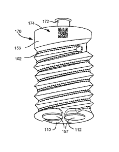
WO 2011/156733 PCT/US2011/040014
result, self-retaining sutures provide easier handling in anatomically tight
or deep places
(such as the pelvis, abdomen and thorax) and make it easier to approximate
tissues in
laparoscopic/endoscopic and minimally invasive procedures; all without having
to
secure the closure via a knot. Greater accuracy allows self-retaining sutures
to be used
for more complex closures (such as those with diameter mismatches, larger
defects or
purse string suturing) than can be accomplished with plain sutures. The
superior
qualities of self-retaining suture are particularly beneficial in endoscopic
and
telesurgical procedures. Self-retaining suture help overcome the limitations
of dexterity
and sensitivity present in endoscopic and telesurgical instruments.
[0072] FIG. 1C shows an endoscopic suture delivery instrument 150 for
delivering a self-retaining suture system 100 to a surgical site within a
patient. Suture
delivery instrument 150 includes, at the proximal end, a handle 152 connected
by an
elongated tubular member 154 to a spool 156. Handle 152 allows for positioning
and
operation of the suture delivery instrument 150 from outside of the body of
the patient.
Handle 152 may include one or more actuators 158 which is, in some
embodiments,
moved relative to one another and/or handle 152 for operating an effector,
such as
surgical scissors, a delivery spool, etc., located on the suture delivery
instrument.
[0073] Elongated tubular member connects handle 152 (proximal end) to
spool
156 (distal end). Elongated tubular member 154 is a rigid member which is
sized to fit
through an access port into the body of the patient. Preferably, the tubular
member 154
is about or less than 12 mm, 8 mm and 5 mm. Elongated tubular member 154 must
be
long enough to reach the desired surgical site through the access port. For
laparoscopic
instruments, for example, elongated tubular member 154 is between 180 mm and
450
mm in length and is typically 360 mm in length for adults and 280 mm in length
for
pediatric surgery. Typically the access port will be 12mm in diameter in less.
Preferably
the access port will be 10 mm in diameter or less. In some case the access
port is, in
some embodiments, 8 mm or 5 mm in diameter or less. In general smaller access
ports
are preferred to reduce trauma to patient tissues however, the parts must be
sufficiently
large to permit entry of instruments having the functionality to perform the
desired
surgical manipulations. The diameter of the elongated tubular member 154 and
spool
156 will be smaller than the inner diameter of the access port so that the
distal portion
of suture delivery instrument is, in some embodiments, introduce through the
access
port.
19
CA 02801271 2012-11-29
WO 2011/156733 PCT/US2011/040014
[0074] FIG. 1D shows a cartridge 170 which includes spool 156 and a
connector 172. Connector 172 allows cartridge 170 to be releasably attached to
the
distal end of elongated tubular member 154. In some embodiments, an actuator
158
controls the attaching and releasing of the cartridge 170. A selection of
sterile cartridges
170 is, in some embodiments, supplied for a procedure each supporting a
different self-
retaining suture. Thus, suture delivery instrument 150 can be used by the
surgeon or
assistant to select and deliver multiple self-retaining suture systems 100 in
the course of
a procedure. In alternative embodiments spool 156 is, in some embodiments,
permanently fixed to the end of suture delivery instrument 150. As shown in
FIG. ID,
spool 156 may also include one or more needle docks 157 for supporting the
needles
110, 112 of self-retaining suture system 100. Needles 110, 112 are releasable
attached
to needle docks 157. The needle 110, 112 are removed from needle docks 157 to
allow
deployment of self-retaining suture thread 102. In some embodiments, needles
110, 112
are replaced in needle docks 157 to allow removal of needles 110, 112 and any
surplus
self-retaining suture thread 102 after deployment of self-retaining suture
thread 102.
[0075] As also shown in FIG. 1D, cartridge 170 includes a marker 174. As
shown in FIG. 1D, marker 174 is a QR code. A QR code is a machine-readable
matrix
code or two-dimensional barcode designed to allow quick decoding of its
contents. In
particular QR codes can be quickly recognized and decoded in camera images.
The QR
code in some embodiments directly identifies properties of the suture and in
other cases
identifies the location (URL or other) of data identifying properties of the
suture. The
properties of the suture are then displayed with the image of the surgical
site provided
to the surgeon (See FIG. 1G). The information displayed allows the surgeon to
verify
that the cartridge is loaded with the desired suture. Although a QR code is
shown in
FIG. ID, potential markers include, but are not limited to: markers visible in
the visible
light frequency range; alphanumeric markers, QR code markers, markers
invisible to
the naked eye but which can be visualized under the conditions of surgical
use; markers
recognizable in the non-visible radiation frequency range; markers detectable
with
ultrasound; markers which are machine readable; markers which are human
readable;
markers which is, in some embodiments, read remotely; markers which are active
markers (including RFID); and markers which are passive markers (including
passive
RFID). The properties of the suture which can be associated with the marker
include,
but are not limited to: length, diameter, material, needles, presence of
retainers, absence
CA 02801271 2012-11-29
WO 2011/156733 PCT/US2011/040014
of retainers, source/brand and/or other fixed properties. In addition to fixed
or static
properties, a marker can be used to identify dynamic properties. For example,
movement of the cartridge and/or suture through forces being placed on the
cartridge
can cause the marker to move, and such movement can be noted by telesurgical
system
in order to track the changing location of the cartridge and the suture. Such
movement
can be translational movement or rotational movement. With the tracking of
rotational
movement of the spool, for example, the amount of suture removed from the
spool can
be tracked. Markings placed additionally on the suture can be used to identify
the
changing location of the suture and also, for example, tension placed on the
suture. The
markings can also be used with a voice-command telesurgical system. The
surgeon
speaks the type of suture desired, and the telesurgical system then loads the
cartridge
onto the end of a tool located on an arm of the telesurgical system for
deployment into a
patient.
[0076] Spool 156 is mounted on the distal end of elongated tubular
member 154
and is sized so that it may slide through an access port into the body of the
patient.
Spool 156 supports self-retaining suture system 100 thus allowing self-
retaining suture
system 100 to be delivered through an access port to the surgical site within
the patient.
FIG. lE shows the distal portion of suture delivery instrument 150 introduced
through
an access port 160 into a patient 162. Suture delivery instrument 150 is
inserted through
a cannula 164 at the access port 160. Suture delivery instrument 150 is, in
some
embodiments, slid in and out of cannula 164 as shown by arrow 166. Suture
delivery
instrument 150 and cannula 164 may also pivot about the access port 160 as
shown by
arrows 168. Thus, suture delivery instrument 150 allows spool 156 to be
delivered to a
surgical site within patient 162.
100771 FIG. IF shows delivery of a self-retaining suture system 100 to a
surgical site in a patient. As shown in FIG. 1F, an endoscope 180 illuminates
the
surgical site with one or more light sources 182. Endoscope 180 also images
the
surgical site through one or more imaging devices 184. Endoscope 180 thereby
illuminates the surgical site. The dashed circle 186 indicates the field of
view that is, in
some embodiments, transmitted to the surgeon. Note that suture delivery
instrument
150 has been inserted so as to position a spool 156 of a cartridge 170 within
the field of
view. The end effectors (scissors, forceps and the like) of one or more
endoscopic
surgical instruments 190 also appear in the field of view. The surgeon may
operate the
21
CA 02801271 2012-11-29
WO 2011/156733 PCT/US2011/040014
endoscopic surgical instruments 190 to grasp the needles 110, 112 supported by
spool
156. The surgeon may then operate the endoscopic surgical instruments 190 to
deploy
self-retaining suture thread 102 into tissue 192. After deployment of self-
retaining
suture thread 102, the surgeon may operate endoscopic surgical instruments 190
to
replace needle 110, 112 in spool 156 and cut off any unused self-retaining
suture thread
102. Suture delivery instrument 150 may then be removed from the surgical site
thereby
removing the needles and any excess self-retaining suture thread 102 from the
patient's
body.
[0078] FIG. 1G shows an example of an image 194 on a display 196 of the
surgical site of FIG. 1F as displayed to a surgeon. The dashed circle 186
indicates the
field of view available from the endoscope (not shown). Note that suture
delivery
instrument 150 has been inserted so as to position a spool 156 of a cartridge
170 within
the field of view 186. Marker 174 of cartridge 170 is visible in the image. A
computer
system associated with display 196 identifies and translates marker 174. As
shown in
FIG. 1G, suture property information 176 associated with marker 174 is
displayed to
the surgeon in the image 194. The information displayed allows the surgeon to
verify
that the cartridge is loaded with the desired suture. The information
displayed can be
static or dynamic information. For example, having identified the suture the
image
display system can also display other suture property information 176 relevant
to the
suture. For example tension sensed by the endoscopic tools or otherwise can be
displayed as a percentage graph of the maximum rated tension of the identified
suture.
Robot-Assisted Suture Delivery System
[0079] As described above, minimally invasive telesurgical systems have
been
developed to increase a surgeon's dexterity when working within an internal
surgical
site, as well as to allow a surgeon to operate on a patient from a remote
location. In a
telesurgery system, the surgeon is provided with an image of the surgical site
at a
console. While viewing an image of the surgical site on a suitable display,
the surgeon
performs the surgical procedures on the patient by manipulating input devices
of the
console. The input devices control a robot arm which positions and manipulates
the
surgical instrument. During the surgical procedure, the telesurgical system
can provide
mechanical actuation and control of a variety of surgical instruments or
instruments
having end effectors such as, e.g., tissue graspers, cautery, needle drivers,
or the like,
22
CA 02801271 2012-11-29
WO 2011/156733 PCT/US2011/040014
that perform various functions for the surgeon, e.g., holding or driving a
needle,
grasping a blood vessel, or dissecting tissue, or the like, in response to
manipulation of
the master control devices. The Intuitive Surgical, Inc. DA VINCI Surgical
System is
one example of a MIS telesurgical system.
[0080] In a telesurgical procedure, sutures, including self-retaining
suture
systems, can in some embodiments be introduced to the surgical site using
suture
delivery instrument 150 previously described with respect to FIGS. 1C-1G. The
suture
delivery instrument could be operated manually by the surgeon. However, this
requires
the surgeon to leave the workstation. Alternatively, the suture delivery
instrument 150
can be operated manually by a surgical assistant. However, this requires the
assistant to
insert the suture delivery instrument manually without the visualization
provided by the
workstation. According to another embodiment of the present invention, a
suture
delivery instrument is provided which interfaces with the telesurgery system.
The suture
delivery instrument is used to deliver the self-retaining suture to the
surgical site under
the command of the surgeon. Such a suture delivery instrument advantageously
leverages the abilities of the telesurgery system to accurately deliver the
self-retaining
suture to the surgical site under the control of the surgeon at the
workstation and using
the visualization capabilities of the telesurgery system. Moreover certain
portions of the
suture delivery operation is, in some embodiments, safely automated to
facilitate the
repeated delivery and extraction of sutures to the surgical site after initial
setup under
the control of the surgeon. The surgeon controls the suture delivery
instrument with one
or more inputs of the console, which can include, for example, switch,
keyboards,
motion controllers and/or voice input devices.
[0081] FIG. 2A shows a suture delivery tool 250 suitable for use with a
telesurgery system. Suture delivery tool 250 includes, at the proximal end, a
case 252
connected by a tool shaft 254 to an end effector including a spool 256. Case
252 can be
mounted to the interface 246 of a manipulator arm 240 to allow for positioning
and
operation of the suture delivery tool 250 from outside of the body of the
patient. Suture
delivery tool 250 includes a spool 256 mounted on the distal end of tool shaft
254.
Spool 256 supports self-retaining suture system 100 thus allowing self-
retaining suture
system 100 to be delivered through a cannula/guide 264 to the surgical site
within the
patient. Spool 256 is sized so that it may slide through cannula/guide 264
into the body
of the patient.
23
CA 02801271 2012-11-29
WO 2011/156733 PCT/US2011/040014
[0082] Tool shaft 254 connects case 252 (proximal end) to the spool 256
(distal
end). Tool shaft 254 is a rigid member which is sized to fit through an access
port into
the body of the patient. Alternatively, the tool shaft can be flexible. The
tool shaft itself
can be controlled by the telesurgical system so that the tool shaft can be
"snaked" to a
desired location. Tool shaft 254 must be long enough to reach the desired
surgical site
through the access port. The diameter of the tool shaft 254 and spool 256 must
be small
enough so that the distal portion of surgical tool 250 is, in some
embodiments,
introduced through the cannula tool guide/access port 264 into the patient.
Tool shaft
254 may contain one or more mechanical linkages for transferring motion from
the
gears 258 in the case to an end effector at the distal end of tool shaft 254.
[0083] FIG. 2A also shows the portion of a manipulator arm 240 to which
a
suture delivery tool 250 is, in some embodiments, mounted. The case 252 of
suture
delivery tool 250 (or another tool) is, in some embodiments, releasably
mounted to the
interface 246 on the manipulator arm 240. Case 252 includes one or more clips
253
which engage mating structures on interface 246 to hold case 252 to interface
246. Note
that interface 246 can be moved up and down track 247 to slide a tool in and
out of
cannula/tool guide 264. The movement of interface 246 along track 247 is
effected by a
transducer/actuator. Track 247 is sufficiently long that when interface 246 is
moved to
the proximal end of the track (the end furthest from the patient) a suture
delivery tool
250 mounted to the interface is completely retracted from the cannulaltool
guide 264.
Thus, a suture delivery tool 250 is, in some embodiments, mated with or
released from
interface 246 when interface 246 is at the proximal end of the track. The
suture delivery
tool 250 may then be inserted through access port 264 using the
transducer/actuator to
advance interface 246 along track 247 towards the access port 264.
[0084] As shown in FIG. 2A, case 252 of suture delivery tool 250 may
include
one or more gears 258 to control movement/operation of portions of the suture
delivery
tool 250. Interface 246 includes a plurality of powered gears 248 which mesh
with the
plurality of gears 258 in case 252 when case 252 is mounted to interface 246.
This
allows the powered gears 248 of the interface to be utilized to rotate tool
shaft 254
and/or spool 256 and/or operate other mechanical operations of the suture
delivery tool.
For example, suture delivery tool 250 in some embodiments includes at its
distal end a
grasper for grasping needles for removal from the patient or a cutter for
cutting suture
24
CA 02801271 2012-11-29
WO 2011/156733 PCT/US2011/040014
during the procedure; the grasper or cutter is, in some embodiments, operated
by the
powered gears 248 through the mating gears 258 of case 252.
[0085] FIGS. 2B and 2C show a suture delivery tool 250 mounted to a
manipulator arm 240. The case 252 is held to interface 246 by one or clips 253
(not
shown). One or more release levers 257 are accessible when case 252 is mounted
to
interface 248 to release clips 253 (not shown) when desired. Spool 256 is, in
some
embodiments, permanently attached to suture delivery tool 250 or may form part
of a
cartridge which is, in some embodiments, releasably attached to suture
delivery tool
250. In some embodiments the spool is permanently or releasably attached at a
fixed
location of the suture delivery tool 250. Spool 256 is fixed (in this example)
at the distal
end of suture delivery tool 250. To introduce spool 256 to a surgical site in
the subject,
the interface 246 is first moved to the proximal end of the track 247 (the end
furthest
from the patient). The case 248 of the suture delivery tool 250 carrying the
spool 256 is
then mated with the interface 246 of the manipulator arm 240 as shown in FIG
2B. The
interface 246 is then advanced linearly towards the patient down the track
247. The
movement of the interface 246 down the track 247 inserts the spool 256 and the
self-
retaining suture 100 through the cannula/tool guide 264 into the patient as
shown in
FIG. 2C. The self-retaining suture 100 may then be positioned at the surgical
site within
the patient by the manipulator arm 240.
[0086] When the spool 256 has been positioned at the surgical site, the
self-
retaining suture 100 is positioned to be removed from the spool 256 by another
instrument. In some embodiments, the needles and surplus suture are reattached
to the
spool after deployment of the suture. The suture delivery tool delivery tool
250 and
spool 256 (and optionally the needles and excess suture) are removed from the
body by
retracting the interface 246 to the proximal end of the track 247 (the end
furthest from
the patient) as shown in FIG 2B. If another suture is required, the suture
delivery tool
250 is exchanged for another suture delivery tool, or a cartridge including
the spool 256
is removed and replaced with a new cartridge having a new spool 256.
[0087] FIG. 2D shows a view of manipulator arm 240 with the distal
portion of
suture delivery tool 250 introduced through an incision 260 into a patient
262. Suture
delivery tool 250 is inserted through a cannula 264 at the incision 260. The
cannula 264
is coupled to the manipulator arm 240. The suture delivery tool 250 is
releasably mated
with the interface 246 on the manipulator arm 240. The manipulator arm 240 can
CA 02801271 2012-11-29
WO 2011/156733 PCT/US2011/040014
position the suture delivery tool 250 and spool 256 in three dimensions and
rotate the
suture delivery tool 250 about the insertion axis while constraining the
motion
(preventing lateral displacement) at the incision 260. Suture delivery tool
250 is slid in
and out of cannula 264 as shown by arrow 266. Suture delivery tool 250 and
cannula
264 is adapted to be pivoted about the incision 260 as shown by arrows 268,
269. The
movements of the suture delivery tool in three or more dimensions within
patient 262
are thus under the control of manipulator arm 240 allowing the spool 256 and
suture
100 to be delivered to a desired position in the operative site and/or within
the field of
vision of the surgeon.
[0088] FIGS. 2E and 2F show an example of a telesurgical system 200
which
includes a plurality of manipulator arms 240 one of which can be used to
position the
suture delivery tool 20 within a patient. FIG. 2E shows a perspective view of
the
telesurgical system whereas FIG. 2F shows a functional block diagram of the
telesurgical system 200. As shown in FIGS. 2E, 2F, telesurgical system 200
comprises
a patient-side manipulator system 204 and a surgeon's console 201. Patient-
side
manipulator system 204 includes a plurality of manipulator arms 240 mounted on
an
adjustable stand 242. The manipulator arms 240 comprise a plurality of
mechanical
linkages and a plurality of transducers/actuators. The transducers/actuators
are, in some
embodiments, electrical motors, for example, stepper motors and/or servo
motors. In
alternative embodiments, the actuators are pneumatic, hydraulic, magnetic or
other
transducers capable of effecting movement of the linkages in response to
control
signals. The position of the linkages is monitored using a plurality of
sensors 270; e.g.
linear or rotary optical encoders. The linkages are adapted to be moved
independently
by a plurality of actuators 272; e.g. stepper motors and shape-memory
actuators. In
some cases, the actuators 272 may also be sensors 270; for example, stepper
motors
function as actuators, position encoders and force sensors.
[0089] The endoscope, suture delivery instrument and one or more
surgical
tools are coupled to the manipulator arms 240. The number of patient-side
manipulators
and instruments used will vary depending on the procedure. A patient-side
manipulator
system 240 in some embodiments includes two mechanical manipulator arms 240
for
operating surgical tools and one manipulator arm 240 for positioning the
endoscope. A
suture delivery tool is in this embodiment positioned and operated by one of
the two
manipulator arms 240 for operating surgical tools. A suture is in some cases
inserted
26
CA 02801271 2012-11-29
WO 2011/156733 PCT/US2011/040014
with the suture delivery tool 250 and then the suture delivery tool exchanged
for
another surgical tool such as a needle driver or grasper. In some systems, a
fourth arm is
provided. In such systems the suture delivery tool is positioned and operated
by the
fourth manipulator arm. The surgeon can switch between control of the surgical
instruments and control of the suture delivery tool without the need for
exchange of the
suture delivery tool with a surgical instrument.
[0090] Surgeon's console 201 comprises a display system 212, a control
system
214 and a processing system 218. The display system 212 includes a 2D or 3D
video
display 213 and one or more of an audio output system, force-feedback system,
touchscreen display and other display elements e.g. lights, buzzers etc. The
display
system 212 provides the surgeon 202 with an image of the surgical site and may
also
provide other information in visual, audible and/or haptic formats. The
control system
214 may include one or more of a variety of input devices; for example hand-
operated
controllers 215, joysticks, gloves, keyboards 216, buttons, case-pedals 217,
touchscreen
displays, mice and the like. A microphone may also be provided so that the
surgeon can
provide voice commands to the control system. Particular components are
elements of
both display system 212 and control system 214; for example, force-feedback
hand
controllers and touchscreen displays which both display information and
receive input.
[0091] The surgeon 202 performs a minimally-invasive surgery procedure
by
manipulating control devices of the control system 214. The output of the
control
system 214 is received by a processing system 218. One function of processing
system
218 is to translate the output of the control system 214 into control signals
for the
operation of the patient-side manipulator system 204. Surgeon's console 201 is
connected by cable 206 to patient-side manipulators 240 and 242. The operation
of the
control devices by the surgeon 204 operates the patient-side manipulator
system 204
and manipulator arms 240 to position and operate surgical tools and an
endoscope
coupled to the manipulator arms 240. The movement of the surgical tools is
imaged by
the endoscope and the image of the surgical tools is transmitted to the
processing
system 218. The processing system transforms the image of the surgical tools
and
transmits it (and other information) to display system 212 so that it can be
observed by
the surgeon 202.
[0092] FIGS. 3A and 3B show an alternative suture delivery tool 350
mounted
to a manipulator arm 240. Suture delivery tool has a load slot 310 for
receiving a
27
CA 02801271 2012-11-29
WO 2011/156733 PCT/US2011/040014
cartridge 320 including a spool 356. Cartridge 320 is sized and configured to
fit through
load slot 310 into the tool shaft 354 as shown in FIG. 3A. Suture delivery
tool 350 has a
transport mechanism 312 for moving cartridge 320 from the loading slot 352 to
the
distal end of suture delivery tool 350 where it is accessible through delivery
slot 314 for
removal of the suture 100. In some embodiments, the driven gears 248 of a
manipulator
arm 240 operate the transport mechanism through mating gears (not shown) in
the case
352. The transport mechanism 312 is for example a cable drive screw drive or
similar
drive for moving a cartridge linearly through the tool shaft 354.
[0093] To introduce spool 356 to a surgical site in a patient, the
distal end of the
suture delivery tool 350 is first positioned at the surgical site under
surgeon control.
Cartridge 320 is then loaded into the loading slot 310 of the suture delivery
tool 350.
The transport mechanism 312 is then operated to move the cartridge 320 from
the
loading slot 310 to the delivery slot 314 at the distal end of suture delivery
tool 350 as
shown by arrow 317. When cartridge 320 reaches the distal end of suture
delivery tool
350, cartridge 320 is exposed sufficiently at delivery slot 314 that the
suture and
needles are exposed to be accessed and removed from the cartridge 320 as shown
in
FIG 3B. The self-retaining suture 100 is then removed from the cartridge 320
by
another surgical instrument at the surgical site. In some embodiments, the
spool 356 can
be rotated at the delivery slot 314 in order to remove the suture from the
spool. In some
embodiments, the needles and surplus suture are re-attached to cartridge 320
after
deployment of the self-retaining suture 100. The cartridge 320 is then removed
from the
body by operating the transport mechanism 356 in the opposite direction to
retract the
cartridge 320 from the delivery slot 314 to the loading slot 310 where the
cartridge 320
is positioned to be removed and replaced with a new cartridge 320.
[0094] Advantageously, one or more cartridges 320 are delivered through
suture
delivery tool 350 automatically. The automatic delivery of cartridges is
rendered safe
by the fact that the cartridge 320 stays within the confines of the suture
delivery tool
350. Additionally, the suture delivery tool itself does not change position
during
automatic delivery of the cartridge (only the cartridge is moved). Once the
suture
delivery tool has been positioned under surgeon guidance, there is little or
no possibility
of the insertion or retraction of a new cartridge 320 causing damage to tissue
at the
surgical site. Thus the presentation and removal of suture cartridges 320 is
performed
28
CA 02801271 2012-11-29
WO 2011/156733 PCT/US2011/040014
automatically or by the surgeon's assistant without the need to check the
position for
delivery of subsequent cartridge deliveries.
[0095] FIG. 3C shows one embodiment of a suture delivery tool 350 which
is
adapted to move a cartridge 320 from a proximal end of the shaft 354 to the
distal end
of the shaft 354. FIG. 3D shows a section through the tool shaft 354. FIGS. 3E
and 3F
show views of one embodiment of a cartridge for use with suture delivery tool
350 of
FIG. 3C. FIG. 3C shows interior components of case 352 and tool shaft 354. As
shown
in FIG. 3C, transport mechanism comprises a threaded rod 316 which runs the
length of
tool shaft 354. The threaded rod 316 is supported at each end by a bushing
which
allows rotation of the threaded rod. A capstan 360 within case 352 is directly
connected
to a gear of case 352 which is adapted to be mated with a driven gear of the
interface of
the manipulator arm. A transmission mechanism transmits rotation of capstan
360 to
rotation of threaded rod 316. The transmission mechanism is, in some
embodiments, a
geared mechanism or may comprise a pulley-driven system as shown in FIG. 3C.
As
shown in FIG. 3C a loop of cable 364 is wrapped around capstan 360 and idler
pulley
364 between capstan 360 and idler pulley 364, cable 364 is also wrapped around
a
pulley 366 mounted to threaded rod 316. Thus, when capstan 360 is turned by a
driven
gear of the manipulator arm the threaded rod is rotated. When a cartridge 320
is inserted
into the loading slot 310 a groove 322 in the cartridge 320 engages the
threaded rod
316.
[0096] When cartridge 320 is engaged with threaded rod 316, rotation of
threaded rod 316 results in movement of the cartridge up or down tool shaft
354
depending on the direction of rotation of the threaded rod 316. Threaded rod
316 can be
rotated in one direction to move cartridge 320 from the load slot 310 to the
delivery slot
316. Threaded rod 316 can be rotated in the opposite direction to move
cartridge 320
from the delivery slot 316 back to the load slot 310 where it can be
removed/replaced.
Note that in some embodiments, what is shown as a cartridge in FIG. 3C is a
shuttle
which is an integral part of the suture delivery tool 350. In such
embodiments, rather
than removing and replacing a cartridge, sutures are loaded and unloaded from
the
shuttle to be transported to or from the distal end of the suture delivery
tool 350.
[0097] FIG. 3D shows a sectional view through shaft 354 along the line
D¨D
of FIG. 3C. The position of a cartridge 320 within the shaft is shown by the
dashed line.
Note that the interior of groove 322 is pushed against and engaged with
threaded rod
29
CA 02801271 2012-11-29
WO 2011/156733 PCT/US2011/040014
316. Note also that shaft 354 has an internal ridge 318 which also engages
groove 322
to prevent rotation of cartridge 320.
[0098] FIGS. 3E and 3F show one embodiment of a cartridge 320 suitable
for
use with the suture delivery tool 350 of FIGS. 3C and 3D. As shown in FIG. 3E
cartridge 320 comprises a cylinder several centimeters long and having a
diameter
slightly less than the interior diameter of shaft 354. Cartridge 320 has a
groove 322 in
the back surface. Groove 322 is sized to fit over the threaded rod 316 and
ridge 318 of
suture delivery tool 350. Groove 322 has surface features 324 for engaging
threaded rod
316. Surface features 325 include, for example, threads, ridges or similar
contact points
which can engage threaded rod 316 to cause linear motion of cartridge 320.
Cartridge
320 has an opening 321 on the front surface through which a self-retaining
suture 100 is
loaded into the hollow chamber 326. Hollow chamber contains needle docks 328
for
releasably securing needles 110, 112 of self-retaining suture 100. Suture
thread 102 is
positioned linearly within hollow chamber 326 passing around pin 329 to hold
self-
retaining suture 100 in position during transfer. Where longer suture is
required self-
retaining suture 100 is coiled up with cartridge 320. Cartridge 320 may
additionally
comprise more needle docks and nubs so that cartridge 320 can hold two or more
self-
retaining sutures 100.
C. Suture Cartridges For Suture Delivery Systems
[0099] FIGS. 4A through 4G show various suture cartridges suitable for
use
with MIS suture delivery tools in accordance with embodiments of the present
invention. In general, the cartridge should have a diameter less than the
diameter of the
access port i.e. typically the cartridge should be 10 mm in diameter or less.
The
cartridge length will be determined by the amount of suture to be contained.
As a
practical matter, the cartridges will preferably be a fraction of the length
of the suture
delivery tool. Thus, the cartridges will preferably be 120 mm or less in
length and more
preferably 80 mm or less in length. In some circumstances, the cartridges are
10 mm or
less in length. The cartridge should also have a coupling, aperture groove or
the like for
attaching the cartridge to the suture delivery tool or engaging the drive
mechanism of
the suture delivery tool. Each of the cartridges and spools discussed herein
is adaptable
for use with each of the suture delivery tools discussed herein by addition of
the
appropriate mating features for engaging the suture delivery tool.
CA 02801271 2012-11-29
WO 2011/156733 PCT/US2011/040014
[00100] FIG. 4A shows a cartridge 410 comprising a spool 412 having a
helical
groove 414. As shown in FIG. 4A, self-retaining suture 100 is wrapped around
spool
412 within helical groove 414. In preferred embodiments, the cartridge
includes a
mechanism for preventing tangling of the suture or self-retaining suture. For
example,
with respect to cartridge 410, the retainers of section 142 facing in a given
direction are
spaced apart from the retainers of section 146 facing in an opposing direction
(and
which is separated from section 142 by transitional segment 144). The self-
retaining
suture 100 is, in some embodiments, wrapped sufficiently tightly around spool
412 that
the retainer pluralities do not overlap with one another; for example, needles
110 and
112 at either end of self-retaining suture 100 are, in some embodiments,
removably
engaged in needle parks 416 and 417, respectively, in order to achieve such
tension.
[00101] Note helical groove 414 is sufficiently deep that the suture 100
does not
protrude above the ridges between turns of groove 414. It is to be understood
that in this
particular embodiment that friction engagement structures can be used to
retain the
suture 100 to the spool 412 in place in grooves. Note that suture 100 must be
unwound
from spool 412 thus requiring that spool 412 is mounted in a fashion that
allows it to
rotate or mounted without obstruction to unwinding the suture 100 from the
spool 412.
As shown in FIG. 4A, cartridge 410 has a central aperture 418 for mounting
cartridge
on a pin on the end of a suture delivery tool (for example suture delivery
tool 150 of
FIG. 1C or suture delivery tool 250 of FIG. 2A. The aperture is provided with
a latching
mechanism such that the pin is releasably retained within aperture 418 while
still
allowing rotation of cartridge 410 during removal of self-retaining suture
100.
[00102] FIG. 4B shows a partial cutaway view of a variation of cartridge
410 of
FIG 4A. In the embodiment of FIG. 4B the spool 412 is provided with a cover
430
which fits over the spool 412, holding suture 100 within groove 414. Suture
100 can be
removed by sliding cover 430 off spool 412 (incrementally or in one go).
Alternatively,
cover 430 is made of a material that can be split by the suture as the suture
100 is
unwound. Cover 430 may, for example, be perforated along the groove 414 so
that
suture 100 is adapted to be pulled through the cover 430 as it is unwound from
the
spool 412. Such a cover may also be used with other cartridges described
herein. A
cover is useful to protect the suture during delivery to the surgical site and
to retain the
suture within the cartridge during delivery. In some embodiments the cover is
removed
31
CA 02801271 2012-11-29
WO 2011/156733 PCT/US2011/040014
by the surgeon using another surgical instrument. In other embodiments, the
cover is
removed by actuation of the suture delivery tool to which the cartridge is
mated.
[00103] As shown in FIG. 4C, self-retaining suture 100 is wrapped around
spool
422 within double helical grooves 424. The retainers of section 142 facing in
a given
direction are spaced apart from the retainers of section 146 facing in an
opposing
direction (and which is separated from section 142 by transitional segment
144). The
self-retaining suture 100 is, in some embodiments, wrapped sufficiently
tightly around
spool 422 that the retainer pluralities do not overlap with one another. For
example,
needles 110 and 112 at either end of self-retaining suture 100 are, in some
embodiments, removably engaged in needle parks 426 and 427 and transitional
segment
144 can be wrapped around pin 423, respectively, in order to achieve such
tension. Note
helical groove 424 is sufficiently deep that the suture 100 does not protrude
above the
ridges between turns of grooves 424. It is to be understood that in this
particular
embodiment that friction engagement structures can be used to retain the
suture 100 to
the spool 422 in place in grooves. Note that suture 100 must be unwound from
spool
422 thus requiring that spool 422 is mounted in a fashion that allows it to
rotate or
mounted without obstruction to unwinding the suture 100 from the spool 422. As
shown
in FIG. 4A, cartridge 420 has a central protrusion 428 for mounting cartridge
on the end
of a suture delivery tool, for example, suture delivery tool 150 of FIG. 1C or
suture
delivery tool 250 of FIG. 2A. The protrusion is provided with a latching
mechanism
429 such that the protrusion is releasably retained within a suture delivery
tool while
still allowing rotation of cartridge 420 during removal of self-retaining
suture 100.
[00104] Groove 414 or grooves 424 are, in some embodiments, provided with
retaining features to releasably retain self-retaining suture 100 to manage
self-retaining
suture 100 while it is being unwound form a spool. For example, as shown in
FIG., 4D,
the groove is shaped so that the adjacent walls of the grooves are
approximately parallel
and slightly closer together than the diameter of the suture 100. The walls
thus serve to
keep suture 100 in the groove even when the tension in suture 100 is released.
The
walls are flexible enough that they can be pushed apart with a slight force to
admit and
release suture 100 as it is being wound on or off of the spool. This
configuration of
helical groove forms what is essentially a continuous helical clip which
lightly holds
suture 100 to the surface of the spool. Other configurations of groove and
clips are used
in alternative embodiments to hold suture 100 in place. For example,
releasable clips
32
CA 02801271 2012-11-29
WO 2011/156733 PCT/US2011/040014
are positioned intermittently along the helical groove. Alternatively, a
releasable
adhesive or gel is provided continuously or intermittently along the helical
grove to
retain suture 100 in the absence of tension. The spools of the various
embodiments
herein hold the sutures so as not to compress or reposition the retainers.
Generally, the
retainers on the suture are proud. That is, the retainers stand out or away
from the body
of the suture. In a container or cartridge such as a spool, it may be
desirable that the
retainers to remain away from the body of the suture and not be compressed
against the
body of the suture. Further, it is to be understood that some materials such
as polymers,
that the suture body is made up can have a memory. That is to say, the suture
can
develop a set shape after being help in a certain shape for awhile, such as
being held in
a container. With a spool, the suture can be set or have a memory of a suture
with many
small loops. This can be advantageous in a situation where tight radius
suturing is
desirable. With the suture set with tight loops, the suture can be managed as
the suture
is in a coil and the tight loops of the suture help facilitate sewing tissue
with tight radius
stitches. Further, with these cartridges, drug can be coated on the internal
surfaces of
the cartridges such that as the suture is removed from the cartridges, drugs
coated on the
surfaces of the helical grooves, for example, can rub off onto the suture for
delivery to
the tissue.
[00105] Cartridges 410 and 420 are preferably 10 mm or less in diameter
so that
they may fit through a cannula/guide into a patient. The length of cartridges
410, 420
may vary depending upon the length of self-retaining suture 100 and the number
of
turns of the groove required to hold self-retaining suture 100. For example,
if spools
412, 422 are 10 mm in diameter, approximately 30 mm of suture will be taken up
by
one wrap around the spool. Thus, a suture of 70 mm total length will require
less than
three turns of groove around the spool. A suture of 140 mm total length will
require less
than five turns of groove around the spool. These three to five turns of
groove may
readily be provided in a cartridge 10 mm in a spool 10 mm or less in length.
This allows
three to five cartridges of 10 mm or less in length to be mounted to the end
of a suture
delivery tool to allow the introduction of multiple self-retaining sutures to
a surgical site
at the same time. As shown in FIG. 4E, a suture delivery tool 450 includes a
spindle
452. A plurality of cartridges 454 each holding a self-retaining suture 100
including
needles is received over spindle 452. Each cartridge 454 has a central
aperture 455
which fits over spindle 452. Spindle 452 has at its distal end a catch 456 for
releasably
33
CA 02801271 2012-11-29
WO 2011/156733 PCT/US2011/040014
retaining the plurality of cartridges onto the spindle 452 during use.
Additional catches
456 is, in some embodiments, provided along the length of spindle 452 so that
less than
the maximum number of cartridges is, in some embodiments, securely held on the
spindle 452.
1001061 FIGS. 4F and 4G show two partial cutaway views of a suture
cartridge
460 suitable for use with MIS suture delivery tools in accordance with
embodiments of
the present invention. In cartridge 460, self-retaining suture 100 is arranged
in a linear
configuration. The linear configuration is suitable for shorter sutures which
are often
used in MIS surgery. For example, in one embodiment, self-retaining suture 100
is, in
some embodiments, approximately 70 mm in total length with each of sections
142 and
146 being approximately 35 mm in length. Cartridge 460 would be approximately
40
mm in length to accommodate the 70 mm suture in the configuration of FIG 4F.
In
another embodiment, self-retaining suture 100 is, in some embodiments,
approximately
140 mm in total length with each or sections 142 and 146 being approximately
70 mm
in length. Cartridge 460 would be approximately 80 mm in length to accommodate
the
140 mm suture in the configuration of FIG 4F.
[00107] As shown in FIGS. 4F and 4G, cartridge 460 is approximately
cylindrical and has a projection 462 on its proximal end for releasably
engaging a suture
delivery tool. Cartridge 460 has an opening 464 at its distal end for allowing
access to
the interior of cartridge 460. Adjacent opening 464 there are a plurality of
needle docks
466 for releasably holding a plurality of needles. As shown in FIGS. 4F and
4G, a self-
retaining suture 100 is contained with cartridge 460 with needles 110 and 112
releasably secured adjacent opening 464 by needle docks 466. A longitudinal
divider
467 separates sections 142 and 146 of self-retaining suture 100. Section 144
of self-
retaining suture 100 passes through a slot 468 in divider 467. When deployed
self-
retaining suture 100 can be pulled along/through slot 468 towards opening 464.
Slot
468 is open with opening 464 allowing self-retaining suture 100 to be released
from the
cartridge 460.
[00108] The cartridge 460 may include several different sutures in
similar
configurations and spaced from one another by dividers or the like. As shown
in FIG.
4H, multiple loops of suture 100 are separated from one anther by dividers 472
so that
each suture is, in some embodiments, removed individually from a cartridge.
The
stacked sutures may then be loaded into a suture cartridge 470. Cartridge 470
has
34
CA 02801271 2012-11-29
WO 2011/156733 PCT/US2011/040014
sufficient needle docks for each of the needles of the sutures. In use, the
surgeon may
remove one suture and its associated needles at a time without disturbing the
other
sutures 100.
Suture Cartridge Magazine For Suture Delivery Systems
1001091 In some situations, it is, in some embodiments, desirable to
automate the
delivery, loading and exchange or suture delivery cartridges to a suture
delivery tool in
a telesurgery system. In one example, where telesurgery is to be performed at
a remote
location, there is, in some embodiments, no patient-side assistant to load
cartridges onto
a suture delivery system. In another example, it is, in some embodiments, more
expedient or reliable to let the surgeon select a suture of choice using the
surgeon's
console as an interface rather than relying upon communication between the
surgeon
and a patient-side assistant. A suture cartridge magazine holds a plurality of
suture
cartridges. A selector mechanism allows a surgeon to select a cartridge to be
loaded into
a suture delivery tool from the cartridges available in the magazine. A
cartridge
load/unload mechanism loads the cartridge onto/into the suture delivery tool
and
unloads the cartridge after deployment of the suture. This allows the surgeon
to choose
a suture and deploy it to the surgical site using the surgeon's console
controls without
the use of an assistant.
1001101 FIGS. 5A-5C show a suture cartridge magazine 500 which is adapted
to
select, load and unload cartridges 520 into a suture delivery tool 550 mounted
to a
manipulator arm 540. The case 552 of suture cartridge magazine 500 is
releasably
mounted to the interface 546 on the manipulator arm 540. Magazine 500 is, in
this
embodiment, mounted to interface 546 adjacent a load slot 522 in suture
delivery tool
550. Thus, in this embodiment, magazine 500 is in a fixed position relative to
suture
delivery tool 550. In other embodiments, such as where a cartridge is loaded
at the
distal end of a suture delivery tool, magazine 500 is, in some embodiments,
mounted in
an affixed position relative to manipulator arm 540. In such case, the suture
delivery
tool retracts to place the distal end of the tool in position for loading and
is then
advanced into the patient.
1001111 As shown in FIGS. 5A-5C, a support 502 is mounted to interface
546.
Support 502 carries a selector mechanism 504 and a load/unload mechanism 506.
Magazine 500 is hooped shaped an has a plurality of spring loaded bays 508
each of
CA 02801271 2012-11-29
WO 2011/156733 PCT/US2011/040014
which is adapted to releasably secure a cartridge. The number of bays can be
selected
based on the needs of the system. The bays 508 are open towards the interior
and
exterior of the magazine. However, a cover 510 substantially covers the
magazine 500
such that only one bay 508 is exposed at an opening 512 at any time. The bays
can
rotate with respect to cover 510 so that any one of the bays 508 is, in some
embodiments, adjacent the opening 512. Magazine 500 mounts to support 502 such
that
opening 512 in cover 510 is adjacent to suture delivery device 550.
[00112] Selector mechanism 504 engages magazine 500 such that it rotates
bays
508 past opening 512 and suture delivery tool 550 as shown by arrow 514 of
FIGS. 5A
and 5B. Selector mechanism 504 may rotate bays 508 passed opening 512 for
sequential loading of cartridges 520. Alternatively, as cartridges 520 is, in
some
embodiments, loaded with different sutures, it is, in some embodiments,
desirable to
allow selectable loading and unloading of any one of the cartridges. Data
regarding the
sutures is, in some embodiments, entered into the system manually upon loading
the
magazine, or more preferably, each cartridge comprises a machine readable
device,
such as a barcode or RFID tag, which identifies the suture loaded on the
cartridge. The
system can thus automatically determine the sutures available in a magazine
and the
position of the respective cartridges in the magazine by reading each machine
readable
device in sequence when the magazine is loaded. The surgeon's console should
display
data regarding the sutures available in cartridges available in the magazine
to the
surgeon and allow the surgeon to select a suture for use in the procedure.
When the
surgeon selects a suture, thus selector system 504 indexes magazine 500 until
the
desired cartridge 520 is positioned adjacent opening 512 and suture delivery
tool 550.
The selector system 504 is preferably operated by one or more
transducers/actuators
controlled by the surgeon from the surgeon's console. In some embodiments
selector
system 504 may coupled to a driven gear 248 (See FIG. 2A) of interface 546.
[00113] When the desired cartridge 520 is positioned adjacent opening 512
and
suture delivery tool 550, load/unload mechanism 506 is activated to push the
cartridge
520 from the spring-loaded bay 508 through opening 512 and into the load slot
522 of
the suture delivery tool 550 (see arrow 516 in FIG 5C). The transport
mechanism of the
suture delivery tool can then be activated to transport cartridge 520 to the
distal end of
suture delivery tool 550 for deployment of the suture. After deployment,
cartridge 520
is transported back to the load slot of suture delivery tool 550. Load/unload
mechanism
36
CA 02801271 2012-11-29
WO 2011/156733 PCT/US2011/040014
506 is then activated to pull cartridge 520 from the load slot into the spring-
loaded bay
508. A new suture may then be selected by the surgeon and the cartridge
indexed and
loaded automatically in response to the suture selected by the surgeon. The
load/unload
mechanism 506 is operated by one or more transducers/actuators controlled by
the
surgeon from the surgeon's console. In some embodiments load/unload mechanism
506
may coupled to a driven gear 248 (See FIG. 2A) of interface 546.
[00114] Note that all of the suture delivery systems described herein
are, in some
embodiments, utilized for the delivery of plain sutures. Moreover, the suture
delivery
systems are, in various embodiments, used for the delivery of self-retaining
sutures in a
wide variety of configurations including unidirectional self-retaining
sutures,
unidirectional self-retaining sutures having an anchor at one end and a needle
at the
other end; and/or bidirectional self-retaining sutures as discussed herein.
[00115] FIG. 6A illustrates an alternative embodiment of a self-retaining
suture
system 600. Self-retaining suture system 600 includes needle 110 and sections
140, 142
and 144 of self-retaining suture system 100 of FIG. 1A. However, self-
retaining suture
system 600 is a single-armed system. As shown in FIG. 6A, filament 120
terminates
following section 144 in a tissue anchor 602a. Tissue anchor 602a is a device
for
engaging tissue and preventing filament 120 from moving through tissue in the
direction of needle 110. Tissue anchor 602a is in some embodiments formed in
one
piece with filament 120 or formed separately and subsequently attached to
filament
120. As shown in FIG. 6A, tissue anchor 602a has a bar-shaped body 610a which
extends approximately perpendicular to the axis of filament 120. Bar-shaped
body 610a
is sufficiently long and stiff to preclude movement of the distal end of
filament 120 in
the direction of needle 110 after tissue anchor 602a has engaged a tissue.
[00116] FIG. 6B shows an alternative anchor 602b which could be used in
place
of tissue anchor 602a of FIG. 6A. As shown in FIG. 6B, tissue anchor 602b
comprises a
conical body 610b. Conical body 610b has a pointed end 612b and tissue
engaging
features 614b which consist of ribs and/or barbs. Tissue anchor 602b is
configured to be
pushed into tissue in order to anchor filament 120 to that tissue and preclude
movement
of the distal end of filament 120 in the direction of needle 110.
[00117] FIG. 6C shows an alternative tissue anchor 602c which could be
used in
place of tissue anchor 602a of FIG. 6A. As shown in FIG. 6C, tissue anchor
602c
comprises a loop 610c. Loop 610c is, in this embodiment, formed by folding
back the
37
CA 02801271 2012-11-29
WO 2011/156733 PCT/US2011/040014
end 612c of filament 120 and securing end 612c to filament 120 by welding,
fusing
and/or adhesive. Loop 610c is thus formed from the material of filament 120.
Loop
610c has an aperture 614c through which needle 110 can pass in order to create
a
noose/cinch which can be used to engage tissue and preclude movement of the
distal
end of filament 120 in the direction of needle 110.
[00118] FIG. 6D
shows an alternative tissue anchor 602d which could be used in
place of tissue anchor 602a of FIG. 6A. As shown in FIG. 6D, tissue anchor
602d
comprises a staple-shaped body 610d. Filament 120 passes through an aperture
in
anchor 602d and is secured by a crimp 614d. Staple-shaped body 610d has two
pointed
ends 612d which can be pushed into tissue and deformed towards each other to
engage
the tissue and preclude movement of the distal end of filament 120 in the
direction of
needle 110.
[00119] FIGS. 6E
and 6F show an exploded and partial cutaway views of a
suture cartridge 660 suitable for use with MIS suture delivery tools in
accordance with
embodiments of the present invention. Cartridge 660 can be used to releasably
hold one
or more unidirectional self-retaining suture 600 with an anchor 602a-d such as
described in FIGS. 6A-6D. In cartridge 660, one or more self-retaining suture
systems
600 is arranged in a linear configuration. The linear configuration is
suitable for shorter
sutures which are often used in MIS surgery. For example, in one embodiment,
self-
retaining suture system 600 is, in some embodiments, approximately 70 mm in
total
length. Cartridge 660 would be approximately 70 mm in length to accommodate
the 70
mm suture in the configuration of FIG 6E. As shown in FIGS. 6E and 6F,
cartridge 660
is approximately cylindrical and has a projection 662 on its proximal end for
releasably
engaging a suture delivery tool. Cartridge 660 has an opening 664 at its
distal end for
allowing access to the interior of cartridge 660. Adjacent opening 664 there
are one or
more needle docks 667 for releasably holding one or more needles 110. As shown
in
FIG. 6F one or more self-retaining suture system 600 can be contained within
cartridge
660 with needles 110 releasably secured adjacent opening 664 by needle docks
666.
Anchors 602a-d of self-retaining suture systems 600 are positioned towards the
proximal end of the cartridge 660.
[00120] Multiple
self-retaining suture systems 600 are, in some embodiments,
loaded in a cartridge 660. As shown in FIG. 6E the self-retaining suture
systems 600 are
stacked and loaded through opening 664. Where multiple self-retaining suture
systems
38
CA 02801271 2012-11-29
WO 2011/156733 PCT/US2011/040014
600 are loaded in a cartridge 600 they can be spaced from one another by
dividers or
the like to prevent entanglement. Cartridge 670 of FIG. 6G and/or dividers 669
have
sufficient needle docks 667 for each of the needles 110. As shown in FIGS. 6E
and 6F,
multiple self-retaining suture systems 600 are separated from one another by
dividers
669. Each of the plurality of self-retaining suture systems 600 can be, in
some
embodiments, removed individually from a cartridge 670. Cartridge 660
containing one
or more self-retaining suture system 600 can be mounted to an endoscopic
suture
delivery tool as previously described for delivery of self-retaining suture
systems 600
through a port to a desired location within a patient's body. In use, the
surgeon may
remove one self-retaining suture system 600 and its associated needle 110 and
anchor
602a-d without disturbing the other self-retaining suture systems 600.
[00121] FIG. 6G shows a cartridge 670 which includes spool 656 and a
connector 672. Connector 672 allows cartridge 670 to be releasably attached to
the
distal end of a tool such as an endoscopic tool. Spool 656 includes a spiral
groove 658
for releasably holding a unidirectional self-retaining suture 600 with an
anchor 602a-d
such as described in FIGS. 6A-6D. As shown in FIG. 6G, spool 656 also includes
one
or more needle docks 657 for supporting the needle 110 of self-retaining
suture system
600. Needle 110 is releasable attached to needle dock 657. The needle 110 is
removed
from needle dock 657 to allow deployment of self-retaining suture system 600.
In some
embodiments, needle 110 is replaced in needle dock 657 to allow removal of
needle
110 and any surplus self-retaining suture thread after deployment of self-
retaining
suture system 600.
[00122] FIG. 6H shows cartridge 670 attached to the elongated member 682
of
an endoscopic instrument 680 for insertion through a port into the body of a
patient. A
selection of sterile cartridges 670 is, in some embodiments, supplied for a
procedure
each supporting a different self-retaining suture system 100, 600. Thus,
endoscopic
suture delivery instrument 680 can be used by the surgeon or assistant to
select and
deliver multiple self-retaining suture systems 100, 600 in the course of a
procedure.
Handle 684 can be manipulated by a surgeon outside the body of a patient to
deliver
spool 656 to a desired site within the patient. In some embodiments, an
actuator 686 is
provided to control the attaching and releasing of the cartridge 670. In
alternative
embodiments spool 656 is fixed to the end of suture delivery instrument 680.
Although
39
CA 02801271 2012-11-29
WO 2011/156733 PCT/US2011/040014
a manual endoscopic instrument 680 is shown, cartridge 670 can also be
delivered by a
robotically-operated endoscopic tool as shown for example in FIG. 3A.
[00123] Suture delivery systems of the present invention include, in some
embodiments, the systems, dispensers, devices and methods described in
paragraphs
124 to 195 below.
[00124] A suture dispenser, comprising:
a self-retaining suture having an elongated suture body with a first
segment having a first plurality of retainers disposed proximally to a first
end, and a
second segment having a second plurality of retainers disposed proximally to a
second
end;
an elongated shaft;
a spool coupled to a distal end of the elongated shaft, the self-retaining
suture being releasably secured to the spool; and
an actuator attached to a proximal end of the elongated shaft by which
the dispenser is manipulated to introduce the spool and distal end of the
elongated shaft
through an access port into a surgical site within a patient.
[00125] The dispenser of paragraph 124, wherein:
the self-retaining suture comprises a first needle at the first end and a
second needle at the second end; and
the spool comprises a first needle dock for the first needle and a second
needle dock for the second needle.
[00126] The dispenser of paragraph 124, wherein:
the spool comprises a plurality of suture containment regions to
segregate the retainers of the first segment from the retainers of the second
segment.
[00127] The dispenser of paragraph 124, wherein:
the spool comprises a plurality of grooves to segregate the retainers of
the first segment from the retainers of the second segment.
[00128] The dispenser of paragraph 124, wherein:
the spool is releasably coupled to the distal end of the elongated shaft
such that the spool is adapted to be replaced by a second spool.
[00129] The dispenser of paragraph 124, further comprising:
a cover positioned over the spool and adapted to protect the self-
retaining suture during introduction of the spool and distal end of the
elongated shaft
through an access port into a surgical site within a patient.
[00130] The dispenser of paragraph 124, further comprising:
a cover positioned over the spool and adapted to protect the self-
retaining suture during introduction of the spool and distal end of the
elongated shaft
through an access port into a surgical site within a patient; and
wherein the cover can be opened to access the self-retaining suture.
[00131] The dispenser of paragraph 124, further comprising:
a cover positioned over the spool and adapted to protect the self-
retaining suture during introduction of the spool and distal end of the
elongated shaft
through an access port into a surgical site within a patient; and
wherein the actuator is coupled to the cover and can open the cover
within a patient to permit access to the self-retaining suture.
[00132] The dispenser of paragraph 124, wherein:
the dispenser is provided with a machine-readable code to identify a
characteristic of the self-retaining suture.
[00133] The dispenser of paragraph 124, wherein:
the spool includes a therapeutic agent to apply to the self-retaining
suture.
[00134] The suture dispenser of paragraph 124 wherein:
said spool has a helical groove to releasably hold said self-retaining
suture.
[00135] The suture dispenser of paragraph 124 wherein:
said spool has a groove to releasably hold said self-retaining suture.
[00136] The suture dispenser of paragraph 124 wherein:
said suture includes an elongated channel to releasably hold said self-
retaining suture.
[00137] The suture dispenser of paragraph 124 wherein:
said suture dispense has a linear channel to releasably hold said self-
retaining suture.
[00138] The suture dispenser of paragraph 124 wherein:
41
CA 2801271 2017-11-29
CA 02801271 2012-11-29
WO 2011/156733
PCT/US2011/040014
first retainers of said first plurality of retainers and second retainers of
said second plurality of retainers all project in the same direction along the
suture.
[00139] The suture dispenser of paragraph 124 wherein:
first retainers of said first plurality of retainers project in a first
direction
along said suture, and second retainers of said second plurality of retainers
project in a
second direction along the suture and wherein said first direction is
different from said
second direction.
[00140] The suture dispenser of paragraph 124 wherein:
said actuator is a handle.
[00141] The suture dispenser of paragraph 124 wherein:
said actuator is a mount that is adapted to mount said suture dispenser
onto a robotic-assisted surgical tool.
[00142] The suture dispenser of paragraph 124 wherein:
said actuator is a mount that is adapted to mount said suture dispenser
onto at least one of an endoscopic surgical tool and a laparoscopic surgical
tool.
[00143] The suture dispenser of paragraph 124 wherein:
said spool has a diameter about 12 mm or less.
[00144] The suture dispenser of paragraph 124 wherein:
said spool has a diameter of about 8 mm or less.
1001451 The suture dispenser of paragraph 124 wherein:
said spool has a diameter of about 5 mm or less.
[00146] The suture dispenser of paragraph 124 wherein:
wherein said shaft is flexible.
[00147] The suture dispenser of paragraph 124 wherein:
said suture includes a pledget.
[00148] The suture dispenser of paragraph 124 wherein:
said suture dispenser is adapted to be operated by a telesurgical system.
[00149] The suture dispenser of paragraph 124 in combination with a
telesurgical
system.
[00150] The suture dispenser of paragraph 124 in combination with one of an
endoscopic surgical tool, a robotic-assisted tool, and a laparoscopic surgical
tool.
[00151] The suture dispenser of paragraph 124 wherein said dispenser is
provided
with a machine readable code to identify a characteristic of said suture, in
combination
42
with a telesurgical system with a code reader and a display, and wherein said
code
reader can read said code and said display can display a representation of
said code.
[00152] The suture dispenser of paragraph 124 wherein said dispenser is
provided
with a machine readable code to identify a characteristic of said suture, in
combination
with a robotic-assisted tool with a code reader and a display, and wherein
said code
reader can read said code and said display can display a representation of
said code.
[00153] The suture dispenser of paragraph 124 wherein said dispenser is
provided
with a machine readable code to identify one of or both of a static
characteristic and a
dynamic characteristic of said suture, in combination with a telesurgical
system with a
code reader and a display, and wherein said code reader can read said code and
said
display can display a representation of said code.
[00154] The suture dispenser of paragraph 124 in combination with a
telesurgical
system that can accept voice commands is order to at least one of select the
spool and
deploy the suture.
[00155] The suture dispenser of paragraph 124 wherein said dispenser can
accept
and dispense a plurality of spools.
[00156] The suture dispenser of paragraph 124 wherein said dispenser is a
magazine
that can accept and dispense a plurality of spools.
[00157] The suture dispenser of paragraph 124 wherein said actuator is an
interface
that is adapted to be releasably attached to one of a robotic-assisted system,
a robot
system and a telesurgical system.
[00158] A suture dispenser comprising:
a self-retaining suture;
a spool that is adapted to be releasably attached to a surgical tool; and
a channel in said spool that can selectively receive said self-retaining
suture.
[00159] The suture dispenser of paragraph 158 wherein said channel is
helical.
[00160] The suture dispenser of paragraph 158 wherein said channel is
straight.
[00161] The suture dispenser of paragraph 158 including a cover to cover said
spool
with the self-retaining suture received in a channel of said spool.
[00162] The suture dispenser of paragraph 158 wherein said spool has a
diameter of
about 12 mm and less.
43
CA 2801271 2017-11-29
CA 02801271 2012-11-29
WO 2011/156733 PCT/US2011/040014
[00163] The suture dispenser of paragraph 158 wherein said spool has a
diameter of
about 8 mm and less.
[00164] The suture dispenser of paragraph 158 wherein said spool has a
diameter of
about 5 mm and less.
1001651 The suture dispenser of paragraph 158 wherein said suture has a
pledget.
[00166] The suture dispenser of paragraph 158 wherein said dispenser has a
machine
readable code that can identify a characteristic of said suture.
[00167] The suture dispenser of paragraph 158 wherein said spool includes at
least
one needle dock and said suture includes at least one needle.
[00168] A suture dispenser, comprising:
a self-retaining suture having an elongated suture body with a first
segment having a plurality of retainers disposed proximally to a first end,
and a second
segment having a plurality of retainers disposed proximally to a second end;
an endoscopic tool comprising an elongated tube;
a suture cartridge positioned within a distal end of the elongated tube the
self-retaining suture being releasably secured to the suture cartridge; and
an interface attached to a proximal end of the elongated shaft by which
the dispenser is manipulated to introduce the distal end of the elongated
shaft through
an access port into a surgical site within a patient.
1001691 The dispenser of paragraph 168, wherein:
the self-retaining suture comprises a first needle at the first end and a
second needle at the second end; and
the suture cartridge comprises a first needle dock for the first needle and
a second needle dock for the second needle.
[00170] The dispenser of paragraph 168, wherein:
the suture cartridge comprises a plurality of suture containment regions
adapted to segregate the retainers of the first segment from the retainers of
the second
segment.
[00171] The dispenser of paragraph 168, wherein:
the suture cartridge is releasably coupled to suture dispenser such that
the spool is adapted to be replaced by a second spool.
[00172] The dispenser of paragraph 168, further comprising a second self-
retaining
suture releasable secured to a second suture cartridge positioned with the
elongate tube.
44
CA 02801271 2012-11-29
WO 2011/156733 PCT/US2011/040014
[00173] The dispenser of paragraph 168, wherein:
the elongate tube comprises a cover positioned over the spool to protect
the self-retaining suture during introduction distal end of the elongated tube
through an
access port into a surgical site within a patient; and
wherein the cover can be opened within a patient to permit access to the
self-retaining suture.
[00174] The dispenser of paragraph 168, further comprising an actuator adapted
to
move the cartridge out of the distal end of the elongate tube to permit access
to the self-
retaining suture.
[00175] The dispenser of paragraph 168, wherein:
the cartridge is provided with a machine-readable code adapted to
identify a characteristic of the self-retaining suture for display to a
surgeon.
[00176] The dispenser of paragraph 168, wherein:
the cartridge includes a therapeutic agent to apply to the self-retaining
suture.
[00177] The dispenser of paragraph 168, wherein:
the interface is adapted to be releasably attached to a surgical robot.
[00178] A system for delivering suture to a surgical site within a patient
with an
endoscopic instrument, the system comprising:
a plurality of cartridges each loaded with a suture;
a cartridge selector that can select a cartridge having a selected suture;
and
a cartridge actuator that can move the selected cartridge through the
endoscopic instrument and exposing the selected cartridge to permit access to
the self-
retaining suture from within a surgical site within a patient.
[00179] The system of paragraph 178, wherein the system further comprises a
surgical robot for positioning the endoscopic instrument and thereby
positioning the
selected cartridge within a patient.
[00180] The system of paragraph 178, wherein the selector is responsive to a
voice
command of a surgeon.
[00181] The system of paragraph 178, wherein the cartridge selection,
comprises:
a housing external to the patient having a plurality of bays to receive the
plurality of cartridges; and
CA 02801271 2012-11-29
WO 2011/156733 PCT/US2011/040014
an aligner that can align one of the plurality of bays and one of the
plurality of cartridges with a lumen of the endoscopic instrument in order to
deliver the
surgical site within a patient.
[00182] The dispenser of paragraph 124 wherein:
said spool includes a plurality of suture containment regions that hold
the suture and prevent the retainers from being repositioned.
[00183] The dispenser of paragraph 158 wherein:
said spool includes a plurality of suture containment regions that hold
the suture and prevent the retainers from being repositioned.
[00184] The dispenser of paragraph 124 wherein:
said suture has acquired a memory set that including a plurality of loops
about the diameter of said spool.
[00185] The dispenser of paragraph 158 wherein:
said suture has acquired a memory set that including a plurality of loops
about the diameter of said spool.
[00186] The dispenser of paragraph 168 wherein:
said suture has acquired a memory set that including a plurality of loops
about the diameter of said spool.
[00187] The dispenser of paragraph 178 wherein:
said suture has acquired a memory set that including a plurality of loops
about the diameter of said spool.
[00188] The dispenser of paragraph 124 wherein:
said suture has acquired a memory set that including a plurality of loops
about the diameter of said spool, which loops assist in tight radius suturing.
[00189] The dispenser of paragraph 158 wherein:
said suture has acquired a memory set that including a plurality of loops
about the diameter of said spool, which loops assist in tight radius suturing.
[00190] The dispenser of paragraph 168 wherein:
said suture has acquired a memory set that including a plurality of loops
about the diameter of said spool, which loops assist in tight radius suturing.
[00191] The dispenser of paragraph 178 wherein:
said suture has acquired a memory set that including a plurality of loops
about the diameter of said spool, which loops assist in tight radius suturing.
46
CA 02801271 2012-11-29
WO 2011/156733 PCT/US2011/040014
[00192] A minimally invasive surgical method comprising:
delivering a cartridge to a surgical site, which cartridge includes a suture
with self-retainers and a needle;
accessing the cartridge with an endoscopic tool;
using the endoscopic tool to remove the suture from the cartridge;
using the endoscopic tool to sew tissue with the suture.
[00193] The method of paragraph 192 wherein:
said delivering step includes delivering said cartridge with one of an
endoscopic tool, a laparoscopic tool, a robotically assisted tool, and a
telesurgical tool.
[00194] The method of paragraph 192 wherein:
said delivering step includes paying out the suture from the cartridge.
[00195] The method of paragraph 193 wherein:
said delivering step includes paying out the suture from the cartridge.
[00196] Although the present invention has been shown and described in detail
with
regard to only a few exemplary embodiments of the invention, it should be
understood
by those skilled in the art that it is not intended to limit the invention to
the specific
embodiments disclosed. Various modifications, omissions, and additions may be
made
to the disclosed embodiments without materially departing from the novel
teachings
and advantages of the invention, particularly in light of the foregoing
teachings.
Accordingly, it is intended to cover all such modifications, omissions,
additions, and
equivalents as is, in some embodiments, included within the spirit and scope
of the
invention as defined by the following claims.
47