Note: Descriptions are shown in the official language in which they were submitted.
CA 02801350 2013-01-09
Title
Separation of living untouched neurons
Field of the Invention
The present invention relates generally to the field of separating cells, in
particular to
processes for separation of untouched neuronal cells.
Background of the Invention
A cell suspension that is obtained after dissociation of neural tissue
comprises a wide
variety of different cell types. But, selective targeting and isolation of a
special cell type is
often the prerequisite for experimental in vitro studies on these cells.
Relatively easy
techniques like density gradient centrifugation or isolation of living cells
by culture
conditions lead to low purities and massive cell loss.
A widely used approach in the field of neurobiology is the use of transgenic
mice
expressing a fluorescent protein controlled by a cell-type specific promoter
in combination
with fluorescence-activated cell sorting that facilitates isolation of a
special cell population
(Nolte et al., 2001: Glia 33:72-86; Tomomura et al., 2001: Eur J Neurosci
14:57-63;
Tamamaki et al., 2003: J Comp Neurol 467, 60-79; Zuo et al., 2004: J Neurosci
24:10999-11009).
But, besides the need of transgenic mice and expensive equipment this
procedure is quite
cumbersome and takes several hours. Fluorescence-activated cell sorting of
neurons was
also described for non-transgenic animals, after labeling the cells with the
neuron specific
marker NeuN (Guez-Barber et al., 2012: J Neurosci Methods 203, 10-18).
Nevertheless,
this approach does not allow for the separation of living neurons as it
requires labelling of
an intracellular marker.
Another technique leading to isolation of neural cells was described by Barres
et al. (1988:
Neuron 1:791-803). The method named immunopanning uses an antibody mediated
cell
adhesion. Originally described for the isolation of Retinal cells using Thy-1
as specific
marker for retinal ganglion cells, the method was also applied for the
isolation of neurons
(Cahoy et al., 2008: J Neurosci 28:264-278) by subsequent depletion of
oligodendrocytes,
1
CA 02801350 2013-01-09
microglia and astrocytes. But, an one step isolation for neurons using this
technique has
not been described so far.
A straightforward method for the isolation of a desired cell type is the
magnetic separation
of cells, e.g. the magnetic activated cell sorting (MACS technology, Miltenyi
Biotec
GmbH, Germany; US5411863, US5543289, US6020210, US6417011). This technology
requires a marker that allows direct separation of the cells of interest by an
antibody
coupled to a magnetic microbead. However, a general neuron specific cell
surface marker
is not known so far and the direct magnetic cell sorting of viable neurons is
therefore not
applicable. Nevertheless, a negative isolation strategy can be applied to
isolate cell
populations that cannot be addressed directly. In this approach, non-target
cells are
magnetically labelled and depleted, thereby isolating the unlabelled cells of
interest.
The object of the present invention is to provide an improved method for
separating living
neuronal cells and removing non-neuronal cells from a cell suspension derived
from
nervous tissue.
Summary of the Invention
The inventors surprisingly identified CD51 (Integrin alpha V) as a marker that
is
expressed by the majority of non-neuronal cells which are present in a cell
suspension
obtained from nervous tissue, e.g. astrocytes, astrocyte precursors,
microglia,
oligodendrocytes, oligodendrocyte precursors, endothelial cells, fibroblasts,
and
lymphocytes. Moreover, the inventors also identified CD51 as a marker that is
expressed
by the majority of non-neuronal cells which are present in spontaneously
differentiated
pluripotent cells. Therefore, this marker can be used as a pan-non-neuronal
cell surface
marker for the depletion of non-neuronal cells.
CD51 represents the integrin alpha chain V (UniProtKB acc. no. P43406 (mouse),
P06756
(human)). It associates non-covalently with the B subunits of the integrin
family including
B1 (CD29), 133 (CD61), B5 and 136 to form functional signaling complexes.
Integrin alpha
V is expressed by a variety of tissues during development and in the adult. It
plays a
crucial role in vasculogenesis, angiogenesis, wound healing, tumorigenesis,
neurogenesis,
and inflammation (Takada et al., 2007: Genome Biology 8:215.1-215.9). CD51 was
also
described to be expressed by fibroblasts (Treese et al., 2008: Cytometry A
73A:351-360)
2
CA 02801350 2013-01-09
and by cells of the skeletal muscle (Hirsch et al., 1994: Dev Dyn 201:108-
120). In the
brain it was found to be crucial for neuron-glial adhesive interactions during
neuronal
migration in the cerebral cortex (Anton etal., 1999: Neuron 22:277-289).
The present invention provides the use of the antigen CD51 as a negative
selection marker
for neuronal cells.
A method for enrichment, isolation and/or detection of living neuronal cells
comprises the
steps a) contacting a sample containing neuronal cells with an antigen-binding
fragment
such as an antibody or an antibody fragment specific for the CD51 antigen
coupled to a
solid phase thereby labelling CD51 positive cells of said sample, and b)
isolating the non-
labelled cells of said sample, i.e. the cells which are not bound by the
antigen-binding
fragment specific for the CD51 antigen. These are the untouched target cells,
i.e. the
enriched neuronal cells substantially free of non-neuronal cells.
The purity can be further increased if an astrocyte specific cell surface
marker is used in
addition to CD5 1 to deplete a subpopulation of CD51 negative remaining
astrocytes.
Brief Description of the Drawings
FIG 1
Flow cytometric analysis of CD51 expression in dissociated postnatal mouse
brain tissue.
FIG 2A and FIG 2B
Flow cytometric characterization of CD51 expression in different neural cell
types in
dissociated postnatal mouse brain by co-staining of CD51 and markers specific
for
different neural subpopulations.
FIG 3
Depletion of CD51 positive cells by magnetic cell separation using an direct
(A) or
indirect (B) labelling strategy.
3
CA 02801350 2013-01-09
FIG 4A and FIG 4B
Efficient depletion of non-neuronal subpopulations by CD5 1-Biotin and Anti-
Biotin
MicroBeads.
FIGS
Increase of the purity by using an astrocyte-specific marker in combination
with CD5 1.
FIG6
Isolation of neurons from different brain regions using CD5 1-Biotin and Anti-
Biotin
MicroBeads.
Detailed Description of the Invention
Unexpectedly, the inventors found that CD51 is expressed by the majority of
non-neuronal
cells, including e.g. astrocytes, astrocyte precursors, microglia,
oligodendrocytes,
oligodendrocyte precursors, endothelial cells, and lymphocytes, but not by
neurons (see
Example 2). Therefore, surprisingly, the cell surface marker CD5 1 is well-
suited as a
negative selection marker for neuronal cells.
In a main aspect the present invention provides the use of CD5 1 as a negative
selection
marker for neuronal cells.
In one aspect the present invention provides a method for enrichment,
isolation and/or
detection of neuronal cells comprising the steps
a) contacting a sample containing neuronal cells with an antigen-binding
fragment
specific for the CD5 1 antigen coupled to a solid phase thereby labelling the
CD5 1
positive cells of said sample,
b) isolating the non-labelled cells of said sample.
The non-labelled or untouched cells are the cells which are not bound by the
antigen-
binding fragment specific for the antigen CD5 1.
The purity can be further increased if an astrocyte specific cell surface
marker is used in
addition to CD5 1 to deplete a subpopulation of remaining astrocytes.
Therefore, in one
4
CA 02801350 2013-01-09
embodiment of the invention a method for enrichment and isolation of neuronal
cells is
provided that comprises the steps
a) contacting a sample containing neuronal cells with an antigen-binding
fragment
specific for the CD51 antigen coupled to a solid phase and with an antigen-
binding
fragment specific for an astrocyte specific cell surface marker, e.g. ACSA-2
(ACSA: astrocyte cell surface antigen) or GLAST (ACSA-1) coupled to a solid
phase, thereby labelling the CD51 positive cells and the cells expressing an
astrocyte specific cell surface marker of said sample
b) isolating the non-immobilised cells of said sample.
Contacting of the sample containing the neuronal cells with an antigen-binding
fragment
specific for the CD51 antigen and with an antigen-binding fragment specific
for an
astrocyte specific cell surface marker such as ACSA-2 or GLAST (ACSA-1) can be
performed simultaneously or subsequently.
In a further aspect the present invention provides a substantially pure
neuronal cell
composition obtainable by the methods disclosed herein. The invention allows
isolation of
all neurons that are present in the mixed neural cell suspensions. The cell
composition
shows only minimal contamination by non-neuronal cells and comprises a variety
of
different neuronal subtype, which cannot be obtained by other methods of the
prior art.
In an additional aspect the present invention provides a kit for enrichment,
isolation and/or
detection of neuronal cells comprising a) an antigen-binding fragment specific
for the
CD51 antigen coupled to a solid phase; and optionally in addition b) an
antigen-binding
fragment specific for an astrocyte specific cell surface marker such as ACSA-2
or GLAST
(ACSA-1) coupled to a solid phase.
The cells achieved by the method of the present invention can be cultured
and/or analysed
(characterised) after enrichment according to all methods known to the person
skilled in
the art.
CA 02801350 2013-01-09
Definitions
Unless defined otherwise, technical and scientific terms used herein have the
same
meaning as commonly understood by one of ordinary skill in the art to which
this
invention belongs.
The term "sample" as used herein refers to a sample containing neuronal and
non-neuronal
cells in any ratio. Preferentially, these cells are viable. But, these cells
can also be fixed
cells which may be used for subsequent nucleic acids or protein extraction.
The samples
may be from animals, especially mammals such as mouse, rats or humans. Tissue
derived
from the nervous system, e.g. whole brain tissue, special brain regions,
spinal cord,
peripheral nervous tissue, embryonic stem (ES) cell derived or induced
pluripotent stem
(iPS) cell derived neural cells, or any tissue that contains neuronal cells
can be used. The
invention is illustrated mainly isolating neuronal cells from dissociated
mouse brain tissue.
However, it encompasses isolation of neuronal cells from any mammalian tissue
in general
using antibodies that detect the CD51 antigen.
Exemplary it is described in Example 9 that the expression profile of CD51 is
equivalent
in humans. All procedures of the embodiments of the present invention and the
compositions obtainable by the methods can also be from human origin or any
other
species than mouse.
The term õtarget cells" as used herein refers to the cells which are the
desired cells
separated by the present invention. Regularly, the target cells are the non-
labelled, CD51
negative neuronal cells generated by the process of the present invention.
The term "non-target cells" as used herein refers to the non-neuronal cells
which are
specifically bound by one antigen-binding fragment which is coupled to a solid
phase.
The term "negative fraction" as used herein refers to the neuronal cells which
are and are
not bound by one antigen-binding fragment coupled to a solid phase and are the
desired
target cells.
6
CA 02801350 2013-01-09
The term "positive fraction" as used herein refers to the non-neuronal cells
which are
bound by one antigen-binding fragment coupled to a solid phase and are the
undesired
non-target cells.
The term "original fraction" as used herein refers to the mixed neural cell
suspension
before the cell separation containing the desired neuronal as well as non-
neuronal cells.
The term "depletion" as used herein refers to a process of a negative
selection that
separates the desired neuronal cells from the undesired non-neuronal cells
which are
labelled by one antigen-binding fragment coupled to a solid phase.
The term "non-labelled" or "untouched" as used herein refers to the neuronal
cells which
are not bound by one antigen-binding fragment coupled to a solid phase. The
non-labelled,
untouched cell fraction contains the desired target cells.
The term "purity" as used herein refers to the percentage of CD51 or CD51/ACSA-
2
negative cells in the negative cell fraction.
The term "neural" as used herein refers to all different subpopulations of
cells derived
from tissue of the nervous system containing neuronal and non-neuronal cells.
The term "marker" as used herein refers to a cell antigen that is specifically
expressed by a
certain cell type. Preferentially, the marker is a cell surface marker, so
that enrichment,
isolation and/or detection of living cells can be performed.
The term "solid phase" as used herein refers to the coupling of the antigen-
binding
fragment, e.g. an antibody, to other molecules, e.g. particles, fluorophores,
haptens like
biotin, or larger surfaces such as culture dishes and microtiterplates. In
some cases the
coupling results in direct immobilization of the antigen-binding fragment,
e.g. if the
antigen-binding fragment is coupled to a larger surface of a culture dish. In
other cases this
coupling results in indirect immobilisation, e.g. an antigen-binding fragment
coupled
7
CA 02801350 2013-01-09
directly or indirectly (via e.g. biotin) to a magnetic bead is immobilised if
said bead is
retained in a magnetic field. In further cases the coupling of the antigen-
binding fragment
to other molecules results not in a direct or indirect immobilization but
allows for
enrichment, separation, isolation, and detection of cells according to the
present invention,
e.g. if the antigen-binding fragment is coupled to a fluorophore which then
allows
discrimination of labelled cells and non-labelled cells, e.g. via flow
cytometry methods,
like FACSsorting, or fluorescence microscopy.
The term "particle" as used herein refers to a solid phase such as colloidal
particles,
microspheres, nanoparticles, or beads. Methods for generation of such
particles are well
known in the field of the art. The particles may be magnetic particles. The
particles may
be in a solution or suspension or they may be in a lyophilised state prior to
use in the
present invention. The lyophilized particle is then reconstituted in
convenient buffer
before contacting the sample to be processed regarding the present invention.
The term "magnetic" in "magnetic particle" as used herein refers to all
subtypes of
magnetic particles which can be prepared with methods well known to the
skilled person
in the art, especially ferromagnetic particles, superparamagnetic particles
and
paramagnetic
particles.
"Ferromagnetic" materials are strongly susceptible to magnetic fields and are
capable of
retaining magnetic properties when the field is removed. "Paramagnetic"
materials have
only a weak magnetic susceptibility and when the field is removed quickly lose
their weak
magnetism. "Superparamagnetic" materials are highly magnetically susceptible,
i.e. they
become strongly magnetic when placed in a magnetic field, but, like
paramagnetic
materials, rapidly lose their magnetism.
The term "antigen-binding fragment" as used herein refers to any moiety that
binds
preferentially to the desired target molecule of the cell, i.e. the antigen.
The term moiety
comprises, e.g., an antibody or antibody fragment. The term "antibody" as used
herein
refers to polyclonal or monoclonal antibodies which can be generated by
methods well
known to the person skilled in the art. The antibody may be of any species,
e.g. murine,
8
CA 02801350 2013-01-09
rat, sheep, human. For therapeutic purposes, if non-human antigen binding
fragments are
to be used, these can be humanized by any method known in the art. The
antibodies may
also be modified antibodies (e.g. oligomers, reduced, oxidized and labelled
antibodies).
The term "antibody" comprises both intact molecules and antibody fragments,
such as
Fab, Fab', F(ab')2, Fv and single-chain antibodies. Additionally, the term
"antigen-
binding fragment" includes any moiety other than antibodies or antibody
fragments that
binds preferentially to the desired target molecule of the cell. Suitable
moieties include,
without limitation, oligonucleotides known as aptamers that bind to desired
target
molecules (Hermann and Pantel, 2000: Science 289: 820-825), carbohydrates,
lectins or
any other antigen binding protein (e.g. receptor-ligand interaction).
The linkage between antibody and particle can be covalent or non-covalent. A
covalent
linkage can be, e.g. the linkage to carboxyl-groups on polystyrene beads, or
to NH2 or SH2
groups on modified beads. A non-covalent linkage is e.g. via biotin-avidin or
a
fluorophore-coupled-particle linked to anti-fluorophore antibody. Methods for
coupling
antibodies to particles, fluorophores, haptens like biotin or larger surfaces
such as culture
dishes are well known to the skilled person in the art.
For enrichment, isolation or selection in principle any sorting technology can
be used. This
includes for example affinity chromatography or any other antibody-dependent
separation
technique known in the art. Any ligand-dependent separation technique known in
the art
may be used in conjunction with both positive and negative separation
techniques that rely
on the physical properties of the cells. An especially potent sorting
technology is magnetic
cell sorting. Methods to separate cells magnetically are commercially
available e.g. from
Invitrogen, Stem cell Technologies, in Cellpro, Seattle or Advanced Magnetics,
Boston.
For example, monoclonal antibodies can be directly coupled to magnetic
polystyrene
particles like Dynal M 450 or similar magnetic particles and used e.g. for
cell separation.
The Dynabeads technology is not column based, instead these magnetic beads
with
attached cells enjoy liquid phase kinetics in a sample tube, and the cells are
isolated by
placing the tube on a magnetic rack. However, in a preferred embodiment for
enriching,
sorting and/or detecting neuronal cells from a sample containing neuronal
cells according
9
CA 02801350 2013-01-09
the present invention monoclonal antibodies are used in conjunction with
colloidal
superparamagnetic microparticles having an organic coating by e.g.
polysaccharides
(Magnetic-activated cell sorting (MACS) technology (Miltenyi Biotec, Bergisch
Gladbach, Germany)). These particles (nanobeads or MicroBeads) can be either
directly
conjugated to monoclonal antibodies or used in combination with anti-
immunoglobulin,
avidin or anti-hapten-specific MicroBeads.
The MACS technology allows cells to be separated by incubating them with
magnetic
nanoparticles coated with antibodies directed against a particular surface
antigen. This
causes the cells expressing this antigen to attach to the magnetic
nanoparticles. Afterwards
the cell solution is transferred on a column placed in a strong magnetic
field. In this step,
the cells attach to the nanoparticles (expressing the antigen) and stay on the
column, while
other cells (not expressing the antigen) flow through. With this method, the
cells can be
separated positively or negatively with respect to the particular antigen(s).
In case of a positive selection the cells expressing the antigen(s) of
interest, which attached
to the magnetic column, are washed out to a separate vessel, after removing
the column
from the magnetic field.
In case of a negative selection the antibody used is directed against surface
antigen(s)
which are known to be present on cells that are not of interest. After
application of the
cells/magnetic nanoparticles solution onto the column the cells expressing
these antigens
bind to the column and the fraction that goes through is collected, as it
contains the cells of
interest. As these cells are non-labelled by an antibody coupled to
nanoparticels, they are
"untouched".
The procedure can be performed using direct magnetic labelling or indirect
magnetic
labelling. For direct labelling the specific antibody is directly coupled to
the magnetic
particle. Indirect labelling is a convenient alternative when direct magnetic
labelling is not
possible or not desired. A primary antibody, a specific monoclonal or
polyclonal antibody,
a combination of primary antibodies, directed against any cell surface marker
can be used
for this labelling strategy. The primary antibody can either be unconjugated,
biotinylated,
or fluorophore-conjugated. The magnetic labelling is then achieved with anti-
immunoglobulin MicroBeads, anti-biotin MicroBeads, or anti-fluorophore
MicroBeads.
CA 02801350 2013-01-09
The method of the present invention allows for both the direct magnetic
labelling and the
indirect magnetic labelling (see Example 3).
The term "substantially pure neuronal cell composition" as used herein refers
to a cell
composition containing at least 80%, more preferentially at least 90%, most
preferentially
at least 95% of CD51 or CD51/ACSA (astrocyte cell surface antigen) negative
cells in the
target cell fraction. CD51 negative cells are in the target cell fraction if
the method of the
present invention is performed by using an antigen-binding fragment specific
for the
CD51 antigen. CD51/ACSA negative cells are in the target cell fraction if the
method of
the present invention is performed by using an antigen-binding fragment
specific for the
CD51 antigen and an antigen-binding fragment specific for an astrocyte
specific surface
marker, e.g. ACSA-2 or GLAST (ACSA-1).
Normally, neuronal cells are integrated in a network of different cell types
in vivo. To
make them accessible to enrichment and sorting techniques the tissue has to be
dissociated
before use of such methods.
In the present invention, brain tissue is enzymatically dissociated with a
trypsin or papain
based procedure using e.g. the MACS Neural Tissue Dissociation Kit (T) (NTDK
(T)) or
the MACS Neural Tissue Dissociation Kit (P) (NTDK (P)) (Miltenyi Biotec). The
tissue
is further mechanically dissociated manually or with an instrument that allows
automated
tissue dissociation, e.g. gentleMACSTm Dissociator (Miltenyi Biotec). Other
methods that
allow generation of a viable single cell suspension from neural tissue can
also be used and
are well known by the person skilled in the art.
The neuronal cells obtainable by the methods disclosed herein may be used for
subsequent
steps such as research, diagnostics, pharmacological or clinical applications
known to the
person skilled in the art. Purification of neurons from the variety of other
cell types in the
brain, is a prerequisite for molecular, biochemical or electrophysiological in
vitro analysis.
Cells can be taken into culture using a Medium optimized for this application,
e.g.
MACS Neuro Medium supplemented with MACS Supplement B27 PLUS (Miltenyi
Biotec). In the present invention isolated cells were seeded onto poly-L-
lysine¨coated
11
CA 02801350 2013-01-09
glass coverslips and maintained in a humidified atmosphere (5% CO2, 95% air)
at 37 C
for 1 week using MACS Neuro Medium (Miltenyi Biotec) supplemented with MACS
Supplement B27 PLUS (Miltenyi Biotec) and L-glutamine (0.5 mM, Invitrogen).
Such neuronal cell cultures can be used to study e.g. neural development,
synaptogenesis,
cell signaling, neurotransmitter release, or to perform electrophysiological
measurements
for the investigation of neural activity.
The enriched neuronal cells can be also used before and/or after cell
culturing as a
pharmaceutical composition in the therapy, e.g. cellular therapy, or
prevention of diseases.
The pharmaceutical composition can be used for the treatment and/or prevention
of
diseases in mammals, especially humans, possibly including administration of a
pharmaceutically effective amount of the pharmaceutical composition to the
mammal.
The disease may be any disease, which can be treated and/or prevented through
the
presence of neuronal cells and/or through increasing the concentration of the
relevant cells
in/at the relevant place, i.e. the brain or spinal cord. The treated and/or
preventively treated
disease may be any brain disorder, e.g. a degenerative disorder of neurons of
a particular
area of the central nervous system. The treatment may be the transplantation
of enriched
neuronal cells to the relevant place of the brain.
Pharmaceutical compositions of the present disclosure may be administered in a
manner
appropriate to the disease to be treated (or prevented). The quantity and
frequency of
administration will be determined by such factors as the condition of the
patient, and the
type and severity of the patient's disease, although appropriate dosages may
be determined
by clinical trials.
Embodiments
Methods which allow for the use of a negative selection marker for enrichment,
isolation
and/or detection of cells are e.g. magnetic cell separation methods,
immunopanning, and
FAC S sorting.
The antigen binding fragments may be labelled with particles, e.g. magnetic
particles,
haptens like biotin, or fluorophores. The antigen binding fragments may be
immobilised,
e.g. by attaching them on the surface of culture dishes or by labelling them
with particles
such as magnetic beads.
12
CA 02801350 2013-01-09
In one embodiment of the present invention the antigen-binding fragment is a
CD51
antibody labelled with magnetic particles (CD51 MicroBeads). The coupling of
antibody
and particle may be covalent or non-covalent. If the coupling is covalent, the
CD51
antibody is directly coupled to the magnetic particle. If the coupling is non-
covalent, the
particle is e.g. an anti-Biotin MicroBead or a Streptavidin MicroBead and the
CD51
antibody is biotinylated. The sample, e.g. the labelled neural cell is loaded
onto a column
which is placed in a magnetic field. The magnetically labelled non-neuronal
cells retain
within the column and the flow through contains the untouched enriched
neurons.
Cultivation of these cells leads to a neuronal cell fraction with only a low
percentage of
contaminating non-neuronal cells (<10%) (see Example 5).
In another embodiment of the present invention the antigen binding fragments
are an anti-
CD51 antibody and an antibody specific for an astrocyte specific cell surface
marker such
as ACSA-2 or GLAST (ACSA-1). These antibodies are coupled covalently to
magnetic
particles. The sample, e.g. the neural cell suspension is labelled
simultaneously with the
Anti-CD51 MicroBeads and e.g. the Anti-ACSA-2 MicroBeads and loaded onto a
column
which is placed in a magnetic field. The magnetically labelled non-neuronal
cells retain
within the column and the flow through contains the untouched enriched
neurons.
Cultivation of these cells leads to a neuronal cell fraction that shows only a
low percentage
of contaminating non-neuronal cells (<5%). In a variant of this embodiment the
anti-CD51
antibody and the antibody specific for an astrocyte specific cell surface
marker are coupled
to the same magnetic particle.
In another embodiment of the present invention the antigen binding fragments
are an anti-
CD51 antibody and an antibody specific for an astrocyte specific cell surface
marker such
as ACSA-2 or GLAST (ACSA-1). These antibodies are coupled covalently to
magnetic
particles. The sample, e.g. the neural cell suspension is labelled first with
the CD51
MicroBeads and loaded onto a column which is placed in a magnetic field. The
magnetically labelled non-neuronal cells retain within the column and the flow
through
contains the untouched enriched neurons. Then, the flow through is labelled
with the
13
CA 02801350 2013-01-09
astrocyte specific MicroBeads and loaded onto a second column which is placed
in a
magnetic field. The magnetically labelled astrocytes retain within the column
and the flow
through contains the further enriched neurons. In a variant of this embodiment
the order of
labelling the sample containing neuronal cells is altered to the first
labelling with e.g. the
Anti-ACSA-2 Microbeads and thereafter with the CD51 MicroBeads.
In another embodiment of the present invention the antigen binding fragments
are an anti-
CD51 antibody and an antibody specific for an astrocyte specific cell surface
marker such
ACSA-2 or GLAST (ACSA-1). These antibodies are biotinylated. The sample, e.g.
the
neural cell suspension is labelled simultaneously or subsequently with the
CD51-Biotin
and e.g. the Anti-ACSA-2-Biotin. The magnetic labelling is then achieved with
anti-Biotin
MicroBeads or Streptavidin MicroBeads and the cells are loaded onto a column
which is
placed in a magnetic field. The magnetically labelled non-neuronal cells
retain within the
column and the flow through contains the untouched enriched neurons.
Cultivation of
these cells leads to a neuronal cell fraction that shows only a low percentage
of
contaminating non-neuronal cells (<5%) (see Example 8).
In another embodiment of the present invention the sample containing neural
cells is
depleted of CD51 negative cells using immunopanning with CD51 antibodies. The
CD51
antibody is immobilised on the surface of a panning plate, e.g. a petri dish
or a multi-well
dish. The sample containing the neuronal cells in panning buffer is incubated
on the CD51
panning plate to bind CD51 expressing cells. The non-adherent cells are
harvested, e.g. by
centrifugation and resuspended in e.g. cell culture medium for subsequent use.
To increase purity of neuronal cells the cells may further be depleted of
astrocytes after
depletion of CD51 positive cells using an astrocyte panning plate, i.e. a
plate on which an
antibody specific for an astrocyte marker such as ACSA-2 or GLAST (ACSA-1) is
immobilised.
In another embodiment of the present invention the sample containing the
neuronal cells is
labelled with a fluorescently tagged CD51 antibody (and optionally with a
further
14
CA 02801350 2013-01-09
fluorescently tagged antibody specific for astrocyte specific markers) and
subjected to a
flow cytometry method, e.g. fluorescence-activated cell sorting (FACS).
The cell separation components necessary to perform the methods disclosed
herein may be
provided as a kit. Each kit contains the components necessary to perform the
separation of
desired cells from a sample containing neuronal cells. A kit for enrichment,
isolation
and/or detection of neuronal cells comprises
a) an antigen-binding fragment specific for the CD51 antigen coupled to a
solid
phase; and optionally
b) an antigen-binding fragment specific for an astrocyte specific cell surface
marker
coupled to a solid phase.
For use in magnetic cell sorting the antigen binding fragments are coupled to
magnetic
particles as described herein. The magnetic particles, e.g. MicroBeads, of the
kit may be in
a solution or suspension or they may be in a lyophilized state prior to use in
a method of
the present invention. The lyophilized particle is then reconstituted in
convenient buffer
before contacting with the sample containing neuronal cells to be processed
regarding the
present invention.
The antigen-binding fragment specific for an astrocyte specific cell surface
marker may be
Anti-ACSA-2 or Anti-GLAST (ACSA-1). Preferentially the astrocyte specific cell
surface
marker is ACSA-2.
Examples
Hereinafter, the present invention is described in more detail and
specifically with
reference to the examples, which however are not intended to limit the present
invention.
Example 1: CD51 expression in postnatal mouse brain tissue
Mouse brain tissue derived from P6 CD1 mice was dissociated with a trypsin or
papain
based procedure using the MACS Neural Tissue Dissociation Kit (T) (NTDK (T))
or the
MACS Neural Tissue Dissociation Kit (P) (NTDK (P)) (Miltenyi Biotec) in
combination
with the gentleMACSTm Dissociator (Miltenyi Biotec) according to the
manufacturer's
instructions. Single cell suspensions were stained with the CD51 antibody
conjugated to
CA 02801350 2013-01-09
APC or PE for flow cytometric analysis. To avoid false-positive staining due
to Fc
receptor interactions, the FcR Blocking Reagent, mouse (Miltenyi Biotec) was
applied
-
prior to antibody labelling. Cell debris and dead cells (identified by
propidium iodide)
were excluded from the analysis. Data was acquired on a MACSQuant Analyzer
(Miltenyi Biotec).
CD51 positive cells were detected in trypsin as well as papain dissociated
brain tissue with
a percentage of 21.8-25.4 %. The percentage of CD51 positive cells slightly
differed
depending on the antibody conjugate and the enzyme used for dissociation, but
no
significant differences were detected. This experiment shows that the CD51
antigen shows
neither papain nor trypsin sensitivity and CD51 positive cells can be clearly
discriminated
from CD51 negative cells in a cell suspension obtained from mouse brain tissue
(see
FIG1).
Example 2: Flow cytometric characterization of CD51 positive cells in
postnatal mouse
brain tissue
Mouse brain tissue derived from P 1 , P2, or P3 CD1 mice was dissociated as
described
before using the NTDK(T) or (P). The resulting single cell suspension was co-
stained with
CD51 and markers specific for different neural subpopulations and subjected to
flow
cytometric analysis. For staining of the protease-sensitive epitops CD31 and
AN2, the cell
suspension was incubated for 2 h at 37 C in MACS Neuro Medium + MACS
Supplement B27 PLUS under continuous rotation to re-express the epitopes. Due
to the
lack of markers that allow to mark living neuronal cell populations, GAD67-GFP
transgenic mice were used to detect GABAergic neurons by their expression of
GFP
(green fluorescent protein). The analysis showed that GAD67-GFP positive
GABAergic
neurons lacked CD51 expression. Furthermore, neuronal progenitor cells,
identified by
PSA-NCAM expression, were identified as CD51 negative (see FIG2A).
In comparison, CD11b-positive microglia, CD45 positive lymphocytes, AN2-
positive
oligodendrocyte precursors, 04-positive oligodendrocytes, as well as CD31
positive
endothelial cells were detected by CD51 (see FIG2A and B). In case of AN2 and
CD31
staining, the total percentage of CD51 positive cells increased due to the
treatment for re-
expression of the AN2 and CD31 epitopes. The majority of astrocytes, which
were
16
CA 02801350 2013-01-09
identified by astrocyte specific antibodies Anti-GLAST (ACSA-1) and Anti-ACSA-
2, was
also CD51 positive. Approximately 50% of A2B5 positive progenitor cells showed
CD51
expression (see FIG2B). All antibody conjugates used in this analysis are
available at
Miltenyi Biotec GmbH, Bergisch Gladbach, Germany.
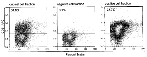
Example 3: Depletion of CD51 positive cells by magnetic cell separation
For depletion of CD51 positive non-neuronal cells a direct labelling strategy
was
compared to an indirect labelling with respect to purity and recovery of the
target cells.
For the direct labelling a CD51 specific antibody was covalently conjugated to
magnetic
particles.
The generation of superparamagnetic particles as used herein is disclosed in
US5543289
which is included herewith by reference. Monoclonal antibodies recognizing the
CD51
antigen were covalently conjugated to magnetic beads, resulting in 251.ig
antibody per mL
of bead suspension at a concentration of 0D450 = 10.
Different concentrations of bead conjugated antibodies (0.75, 1.5, 3, 6
OD450/m1) were
given to 100 t1 of a neural cell suspension containing lx107 cells. Cells were
incubated for
15 minutes at 4 C, washed once with 1 ml PBS+0.5% BSA buffer, then resuspended
in
lml of the same buffer and loaded on an LD column placed in the magnetic field
of a
MidiMACSTm Separator (Miltenyi Biotec). The column was washed twice with 1 ml
of
the same buffer. The magnetically labelled CD51 positive cells were retained
within the
column, whereas the flow through contained the CD51 negative target cells.
CD51
positive cells retained within the column and were eluted as positively
selected cell
fraction after removing the column from the magnet.
To determine the depletion efficiency the original as well as the negative and
positive cell
fraction were stained with CD51-APC and analyzed by flow cytometry. A
MicroBead
concentration of 3 0D450/m1 resulted in a high purity of CD51 negative cells.
FIG 3A
shows the original, negative, and positive fraction of one representative
experiment. 96.7
0.5% of the cells in the negative cell fraction were identified as CD51
negative. 88.4
2.2% of the target cells contained in the original fraction were collected in
the negative
fraction. One representative experiment is shown in FIG 3A.
17
CA 02801350 2013-01-09
,
Furthermore, an indirect labelling strategy was tested. Therefore, 1x10⁷ cells
were first
labelled with the biotinylated CD51 specific antibody for 10 minutes and
washed once
with 1 ml PBS+0.5% BSA buffer. Then, superparamagmetic MicroBeads coupled to
an
Anti-Biotin antibody were applied. Cells were incubated for 15 minutes at 4 C
and then
washed once. Separation was carried out as described before. Different
concentrations of
the CD51-Biotin conjugate were tested (1, 2, 4, 6, 8 gimp. The highest purity
of CD51
negative cells was obtained with a CD51-Biotin concentration of 6 gernl. One
representative experiment is shown in FIG3B.
Analysis showed that 98.6 0.17% of the cells in the negative cell fraction
were CD51
negative. Recovery of the target cells was 76.7 2.6% (see FIG 3B).
Example 4: Efficient depletion of non-neuronal cells
CD51 positive cells were depleted using whole mouse brain derived from P2 or
P3 CD1
mice and an indirect labelling strategy as described before. The original,
negative as well
as positive cell fraction were co-stained with CD51 and exemplary A2B5, 04,
GLAST
(ACSA-1), ACSA-2, CD1 1 b specific antibodies to determine the percentage of
different
neural subtypes in the original, negative as well as positive cell fraction.
Use of GAD67-
GFP mice allowed detection of GABAergic neurons. FIG4A shows that GAD67-GFP
positive GABAergic neurons were enriched in the negative target cell fraction,
whereas
non-neuronal cells, like AN2 positive oligodendrocyte precursors, 04 positive
oligodendrocytes, and CD1 lb positive microglia were depleted and found in the
positive
fraction (see FIG4A,B). The majority of GLAST (ACSA-1) and ACSA-2 positive
astrocytes was also depleted, but approximately 6% of these cells retain
within the
negative fraction (see FIG4B). All antibody conjugates used within this
analysis are
available at Miltenyi Biotec GmbH, Bergisch Gladbach, Germany.
Example 5: Cultivation of the negative cell fraction
Cortical hemispheres were obtained from 1 day old mice and dissociated using
the NTDK
(P). CD51 positive cells were labeled using the biotinylated primary CD51
specific
antibody at a concentration of 6 1.1g/m1 and Anti-Biotin MicroBeads and then
depleted as
described before. The negative as well as the positive cell fraction was
cultivated.
18
CA 02801350 2013-01-09
Therefore, cells were seeded onto poly-L-lysine-coated glass coverslips and
maintained in
a humidified atmosphere (5% CO2, 95% air) at 37 C for 5 days using e.g. MACS
Neuro
Medium (Miltenyi Biotec) supplemented with MACS Supplement B27 PLUS (Miltenyi
Biotec) and L-glutamine (0.5 mM, Invitrogen). Cultures were then fixed with 4%
paraformaldehyde (PFA) in PBS (pH 7.4) for 20 min at 4 C. For immunostaining
primary
antibodies against GLAST (ACSA-1, mouse IgG2a, Miltenyi Biotec), GFAP (mouse
IgGl,
Millipore), Myelin Basic Protein (MBP) (mouse IgG2a, Millipore), and
Microtubuli-
Associated Protein 2 (MAP2) (rabbit polyclonal, Millipore) were applied
overnight at 4 C
or for 3 h at room temperature. After rinsing 3 times with PBS, samples were
incubated
for 3h at 4 C or 1 h at room temperature with the corresponding secondary
antibodies
(Invitrogen). Cover slips were mounted onto glass slides using fluorescence
mounting
medium (Dako) and samples were analyzed by confocal fluorescence microscopy
(Leica
TCS SP2).
MAP2 immunostaining detected a lot of neurons in the negative cells fraction
and only
few neurons in the positive fraction. Co-staining for MAP2 and the astrocyte
specific
markers GFAP as well as GLAST (ACSA-1) showed that that some astrocytes were
found
in the negative fraction, but far more were detected in the positive fraction.
Almost no
MBP positive oligodendrocytes were detected in the negative fraction and
mainly found in
the positive fraction. The percentage of contaminating astrocytes and
oligodendrocytes
was <10%.
Example 6: Increase of the purity using an astrocyte-specific marker in
addition to CD51
Whole mouse brain tissue derived from P3 CD1 mice was dissociated using the
NTDK (P)
as described before. To further increase the purity of the neuronal cell
fraction and to
deplete also the contaminating astrocytes, the astrocyte specific antibody
Anti-ACSA-2
was used in combination with CD5 1 at different concentrations. Therefore,
different
concentrations of the biotin conjugated CD51 and Anti-ACSA-2 antibody were
applied
simultaneously to 100 111 of a neural cell suspension containing 1x10⁷ cells
and then
incubated for 10 minutes at 4 C. Cells were then further processed and
separated using
one LD Column as described before.
19
CA 02801350 2013-01-09
To determine depletion efficiency, the original as well as the negative and
positive cell
fraction were stained with the fluorochrome conjugated CD51 and Anti-ACSA-2
antibody
and then analyzed by flow cytometry. The best purity and recovery of target
cells was
obtained when a concentration of 4 12g/m1 of the CD51-Biotin conjugate and a
concentration of 1 Kg/m1 of the Anti-ACSA-2-Biotin was applied. FIG5 shows one
representative experiment. CD51/ACSA-2 positive cells detected in the original
fraction
were almost completely depleted in the negative target cell fraction (see FIG
5). The
average purity of CD51/ACSA-2 negative cells obtained with this antibody
composition
was 98.55 1.2%, whereas 69.5 3.15% of the target cells were recovered in the
negative
fraction.
Example 7: Separation of neurons from different brain regions
Brains from P4 CD1 mice were removed and cortical hermispheres, cerebellum,
midbrain,
or olfactory bulb were dissociated separately using the NTDK (P). Cells were
labelled as
described before with the CD51 and Anti-ACSA-2-Biotin conjugated antibodies at
a
concentration of 4 p.g/m1 and 1 lg/ml, respectively. Then, Anti-Biotin
MicroBeads were
applied for 15 minutes. After the separation, the original, negative as well
as positive cell
fraction were stained with ACSA-2 and CD51 specific fluorochrome conjugated
antibodies and analysed by flow cytometry to determine purity. The frequency
of CD51
and ACSA-2 positive cells differed in the original cell fraction depending on
the brain
region. In case of neural cells derived from olfactory bulb tissue, CD51/ACSA-
2 positive
cells showed a percentage of only 8%. In the cerebellum the percentage
increased to
approximately 15%, whereas in case of cortical hemispheres 40% and in midbrain
even
60% of all cells were CD51/ACSA-2 positive non-neuronal cells. Nevertheless,
purity of
neuronal cells in the negative fraction was around 99% in case of olfactory
bulb,
cerebellum and cortical hemispheres and 97% for midbrain tissue (see FIG6).
Example 8: Cultivation of neuronal cells isolated from mouse brain tissue
derived from
mice of different age
Cortical hemispheres were obtained from 1, 3 or 5 day old mice and dissociated
using the
NTDK (P) . Cells were indirectly labelled and separated as described before
with the
CA 02801350 2013-01-09
,
,
CD5 1 and Anti-ACSA-2 biotinylated antibodies first and then Anti-Biotin
MicroBeads.
The negative as well as the positive cell fraction was cultivated. Therefore,
cells were
seeded onto poly-L-lysine-coated glass coverslips and maintained in a
humidified
atmosphere (5% CO2, 95% air) at 37 C for 5 days using e.g. MACS Neuro Medium
(Miltenyi Biotec) supplemented with MACS Supplement B27 PLUS (Miltenyi
Biotec)
and L-glutamine (0.5 mM, Invitrogen). Cultures were then fixed with 4%
paraformaldehyde (PFA) in PBS (pH 7.4) for 20 min at 4 C. For immunostaining,
primary antibodies against GLAST (ACSA-1, mouse IgG2a, Miltenyi Biotec), GFAP
(mouse IgG 1 , Millipore), Myelin Basic Protein (MBP) (mouse IgG2a,
Millipore),
Microtubuli-Associated Protein 2 (MAP2) (rabbit polyclonal, Millipore), and
NeuN
(mouse IgG 1, Millipore) were applied overnight at 4 C or for 3 h at room
temperature.
After rinsing 3 times with PBS, samples were incubated for 3h at 4 C or 1 h at
room
temperature with the corresponding secondary antibodies (Invitrogen). Cover
slips were
mounted onto glass slides using fluorescence mounting medium (Dako) and
samples were
analyzed by confocal fluorescence microscopy (Leica TCS SP2).
Immunostaining of the negative as well as positive cell fraction showed that
mainly
neurons identified by MAP2 and NeuN immunostaining were present in the
neuronal cell
fraction. Only a very low number of contaminating GLAST or GFAP positive
astrocytes
and MBP positive oligodendrocytes was detected in the target cell fraction
(<5%).
Neurons isolated from Pl, P3, and P7 mouse brain tissue were successfully
cultivated (see
FIG8).
Example 9: CD5 1 expression in human induced pluripotent stem (iPS) cell
derived neural
cells
Immunocytochemical staining experiments using human induced pluripotent stem
(iPS)
cells that were maintained under conditions promoting spontaneous
differentiation showed
that neurons identified by MAP2 and NeuN immunostaining lacked CD51
immunoreactivity. In contrast, non-neuronal cells like astrocytes and
oligodendrocytes
were found to express CD51.
21