Note: Descriptions are shown in the official language in which they were submitted.
=
MAGNETIC RESONANCE IMAGING METHODS
BACKGROUND
Field
[0001] This case relates to nuclear Magnetic resonance (N'MR) imaging methods.
More particularly, this case relates to NMR imaging methods that provide,
among other
things, an ability to resolve short "T2" Components. This case has
applicability to the
imaging of rocks, including rocks previously or presently bearing
hydrocarbons, although
it is not limited thereto.
Description of Related Art '
[0002] Nuclear magnetic resonance (NMR) involves the application of a magnetic
field to an object that impacts the magnetic moment (spin) of an atom in the
object. In
general, the magnetic field causes the. atoms in the object to align along and
oscillate
(J)recess) about the axis of the applied magnetic field. The spin of the atoms
can be
measured. Of particular interest is the return to equilibrium of this
magnetization; i.e.,
relaxation. For example, a state of non-equilibrium occurs after the magnetic
field is
released and the atoms begin to relax from their forced alignment.
Longitudinal
relaxation due to energy exchange between the spins of the atoms and the
surrounding
lattice (spin-lattice relaxation) is usually denoted by a time Ti when the
longitudinal
magnetization has returned to a predetermined percentage (i.e., 63%) of its
final value.
Longitudinal relaxation involves the component of the spin parallel or anti-
parallel to the
direction of the magnetic field. Transverse relaxation that results from spins
getting out
of phase is usually denoted by time T2 when the transverse magnetization has
lost a
predetermined percentage (i.e., 63%) of its original value. The transverse
relaxation
involves the components of the spin oriented orthogonal to the axis of the
applied
magnetic field. The 12 measurement is often performed using a well-established
Carr-
Purcell-Meiboom-Gill (CPMG) pulse sequence which utilizes an initial 90 degree
excitation pulse followed by a series of 180 degree (pi) refocusing pulses,
and the data is
typically analyzed using a Laplace inversion technique or an exponential curve
fit.
1
CA 2801439 2019-05-13
_ .
=
[00031 NMR relaxation such as measured by T2 has been shown to be directly
proportional to the surface-to-volume ratio of a porous material,
1 5
T2n V
(1)
where S is the total surface area of the material,
Vp is the pore volume, and
p is the surface relaxivity.
Surface relaxivityp is a quantity (in micron/second) that defines the strength
of the
surface relaxation phenomenon. Because of this relationship, NMR is
extensively used in
petroleum exploration to obtain estimates of porosity, pore size, bound
fluids,
permeability, and other rock and fluid properties (i.e., "petrophysical
data"). For
example, it is known that the T2 distribution i closely related to the pore
size
distribution. Reservoir rocks often exhibit a wide range of T2 due to the
difference in
pore sizes, with observed T2s from several seconds down to tens of
microseconds.
Typically, signals at long T2 (e.g. >100 milliseconds) are from large pores
and such
fluids are considered to be producible. For shorter T2 signals, 3-50
milliseconds, the
fluids are often considered to be bound by capillary force of the pores. For
example, in
sandstone rocks, signals at T2 below 30 ms are considered bound by capillary
force and
will not produce. Thus, a cutoff value, T2cut, e.g., T2cut = 30 ms can be used
to calculate
the bound fluid volume
2mir =
BFV fer2)d T2
(2)
rzmin
where f(T2) is the T2 distribution, and
7'2õ,,õ is the minimum 12 obtained in-the T2 distribution.
Iff(T2) is the T2 distribution for the fully saturated sample, then the
porosity 0 can be
obtained by integrating f(T2) according to
=max
= fel"2)672. (3)
Zmin
where T2õ,õ is the maximum T2 exhibited in the sample. Signals with even
shorter T2,
such as T2 <3 milliseconds, are often due to clay bound water or viscous
(heavy)
2
CA 2801439 2019-05-13
_
1111
hydrocarbon. Some rocks contain a significant amount of kerogen that is solid
organic
matter and which may exhibit T2s down to tens of microseconds.
[0004] Conventional magnetic resonance imaging (MRI) techniques that work well
for long T2 signals fail for short T2 signals. In particular, conventional
methods such as
the Multiple-Slice-Multiple-Echo (MSME) imaging technique use slice selection
(discussed below), frequency encoding and phase encoding. Both frequency and
phase
encoding require that the gradient pulses be switched on and off between each
of the
adjacent refocusing pulses (pi pulses). Gradients for slice selection must
also be turned
on and off for each refocusing pulse as they will interfere with the frequency
encoding
pulses. Each switching procedure typically takes several hundred microseconds.
As a
result, the minimum echo time that can be achieved by the fiequency encoding
and phase
encoding techniques is generally on the order of several milliseconds,
preventing the .
resolution of shorter T2 values. "Lengthy" echo times (on the order of several
milliseconds) also pose the problem that in order to obtain a sufficient
signal to noise
ratio (SNR) required to resolve each of the image elements mm3), relatively
higher
magnetic fields are necessary. However, with rock samples, at higher fields, a
competing
source of decay due to diffusion of the fluid and the induced magnetization of
the rock
will dominate and artificially shorten the apparent T2. The lengthy echo time
of
conventional MRI worsens the effect and further limits the samples appropriate
for
analysis.
[0005] Slice selection refers to the use of the differences in frequency
response of the
spins to a particular radio frequency (RF) pulse in the presence of an
inhomogeneous
magnetic field, and is a common component of MRI imaging. Typically, as in
MSME,
this is done to isolate a slice in the sample for imaging the sample with
other image
encoding techniques, i.e. phase encoding and frequency encoding. A gradient
pulse will
generate an approximately linear ramp in magnetic field strength that changes
along a
chosen direction in space. Because the frequency of the spins is proportional
to field
strength, the spin frequency will also form a linear ramp across the sample.
As an RF
pulse of finite duration and power will interact with spin of a limited range
of
frequencies, in the presence of a gradient this will interact with spins in a
limited region
3
CA 2801439 2019-05-13
- - _ _
== [sitc
of the sample and hence an MRI sequence will only image this portion of the
sample. As
the shape of the amplitude profile, the length, and frequency of the RF pulse
will
determine the exact nature of the response of spins at different frequencies
and the
amplitude and direction of the applied gradient can be controlled, the
position and width
of the slice can determined. Furthermore, the profile of excitation within the
slice (as in
Hadamard imaging) can also be controlled for further resolution as a function
of slice
depth. However, these techniques are combined with other image encoding
methods (i.e.
frequency encoding, phase encoding).
SUMMARY
[0006] This summary is provided to introduce a selection of concepts that
are further
described below in the detailed description. This summary is not intended to
identify key
or essential features of the claimed subject matter, nor is it intended to be
used as an aid
in limiting the scope of the claimed subject matter.
[0007] According to one aspect, a method is provided for performing NMR
imaging
on rocks that reliably provides indications of NMR properties, such as 12
distribution.
[0008] In another aspect methods are provided for conducting NMR imaging in a
manner that permits resolving short T2 components (e.g., T2 <3 milliseconds).
[0009] In one embodiment magnetic resonance imaging (MRI) of an object is
conducted according to the following steps: (1) generating with NMR apparatus
a field
gradient along the object in a set direction, (2) obtaining a series of one-
dimensional
profiles (projections) of the object by subjecting the object under the field
gradient to a
series of RF pulse sequences, each sequence including an excitation pulse and
refocusing
pulses and recording the resulting echo train signals, each one-dimensional
projection
corresponding to a particular echo; (3). rotating the field gradient direction
to different set
directions while maintaining the magnitude of the field gradient, and
repeating step (2)
for each different direction, and obtaining one-dimensional projections for
each echo for
each field gradient direction; and (4) using the one-dimensional projections,
obtaining an
NMR image of the object or indications thereof for each of a plurality of
echoes.
4
CA 2801439 2019-05-13
81605701
[0010] In one embodiment, the NMR images or indications thereof for one or
more of the
plurality of echoes is displayed.
[0011] In one embodiment, using the image or indications thereof for a
plurality of
echoes, T2 decay data is obtained for one or more locations in the object. The
T2 decay data
may be displayed as a number and/or as a plot. In another embodiment, T2 decay
data is
obtained for multiple locations of the object. The T2 decay data may be
displayed as numbers
and/or as plots.
[0012] In one embodiment, the T2 decay data is converted to a T2
distribution. The T2
distribution may be displayed in graphic form as a plot.
[0013] In one embodiment, at least one petrophysical information product
dependent on
the T2 decay data is obtained using the NMR image or indications thereof.
[0014] In one embodiment, the image of the object or indications thereof is
obtained from
the projections using an inverse Radon transformation.
[0015] In one embodiment, T2 decay data is converted to a T2 distribution
using a
Laplace inversion.
[0016] In one embodiment, the RF pulse sequence is a CPMG or a modified
CPMG pulse
sequence.
[0016a] According to an aspect, there is provided a method of investigating an
object
using nuclear magnetic resonance (NMR) equipment, comprising the steps of: a)
generating a
field gradient along a set direction; b) obtaining a series of one-dimensional
profiles of the
object by subjecting the object under the field gradient to a series of RF
pulse sequences, each
RF pulse sequence generating a series of echoes and recording echo train
signals resulting
from the series of RF pulse sequences interacting with the object, each one-
dimensional
profile corresponding to a particular echo; c) changing the field gradient
direction to different
set directions while maintaining the magnitude of the field gradient, and
repeating step b) for
each different field gradient direction, thereby obtaining one-dimensional
projections for each
CA 2801439 2019-05-13
81605701
echo for each field gradient direction; and d) using the projections,
generating NMR image
data for each of a plurality of echoes for at least one location in the
object.
[0016b] According to another aspect, there is provided a method of
investigating a
hydrocarbon-bearing rock using nuclear magnetic resonance (NMR) equipment,
comprising
the steps of: a) with a field gradient set along a first direction,
sequentially setting the NMR
RF pulse frequency to a plurality of different frequencies, and at each
different frequency
generating an NMR pulse sequence with an excitation pulse and refocusing
pulses and
acquiring and storing indications of echo train signals resulting from the NMR
pulse sequence
interaction with the rock; b) changing the direction of the field gradient a
plurality of times,
and repeating step a) for each field direction; c) generating a one-
dimensional projection of
the rock for each of a plurality of echoes utilizing the indications of echo
train signals, thereby
obtaining a plurality of one-dimensional projections; d) utilizing the
plurality of one-
dimensional projections, for each of the plurality of echoes, generating NMR
image data for at
least one location in the rock; and e) displaying the NMR image data.
[0016c] According to another aspect, there is provided a method of
investigating an object
using nuclear magnetic resonance (NMR) equipment, comprising the steps of: a)
generating a
field gradient along a set direction; b) setting the NMR RF pulse frequency to
a set frequency;
c) generating an NMR CPMG pulse sequence with the field gradient at the set
direction and
the RF pulse frequency at the set frequency; d) acquiring and storing
indications of echo train
signals as a result of the NMR CPMG pulse sequence interaction with the
object; e) changing
the RF pulse frequency to a new set frequency and repeating steps c) and d) at
the new set
frequency; 0 repeating step e) a plurality of times; g) changing the set
direction of the field
gradient to a new set direction and repeating steps b) through 0 at the new
set direction;
h) repeating step g) a plurality of times; i) for each field gradient
direction, generating a one-
dimensional projection of the object for each of a plurality of echoes
utilizing the indications
of echo train signals acquired as a result of the NMR CPMG pulse sequence
interaction with
the object at multiple NMR RF pulse frequencies, thereby obtaining a plurality
of one-
dimensional projections; j) from the plurality of one-dimensional projections,
for each of the
plurality of echoes, generating NMR image data for at least one location in
the object.
5a
CA 2801439 2019-05-13
81605701
BRIEF DESCRIPTION OF THE DRAWINGS
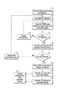
[0017] Figure 1 is a flow diagram of one embodiment of a method.
[0018] Figure 2 is a time diagram displaying indications of RF pulses, a
gradient, and an
acquired echo train useful in the method of Figure 1.
[0019] Figure 3 is a diagram helpful in understanding slicing in multiple
projections.
[0020] Figure 4 is a diagram illustrating a two-dimensional Radon transform
as line
integrals.
5b
CA 2801439 2019-05-13
=
4110 16.1
1.11
[0021] Figure 5 is a diagram useful in understanding the Projection-
Slice Theorem.
[0022] Figures 6A ¨61) are magnetic resonance images generated for four
different
echoes using the method of Figure f showing two tubes of water doped with
different
amounts of copper sulfate.
[0023] Figures 7A and 78 are plots of the T2 signal and the T2 distribution of
a
specific pixel of one of the copper sulfate doped tubes of water imaged in
Figures 6A-6D.
[0024] Figures 7C and 7D are plots of the T2 signal and the T2 distribution of
a
specific pixel of the second of the tubes of copper sulfate doped water imaged
in Figures
6A - 6D.
[0025] Figure 8 is a magnetic resonance image generated using the method of
Figure
1 of a shale sample.
[0026] Figures 9A and 9B are plots of the T2 signal and the T2 distribution of
a
specific pixel of the image of Figure 8.
DETAILED DESCRIPTION
[0027] A flow diagram of one embodiment of an imaging method is seen in Fig.
1.
More particularly, an object is investigated using an NMR apparatus (not
shown), where
a field gradient Gxy(0) is generated by the NMR apparatus at 12 along a set
(first)
direction. At 14, the RF pulse frequency is set to a set (first) frequency. At
15, a CPMG
pulse sequence is generated, and echo train signals are acquired and stored by
the NMR
apparatus as discussed below with reference to Fig. 2. At 16, a determination
is made as
to whether echo train signals have been acquired for a desired number of
different RF
frequencies. If not, the RF frequency is changed to a new set frequency at 18
thereby
changing the object slice position as discussed below with reference to Fig.
3, and the
method returns to 15 where a CPMG pulse sequence is generated with the new RF
frequency and echo train signals are acquired and stored. Steps 15, 16, and 18
are
repeated until a determination is made at 16 that signals resulting from CPMG
pulse
sequences at a sufficient number of RF frequencies have been recorded. Then at
20,
6
CA 2801439 2019-05-13
_ _ _
IS11.01110
using the echo train signals, a one-dimensional projection (profile) is
generated for each
echo as discussed below with reference to Fig. 3. At 22, a determination is
made as to
whether one-dimensional projections have been generated for a sufficient
number of field
gradient directions. If not, at 24, the gradient direction is changed (e.g.,
rotated) to a
different set direction, and the method returns to step 14 et seq., to obtain
additional one-
dimensional projections. It will be appreciated that the direction may be
changed not
only in two dimensions (x-y space), but in three dimensions (x-y-z space) as
desired.
Regardless, when data for sufficient gradient directions have been obtained,
the method
continues at 26, where the one-dimensional projections are used to obtain an
NMR image
of the object or indications thereof (data) for each of a plurality of echoes.
Images or
indications of the object for one or m-ire of the echoes are optionally
displayed as
discussed in more detail below with reference to Figs. 6A-6D. In one
embodiment, T2
NMR images are obtained using a transform such as a Radon transformation
discussed in
more detail below with reference to Fig. 4.
[0028] In one embodiment, at 28, T2 decay data is obtained for a location in
the
object using the image or indications thereof for a plurality of echoes. The
T2 decay data
is optionally displayed in the form of a number and/or as a plot as discussed
in more
detail below with reference to Figs. 7A, 7C and 9A. In another embodiment, at
28, T2
decay data are obtained for multiple locations of the object and are
optionally displayed
in the form of a number and/or as a plot.
[0029] In one embodiment, at 30, the T2 decay data for one location in the
object are
processed, e.g., using a Laplace inversion to generate a T2 distribution. The
T2
distribution may be displayed in graphic form. Additionally or alternatively,
a 12 value
may be obtained from the T2 distribution data. In another embodiment, at 30,
T2 decay
data for multiple locations in the object are converted to a plurality of T2
distributions
and are optionally displayed as discussed in more detail below with reference
to Figs. 7B,
7C and 9B.
[0030] In one embodiment, the T2 decay data and/or distributions obtained at
28
and/or 30 are used at 32 to generate indications of rock parameters
(attributes). By way
=
7
CA 2801439 2019-05-13
81605701
of example and not limitation, as described in U.S. Patent 5,387,865,
the fluid flow permeability of the porous object (rock) under study may
be determined using inter alia a T2 value determination, The fluid flow
permeability may be displayed. As another example, as described in U.S.
Patent 5,363,041, unbound fluid volume (and bound fluid volume)
of the object (formation) under study may be determined using inter alia
a T2 determination. The unbound fluid volume and/or bound fluid volume may be
displayed. As disclosed in A. Timur, "Pulsed Nuclear Magnetic Resonance
Studies of Porosity, Movable Fluid and Permeability of Sandstones", Journal of
Petroleum Technology, June 1979, p. 775, indications of T2 may be used in
making
determinations of porosity, permeability, and movable fluid of sandstones
which may be
displayed.
[0031] While Fig. 1 provides one embodiment in steps 14-20 for generating
one-
dimensional projections (profiles) for a desired number of echoes, it should
be
appreciated that the one-dimensional projections can be obtained in other
manners. By
way of example and not by way of limitation, instead of repeatedly changing
the RF
frequency and acquiring resulting echo train signals in order to generate the
one-
dimensional projections, the position of the sample can be varied (e.g, slid)
so that
different slices of the sample are investigated without changing the RF
frequency. By
way of another example, and not by way of limitation, instead of changing the
RF
frequency and acquiring echo train signals, an offset pulse BO may be applied
on top of
the gradient so that the same RF frequency can correspond to a different
position. Thus,
by applying different offset pulses BO, B1 ..., different slices of the sample
are
investigated without changing the RF frequency. Other methods and mechanisms
can be
used as well, provided that one-dimensional projections are generated for the
desired
number of echoes while avoiding gradient switching during the CPMG pulse
sequencing.
[0032] As previously mentioned, one embodiment entails conducting CPMG pulse
sequences under a field gradient Gxy(0) and acquiring echo signals. While the
CPMG
pulse sequence refers to a specific sequence (described in more detail below
with
reference to Fig. 2), other "improved" pulse sequences that will generate an
appropriate
8
CA 2801439 2019-05-13
81605701
echo train may be utilized. Thus, by way of example and not by way of
limitation, a
modified CPMG as described in M.D. Hurlimann, "Carr-Purcell Sequences with
Composite Pulses", Journal of Magnetic Resonance, Vol. 152, Issue 1, Sept.
2001,
pp. 109-123, may be utilized. Also, by way of example and not by way of
limitation, U.S. Patent 6,580,272 discloses a split-180 signal in order to
detect a steady state free precession signal. Another example is described
in T.W. Bornemana et al., "Pulses Derived from Optimal Control Theory",
Journal of Magnetic Resonance, Vol. 207, Issue 2, Dec. 2010, pp. 220-223.
Other pulse
sequences can be used as well such as complex (shaped) RF pulses or composite
pulses,
provided that they are slice selective'or their frequency profiles can be used
to invert for
one-dimensional projections for a desired number of echoes.
[00331 Turning now to Fig. 2, a standard CPMG pulse sequence is seen with an
initial
90 degree frequency-selective (excitation) pulse followed by a series of
frequency-
selective 180 degree (refocusing) pulses. As seen in Fig. 2, the field
gradient Gxy(0) is
maintained constant during the CPMG pulse sequence. Also, as seen in Fig. 2,
following
each selective refocusing pulse, an echo signal AQ is acquired. It will be
appreciated
that the CPMG pulse sequence may utilize tens, hundreds, or thousands of
refocusing
pulses, and therefore tens, hundreds, or thousands of echo signals can be
acquired by the
NMR equipment.
[00341 For any given field gradient direction, changing the RF frequency
changes the
slice (as seen in Fig. 3) of the object being investigated, and this may be
done at desired
granularity. Thus, while six slices are shown in Fig. 3, it will be
appreciated that a
different number of slices may be generated. Similarly, for a particular echo,
the number
of one-dimensional projections generated depends on the number of times the
gradient
direction was changed (e.g., rotated) at step 24 of Fig. I. Thus, while only
two one-
dimensional projections are seen in Fig. 3 at ninety degree angles relative to
each other, it
will be appreciated that many more projections may be generated by changing
the field
gradient direction with more granularity. It will be appreciated that in order
to change the
field gradient, the sample may be rotated, or the NMR equipment may be moved
or
adapted to generate field gradients of different directions.
9
CA 2801439 2019-05-13
[0036] According to one aspect, and as previously suggested, the CPMG sequence
utilized can be tailored as desired for the context of the object
investigation. For
example, for imaging of hydrocarbon-bearing rock, specific dynamics known with
respect of NMR logging as discussed by M. Hurlimann et al., "Diffusion and
Relaxation
Effects in Generaly Stray Field NMR Experiments, Journal of Magnetic
Resonance, Vol.
148(2), pp. 367-378, may be applicable. Likewise, modifications to improve the
CPMG,
(e.g., improving the echo refocusing, controlling the bandwidth, minimizing
interference
between different slices, diffusion editing to provide additional information
on diffusion,
and saturation recovery for T1 information, as is done in wireline and LWD NMR
logging) may be utilized.
[0036] As previously mentioned, data points are derived from echoes. A single
intensity value is extracted from each echo to get the value of the
projection. In one
embodiment, the value of the data point is taken from the intensity of the
echo at the
= center of the echo signal AQ. In another embodiment, a maximum value may
be taken.
In yet another embodiment, the echo is acquired with the same frequency as the
CPMG
pulse, and any of many techniques may be used to extract echo intensities such
as, by
way of example and not by way of limitation, integrating over a range of
frequencies, or
using a matched filter. It will be appreciated that depending upon the details
of CPMG
pulse sequence utilized, different sensitivities may result across the slice's
profile (for
example a square pulse will have a slice profile similar to a sinc function, a
Gaussian
pulse a Gaussian profile). In one embodiment, knowledge of the slice profile
and the use
of overlapping slices in combination with deconvolution can be used to enhance
the
resolution of any set of projections beyond the natural slice width.
[0037] As previously mentioned, one-dimensional projections are used to obtain
an
NMRimage of the object or indications thereof (data values for each point or
pixel of
interest) for each of a plurality of echoes. In one embodiment, the images are
obtained
using a transform such as a Radon transformation which is the mathematical
basis for
tomographic imaging from projections. The two-dimensional Radon transform set
forth
in Equation (4) below is simply a line integral, as shown in Fig. 4,
CA 2801439 2019-05-13
r.
ge, el) = -1¨ica yY6(xcos6 ysitte Otixcly (4)
--os
where 0 is the gradient direction (angle) of the signal, and / is the slice
position set by the
RF frequency, and Sis a Dirac delta.
[0038] The Radon transform is closely related to the Fourier transform by
the
Projection-Slice Theorem which is the basis for image reconstruction
algorithms. The
Projection-Slice Theorem simply states, as seen in Fig. 5, that the one-
dimensional (1D)
Fourier transform of the projection equals the two-dimensional (2D) Fourier
transform
along the radial line at that angle (0). According to the Projection-Slice
Theorem, to
reconstruct the image, an inverse of the 2D Fourier transform can be taken in
polar
coordinates which is known as a filtered back-projection or inverse Radon
transform.
The inverse 2D Fourier transform in Cartesian coordinates can be represented
by:
frx, 1.7)eilirf -xu +3'1') dud r (5)
The transform is implemented in most mathematics software packages, for
instance
MATLAB (a trademark of MathWorks, Inc. of Natick, Massachusetts, USA), as a
last'
algorithm that computes the same result as the integral form. The inverse 2D
Fourier
transform in polar coordinates can be represented as:
fin = (ir eir' 2rt I p[--j'
0)e-P4c1411,(271-pr)dp
22T
(6)
where p is the Fourier conjugate variable to 1 and G(0- 0) is the 1D Fourier
transform of
the 1D projection data collected for each gradient angle 0 . It is noted that
multiple 'fast'
algorithms exist to solve the discrete form of this equation without directly
computing the
integrals and are implemented in common mathematics software suites such as
MATLAB .
[0039] Once the image of each echo time is reconstructed, e.g., using
inverse Radon
transformations, the individual image elements (pixels) or averages over
regions of the
image may be used to construct individual T2 decay curves. For example, and as
11
CA 2801439 2019-05-13
111110
discussed in more detail below with reference to Figs. 6A-6D and 7A-7D, with
the
reconstructions, values are obtained for each echo for each point or pixel in
the image.
The value for each pixel may then be plotted as a function of (echo) time to
provide a T2
decay curve. The curve for each pixel may then be analyzed by any number of
the
existing analysis techniques for CPMG data such as exponential or stretched
exponential
curve fitting, or inverse Laplace transformation, to generate "answer
products" such as
T2 distributions. For CPMG analysis techniques that are equivalent to taking a
linear
=
transform for the CPMG decay, this analysis step may be done before the
inverse Radon
transform (or equivalent image reconstruction technique) and instead of the
image
reconstruction for the individual echoes, and the same image reconstruction
technique
may be applied directly to values extracted from the CPMG decays.
[0040] Turning now to Figs. 6A-6D, four reconstructed images are shown
resulting
from an NMR investigation of two tubes of water doped with different amounts
of copper
sulfate at different echo times (e.g., the first, eighth, thirty-second, and
one hundred
twenty-eighth echoes). The images were obtained and reconstructed using the
imaging
method described above with respect to Fig. 1. The intensities of particular
pixels were
then plotted as a function of time. For example, the intensities (magnitudes)
of the
echoes for pixel 10x7 which appears located at the center of the left tube of
doped water
were plotted in Fig. 7A. The resulting curve of Fig. 7A was then analyzed
using an
inverse Laplace transform to generate the T2 distribution shown in Fig. 7B. As
will be
appreciated, Fig. 7B indicates that the T2 decay for that pixel is centered
about 2.5
milliseconds which is a very short T2 time. Similarly, the intensities of the
echoes for
pixel 10x15 which appears located at the center of the right tube of doped
water were
plotted in Fig. 7C. The resulting curve of Fig. 7C was then analyzed to
generate the 12
distribution shown in Fig. 7D. As will be appreciated, Fig. 7D indicates that
the T2
decay for that pixel is centered about 19 milliseconds which is a short T2
time.
[0041] A shale sample was subjected to the NMR pulse sequence and image
reconstruction discussed above with reference to Fig. I. The resulting first
echo image
for a particular pixel (pixel 11x11) is seen in Fig. 8. Using the image
reconstructions for
sixteen echoes, the T2 decay curve for pixel 11x11 was generated as seen in
Fig. 9A.
= 12
CA 2801439 2019-05-13
S1S11Ø
The curve of Fig. 9A was then analyzed to generate the T2 distribution shown
in Fig. 9B.
As will be appreciated, Fig. 9B indicates that the 12 decay for that pixel is
centered at
approximately 500 microseconds, a very short12 time that may be indicative of
viscous
oil, bitumen, and/or kerogen.
[0042] It should be appreciated that using the NMR pulse sequence and image
reconstruction techniques discussed above, various valuable answer products
may be
obtained. For example, T2 decay curves may be obtained as shown and described
with
respect to Figs. 7A, 7C, and 9A. Alternatively, or in addition, T2
distribution curves may
be obtained as shown and described with respect to Figs. 7B, 7D, and 9B.
Alternatively,
or in addition, 12 values (times) may be obtained as described with reference
to the
center of each of the distribution curves=of Figs. 7B, 7D, and 9B. The T2
values may be
shown as numerical answer products. or on graphs or plots. By way of example
and not
limitation, the T2 values for different pixels may be represented as numbers
for each
pixel on a chart or on a plot, or as different colors or intensities with an
appropriate key
on a chart or on a plot. Alternatively, or in addition, petrophysical data
such as
determinations of permeability (estimates), porosity, or bound or unbound
water, and/or
the heterogeneity of any of these properties, may be determined using the T2
information
obtained, and may be displayed numerically, on graphs or plots, or otherwise.
[0043] There have been described and illustrated herein several
embodiments of
investigating objects using NMR measurements. While particular embodiments
have
been described, it is not intended that the embodiments limit the scope
hereof. Thus,
many changes may be made. For example, while the gradient was described with
respect
to Fig. 2 as being constantly on, the gradient may be temporarily turned off
in order to
reduce duty cycle for the gradient amplifier. This may be done between CPMG
trains.
Also, the inverse Radon transform may be accomplished by different algorithms,
and
while filtered back-projection was described, other reconstruction techniques
such as
compressed sensing may be used to similar effect. Further, while a particular
sequence
of steps was described with respect to Fig. 1 where one-dimensional
projections are
generated for the echoes prior to changing the field gradient direction, it
will be
appreciated that the sequence of steps is not limiting and may be changed. For
example,
= 13
=
CA 2801439 2019-05-13
IS11.011
all echo data may be stored for all gradient directions prior to generating
the one-
dimensional projections. In addition, while the embodiments were described as
generating particular "answer products", it will be appreciated that other
answer products
could be generated and displayed. It will therefore be appreciated by those
skilled in the
art that yet other modifications could be made. Accordingly, all such
modifications are
intended to be included within the scope of this disclosure as defined in the
following
claims. In the claims, means-plus-function clauses, if any, are intended to
cover the
structures described herein as performing the recited function and not only
structural
equivalents, but also equivalent structures. It is the express intention of
the applicant not
to invoke 35 U.S.C. 112, paragraph 6 for any limitations of any of the
claims herein,
except for those in which the claim expressly uses the words 'means for'
together with an
associated function.
14
CA 2801439 2019-05-13