Note: Descriptions are shown in the official language in which they were submitted.
CA 02801484 2012-12-03
- 1 -
DESCRIPTION
"DEVICE FOR MEASURING THE KNEE LAXITY"
FIELD OF THE INVENTION
The present invention relates to a device for
evaluating the instability of the knee due to rupture of
any ligament, namely the anterior cruciate ligament,
posterior cruciate ligament, postero-internal capsule,
postero-external capsule, as well as in all the axis of
orthonormal, anterior, posterior, antero-internal, antero-
external, postero-internal and postero-external
referentials.
In recent decades, the number of injuries in
lower limb joints has significantly increased. This
problem affects both athletes and any other individuals who
may be victims of accidents and/or congenital diseases.
This type of injuries and traumas leads to an
individual inability since it creates instability of the
knee, preventing their normal functions (rotation, impulse
and extension) thus creating an incapacity for the
individual to execute his daily tasks.
CA 02801484 2012-12-03
- 2 -
In fact, the treatment of the knee ligaments
ruptures varies depending on the severity and can involve
different therapies, ranging from physiotherapy to surgery.
However, in order to apply the appropriate
treatment, it is essential to perform an accurate and
precise diagnosis of the injury type.
Nowadays, the injury diagnosis is achieved
through an inquiry presented to the patient (where and how
the incident occurred, what type of activity was being
executed and how often), analysis of the injured area and
visualization of the injury through clinical examination
using various diagnostic methods.
However, these diagnostic tests, which may
include radiographs (x-rays), computed axial tomography
scan (CAT-scan), magnetic resonance imaging (MRI) and
arthroscopy (injured joint observation through insertion of
a small instrument into the joint), can't accurately
measure the severity of the injury.
The present invention allows an accurate
assessment of the laxity, not only for the anterior and
posterior cruciate ligaments but for all the others knee
ligaments.
The device for measuring the knee laxity is not
just intended to overcome all the drawbacks with state-of-
CA 02801484 2012-12-03
- 3 -
art devices, but it represents a new concept and a major
scientific and technical improvement in measuring the knee
laxity (anterior - posterior and rotations) and in
subsequent treatment.
By making these measurements, it will be possible
to define exactly who are the patients, that because of the
ligaments rupture, will need surgery and those which only
require preservation treatment.
TECHNICAL BACKGROUND
Devices KT1000 and KT2000 sold by MEDmetric
Corporation are known in the market and they roughly
measure the anterior and posterior tibial translation,
which is measured from the outside of the leg and has the
muscle mass and the soft tissues (more or less compressible
and variable from individual to individual) as an error
factor, being this measurement taken only in mm.
Ferromagnetic materials are used in these
devices, what makes them unusable inside MRI or CAT-scan
devices is they interfere with the image processing. These
devices are used only for evaluation of the anterior
cruciate ligament rupture and posterior cruciate ligament
rupture.
EP 1 125 567 discloses a device allowing an
accurate measuring of the tibial anterior translation and
CA 02801484 2012-12-03
- 4 -
internal rotation because it can be used inside MRI or CAT-
scan devices, not interfering with image processing since
it doesn't include ferromagnetic materials.
The measurements are made in the MRI or CAT-scan
images, between two bone structures (femur and tibia) which
grants them great accuracy. These measurements can be
related to the associated injuries observed in the images
(meniscus, cartilage, etc.).
This device disclosed in patent EP1125567 is
only used in the ruptures of the anterior cruciate
ligament. On account of this uniqueness, it doesn't allow
an accurate diagnosis of associate instabilities.
On account of those limitations, the device
disclosed in patent EP1125567 does not allow distinction
of those patients who need a surgical technique with
greater or lesser rotation control, and also does not allow
the evaluation of patients with associate ligament injuries
(most common).
The device described in this invention allows an
accurate measuring of the tibial anterior translation and
tibial posterior translation, the internal rotation and
external rotation because it can be used inside devices of
MRI or CAT-scan, not interfering with image processing
since it doesn't include ferromagnetic materials.
CA 02801484 2012-12-03
- 5 -
The device for measuring the knee laxity
described in the invention includes materials suitable for
use in MRI or CAT-scan for instance, inter alia,
composites, plastics, resins and carbon fibers.
With the assistance of the device described in
this invention, the measurements are made in the images
from MRI and CAT-scan between two bone structures, femur
and tibia.
These measurements can be related to associated
injuries observed in the images, like inter alia, with
meniscus, cartilages, etc.
The device for measuring the knee laxity
described in the present invention has the ability to
measure the knee rotation without using images from MRI and
CAT-scan.
The device is used for ruptures of the anterior
cruciate ligament and posterior cruciate ligament, antero-
external, postero-external, antero-internal and postero-
internal instabilities, instabilities in all directions,
and also to evaluate all possible rotational instabilities
of the knee.
It is therefore possible to obtain accurate
diagnoses and subsequent modifications of the surgical
indications, allowing the separation of those patients who
CA 02801484 2012-12-03
- 6 -
need surgery from those who just need physiotherapy.
This device is quite innovative and will
radically change the orthopedics market since:
- Until now, with prior art technique, the
instability measurement is made roughly and by
approximation, while with this new device it will be
possible to measure the knee instability with accuracy.
- Until now, the treatment and surgeries for knee
traumatic injury were generic, but with this new device it
will be possible to get and set a real diagnosis, leading
to more appropriate treatments which means one can decide
whether or not to perform surgery with greater precision,
and can choose between different types of surgery depending
on the needs, leading to a higher success rate in patients'
recovery.
SUMMARY OF THE INVENTION
The device of the present invention for measuring
the knee laxity is a medical or surgical equipment for
measuring accurately and directly the abnormal movement of
the injured knee joint, and can be used inside the CAT-scan
and MRI devices.
This device for measuring the knee laxity, with
or without use of additional means of diagnosis as CAT-scan
and MRI, evaluates knee instability caused by rupture of
any ligament, namely the anterior cruciate ligament,
CA 02801484 2012-12-03
- 7 -
posterior cruciate ligament, postero-internal capsule,
postero-external capsule, as well as around axis
orthonormal, anterior, posterior, antero-internal, antero-
external, postero-internal and postero-external
referential. Essentially it comprises a part to receive
and hold the thigh with belts, a part to receive and hold
the leg and a part to receive and hold the foot with belts.
The present invention comprises independent means
to push the anterior zone of the leg backward and to push
the posterior zone of the leg forward, and the means to
push the foot to the left and right sides.
The device also includes a part for posterior
support and fixation of the foot, allowing it to
alternatively rotate to the left or to the right according
to the force exerted on the part for posterior support and
fixation of the foot, around a shaft that runs through the
said part for posterior support and fixation of the foot
and through a supporting part which is articulated.
The present invention can be placed inside a MRI
or CAT-scan device to help measuring the laxity of a knee
with ligament rupture, in order to obtain one or more
images of antero-posterior translation.
Therefore, a rotation will be executed, which may
be measured between two bony points on the tibia and femur
with high precision, the measurement being performed on the
CA 02801484 2012-12-03
- 8 -
image without distortion since the materials used will be
tested in order not to interfere with the image.
Additionally, the device object of this invention
will allow measuring the rotation between femur and tibia
caused by ligament ruptures, the most serious knee
injuries, which is the most important cause of instability
and knee failure.
By measuring the rotation between tibia and femur
inside the MRI and CAT-scan device, one can select exactly
who are the patients which need surgery and those which
only require physiotherapy treatment.
Since the device object of the present invention
can be used in an of MRI or CAT-scan environment, all
associated injuries, occurring for instance and inter alia
with the menisci, cartilage, etc., can be correlated with
an injury severity that can be quantified.
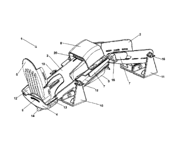
BRIEF DESCRIPTION OF THE DRAWINGS
The following description is made with reference
to the accompanying drawings which are presented just as a
non-limiting example, in which:
- Figures 1 and 2 are perspective views of the device
described in the present invention;
-Figure 3 is a side view of the device described in
CA 02801484 2012-12-03
- 9 -
the present invention;
- Figure 4 is a top view of the device described in
this invention, and
- Figure 5 is a front view of the device described in
the present invention, in which one can see a scale,
being the support part in an angle.
LEGEND OF THE FIGURES
- device (1) for measuring the knee laxity
- part (2) for posterior support and fixation of the thigh
- part (3) for posterior support and fixation of the leg
- supporting piece (4)
- part (5) for posterior support and fixation of the foot
- scale (6) placed in the part (5) for posterior support
and fixation of the foot
- articulation elements (7)
- removable part (8) for front fixation of the thigh
- removable part (9) for posterior fixation of the thigh
- moving part (10) with flat base
- moving part (11) with flat base
- means (12) to push the foot in clockwise direction
- means (13) to push the foot in anticlockwise direction
- shaft (14)
- tightening nuts (15) for the articulation elements
- tightening nuts (16, 19) for the moving parts (10, 11)
- means (20) to push the leg backwards
- means (21) to push the leg forward
CA 02801484 2012-12-03
- 10 -
DETAILED DESCRIPTION OF THE INVENTION
As can be seen in Figures 1, 2, 3 and 4, the
device (1) described in this invention is comprised
essentially of four parts, namely a part (2) for the
posterior support and fixation of the thigh, a part (3) for
the posterior support and fixation of the leg, a supporting
part (4) and a part (5) for posterior support and fixation
of the foot.
These four parts respectively include multiple
belts, not represented in the figures, which together with
parts (8) and (9) will ensure that the leg, thigh and foot
lay and remain fixed against the device (1), object of this
invention.
To ensure that the images obtained with the
device (1) by computed axial tomography scan or magnetic
resonance imaging do not show distortions, all materials
used in the present invention are plastics, resins and
composites.
The device (1) comprises independent means (20)
to push backwards the anterior zone of the leg and means
(21) to push forward the posterior zone of the leg (21),
shown in Figure 2, respectively located in the inner wall
of a removable part (8) for posterior support and fixation
of the leg and in the inner wall of a removable part (9)
for front support and fixation of the leg.
CA 02801484 2012-12-03
- 11 -
On the other hand, in the foot zone (1), as can
be seen in Figure 4, the device also comprises means (12)
to push the foot in clockwise direction, and means (13) to
push the foot in anti-clockwise direction, respectively
located on the inner wall on the right and left sides of a
support piece (4).
These means (20), (21), (12) and (13) to push
their respective leg and foot zones can be of any kind
namely manually inflatable bags or compressor filled ones,
or by hydraulic means.
The means (20), (21), (12) and (13) to push their
respective leg and foot zones, so as to position and hold
the patient foot and/or leg into position, can work
independently and alternatively from each other.
Moreover, the means (20), (21) to push their
respective leg zones can work in conjunction with the means
(12) and (13) to push the foot, so as to position and hold
the foot and/or the patient's leg into position.
Thus, as described, the present invention can
position and hold the foot and/or the leg of the patient in
various positions.
After applying a force on the posterior surface
of the leg, or on the anterior surface of the leg, applying
a force to rotate the foot internally or externally, or any
CA 02801484 2012-12-03
- 12 -
one of these movements combined two by two, the device (1)
will allow the measurements of translation and rotation of
the knee, of the tibia over the femur, into the MRI or CAT-
scan device, or any other imaging device that permits these
measurements. These measurements include the evaluation in
mm and/or degrees of translation and/or rotation of the
tibia over the femur.
As shown in Figure 5, the part (5) for posterior
support and fixation of the foot can rotate clockwise or
anticlockwise around a shaft (14), which runs through this
part (5) and through a supporting part (4).
In Figures 1 and 5 one can also see a scale (6)
located in the supporting piece (4)
In the device (1), the parts of the leg and thigh
can be positioned at different angles, through the
articulated elements (7) held in the desired positions by
tightening nuts (15) . The parts of the leg and thigh can
also be adapted to various anatomical dimensions of the
patient by sliding said elements (7) and parts (2, 3), and
element(4), which are then fixed in the desired positions
by mechanical means, not represented in the Figures, placed
in openings or holes existing in those elements and parts.
In Figures 1, 2 and 3 one can see the moving
parts (10) and (11) comprising flat bases which will
provide support and stability to the device (1) on a
CA 02801484 2012-12-03
- 13 -
horizontal plane.
These moving parts (10) and (11) are fastened to
the sides of parts (2) and (3) through tightening nuts (16-
19), as shown in Figure 4.
As it will be apparent to those skilled in the
art, various detail modifications can be made, which
however are considered to be included in the spirit of the
invention.
The invention should be limited only by the scope
of the appended claims.