Note: Descriptions are shown in the official language in which they were submitted.
CA 02801559 2013-02-14
02/14/2013 THU 11:34 FAX 1 705 652 6074 Gastle Image Systems
2009/064
MASK AND METHOD FOR USE IN RESPIRATORY MONITORING AND
DIAGNOSTICS
FIELD OF THE DISCLOSURE
[0001] The present disclosure relates to respiratory diagnostic and
monitoring
systems, and in particular, to a mask and method for use in respiratory
monitoring and
diagnostics.
BACKGROUND
100021 Several clinical conditions require close monitoring of respiratory
activity
including respiratory failure, respiratory tract infections as well as
respiratory depression
i 0 associated with anesthesia and sedatives. Also, respiratory disorders are
known to disturb
=
= sleep patterns. For example, recurrent apneas and hypopnea
lead to intermittent hypoxia
that provokes arousals and fragmentation of sleep, which in turn may lead to
restless
sleep, and excessive daytime sleepiness. Repetitive apneas and intermittent
hypoxia may
=
also elicit sympathetic nervous system activation, oxidative stress and
elaboration of
inflammatory mediators which may cause repetitive surges in blood pressure at
night and
increase the risk of developing daytime hypertension, atherosclerosis, heart
failure, and
stroke independently from other risks.
[0003] There remains a need for improved tools and methods for monitoring
respiratory activity, for example in a clinical setting, or again in
diagnosing and/or
monitoring respiratory disorders, as discussed above, in order to reduce or
even obviate
the risks that may be associated therewith.
[0004] Namely, while some have proposed diagnostic tools and methods for
diagnosing, monitoring and/or generally investigating certain breathing
disorders, these
tools and methods are often particularly invasive and/or uncomfortable for the
subject at
hand, and therefore, can yield unsatisfactory results. For instance, many
diagnostic
I-M P5/PCT-CDA
PAGE 9/64* RCVD AT 2/1412013 11:40:51 AM [Eastern Standard Time]* SVR:F0000315
" DNIS:3906* CSID:1 705 652 6074 DURATION (mm-ss):28-13
02/14/2013 THU 1135 FAX 1 705 652 6074 Gastle Image Systems CA 02801559 2013-
02-14 2010/064
A
procedures are solely implemented within a clinical environment, which amongst
other
deficiencies, do not allow for monitoring a subject in its natural
environment, leading to
skewed or inaccurate results, or in the least, forcing the subject through an
unpleasant and
mostly uncomfortable experience.
5 100051 Alternatively, different portable devices have been
suggested for the diagnosis
of sleep apneas; however, these solutions generally require the subject to
position and
attach several wired electrodes themselves in the absence of a health care
provider.
Unfortunately, subject-driven electrode positioning and installation often
leads to a
reduction in subject comfort and compliance, and increases the chance that the
electrodes
to will be detached or displaced in use. Since accurate positioning and
installation of such
electrodes are paramount to proper diagnostics, captured signals in such
situations are
often unreliable, a measure which can only effectively be determined once the
data is
transferred back to a health center, at which point, such data, if properly
identified, must
be withdrawn from the study. Furthermore, such devices regularly need to be
shipped
15 back to the health center for processing and, given their generally
invasive nature, for
= hygienic reconditioning, e.g. disinfection.
[0006] Similarly, in a clinical setting, while the positioning and
attachment of
monitoring electrodes may be completed by an experienced health care
professional, the
devices currently used in such settings generally at best leave the subject
physically wired
20 to one or more monitoring devices, if not via more invasive techniques,
which wiring can
be a particular nuisance to the subjects general comfort and mobility, and
obtrusive to
individuals or health care practitioners maneuvering around the subject. For
example,
International Application Publication No. WO 01/15602 describes a clinical
system
wherein a microphone is suspended from the ceiling above the subject, the
recorded data
25 of which is combined with readings from an esophageal pressure catheter
and nasal
airflow monitoring.
2
TRI-MP5/PCI-CDA
L....PAGE 10164' RCVD AT 2/1412013 11:40:51 AM [Eastern Standard Time]
SVR:F0000315* DNIS:3906* CSID:1 705 6526074' DURATION (mm-ss):28-13
CA 02801559 2013-02-14
02/14/2013 THU 11:35 FAX 1 705 652 6074 Gastle Image Systems
2011/064
100071 Less intrusive methods have been proposed, for example in US Patent
No.
5,797,852, wherein a microphone is suspended from a base device sitting on the
headboard of the subject's bed to record sound produced by the subject's
breathing,
which base device further comprises a second microphone to record ambient
noise in the
subject's room. Clearly, the accuracy of the recordings is highly dependent on
the
subject's position, which will most likely vary during a given sleeping
period. Other
examples found in US Patent No. 6,142,950 and US Patent Application
Publication No.
2002/0123699 provide facially mounted devices configured for either airflow or
sound
recordal, respectively. While these latter devices may be less dependent on
subject
positidning, they are equally limited in the type of data acquired for
processing, as only
one of airflow or sound can be accessed by any one of these designs.
Similarly,
International Application Publication No. WO 2006/008745 describes the use of
a
standard headset having a microphone disposed in front of the subject's mouth
to monitor
expiratory airflow, with other subject driven and ambient sounds being
expressly filtered
out as parasitical to the intended system. Furthermore, each of the above
examples
proposes a configurationally limited design that generally suffers from
various
deficiencies which, in operation, limit its effectiveness in capturing
accurate and usable
data.
[00081 Accordingly, there is a need for a new mask and method for use in
respiratory
monitoring and/or diagnostics that overcome some of the drawbacks of known
techniques, or at least, that provide the public with a useful alternative.
Furthermore,
improvements and/or alternative approaches in the type and quality of
information
collected in monitoring and/or diagnosing a subject, as well as in the methods
and
procedures implemented in processing and analyzing this information are needed
to yield
better results without, for example, necessarily requiring further data
diversity which,
ultimately, can result in greater constraints to the subject's mobility and/or
comfort.
[0009] This background information is provided to reveal information
believed by the
applicant to be of possible relevance to the present invention. No admission
is necessarily
3
TRI-MPS/PCT-CDA
. _PAGE 11164 RCVD AT 2/1412013 11:40:51 AM [Eastern Standard Time]'
SVR:F0000315 * DNIS:3906 CSID:1 705 652 6074' DURATION (mm-ss):28-13
CA 02801559 2013-02-14
02/14/2013 THU 11:36 FAX 1 705 652 6074 Gastle Image Systems
2012/064 _
intended, nor should be construed, that any of the preceding information
constitutes prior
art against the present invention.
SUMMARY
[0010] An object of the invention is to provide a mask and method for use
in
5 diagnosing breathing disorders. In accordance with an aspect of the
invention, there is
provided a mask to be worn by a subject on its face for use in respiratory
monitoring, the
= mask comprising: at least one transducer responsive to
sound and airflow for generating a =
data signal representative thereof; and a support structure shaped and
configured to rest
on the subject's face and thereby delineate a nose and mouth area thereof, and
comprising
10 two or more outwardly projecting limbs that, upon positioning the mask,
converge into a
transducer supporting portion for supporting said at least one transducer at a
distance
from said area, thereby allowing for monitoring via said at least one
transducer of both
sound and airflow produced by the subject while breathing.
100111 In accordance with another embodiment of the invention, there is
provided a
15 mask to be worn by a subject on its face for use in respiratory
monitoring, the mask
comprising: a transducer responsive to airflow for generating a data signal
representative
thereof; and a support structure shaped and configured to rest on the
subject's face and
thereby delineate a nose and mouth area thereof, and comprising two or more
outwardly
projecting limbs that, upon positioning the mask, converge into a transducer
supporting
20 portion for supporting said transducer at a distance above said area,
each of said two or
more outwardly projecting limbs having, along at least a portion thereof, an
inward-
facing channel defined therein for channeling toward said transducer, air flow
produced
by the subject while breathing, thereby allowing for monitoring of said
airflow.
[0012] In accordance with another embodiment of the invention, there is
provided a
25 method for remotely diagnosing a breathing disorder of a subject, the
method comprising
the steps of: providing the subject access to a self-contained diagnostic mask
to be worn
on the subject's face while breathing, said mask comprising at least one
transducer
4
TRI-tvIP5/PCT-CDA
1..õ_..PAGE 12164" RCVD AT 211412013 11:40:51 AM [Eastern Standard Time]'
SVR:F00003/5" DNIS:3906" CSID:1 705 652 6074 * DURATION (mm-ss):28:13
02/14/2013 THU 11:36 FAX 1 705 652 6074 Gastle Image Systems CA 02801559
2013-02-14
E1013/064
responsive to sound and airflow for generating a signal representative'
thereof, and a
recording device operatively coupled thereto; recording on said recording
device sound
and airflow signals produced by the subject while breathing; transferring said
recorded
signals to a remotely located diagnostic center for processing; and diagnosing
the
5 breathing disorder solely on the basis of said processed sound and
airflow signals.
[0013] In accordance with another embodiment of the invention, there
is provided a
mask to be worn by a subject on its face for use in respiratory monitoring,
the mask
comprising: a transducer responsive to airflow for generating a signal
representative
thereof; and a support structure shaped and configured to rest on the
subject's face and
10 extend outwardly therefrom over a nose and mouth area thereof to
provide a transducer
supporting portion for supporting said transducer, upon positioning the mask,
at a
distance from said area and at a preset orientation in relation thereto,
thereby allowing for
monitoring via said transducer of airflow produced by both the subject's nose
and mouth
while breathing.
15 [0014] In accordance with another embodiment, there is
provided a mask to be worn
on a subject's face for use in respiratory monitoring, the mask comprising: a
transducer
responsive to airflow for generating a signal representative thereof; and a
support
structure shaped and configured to rest on the subject's face, and comprising
two or more
outwardly projecting air guiding or redirecting limbs that, upon positioning
the mask,
20 converge into a transducer supporting portion supporting said at least
one transducer at a
distance from a nose and mouth area of the subject's face, said two or more
outwardly
projecting air guiding or redirecting limbs shaped to guide or redirect
airflow produced
by the subject while breathing toward said transducer when said support
structure rests on
the subject's face, thereby improving responsiveness of said transducer to
airflow
25 produced by the subject while breathing.
[0015] In accordance with another embodiment, there is provided a mask
to be worn
on a subject's face for use in respiratory monitoring, the mask comprising: a
transducer
5
TRI-MP5/PCT-CDA
=
PAGE 13164' RCVD AT 2114/2013 11:40:51 AM [Eastern Standard Time]'
SVR:F0000315* DNIS:3906 * CSID:1 705 652 6074 * DURATION (mm-ss):28-13
02/14/2013 THU 11:36 FAX 1 705 652 6074 Gastle Image Systems CA 02801559 2013-
02-14 2014/064
responsive to airflow for generating a signal representative thereof; and a
support
structure shaped and configured to rest on the subject's face and extend
outwardly
therefrom over a nose and mouth area thereof to provide a transducer
supporting portion
supporting said transducer, upon positioning the mask, at a distance from and
directed
5 toward said area, and at a preset position substantially laterally
centered relative to the
subject's face and longitudinally substantially in line with or below the
subject's mouth.
100161 Other aims, objects, advantages and features of the invention
will become
more apparent upon reading of the following non-restrictive description of
specific
embodiments thereof, given by way of example only with reference to the
accompanying
10 drawings.
BRIEF DESCRIPTION OF THE FIGURES
100171 Several embodiments of the present disclosure will be provided,
by way of
examples only, with reference to the appended drawings, wherein:
[0018] Figure 1 is a plot of an exemplary microphone response curve of
an exemplary
15 embodiment;
100191 Figure 2a is side view of an exemplary embodiment of a
microphone and
transducer set-up on an individual wherein the microphone is attached to a
face mask
located on the front of an individual's face;
[00201 Figure 2b is side view of an exemplary embodiment of a 2-
microphone and
20 transducer set-up on an individual wherein the microphones arc attached
to a face mask
located on the front of an individual's face;
100211 Figure 3 is a schematic computer system in accordance with an
apparatus for
transforming breathing sounds in inspiration and expiration phases;
ni-vipstPcT-coA 6
1. _PAGE 14/64' RCVD AT 2/1412013 11:40:51 AM [Eastern Standard Time]*
SVR:F0000315* DNIS:3906* CSID:1 705 652 6074 * DURATION (mm-ss):28-13
CA 02801559 2013-02-14
02/14/2013 THU 11:37 FAX 1 705 652 6074 Gastle Image Systems
Z015/064
[00221 Figure 4 is a block diagram of a computer system in accordance
with the
apparatus of figure 3;
[00231 Figure 5 is a digitized raw data wave plot representative of
breathing sound
amplitude versus time;
5 [0024] Figure ba is an exemplary set-up of Respiratory
Inductance Plethysmogrphy
(RIP) on an individual and the microphone and transducer equipment of figures
2a and
2b;
[0025] Figure 6b is an exemplary plot of 25-second long recording of
breathing
sounds and simultaneous RIP signals from a representative individual wherein
the dashed
it) line indicates the separation of inspiration and expiration cycles;
[00261 Figure 7a is a representative digitized raw data breathing
sound amplitude
versus time plot of a single breathing cycle with the three phases of
respiration;
[00271 Figure 7b is a representative frequency spectrum of the
inspiration phase of
figure 7a;
15 [0028] Figure 7c is a representative frequency spectrum of the
expiration phase of
figure 7a;
[0029] Figure 8a is a representative plot of the average frequency
magnitude
spectrum and standard deviations of breathing sounds for inspiration in an
individual;
[0030] Figure 8b is a representative plot of the average frequency
magnitude
20 spectrum and standard deviations of breathing sounds for expiration in
an individual;
[0031] Figure 9 is a flow diagram of the method for monitoring,
identifying and
determining the breathing phases from breathing sound data;
7
T RI-MP5/PCT-C DA
=
, PAGE 15/64* RCVD AT 2/1412013 11:40:51 AM [Eastern Standard Timel*
SVR:F0000315* DNIS:3906* CSID:1 705 652 6074* DURATION (mm-ss):28-13
=
CA 02801559 2013-02-14
02/14/2013 THU 11:37 FAX 1 705 652 6074 Gastle Image Systems
2016/064
100321 Figure 10a is representative amplitude versus time plot of breathing
sound
data and simultaneous RIP data;
[00331 Figure 10b is a comparative plot of the RIP data of figure 10a and the
breathing phases found using the method of figure 9 for monitoring,
identifying and
determining breathing phases wherein the positive values of the dashed line
represent
inspiration and the negative values of the dashed line represent expiration;
100341 Figure 11 is a perspective view of a mask for use in respiratory
monitoring
and/or diagnostics, in accordance with one embodiment of the invention;
[0035] Figure 12 is a side view of the mask of Figure 11 when positioned on a
subject's face, in accordance with one embodiment of the invention;
100361 Figure 13 is a front perspective view of an outwardly projecting
portion of a
respiratory monitoring and/or diagnostic mask, for example as shown in Figure
11,
showing in stippled lines limb extremities and 'reinforcements, and a
transducer
supporting extension thereof;
[0037] Figure 14 is a rear perspective view of the outwardly projecting
portion of
Figure 13;
[0038] Figure 15 is a top plan view of the outwardly projecting portion of
Figure 13;
100391 Figure 16 is a rear view of the outwardly projecting portion of Figure
13;
[0040] Figure 17 is a front view of the outwardly projecting portion of Figure
13;
[0041] Figure 18 is a bottom plan view of the outwardly projecting portion of
Figure
13;
[0042] Figure 19 is a left side view of the outwardly projecting portion of
Figure 13;
[0043] Figure 20 is a right side view of the outwardly projecting portion of
Figure 13;
8
"1-12I-MP5/PCT-CDA
_RAGE 16164 RCVD AT 2/1412013 11:40:51 AM [Eastern Standard Time] SVR:F0000315
* DNIS:3906 " CSID:1 705 6526074' DURATION (mm-ss):28-13
CA 02801559 2013-02-14
02/14/2013 THU 11:38 FAX 1 705 652 6074 Gastle Image Systems
2017/064
100441 Figure 21 is a right side view of the outwardly projecting portion of
Figure 13,
showing in stippled lines coupling of same to a face resting portion and
restraining
mechanism of the mask when positioned on the face of a subject, as well as a
microphone
mounted within a transducer supporting portion of the outwardly projecting
portion for
capturing sound and airflow produced by the subject while breathing;
[00451 Figure 22 is a cross section of the outwardly projecting portion of
Figure 13,
showing in stippled lines positioning of same on the face of a subject;
100461 Figure 23 is a schematic diagram of a process for decoupling a data
stream
representative of airflow from a combined data stream representative of both
airflow and
sound, in accordance with one embodiment of the invention;
100471 Figure 24 is a schematic diagram comparing a standard respiratory
diagnosis
approach with a respiratory diagnostic method in accordance with one
embodiment of the
invention;
100481 Figure 25 is a front view of a self-contajned mask for use in
respiratory
monitoring and/or diagnostics, in accordance with one embodiment of the
invention;
[0049] Figure 26 is a side view of the mask of Figure 25, as worn on by a
candidate's
on its face;
10050] Figure 27 is a side view diagram of exemplary candidate oral and nasal
airflow produced while breathing, in accordance with one embodiment of the
invention;
[0051] Figure 28 is a side view of the mask of Figure 26, showing in dash-dot
lines
overlapped thereon, the exemplary candidate oral and nasal airflow of Figure
27 of an
estimated candidate oral and nasal airflow, and intersection thereof, in
accordance with
one embodiment of the invention;
9
TRI-MP5/PCT-CDA
PAGE 17/64* RCVD AT 2/14/2013 11:40:51 AM [Eastern Standard Time]
SVR:F0000315* DNIS:3906 CSID:1 705 652 6074* DURATION (mm-ss):28-13
CA 02801559 2013-02-14
02/14/2013 THU 11:38 FAX 1 705 652 6074 Gastle Image Systems
2018/064
[0052] Figure 29 is a side view diagram of multiple overlapped oral and
nasal
airflows, and their respective intersections, in accordance with one
embodiment of the
invention;
f00531 Figures 30 and 31 are front and partially cut-away side views
respectively of
5 the mask of Figure 25, showing an illustrative laterally diverging nasal
airflow portion =
being redirected by a funneling shape of the mask, in accordance with one
embodiment of
the invention.
DETAILED DESCRIPTION
[0054] It should be understood that the disclosure is not limited in its
application to
10 the details of construction and the arrangement of components set forth
in the following
description or illustrated in the drawings. The disclosure is
capable of other
embodiments and of being practiced or of being carried out in various ways.
Also, it is to
be understood that the phraseology and terminology used herein is for the
purpose of
description and should not be regarded as limiting. The use of "including,"
"comprising,"
15 or "having" and variations thereof herein is meant to encompass the
items listed thereafter
and equivalents thereof as well as additional items. Unless limited otherwise,
the terms
"connected," "coupled," and "mounted," and variations thereof herein are used
broadly
and encompass direct and indirect connections, couplings, and mountings. In
addition,
the terms "connected" and "coupled" and variations thereof are not restricted
to physical
20 or mechanical or electrical connections or couplings. Furthermore, and
as described in
subsequent paragraphs, the specific mechanical or electrical configurations
illustrated in
the drawings are intended to exemplify embodiments of the disclosure. However,
other
alternative mechanical or electrical configurations are possible which are
considered to be
within the teachings of the instant disclosure. Furthermore, unless otherwise
indicated,
25 the term "or" is to be considered inclusive.
[0055] With reference to the disclosure herein and the appended figures,
a mask and
method for use in respiratory monitoring and diagnostics is henceforth
described, as well
10
TRI-MPS/PCT-CDA
=
. PAGE 18164* RCVD AT 2/14/2013 11:40:51 AM [Eastern Standard Time]
SVR:F0000315* DNIS:3906* CSID:1 705 6526074'DURATION (mm-ss):28-13
CA 02801559 2013-02-14
02/14/2013 THU 11:38 FAX 1 705 652 6074 Gastle Image Systems
2019/064
as a method for monitoring, identifying and/or determining characteristics of
an
individual's breathing, including breathing phases thereof, using a processed
acoustic
signal data stream collected and/or recorded waveform data. In one example,
the
waveform data is collected from or is associated with breathing sounds and
other sounds
from one or more microphones or other sound wave collecting equivalents
thereof.
[0056] In some embodiments, various systems and methods, or subsystems and
procedures, may involve the use of a control unit or other such computing
device, in
which some or all of its associated components are computer implemented that
may be
provided in a number of forms. They may be embodied in a software program
configured to run on one or more general purpose computers, such as a personal
computer, or on a single custom built computer, such as a programmed logic
controller
(PLC) which is dedicated to the function of the system alone. The system may,
alternatively, be executed on a more substantial computer mainframe. The
general
purpose computer may work within a network involving several general purpose
computers, for example those sold under the trade names APPLE or IBM, or
clones
thereof, which are programmed with operating systems known by the trade names
WINDOWSTM, LINUXTM, MAC 0/STm or other well known or lesser known equivalents
"-
of these. The system may involve pre-programmed software using a number of
possible
languages or a custom designed version of a programming software sold under
the trade
name ACCESS or other programming software. The computer network may be a wired
local area network, or a wide area network such as the Internet, or a
combination of the
two, with or without added security, authentication protocols, or under "peer-
to-peer" or
"client-server" or other networking architectures. The network may also be a
wireless
network or a combination of wired and wireless networks. The wireless network
may
operate under frequencies such as those dubbed 'radio frequency' or "RF" using
protocols such as the 802.11, TCP/IP, BLUE TOOTH and the like, or other well
known
Internet, wireless, satellite or cell packet protocols. Also, the present
method may also be
implemented using a microprocessor-based, battery powered device.
11
TRI-MP5/PCT-CDA
PAGE 19/64' RCVD AT 2/1412013 11:40:51 AM [Eastern Standard Timel*
SVR:F00003/5 * DNIS:3906* CSID:1 705 652 6074 * DURATION (mm-ss):28-13
CA 02801559 2013-02-14
02/14/2013 THU 11:39 FAX 1 705 652 6074 Gastle Image Systems 0020/064
[0057) FIG. 3 shows a general computer system on which embodiments may be
practiced. The general computer system comprises information relay module
(1.1). In
some embodiments, the information relay module (1.1) comprises a means for
providing
audible cues, such as speakers. In some embodiments, the information relay
module is
comprised of a display device or module (1,1) with a display screen (1,2).
Examples of
= display device are Cathode Ray Tube (CRT) devices, Liquid Crystal Display
(LCD)
Devices etc. The general computer system can also have other additional output
devices
like a printer. The cabinet (1.3) houses the additional basic components of
the general
computer system such as the microprocessor, memory and disk drives. In a
general
computer system the microprocessor is any commercially available processor of
which
x86 processors from Intel and 680X0 series from Motorola are examples. Many
other
microprocessors are available. The general computer system could be a single
processor
system or may use two or more processors on a single system or over a network.
The
microprocessor for its functioning uses a volatile memory that is a random
access
memory such as dynamic random access memory (DRAM) or static memory (SRAM).
The disk drives are the permanent storage medium used by the general computer
system.
This permanent storage could be a magnetic disk, a flash memory and a tape.
This storage
could be removable like a floppy disk or permanent such as a hard disk.
Besides this the
cabinet (1.3) can also house other additional components like a Compact Disc
Read Only
Memory (CD-ROM) drive, sound card, video card etc. The general computer system
also
includes various input devices such as, for example, a keyboard (1.4) and a
mouse (1.5).
The keyboard and the mouse are connected to the general computer system
through wired
or wireless links. The mouse (1.5) could be a two-button mouse, three-button
mouse or a
scroll mouse. Besides the said input devices there could be other input
devices like a light
pen, a track ball, etc. The microprocessor executes a program called the
operating system
for the basic functioning of the general computer system. The examples of
operating
systems are UNIXTM, WINDOWSTM and OS XTM. These operating systems allocate the
computer system resources to various programs and help the users to interact
with the
12
TR1-MP5/PCT.CDA
PAGE 20/64* RCVD AT 2/1412013 11:40:51 AM [Eastern Standard Time)*
SVR:F00003/5* DNIS:3906* CSID:1 705 652 6074 DURATION (rnm-ss):28-13
02/14/2013 THU 11:39 FAX 1 705 652 6074 Gastle Image Systems CA 02801559
2013-02-14
0021/064
=
system. It should be understood that the disclosure is not limited to any
particular
hardware comprising the computer system or the software running on it.
100581 FIG. 4 shows the internal structure of the general computer
system of FIG. 3.
The general computer system (2.1) includes various subsystems interconnected
with the
5 help of a system bus (2.2). The microprocessor (2.3) communicates and
controls the
functioning of other subsystems. Memory (2.4) helps the microprocessor in its
functioning by storing instructions and data during its execution. Fixed Drive
(2.5) is
used to hold the data and instructions permanent in nature like the operating
system and
other programs. Display adapter (2.6) is used as an interface between the
system bus and
0 the display device (2.7), which is generally a monitor. The network
interface (2.8) is used
to connect the computer with other computers on a network through wired or
wireless
means. The system is connected to various input devices like keyboard (2.10)
and mouse
(2.11) and output devices like a printer (2.12) or speakers. Various
configurations of
these subsystems are possible. It should also be noted that a system
implementing
15 exemplary embodiments may use less or more number of the subsystems
than described
above. The computer screen which displays the recommendation results can also
be a
separate computer system than that which contains components such as database
360 and
the other modules described above.
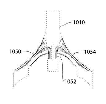
[0059] Referring now to Figures 11 and 12, and in accordance with an
illustrative
20 embodiment of the invention, a mask to be worn on a subject's face for
use in respiratory
monitoring and/or diagnostics will be described. The mask, generally referred
to using
the numeral 1000, comprises at least one transducer, such as microphones 1002
and 1004
in this example, and a support structure 1006 for supporting same above a nose
and
mouth area of the subject's face. The support structure 1006 is generally
shaped and
25 configured to rest on the subject's face and, in this example, thereby
delineate the nose
and mouth area thereof (e.g. see Figure 12), and comprises two or more
outwardly
projecting limbs 1008 (e.g. three limbs in this example) that, upon
positioning the mask
13
TRI-m1,5/PCI-CDA
PAGE 21/64* RCVD AT 2114/2013 11:40:51 AM [Eastern Standard Time)*
SVR:F00003/5 DNIS:3906* CSID:1 705 652 6074* DURATION (mm-ss):28-13
CA 02801559 2013-02-14
02/14/2013 THU 11:40 FAX 1 705 652 6074 Gastle Image Systems 12022/064
1000, converge into a transducer supporting portion 1010 for supporting
microphones
1002 and 1004 at a distance from this area,
[0060] In general, the at least one transducer is responsive to sound and/or
airflow for
generating a data signal representative thereof, so to effectively monitor
sound and/or
airflow produced by the subject while breathing. For example, in the
illustrated
embodiment, two microphones 1002 and 1004 are provided in the transducer
support
portion 1010, wherein one of these microphones may be predominantly responsive
to
sound, whereas the other may be predominantly responsive to airflow. For
example, the
microphone configured to be predominantly responsive to airflow may be more
sensitive
to air pressure variations than the other. In addition or alternatively, the
microphone
configured to be predominantly responsive to sound may be covered with a
material that
is not porous to air. In addition or alternatively, the microphone configured
to be
predominantly responsive to sound may be oriented away from the subject's nose
and
mouth so to reduce an air impact on the diaphragm of this microphone produced
by the
subject's breathing airflow. In other embodiments, a microphone predominantly
responsive to airflow may be positioned in the transducer support portion in
line with the
subject's nose and mouth, while another microphone may be positioned to the
side or on
the periphery of the mask to thereby reduce an influence of airflow thereon.
In some of
these embodiments, the recorded sound from the peripheral microphone, or again
from
the microphone predominantly responsive to sound, may in fact be used to
isolate the
airflow signal recorded in the nosepiece, by filtering out the sound signal
recorded
thereby, for example. An example of this process is schematically depicted in
Figure 23,
wherein a sound signal recorded via microphone 2 is used as reference for
microphone 1
to further isolate an airflow signal picked up via microphone 1. It will be
appreciated that
this type of processing may occur locally, via one or more microprocessors
disposed
directly within the mask, for example, or again via a downstream processing
platform, for
example implemented at a remotely located diagnostic center.
14
TR1-MP5/PCT-CDA
PAGE 22164' FICVD AT 2114/2013 11:40:51 AM [Eastern Standard Timel*
SVR:F00003/5 * DNIS:3906* CSID:1 705 652 6074* DURATION (mm-ss):28-13
CA 02801559 2013-02-14
02/14/2013 THU 11:40 FAX 1 705 652 6074 Gastle Imau Systems
12023/064
[00611 In yet another embodiment, a single microphone may alternatively be
used to
capture both sound and airflow, wherein each signal may be distinguished and
at least
partially isolated via one or more signal processing techniques, for example,
wherein a
turbulent signal component (e.g. airflow on microphone diaphragm) could be
removed
from other acoustic signal components (e.g. snoring). Such techniques could
include, but
are not limited to adaptive filtering, harmonics to noise ratio, removing
harmonics from a
sound recording, wavelet filtering, etc.
1.00621 In each of the above examples, the device may be implemented using a
single
type of transducer, for example one or more microphones which may in fact be
identical.
It will be appreciated however that other types of transducers, particularly
responsive to
airflow, may be considered herein without departing from the general scope and
nature of
the present disclosure. For example, a pressure sensor or airflow monitor may
be used
instead of a microphone to yield similar results in capturing an airflow
produced by the
subject while breathing.
[0063] Furthermore, while the above examples contemplates the provision of
one or
more transducers for the recordal of both sound and airflow, it may be
desirable, in
accordance with other embodiments of the invention, to include only a single
transducer
for acquiring data representative of only one of sound or airflow. For
example, in the
illustrative embodiments depicted and described in greater detail below,
improved airflow
measurements may in fact be used in isolation to provide a certain level of
monitoring
and diagnosis, without departing from the general scope and nature of the
present
disclosure.
[0064] It will also be appreciated by the skilled artisan that the exact
location of the
transducer(s) / microphone(s) may, depending on the subject, application
and/or further
experimentation, be subject to change. For example, the mask may be
reconfigured to
adjust the position of the at least one transducer, together or independently
when
considering multiple-transducer embodiments, to be closer to the nose, closer
to the
15
TRI-MPS/PCT-CDA
PAGE 23164* RCVD AT 2114/2013 11:40:51 AM [Eastern Standard Time]'
SVR:F0000315* DNIS:3906* CSID:1 705 652 6074 DURATION (mm-ss):28-13
02/14/2013 THU 11:40 FAX 1 705 652 6074 Gastle Image Systems CA 02801559
2013-02-14
2024/064
=
mouth, between the nose and mouth, in the upper lip or mustache area of the
subject's
face, etc. Ultimately, the mask will provide for the ability to capture both
sound and
airflow, both useful in respiratory monitoring and diagnostics.
10065] Still referring to the embodiment of Figures 11 and 12, the
support structure
5 further comprises an optional frame 1012 and face resting portion 1014
shaped and
configured to contour the face of the subject and at least partially
circumscribe the nose
and mouth area of the subject's face, thereby facilitating proper positioning
of the mask
on the subject's face and providing for greater comfort. A restraining
mechanism, such as
head straps 1016 and 1018, can be used to secure the mask to the subject's
face and
10 thereby increase the likelihood that the mask will remain in the
proper position and
alignment during use, even when the subject is sleeping, for example, in
monitoring and
diagnosing certain common breathing disorders. It will be appreciated that the
mask and
diagnostic approaches described below are also applicable, in some conditions,
in
monitoring and diagnosing a subject's breathing when awake.
15 [0066] In this embodiment, the mask 1000 further comprises a
recording device 1020,
such as a digital recording device or the like, configured for operative
coupling to the at
..=
least one transducer, such as microphones 1002 and 1004, such that sound
and/or airflow
signals generated by the at least one transducer can be captured and stored
for further
processing. In this particular embodiment, the recording device 1*020 is
disposed on a
20 frontal member 1022 of the support structure 1006, thereby reducing an
obtrusiveness
thereof while remaining in close proximity to the at least one transducer so
to facilitate
signal transfer therefrom for recordal. In providing an integrated recording
device, the
mask 1000 can effectively be used as a self-contained respiratory monitoring
device,
wherein data representative of the subject's breathing can be stored locally
on the mask
25 and transferred, when convenient, to a remotely located respiratory
diagnostic center.
[0067] Referring now to Figures 13 to 22, the general shape and
structural features of
support structure 1006, in accordance with one embodiment of the invention,
will be
16
TRI-MPS/PCT-CDA
: PAGE 24164 RCVD AT 211412013 11:40:51 AM [Eastern Standard Time]'
SVR:F0000315 * DNIS:3906* CSID:1 705 6526074'DURATION (mm-ss):28-13
CA 02801559 2013-02-14
02/14/2013 THU 11:41 FAX 1 705 652 6074 Gast1e Image Systems 2025/064
described in greater detail. In this embodiment, the support structure
comprises three (3)
outwardly projecting limbs, namely two opposed limbs 1050 and a central limb
1052,
which converge into the transducer supporting portion 1010, thereby forming a
tripod-
like structure extending from the nose and mouth area of the subject's face
when the
mask is in position. Each of these limbs has, along at least a portion thereof
and in
accordance with one embodiment, an inward-facing channel 1054 defined therein
for
channeling at least a portion of airflow produced by the subject while
breathing, toward
the at least one transducer disposed within the transducer supporting portion
1010. To
further accentuate this feature, the transducer supporting portion 1010 of
this particular
embodiment is shaped and oriented to further funnel the airflow channeled by
the limbs
1050 and 1052 toward the at least one transducer, depicted generically in
Figure 21 as
transducer 1056. For instance, the funneling shape may fluidly extend into
each of these
inward-facing channels 1054 to provide a continuous airflow guide toward the
at least
one transducer 1056 positioned within the transducer support portion 1010.
Furthermore,
as will be appreciated by the person of ordinary skill in the art, the
provision of limbs
1050 and 1052, as compared to an enclosed mask, provides for reduced airflow
resistance, resulting in substantially reduced dead space. As will be
appreciated by the
skilled artisan, while the limbs and transducer support portion are described
as distinct
components of the support structure, these terms are merely used herein for
the purpose
of illustrating a general progression, in this embodiment, of outwardly
projecting
structures ultimately converging toward one or more adequately supported
transducers.
Accordingly, while the above describes a substantially funneling transducer
support
portion, a similar embodiment may rather define a substantially funneling
support
structure and/or limbs converging to a supported transducer, for example as
described in
accordance with the following embodiment, and that, without departing from the
general
scope and nature of the present disclosure.
100681 Referring now to Figures 25 and 26, and in accordance with another
illustrative embodiment of the invention, a mask to be worn on a subject's
face for use in
17
TRI-MP5/PCT-CDA
PAGE 25/64' RCVD AT 2114/2013 11:40:51 AM [Eastern Standard Time]
SVR:F0000315* DNIS:3906* CSID:1 705 652 6074' DURATION (mm-ss):28-13
02/14/2013 THU 11:41 FAX 1 705 652 6074 Gast1e Image Systems CA 02801559 2013-
02-14 J2026/064
respiratory monitoring and/or diagnostics will be described. The mask,
generally referred
to using the numeral 2000, comprises at least one transducer, such aS
microphone 2002 in
this example, and a support structure 2006 for supporting same above a nose
and mouth
area of the subject's face. The support structure 2006 is generally shaped and
configured
5 to rest on the subject's face and extend outwardly therefrom over a nose
and mouth area
thereof to provide a transducer supporting portion 2010 for supporting the
microphone
2002, upon positioning the mask, at a distance from this area.
[0069] In this example, the support structure 2006 is shaped and
configured to
support the transducer 2002 above the nose and mouth area at a preset
orientation in
10 relation thereto, wherein the preset orientation may comprise one or
more of a preset
position and a preset angle to intercept airflow produced by both the
subject's nose and
mouth.
[0070] For example, in one embodiment, the preset orientation may be
preset as a
function of an estimated intersection between nasal and oral airflow, for
example based
I 5 on an observed or calculated average intersection between such
airflows.
[0071] For instance, in one embodiment, the preset orientation may
comprise a preset
position that, upon positioning the mask on the subject's face, is
substantially laterally
centered relative to the subject's face and longitudinally substantially in
line with or
below the subject's mouth, thus generally intercepting oral and nasal airflow.
20 [0072] In a same or alternative embodiment, the preset
orientation may comprise a
preset angle that aligns the microphone, or a principle responsiveness axis
thereof, along
a line more or less representative of an averaging between general oral and
nasal airflows,
For instance, in one embodiment, the orientation angle is preset to more or
less bisect an
angle formed by the transducer's preset position relative to the subject's
nose (i.e.
25 nostrils) and mouth. As will be described below, this bisecting angle,
which should be
construed within the present context to represent an angle more or less
directing the
transducer's principal responsiveness axis toward a point somewhere between
the
18
TRI-MPS/PCT-CDA
PAGE 26/64 RCVD AT 2/14/2013 11:40:51 AM [Eastern Standard Time]' SVR:F0000315
* DNIS:3906 = CSID:1 705 6526074'DURATION (mm-ss):28-13
CA 02801559 2013-02-14
02/14/2013 THU 11:42 FAX 1 705 652 6074 Gastle Imaqe Systems 2027/064
wearer's nose and mouth, may be determined as a function of measured, observed
and/or
otherwise estimated nasal and oral breathing patterns, so to improve or
enhance the
= transducer's general responsiveness to airflow originating from the
nose and/or mouth of
the candidate. Generally, the preset orientation may thus, in accordance with
one
embodiment of the invention, comprise a preset angle that, upon positioning
the mask on
the subject's face, substantially aligns the transducer with a point between
the subject's
nose and mouth.
[0073] With reference to Figure 27, an exemplary depiction 2100 of a general
nasal
(2150) and oral (2152) airflow overlap pattern is shown, in a vertical plane,
whereby air
directed by either of the nose and mouth is shown to generally spread
conically and
intersect at a point or in a general intersection area 2154. With reference to
Figure 28, in
which the airflow patterns 2100 of Figure 27 superimpose the mask 2000 of
Figure 27,
and in accordance with one embodiment of the invention, the preset orientation
of the
transducer 2002 is generally selected as a function of the airflow
intersection point or
area 2154 so to fall in a vicinity thereof', thus effectively improving
airflow detection.
[0074] In one example, and with reference to Figure 29, multiple nasal and
oral
airflow patterns (2250 and 2252, respectively) were traced, and their
respective
intersections, such as intersection point or area 2254, noted. From these
traced patterns
and observed intersections, an estimated general intersection point or area
could be
defined, as a function of which, a preset transducer orientation could then be
defined to
improve, if not maximized, a responsiveness thereof to nasal and oral airflow
produced
by different candidates while breathing. As will be appreciated by the skilled
artisan,
while various observations can be conducted in optimizing transducer
orientation in
respect of an estimated or anticipated most likely nasal and oral airflow
intersection area,
other considerations in developing a specific mask design may also affect the
ultimate
orientation of the transducer. Selecting a preset orientation as a function of
such
observations, however, may nonetheless improve an overall responsiveness and
usability
of the mask for breath monitoring and/or diagnostics.
19
TRI-MP5/PCT-CDA
PAGE 27164' RCVD AT 211412013 11:40:51 AM [Eastern Standard Time]
SVR:F0000315* DNIS:3906 * CSID:1 705 6526074'DURATION (mm-ss):28-13
CA 02801559 2013-02-14
02/14/2013 THU 11:42 FAX 1 705 652 6074 Gastle Image Systems 2028/064
[0075j Referring now to Figures 30 and 31, the support structure 2006
generally
comprises two outwardly projecting limbs 2008 that flow continuously one
within the
other toward the transducer supporting portion 2010 in defining a funneling
shape that
substantially converges toward this transducer supporting portion, thus
effectively
redirecting nasal and/or oral airflow toward the transducer 2002 and allowing
for
effective monitoring of airflow produced by both the subject's nose and mouth
while
breathing. As particularly shown in these figures, an illustrative nasal
airflow 2350,
which will generally more or less diverge laterally from the candidate's
nostrils as it is
projected more or less obliquely downward therefrom (e.g. as shown in Figures
27 to 29),
In can be effectively collected, at least partially, by the generally concave
support structure
2006 to be substantially funneled thereby toward .the transducer 2002.
Accordingly, in
this embodiment, not only is the transducer's preset orientation generally
selected as a
function of an estimated nasal and oral airflow intersection, the general
funneling shape
of the support structure 2006 will further redirect at least a portion of
laterally diverging
nasal (and oral) airflow toward the transducer 2002. Similarly, though not
explicitly
depicted herein, the same generally concave shape of the funneling support
structure
2006 will also, partly due to its upwardly titled orientation in this
embodiment, also at
least partially redirect longitudinally divergent airflow toward the
transducer 2002.
[0076] With particular reference to Figure 30, and in accordance with one
embodiment, the transducer supporting portion 2010 of the support structure
2006
comprises one or more (three in this embodiment) transducer supporting bridges
or limbs
2026 extending from a transducer-surrounding aperture 2028 defined within the
support
structure 2006. In this embodiment, the provision of bridging limbs 2026 may
allow for a
general reduction in airflow resistance, which may result in substantially
reduced dead
25. space. For example, as schematically illustrated in this Figure, while the
general
funneling shape of the support structure 2006 allows for a redirection of
airflow 2350
toward the transducer 2002, the bridged aperture 2028 allows for this flow of
air to
continue beyond the transducer 2002, and thereby reduce the likelihood of this
flowing
20
TRI-MP5/PCT-CDA
PAGE 28/64 RCVD AT 211412013 11:40:51 AM [Eastern Standard Time] SVR:F0000315*
DNIS:3906* CSID:1 705 652 6074 * DURATION (mm-ss):28-13
02/14/2013 THU 11: 43 FAX 1 705 652 6074 Gastle Image Systems CA 02801559
2013-02-14
2029/064
=
air pooling within the mask and/or flowing back onto itself, which could
otherwise lead
to a generally uncomfortable warm/humid flow of breath back in the candidate's
face
(and which could thus be breathed in again), and/or lead to unusual flow
patterns and/or
sounds that could further complicate data processing techniques in accounting
for these
5 patterns.
[0077[ The person of ordinary skill in the art will readily appreciate
that while the
above describes one example of a particular mask shape and orientation, other
shapes and
orientations may be exploited to achieve similar results, and that, without
departing from
the general scope and nature of the present disclosure.
10 [0078] Referring generally to Figures 25 and 26, the
transducer 2002 is at least
responsive to airflow for generating a signal representative thereof, so to
effectively
monitor airflow, and optionally sound, produced by the subject while
breathing. For
example, in the illustrated embodiment, a single microphone 2002 is provided
in the
transducer support portion 2010, wherein both sound and airflow may be
recorded, or
15 again, wherein either of these signals may be predominantly recorded
based on the
application at hand. It will be appreciated that the considerations discussed
above with
respect to the provision different numbers and/or types of transducers will be
readily
applicable in the context of this embodiment, as can the single or multiple
signal
processing techniques discussed above, and their equivalents, be considered in
the
20 context of the implementation of this embodiment.
100791 The support structure 2096 further comprises an optional frame
2012 and face
resting portion 2014 shaped and configured to contour the face of the subject
and at least
partially circumscribe the nose and mouth area of the subject's face, thereby
facilitating
proper positioning of the mask on the subject's face and providing for greater
comfort. A
25 restraining mechanism, such as head straps 2016, can be used to
secure the mask to the
subject's face and thereby increase the likelihood that the mask will remain
in the proper
position and alignment during use, even when the subject is sleeping, for
example, in
TRI-MPS/PCT-CDA 21
=
1
PAGE 29/64 RCVD AT 2/1412013 11:40:51 AM [Eastern Standard Time)* SVR:F0000315
DNIS:3906* CSID:1 705 652 6074 DURATION (mm-ss):28-13
CA 02801559 2013-02-14
02/14/2013 THU 11:43 FAX 1 705 652 6074 Gastle Image Systems
2030/064
monitoring and diagnosing certain common breathing disorders. It will be
appreciated
that the mask and diagnostic approaches described below are also applicable,
in some
conditions, in monitoring and diagnosing a subject's breathing when awake.
100801 In this embodiment, the mask 2000 further comprises a recording device
2020,
such as a digital recording device or the like, configured for operative
coupling to the at
least one transducer 2002, such that sound and/or airflow signals generated by
the at least
one transducer can be captured and stored for further processing. In this
particular
embodiment, the recording device 2020 is disposed on one of the limbs 2008 of
the
support structure 2006, thereby reducing an obtrusiveness thereof while
remaining in
close proximity to the at least one transducer so to facilitate signal
transfer therefrom for
recordal. A battery pack 2024, operatively coupled to the recording device
2020, is
provided on a frontal member 2022 of the mask 2000 to power the recording
device.and
transducer in acquiring data free of any external wiring or the like. In
providing an
integrated and self-supported recording device, the mask 2000 can effectively
be used as
a self-contained respiratory monitoring device, wherein data representative of
the
subject's breathing can be stored locally on the mask and transferred, when
convenient, to
a remotely located respiratory diagnostic center.
[0081] As will be appreciated by the person of ordinary skill in the art, the
general
shape and design of the above-described masks (1000, 2000) can provide, in
different
embodiments, for an improved responsiveness to airflow produced by the subject
while
breathing, and that irrespective of whether the subject is breathing through
the nose or
mouth, predominantly through one or the other, or through both substantially
equally.
Namely, the ready positioning of an appropriate transducer responsive to
airflow relative
to the nose and mouth area of the subject's face is provided for by the
general spatial
configuration of these masks. Accordingly, great improvements in data quality,
reliability
and reproducibility can be achieved, and that, generally without the
assistance or
presence of a health care provider, which is generally required with
previously known
systems.
22
TRI-MP5/PCT-CDA
=
PAGE 30164' RCVD AT 2/1412013 11:40:51 AM [Eastern Standard Time]'
SVR:F0000315* DNIS:3906* CSID:1 705 6526074'DURATION (mm-ss):28-13
02/14/2013 THU 11:44 FAX 1 705 652 6074 Gastle Image Systems CA 02801559
2013-02-14
2031/064
[0082] Furthermore, it will be appreciated that different
manufacturing techniques
and materials may be considered in manufacturing the above and similar masks,
for
example as described below, without departing from the general scope and
nature of the
present disclosure. For example, the entire mask may be molded in a single
material, or
5 fashioned together from differently molded or otherwise fabricated
parts. For example,
the outwardly projecting nosepiece of the mask may comprise one part, to be
assembled
with the frame and face-resting portion of the mask. Alternatively, the frame
and
nosepiece may be manufactured of a single part, and fitted to the face-resting
portion
thereafter. As will be further appreciated, more or less parts may be included
in different
10 embodiments of these masks, while still providing similar results. For
example, the nose
piece, or an equivalent variant thereto, could be manufactured to rest
directly on the
subject's face, without the need for a substantial frame or face resting
portions, as
illustrated in the above described embodiments. Alternatively or in addition,
different
numbers of outwardly projecting limbs (e.g. two, three, four, etc.) or
structures may be
15 considered to provide similar results.
[0083] As discussed hereinabove, breathing disorders are traditionally
monitored and
diagnosed using data acquired at sleep centers, where subjects are fitted with
a number of
electrodes and other potentially invasive monitoring devices, and monitored
while they
sleep. Clearly, as the subject is both required to sleep in a foreign setting
with a number
20 of relatively invasive and obtrusive monitoring devices attached to
them, the data
collected can often be misleading, if the subject even ever manages to get any
sleep to
produce relevant data. Clearly, other respiratory monitoring and diagnostic
approaches
can be implemented while the subject is awake, and such approaches are fully
within the
realm of the present disclosure as the masks and methods disclosed herein may,
in some
25 embodiments, be rendered equally useful in monitoring or diagnosing
sleeping and awake
subjects.
[0084] Furthermore, known respiratory diagnostic systems, for
example as depicted
in Figure 24, generally require the acquisition of multiple sensory data
streams to produce
23
TRI-MPS/PCT-CDA
1
= PAGE 31/64* RCVD AT 211412013 11:40:51 AM [Eastern Standard Time]
SVR:F0000315 * DNIS:3906* CSID:1 705 652 6074 * DURATION (mm-ss):28-13
CA 02801559 2013-02-14
02/14/2013 THU 11:44 FAX 1 705 652 6074 Gastle Image Systems Z032
/ 064
workable results that may include breath sounds, airflow, chest movements,
esophageal
pressure, heart rate, etc. Similarly, known portable monitoring devices
proposed for the
diagnosis of sleep apnea generally require subjects to. adequately position
and attach
several wired electrodes responsive to a number of different biological
parameters, such
as listed above, which generally reduces the comfort and compliance of
subjects and
increases chances of detachment and/or displacement of the electrodes. Given
that
portable sleep apnea monitors are used in the absence of an attending health
care
professional, inaccurate placement or displacement of electrodes cannot be
easily
detected until the data is transferred to the health center. On the other
hand, simplified
portable respiratory monitoring devices, as discussed above, only produce data
with
respect to either airflow or sounds generated during breathing, which limited
data sets are
generally insufficient in adequate respiratory disorder diagnostics.
[0085] in comparison, the respiratory monitoring and/or diagnostic masks
described
above in accordance with different embodiments of the invention may provide a
number
Is of advantages over known techniques. For example, all elements of these
self-contained
diagnostic masks are contained in a single unit including for instance, the at
least one
transducer, power supply, electronics, and data storage. The at least one
transducer is
embedded within the mask structure and thus readily positioned on the
subject's face by
the very nature of the mask's spatial configuration. Accordingly, proper
positioning is
generally guaranteed, allowing for adequate capture of both sound and/or
airflow
produced by the subject while breathing, while reducing the number of required
electrodes. Furthermore, as all wiring and circuitry may be embedded within
these masks,
problems traditionally associated with disconnection of sensory electrodes are
practically
eliminated. The subject is also free of external wiring, thereby reducing
subject
discomfort and increasing compliance. This advantage is diagrammatically
illustrated in
Figure 24, wherein a single physical data channel can be produced locally
using a self-
contained mask, and communicated to a diagnostic center where signal
processing, for
example as described below, enables extraction of a number of clinical
measures useful
24
TRI-MPS/PCT-CDA
= PAGE 32/64 RCVD AT 2/14/2013 11:40:51 AM [Eastern Standard Time]
SVR:F0000315 DNIS:3906 " CSID:1 705 652 6074 * DURATION (mm-ss):28-13
CA 02801559 2013-02-14
02/14/2013 THU 11:45 FAX 1 705 652 6074 Gastle Image Systems
jap33/064
in providing similar diagnostics as that only previously available using
multiple
electrodes in conventional systems. It will be appreciated that reducing the
number of
physical channels provides great advantage in deploying a portable device
wherein a
layman is required to wear the device in the absence of a trained health care
provider. In
the present diagram, it will be appreciated that reference to a "single
channel" in fact
generally represents a single physical link between the subject, and what
could ultimately
result in a full respiratory diagnosis. Namely, the subject in this embodiment
is only
requested to wear a mask which allows for recordal of sound and/or airflow via
one or
more transducers, while allowing for the downstream processing of multiple
clinical
measures from this single data acquisition device type. To the contrary,
clinical and
known portable devices generally require multiple data outputs provided by a
multiplicity
of data acquisition devices and types so to access multiple clinical measures,
which, as
discussed above, reduces subject comfort and compliance, and may therefore
reduce data
reliability and reproducibility. The alternative in the art, is to reduce data
acquisition to a
single measure, which, in general, has limited value.
100861 In one embodiment, the recorded data is stored, and optionally
encrypted on a
removable data storage device, such as an SD card or the like. For example,
analog data
acquired by the one or more transducers can be locally pre-amplified,
converted into
digital data (e.g. via a local A/D converter) and stored in the removable
memory device.
The stored data can then either be uploaded from the memory card to a local
computing
device (e.g. laptop, desktop, palmtop, smartphone, etc.) for transmittal to a
remotely
located diagnostic center via one or more wired and/or wireless communication
networks,
or physically shipped or delivered to the remotely located diagnostic center
for
processing. Namely, the acquired data can be processed via one or more
diagnostic
software platforms, or the like (e.g. as discussed hereinbelow), to evaluate
the subject's
breathing and provide, as appropriate, diagnosis of relevant breathing
disorders.
Furthermore, given this system's generally distributed architecture, various
distinct
and/or complimentary processing techniques and algorithms may be applied to a
same
25
TRI-MP5/PCT-CDA
PAGE 33164 RCVD AT 2114/2013 11:40:51 AM [Eastern Standard Time]'
SVR:F0000315* DNIS:3906* CSID:1 705 652 6074 * DURATION (mm-ss):28-13
CA 02801559 2013-02-14
02/14/2013 THU 11:45 FAX 1 705 652 6074 Gastle Image Sytems
2034/064
data set to increase diagnostic complexity and/or reliability, for example. In
such
embodiments, given that the data storage device retains all relevant data,
once the data is
shipped, the mask itself may be disposed of, or again, reused by the same
subject to
acquire further data in respect of a same or similar breathing study.
[00871 It will be appreciated that different types of data transfer and
communication
techniques may be implemented within the present context without departing
from the =
general scope and nature of the present disclosure. For example, while the
above
examples contemplate the use of a digital recording device having a removable
data
storage medium, such as a memory card of the like, alternative techniques may
also be =
0 considered. For example, the recording device may rather include a wireless
communication interface wherein data integrally recorded thereon can be
wirelessly
uploaded to a computing device in close proximity thereto. For example, Wi-Fi
or
Bluetooth applications may be leveraged in transferring the data for
downstream use.
Alternatively, the device may include a communication port wherein recorded
data may
be selectively uploaded via a removable communication cable, such as a USB
cable or
the like. In yet another example, the recording device itself may be removably
coupled to
the mask and provided with a direct communication interface, such as a USB
port or the
like for direct coupling to an external computing device. These and other such
examples
= are well within the realm of the present disclosure and
therefore, should not, nor should
their equivalents, be considered to extend beyond the scope of the present
disclosure.
[0088] As will be appreciated from the proposed diagnostic procedures
described
below, the provision of a respiratory monitoring and diagnostic mask, as
described
herein, provides for the implementation of a method for remotely diagnosing a
breathing
disorder of a subject. Namely, upon providing the subject access to a self-
contained
mask, as described herein, the subject may then proceed to wear the mask, when
appropriate for the condition to be monitored, and integrally record sound
and/or airflow
produced during breathing. Once this data is transferred to a remotely located
diagnostic
center, a breathing disorder may be diagnosed on the basis of the processed
sound and/or
26
TRI-N1135/PCT-CDA
PAGE 34164* RCVD AT 2/14/2013 11:40:51 AM [Eastern Standard Time)*
SVR:F0000315* DNIS:3906* CSID:1 705 652 6074 * DURATION (mm-ss):28-13
CA 02801559 2013-02-14
0 2/14/ 2013 THU 11: 45 FAX 1 7 0 5 6 52 6 0 7 4 Gas tle Image ystems
2035/064
airflow signals recorded by the mask. Namely, no additional sensors or
recordings are
required to achieve workable results, leaving the subject to conduct all
relevant
recordings at home, if so desired, remote from any qualified health care
practitioner.
Furthermore, the general improvements in transducer positioning achieved by
the design
5 of the various embodiments of the masks described herein, allow for
greater data
reliability and reproducibility, while significantly reducing discomforts or
inconveniences
to the subject.
[00891 In accordance with another embodiment, a microphone 12 is
located in a
position proximal to an individual's mouth as shown in FIGS. 2a and 2b, in
this case by a
10 dimension A of approximately 3 cm in front of the individual's face,
i.e. at a distance
from a nose and mouth area of the subject's face. The microphone 12 may be
configured
to communicate with the microprocessor by way of an interface or other data
acquisition
system, via a signal transducing link or data path 18 to provide one or more
data
collection modules with the microphone 12. Thus, such data collection !nodules
and the
15 microphone are operable to collect breathing sounds emanating from the
individual's
mouth and nose, during the inspiration and/or expiration phases of breathing.
For
example, an exemplary microphone response curve is shown in FIG. 1. The
acoustic
signal data breathing sounds collected from the individual may be comprised of
both
airflow sounds from the individual's breathing applying air pressure to the
microphone
20 diaphragm and actual breathing sounds resultant from the individual's
breathing being
recorded and/or collected by the microphone 12. Furthermore, the acoustic
signal data
breathing sounds collected from the individual may be, in another exemplary
embodiment, comprised of substantially only actual sounds resultant from the
individual's breathing being recorded and/or collected by the microphone 12.
In still yet
25 another embodiment, the acoustic signal data breathing sounds
collected from the
individual may be comprised of substantially only airflow sounds resultant
from the
individual's breathing applying air pressure to the microphone diaphragm and
being
recorded and/or collected by the microphone 12. As used herein, term "airflow
sounds"
27
TRI-MP5/FCT-CDA
PAGE 35/64* RCVD AT 2114/2013 11:40:51 AM [Eastern Standard
Time]*SVR:F0000315* DNIS:3906 CSID:1 705 6526074' DURATION (mm-ss):28-13
'
CA 02801559 2013-02-14
02/14/2013 THU 11:46 FAX 1 705 652 6074 Gastle Ima_ge pysterris
2_036/064
=
refers to the air pressure resultant from an individual's breathing being
applied to and
= causing the microphone's diaphragm to move such that the
microphone collects and
produces data for the audio recording.
[00901 The microphone 12, for example, may be coupled in or to a loose
fitting full
5 face mask 16 as shown in FIGS. 2a and 2b. Furthermore, the face mask 16
may include
at least one opening 14 to allow for ease of breathing of an individual 20.
For example,
the microphone 12 may be in a fixed location with a spacing of dimension "A",
of about
3 cm in front of the individual's face as shown schematically in FIG. 2a;
however other
distances in front of the individual's face may be desirable in some
embodiments. The
la microphone 12, in this case, is embedded in a respiratory mask 16 which
is modified by
cutting away material so as produce opening 14 such that only a structural
frame portion
remains to keep the microphone 12 in a fixed location relative the nostrils
and the mouth =
of an individual 20. In one example, the audio signals from the microphone may
be
digitized using an audio signal digitizing module and digitized sound data to
be
15 transferred via transducing link 18 to the computer using a USB
preamplifier and audio
interface (M-Audio, Model Fast Track Pro USB) with a sampling rate of 22,050
Hz and
resolution of 16 bits. Although various types of audio interfaces may be used,
in the
instant exemplary embodiment, an external audio interface provides suitable
results over
the other types of audio adapters, for example, built-in audio adapters due to
the superior
20 signal to noise (S/N) ratio of the external adaptor which is about 60
dI3 at 1 kHz. Sound
recordings may then be passed through a 4111 order band-stop digital filter
with a centre
frequency of about 60 Hz to suppress line interference. Other structures may
also be used
to locate the microphone in position, as including support structures
positioned against a
plurality of locations on the individual or stationed adjacent the individual
as required. .
25 [00911 Furthermore, in another exemplary embodiment, a two
microphone system
may be useful. In such a system, as shown in FIG 2b, one of the microphones, a
first
microphone 12b, may be configured to collect actual breathing sounds and
airflow sounds
whereas the other microphone, a second microphone 12c may be configured to
collect
28
TRI-MP5/PCI-CDA
= PAGE 36164 RCVD AT 211412013 11:40:51 AM [Eastern Standard Time]'
SVR:F0000315 * DNIS:3906* CSID:1 705 652 6074' DURATION (mm-ss):28-13
02/14/2013 THU 11:46 FAX 1 705 652 6074 Gastle Image Systems
CA 02801559 2013-02-14
Z037/064
substantially only actual breathing sounds. In this embodiment, the waveform
sounds
and/or data collected from the second microphone 12c may be subtracted or
filtered from
the waveform sounds collected from the first microphone 12b, thereby resulting
in a
waveform data stream of substantially only airflow sounds. The airflow sounds
may be
5 resultant of pressure air from an individual's breathing being
collected as applied to the
diaphragm of a microphone as noted above. Subsequently, the airflow sounds may
then
be used as a waveform amplitude acoustic data stream in accordance with the
forgoing
method.
[00921 A raw acoustic data stream of breathing sounds, as
shown in a representative
10 plot, for example in FIG. 5, is then collected for each of a
plurality of respiratory phases
to form a bioacoustics signal recording, wherein the acoustic data stream is
subsequently
transformed.
[00931 As will be described below, in at least one embodiment,
a method and an
apparatus are provided to monitor, identify and determine the inspiratory
and/or
15 expiratory phases of the respiratory cycle of an individual 20
from the frequency
characteristics breathing sounds. It is understood that a numerical
comparative analysis
of the frequency spectrum as transformed from waveform amplitude data of
breathing
sounds and/or airflow sounds of an individual 20 may be useful to
differentiate between
the inspiration and expiration phases of breathing.
20 [0094] It will be appreciated by the person of
ordinary skill in the art that while the
below example describes a method in which a mask as depicted in Figures 2a and
2b was
used for data acquisition and breath monitoring/diagnostics, a mask as
described above
. with reference to Figures 11 to 22, or with reference to Figures 26 and 27,
could also be
used to produce similar effects, and that, without departing from the general
scope and
25 nature of the present disclosure. Furthermore, while the below
predominantly proposes a
wired solution for real-time monitoring, a similar approach may be applied,
for example
with respect to a self-contained mask as described above, wherein processing
steps
TRI-MPS/PCT-CDA
29
PAGE 37164 RCVD AT 211412013 11:40:51 AM [Eastern Standard Time]' SVR:F0000315
DNIS:3906* CSID:1 705 6526074'DURATION (mm-ss):28-13
CA 02801559 2013-02-14
0V14/2013 THD 11:47 FAX 1 705 652 6074 Gastle Image Systems
_Z038/064
applied to the locally acquired data could be implemented remotely at an
appropriate
diagnostic center.
[00951 It will also be appreciated that while the below description provides
one
example of a breath monitoring application of the herein-described masks,
other similar
or distinct breath monitoring and/or diagnostic approaches may be applied
using the data
acquired via different embodiments of these masks, and that, without departing
from the
general scope and nature of the present disclosure. For example, different
monitoring
and/or diagnostic methods relying on breath sound and/or airflow measurements
may be
implemented with and rely on data acquired using different mask embodiments as
described herein, which studies may include, but are not limited to, sleep
disorders such
as apneas and/or hypopneas, breathing disorders, snoring, and other such
conditions as
will be readily apparent to the person of ordinary skill in the art.
Accordingly, the below
example should not be construed as limiting to the above embodiments, hut
rather as a
means to exemplify it's possible utility within a particular context.
;
DATA ACQUISITION
[00961 Data were collected from 10 consecutive men and women at least 18
years of
age referred for overnight polysomnography (PSG). The subjects'
characteristics are
shown in Table I. Breath sounds were recorded by a cardoid condenser
microphone
(Audi-Technica condenser microphone, Model PRO 35x). The microphone's cardioid
polar pattern reduces pickup of sounds from the sides and rear, improving
isolation of the
sound source. The microphone 12 used for recording breath sounds has a
relatively flat
frequency response up to 2000 Hz as shown in FIG. 1. Furthermore, the
microphone 12,
as used herein has a higher output when sound is perpendicular to the
microphone's
diaphragm as shown by the solid line in FIG. 1, which helps reduce low
frequency
ambient noise interference. In this example, the microphone 12 was embedded in
the
centre of a loose fitting full face mask 16 modified to reduce airflow
resistance and
eliminate dead space by way of large openings 14 as shown in FIGS. 2a and 2b.
The
30
TRI-MPS/PCT-CDA
PAGE 38164' RCVD AT 211412013 11:40:51 AM [Eastern Standard Time] SVR:F0000315
DNIS:3906* CSID:1 705 6526074' DURATION (mm-ss):28-13
CA 02801559 2013-02-14
02/14/2013 THU 11:47 FAX 1 705 652 6074 Gastle Image Systems
0039/064
microphone 12 attached to the face mask 16, and was located in front of the
individual's
face. The mask 16 provides a structural frame portion to keep the microphone
in a fixed
location, at a dimension A of approximately 3 cm in front of the individual's
face, so as
to record breathing sounds to an audio recording device, such as a computer as
described
5 above, to make an audio recording thereof. In some exemplary embodiments,
the audio
recording of breathing sounds may be made and recorded in analog format prior
to
digitizing the audio recording. However, in other embodiments the audio
recording of
breathing sounds may be digitized in real-time. Furthermore, in some exemplary
embodiments, the processing of the audibly recorded waveform data or acoustic
signal
data may be performed in real-time, so as to provide substantially
instantaneous
= information regarding an individual's breathing. In an
exemplary embodiment, digitized
sound data were transferred to a computer using a USB preamplifier and audio
interface
(M-Audio, Model MobilePre USB) with a sampling rate of 22,050 Hz and
resolution of
16 bits. Although various types of audio interfaces may be used, in the
instant exemplary
embodiment, an external audio interface was preferred over a built-in audio
adapter due
to the better signal to noise (SIN) ratio of the external audio interface,
which was 91 dB.
FIG. 5 shows a 25-second waveform amplitude recording plot. However, in other
exemplary embodiments, it may be desirable to record breathing sounds for a
time period
of from about 10 seconds to 8 hours. In some exemplary embodiments it may be
desirable to record breathing sounds for a time period of from about 10 second
to about
20 minutes. In other exemplary embodiments, it may be desirable to record
breathing
sounds for greater than 20 minutes.
BREATHING ACOUSTICS ANALYSIS
[00971 In an exemplary embodiment, full night breath sound recordings were
displayed on a computer screen similar to the computer screen 1.2 of FIG. 3. A
representative raw acoustic data waveform plot, as may be shown on a computer
screen
1.2, is provided in FIG. 5 for a 25-second recording. Each increase in
amplitude
represents a single breath. The individual phases of a breathing cycle are not
readily
31
TRI-MPS/PCT-CDA
PAGE 39164* RCVD AT 2114/2013 11:40:51 AM [Eastern Standard Time]'
SVR:F0000315 DNIS:3906* CSID:1 705 6526074' DURATION (mm-ss):28-13
CA 02801559 2013-02-14
02/14/2013 THu 1148 FAx 1 705 652 6074 Gastle Image Systems
1Z1040/064
resolvable in FIG. 5 owing to the time scale being too large to resolve single
breath
details. For example, FIG. 7a more clearly shows the inspiration and
expiration phases of
a breathing cycle in a waveform amplitude versus time plot. The recordings
were visually
scanned to identify periods of regular breathing. After visual scanning, the
recordings
were played back for auditory analysis,
[0098] Sequences of normal breaths that did not have signs of obstructive
breathing
such as snoring and interruptions, or other irregularities such as tachypnea
(rapid
breathing), or hyperventilation (deep breathing) were then included in the
subsequent
frequency analysis. However, snoring and other types of noisy breathing can
also be
included in this analysis by applying a pre-processing technique that isolates
turbulent
from non-turbulent components, (e.g. as shown in Figure 23) whereby
ultimately, the
turbulent component may be selected for further processing. This process was
repeated to
select three random parts of an individual's sleep. If a portion of the
recording fulfilled
the aforementioned inclusion criteria, then 3 to 4 consecutive breaths were
selected from
that portion. A total of 10 breaths were selected from each individual. During
the process
of selecting the individual's breathing sound portions, the investigator did
not have a
previous knowledge of the sleep stage. Therefore, the investigator was blind
to the sleep
stage of an individual while selecting the analyzed breaths except for knowing
that
sampling started after the onset of sleep. The real-time stamp of each breath
was
registered in order to retrieve the sleep stage in which it took place in
afterwards.
Subsequently, the investigator listened to these breathing sounds again to
divide each
breath into its inspiratory, expiratory and interbreath phases. Each phase was
labeled
manually.
[00991 The data array of each breathing phase was passed through a hamming
window and a 2048-point Fast Fourier Transform (FFT) of the windowed data with
50%
overlap was calculated. The resultant frequency spectrum was displayed on a
computer
screen for visual analysis. The frequency spectra of the interbreath pauses
were also
calculated and incorporated in the analysis to control for the effect of
ambient noise.
32
TRI-MPS/PCT-CDA
PAGE 40/64 RCVD AT 211412013 11:40:51 AM [Eastern Standard Time]' SVR:F0000315
* DNIS:3906* CSID:1 705 652 6074* DURATION (mm-ss):28-13
CA 02801559 2013-02-14
02/14/2013 THU 11:48 FAX 1 705 652 6074 Gastle Image Systems
2041/064
Careful visual examination of spectra revealed that during inspiration, the
amplitude of
signals above 400 Hz was consistently higher than during expiration.
Therefore, it was
determined that the bands ratio (BR) of frequency magnitude between 400 to
1000 Hz, to
frequency magnitude between 10 to 400 Hz is higher in the inspiration phase as
5 compared to the expiration phase. It will be appreciated that the
above-noted threshold of
400 Hz is not necessarily to be strictly applied as this value can be varied
generally
between 200 Hz and 900 Hz depending on the microphone acoustic
characteristics, and
specificities of the application. The BR of each breathing cycle was then
calculated using
equation (1).
moth' 400117.
I ID BR -= 40011: FFT(f)I EFFT(f) 10H:
(I)
[001001 Using equation (1), the numerator represents the sum of FFT
higher frequency
magnitude bins which lie between 400 and 1000 Hz, and the denominator
represents the
sum of FFT lower frequency magnitude bills which lie between 10 and 400 Hz,
Bins
bellow 10 Hz were not included to avoid any DC contamination (referring to
drift from a
15 base line), and frequencies above 1000 Hz, can also, in some
embodiments, be neglected
since preliminary work (not shown) revealed insignificant spectral power at
frequencies
above 1000 Hz, in which case the computation may also be reduced. It will be
appreciated, however, that higher frequencies above 1000 Hz may nonetheless be
included depending on the calculation power or the instruments being used, To
verify
20 repeatability of the results, BR was calculated for 3 to 4 successive
breaths in the
included sequence and for a total of three sequences from different parts of
the
individual's sleep. A total of 100 breaths were collected from the 10
subjects. The mean
number of breaths per subject was 10 0.
1001011 It will be appreciated by the person of ordinary skill in
the art that other
25 methods may be employed to achieve similar results. For example,
while taking the ratios
of sub-bands of an FFT spectrum to measure sub-band energy distributions
provides a
33
TRI-MPS/PCT-CDA
PAGE 41/64 RCVD AT 2/14/2013 11:40:51 AM [Eastern Standard Time]*
SVR:F0000315* DNIS:3906* CSID:1 705 6526074' DURATION (mm-ss):28-13
02/14/2013 Tim 11: 48 FAx 1 705 652 6074 Gas-hie Image Systems CA 02801559
2013-02-14
[2042/064
useful approach, other statistical methods and pattern recognition tools can
be used to
distinguish the relative distribution of sub-band ratios in FFT. Furthermore,
FFT could
also be replaced, in some embodiments, by implementing a series of digital
filters that
measure signal energy in the bands mentioned in this work, for example.
Additionally, it
5 will be appreciated that the entire digital processing stream, could,
in some embodiments,
be replaced by analogue signal processing techniques, such as by deploying a
series of
analog filters to achieve similar results.
SLEEP STAGING
1001021 Sleep stages were recorded during the course of' the night
using standard
10 polysomnographic techniques that included electro-encephalography
(EEG), electro-
oculography and submental electro-myography (Rechtschaffen A and Kales A 1968
A
Manual of Standardized Terminology, Techniques and Scoring System Pr Sleep
Stages of
Human Subjects. (Los Angeles: UCLA Brain Information Service/Brain Research
Institute). The corresponding sleep stage for the selected breath samples was
determined
t5 from the PSG recording (not shown).
STATISTICAL ANALYSIS
[00103] Data are expressed as rnean SD unless otherwise stated. A
Wilcoxon Signed
Ranks Test was performed using SPSS statistical package (SASS, Chicago,
Illinois). This
test compares two related variables drawn from non-normally distributed
populations.
20 One-sample sing test was performed using Minitab 15 statistical package
(Minitab
State College, PA).
COMPARISION OF BANDS RATIO TO RESPIRATORY INDUCTANCE
PLETHYSMOGRAPHY
SUBJECTS
34
TRI-MP5/Per-CDA
PAGE 42164' RCVD AT 2114/2013 11:40:51 AM [Eastern Standard Time]*
SVR:F0000315* DNIS:3906* CSID:1 705 652 6074* DURATION (mm-ss):28-13
02/14/2013 THU 11:49 FAX 1 705 652 6074 Gastle Image Systems CA 02801559 2013-
02-14 0043/064
1001041 Healthy subjects at least 18 years of age were recruited with no
history of
respiratory or cardiopulmonary disease in , addition to being free from
prescribed
medications. Data were collected from 15 subjects, 6 men and 9 women, healthy
volunteers. Individuals used in the study were recruited by advertisement and
were
5 divided randomly intro 2 groups with 5 subjects in one grotip (test
group) and 10 in the
other (validation group). The data from the 5 subjects in the test group were
used to
examine acoustic characteristics of breathing phases, which were then
incorporated into a
method having an algorithm as described below. The resultant method was tested
on the
data of 10 subjects in the validation group to determine the validity of the
method for
to determining the inspiration and expiration phases of an individual's
breathing sounds.
BREATH SOUND RECORDING
1001051 Breath sounds in this particular example were recorded using a
unidirectional,
electret condenser microphone (Knowles Acoustics, Model MB6052USZ-2). The
microphone's unidirectional pattern reduces the pickup of sounds from the
sides and rear
15 thereby improving isolation of the sound source. In this example, the
microphone 12 was
embedded in a respiratory mask 16, as shown in Figures 2a and 2b, that was
modified by
cutting away material so as to produce opening 14 such that only a structural
frame
remained to keep the microphone 12 in a fixed location relative the nostrils
and the mouth
of an individual 20 at a dimension "A" of approximately 3 ern in front of the
individual's
20 face as shown in FIG. 2a. The audio signal was digitized using an audio
signal digitizing
module and digitized sound data were transferred via transducing link 18 to a
computer
using a USB preamplifier and audio interface (M-Audio, Model Fast Track Pro
USB)
with a sampling rate of 22,050 Hz and resolution of 16 bits. Although various
types of
audio interfaces may be used, in the instant exemplary embodiment, an external
audio
25 interface was preferred over the other types of audio adapters, for
example, built-in audio
adapters due to the superior signal to noise (S/N) ratio of the external
adaptor which was
about 60 dB at I kHz. Sound recordings were then passed through a 4th order
band-stop
digital filter with a centre frequency of about 60 Hz to suppress line
interference.
TRI-MP5/PCT-CDA 35
PAGE 43/64' RCVD AT 2/1412013 11:40:51 AM [Eastern Standard Timel*
SVR:F0000315* DNIS:3906* CSID:1 705 652 6074 * DURATION (mm-ss):28-13
CA 02801559 2013-02-14
02/14/2013 THU 11:49 FAX 1 705 652 6074 Gastle Image Systems
2044/064
=
RESPIRATORY INDUCTANCE PLETHYSMOGRAPHY
[OM] Respiratory inductance plethysmography (RIP), (Respitrace Ambulatory
Monitoring Inc., White Plains, NY, USA) was used to monitor respiratory
pattern of
individuals and the timing of the breathing phases. In contrast to other
breathing
5 monitoring apparatus such as pneumotacography, RIP has the advantage
of being applied
away from the face of an individual to allow capture of breathing phases.
Briefly, RIP is
a system comprising two flexible sinusoidal wires. Each wire is embedded in
stretchy
fabric band. One band 28 is placed around the chest of an individual and the
other band
30 is placed around the abdomen of the individual as shown in FIG. 6a. The
inductance
Jo of each band changes upon rib cage and abdomen displacements and
generates a voltage
signal proportional to its inductance. The signals from the RIP bands 28 and
30 were
digitized at 150 Hz and stored in a computer memory as substantially describe
above with
reference to FIGS. 3 and 4. The electrical sum of the ribcage and abdominal
signals is
displayed on a readable medium, for example a computer screen or a physical
plot, and
15 provides the total thoracoabdominal displacement. The
thoracoabdominal displacement
recorded from the RIP system reflects changes of tidal volume during
respiration.
[00107] In order to compare the inspiration and expiration
phases of an individual's
breathing to RIP, the microphone 12, as noted above, was coupled in this
example to a
modified mask 16 in front of the subject's face. Simultaneously, the RIP bands
28 and 30
20 were placed around the subject's chest and abdomen to measure
thoracoabdominal
motion as noted above. Recording were captured from both the microphone 12 and
the
RIP bands 28 and 30 simultaneously to assess the timing of breath sounds
against the RIP
waveform data.
STUDY PROTOCOL
25 [00108] Individuals were studied in the supine
position and were instructed to breathe
normally. Microphone holding frame 16 was placed on individual's face. Each
individual
was asked to breath for two minutes at their regular breathing rate. In order
to mimic all36
TRI-MP5/PCI-CDA
PAGE 44/64 * RCVD AT 2114/2013 11:40:51 AM [Eastern Standard Time]
SVR:F00003/5 DNIS:3906 CSID:1 705 652 6074 * DURATION (mm-ss):28-13
CA 02801559 2013-02-14
OZ./14/2013 THU 11:50 FAX 1 705 652 6074 Gastle Image Systems
20 4 5 / 0 6 4... ...
possible breathing conditions, the individuals were asked to breath through
their nose
only for half of the experiment time, and through their nose while mouth was
slightly
open in the other half Incomplete breaths at the beginning and end of
recording were
discarded and all the breaths in between were included in the analysis.
5 ANALYSIS OF BREATH ACOUSTICS
[00109] In a first stage, spectral variables of breath sounds that
characterize the
inspiratory and expiratory phase components of a respiratory cycle were
determined. The
data of five subjects, 3 females and 2 males was chosen randomly from total 15
subjects
and used to study the frequency characteristics of the acoustic signals of
different
10 respiratory phases. Inspiratory and expiratory segments of breath
sounds were determined
and extracted from the acoustic data by comparing it to the inspiratory
(rising edge) and
expiratory (falling edge) of the RIP trace as shown in FIG. 6b. A 25-second
long
recording of breath sounds and simultaneous summed thoracoabdominal RIP
signals
from a representative subject is shown, for example, in FIG. 6b. Dashed
vertical lines are
is shown to separate inspiration and expiration phases of the second cycle
at 32.
[00110] The first 10 complete breaths of each subject were analyzed, which
yielded a
total of 50 inspirations and 50 expirations acoustic data sets from the 5
subjects.
Subsequently, the frequency spectrum of each phase was calculated separately
using
Welch's method (i.e. the average of a 2048-point Fast Fourier Transform (FFT)
of sliding
20 hamming windows with 50% overlap). FFT arrays were normalized in
amplitude in order
to compare the relative changes in power spectrum among resultant spectral
arrays.
[00111] Using variables derived from frequency spectra of the 5 test
individual's noted
above, the inspiratory and expiratory phases of the breathing cycle were
determined for
the remaining 10 individuals in order to test the validity of the method.
Furthermore, the
25 method was tested for the ability to determine breathing. phases from
acoustic data
independently from other inputs. The data analysis was performed with Matlab
R2007b
software package (Mathworks, Natick, Massachusetts).
37
TRI-M Rs/pc-1*-c DA
=
PAGE 45/64' RCVD AT 2/14/2013 11:40:51 AM [Eastern Standard Time]*
SVR:F0000315* DNIS:3906* CSID:1 705 6526074' DURATION (mm-ss):28-13
O2/14/2013 THU 11:50 FAX 1 705 652 6074 Gastle Image S.y_stems CA 02801559
2013-02-14
2046/064
RESULTS
1001121 The characteristics of the individuals in this study are shown
in Table 1. A
total of 100 breaths were sampled from 10 patients with a mean number of 10
breaths per
subject. Seventy percent of' the breaths analyzed were from non-rapid-eye
movement
5 sleep (NREM), and 18% from rapid eye movement sleep (REM), and 12% while
patients
were awake according to the polysomnographie criteria.
Table 1. Characteristics of subjects.
Subject Age (years) Sex Body Mass Index
Subject 1 51 F 39.1
Subject 2 43 M 25.6
Subject 3 49 M 23.7
Subject 4 27 M 36.8
Subject 5 64 M 26.3
Subject 6 60 M 33.0
Subject 7 68 F 28.5
Subject 8 31 M 30.3
Subject 9 48 F 31.6
Subject 10 56 M 26.7
[001131 The bands ratio (BR) value was calculated for the inspiration phase
bands
ratio (BRi) 24, the expiration phase bands ratio (BRe) 26, and the interbreath
pause bands
TRI-MP5/PCT-CDA 38
4
PAGE 46/64* RCVD AT 2114/2013 11:40:51 AM [Eastern Standard Time]'
SVR:F0000315* DNIS:3906* CSID:1 705 6526074' DURATION (mm-ss):28-13
02/14/2013 THU 11:50 FAX 1 705 652 6074 Gastle Image Systems CA 02801559 2013-
02-14 Q1 0 4 7 / 6 4
=
ratio (BRp) 22 using equation I. Inspiration and expiration showed consistent
patterns of
their frequency spectra as depicted in FIG. 7a for a given breathing cycle.
[001141 As shown in a representative example in FIG. 7b, there was a
sharp narrow
band of harmonics usually below 200Hz for inspiration. The spectrum exhibited
a valley
5 between 200Hz and 400Hz and a peak again after 400Hz as shown in FIG.
7b. Another
variation of the inspiratory spectrum was the same initial narrow band
followed by a
relatively smooth spectrum without the 400 Hz drop (not shown). The expiratory
spectrum, as shown in a representative example in FIG. 7c, on the other hand,
formed a
wider band that spanned frequencies up to 500Hz and whose power dropped off
rapidly
10 above this frequency. The inspiratory spectrum (FIG. 7b) showed a peak
close to the line
frequency. The spectrum of the interbreath pause (not shown) was inconsistent
and
showed random variations without any consistent pattern. To rule out the
effect of line
frequency on inspiration bands ratio (BRi), a Wilcoxon signed rank test was
used to test
the relation between BRi and bands ratio interbreath pause (BRp). The test was
15 significant (p<0.00I), thus it was determined that BRi is different
from BRp and that line
interference does not significantly contribute to the frequency spectrum of
inspiration.
[001151 The relationship between BRi and BRe was examined using the Wileoxon
Signed Ranks Test. The test showed that a BRi is not equal to BRe (P<0.001)
with 95%
of breathes having BRi greater than BRe. Since minute differences between BRi
and BRe
20 might be attributed to randomness, two thresholds of 50% and 100%
difference between
BRi and BRe were tested. The ratio BRi/BRe was calculated for each breath. By
taking
the ratio, BRi and BRe may be treated as dependant pairs. These ratios were
then tested
for being greater than 1.5 (50% difference) and greater than 2 (100%
difference). The
one-sample sign test showed that BRi/BRe is greater than 1.5 (p<0.001) and
greater than
25 2 (p<0.001). In order to account for potential differences between
subjects in the analysis,
the mean BRi/BRe was calculated for each individual subject as displayed in
Table 2.
The one-sample sign test of the median was significant for mean BRi/BRe
greater than
1.5 (p=0.001) and significant for mean BRi/BRe greater than 2 (p=0.001).
Breaths that
TRI-MP5/PCT-CDA 39
PAGE 47164 RCVD AT 2/14/2013 11:40:51 AM [Eastern Standard Time)* SVR:F00003/5
DNIS:3906* CSID:1 705 652 6074 * DURATION (mm-ss):28-13
CA 02801559 2013-02-14
02/14/2013 THU 11: 51 FAX 1 705 652 6074 Gastle Image Systems
1048/064
=
=
were drawn when subjects were polysomnographically awake did not differ
significantly
in terms of BRi/BRe from the rest of breaths (p=0.958) and, therefore, were
included in
the aforementioned analysis.
Table 2. Mean BRi/BRe for the subjects,
Subject Mean BRi/BRe
(value SD)
Subject 1 1.66 0.60
Subject 2 2.30 1.33
Subject 3 2.43 0.71
Subject 4 3.17 1.17
Subject 5 2.67 1.60
Subject 6 3.86 2.65
Subject 7 23.01 9.65
Subject 8 14.99 8.86
Subject 9 15.66 9.42
Subject 10 11.56 2.60
[00116] The sensitivity of this method was tested for each of the two cut-
offs. Out of
100 breath samples, 90 had BRi 50% greater than BRe, and 72 had BRi 100%
greater
than BRe thereby giving an overall sensitivity of 90% and 72% respectively.
[00117] A total of 346 breaths met the inclusion criteria. The average number
of
breaths per individual was 23.0 7.79. Only the first 10 complete breaths
were used to
40
TRI-M1)5/PCI-CDA
PAGE 48/64 RCVD AT 2114/2013 11:40:51 AM [Eastern Standard Time] SVR:F00003/5
DNIS:3906* CSID:1 705 652 6074 * DURATION (mm-ss):28-13
CA 02801559 2013-02-14
02/14/2013 THU 11:51 FAX 1 705 652 6074 Gastle Image Systems
2049/064
study the spectral frequency characteristics from the 5 individuals in the
test group. From
the validation group 218 breaths (i.e. 436 phases) were included in the
analysis with an
average of 21.8 8.2 breaths per subject.
ANALYSIS OF BREATH SOUNDS
[00118] Data obtained from the test group of 5 individuals yielded 100 arrays
of FFT
magnitude bins normalized in amplitude with one half being from inspiratory
acoustic
inputs or phases and the other half from expiratory acoustic inputs or phases.
The average
spectrum of all normalized arrays belonging to the inspiration and expiration
phases with
the corresponding standard deviation are shown in FIGS. 8a and 8b
respectively. FIGS.
8a and 8b demonstrate that the frequency spectra of the 2 phases have
different energy
distributions. The mean inspiratory spectrum, shown in FIG. 8a peaked between
30 Hz
= and 270 Hz. The spectrum exhibited flatness between 300 Hz and
1100 Hz before the
next major peak with a center frequency of 1400 Hz. The expiratory spectrum,
on the
other hand, peaked between 30 to 180 Hz as shown in FIG. 8b. Its power dropped
off
exponentially until 500 Hz after which it flattened at low power.
[00119] The signal power above 500 Hz was consistently higher in inspiration
than
expiration. Since the ratio of frequency magnitudes between 500 to 2500 Hz,
the higher
frequency magnitude bins, to frequency magnitude between 0 to 500 Hz, the
lower
frequency magnitude bins, is higher during the inspiration phase than during
the
= 20 expiration phase for each breathing cycle, frequency ratio can be
used to differentiate the
two phases of the breathing cycle. This ratio is presented in equation (2) as
the frequency
bands ratio (BR).
2500/1: 5001Iz
BR= E FFT(f)I EFFT(f) (2)
5001/z OH:
[00120] The numerator of equation (2) represents the sum of FFT higher
magnitude
bins between 500 to 2500 Hz, and the denominator represents the sum of FFT
lower
41
TRI-MP5/PCT-CDA
PAGE 49164 RCVD AT 211412013 11:40:51 AM [Eastern Standard Time]' SVR:F0000315
* DNIS:3906* CSID:1 705 652 6074 * DURATION (mm-ss):28-13
CA 02801559 2013-02-14
0V14/2013 THU 11:52 FAX 1 705 652 6074 Gastle Inge Systems
Z050/064
magnitude bins below 500 Hz. BR was calculated for each of the six curves
shown in
FIGS. 8a and 8b which include the curve of the mean and the positive and
negative
standards deviation for both inspiration and expiration. These results are
presented in
Table 3:
5 Table 3. BR calculated for inspiration and expiration
spectra.
Inspiration BR Expiration BR
Mean inspiration spectrum 2.27 Mean expiration spectrum
0.15
Mean inspiration spectrum+ 2.34 Mean expiration spectrum +
0.21
Std Std
Mean inspiration spectrum ¨ 2.14 Mean expiration spectrum ¨
0.02
Std Std
001211 The numbers in Table 3 represent the BR which is a ratio calculated
from
various curves.
1001221 Table 3 shows that the mean BR for inspiration (BRi) is 15.1 times
higher
than mean BR for expiration (BRe). BRi is higher than that for BRe. For
example, by
10 comparing the two extremes, 'BR for mean inspiration - Std', and 'BR
for mean
expiration 1- Std', as noted in Table 3 and shown in FIGS. 8a and 8b, BRi may
be 10.2
time greater than that for BRe. However, other predetermined multipliers may
be
acceptable for determining the inspiration and expiration phases of breathing.
For
example, the multiplier maybe from about 1 to about to about 20. Therefore,
the
15 frequency-based variable BR may be used to distinguish the various
phases of a given
breathing cycle.
[001231 In order to validate the results of the procedure as found using
the test group,
the BR parameters as determined above were utilized to track the breathing
phases in the
42
TRI-MPS/PCT-CDA
=
PAGE 50/64 RCVD AT 2114/2013 11:40:51 AM [Eastern Standard Time)' SVR:F00003/5
DNIS:3906* CSID:1 705 652 6074' DURATION (mm-ss):28-13
CA 02801559 2013-02-14
02/14/2013 THU 11:52 FAX 1 705 652 6074 Gastle Ima_ge aystems to 0
5 1 / 0 6 4
=
individuals in the validation group. A method that depends on past readings of
acoustic
data was developed to predict the current phase. A flow diagram of this method
is shown
schematically in FIG. 9. For example, a benefit of using past values rather
than post-
processed statistics is that the technique can be adopted for real-time
implementation.
According to this exemplary embodiment, the acoustic data stream is segmented
into 200
ms segments. However, it may be desirable for the segments to be of a length
greater than
or less 200 ms. For example the segments may be from about 50 ms to about I
second.
Preferably, the segments are from about 100 ms to about 300 ms. Each segment
is then
treated as described above in relation to the test group. For example, Welch's
method
was applied to calculate frequency spectrum and it's BR, a first bands ratio
(first BR).
Subsequently the mean BR of the past 1.4 seconds (7 segments x 200 ms) or the
mean of
all the past BR's, whichever is greater, was calculated. Each newly found BR,
said first
BR, was then compared with the past BR average or mean bands ratio. If the
first BR is
greater than the mean BR by at least a predetermined multiplier, then it is
labeled as
is inspiration. The predetermined multiplier may be from about 1.1 to about
10. Preferably
the multiplier is from about 1 to about 5. Most preferably, the multiplier is
from about
1.5 to 2. For example, if the first BR is twice the past 1.4 seconds BR
average (mean BR)
then it is labeled as inspiration. Likewise, if the first BR is less than mean
BR by at least a
predetermined multiplier, then it is labeled as expiration. Therefore, for
example, a
segment is labeled as expiration if the corresponding BR is 2 times below the
average of
the past two segments. FIG. 10a shows an exemplary representative plot of an
embodiment of all BR values calculated .from the acoustic data with the
corresponding
RIP for comparison. Visual examination shows that there is a correlation
between BR
waveform and its RIP counterpart. Averaging of the BR's is performed in order
to
smooth out intra-phase oscillations in BR such as in the case of the BR curve
at time 5-10
seconds seen in FIG. 10a
[001241 The method was tested prospectively on the breathing acoustic data of
10
subjects in the validation group. The breathing phases found using the
presently
43
TRI-MPS/PCT-CDA
PAGE 51164 RCVD AT 2114/2013 11:40:51 AM [Eastern Standard Time]'
SVR:F00003/5* DNIS:3906* CSID:1 705 652 6074 * DURATION (mm-ss):28-13
02/14/2013 THU 11:52 FAX 1 705 652 6074 Gastle Image Systems CA 02801559
2013-02-14
Z052/064
described method as applied to the data of FIG. 10a are shown in FIG. lob.
With
reference to FIG. 10b, the dashed line represents the respiratory or breathing
phases
found utilizing the currently described method. Out of 436 breathing phases,
425
breathing phases were labeled correctly, 8 phases were partially detected, and
3 phases
5 were labeled as being the opposite phases. Therefore, utilizing the
method, about 97.4%
of the breathing phases were detected correctly using acoustic data as
compared with RIP
trace.
1001251 With reference to FIG. I0b, the breathing cycles are shown as a
processed
wave amplitude versus time plot. The processed wave amplitude data are shown
by the
10 dashed line and indicate the respiration phase of an individual's
breathing. In an
exemplary embodiment, the processed wave amplitude versus time plot may be
displayed
on a display module such as that shown in FIG. 3 at 1.1. The processed wave
amplitude
versus time plot may also be, in some exemplary embodiments, provided to an
operator
by way of an information relay or relaying module in a printed form or other
suitable
15 form, for example audio cues, such that the breathing of an individual
may be monitored
in accordance with the method by an operator. In some exemplary embodiments,
the
information relay module may display or provide the processed data in terms or
inspiration and/or expiration indicia.
1001261 The frequency spectrum of inspiration may be characterized by a narrow
band
20 below 200 Hz, a trough starting from about 400 Hz to about 600 Hz. In
the exemplary
embodiments noted herein, the trough begins at about 400 Hz in one, the first,
embodiment (FIG. 7b) and at about 500 Hz in another, second, embodiment (FIG.
8a). A
wider but shorter peak above may be seen at about 400 Hz to about 600 Hz. The
peak is
seen at about 400 Hz in the first embodiment (FIG. 7b) and at about 500 Hz in
the second
25 embodiment (FIG. 8a). In the embodiments noted herein, a smooth
frequency
distribution is noted after the decline of the initial narrow peak (FIGS. 7b
and 8a).
However, it maybe desirable in order embodiment to utilize various other
frequencies and
TRI-tv1P5/PCI-CDA 44
=
PAGE 52/64 RCVD AT 2/1412013 11:40:51 AM [Eastern Standard Time]'
SVR:F0000315* DNIS:3906* CSID:1 705 652 6074 * DURATION (mm-ss):28-13
CA 02801559 2013-02-14
02/14/2013 THU 11:53 FAX 1 705 652 6074 Gast1e Image Systems
[ZIO 5 3 / 0 6 4
=
frequency ranges, for example by way of illustration and not limitation,
greater than or
less than about 400 Hz or 500 Hz.
1001271 Expiration, on the other hand, may be characterized by a wider peak
with a
relatively sharp increase from about 10 to 50 Hz and a smooth drop from about
50 to 400
Hz as seen in the first embodiment shown in FIG. 7c or in the second exemplary
embodiment as shown in FIG. 8b, above about 500 Hz. There is a relatively
sparse
frequency content above about 400 Hz in the first exemplary embodiment of FIG.
7c and
likewise in the exemplary second embodiment of FIG. 8b above about 500 Hz. A
cut-off
point of 400 Hz in the first exemplary embodiment and 500 Hz in the second
exemplary
icr embodiment was chosen to distinguish between inspiration and expiration
phases based
upon these observations. Although recordings of breathing sounds have
frequency
content up to 10 kHz, most of the power lies below 2 kHz, and therefore higher
frequencies may not be required to be considered. Additionally, frequencies
below 10 Hz
may also be excluded in order to avoid the effect of baseline shift (DC
component).
is Therefore, a considering the aforementioned factors a simple ratio between
the sums of
magnitudes of bins of higher frequency (above about 400 Hz in the first
embodiment and
above about 500 Hz in the second embodiment) to those of lower frequency
(about 10 Hz
to about 400 Hz in the first embodiment and about 0 Hz to about 500 Hz in the
second
embodiment) distinguished the inspiration phase from the expiration phase of
breathing.
20 However, as the preceding embodiments are for exemplary purposes only and
should not
be considered limiting, other frequency ranges may be utilized. Additionally,
the method
may be fine tuned and/or modified as desired according to the location and
type of the
microphone.
1001281 As shown by way of the exemplary embodiments disclosed herein
expiration
25 may have a lower BR Value than inspiration. Therefore the ratio of BRi/BRe
for each
breathing cycle was calculated in order to determine the intra-breath
relationship between
BRi and BRe. BRi/BRe was surprisingly found to be significantly greater than
one. In
other words, for each individual breath BRi is significantly higher than BRe.
Since this
45
TR I -NA PS/PCT-CDA
PAGE 53164 RCVD AT 2/14/2013 11:40:51 AM [Eastern Standard Time] SVR:F0000315
DNIS:3906* CSID:1 705 652 6074 * DURATION (mm-ss):28-13
02/14/2013 THU 11:53 FAX 1 705 652 6074 Gastle Image S_ystems CA 02801559
2013-02-14
2054/064
exemplary method employs relative changes in spectral characteristics, it is
not believed
to susceptible to variations in overall signal amplitude that result from
inter-individual
variations.
=
[00129] The sensitivity of the exemplary method in certain embodiments
is about 90%
5 and 72% for I.5-fold and 2-fold difference between the two phases
respectively.
However, there may be a trade-off between sensitivity and robustness; choosing
a higher
frequency cut-off may make the method more specific and less susceptible to
noise but
sensitivity may decrease.
[00130] As disclosed herein, a method for monitoring breathing by
examining BR
10 variables of short segments of breathing acoustic data is provided.
The data was divided
into 200 ins segments with subsequent Welch's method applied on each segment.
However, longer or shorter segments may be desirable in various applications.
The
method involves applying FFT's on each segment and averaging the resultant
arrays.
Averaging FFT results within the segment further provides a random-noise-
cancelling
15 effect. The method of utilizing BRi/BRe in order to determine the
breathing phase sound
data a showed correlation with thoracoabdorninal movement as seen in FIGS. 10a
and
10b. Therefore, the currently provided method may be useful for monitoring,
identifying
and determining the breathing cycle phases of an individual. The method may,
for
example, be utilized for monitoring, identifying and determining the breathing
phase
20 from a pre-recorded audio track, or the method may also be utilized,
for example for real-
time monitoring of breathing.
[00131] For example, in a real-time breathing monitoring situations,
BR variables may
be examined in sequence and each BR variable is compared with a predetermined
number
of preceding BR values or preceding BR values. The preceding BR variables may
be
25 subject to a moving averaging window with the length of a breathing
phase, which is
approximately, for example 1.4 seconds. However, a longer or shorter window
may be
utilized as required. Although in one exemplary embodiment, there is shown a
10-15 fold
TRI-m1)5/PCT-CDA 46
PAGE 54164' RCVD AT 2/1412013 11:40:51 AM [Eastern Standard Time]
SVR:F0000315* DNIS:3906 CSID:1 705 652 6074* DURATION (mm-ss):28-13
02/142013 THU 11:54 FAX 1 705 652 6074 Gastle Image Systems = CA
02801559 2013-02-14
20.55/064
difference in the BR between the breathing phases, a lower threshold may be
considered.
For example, since the moving averaging window incorporates transitional BR
points
between the inspiration and expiration phases which dilute the BR average of a
pure
breathing phase a greater or less fold-difference than that noted herein in
the exemplary
5 embodiments may be observed. Accordingly, an empirical threshold of 2
was chosen for
the testing and illustration purposes of an example of the present method.
Utilizing the
method as provided herein, about 97.4% of the breathing phases were classified
correctly.
It will be appreciated that while a moving averaging technique is proposed
above, other
techniques may be applied to distinguish BR Variables that have higher values
10 (inspiration) from those that have lower ones (expiration). Exemplary
techniques may
include, but are not limited to k-means clustering, fuzzy e-means, Otsu
clustering, simple
thresholds, etc.
[00132] The method and apparatus as defined herein may be useful for
determining the
breathing phases in sleeping individuals as well as being useful for
determining the
15 breathing phases of awake individuals. It provides a numerical method
for distinguishing
each phase by a comparison of segments of the frequency spectrum. The present
exemplary method may, if 'desired, be used for both real-time and offline
(recorded)
applications. In both cases (online and offline) phase monitoring may be
accomplished by
tracking fluctuations of BR variables.
20 [00133] The present exemplary method may be applied to other
applications which
require close monitoring of respiration such as in intensive care medicine,
anesthesia,
patients with trauma or severe infection, and patients undergoing sedation for
various
medical procedures. The present exemplary method and apparatus provides the
ability of
integrating at least one transducer, such as a microphone, and a transducing
link with a
25 medical mask, for example as shown in Figures 2a and 2b, and II to
22, thereby
eliminating the need to attach a standalone transducer on the patients' body
to monitor
respiration. The present exemplary method may also be used for accurate online
breathing rate monitoring and for phase-oriented inhaled drug delivery, for
classification
47
TRI-MP5/PCT-CDA
PAGE 55/64' RCVD AT 2/14/2013 11:40:51 AM [Eastern Standard Time] SVR:F0000315
DNIS:3906*CSID:1 705 652 6074' DURATION (mm-ss):28-13
CA 02801559 2013-02-14
02/14/2013 THU 11: 54 FAX 1 705 652 6074 Gastle Image Sy_stems
2056/064
of breathing phases during abnormal types of breathing such as snoring,
obstructive sleep
apnoea, and postapnoeic hyperventilation.
[001341 Thus, the present method may thus be useful to classify breathing
phases
using acoustic data gathered from in front of the mouth and nostrils distal to
the air
.5 outlets of an individual. A numerical method for distinguishing each phase
by simple
comparison of the frequency spectrum is provided. Furthermore, a method which
employs relative changes in spectral characteristics, and thus it is not
susceptible to
variations in overall signal amplitude that result from inter-individual
variations is
provided and may be applied in real-time and recorded applications and
breathing phase
analysis.
[00135] While the present disclosure describes various exemplary embodiments,
the
disclosure is not so limited. To the contrary, the disclosure is intended to
cover various
modifications and equivalent arrangements included within the scope of the
appended
claims. The scope of the following claims is to be accorded the broadest
interpretation so
as to encompass all such modifications and equivalent structures and
functions.
48
TRI-MPS/PCT-CDA
PAGE 56164 RCVD AT 211412013 11:40:51 AM [Eastern Standard Time]' SVR:F0000315
" DNIS:3906* CSID:1 705 652 6074 * DURATION (mm-ss):28-13