Note: Descriptions are shown in the official language in which they were submitted.
CA 02801969 2013-01-10
VASCULAR WOUND CLOSURE DEVICE
Background of the Invention
Field of the Invention
The present invention generally relates to a system that facilitates closure
of
openings in blood vessels. More specifically, the present invention delivers a
material
adjacent a vessel.
Description of the Related Art
In many medical procedures, it is necessary to locate an opening in tissue so
that
some form of treatment, diagnosis or revision, can be applied to that opening.
For example,
in order to perform transluminal balloon angioplasty, an. opening must be
created in an
artery in order to insert a balloon. This opening must later be closed.
Transluminal balloon angioplasty is used in the treatment of peripheral
vascular
disease to increase or restore blood flow through a sipiificantly narrowed
artery in a limb; it
is also used in the treatment of blockage of the coronary arteries. In fact,
coronary
angioplasty has emerged as a major viable alternative to bypass surgery for
revascularization of stenotic and occluded coronary arteries. Unlike bypass
surgery,
angioplasty does not require general anesthesia, opening of the chest wall,
use of a heart-
lung machine, or transfusion of blood. Angioplasty is not only less invasive
and less
traumatic to the patient, but is also less expensive because of the shorter
hospital stay and
shorter recovery time.
Transluminal balloon angioplasty is performed by first inserting a hollow
needle
through the skin and surrounding tissues and into the patient's femoral
artery. A guidewire
is advanced through the hollow needle and into the artery, then along the
patient's
vasculature toward the site of the blocked blood vessel or valve to be
treated. X-ray
imaging is used to help guide the guidewire through the vascular system and
into position
-1-
CA 02801969 2013-01-10
adjacent the stenosis to be treated. A balloon catheter is then threaded over
the guidewire
and advanced until the deflated balloon is within the stenosis, The balloon is
then
repeatedly inflated to widen the narrowed blood vessel. After the procedure is
complete,
the catheter and guidewire are withdrawn from the blood vessels and the
patient.
After the catheter used during angioplasty is removed, the puncture wound in
the
femoral artery must be closed and the bleeding through the puncture site in
the artery
stopped. Often, ice packs and/or pressure are applied to the area surrounding
the wound for
a period lasting up to several hours in an attempt to stop the bleeding. There
exists,
however, a significant chance that the wound will reopen and begin bleeding
again when
the patient moves. Another possible complication is the development of a false
aneurysm,
which increases the risks of both infection and reopening.
Efforts have been made to close the puncture wound using staples, clips,
collagen
plugs, and sutures. These efforts, and the devices incident thereto, tend to
be cumbersome
and achieve only limited success.
Other wounds in the vasculature of a patient can also be difficult to locate,
access
and close. Thus, a device and method to facilitate locating and. closing such
wounds in the
vasculature of a patient would be beneficial. A device having the ability to
consistently and
reliably locate, isolate and close the puncture wound would eliminate the
prolonged
bleeding currently associated with such wounds.
Summary of the Invention
Accordingly, there is a need in the art for a device and method for precisely
locating
a blood vessel, wound and sealing the wound.
In accordance with one embodiment, an apparatus is provided for subcutaneously
delivering a material. The apparatus comprises an elongate delivery tube
having a chamber
configured to accommodate a material therewithin, an elongate pusher member
having a
distal portion configured to slidably extend through at least a portion of the
delivery tube so
as to push at least a portion of the material out of the delivery tube, and a
flexible locking
member configured to fit at least partially around the pusher member and
adapted to expand
in a transverse direction when subjected to generally longitudinal
compression. The
flexible locking member is disposed adjacent the pusher member so that when
the locking
member is subjected to generally longitudinal compression, the locking member
expands
-2-
CA 02801969 2014-05-26
transversely to engage the pusher member to increase friction between the
pusher member
and the locking member.
In accordance with another embodiment, the pusher member has at least one
protuberance. The flexible locking member is disposed adjacent the
protuberance so that
when the locking member is subjected to longitudinal compression, the locking
member
expands transversely to engage the pusher member protuberance so that the
pusher member
is restrained Lom moving relative to the locking member.
In accordance with yet another embodiment, the present invention describes an
assembly for closing a vascular wound. The assembly includes a delivery tube
configured
to accommodate a hemostatic material therewithin, an. apparatus configured to
position a
distal end of the delivery tube adjacent the vascular wound, a pusher Member
having a
distal portion configured to fit at least partially through the proximal end
of the delivery
tube, and an adjustable stopper dispcised about a surface of the pusher
member. A portion
of the pusher member has a diameter larger than the diameter of at least a
portion of the
delivery tube. The adjustable stopper is configured to engage the surface of
the pusher
member and to selectively move proximally or distally along the surface of the
pusher
member to adjustably couple the apparatus and the delivery tube.
For purposes of summarizing the preferred embodiments and the advantages
achieved over the prior art, certain embodiments and advantages have been
described
herein above. Of course, it is to be understood that not necessarily all such
advantages may
be achieved in accordance with any particular embodiment Thus, for example,
those
skilled in. the art iU recognize that the invention may be embodied or carried
out in a
manner that achieves or optimives one advantage or group of advantages as
taught herein
without necessarily achieving other objects or advantages as may be taught or
suggested
herein.
. -3-
.
CA 02801969 2014-05-26
In selected embodiments, the invention provides an apparatus for
subcutaneously
delivering a material. The apparatus may include an elongate catheter, an
elongate pusher
member, and a flexible locking member. The elongate delivery tube may have a
chamber
configured to accommodate a malleable material therewithin. The elongate
catheter may
extend longitudinally through the delivery tube, the chamber being defined
between the
catheter and the delivery tube. The elongate pusher member may have a distal
portion
configured to slidably extend through at least a portion of the delivery tube
chamber so as to
push at least a portion of the material out of the delivery tube, the pusher
member having a
surface with at least one protuberance disposed proximal a distal end of the
pusher member
and distal a proximal end of the pusher member so that the surface extends
both proximally
and distally of the protuberance, the pusher member protuberance having a
protuberance
length along the elongate pusher member; and, a flexible locking member
configured to fit at
least partially around the pusher member and adapted to expand in a transverse
direction
when subjected to generally longitudinal compression, the flexible locking
member having a
longitudinal length that is greater than the protuberance length. The flexible
locking member
may be disposed adjacent the pusher member protuberance so that when the
locking member
is subjected to generally longitudinal compression, the locking member expands
transversely
to simultaneously engage the pusher member protuberance and the pusher member
surface
adjacent the protuberance to increase friction between the pusher member and
the locking
member so that the pusher member is restrained from moving relative to the
locking member.
In an alternative embodiment, the invention provides an apparatus for
subcutaneously
delivering a material, the apparatus including an elongate delivery tube, an
elongate catheter,
an elongate pusher member, and a flexible locking member. The elongate
delivery tube may
have a chamber enclosing a malleable fibrous hemostatic material therewithin,
a distal portion
of the delivery tube having a smaller diameter than a proximal portion of the
delivery tube.
The elongate catheter may extend through the delivery tube, the chamber being
defined as
space between the catheter and the delivery tube, the distal portion of the
delivery tube
engaging the catheter. The elongate pusher member may have a surface with at
least one
protuberance, and additionally comprising a distal portion configured to
slidably extend
through at least a portion of the delivery tube so as to push at least a
portion of the material
- 3a -
CA 02801969 2014-05-26
out of the delivery tube, a portion of the pusher member having a diameter
less than the
proximal portion of the delivery tube but greater than the distal portion of
the delivery tube so
that when the pusher is moved distally relative to the delivery tube, the
pusher member
portion engages an inner surface of the distal portion of the delivery tube
and deforms the
delivery tube, the pusher member portion being disposed within the delivery
tube chamber
proximal of the hemostatic material so that the hemostatic material is
enclosed between the
pusher member portion, delivery tube and catheter. The flexible locking member
may be
configured to fit at least partially around the pusher member portion and
adapted to expand in
a transverse direction when subjected to generally longitudinal compression;
wherein the
flexible locking member is disposed adjacent the protuberance on the pusher
member portion
so that when the locking member is subjected to generally longitudinal
compression, the
locking member expands transversely to engage the pusher member protuberance
and the
pusher member surface adjacent the protuberance so as to increase friction
between the pusher
member and the locking member so that the pusher member is restrained from
moving
relative to the locking member, and wherein the locking member simultaneously
engages the
pusher member and the delivery tube.
In another embodiment, the invention provides an apparatus for subcutaneously
delivering a material, comprising: an elongate delivery tube comprising first
and second
elongate tube members configured to move between an engaged closed position
and an open
position, the tube members forming the delivery tube when in the engaged
closed position, the
delivery tube having a chamber configured to accommodate a material
therewithin;
an elongate pusher member having a distal portion configured to slidably
extend through at least a
portion of the delivery tube chamber so as to push at least a portion of the
material out of the
delivery tube chamber; a flexible collar disposed about the tube members, the
collar being biased
to exert an inwardly-directed force on the delivery tube to hold the tube
members in the closed
position; wherein the delivery tube generally tapers along its length, and as
the delivery tube
tapers in a proximal direction a diameter of the chamber increases; wherein
the distal portion of
the elongate pusher member is configured to fit in a proximal portion of the
delivery tube
chamber; and wherein a diameter of the distal portion of the pusher member is
greater than a
diameter of a distal portion of the chamber near a distal end of the delivery
tube.
- 3b -
CA 02801969 2014-05-26
In another embodiment, the invention provides an apparatus for use in closing
a
subcutaneous wound, comprising: an elongate delivery tube comprising first and
second
elongate tube members configured to move between an engaged closed position
and an open
position, the tube members forming the delivery tube when in the engaged
closed position, the
delivery tube having a chamber configured to accommodate a material
therewithin, the
chamber tapering along its length so that a diameter of the chamber increases
moving
proximally from a distal end of the chamber, the chamber having a proximal
portion and a
distal portion; a flexible collar disposed about the tube members, the collar
being biased to exert
an inwardly-directed force on the delivery tube to hold the tube members in
the closed position;
an elongate pusher having a distal portion configured to fit in the proximal
portion of the chamber
when the delivery tube is closed, the pusher distal portion having a diameter
greater than a
diameter of the distal portion of the chamber; a distal tip of the delivery
tube configured to be
arranged at or adjacent a subcutaneous wound; wherein the elongate pusher is
configured to be
movable distally relative to the delivery tube so that the distal portion of
the pusher engages the
first and second elongate tube members and forces the first and second
elongate tube members
apart and into an open position and also pushes material within the chamber
distally out of the
chamber.
The embodiments discussed above and other embodiments will become readily
apparent to those skilled in the art from the following detailed description
of the preferred
embodiments having reference to the attached figures, the invention not being
limited to any
particular preferred embodiment(s) disclosed.
Brief Description of the Drawings
Figure 1 is a side view of an embodiment of a vascular closure apparatus shown
assembled and ready for use.
- 3c -
CA 02801969 2013-01-10
Figure 2 is a side view of a distal portion of the apparatus of Figure I,
Figure 3 is a side view of a push member having features in accordance with
the
present invention.
Figure 4 shows ,the apparatus of Figure 1 advanced over a guidewire into a
blood
vessel of a patient.
Figure 5 shows the arrangement of Figure 4 with the retractor arms open and a
suction tool in use.
Figure 6 shows the arrangement of Figure 5, wherein a hemostatic sponge has
been
advanced into contact with the blood vessel wall.
Figure 7 shows the arrangement of Figure 6, with the retractor arms removed.
Figure 8 shows the arrangement of Figure 7 with the catheter and guidewire
removed.
Figure 9 shows the arrangement of Figure 8, wherein a flowable adhesive is
being
delivered to the sponge.
Figure 10 shows the arrangement of Figure 8, wherein the push member is being
removed from the patient.
Figure 11 shows a sealed puncture wound after treatment with an embodiment of
. the device and method.
Figure 12 shows another embodiment of a vascular wound closure apparatus.
Figure 13 shows a side view of a catheter for use according to the embodiment
illustrated in Figure 12.
Figure 14 shows a retractor portion of the apparatus of Figure 12 with the
retractor
arm _____ in an open position.
Figure 15 shows a side plan view of one of the retractor arms illustrated in
. 25 Figure 14.
Figure 16 shows the catheter of Figure 13 disposed in the retractor arm of
Figure 15.
Figure 17 shows a partially cutaway view of another embodiment of a vascular
wound closure apparatus.
Figure 18 shows a side view of a catheter according to the embodiment
illustrated
in Figure 17.
-4-
CA 02801969 2013-01-10
Figure 19 shows a partially cutaway view of a pusher member according to the
embodiment illustrated in Figure 17.
Figure 20 shows a partially cutaway view of a delivery tube according to the
embodiment illustrated in Figure 17.
Figure 21 shows a cross section of the delivery tube of Figure 20 taken along
line
21-21.
= Figure 22 shows a wall portion of the delivery tube of Figure 20 having a
detent
catch coupling portion.
Figure 23 shows the apparatus of Figure 12 during use.
Figure 24 shows a wall portion of another embodiment of a delivery tube having
a
j-lock coupling portion.
Figure 25 shows another embodiment of a vascular wound closure apparatus.
Figure 26 shows a partially cutaway side view of a catheter according to the
embodiment illustrated in Figure 25.
Figure 27 shows a partially cutaway cross-sectional view of a pusher member =
according to the embodiment illustrated in Figure 25.
Figure 28 shows a partially cutaway view of a delivery tube according to the
embodiment illustrated in Figure 25.
= Figure 29 shows another embodiment of a vascular wound closure apparatus.
Figure 30 shows a partially cutaway side view of a catheter according to the
embodiment illustrated in Figure 29.
Figure 31 shows a partially cutaway cross-sectional view of a pusher member
according to the embodiment illustrated in Figure 29.
Figure 32a shows a side view of a handle support for use in connection with
the
pusher member of Figure 31.
Figure 32b shows a top view of the handle support of Figure 32a.
Figure 33 shows a top view of the handle for use in connection with the pusher
member of Figure 31.
Figure 34 shows a top view of the coupling member for use in connection with
the
pusher member of Figure 31.
Figure 35 shows a partially cutaway view of a delivery tube according to the
embodiment illustrated in Figure 29.
-5-
CA 02801969 2013-01-10
Figure 36 shows the delivery tube of Figure 35 separated into two halves.
Figure 37 is a side view of a fully-assembled vascular wound closure device
having
features in accordance with the embodiment illustrated in Figure 29.
Figure 38 shows the apparatus of Figure 37 with the catheter, pusher member
and
delivery tube uncoupled from one another.
Figure 39 shows the apparatus of Figure 37 deploying a hemostatic agent.
Figure 40 is a side view of another embodiment of a vascular wound closure
apparatus.
Figure 41 is a cross sectional view of the apparatus of Figure 40.
Figure 42a is a close up view of a portion of the apparatus of Figure 41.
Figure 42b is a close up view of another portion of the apparatus of Figure
41.
Figure 43 shows a portion of the apparatus of Figure 41 in position adjacent a
vascular wound.
= Figure 44 shows a delivery tube portion of the apparatus of Figure 41
separated
into halves.
Figure 45 shows a pusher member of the apparatus of Figure 41.
Figure 46 is a close up view showing a distal end of the pusher member of
Figure
44 fit into a proximal end of the delivery tube of Figure 44.
Figure 47 shows a catheter portion of the apparatus of Figure 41.
Figure 48 is a close up view showing the catheter of Figure 47 attached to a
proximal end of the pusher member of Figure 45.
Figure 49 is a perspective view of a collar portion of the apparatus of Figure
41.
Figure 50 shows a portion of the apparatus of Figure 41 being advanced toward
a
tissue wound.
Figure 51 shows the arrangement of Figure 50 with the apparatus in position
adjacent the wound and deploying a hemostatic agent.
Detailed Description of Preferred Embodiments
The present embodiments are especially useful for closing vascular puncture
wounds that are difficult to access and/or visualize. It is difficult to
directly and accurately
modify a wound in a blood vessel in order to close such wounds. Additionally,
there are
pitfalls associated with directly modifying the blood vessel. For example,
since the
clinician cannot see the wound, it is difficult to correctly place closure
media such as
-6-
CA 02801969 2013-01-10
sutures, staples, or clips. Incorrect placement of such closure media likely
results in
inadequate closure; the puncture wound remains open, perhaps without the
clinician being
aware. Additionally, incorrect placement of closure media may cause permanent
damage to
the vessel, including tearing and additional puncture wounds. Further, if
closure media
extends through the wound and into the blood flow, this media can increase the
likelihood
of thrombus formation or could introduce potentially toxic substances into the
bloodstream.
Of course, closure media inadvertently released into the bloodstream could
lead to serious
blood vessel blockage complications.
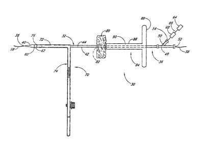
With reference to Figure 1, a vascular wound closure assembly 30 includes an
elongate catheter 32 having a distal end 34 and a proximal end 36 of the
catheter 32. A
distal opening 38 is formed through the distal end 34 of the catheter 32 and
opens along a
longitudinal axis of the catheter 32. The catheter 32 includes a tapered tip
40 at the distal
end 34. An elongate main. body 42 of the catheter 32 is disposed proximal the
tapered tip
40. Preferably the main body 42 has a substantially uniform diameter along its
length. A
lumen 44 extends longitudinally within the catheter 32 from the distal opening
38 to the
proximal end 36.
A connector portion 46 is provided on the proximal end 36. The connector
portion
46 includes a main lumen 48 and a secondary lumen 50. The main lumen 48
extends along
the longitudinal axis of the catheter 32 and is coextensive with the catheter
lumen 44. The
secondary lumen 50 extends outwardly from the main lumen 48, but communicates
with
the main lumen 48 and the catheter lumen 44. A proximal opening .52 is
provided at the
proximal end of the main lumen 48 and, like the distal opening 38, opens along
the
longitudinal axis. A secondary opening 54 opens into the secondary lumen 50.
The distal and proximal openings 38, 52 are sized and adapted to accommodate a
guidewire 58 such as the guidewire used in angioplasty and other vascular
surgeries. As
such, the guidewire 58 can be threaded through the catheter 32 and the
catheter can be
advanced over the guidewire 58.
Holes 60 are formed through a side wall of the catheter 32 near the distal end
34 of
the catheter 32. Preferably, at least two holes 60 are provided. All of the
holes 60
preferably are disposed substantially the same distance from the distal end 34
of the
catheter 32. Preferably, a raised portion 62 of the catheter 32 is provided in
the region
around the holes 60, which region is proximal of the tip 40 and distal of the
main body 42.
-7-
CA 02801969 2014-05-26
At the raised portion 62, the catheter 32 has an outer diameter that is
slightly larger than the
outer diameter throughout the catheter main body 42.
With continued reference to Figure 1, a vacuum or other source of suction 64
is
provided and communicates, through tubing 66, with the secondary lumen 50 of
the
catheter connector portion 46. Thus, a vacuum is drawn through the catheter
lumen 44.
Preferably, the distal and proximal openings 38, 52, which accommodate the
guidewire 58,
are sized so that the guidewire 58 substantially plugs the openings; thus, the
vacuum is
drawn through the holes 60. A viewing port 68 is arranged between the source
of suction
64 and the catheter 32. The viewing port 68 is configured to allow a clinician
to view the
material that is drawn by suction through the holes 60 and through the
catheter lumen 44.
The viewing port 68 will be discussed in more detail below.
With reference to Figures 1 and 2, a retractor 70 preferably is mounted on the
catheter 32. The retractor 70 includes opposing elongate retractor arms 72
that are aligned
longitudinally on the catheter 32. A retractor body 74 is configured to
selectively open and
close the retractor arms 72 when operated by a clinician. The elongate
retractor arms 72 of
the retractor 70 are positioned on the catheter 32 so that distal ends 76 of
the arms are
positioned proximal of the catheter holes 60 a distance that is at least the
same as the width
of an artery wall, preferably at least about .5 to 2 millimeters.
It is to be understood that the present device can include structure that is
somewhat
different than the particular structure shown in Figures 1 and 2. For example,
other
catheter and retractor structures can appropriately be used. For example, some
acceptable
catheter and retractor embodiments are presented in U.S. Application Serial
No. 09/325,982, filed on June 4, 1999, now U.S. Patent No. 6,287,322.
With reference again to Figure 1, a hemostatic member 80 is arranged on the
catheter 32 proximal of the retractor 70. As will be discussed in more detail
below, the
hemostatic member comprises a material that is made of or includes a
hemostatic agent.
The hemostatic agent is adapted to aid blood clotting. In one embodiment, the
hemostatic
member 80 comprises a sponge or sponge-like material. In this description, the
term
sponge is intended to be a broad term that is used in accordance with its
ordinary meaning
and refers to, without limitation, a material that is at least partially
porous and is adapted to
allow at least some blood to flow into and within the material so as to soak
the material
-8-
CA 02801969 2013-01-10
with blood. For example, a sponge may include a natural or artificial sponge,
a woven or
non-woven cloth, a fibrous puff or the like. Additionally, a sponge may
comprise a
material that soaks up at least a portion of blood that may come in contact
with the material,
or may comprise a material that doesn't soak up blood. =
For purposes of this description, the hemostatic member 80 is referred to as
the
sponge 80. However, it is to be understood that use of the term "sponge" does
not limit the
scope of materials that can be used as the hemostatic member. In fact, any
material that
aids or facilitates blood clotting can be used as the hemostatic member.
Throughout this description, the term hemostatic agent is used as a broad term
in its
ordinary sense and refers to, without limitation, an agent that promotes blood
clotting.
Such an agent may take many forms, including liquid, powder, beads, etc. and
can include
= or be combined with a substrate or carrier, The term hemostatic material
is also used in this
description as a broad term used in its ordinary sense. It refers to, without
limitation, any
= material having properties that promote blood clotting. Thus, hemostatic
material can
- - include a hemostatic agent taken alone or in combination with a substrate
or carrier that is
= formed separately from the agent. The term hemostatic material includes
hemostatic
sponges.
Preferably, the sponge 80 extends circumferentially around the catheter main
body
42, and is arranged so that it can be slid longitudinally along the catheter
32. Most
= 20 preferably, the catheter 32 extends through a passageway 82 through
the sponge 80. The
passageway 82 is formed as the catheter 32 is forced through the sponge 80.
A push member 84 is also arranged on the catheter 32 proximal of the sponge
80.
With reference also to Figure- 3, the push member 84 comprises a body portion
86 and a
proximal handle portion 88. An elongate lumen 90 is formed through the body
portion 86.
As shown in Figure 1, the lumen 90 preferably encircles the catheter 32 so as
to allow the
push member 84 to slide relative to the catheter 32. A plurality of holes 92
are formed
through the body portion 86 at a point near the distal end of the push member
84.
As will be discussed in more detail below in connection with Figure 4, the
vascular
wound closure assembly 30 enables a clinician to precisely locate a
subcutaneous vascular
wound "w", access the wound w, and deliver the hemostatic sponge 80 to, the
wound site.
The hemostatic sponge 80 includes a hemostatic agent that helps facilitate
closure of the
wound w.
-9..
CA 02801969 2013-01-10
In order to properly apply the hemostatic sponge 80, the -vascular closure
assembly
= 30 first precisely locates and provides access to the vascular wound w.
It is to be
understood that the present method and apparatus can be used to close various
vascular and
other wounds. Figures 1-11, and the accompanying discussion, present an
example using
an embodiment to close a puncture wound w in a patient's femoral artery 94.
With specific reference to Figures 1, 2, 4 and 5, in order to precisely locate
and
provide access to a femoral artery puncture wound w, the catheter 32 is first
threaded over a
guidewire 58 that has been previously inserted into the patient's femoral
artery 94 through
the puncture wound w. The lumen 44 is attached to the source of suction 64 and
the
assembly 30 is advanced over the guidewire 58 through. a patient's tissue 96
so that the
distal tip 40 of the catheter 32 extends through the vascular puncture wound
w.
As the assembly 30 is advanced, the source of suction 64 draws bodily fluids
through the holes 60. The fluids pass through the viewing port 68, which
allows the
clinician to identify the fluids being withdrawn. The viewing port 68 can have
any suitable
structure or location. For example, the -viewing port can comprise clear
tubing attached to
the catheter, a substantially transparent syringe that functions as both a
source of suction
and a viewing port, or a Portion of the catheter that is substantially
transparent. Most
preferably, the catheter 32 is formed of a transparent material so that the
clinician becomes
aware as soon. as blood begins to be drawn through the catheter.
When the holes 60 pass the artery wall 98 and enter the blood vessel 94, as
shown in
Figure 4, blood "b" begins to be drawn through the holes 60 into the catheter
32 and is
conducted past the viewing port 68. Thus, when blood b is observed in the
viewing port 68,
the clinician will know that the holes 60 have just passed into the puncture
wound w and
that the distal ends 76 of the retractor arms 72 are thus positioned adjacent
the outer wall 98
of the artery 94, preferably within about 2 mm of the artery wall 98. The
retractor arms 72
are then separated as shown in Figure 5, thus drawing surrounding tissue 96
away from the
wound w and creating a field 100 around the puncture wound w. The catheter 32
remains
disposed partially within the puncture wound w, effectively plugging the wound
and
preventing blood from flowing through the wound. The raised portion 62 flexes
the edges
of the wound w to enhance the seal between the catheter 32 and the puncture
wound edges.
-10-
CA 02801969 2013-01-10
With continued reference to Figure 5, a suction tool 102 can be used to clear
away
bodily fluids and other matter that may be within the field 100 and to clean
the wall 98 of
the blood vessel 94 adjacent the puncture wound w.
With reference next to Figure 6, once the puncture wound w has been precisely
located, the push member 84 is advanced distally along the catheter 32, thus
advancing the
sponge 80 into contact with the vessel wall 98 so as to surround the puncture
wound w. As
mentioned above and discussed in more detail below, the sponge 80 comprises a
hemostatic
agent that will help accelerate blood clot formation at the wound site w in
order to help the
wound heal faster.
Preferably, the sponge 80 is at least partially coated with an adhesive so
that the
sponge will at least partially bond to the vessel wall 98. Alternatively, or
in addition,
flowable adhesive can be delivered into the field around the puncture wound
before the
sponge is advanced into contact with the vessel wall. Of course, the sponge
can be
delivered without using any adhesive.
The sponge 80 preferably is mounted onto the catheter 32 so as to
substantially
encircle the catheter 32. Thus, since the tip 4001 the catheter is disposed in
the wound, the
sponge 80 substantially surrounds the wound w when the sponge is positioned
adjacent the
vessel wall 98.. When the sponge 80 is in place adjacent the wound w, the
retractor 70 can
be removed, as shown in Figure 7. When the retractor 70 is removed, the
surrounding
body tissues 96 collapse around the sponge 80 and push member 84. The push
member 84
holds the sponge 80 in position while body tissue 96 surrounds the sponge 80
and while the
adhesive cures.
With reference next to Figure 8, with the push member 84 in place, the
catheter 32
and guidewire 58 can also be removed from the patient. The passage 82 through
the sponge
80, which had been occupied by the catheter 32, collapses onto itself so that
it is
substantially closed. The vessel wound w is no longer plugged by the catheter
32, and it is
anticipated that blood b from the vessel 94 will flow into the sponge 80, at
least partially
soaking the sponge 80. Although the retractor 70 is removed prior to the
catheter 32 in the
above-discussed embodiment, it is to be understood that, in another
embodiment, the
catheter may be removed prior to the retractor.
In still another embodiment, additional pressure can be applied to the push
member
84 in order to at least partially block blood flow through the blood vessel
94. In this
-11-.
CA 02801969 2013-01-10
manner, the clinician can control how quickly blood will flow through the
wound w and
into the sponge 80. Of course, other methods and apparatus can be used to
temporarily
reduce or stop blood flow through the vessel.
In a preferred embodiment, the sponge 80 comprises a material made of, soaked
in
or otherwise treated with a hemostatic agent. The agent is specially adapted
to aid blood
clotting. Thus, blood that flows into the sponge encounters the agent and will
quickly
become clotted, causing natural sealing of the wound through blood clotting.
Sponge-like
hemostasis agents are available and can include products such as GelfoamTM,
OxycellTM
and AviteneTM. Another material that can be used as a sponge is chitosan.
These and other
appropriate sponges may be impregnated with agents such as thrombin, a liquid
clotting
= agent, to help accelerate blood clot formation and HemadexTm, which is
available from
Medafor, Inc. Another material that may advantageously be used is a collagen
UltrafoamTM
sponge marketed by C.R. Bard/Davol, Inc. The UltrafoamTm sponge is made from
Avitenena collagen, a natural clotting agent, and does not require the
addition of thrombin.
This reduces preparation time and the risk that a patient will experience a
potentially
hazardous reaction to bovine thrombin. Other medicaments can also be included
in the
= sponge. For example, antibiotic medicines, anti-inflarnmatory drugs,
healing aids, and the
like can be impregnated into the sponge material.
In a particularly preferred embodiment, the hemostatic agent comprises a
starch
such as bioabsorbable microporous polysaccharide microspheres (e.g.,
TRAUMADEXTm
marketed by Emergency Medical Products, Inc. of Waukesha, WI). The
microspheres have
micro-replicated porous channels. The pore size of the microsph.eres
facilitates water
absorption and hyperconcentration of albumin, coagulation factors, and other
protein and
cellular components of the blood. The microspheres also affect platelet
function and
enhance fibrin formulation. In addition, the microspheres are believed to
accelerate the
coagulation enzymatic reaction rate. When applied directly, with pressure, to
an actively
bleeding wound, the particles act as molecular sieves to extract fluids from
the blood. The
controlled porosity of the particle excludes platelets, red blood cells, and
serum proteins
larger than 25,000 Daltons, which are then concentrated on the surface of the
particles.
This molecular exclusion property creates a high concentration of platelets,
thrombin,
fibrinogen, and other proteins on the particle surface, producing a gelling
action. The
gelled, compacted cells and constituents accelerate the normal clotting
cascade. The fibrin
-12-
CA 02801969 2014-05-26
network formed within this dense protein-cell matrix adheres tightly to the
surrounding
tissue. The gelling process initiates within seconds, and the resulting clot,
while
exceptionally tenacious, breaks down normally along with the microparticles.
Such
microporous polysaccharide microspheres, and additional hemostatic agents, are
discussed
in more detail in Applicants' copending application entitled "Deployable
Multifunctional
Hemostatic Agent," U.S. Application Serial No. 10/868,201, filed June 14,
2004.
Any suitable hemostatic substrate can be employed as a support for the
hemostatic
agents of preferred embodiments. However, in a particularly preferred
embodiment the
hemostatic substrate comprises chitosan. Chitosan is obtained from chitin, a
biopolymer
obtained principally from shrimp and crab shell waste. Chitosan is the main
derivative of
chitin, and is the collective term applied to deacetylated chitins in various
stages of
deacetylation and depolymerization. The chemical structure of chitin and
chitosan is
similar to that of cellulose. The difference is that instead of the hydroxyl
group that is
bonded at C-2 in each D-glucose unit of cellulose, there is an acetylated
amino group (-
NHCOCH3) at C-2 in each D-glucose unit in chitin and an amino group at C-2 in
each D.-
glucose unit of chitosan.
H Cligli I It
HO = HO =
Ha 0
MOH OR CH3011
Cellulose
Nueocth ("Hz0H IN:HCOCH-,
HO
7
0
CH2OH NHCOCH:4 I2011
Chitin
NU. CH:OH N,
HO---
HO 0,
CH7OH N112
abon
Chi tosan
-13-
CA 02801969 2013-01-10
Chitin and chitosan are both nontoxic, but chitosan is used more widely in
medical
and pharmaceutical applications than chitin. Chitosan exhibits good
biocompatibility and
is biodegradable by chitosanase, papain, cellulase, and acid protease.
Chitosan exhibits
anti-inflammatory and analgesic effects, and promotes hernostasis and wound
healing.
Chitosan has also been used as a hemostatic agent in surgical treatment and
wound
protection. The hemostatic effect of chitosan has been described in U.S.
Patent No.
4,394,373.
A single hemostatic substrate or combination of hemostatic substrates of
different
forms and/or compositions can be employed in the devices of preferred
embodiments.
Different substrate forms can be preferred, for example, fibrous puff, fleece,
fabric, sheet,
suture, or powder. A homogeneous mixture of different substrate-forming
materials can be
employed, or composite substrates can be prepared from two or more different
formed
substrates. A preferred composite comprises chitosan and collagen. Additional
details
concerning chitosan and other suitable substrates are discussed in more detail
in
Applicants' copending application "Deployable Multifunctional Hemostatic
Agent."
The sponge-like substrate material preferably is soft and pliable and will
conform to
the structure of the blood vessel, the wound and the field around the blood
vessel. Thus,
the sponge-like material is specially suited for use in the confined space
surrounding a
vascular puncture. Additionally, the hemostatic sponge 80 will be held in
place by the
tissue 96 surrounding the puncture wound w, which tissue 96 collapses over the
sponge 80
when tools such as the retractor 70 are removed.
To further help hold the sponge go in place, flowable adhesive 106 from a
source of
adhesive 108 can be delivered through the lumen 90 of the push member 84 and
onto the
sponge 80, as shown in Figure 9. The adhesive 106 flows through the open
distal end of
the push member 84 and also through the. holes 92 through the push member body
portion
86. Upon curing, the adhesive 106 can form a sealing layer around and within
the sponge
80, thus confining the blood b to the sponge area. This helps minimize
bleeding and even
further speeds clot formation. In one embodiment, adhesive, when cured, is
substantially
non-porous, and thus confines blood to a desired area. Adding adhesive 106
will also
facilitate more complete closure of the passage through the sponge, which
passage was
vacated by the catheter 32. Further, the adhesive 106 will help hold the
sponge 80 in place
relative to the puncture wound w and the surrounding tissue 96.
-14-
CA 02801969 2013-01-10
= As discussed above, prior to being advanced into contact with the blood
vessel wall,
the sponge 80 may be soaked in an adhesive or, more preferably, coated with a
layer of
adhesive. In this manner, adhesive distribution on the sponge can be
controlled. By
controllably applying a coating of adhesive around the outer surface of the
sponge, the
adhesive will bond the sponge to the area surrounding the blood vessel wound
w, including
the vessel 94 itself, and also can form a perimeter seal of the sponge when
the adhesive
= cures, The coating of adhesive can act as a non-porous or selectively-
porous membrane
confining the blood b to the sponge 80. It is to be understood that a coating
of adhesive
may be used instead of or in addition to applying additional adhesive 106
through the push
member 84.
Various kinds of flowable adhesives may be acceptable for use with the sponge.
For example, fibrin tissue sealants such as Tisseel , which is available from
Baxter
Healthcare Corp., may be appropriate. Other commercially available adhesives
that may be
appropriate include BioglueTM, available from Cryolife, Inc., and FlosealTM,
which is
= available from Fusion Medical Technologies. Various cyanoacrylate adhesives
are
currently commercially available and can be used with this invention. Of
course, any
product that is capable of sealing the sponge or at least retarding blood flow
through or
= beyond the sponge would be acceptable. It is also to be understood that
certain adhesives
will not require that the field and/or the outer wall of the blood vessel be
cleared before the
adhesive is injected.
Curing time and ease of use will vary depending on the adhesive used. For
example, some adhesives cure to a malleable gel-like state within a few
seconds, while
-
others will cure directly to a hardened state in a few minutes. The time
period for curing is
chosen to allow the clinician to advance the sponge into position adjacent the
wound and in
contact with the artery, at which time the sponge will begin to be bonded to
the vessel wall
and substantially sealed by the adhesive. It should be appreciated that any
acceptable
adhesive having any acceptable curing time may be used. In accordance with
this
description, an adhesive is considered to be cured when it is adhered to
surrounding tissue,
and when it does not spontaneously flow.
, The
push member 84 may be kept in place for any reasonable time period in order to
allow the adhesive 106 to cure. Also, multiple sponges can be used, if
desired. Preferably,
however, the adhesive 106 will cure sufficiently in about five minutes or
less. Other tools,
-15-
CA 02801969 2013-01-10
such as an ultraviolet light source or a heat application device, may be used
to help speed
adhesive curing.
Once the sponge 80 is correctly placed, the push member 84 can be removed.
Removal of the push member 84 can be aided by a release rod 110 which, as
shown in.
Figure 10, is advanced through the push member lumen 90 and into contact with
the
sponge 80. The release rod 110 holds the sponge 80 in place as the push member
84 is
withdrawn from the patient. Thus, the release rod 110 engages the sponge SO so
as to
provide counter traction when the push member 84 is withdrawn. In this way,
the push
member 84 can be removed even if' some adhesion occurs between the sponge 80
and the
push member= 84. With reference next to Figure 11, once the release rod 110 is
withdrawn,
the patient's skin 112 is closed by any appropriate closure media such as, for
example,
sutures 114. The hemostatic sponge 80 is left in place. The body's natural
blood clotting
process will plug and repair the vascular wound w with the aid of the
hemostatic sponge 80.
Thus, healing will proceed without the danger of false aneurysms, missed or
faulty wound
closure, or the like.
In the embodiment illustrated in Figures 1-9, the catheter comprises a single-
lumen
catheter. In another embodiment (not shown), the elongate catheter has a first
lumen
comprising a tube that extends from the distal end opening to the proximal end
opening and
slidingly accommodates the guidewire therewithin. The outer wall of the
catheter defines a
second lumen that concentrically surrounds the first lumen. The holes through
the outer
wall of the catheter open into the second lumen. Additionally, an access lumen
communicates with the second lumen. In this embodiment, the distal and
proximal
openings, which accommodate the guidewire, do not communicate with the second
lumen,
which lumen communicates with the source of suction through the access lumen.
Accordingly, in this embodiment, there may be less of a chance that body
fluids will be
drawn into the catheter through the distal and proximal guidewire openings
than in an
embodiment employing a single lumen. However, the single-lumen catheter can be
less
expensive to manufacture and can be expected to have a smaller diameter than
the dual-
lumen catheter.
With reference next to Figures 12-16, another embodiment of a vascular wound
closure apparatus is presented. The apparatus includes a retractor 200 and an
elongate
catheter 250.
-16-
.
CA 02801969 2013-01-10
With particular reference to Figure 13, the catheter 250 has a proximal end
250a
and a distal end 250b. A distal opening is fonn.ed through the distal end of
the catheter and
opens along a longitudinal axis of the catheter. A lumen 250c is defined
within the
catheter. A tip. 256 at the distal end 250b of the catheter 250 preferably is
tapered. A
= 5
connector portion is provided on the proximal end 250a, which connector
portion
preferably includes a main lumen and a secondary lumen. The main lumen extends
along
the longitudinal axis of the catheter and is coextensive with the catheter
lumen 250c. At
least one indicator hole 258 is formed through a side wall of the catheter
near the distal end.
Preferably the catheter 250 is generally straight and is sized between about 4-
8F and more
preferably about 6F.
An outer surface 252 of the catheter 250 preferably has a generally
cylindrical shape
and includes a raised portion 254. In one preferred embodiment, the raised
portion 254
defines a connection between two separate sections (not shown) of the catheter
250. In the
- illustrated embodiment, the raised portion 254 is cylindrical and
includes a length 254a.
With continued reference to Figure 13 a pusher member 260 preferably is
movably
= disposed about the outer surface 252 of the catheter. The pusher member
260 preferably is
= configured to slide over the catheter 250. The pusher member 260
preferably has an inner
lumen having a diameter greater than the raised portion 254 of the catheter
250 so that the
pusher member 260 can slide over the raised portion 254.
With reference next to Figures 14-16, the retractor 200 preferably is
configured to
be mounted onto the catheter 250. In the illustrated embodiment, the retractor
200
preferably has two retractor arms 202 movably connected to each other, each
having a
length 204 from a proximal end 206 to a distal end 208. The retractor arms 202
preferably
are capable of being moved between an open position (see Figure 14) and a
closed position
(see Figure 12). When in the closed position, as illustrated in Figure 12, the
retractor arms
202 preferably enclose at least a portion of the catheter 250. Although the
illustrated
embodiment of the retractor 200 shows only two retractor arms 202, it should
be
understood that the retractor 200 can have more than two retractor arms 202.
With continued reference to Figures 14 and 15, each of the retractor arms 202
preferably defines an inner surface 210 generally facing the inner surface 210
of the other
arm 202. Each inner surface 210 defines edges 212 that preferably extend along
the length
204 of the arms 202. The inner surface 210 also preferably defines a cavity or
channel 220
-17-
=
CA 02801969 2013-01-10
extending between the edges 212. The channel 220 preferably extends the length
of the
retractor arms 202. When the retractor arms 202 are in the closed position, as
shown in
Figure 12, the channels 220 on the retractor arms 202 preferably combine to
define a canal
221 extending the length 204 of the arms 202.
With reference to Figures 14-15, the channel 220 preferably comprises a
proximal
portion 222 disposed at the proximal end 206 of the retractor arms 202. In a
preferred
embodiment, the proximal portion 222 has a generally curved shape configured
to
removably receive and substantially contact and hold at least a portion of the
catheter 250
in a fixed position when the retractor arms 202 are in the closed position.
The proximal
portion 222 also has a depth 222a generally orthogonal to the length 204 of
the retractor
arms 202. For example, the proximal portion 222 can have a semi-circular cross-
section
with a radius 222a about the same as that of an outer surface 252 of the
catheter 250.
However, the proximal portion 222 can have any shape configured to
substantially contact
the catheter 250 when the retractor arms 202 are in the closed position. Most
preferably,
the proximal portion 222 is sized and configured generally complementary to
the catheter
.250 so that the retractor 200 holds the catheter 250 generally snugly at the
proximal portion
= 222.
With continued reference to Figures 15 and 16, the channel 220 preferably
comprises a receiver portion 224 adjacent the proximal portion 222. The
receiver portion
224 preferably has a generally curved shape and has a depth 224a generally
orthogonal to
the length 204 of the arras 202 that is greater than the depth 222a of the
proximal portion
222. Accordingly, the receiver portion 224 defines an edge 224b between the
receiver
portion 224 and the proximal portion 222. The illustrated receiver portion 224
has a semi-
circular cross-section with a radius 224a that is greater than the radius 222a
of the proximal
portion 222. Most preferably, the receiver portion 224 is generally
complementary to the
catheter raised portion 254 so As to receive the raised portion 254 therein.
The channel 220 also preferably comprises a contact portion 226 adjacent the
receiver portion 224. Similar to the proximal portion 222, the contact portion
226
preferably is generally complementary to the catheter outer surface 252 and is
configured to
removably receive, and to substantially contact and hold the catheter 250 when
the retractor
arms 202 are in the closed position. The contact portion 226 preferably has a
depth 226a
generally orthogonal to the length 204 of the retractor arms 202. In one
preferred
-18-
.
CA 02801969 2013-01-10
embodiment, the depth 216a is similar to the depth 222a of the proximal
portion 222. For
example, the contact portion 226 can have a semi-circular cross-section with a
radius 226a
about the same as the radius 222a of the proximal portion 222. The depth 226a
of the
contact portion 226 is also preferably smaller than the depth 224a of the
receiver portion
224, so that the receiver portion 224 defines an edge 224c between the
receiver portion 224
and the contact portion 226.
With reference still to Figures 15 and 16, in the illustrated embodiment, the
proximal portion 222 and contact portion 226 each are smaller than the
receiver portion
224. Most preferably, the proximal portion 222 and contact portion 226 are
configured so
that the catheter raised portion 254 cannot slide through either portion 222,
226. Thus,
when the raised portion 254 is disposed in the receiver portion 224 as shown
in Figure 16,
the raised portion is constrained from moving proximally or distally. As such,
the entire
catheter 250 is longitudinally locked in place relative to the retractor 200
when the retractor
arms 202 are closed about the catheter as shown in the Figure 12.
The channel 220 further preferably comprises a compartment portion or chamber
228 adjacent the contact portion 226. The chamber 228 preferably has a
generally curved
shape and a depth 228a generally orthogonal to the length 204 of the retractor
arms 202
greater than the depth 226a of the contact portion 226. For example, the
chamber 228 can
have a semi-circular cross-section with a radius 228a greater than the radius
226a. Further,
the contact portion 226 defines an edge 226b between the contact portion 226
and the
chamber 228. The chamber 228 is configured to receive a portion of the
catheter 250
therein and to define a space 228h between the catheter 250 and the retractor
arms 202.
When the retractor arms 202 are in the closed position, the space 228b extends
generally
about the entire circumference of the catheter 250. The space 228b is
configured to receive
and accommodate a hemostatic material 270 therein so that it surrounds at
least a portion of
the outer surface 252 of the catheter 250. The hemostatic material 270 is
further described
below.
A distal portion 230 of the channel is defined adjacent the chamber 228 and
has a
depth 230a generally orthogonal to the length 204 .of the retractor arms 202
smaller than the
depth 228a of the chamber 228. The distal portion 230 preferably is generally
complementary to the catheter outer surface 252 so as to substantially contact
and hold the
catheter 250 when the retractor arms 202 are in the closed position. For
example, the distal
-19-
CA 02801969 2013-01-10
portion 230 can have a semi-circular cross-section with a radius 230a. In one
preferred
embodiment, the radius 230a is about the same as the radius 226a of the
contact portion 226
and/or the radius 222a of the proximal portion 222. A generally smooth
transition section
230b preferably connects the chamber portion 228 and the distal portion 230.
With reference again to Figures 12-16, in practice, the hemostatic material
270 is
preferably disposed about the outer surface 252 of the catheter 250 at a
location between
the raised portion 254 and the catheter holes 258. The catheter 250 is placed
in the channel
220, while the arms 202 are in the open position, so that the raised portion
254 is disposed
in the receiver portion 224 and the hemostatic material 270 is housed in the
chamber 228.
- 10 Preferably, the catheter 250 and retractor 200 are configured so that,
when assembled, the
distance between the distal end 208 of the retractor arms 202 and the
indicator holes 258 is
at least the same as the width of an artery wall. Preferably, said distance is
at least about
0.5 to 2 millimeters.
When the retractor arms 202 are moved into the closed position with the raised
portion 254 disposed in the receiver portion 224, the catheter 250 is
longitudinally locked
relative to the retractor 200. Thus, the catheter 250 and retractor 200 will
move together
even if longitudinal forces are exerted upon one or the other structure. In
use, the apparatus
is advanced into the patient so that the catheter 250 is advanced into the
wound "w" as
discussed above in connection with the embodiment discussed in connection with
Figures
1-4. When blood "b" is observed in a viewing port (not shown) connected to the
catheter
250, the retractor arms 202 are then preferably moved into the open position.
The pusher
member 260 is then advanced toward the distal end 250b of the catheter 250 to
engage and
advance the hemostatic material 270 into contact with the wound w.
In. the embodiment illustrated in Figures 12-16, the hemostatic material 270
preferably comprises a malleable, fibrous material. For example, preferably
the substrate
comprises a puff - a fibrous, cotton-like material that can be manipulated
into a suitable
shape or size so as to accommodate a particular wound configuration. Most
preferably, the
hemostatic material 270 comprises a puff prepared from chitosan fibers and
infused with
microporous polysaccharide microspheres. Applicants' copending application
"Deployable
Multifunctional Hemostatic Agent" discusses such a hemostatic puff and methods
of
depositing microporous polysaccharide microspheres thereon. Other fibrous
substrates and
hemostatic agents may also be employed in. other embodiments.
-20-
CA 02801969 2013-01-10
In still other embodiments, the hemostatic material 270 can be infused with
any
number of medications associated with the treatment of wounds. For example,
antibiotic
and anti-inflammatory medications may be fin-tiler infused or deposited on a
substrate.
It is further to be understood that, in accordance with further embodiment,
the raised
portion 270 can have various configurations. For example, the raised portion
may not
extend circumferentially about the catheter 250, and a plurality of raised
portions may be
employed. Preferably, the receiver portion of the retractor 200 is shaped
complementary to
the raised portion. Additionally, the inner lumen of the pusher member 260 may
have a
cross sectional shape configured to fit over the raised portion, and may in
some
embodiments be non-circular.
With reference next to Figures 17-23, another embodiment of a vascular wound
closure assembly 300 comprises a catheter 310 having a proximal end 312 and a
distal end
314, and defining a lumen (not shown) therebetween. A pusher member 330 having
a
proximal end 332 and a distal end 334 is slidably disposed on the catheter
310. A delivery
15; tube 350 having a proximal end 352 and a distal end 354 is slidably
disposed on the
catheter 310 and is positioned distal of the pusher member 330. The closure
assembly 300
. preferably is made of a polymeric material, such as polypropylene.
Preferably, the
assembly 300 is also made of hypoallergenic materials.
. .
With particular reference to Figure 18, the catheter 310 preferably comprises
a stop
member 316 disposed in a fixed position about the catheter surface 310a. The
distal end
314 preferably is tapered, and catheter holes 318 are formed through a side of
the catheter
310 proximal of the distal end 314. In one embodiment, the catheter 310
preferably
comprises a secondary branch 319 disposed at the proximal end 312, and having
a
secondary lumen (not shown) connected to the lumen of the catheter 310. The
secondary
branch 319 preferably is configured to operatively connect to a variety of
devices used in
the closure of vascular wounds, such as a suction device, For example, in one
embodiment,
a syringe can be connected to the secondary branch 319 to pull a vacuum
through the
catheter 310.
A coupling member 320 preferably is movably disposed about the catheter 310
and
is configured to mechanically couple to the stop member 316. In the
illustrated
embodiment, the stop member 316 is threaded on its outer surface and the
coupling
member 320 is threaded on its inner surface so that the respective threads are
engageable so
-21-
CA 02801969 2013-01-10
that the coupling member 320 and catheter 310, when engaged, do not move
longitudinally
relative to one another. As such, the member 320 and catheter 310 are
releasably coupled
to one another. In other embodiments, other suitable mechanical coupling
mechanisms can
be used. For example, a detent and catch Mechanism or a j-lock mechanism can.
also be
acceptably employed.
In this description, the term releasably coupled is a broad term used in its
ordinary
sense and referring to, without limitation, to members being attached or
affixed to one
another in a manner so that they can be decoupled from one another. For
example, without
limitation, members can be coupled with threads, a detent mechanism, a
conformed yet
breakable bridge, such as flashing from injection-molding, an adhesive, or the
like.
With particular reference next to Figure 19, the pusher member 330 preferably
comprises a generally cylindrical central portion 336, a generally conical
transition portion
337 and a generally cylindrical distal portion 338. The diameter of the
central portion 336
preferably is larger than the diameter of the distal portion 338. The pusher
member 330
preferably defines a canal 330a that extends from the proximal end 332 to the
distal end
334 and which is preferably configured to slidably receive the catheter 310
therethrough.
For example, the canal 330a can have a circular cross-section with a diameter
larger than
the diameter of the catheter surface 310a. However, the canal 330a is not
large enough to
fit over the catheter stop member 316. As such, the pusher member 330 cannot
be moved
proximally over the catheter 310 beyond the stop member 316.
The pusher member 330 preferably comprises a handle 340 near the proximal end
332. It is to be understood that the pusher member 330 can comprise more than
one
handle 340.
A proximal coupling member 342 is disposed at the proximal end 332. In the
illustrated embodiment, the proximal coupling member 342 comprises threads on
its outer
surface sized and configured to engage the threads of the coupling member 320.
As shown
in Figure 27, the catheter coupling member 320 is configured to engage both
the stop
member 316 and the pusher member proximal coupling member 342 so as to
selectively
hold the pusher 330 longitudinally fixed relative to the catheter 310,
A distal coupling member 344 is disposed proximal the transition portion 337.
In
the illustrated embodiment, the distal coupling member 344 comprises a
generally
hemispherical raised portion.
-22-
CA 02801969 2013-01-10
With particular reference next to Figures 20-21, the delivery tube 350
preferably
= has a body 350a with a conical outer surface 350b having a generally
decreasing diameter
350c between a top edge 357 at the proximal end 352 and the distal end 354. A
wall 350e
of the delivery tube 350 has a thickness "t". The delivery tube wall 350e
preferably defines
a chamber 350d extending from the proximal end 352 to the distal end 354 The
chamber
350d preferably is conical in shape, and preferably is configured to receive
hemostatic
material 270 therein between the catheter and the wall. The proximal end 352
of the
delivery tube 350 also is preferably configured to receive at least a distal
portion of the
pusher member 330. The distal end 354 of the delivery tube 350 has a distal
opening that is
configured to receive the catheter 310 extending therethrough.
With particular reference to Figure 21, the delivery tube 350 preferably
comprises
weakened portions 356. In the illustrated embodiment, the weakened portions
356
comprise portions of the tube 350 having a reduced thickness "t. The reduced
thickness
weakened portions 356 preferably extend from at or near the proximal end 352
to the distal
= end 354 of the delivery tube 350. The weakened portions 356 define a
preferential breaking
= or deformation zone of the delivery tube 350 so that when a force beyond
a specified
threshold is applied, the tube will deform or break in the vicinity of the
weakened portions
356. In the illustrated embodiment, the delivery tube 350 has two weakened
portions 356
comprising elongate sections of reduced thickness "t" diametrically opposed to
each other.
Preferably, the elongate weakened portions 356 extend the entire length of the
delivery tube
350.
In accordance with this description; the term weakened portion is a broad term
used
in its ordinary sense and referring to, without limitation, a zone or area
that preferentially
breaks, bends, stretches, expands or otherwise deforms upon application of a
threshold
force. In the illustrated embodiment, the weakened portions comprise portions
that are
relatively thin. In. accordance with other embodiments, a weakened portion can
include,
without limitation, a portion of material that is scored, perforated,
physically or chemically
treated, or the like. Further, a weakened portion can comprise an elastic or
easily
deformable material that may or may not be a different material than the rest
of the
member.
-23-
CA 02801969 2013-01-10
In the illustrated embodiment, as shown in Figure 21, the delivery tube 350
has two
weakened portions 356. However, it is to be understood that the delivery tube
356 can have
one or a plurality of weakened portions 356.
In one embodiment, the delivery tube 350 preferably comprises separation
starter
portions 358 disposed at the proximal end 352. The starter portions 358 are
preferably
disposed adjacent and aligned with the weakened portions 356. In the
illustrated
embodiment, the starter portions 358 are notches 358 aligned with the weakened
portions
356. In other embodiments starter portions 358 can be provided having other
shapes.
The delivery tube 350 farther comprises a coupling portion 360 disposed at the
proximal end 352. The coupling portion 360 preferably is configured to
mechanically
couple to the pusher member distal coupling member 344. With reference to
Figure 22,
the illustrated coupling portion 360a comprises a catch configured to
releasably hold the
raised portion of the pusher member distal coupling member 344. To engage the
coupling
portions 244, 260, the delivery tube 350 is moved longitudinally relative to
the pusher
member 344 until the catch is aligned with the raised portion, at which time
the raised
portion will enter the catch. The catch and raised portion are configured so
that the raised
portion will exit the catch only upon application of a threshold force. Thus,
the pusher
member 330 and tube 350 are releasably coupled and longitudinally fixed
relative to one
another.
With reference again to Figure 20, the delivery tube 350 comprises a handle
362
disposed adjacent the proximal end 352. The handle 362 preferably comprises
two
opposing support anus that extend outward from the conical outer surface 350b
at locations
spaced from the weakened portions 356. In the illustrated embodiment, the
delivery tube
handle 362 comprises two support arms diametrically opposed to each other and
disposed
generally 90 from the weakened portions 356.
With reference again to Figure 17, the vascular wound closure assembly 300 is
assembled by sliding the distal end 314 of the catheter 310 through the canal
330a of the
pusher member 330 so that the proximal end 332 of the pusher member 330
preferably
abuts the stopper member 316, and so the distal end 314 of the catheter 310
extends out
from the distal end 334 of the pusher member 330. The coupling member 320
engages the
stop member 316 and pusher member proximal coupling member 342 so that the
pusher
member 330 is fixed longitudinally to the catheter 310.
-24-
CA 02801969 2013-01-10
The proximal end 352 of the delivery tube 350 is slid over the distal end 314
of the
catheter 310 so that the catheter 310 travels through the opening 350d. As the
delivery tube
350 is slid proximally over the catheter 310 the coupling portion 360
mechanically engages
the distal coupling member 344 of the pusher member 330. As such, the catheter
310,
pusher member 330 and delivery tube 350 are fixed longitudinally to one
another. Thus,
the pusher member and tube move together as a unit. The hemostatic material
270 can be
added to the chamber 350d of the delivery tube 350 before or during the
assembly process.
With continued reference to Figure 17, when the apparatus is assembled, the
distal
end 314 of the catheter 310 extends from the distal end 354 of the delivery
tube 350, and
the catheter holes 318 preferably are spaced from the distal end 354 a
distance at least the
same as the width of an artery wall. Preferably, the distance is about 0.5 to
2 millimeters.
To use the apparatus, the assembled device is advanced into the vascular wound
"w"
in a manner similar to that discussed above in connection with Figures 1-4.
When the
=
. device is positioned so that the distal end 354 of the delivery tube 350
is generally adjacent
= 15 the wound "w", the coupling member 320 preferably is disengaged from
the stop member
316 of the catheter 310 and the proximal coupling member 342 of the pusher
member 330.
Similarly, the coupling portion 360 of the delivery tube 350 preferably is
disengaged from
the distal coupling member 344 of the pusher member 330. Accordingly, the
pusher
member 330 and delivery tube 350 are no longer longitudinally fixed relative
to each other.
With reference next to Figure 23, the pusher member 330 is then preferably
advanced distally into the opening 350d of the delivery tube 350 while the
delivery tube
350 is held generally stationary adjacent the wound w. Since the pusher member
330 is
generally larger in diameter than the delivery tube 350, the delivery tube 350
breaks along
the weakened portions 356 as the pusher member 330 is advanced. In one
embodiment, a
user grasps the handle 340 of the pusher member 330 and the handle 362 of the
delivery
tube 350 to drive the pusher member 330 through the delivery tube 350.
As the delivery tube 350 breaks, openings are created so that the hemostatic
material
270 is free to exit the chamber. As the pusher member 330 advances, it engages
and
advances the hemostatic material 270 out of the tube 350 and into contact with
the wound
"w". Preferably, the broken portions of the delivery tube 350 are removed from
the wound
location.
-25-
CA 02801969 2013-01-10
As described above in connection with other embodiments, the catheter 310 can
be
slidably withdrawn through the canal 330a of the pusher member 330. Further, a
release
rod (not shown) can also be used to provide counter traction to help remove
the pusher
member 330 from the wound location. For example, the release rod can be
slidably
- 5
inserted through the canal 330a of the pusher member 330 so that it engages
the hemostatic
material 270 against the wound location. A user can then remove the pusher
member 330
without disturbing the hemostatic material 270 because the counter traction
provided by the
release rod will keep the hemostatic material 270 in place as the pusher
member is
removed.
In the embodiment discussed above, the coupling members are disengaged before
= advancing the pusher member relative to the delivery tube. It is to be
understood that, in
other embodiments, the coupling members can be adapted so that mere
application of a
force above a threshold force level will defeat the coupling members so as to
release the
releasably coupled members from one another. Thus, as the user applies force
to advance
the pusher member, the user simultaneously disengages the coupling members and
advances the pusher member.
In another embodiment, the distal coupling member of the pusher member is
threaded on its outer surface, and the proximal coupling member of the
delivery tube is
threaded on its inner surface. As such the pusher member and delivery tube are
threadably
affixed to each other. In this arrangement, the pusher member is advanced
relative to the
delivery tube by threading the pusher member. This arrangement allows the user
to adjust
the distance between the distal ends of the delivery tube and the catheter
indicator holes.
When the device is positioned so that the delivery tube is adjacent the wound,
the pusher
member is advanced by continuing to thread the pusher member into the delivery
tube as
. 25 the delivery tube is held in place. As such, the pusher member will
advance, and will
eventually break the tube at the weakened portions. The pusher member can then
be
advanced further by using the handles.
In still other embodiments, other types and structures of coupling members can
be
employed. For example, various releasable locking structures can be employed,
such as a
Mock or an L'-lock (see Figure 24). Additionally, in still further
embodiments, the
coupling members can have still different structure. For example, the coupling
member can
comprise an adhesive between the pusher member and catheter, which adhesive is
-26-
CA 02801969 2013-01-10
configured to be defeated upon application of a threshold force. In yet a
further
embodiment, the pusher member and catheter are lightly heat bonded or
otherwise bonded
together. As such, the bond between the pusher member and catheter will be
overcome
upon application of a threshold force.
Figures 25-28 illustrate another embodiment of a vascular closure apparatus
300'
having many aspects similar to the embodiment described above with reference
to Figures
17-23. Where possible, the same reference numerals are used to identify
similar elements,
but elements of the present embodiment include the appellation
With specific reference to Figure 25, the 'closure apparatus 300' preferably
comprises a catheter 310', a pusher member 330', and a delivery tube 350'
releasably
connected to each other. Additionally, the apparatus 300' preferably comprises
a threaded
coupling member 344' slidably disposed about the pusher member 330'.
With specific reference next to Figure 26, the catheter 310' preferably
comprises an
unthreaded stop member 316'. Additionally, a coupling member 320' preferably
is
configured so that a portion of the coupling member is slidable over the stop
member 316'
so as to enclose it. However, a proximal portion of the coupling member cannot
slide over
= the stop member 316', and thus the stop member limits the distal travel
of the coupling
member.
With reference next to Figure 25 and 27, the proximal end 332 of the pusher
member 330' is configured to abut against the stop member 316' when the pusher
member
330' is slidably disposed on the catheter 310'. The threaded outer surface of
the proximal
coupling member 342 preferably is configured to mechanically engage the
threads of the
coupling member 320' when said member 320' is advanced over the proximal end
332 of
the pusher member 330'.
The pusher member 330' preferably comprises a transition portion 337' adjacent
the
distal portion 338. The transition portion 337' preferably comprises a
cylindrical raised
portion 337a' and a generally conical portion 337b'. The raised portion 337a`
comprises an
unthreaded outer surface, and preferably is configured to slidably receive a
distal portion of
the coupling member 344' about and over its outer surface so that the coupling
member
= 30 344' encloses the raised portion 337e, A proximal portion of the
coupling member 344'
cannot slide over the raised portion 337a.', and thus the raised portion 337a'
limits distal
travel of the coupling member 344' over the pusher member 330'.
-27-
CA 02801969 2013-01-10
With reference to Figures 25 and 28, the delivery tube 350' preferably
comprises a
coupling portion 360' at its proximal end 352. The coupling portion 360'
preferably
includes a threaded outer surface 360a' configured to mechanically engage the
coupling
member 344'. In one preferred embodiment, the delivery tube 350' comprises
weakened
portions 356, as previously discussed. In another preferred embodiment, the
body 350a' of
the delivery tube 350' comprises two separate halves 350f, 350g configured to
abut against
each other about an axis "z" and be held in a generally fixed position
relative to each other
when the coupling member 344' is threaded. onto the coupling portion 360'. In
yet another
preferred embodiment, the delivery tube 350' comprises a body 350a` with two
halves 350f,
350g joined at the proximal end 352 of the delivery tube 350' by weakened
portions 356'.
In another embodiment, the two halves are joined by an elastic member which
helps hold
the halves together before the hemostatic material is deployed.
With reference again to Figure 25, the vascular closure apparatus 300'
preferably is
assembled so that the catheter 310', the pusher member 330', and the delivery
tube 350' are
releasably coupled by the coupling members so as to be fixed relative to each
other. For
example, the catheter 310' is slidably inserted into the pusher member 330'
until the
stopper member 316' abuts against the proximal end 332 of the pusher member
330'. The
coupling member 320' is then slid over the stopper member 316' and threaded
onto the
proximal coupling member 342 of the pusher member 330'. The delivery tube 350'
is
similarly slid over the catheter 310' and pusher member 330' until the
proximal end 352 of
the delivery tube 350' abuts against the transition portion 337', wherein the
delivery tube
350' preferably encloses the hemostatic material 270 therein. The coupling
member 344' is
then slid over the raised portion 337a' and threadably engages the coupling
portion 360'.
As previously discussed, once the device is in place adjacent the wound "w",
the
coupling members 344', 320' are disengaged so that the pusher member 330' is
uncoupled
from the catheter 310' and the delivery tube 350'. The user advances the
pusher member
330' into the delivery tube 350' to deform the tube and. engage and advance
the hemostatic
material 270 adjacent the wound "w".
In the embodiments just discussed, the delivery tube is configured to break
when the
pusher member is advanced. In other embodiments, the delivery tube may not
break, but
deforms sufficiently so that material within the tube can be dispatched
therefrom. For
example, at least a portion of the tube may be formed of an elastic material,
such as
-28-
CA 02801969 2013-01-10
silicone, so that the pusher member deforms the tube and forces material out
of the tube and
adjacent the wound. Additionally, in one embodiment wherein the tube is formed
of an
elastic material, the tube does not necessarily include a weakened portion.
This principle
can also be employed in connection with other embodiments discussed herein,
including the
following embodiments.
Figures 29-37 illustrate another embodiment of a vascular closure apparatus
300"
= having many aspects similar to the embodiments described above with
reference to Figures
17-23 and to Figures 25-29. Where possible, the same reference numerals are
used to
identify similar elements, but elements of the present embodiment include the
appellation
". Of course, it is to be understood that similar elements do not necessarily
share
identical structure.
= With specific reference to Figure 29, the closure apparatus 300"
preferably
comprises a catheter 310", a pusher member 330", and a delivery tube 350"
releasably
connected to each other.
With reference also to Figure 30, the catheter 310" preferably comprises an
unthreaded stop member 316" that is raised relative to the catheter body.
Additionally, a
coupling member 320" preferably is configured so that a portion of the
coupling member is
slidable over the stop member 316" so as to enclose it. However, a proximal
portion of the
= coupling member 320" cannot slide over the stop member 316", and thus the
stop member
limits the distal travel of the coupling member 320".
With reference next to Figures 29 and 31, the pusher member 330" preferably is
= elongate and is slidable over the catheter 310". A proximal end 332" of
the pusher
. member 330" is configured to abut against the stop member 316" when the
pusher
member 330' is slid proximally over the catheter 310". The threaded outer
surface of the
proximal coupling member 342" of the pusher 330" preferably is configured to
mechanically engage the threads of the coupling member 320" when said coupling
member
320" is advanced over the proximal end 332" of the pusher member 330". As
such, the
pusher member 330" is releas ably coupled to the catheter 310" with the
proximal end
332" abutting the stop member 316".
The threaded outer surface of the proximal coupling member 342" preferably is
also configured to mechanically engage a threaded handle 340". With reference
also to
Figures 32a, 32b, and 33, the illustrated handle 340" comprises a handle
support 370 and
-29-
CA 02801969 2013-01-10
a handle arna 371. The handle support 370 comprises an elongate body 365 and a
flange
367. Preferably, the body 365 has a square or rectangular shaped profile that
corresponds
with a square or rectangular shaped hole 369 defined in the 'handle arm 371,
such that in an
engaged configuration, the handle support 370 rotates together with the handle
arm 371.
The flange 367 also preferably has a square or rectangular profile. The handle
arm 371
= preferably is slidable relative the handle support body 365 in an axial
direction, and thus
preferably can be removed from the handle support 370 and passed over the stop
member
316" of the catheter when the coupling member 320" is uncoupled from the
proximal
coupling member 342". It is to be understood that, in other embodiments, any
suitable
mechanism, such as a detent, can be used to releasably couple the arm handle
371 to the
support 370.
The pusher member 330" preferably comprises a central shaft section 372 that
is
unthreaded and has a reduced diameter relative the threaded outer surface of
the proximal
coupling member 342". The central shaft section 372 preferably is located
between the
proximal coupling member 342", having a raised threaded outer surface, and a
distal
coupling member 374, also having a raised threaded outer surface.
With continued reference to Figures 29 and 31, the pusher member 330"
preferably comprises an adjustable stopper 337" adjacent the distal portion
338". The
adjustable stopper 337" preferably comprises a cylindrical raised portion
337a" and a
generally conical portion 337b". The raised portion 337a" comprises an
unthreaded outer
surface and a threaded inner surface. The threaded outer surface of the distal
coupling
member 374 of the pusher member 330" preferably is configured to mechanically
engage
the threads of the adjustable stopper 337" when the adjustable stopper 337" is
advanced
over a distal portion 338" of the pusher member 330",
A coupling member 344" is slidably disposed about the pusher member 330". The
raised portion 337a" of the adjustable stopper 337" preferably is configured
to slidably
receive a distal portion of the coupling member 344" about and over its outer
surface so
that the coupling .member 344" encloses the raised portion 337a". However, a
proximal
portion of the coupling member 344" cannot slide over the raised portion
337a", and thus
the raised portion 337a" limits distal travel of the coupling member 344" over
the pusher
member 330".
-30-
CA 02801969 2013-01-10
=
With reference next to Figure 34, the proximal portion of the coupling member
344" preferably defines a hole 368 that is sized and configured such that the
coupling
member 344" cannot slide distally over the adjustable stopper 337", but can
slide
proximally over the central shaft section 372, the handle support 370, and the
catheter stop
member 316". In the illustrated embodiment, the hole 368 is generally
complementary to
the handle support flange 367. Thus, when the coupling member 320" and the
handle 371
have been removed, the coupling member 344" can also be removed if desired.
The handle
arms 371 can be replaced on the handle support 370 after the coupling member
344" has
been removed.
The distal portion of the coupling member 344" preferably comprises a threaded
portion 376 on an inner surface. The threaded portion 376 preferably comprises
threads
covering a proximal portion of the inner surface of the coupling member 344".
Preferably,
a distal portion of the inner surface of the coupling member 344" is not
threaded.
With reference next to Figures 29 and 35-36, the delivery tube 350" comprises
a
body 350a" made up of two separate halves or segments 350", 350g'.' The
segments
350f", 350g" are formed separately but are configured to abut against one
another about an
axis "z" to form the delivery tube 350". Preferably a first segment 350g" has
guide
= portions 380 that are configured to fit into recesses 381 formed in a
second segment 350f"
in order to properly align the segments to form the delivery tube 350".
Each segment 350f", 350g" has a generally concave inner surface con-figured so
that the tube 350", when assembled, fits concentrically about the catheter
310". In the
illustrated embodiment, the inner surface defines a chamber between the
catheter 310" and
inner surface when the tube 350" is in place. Preferably, at least a distal
portion of the
chamber is tapered so that the diameter of the chamber decreases smoothly
toward the distal
end thereof.
The delivery tube 350" has a coupling portion 360" at its proximal end 352",
The
coupling portion 360" preferably includes a threaded outer surface 360a"
configured to
mechanically engage the coupling member 344". When the coupling member 344" is
threaded onto the coupling portion 360", as shown in Figure 37, the segments
3501',
350g" are held in a generally fixed position relative to each other. It is to
be understood
that other structures may be used to releasably couple the segments 350f",
350g". Also, in
-31-
CA 02801969 2013-01-10
other embodiments, the delivery tube 350" may comprise a body 350a" having
several
releasably coupled segments at or near the proximal end 352" of the delivery
tube 350".
With reference again to Figure 29, the adjustable stopper 337" preferably
contacts
an upper portion of the assembled delivery tube 350". The adjustable stopper
337" can be
advanced proximally or distally relative the pusher member 330" by rotating
the adjustable
stopper 337". When the catheter 310", the pusher member 330" and the delivery
tube
350" are to be coupled together, moving the adjustable stopper 337" proximally
or distally
relative the pusher member 330" prior to coupling the pusher member 330" with
the
delivery tube 350" effectively adjusts the position of the delivery tube 350"
relative the
catheter 310". Accordingly, another feature of this embodiment is that the
position of the
distal end of the delivery tube 350" can be adjusted relative the holes in the
catheter 310"
by movement of the adjustable stopper 337". This may be desirable to adjust
the delivery
tube 350" position relative the holes in the catheter 310" based on the
thickness of a
particular blood vessel wall and/or the preference of a clinician. For
example, a clinician
may adjust the stopper 337" so as to position the distal end of the delivery
tube 350" at a
desired position between about 1 mm and 1 cm proximal of the holes of the
catheter 310".
The threaded inner surface 376 of the coupling member 344" preferably is
configured to mechanically engage the threads of the coupling portion 360"
when the
coupling member 344" is advanced over the stopper 337" and further over a
proximal end
352" of the delivery tube 350". Accordingly, another feature of this
embodiment is that
the delivery tube 350" is securely coupled with the pusher member 330" through
the
coupling member 344". The coupling member 344" also helps hold the body
segments
350f', 350g" together. Because only a portion of the inner surface of the
coupling member
344" is threaded, the pusher member 330" and the delivery tube 350" can be
quickly and
easily decoupled.
With reference next to Figures 37-39, the vascular closure apparatus 300"
- preferably is assembled so that the catheter 310", the pusher member 330",
and the
delivery tube 350" are releasably coupled by the coupling members so as to be
fixed
relative to each other. For example, the catheter 310" is slidably inserted
into the pusher
member 330" until the stopper member 316" abuts against the proximal end 332"
of the
pusher member 330". The coupling member 320" is then slid over the stopper
member
316" and threaded onto the proximal coupling member 342" of the pusher member
330".
-32-
CA 02801969 2013-01-10
The hemostatic material 270 preferably is wrapped about the catheter 310"
adjacent a distal
end of the pusher member 330", and the delivery tube 350" is positioned over
the catheter
310" so that the hemostatic material is enclosed within the chamber. The
delivery tube
350" is positioned over part of the catheter 310" and pusher member 330" so
that the
proximal end 352" of the delivery tube 350" abuts against the adjustable
stopper 337".
The coupling member 344" is then slid over the raised portion 337a" and
threadably
engages the coupling portion 360". Before coupling the pusher and tube, the
clinician may
adjust the position of the tube relative to the catheter by advancing or
retracting the
adjustable stopper 337". Preferably, the delivery tube 350" is arranged on the
catheter
310" so that the distal end of the tube 350" is between about 1 mm to 1 cm
proximal of the
catheter holes.
As previously discussed, once the fully assembled device is in place adjacent
the
= wound "w", the coupling members 344", 320" are disengaged_ so that the
pusher member
330" is uncoupled from the catheter 310" and the delivery tube 350", as shown
in Figure
. 38. The handle 340" preferably is removed momentarily so that the coupling
member
= -=-=
344" can be removed over the handle support 370. The handle 340" preferably
is replaced
and the adjustable stopper 337" preferably is moved proximally to the central
shaft section
372 so that it can slide freely and will not restrict advancement of the
pusher member 330"
relative the delivery tube 350".
. 20
As best shown in Figure 39, as the clinician advances the pusher member 330"
into
the delivery tube 350", the distal end of the pusher member 330" engages the
hemostatic
material 270 and advances toward the wound "w". As the user advances the
pusher
member 330" into the delivery tube 350", a 'distal portion 338" of the pusher
member
330" preferably contacts the tapered inside surface of the delivery tube
chamber, thus at
least partially separating the segments 350f", 350g' of the delivery tube
350". This allows
the hemostatic material 270 to be deployed from the chamber and onto the blood
vessel at
or adjacent the wound. =
The distal portion 338" of the pusher member 330" preferably contacts the
inside
surface of the delivery tube 350" in a distal portion of the tube. For
example, in one
embodiment, the pusher member 330" first engages the tapered inner surface at
a point
distal the halfway point of the length of the tube. In another embodiment, the
first
engagement point is between about 2/3 and 3/4 of the way into the delivery
tube 350".
-33-
CA 02801969 2013-01-10
In accordance with one embodiment, preferably the apparatus is configured so
that
the stopper 337" is advanced over just one or a few threads of the pusher
member distal
coupling member 374 when the apparatus is assembled. As such, to deploy the
hemostatic
material 270, rather than first retracting the stopper 337", the clinician may
simply twist the
handle to advance the pusher relative to the stopper 337". Soon the stopper
337" will
become unthreaded from the coupling member 374 and will slide freely over the
central
shaft section 372, allowing the clinician to quickly and easily advance the
pusher member
330".
In the embodiments just discussed, the delivery tube segments 350f", 350g" are
configured to separate when the pusher member 330" is advanced. In some
embodiments,
the delivery tube segments may completely separate, while in other
embodiments, only
portions of the delivery tube 350" may separate. Preferably, at least the
distal portions of
the tube separate. The segments preferably are configured such that
advancement of the
pusher member 330" engages the delivery tube 350" so that material within the
tube can
be dispatched therefrom.
With reference to Figures 40, 41 and 42a-b, a vascular wound closure assembly
430 includes an elongate catheter 432 having a distal end 434 and a proximal
end 436. A
distal opening 438 is formed through the distal end 434 of the catheter 432
and opens along
a longitudinal axis of the catheter 432. The catheter 432 includes a tapered
tip 4440 at the
distal end 434. An elongate main body 42 of the catheter 432 is disposed
proximal the
tapered tip 4440. Preferably the main body 42 has a substantially uniform
diameter along
its length. A. lumen 444 extends longitudinally within the catheter 432 from
the distal
opening 438 to the proximal end 436.
A connector portion 446 is provided on the proximal end 436. The connector
portion 446 includes a main lumen 448 and a secondary lumen 450. The main
lumen 448
extends along the longitudinal axis of the catheter 432 and is coextensive
with the catheter
lumen 444. The secondary lumen 450 extends outwardly from the main lumen 448,
but
communicates with the main lumen 448 and the catheter lumen 444. A proximal
opening
452 is provided at the proximal end of the main lumen 448 and, like the distal
opening 438,
opens along the longitudinal axis. A secondary opening 454 opens into the
secondary
lumen 450.
-34-
CA 02801969 2013-01-10
The distal and proximal openings 438, 452 are sized and adapted to accommodate
a
guidewire 458 such as the guidewire used in angioplasty and other vascular
surgeries. As
such, the guidewire 458 can be threaded through the catheter 432 and the
catheter can be
advanced over the guidewire 458.
A hole 460 is formed through a side-wall of the catheter 432 near the distal
end of
the catheter. In another embodiment, at least two holes are provided. All of
the holes
preferably are disposed substantially the same distance from the distal end of
the catheter.
= With continued reference to Figures 40,41 and 42a-b, a vacuum or other
source of
suction 464 is provided and communicates, through tubing 466, with the
secondary lumen
450 of the catheter connector portion 446. Thus, a vacuum is drawn through the
catheter
lumen 44. Preferably, the distal and proximal openings 438, 452, which
accommodate the
guidewire 458, are sized so that the guidewire 458 substantially plugs the
openings; thus,
the vacuum is drawn through the hole 460. A viewing port 468 is arranged
between the
source of suction 464 and the catheter 432. The viewing port 468 is.
configured to allow a
clinician to view the material that is drawn by suction through the hole and
through the
catheter lumen 44.
A delivery tube 490 is disposed over the catheter 432 proximal of the hole
460.
A pusher member 500 also is disposed over the catheter 432 generally proximal
of the
delivery tube 490. The delivery tube 490 and pusher member 500 will be
discussed in more
detail below. The delivery tube 490 and pusher member 500 preferably are
selectively
secured to the catheter 432 so that they are in a fixed position relative to
the catheter. More
specifically, the delivery tube 490 preferably is releasably secured to the
catheter 432 so
that a distal end 502 of the delivery tube 490 is spaced a distance between
about 0.5 to 1.5
cm proximal of the hole 460. More preferably, the distal end 502 of the
delivery tube 490
is spaced less than about 1 cm from the hole.
With reference also to Figure 43, the illustrated vascular wound closure
assembly
430 can be precisely positioned adjacent a subcutaneous vascular wound "w" in
order to
close the wound. With reference to Figures 40-43 and 50, in order to precisely
locate and
provide access to a femoral artery puncture wound w, the catheter 432 is first
threaded over
the guidewire 458, which has been previously inserted into the patient's
femoral artery 94
through the puncture wound w. The lumen 444 is attached to the source of
suction 464 and
-35-
CA 02801969 2013-01-10
the assembly 430 is advanced over the guidewire 458 through a patient's tissue
96 so that
the distal tip 440 of the catheter 432 extends through the vascular puncture
wound w.
As the assembly 430 is advanced, the sow-ce of suction 464 draws bodily fluids
through the hole 460. The fluids pass through the viewing port 468, which
allows the
clinician to identify the fluids being withdrawn. The viewing port 468 can
have any
suitable structure or location. For example, the viewing port can comprise
clear tubing
attached to the catheter, a substantially transparent syringe that functions
as both a source of
suction and a viewing port, or a portion of the catheter that is substantially
tronsparent.
Most preferably, the catheter 432 is formed of a transparent material so that
the clinician
becomes aware as soon as blood begins to be drawn through the catheter.
When the hole 460 passes the artery wall 98 and. enters the blood vessel 94,
as
shown in Figure 43, blood "b" begins to be drawn through the hole 460 into the
catheter
432 and is conducted past the viewing port 468. Thus, when blood b is observed
in the
viewing port 468, the clinician will know that the hole 460 has just passed
into the puncture
wound w and that the distal end 502 of the delivery tube 490 thus positioned
adjacent the
outer wall 98 of the artery 94, preferably within about 1 cm of the artery
wail 98.
With reference next to Figure 44, the delivery tube 490 is generally elongate
and
comprises first and second separately-formed members 492, 494that engage one
another to
form the delivery tube 490. Each tube member 492, 494 has a distal end 502, a
proximal
. end 504, an outer surface 506 .and an inner surface 508. Guide posts 510
formed on one of
the tube members 492, 494 fit into. guide recesses 512 formed in the other
member so as to
align the tube members 492, 494. When connected and aligned as shown in Figure
4, the
tube members 492, 494 form the delivery tube 490. As shown, the proximal end
504 of the
delivery tube 490 preferably is threaded on its outer surface 506. A handle
portion 514 is
disposed distal of the proximal end 504, and the delivery tube 490 generally
tapers from the
handle 514 to the distal end 502.
= A chamber 520 is formed within the delivery tube 490, and the catheter
432 extends
.
therethrough. At the distal end 502 of the delivery tube 490, the chamber 520
is just large
enough to accommodate the catheter 432. However, as the tube tapers in a
proximal
direction, a space 522 is defined between the catheter 432 and the inner
surface 508 of the
tube 490. In the illustrated embodiment, the space 522 is packed with a
therapeutic agent,
preferably a hemostatic material 270 that can be delivered from the tube 490
-36-
CA 02801969 2013-01-10
subcutaneously and adjacent the vascular wound w. A distal end 528 of the
pusher member
500 is accommodated within the .proximal end 504 of the delivery tube 490.
In a preferred embodiment, the hemostatic material 270 comprises a hydrophilic
fibrous fleece. Throughout this description, the term fleece is used as a
broad term in its
ordinary sense and refers to, without limitation, in a non-woven or a woven
cloth form or in
a puff or ball form. It is to be understood that the fibrous fleece may be
treated or coated in
any suitable rammer to enhance its hydrophilic properties and/or its
hemostatic properties.
In a preferred embodiment, fibrous chitosan fleece is treated to deposit a
hemostatic agent
thereon. Most preferably, microporous polysacchaiide microspheres are
deposited on the
fleece.
With reference next to Figure 45, the pusher member 500 comprises an elongate
body 530 and has distal and proximal ends 528, 532. A lumen 534 is formed
longitudinally
through the pusher member 500, and preferably is sized to slidably accommodate
the
catheter 432 therethrough. Preferably, the pusher member 500 is rigid enough
so that it can
= be grasped at or near its proximal end 532 and pushed forward, in turn
engaging and
pushing the hemostatic material 270 within the delivery tube 490, without
binding or
bending excessively. The distal portion 528 of the pusher member 500 is
configured to fit
.
...within a proximal portion 504 of the delivery tube 490. However, the distal
portion 528 of
the pusher member 500 preferably has a greater diameter than at least a
portion of the
delivery tube 490 near the distal end 502 of the delivery tube 490. As such,
when the
pusher member 500 is advanced relative to the delivery tube 490, the pusher
member 500
engages the inner surface 508 of the tube members 492, 494 and forces them
apart so as to
deploy the hemostatic material 270 from within the delivery tube 490.
In the illustrated embodiment, the pusher member 500 is threaded along its
proximal end 532. An annular ridge 540 is formed a distance "d" from the
distal end 528
of the pusher member 500. The annular ridge 540 projects radially outwardly a
very small
distance from an outer surface of the pusher member 500. Since the annular
ridge 540
projects only a very small distance from the surface of the pusher member 500,
it does not
interfere with the pusher member's slidability into the proximal end 504 of
the delivery
tube 490.
In the illustrated embodiment, the pusher member 500 has a diameter of about 4
nun and a lumen diameter 534 of about 2 nun. The annular ring 540 extends
outwardly
-37..
CA 02801969 2013-01-10
from the outer surface a distance of between about 0.1 mm to 0.25 mm, and,
*more
preferably, about 0.15 mm.
With reference also to Figure 46, which shows a close up view of the distal
portion
528 of the pusher member 500 as installed on the delivery tube 490, preferably
a pair of
elastomeric annular locking members 544 are disposed around the pusher member
500.
The locking members 544 preferably are arranged immediately adjacent either
side of the
annular ridge 540, and are sized so as to engage the proximal end 504 of the
delivery tube
490 so as not to slide into or over the delivery tube 490. Preferably, the
elastomeric locking
members 544 are fit about the pusher members 500 so that they can be slid
along the pusher
member 500, and even can slide over the annular ridge 540.
An internally threaded locking cap 546 is configured to be threaded onto the
proximal end 504 of the delivery tube 490. The locking cap 546 has proximal
wall 548
having a hole 549 formed therethrough. The hole 549 is sized to accommodate
and slide
over the pusher member body 530. As shown, the locking members 544 are
arranged on
the pusher member body 530 adjacent either side of the annular ridge 544, and
the pusher
member 500 is inserted into the delivery tube 490 until the locking members
544 engage
the proximal end 504 of the delivery tube 490. The cap 546 is then advanced
over the
pusher member 500 and threaded into place on the delivery tube 490. As the cap
546 is
tightened, the proximal wall 548 of the cap 546 engages the locking members
544, which
are then compressed longitudinally between the cap proximal wall 548 and
delivery tube
= proximal end 504. Due to their elastomeric properties, as the locking
members 544 are
compressed longitudinally, they expand laterally, and thus tightly engage the
pliRlier
member 500 at and adjacent the annular ridge 544.
In the illustrated embodiment, the locking members 544 tightly engage the
annual
ridge 540 such that they resist sliding over the ridge. Since a locking member
544 is
disposed on each side of the ridge 540, the pusher 500 is thus prevented from
sliding in
either a proximal or a distal direction relative to the tube 490, However,
once the cap 546
is loosened and the locking members 544 are released from compression, the
annular ridge
540 is slidable through the locking members 544, and the pusher 500 is
correspondingly
slidable,
-38-
CA 02801969 2013-01-10
In the illustrated embodiment, the locking cap 546 and delivery tube 490 are
threaded. It is to be understood that any other fastening mechanism may be
employed, such
as for example a J-lock or detent.
The illustrated embodiment employs an annular ridge 540 disposed on the pusher
member 500. It is to be understood, however, that other configurations
employing a similar
principle can be acceptable. For example, any type of protuberance, including
a bump, a
series of bumps, spikes or any other protuberance that projects from a surface
of the pusher
member 500 can be employed. Further, protuberances can be employed at only one
area
disposed a predictable distance from the distal end of the pusher member as
shown in the
illustrated embodiment, or, in other embodiments, can be disposed at various
locations or
even continuously along the pusher member so as to allow customization and
optimization
of the placement and locking position of the pusher member relative to the
delivery tube.
Further, in other embodiments, rather than a series of bumps or the like, the
pusher member
surface can be treated to create a surface roughness, such as by being sanded
with a low grit
.
15 .sandpaper, or to be pitted. In such an instance, protuberances are
considered to extend from
the lowest portions of' the pits, grooves or the like. When the locking
members 544 are
longitudinally compressed, the locking members will expand transversely and
tightly
engage at least portions of the pits and protuberances so as to fix the pusher
member 500 in
position relative to the delivery tube 490, As such, a protuberance is
considered to be any
surface aspect upon which a locking member may obtain purchase to grip the
pusher
. member when the cap is tightened.
In the illustrated embodiment, the locking members 544 comprise elastomeric
rings.
It is to be understood that, in other embodiments, the locking members may be
shaped
differently, and may extend around only a portion of the pusher member,
Further, although
the illustrated embodiment shows two locking members 544 disposed one on
either side of
the annular ridge 540, it is to be understood that other embodiments may
employ only a
single locking member, or more than two locking members, configured to
releasably
engage an annular ridge or other protuberance configuration. In still another
embodiment,
one or more locking members are employed, but no protuberances are formed on
the pusher
member surface. In this embodiment, the locking members are pushed tightly
against the
pusher member when the cap is tightened so as to increase the friction between
the locking
-39-
=
CA 02801969 2013-01-10
members and the pusher member, and accordingly resist movement of the pusher
member
relative to the delivery tube.
With reference next to Figure 47, the catheter 432 preferably comprises a stop
member 550 extending radially outwardly from the catheter surface. In the
illustrated
embodiment, the stop member 550 comprises an annular ring; however, it is
anticipated
. that any sort of protuberance can be employed. With reference also to Figure
48, a
coupling member 552 preferably is movably disposed about the catheter 432 and
is
configured to mechanically couple to the proximal end 532 of the pusher member
500. In
the illustrated embodiment, the coupling member 552 is threaded on its inner
surface in
order to engage the threaded proximal end 532 of the pusher member 500. When
the
coupling member 552 and. pusher member 500 are engaged, the catheter stop
member 550
is locked between the proximal end 532 of the pusher member 500 and a proximal
wall 554
of the coupling member 552. As such, the catheter =432 is selectively fixed in
position
relative to the pusher member 500. As discussed above, the pusher member 500
is
= 15 selectively fixed in position relative to the delivery tube 490, As
such, when the locking
.cap 546 and coupling member 552 are engaged as discussed above, the catheter
432, pusher
member 500 and delivery tube 490 are all in fixed positions relative to one
another.
In another embodiment, the catheter 432 comprises a protuberance, such as an
annular ring, and one or more locking members are provided so as to releasably
secure the
pusher member 500 to the catheter 432 when the coupling member 552 is engaged.
With reference next to Figure 49, a collar 560 is illustrated. The illustrated
collar
560 preferably is made of a polymer formed as a broken ring. As such, the
collar 560 is
resilient and circumferentially expandable.
With reference also to Figures 40-43, 50 and 51, the collar 560 preferably is
configured to fit about the delivery tube 590. A relaxed diameter of the
collar 560 is less
than the diameter of at least most of the tapered portion of the delivery tube
490. Thus, the
collar 560 is circumferentially expanded in order to fit over the delivery
tube 490. Such
circumferential expansion is resisted by the collar 560 so that the collar 560
exerts an
=
inwardly-directed force on the delivery tube 490. In order to ease advancing
of the tube
through tissues, the outer diameter of the delivery tube 490 is made quite
small. As a
result, the width of the walls of the tube members 492, 494 preferably is
quite small, In
sonic embodiments, the thin-walled tube members are somewhat flexible. The
inwardly-
-40-
CA 02801969 2013-01-10
directed force exerted by the collar 560 helps hold the tube members 492, 494
together so
as to fit closely about the catheter 432 and to contain the hemostatic
material 270 within the
chamber 522.
In the illustrated embodiment, the collar 560 is configured to be slidable
over the
delivery tube 490. Preferably both the collar 560 and delivery tube 490 have
smooth
engaging surfaces. It is to be understood that other surface configurations
can be used as
appropriate. With reference next to Figures 43, 50 and 51, to correctly
position the vessel
closure device 430, the assembly is advanced over a guidewire 458 into
position adjacent
the wound w. Figure 50 shows the assembly 430 partially advanced through body
tissues
96 toward a puncture wound w. As the apparatus 430 is advanced, the collar 560
engages
the patient's skin, as shown in Figure 50. As the apparatus is further
advanced, the collar
560 continues to engage the patient's skin and the delivery tube 490 slides
distally relative
to the collar 560, as shown in Figure 43. The collar expands with the diameter
of the
tapered delivery tube 490 as the tube slides relative to the collar 560, and
the collar
continues to exert a circumferential force to help hold the tube closed. It is
anticipated that
= the body tissue 96 surrounding the portion of the delivery tube 490
distal of the collar 560
also helps to keep the tube closed.
When the apparatus 430 is in a desired position at or adjacent a wound w, as
shown
in Figure 43, the coupling member 552 and cap 546 are disengaged so that the
pusher
member 500 can be advanced relative to the delivery tube 490 and catheter 432.
With
particular reference to Figure 51, as the pusher member 500 is advanced, the
distal end 528
of the pusher member 500 engages inner surfaces 508 of the tube members 492,
494, thus
forcing the tube members 492, 494 apart and deploying the hemostatic material
270 from
within the delivery tube chamber 522. In the embodiment illustrated in Figure
51, the tube
members 492, 494 are flexible so that they will bend outwardly upon urging
from the
advancing pusher member 500.
As discussed above, in one embodiment, the hemostatic material 270 comprises a
hydrophilic fibrous chitosan fleece. Since the fleece is hydrophilic, it
sticks to the blood
vessel 98 surrounding the wound and to surrounding body tissues 96. Further,
since the
fleece 270 is fibrous, and since the catheter 432 effectively plugs the wound
w as the
material is deployed, none of the fibrous material passes through the wound
into the blood
vessel 94. Further, as the catheter 432 is removed from the wound w, the
fleece readily
-41-
CA 02801969 2013-01-10
collapses into the space previously taken by the catheter. The fleece has
hemostatic
properties, and fully surrounds the wound w, thus aiding relatively quick hemo
stasis of the
wound.
In some embodiments the closure device 430 is assembled so that the distance
from
the catheter hole 460 to the distal end 502 of the delivery tube 490 is about
or slightly
.greater than the width of a blood vessel wall 98. As such, the delivery tube
490 is arranged
immediately adjacent the wound w. With continued reference to Figures 43 and
51, in the
illustrated embodiment, the distance from the catheter holes 460 to the distal
end 502 of the
delivery tube 490 is much greater than the width of a blood vessel wall 98,
but less than
about 1.5 cm. More preferably the distance is about 1 cm or less. As such,
when the
- catheter holes 460 enter the blood vessel 94 and the clinician sees blood
enter the viewing
port 468, the delivery tube 490 is positioned close to but spaced from the
vessel wall 98. In
the illustrated embodiment, this is a safety feature to ensure than. the
distal ends 502 of the
delivery tube members 492, 494 do not enter or damage the wound site w. Upon
. 15 . deployment of the hemostatic material 270, the pusher member 500 pushes
the material
.
over the catheter 432 and into contact with, or into close prwdmity to, the
vessel wall 98
and the wound w. In accordance with another embodiment, the delivery tube 490
is spaced
from the vessel wall 98 a distance of at least about three times the thickness
of the vessel
wall.
With continued reference to Figure 51, when the tube members 492, 494 are
expanded upon deployment as illustrated, there is further resistance to distal
movement of
the tube members 492, 494, thus further contributing to safety. Still further,
although the
-
collar 560 is slidable over the delivery tube 490, it contributes some
frictional resistance so
further distal movement of the delivery tube 490 relative to the collar 560.
The most common sizes of catheters used for interventions through the femoral
artery are sized about 6F or less. In the embodiment illustrated in Figures 40-
51, the
catheter 432 preferably is about 6.5F in size. Since the catheter 432 of the
closure device is
of greater diameter than the catheters used by the clinician prior to closure
of the wound,
the catheter 432 is large enough to tightly engage the wound edges and
effectively plug the
wound. This tight fit of the catheter 432 relative to the wound w helps
prevent hemostatic
material 270 from passing between the catheter and the wound edges and into
the blood
vessel 94. It is to be understood that different sizes of catheters may be
used for
-42-
CA 02801969 2013-01-10
interventions, and for the closure apparatus. Preferably the closure apparatus
catheter 432
has a greater diameter than catheter(s) and other surgical implements used in
the procedure
prior to closure. Preferably, the catheter 432 has a diameter about 0-1F, and
more
preferably about 0.5F, greater than earlier-used catheters.
In accordance with still another embodiment, the delivery tube 490 comprises
indicia printed or otherwise marked thereon. In use, the clinician notes,
during initial
vascular puncture, the depth of the puncture. Later, during vessel closure,
the indicia on the
delivery tube 490 serves as a reference for the clinician to verify the depth
of the tube and
its position relative to the vascular wound. It is to be understood that such
indicia may be
printed on the delivery tube or may be physically formed as raised or lowered
portions of
the tube.
In accordance with another embodiment, a vascular wound closure apparatus
having
features as discussed above in connection with Figures 12-23, 25-28, 29-37, or
40-51 is
provided in a kit for use by a clinician. In this embodiment, the apparatus is
formed of a
= 15 disposable, yet suitable material, such as a medical grade plastic,
and is assembled and
loaded so that the members are releasably coupled to one another and
hemostatic material is
disposed in. the delivery tube. Although the apparatus may be provided pre-
assembled, a
=
clinician may still adjust the position of the tube relative to the catheter
by decoupling the
tube and pusher member, making the adjustment, and then recoupling the tube
and pusher
member. The apparatus is sterilized and preferably is disposed within a
closed, sterilized
container (not shown) which is configured to be opened in a sterile
environment such as an
operating room or catheter lab.
Although this invention has been disclosed in the context of certain preferred
embodiments and examples, it will be understood by those skilled in the art
that the present
invention extends beyond the specifically disclosed embodiments to other
alternative
embodiments and/or uses of the invention and obvious modifications and
equivalents
thereof. In addition, while a munber of variations of the invention have been
shown and
described in detail, other modifications, which are within the scope of this
invention, will
be readily apparent to those of skill in the art based upon this disclosure,
It is also
contemplated that various combinations or subcombinations of the specific
features and
aspects of the embodiments may be made and still fall within the scope of the
invention.
Accordingly, it should be understood that various features and aspects of the
disclosed
-43-.
CA 02801969 2013-01-10
embodiments can be combined with or substituted for one another in order to
form varying
modes of the disclosed invention. Thus, it is intended that the scope of the
present
invention herein disclosed should not be limited by the particular disclosed
embodiments
described above, but should be determined only by a fair reading of the claims
that follow.
-44-