Note: Descriptions are shown in the official language in which they were submitted.
81543849
1
ALBUMIN-BINDING POLYPEPTIDES, FUSION PROTEINS,
AND COMPOSITIONS THEREOF
Technical field
The present disclosure relates to a class of engineered polypeptides having a
binding
affinity for albumin. It also relates to new methods and uses that exploit
binding by these and
other compounds to albumin in different contexts, some of which have
significance for the
treatment of disease in mammals including humans.
Background
Serum albumin
Serum albumin is the most abundant protein in mammalian sera (40 g/I;
approximately 0.7 mM in humans), and one of its functions is to bind molecules
such as lipids
and bilirubin (Peters, Advances in Protein Chemistry 37:161, 1985). Serum
albumin is devoid
of any enzymatic or immunological function. Furthermore, human serum albumin
(HSA) is a
natural carrier involved in the endogenous transport and delivery of numerous
natural as well
.. as therapeutic molecules (Sellers and Koch-Weser, Albumin Structure,
Function and Uses,
eds Rosenoer et at, Pergamon, Oxford, p 159, 1977). The half life of serum
albumin is
directly proportional to the size of the animal, where for example human serum
albumin has a
half life of 19 days and rabbit serum albumin has a half life of about 5 days
(McCurdy et al,
J Lab Clin Med 143:115, 2004). HSA is widely distributed throughout the body,
in particular in
the interstitial and blood compartments, where it is mainly involved in the
maintenance of
osmolarity. Structurally, albumins are single-chain proteins comprising three
homologous
domains and in total 584 or 585 amino acids (Dugaiczyk at al, Proc Natl Acad
Sci USA 79:71,
1982). Albumins contain 17 disulfide bridges and a single reactive thiol,
cysteine in
position 34, but lack N-linked and 0-linked carbohydrate moieties (Peters,
1985, supra;
Nicholson et a/, Br J Anaesth 85:599, 2000).
Fusion or association with HSA results in increased in vivo half life of
proteins
Several strategies have been reported to either covalently couple proteins
directly to
serum albumins or to a peptide or protein that will allow in vivo association
to serum
albumins. Examples of the latter approach have been described e.g. in
W091/01743,
in W001/45746 and in Dennis et a/ (J
CA 2804002 2017-09-08
CA 02804002 2012-12-27
WO 2012/004384 PCT/EP2011/061623
2
Biol Chem 277:35035-43, 2002). The first document describes inter alia the
use of albumin binding peptides or proteins derived from streptococcal protein
G (SpG) for increasing the half life of other proteins. The idea is to fuse
the
bacterially derived, albumin binding peptide/protein to a therapeutically
interesting peptide/protein, which has been shown to have a rapid elimination
from blood. The thus generated fusion protein binds to serum albumin in vivo,
and benefits from its longer half life, which increases the net half life of
the
fused therapeutically interesting peptide/protein. W001/45746 and Dennis et
al relate to the same concept, but here, the authors utilize relatively short
peptides to bind serum albumin. The peptides were selected from a phage
displayed peptide library. In Dennis eta!, earlier work is mentioned in which
the enhancement of an immunological response to a recombinant fusion of
the albumin binding domain of streptococcal protein G to human complement
receptor Type 1 was found. US patent application published as
US2004/0001827 (Dennis) also discloses the use of constructs comprising
peptide ligands, again identified by phage display technology, which bind to
serum albumin and which are conjugated to bioactive compounds for tumor
targeting.
Albumin binding domains of bacterial receptor proteins
Streptococcal protein G (SpG) is a bi-functional receptor present on the
surface of certain strains of streptococci and is capable of binding to both
IgG
and serum albumin (Bjorck eta!, Mol Immunol 24:1113, 1987). The structure
is highly repetitive with several structurally and functionally different
domains
(Guss et al, EMBO J 5:1567, 1986), more precisely three Ig-binding domains
and three serum albumin binding domains (Olsson et al, Eur J Biochem
168:319, 1987). The structure of one of the three serum albumin binding
domains in SpG has been determined, showing a three-helix bundle fold
(Kraulis eta!, FEBS Lett 378:190, 1996, Johansson eta!, J. Biol. Chem.
277:8114-20, 2002). A 46 amino acid motif was defined as ABD (albumin
binding domain) and has subsequently also been designated G148-GA3 (GA
for protein G-related albumin binding). In for example W009/016043, albumin
binding variants of the 46 amino acid motif ABD are disclosed.
Other bacterial albumin binding domains than the ones in protein G
have also been identified, some of which are structurally similar to the ones
of
protein G. Examples of proteins containing such albumin binding domains are
the PAB, PPL, MAG and ZAG proteins (Rozak et al, Biochemistry 45:3263-
CA 02804002 2012-12-27
WO 2012/004384 PCT/EP2011/061623
3
3271, 2006). Studies of structure and function of such albumin binding
domains have been carried out and reported e.g. by Johansson and co-
workers (Johansson eta!, J Mol Biol 266:859-865, 1997). Furthermore, Rozak
eta! have reported on the creation of artificial variants of G148-GA3, which
were selected and studied with regard to different species specificity and
stability (Rozak et al, Biochemistry 45:3263-3271, 2006), whereas Jonsson et
al developed artificial variants of G148-GA3 having very much improved
affinity for human serum albumin (Jonsson eta!, Prot Eng Des Sel 21:515-27,
2008). For some of the variants a higher affinity was achieved at the cost of
reduced thermal stability.
In addition to the three-helix containing proteins described above, there
are also other unrelated bacterial proteins that bind albumin.
ABD and immunization
Recently, a few T- and B-cell epitopes were experimentally identified
within the albumin binding region of Streptococcal protein G strain 148 (G148)
(Goetsch eta!, Clin Diagn Lab Imnnunol 10:125-32, 2003). The authors
behind the study were interested in utilizing the T-cell epitopes of G148 in
vaccines, i.e. to utilize the inherent immune-stimulatory property of the
albumin binding region. Goetsch eta/additionally found a B-cell epitope, i.e.
a
region bound by antibodies after immunization, in the sequence of G148.
In pharmaceutical compositions for human administration no immune-
response is desired. Therefore, the albumin binding domain G148 is as such
unsuitable for use in such compositions due to its abovementioned immune-
stimulatory properties.
Description
The above drawbacks and deficiencies of the prior art are overcome or
alleviated by, in a first aspect, an albumin binding polypeptide, comprising
an
amino acid sequence selected from
i) LAX3AKX6X7ANX10 ELDX14YGVSDF YKRLIX26KAKT VEGVEALKX39X40
ILX43X44LP
wherein independently of each other
X3 is selected from E, S, Q and C;
X6 is selected from E, S and C;
81543849
4
X7 is selected from A and S;
X10 is selected from A, S and R;
X14 is selected from A, S, C and K;
X26 is selected from D and E;
X39 is selected from D and E;
X40 is selected from A and E;
X43 is selected from A and K;
X44 is selected from A, S and E;
L in position 45 is present or absent; and
P in position 46 is present or absent;
and
ii) an amino acid sequence which has at least 95% identity to the sequence
defined in i).
Embodiments include an amino acid sequence which has at least 95% identity to
the
sequence defined in i), provided that X7 is not L, not E and not D.
The above defined class of sequence related polypeptides having a binding
affinity for
albumin is derived from a common parent polypeptide sequence, which folds into
a three
alpha helix bundle domain. More specifically, the polypeptides as described
above are
derived from a model building based on a structure of a complex between serum
albumin
and the albumin binding domain G148-GA3 (Lejon eta!, J Biol Chem 279:42924-8,
2004), as
well as analyses of binding and structural properties of a number of
mutational variants of the
common parent polypeptide sequence. The above defined amino acid sequence i)
comprises
amino acid substitutions as compared to the parent polypeptide sequence that
result in a
class of polypeptides which are expected to fold into an almost identical
three helix bundle
domain. While the parent polypeptide sequence already comprises a binding
surface for
interaction with albumin, that binding surface is modified by some of the
substitutions
according to the above definition. The substitutions according to the above
definition provide
an improved albumin binding ability as compared to the parent polypeptide
sequence.
The albumin binding polypeptides according to the first aspect of the
disclosure
exhibit a set of characteristics, which, for example, make them suitable for
use as fusion or
conjugate partners for therapeutic molecules for human administration. The
albumin binding
polypeptides according to the present disclosure demonstrate, for example in
comparison
with related albumin binding polypeptides such as the albumin binding domain
G148-GA3
CA 2804002 2017-09-08
CA 02804002 2012-12-27
WO 2012/004384 PCT/EP2011/061623
and the albumin binding polypeptides disclosed in W009/016043, at least five
of the following six characteristics:
= The polypeptides display a different surface compared to, for example,
G148-GA3 and other bacterially derived albumin binding domains. The
5 difference may decrease or eliminate any risk for antibody reactions
in
a subject, such as a human, which has been previously exposed to
such bacterial proteins.
= The polypeptides comprise fewer potential T-cell epitopes than, for
example, G148-GA3 and other related, but different, mutational
variants of the common parent polypeptide sequence, and hence
exhibit low immunogenicity when administered to a subject, such as a
human.
= The polypeptides display a lower reactivity with circulating antibodies
when administered to a subject, such as a human. Thus, by amino acid
substitutions in the surface of the polypeptides exposed to circulating
antibodies, i.e. in the polypeptide surface not involved in the binding
interaction with albumin, antibody cross-reactivity is reduced as
compared to, for example, antibody cross-reactivity caused by G148-
GA3 as measured in a test set of human sera.
= The polypeptides have a high albumin binding ability, both in terms of a
higher binding affinity, as defined by a KD value, and in terms of a
slower off-rate, as defined by a koff value, than, for example, known
naturally occurring albumin binding polypeptides, such as the albumin
binding domains derived from bacterial proteins.
= The polypeptides comprise fewer amino acid residues that are
associated with stability problems of polypeptides than, for example,
known naturally occurring albumin binding polypeptides, such as the
albumin binding domains derived from bacterial proteins. Thus, the
polypeptides comprise, for example, no oxidation-prone methionines or
tryptophanes and only one asparagine.
= The polypeptides have a higher structural stability, as defined by a
melting point of above 55 C, than previous albumin binding
polypeptides, such as those disclosed in W009/016043.
In one embodiment, the albumin binding polypeptide according to the
first aspect display all six of the above listed characteristics. In another
embodiment, the albumin binding polypeptide according to the first aspect
displays, when bound to albumin, a more hydrophilic profile than, for
CA 02804002 2012-12-27
WO 2012/004384 PCT/EP2011/061623
6
example, previous albumin binding polypeptides, such as those disclosed in
W009/016043. The surface of the albumin binding polypeptide which is
exposed to the surroundings when the polypeptide interacts with albumin
comprises fewer amino acid residues that confer surface hydrophobicity.
As the skilled person will realize, the function of any polypeptide, such
as the albumin binding capacity of the polypeptides according to the first
aspect, is dependent on the tertiary structure of the polypeptide. It is
however
possible to make changes to the sequence of amino acids in an a-helical
polypeptide without affecting the structure thereof (Taverns and Goldstein, J
Mol Biol 315(3):479-84, 2002; He eta!, Proc Natl Acad Sci USA
105(38):14412-17, 2008). Thus, modified variants of i), which are such that
the resulting sequence is at least 95 % identical to a sequence belonging to
the class defined by i), are also encompassed by the first aspect. For
example, it is possible that an amino acid residue belonging to a certain
functional grouping of amino acid residues (e.g. hydrophobic, hydrophilic,
polar etc) could be exchanged for another amino acid residue from the same
functional group.
The term "(Y0 identitical" or "(Y0 identity", as used in the specification and
claims, is calculated as follows. The query sequence is aligned to the target
sequence using the CLUSTAL W algorithm (Thompson, J.D., Higgins, D.G.
and Gibson, T.J., Nucleic Acids Research, 22: 4673-4680 (1994)). A
comparison is made over the window corresponding to the shortest of the
aligned sequences. The shortest of the aligned sequences may in some
instances be the target sequence, such as the albumin binding polypeptide
disclosed herein. In other instances, the query sequence may constitute the
shortest of the aligned sequences. The query sequence may for example
consist of at least 10 amino acid residues, such as at least 20 amino acid
residues, such as at least 30 amino acid residues, such as at least 40 amino
acid residues, for example 45 amino acid residues. The amino acid residues
at each position are compared, and the percentage of positions in the query
sequence that have identical correspondences in the target sequence is
reported as % identity.
In one embodiment of the albumin binding polypeptide according to the
first aspect, X6 is E.
In another embodiment of the albumin binding polypeptide according to
this aspect, X3 is S.
CA 02804002 2012-12-27
WO 2012/004384 PCT/EP2011/061623
7
In another embodiment of the albumin binding polypeptide according to
this aspect, X3 is E.
In another embodiment of the albumin binding polypeptide according to
this aspect, X7 is A.
In another embodiment of the albumin binding polypeptide according to
this aspect, X14 is S.
In another embodiment of the albumin binding polypeptide according to
this aspect, X14 is C.
In another embodiment of the albumin binding polypeptide according to
this aspect, X10 is A.
In another embodiment of the albumin binding polypeptide according to
this aspect, Xi0 is S.
In another embodiment of the albumin binding polypeptide according to
this aspect, X26 is D.
In another embodiment of the albumin binding polypeptide according to
this aspect, X26 is E.
In another embodiment of the albumin binding polypeptide according to
this aspect, X39 is D.
In another embodiment of the albumin binding polypeptide according to
this aspect, X39 is E.
In another embodiment of the albumin binding polypeptide according to
this aspect, X40 is A.
In another embodiment of the albumin binding polypeptide according to
this aspect, X43 is A.
In another embodiment of the albumin binding polypeptide according to
this aspect, X44 is A.
In another embodiment of the albumin binding polypeptide according to
this aspect, X44 is S.
In another embodiment of the albumin binding polypeptide according to
this aspect, the L residue in position 45 is present.
In another embodiment of the albumin binding polypeptide according to
this aspect, the P residue in position 46 is present.
In another embodiment of the albumin binding polypeptide according to
this aspect, the P residue in position 46 is absent.
In another embodiment, the albumin binding polypeptide according to
this aspect is subject to the proviso that X7 is neither L, E nor D.
CA 02804002 2012-12-27
WO 2012/004384
PCT/EP2011/061623
8
The albumin binding polypeptide according to the first aspect may be
prepared for conjugation with a suitable conjugation partner by the
replacement of surface exposed amino acid residues with, for example, either
a cysteine or a lysine. These replacements may be introduced into the N-
terminal helix, i.e. helix one, of the polypeptide, which is the helix
situated
furthest away from the serum albumin when the albumin binding polypeptide
is bound to serum albumin. Thus, a lysine residue in position X14 of the
sequence defined in i) may be used to enable site-directed conjugation. This
may furthermore be advantageous when the molecule is made by chemical
peptide synthesis, since orthogonal protection of the epsilon-amino group of
said lysine may be utilized. Furthermore, a cysteine residue may be
introduced into the amino acid sequence to enable site-directed conjugation.
For example, a cysteine residue may be introduced into any one of the
positions X3, X6 and/or X14 in accordance with the above definition.
Coupling of a conjugation partner to the epsilon-amine of a lysine or
the thiol group of a cysteine represents two chemically different alternatives
to
obtain site-directed conjugation using an amino acid residue within the amino
acid sequence l). As the skilled person understands, other chemical
alternatives for preparing an amino acid sequence for conjugation exist, and
are as such also within the scope of the present disclosure. One example of
such a chemistry is the click-like chemistry enabled by the introduction of a
tyrosine as presented by Ban et al (J Am Chem Soc 132:1523-5, 2009).
The terms "albumin binding" and "binding affinity for albumin" as used
in this specification refer to a property of a polypeptide which may be tested
for example by the use of surface plasmon resonance technology, such as in
a Biacore instrument. For example as described in the examples below,
albumin binding affinity may be tested in an experiment in which albumin, or a
fragment thereof, is immobilized on a sensor chip of the instrument, and the
sample containing the polypeptide to be tested is passed over the chip.
Alternatively, the polypeptide to be tested is immobilized on a sensor chip of
the instrument, and a sample containing albumin, or a fragment thereof, is
passed over the chip. Albumin may, in this regard, be a serum albumin from a
mammal, such as human serum albumin. The skilled person may then
interpret the results obtained by such experiments to establish at least a
qualitative measure of the binding affinity of the polypeptide for albumin. If
a
quantitative measure is desired, for example to determine a KD value for the
interaction, surface plasmon resonance methods may also be used. Binding
CA 02804002 2012-12-27
WO 2012/004384 PCT/EP2011/061623
9
values may for example be defined in a Biacore2000 instrument (GE
Healthcare). Albumin is suitably immobilized on a sensor chip of the
measurement, and samples of the polypeptide whose affinity is to be
determined are prepared by serial dilution and injected. KD values may then
be calculated from the results using for example the 1:1 Langmuir binding
model of the BlAevaluation 4.1 software provided by the instrument
manufacturer (GE Healthcare).
In one embodiment, the albumin binding polypeptide according to this
aspect binds to albumin such that the koff value of the interaction is at most
5 x 10⁻⁵ s-1, such as at most 5 x 10⁻⁶ s-1.
As described above, the albumin binding polypeptides as defined by
the amino acid sequence i) are derived from a common parent polypeptide
sequence which folds into a three alpha helix bundle domain. In one
embodiment, the three helix domain of this parent polypeptide sequence
forms part of protein G from Streptococcus strain G148, in particular domain
GA3.
In another embodiment, the amino acid sequence of the albumin
binding polypeptide is selected from any one of SEQ ID NO:1-144 and SEQ
ID NO:164-203, such as selected from any one of SEQ ID NO:1-144. More
specifically, the amino acid sequence is selected from SEQ ID NO:4-5, SEQ
ID NO:7-8, SEQ ID NO:10-11, SEQ ID NO:13-14, SEQ ID NO:16-17, SEQ ID
NO:19-20, SEQ ID NO:22-23, SEQ ID NO:25-26, SEQ ID NO:28-29, SEQ ID
NO:31-32, SEQ ID NO:34-35, SEQ ID NO:37-38, SEQ ID NO:41-42, SEQ ID
NO:49-50, SEQ ID NO:164-170 and SEQ ID NO:192-203. Thus, the amino
acid sequence may be selected from SEQ ID NO:4-5, SEQ ID NO:7-8, SEQ
ID NO:10-11, SEQ ID NO:13-14, SEQ ID NO:16-17, SEQ ID NO:19-20, SEQ
ID NO:22-23, SEQ ID NO:25-26, SEQ ID NO:28-29, SEQ ID NO:31-32, SEQ
ID NO:34-35, SEQ ID NO:37-38, SEQ ID NO:41-42 and SEQ ID NO:49-50.
In one embodiment, the albumin binding polypeptide according to this
aspect further comprises one or more additional amino acid residues
positioned at the N- and/or the C-terminal of the sequence defined in i).
These additional amino acid residues may play a role in enhancing the
binding of albumin by the polypeptide, and improving the conformational
stability of the folded albumin binding domain, but may equally well serve
other purposes, related for example to one or more of production,
purification,
stabilization in vivo or in vitro, coupling, labeling or detection of the
polypeptide, as well as any combination thereof. Such additional amino acid
CA 02804002 2012-12-27
WO 2012/004384 PCT/EP2011/061623
residues may comprise one or more amino acid residue(s) added for
purposes of chemical coupling, e.g. to a chromatographic resin to obtain an
affinity matrix or to a chelating moiety for complexing with a radiometal.
The amino acids directly preceding or following the alpha helix at the
5 N-or C-terminus of the amino acid sequence i) may thus in one embodiment
affect the conformational stability. One example of an amino acid residue
which may contribute to improved conformational stability is a serine residue
positioned at the N-terminal of the amino acid sequence i) as defined above.
The N-terminal serine residue may in some cases form a canonical S-X-X-E
10 capping box, by involving hydrogen bonding between the gamma oxygen of
the serine side chain and the polypeptide backbone NH of the glutamic acid
residue. This N-terminal capping may contribute to stabilization of the first
alpha helix of the three helix domain constituting the albumin binding
polypeptide according to the first aspect of the disclosure.
Thus, in one embodiment, the additional amino acids comprise at least
one serine residue at the N-terminal of the polypeptide. The amino acid
sequence is in other words preceded by one or more serine residue(s). In
another embodiment of the albumin binding polypeptide, the additional amino
acids comprise a glycine residue at the N-terminal of the polypeptide. It is
understood that the amino acid sequence i) may be preceded by one, two,
three, four or any suitable number of amino acid residues. Thus, the amino
acid sequence may be preceded by a single serine residue, a single glycine
residue or a combination of the two, such as a glycine-serine (GS)
combination or a glycine-serine-serine (GSS) combination. Examples of
albumin binding polypeptides comprising additional amino residues at the N-
terminal are set out in SEQ ID NO:145-163, such as in SEQ ID NO:145-148
and SEQ ID NO:162-163. In yet another embodiment, the additional amino
acid residues comprise a glutamic acid at the N-terminal of the polypeptide as
defined by the sequence i).
Similarly, C-terminal capping may be exploited to improve stability of
the third alpha helix of the three helix domain constituting the albumin
binding
polypeptide. A proline residue, when present at the C-terminal of the amino
acid sequence defined in i), may at least partly function as a capping
residue.
In such a case, a lysine residue following the proline residue at the C-
terminal
may contribute to further stabilization of the third helix of the albumin
binding
polypeptide, by hydrogen bonding between the epsilon amino group of the
lysine residue and the carbonyl groups of the amino acids located two and
CA 02804002 2012-12-27
WO 2012/004384 PCT/EP2011/061623
11
three residues before the lysine in the polypeptide backbone, e.g., when both
L45 and P46 are present, the carbonyl groups of the leucine and alanine
residues of the amino acid sequence defined in i). Thus, in one embodiment,
the additional amino acids comprise a lysine residue at the C-terminal of the
polypeptide.
As discussed above, the additional amino acids may be related to the
production of the albumin binding polypeptide. In particular, when an albumin
binding polypeptide according to an embodiment in which P46 is present is
produced by chemical peptide synthesis, one or more optional amino acid
residues following the C-terminal proline may provide advantages. Such
additional amino acid residues may for example prevent formation of
undesired substances, such as diketopiperazine at the dipeptide stage of the
synthesis. One example of such an amino acid residue is glycine. Thus, in
one embodiment, the additional amino acids comprise a glycine residue at the
C-terminal of the polypeptide, directly following the proline residue or
following an additional lysine and/or glycine residue as accounted for above.
Alternatively, polypeptide production may benefit from amidation of the C-
terminal proline residue of the amino acid sequence i), when present. In this
case, the C-terminal proline comprises an additional amine group at the
carboxyl carbon. In one embodiment of the polypeptides described herein,
particularly those ending at its C-terminus with proline or other amino acid
known to racemize during peptide synthesis, the above-mentioned addition of
a glycine to the C-terminus or amidation of the proline, when present, can
also counter potential problems with racemization of the C-terminal amino
acid residue. If the polypeptide, amidated in this way, is intended to be
produced by recombinant means, rather than by chemical synthesis,
amidation of the C-terminal amino acid can be performed by several methods
known in the art, e.g. through the use of amidating PAM enzyme.
Examples of albumin binding polypeptides comprising additional amino
acid residues at the C-terminal are set out in SEQ ID NO:145-152, such as in
SEO ID NO:148-150. The skilled person is aware of methods for
accomplishing C-terminal modification, such as by different types of pre-made
matrices for peptide synthesis.
In another embodiment, the additional amino acid residues comprise a
cysteine residue at the N- and/or C-terminal of the polypeptide. Such a
cysteine residue may directly precede and/or follow the amino acid sequence
as defined in i) or may precede and/or follow any other additional amino acid
CA 02804002 2012-12-27
WO 2012/004384 PCT/EP2011/061623
12
residues as described above. Examples of albumin binding polypeptides
comprising a cysteine residue at the N- and/or C-terminal of the polypeptide
chain are set out in SEQ ID NO:149-150 (C-terminal) and SEQ ID NO:151-
152 (N-terminal). By the addition of a cysteine residue to the polypeptide
chain, a thiol group for site directed conjugation of the albumin binding
polypeptide may be obtained. Alternatively, a selenocysteine residue may be
introduced at the C-terminal of the polypeptide chain, in a similar fashion as
for the introduction of a cysteine residue, to facilitate site-specific
conjugation
(Cheng eta!, Nat Prot 1:2, 2006).
In one embodiment, the albumin binding polypeptide comprises no
more than two cysteine residues. In another embodiment, the albumin binding
polypeptide comprises no more than one cysteine residue.
In another embodiment, the additional amino acid residues of the
albumin binding polypeptide comprise a "tag" for purification or detection of
the polypeptide, such as a hexahistidyl (His6) tag, or a "myc" ("c-Myc") tag
or
a "FLAG" tag for interaction with antibodies specific to the tag and/or to be
used in purification. The skilled person is aware of other alternatives.
In yet another embodiment, the albumin binding polypeptide according
to this aspect binds to human serum albumin. In other embodiments, the
albumin binding polypeptide binds to albumin from other species than the
human species, such as albumin from mouse, rat, dog and cynomolgus
macaques.
The "additional amino acid residues" discussed above may also
constitute one or more polypeptide domain(s) with any desired function, such
as the same binding function as the first, albumin binding domain, or another
binding function, or a therapeutic function, or an enzymatic function, or a
fluorescent function, or mixtures thereof.
There is consequently in another, related aspect, provided a fusion
protein or conjugate comprising
i) a first moiety consisting of an albumin binding polypeptide according to
the first aspect as described herein; and
ii) a second moiety consisting of a polypeptide having a desired
biological
activity.
A fusion protein or conjugate comprising an albumin binding
polypeptide according to the first aspect of the disclosure and a second
moiety may increase the in vitro and/or the in vivo half life of the second
moiety, as compared to the in vivo half life of the second moiety per se. As a
CA 02804002 2012-12-27
WO 2012/004384 PCT/EP2011/061623
13
consequence, when a fusion protein or conjugate according to this aspect is
administered to a subject, such as a human subject, the in vivo exposure to
the second moiety may increase, which may lead to improved potency of the
biological activity of the second moiety, as compared to the potency of in
vivo
exposure of the second moiety when administered by itself.
The desired biological activity may, for example, be a therapeutic
activity, a binding activity or an enzymatic activity. When the desired
biological activity is a therapeutic activity, the second moiety showing this
activity may be a therapeutically active polypeptide. Non-limiting examples of
therapeutically active polypeptides are bionnolecules, such as molecules
selected from the group consisting of human endogenous enzymes,
hormones, growth factors, chemokines, cytokines and lymphokines, and non-
human biologically active proteins, such as proteins selected from the group
consisting of bacterial toxins (e.g. pseudomonas exotoxin and staphylococcal
and streptococcal superantigens), enzymes (e.g. RNase and beta-lactamase)
and activating proteins (e.g. streptokinase). Non-limiting examples of
therapeutically active bionnolecules which may prove useful in a fusion or
conjugate with the albumin binding polypeptide are selected from the group
consisting of IL-2, GLP-1, BNP (Alb-beta-natriuretic peptide), IL-1-RA
(interleukin-1 receptor antagonist), KGF (keratinocyte growth factor),
Stemgen , growth hormone (GH), G-CSF, CTLA-4, myostatin, Factor VII,
Factor VIII and Factor IX.
The leaky defective blood vessels of tumor tissue make its vasculature
(endothelial barrier) permeable for macromolecules, whereas in blood vessels
of healthy tissue only small molecules can pass the endothelial barrier.
Likewise, the permeability of the blood-joint barrier for albumin in inflamed
joints of rheumatoid arthritis patients is markedly increased. Thus, fusion
proteins or conjugates according to this aspect are likely to permeate blood
vessels in tumor tissue and the blood-joint barrier in inflamed joints.
When said desired biological activity of the second moiety is a binding
activity, said second moiety may be a binding polypeptide capable of
selective interaction with a target molecule. Such a binding polypeptide may
for example be selected from the group consisting of antibodies and
fragments and domains thereof substantially retaining antibody binding
activity; nnicrobodies, maxybodies, avimers and other small disulfide-bonded
proteins; and binding proteins derived from a scaffold selected from the group
consisting of staphylococcal protein A and domains thereof, other three helix
CA 02804002 2012-12-27
WO 2012/004384 PCT/EP2011/061623
14
domains, lipocalins, ankyrin repeat domains, cellulose binding domains, y
crystallines, green fluorescent protein, human cytotoxic T lymphocyte-
associated antigen 4, protease inhibitors such as Kunitz domains, PDZ
domains, SH3 domains, peptide aptamers, staphylococcal nuclease,
tendamistats, fibronectin type III domain, transferrin, zinc fingers and
conotoxins.
In some embodiments, the target molecule for binding of said target
binding polypeptide may be selected from the group consisting of amyloid 11
(A11) peptide of Alzheimer's disease; other disease-associated amyloid
peptides; toxins, such as bacterial toxins and snake venoms; blood clotting
factors, such as von Willebrand factor; interleukins, such as IL-13;
myostatin;
pro-inflammatory factors, such as TNF-a, TNF-a receptor, IL-1, IL-8 and
IL-23; complement factors, such as 03 and 05; hypersensitivity mediators,
such as histamine and IgE; tumor-related antigens, such as CD19, CD20,
0022, CD30, CD33, CD40, CD52, CD70, cMet, HER1, HER2, HER3, HER4,
CAIX (carbonic anhydrase IX), CEA, IL-2 receptor, MUC1, PSMA, TAG-72;
and other biological molecules such as G-CSF, GM-CSF, growth hormone
(GH), insulin and somatostatin.
As the skilled person understands, the albumin binding polypeptide
according to the first aspect may be useful in a fusion protein or as a
conjugate partner to any other moiety. Therefore, the above lists of
therapeutically active polypeptides, binding polypeptides and target molecules
should not be construed as limiting in any way.
In one embodiment of a fusion protein or conjugate according to the
present disclosure, the second moiety is conjugated to the albumin binding
polypeptide via a lysine or cysteine residue added to the N- or C-terminal of
the albumin binding polypeptide or via a lysine or cysteine residue at a
position within the albumin binding polypeptide, such as at a position
selected
from X3, X6 and X14. If the conjugation site is one within the amino acid
sequence i) of the albumin binding polypeptide, such as a cysteine in position
X14, no additional amino acids need to be added to the albumin binding
polypeptide for the purpose of enabling conjugation to the second moiety. A
conjugation site within the polypeptide chain as defined by i) may moreover
shield the polypeptide against cross-reacting antibodies, in particular the
portion of the polypeptide close to the conjugation site. Without wishing to
be
bound by theory, when the conjugate via the albumin binding polypeptide is
bound to serum albumin in vivo, i.e. situated in the binding pocket of serum
CA 02804002 2012-12-27
WO 2012/004384 PCT/EP2011/061623
albumin, the second moiety, conjugated at a position within for example helix
one of the three helix domain constituting the albumin binding polypeptide, is
likely to point away from the serum albumin to which the albumin binding
polypeptide is bound. In addition, a conjugation site within the albumin
binding
5 polypeptide may impair the presentation of the portion of the peptides
otherwise derived from this portion of the polypeptide to T-cells due to, for
example, effects on processing in the antigen presenting cell, impaired fit of
potential peptides in the peptide binding grove of the MCH class II molecule,
and remodeled peptide surface available to the T-cell receptor (due to the
10 conjugated portion sticking out). Thus, the immunogenicity of the
portion of
the conjugate near the conjugation site is expected to become further
reduced after conjugation.
Due to the high affinity between the albumin binding polypeptide of the
present disclosure and serum albumin, a conjugate or fusion protein
15 comprising such an albumin binding polypeptide might be regarded as an
indirect complex with serum albumin. A conjugate or a fusion protein
according to the present disclosure thus provides an alternative to the
frequently used method of exploiting direct conjugates or fusions with serum
albumin. Such direct conjugates with serum albumin are frequently
inhomogeneous, irrespective of what method is used for coupling. When a
specific molecule is coupled to serum albumin via an amino group of a lysine
residue, any one of a large number of lysines on the surface of the serum
albumin molecule may be targeted, which gives a random conjugation site
and a random product. Although thiol coupling via the single unpaired
cysteine in human serum albumin (in position 34, Peters, 1985, supra) seems
to offer an alternative method for obtaining a direct conjugate, such a
methodology frequently does not lead to a homogeneous product. Only 20-
60% of the molecules in commercially available (human) serum albumin
display a free thiol group, whereas the rest are blocked by thiol compounds
such as cysteine, homocysteine or glutathione. In contrast, conjugation to the
three helix domain of the albumin binding polypeptide according to the
present disclosure may be performed site-specifically. This may be
accomplished, as discussed above, either by coupling to one or more
cysteines, to a selenocysteine, or to a designated lysine (orthogonally
protected during synthesis).
According to this aspect of the present disclosure, the second moiety
having the desired biological activity may either be conjugated to the three
CA 02804002 2012-12-27
WO 2012/004384 PCT/EP2011/061623
16
helix domain of the albumin binding polypeptide or produced as a fusion
protein with the same. A non-limiting example of a conjugate according to the
present disclosure is given below. Glucagon-like peptide 1 (GLP-1), or a
derivative thereof, is a small polypeptide which may suitably be present as a
second moiety in a conjugate with the albumin binding polypeptide.
Conjugation of GLP-1 to the albumin binding polypeptide may be performed
in any one of the positions of the polypeptide sequence as described above.
The conjugate may as such be produced in a non-biological process and is
expected to display a significantly enhanced potency as compared to the
potency of GLP-1 per se. Conjugation may be employed with both small
polypeptides or proteins, such as GLP-1, or with larger polypeptides or
proteins. A conjugate according to the present disclosure may typically
comprise a non-amino acid spacer moiety, such as polyethylene glycol
(PEG).
Other polypeptides or proteins may be combined with the amino acid
sequence of the albumin binding polypeptide in the form of a fusion protein.
Such a fusion protein may furthermore comprise one or more spacer amino
acid residues between the first and second moieties.
As described above, the albumin binding polypeptide according to the
first aspect binds serum albumin from several species, including mouse, rat,
dog and cynomolgus macaques. Thus, a fusion protein or conjugate
according to the present disclosure may contribute to enhancing the biological
effect of a second moiety, not only in a human subject, but also in animal
models. A number of endogenous proteins have been produced as direct
fusions with human serum albumin, examples of such proteins include G-
CSF, GH, interferons, CD4, IL-2, insulin, glucagon, GLP-1, antibody Fab
fragments and protease inhibitors like Kunitz-domain derived proteins. Such
direct fusions may however not be fully evaluated in animal models. This is
due to the fact that human serum albumin does not interact properly with the
endogenous Fc neonatal receptor (FcRn), e.g. in the commonly used animal
models mouse and rat, and that this interaction is an important factor
contributing to the long circulation time of serum albumin. As described
above, a conjugate or a fusion protein according to the present disclosure
may, in the presence of serum albumin, combine with albumin and function as
an indirect fusion with albumin. This makes a conjugate or a fusion protein
comprising an albumin binding polypeptide according to the first aspect useful
CA 02804002 2012-12-27
WO 2012/004384 PCT/EP2011/061623
17
in preclinical model species, provided that the second moiety is biologically
active in the selected species.
In one embodiment, there is provided a fusion protein or conjugate
comprising a further moiety consisting of a polypeptide having a further,
desired biological activity, which may be the same as or different from that
of
the second moiety. One specific example of such a fusion protein or
conjugate comprises a therapeutically active polypeptide as defined above as
a second moiety and a binding polypeptide as defined above as a further
moiety.
With regard to the description above of fusion proteins or conjugates
incorporating an albumin binding polypeptide according to the first aspect, it
is
to be noted that the designation of first, second and further moieties is made
for clarity reasons to distinguish between albumin binding polypeptide or
polypeptides according to the present disclosure on the one hand, and
moieties exhibiting other functions on the other hand. These designations are
not intended to refer to the actual order of the different domains in the
polypeptide chain of the fusion protein or conjugate. Thus, for example, said
first moiety may without restriction appear at the N-terminal end, in the
middle, or at the C-terminal end of the fusion protein or conjugate.
In a related aspect, there is provided an albumin binding polypeptide,
fusion protein or conjugate as defined in the present disclosure, further
comprising an organic molecule, such as a cytotoxic agent. Non-limiting
examples of cytotoxic agents which may be fused or conjugated to an
albumin binding polypeptide according to the first aspect, or combined with a
fusion protein or conjugate according to the second aspect, are selected from
calicheamycin, auristatin, doxorubicin, maytansinoid, taxol, ecteinascidin,
geldanamycin, methotrexate and their derivatives, and combinations thereof.
Previously, attempts have been made to treat various disorders with direct
albumin conjugates. Such direct albumin conjugates have been exploited e.g.
with doxorubicin in cancer (Kratz et al, J Med Chem 45: 5523-33, 2002) and
metotrexate in rheumatoid arthritis (Wunder et al, J Immunol 170:4793-4801,
2003). It is to be understood that the albumin binding polypeptide, either by
itself or as a moiety in a fusion protein or conjugate, by its high albumin
binding ability provides indirect means of construing albumin complexes, and
thus may provide an alternative treatment method compared to the attempts
mentioned above.
CA 02804002 2012-12-27
WO 2012/004384 PCT/EP2011/061623
18
The above aspects furthermore encompass polypeptides in which the
albumin binding polypeptide according to the first aspect, or the albumin
binding polypeptide as comprised in a fusion protein or conjugate according
to the second aspect, has been provided with a label group, such as a label
selected from the group consisting of fluorescent dyes and metals,
chromophoric dyes, chemiluminescent compounds and bioluminescent
proteins, enzymes, radionuclides and particles, for example for purposes of
detection of the polypeptide. In particular, the disclosure encompasses a
radiolabeled polypeptide consisting of a radiochelate of an albumin binding
polypeptide, fusion protein or conjugate as described herein and a
radionuclide, such as a radioactive metal.
In embodiments where the labeled albumin binding polypeptide
comprises an albumin binding polypeptide according to the first aspect of the
disclosure and a label, the labeled polypeptide may for example be used for
labeling serum albumin indirectly. Due to the strong association between the
labeled polypeptide and serum albumin, the labeled polypeptide may be used
for example to study vascular permeability and blood pool.
In other embodiments, the labeled albumin binding polypeptide is
present as a moiety in a fusion protein or conjugate also comprising a second
moiety having a desired biological activity. The label may in some instances
be coupled only to the albumin binding polypeptide, and in some instances
both to the albumin binding polypeptide and to the second moiety of the
conjugate or fusion protein. When reference is made to a labeled polypeptide,
this should be understood as a reference to all aspects of polypeptides as
described herein, including fusion proteins and conjugates comprising an
albumin binding polypeptide and a second and optionally further moieties.
Thus, a labeled polypeptide may contain only the albumin binding polypeptide
and e.g. a therapeutic radionuclide, which may be chelated or covalently
coupled to the albumin binding polypeptide, or contain the albumin binding
polypeptide, a therapeutic radionuclide and a second moiety such as a small
molecule having a desired biological activity such as therapeutic efficacy.
In embodiments where the albumin binding polypeptide, fusion protein
or conjugate is radiolabeled, such a radiolabeled polypeptide may comprise a
radionuclide. A majority of radionuclides have a metallic nature and metals
are typically incapable of forming stable covalent bonds with elements
presented in proteins and peptides. For this reason, labeling of proteins and
peptides with radioactive metals is performed with the use of chelators, i.e.
CA 02804002 2012-12-27
WO 2012/004384 PCT/EP2011/061623
19
multidentate ligands, which form non-covalent compounds, called chelates,
with the metal ions. In an embodiment of the albumin binding polypeptide,
fusion protein or conjugate, the incorporation of a radionuclide is enabled
through the provision of a chelating environment, through which the
radionuclide may be coordinated, chelated or connplexed to the polypeptide.
One example of a chelator is the polyaminopolycarboxylate type of
chelator. Two classes of such polyaminopolycarboxylate chelators can be
distinguished: macrocyclic and acyclic chelators. In one embodiment, the
albumin binding polypeptide, fusion protein or conjugate comprises a
chelating environment provided by a polyaminopolycarboxylate chelator
conjugated to the albumin binding polypeptide via a thiol group of a cysteine
residue or an epsilon amine group of a lysine residue.
The most commonly used macrocyclic chelators for radioisotopes of
indium, gallium, yttrium, bismuth, radioactinides and radiolanthanides are
different derivatives of DOTA (1,4,7,10-tetraazacyclododecane-1,4,7,10-
tetraacetic acid). In one embodiment, the chelating environment of the
albumin binding polypeptide, fusion protein or conjugate is provided by DOTA
or a derivative thereof. More specifically, one group of chelating
polypeptides
encompassed by the present disclosure is made by reacting the DOTA
derivative 1,4,7,10-tetraazacyclododecane-1,4,7-tris-acetic acid-10-
maleimidoethylacetamide (maleimidomonoamide-DOTA) with, for example, a
thiol group of the albumin binding polypeptide, for example as described in
Example 5.
The high kinetic inertness, i.e. the slow rate of dissociation of metal
from DOTA, favors stable attachment of a radionuclide. However, elevated
temperatures are required for labeling due to a slow association rate. For
this
reason, DOTA derivatives are widely used for labeling of short peptides, such
as the albumin binding polypeptides of the present disclosure, which display
binding functionality following incubation at temperatures required for the
labeling reaction.
The most commonly used acyclic polyaminopolycarboxylate chelators
are different derivatives of DTPA (diethylenetriamine-pentaacetic acid).
Hence, polypeptides having a chelating environment provided by
diethylenetriaminepentaacetic acid or derivatives thereof are also
encompassed by the present disclosure.
It has been found that backbone-modified semi-rigid variants of DTPA
provide adequate stability for labeling with 90Y of e.g. Zevalin . Though
CA 02804002 2012-12-27
WO 2012/004384
PCT/EP2011/061623
acyclic chelators are less inert, and consequently, less stable than
macrocyclic ones, their labeling is rapid enough even at ambient temperature.
For this reason, they might be used for labeling of fusion proteins or
conjugates according to the present disclosure. Detailed protocols for
5 coupling of polyaminopolycarboxylate chelators to targeting proteins and
peptides have been published by Cooper et al (Nat Prot 1: 314-7, 2006) and
by Sosabowski and Mather (Nat Prot 1:972-6, 2006).
An albumin binding polypeptide, a fusion protein or conjugate
according to the aspects described herein coupled to a
10 polyanninopolycarboxylate chelator may be used to provide a radiolabeled
polypeptide consisting of a radiochelate of the albumin binding polypeptide,
fusion protein or conjugate coupled to the chelator and a radionuclide
suitable
for medical imaging, the radionuclide being selected from the group
consisting of 61cu, 64cu, 66Ga, 67Ga, 68Ga, 110min, 1111-in, 44
Sc and 86Y, or with a
15 radionuclide suitable for therapy, the radionuclide being selected from
the
group consisting of 225Ac, 212Bi, 213Bi, 67cLI, 166H0, 177Lu, 212pb, 149pm,
153Bm,
227Th and 90Y, wherein the radionuclide is complexed with the albumin binding
polypeptide via a chelator.
In embodiments thereof, the polypeptide may also be radiolabeled with
20 non-metal radioisotopes using so called indirect labeling. Thus, for
labeling
with for example 76Br,
different iodine isotopes and 211At, intermediate
"linker molecules" are used for labeling. Such a linker molecule should
contain two functional moieties, one providing rapid and efficient
radiolabeling, and another enabling rapid and efficient coupling to the
proteins, e.g. to amine groups, or preferably to the thiol group of a unique
cysteine, such as in position X14 of the albumin binding polypeptide. For
example a nnalennide group reacts with thiol groups to form a stable thioether
bond. The "linker molecule" may first be reacted with the radiolabel and
subsequently with the thiol or the selenothiol group of the protein.
In another aspect, there is provided a polynucleotide encoding an
albumin binding polypeptide or a fusion protein as described herein. Also
encompassed is a method of producing an albumin binding polypeptide or a
fusion protein as described above, comprising expressing the polynucleotide,
an expression vector comprising the polynucleotide and a host cell
comprising the expression vector.
The albumin binding polypeptide of the present disclosure may
alternatively be produced by non-biological peptide synthesis using amino
CA 02804002 2012-12-27
WO 2012/004384 PCT/EP2011/061623
21
acids and/or amino acid derivatives having protected reactive side-chains, the
non-biological peptide synthesis comprising
step-wise coupling of the amino acids and/or the amino acid
derivatives to form a polypeptide according to the first aspect having
protected reactive side-chains,
removal of the protecting groups from the reactive side-chains of the
polypeptide, and
folding of the polypeptide in aqueous solution.
Thus, normal amino acids (e.g. glycine, alanine, phenylalanine,
isoleucine, leucine and valine) and pre-protected amino acid derivatives are
used to sequentially build a polypeptide sequence, in solution or on a solid
support in an organic solvent. One specific example of peptide synthesis on
solid support is described in Example 5. When a complete polypeptide
sequence is built, the protecting groups are removed and the polypeptide is
allowed to fold in an aqueous solution. Each polypeptide according to the
present disclosure reversibly folds into a three helix bundle domain without
added factors, and hence folds spontaneously.
The conjugate according to the second aspect may be produced by a
method comprising producing an albumin binding polypeptide according to
any of the methods described above, such as by non-biological peptide
synthesis, and conjugating the produced albumin binding polypeptide with a
second and/or further moiety as defined in connection with the second
aspect.
In one embodiment of a fusion protein or conjugate, there is moreover
provided a fusion protein or conjugate as defined herein for use in therapy,
e.g. for use as a medicament. Such a fusion protein or conjugate may exhibit
a half-life in vivo which is longer than the half-life in vivo of the
polypeptide
having a desired biological activity per se. The fusion protein or conjugate
may moreover elicit no or a reduced immune response upon administration to
the mammal, such as a human, as compared to the immune response elicited
upon administration to the mammal of the polypeptide having a desired
biological activity per se. Alternatively speaking, this provides a method for
decreasing the immunogenicity of a polypeptide having a desired biological
activity, through the fusion or conjugation of such a polypeptide to an
albumin
binding polypeptide, fusion protein or conjugate according to aspects
disclosed herein. In addition, this may enable enhancement of the biological
activity of a second moiety.
81543849
22
In another embodiment, there is provided a fusion protein or conjugate
according
to the present disclosure, for use in diagnosis, e.g. for use as a diagnostic
agent.
The present disclosure also concerns different aspects of using the above-
described albumin binding polypeptide, as well as various methods for
treatment,
diagnosis and detection in which the polypeptide is useful due to its binding
and other
characteristics. When referring to the "albumin binding polypeptide" in the
following
description of these uses and methods, this term is intended to encompass the
albumin
binding polypeptide alone, but also all those molecules based on this
polypeptide
described above that e.g. incorporate the albumin binding polypeptide as a
moiety in a
fusion protein or conjugate, and/or are conjugated to a label, a chelator, a
therapeutic
and/or diagnostic agent and/or are provided with additional amino acid
residues as a tag
or for other purposes. As explained above, such fusion proteins, derivatives
etc form a
part of the present disclosure.
Another set of aspects concern the provision of new means to increase the
solubility in aqueous solution of a poorly soluble compound, through
conjugation thereof
to an albumin binding polypeptide, fusion protein or conjugate. The ensuing
complex of
poorly soluble compound and an albumin binding polypeptide, alone or
incorporated as a
moiety in a fusion protein or conjugate, is able to associate with albumin in
vivo or
in vitro, which association increases the solubility in aqueous solution.
Thus, in an
embodiment of this further aspect, there is provided a composition, comprising
a
compound which per se has a solubility in water of no more than 100 pg/ml;
covalently
coupled to an albumin binding polypeptide, a fusion protein or conjugate as
described
herein, and a pharmaceutically acceptable diluent, carrier and/or excipient.
In one embodiment, the compound per se has a solubility of no more than
10 pg/ml. In yet another embodiment, said solubility is no more than 1 pg/ml.
In some embodiments, the compound may be a pharmaceutically active
compound, for example a cytotoxic agent. Non-limiting examples of cytotoxic
agents are
those selected from calicheamycin, auristatin, doxorubicin, maytansinoid,
taxol,
ecteinascidin, geldanamycin and their derivatives, and combinations thereof.
Alternatively, the cytotoxic agent may
CA 2804002 2019-08-21
CA 02804002 2012-12-27
WO 2012/004384 PCT/EP2011/061623
23
be a synthetic chemotoxin made by organic synthesis and not derived from a
naturally occurring compound.
In addition to the poorly soluble compound and albumin binding
polypeptide, fusion protein or conjugate, the composition according to this
aspect of the disclosure may, in some embodiments, also comprise a binding
polypeptide with an affinity for a clinically relevant target. This binding
polypeptide is suitably different from the albumin binding polypeptide, and
may be non-covalently or covalently coupled to the other components of the
inventive composition. As non-limiting examples, the binding polypeptide with
an affinity for a clinically relevant target may be selected from the group
consisting of antibodies and fragments and domains thereof substantially
retaining antibody binding activity; microbodies, nnaxybodies, avinners and
other small disulfide-bonded proteins; and binding proteins derived from a
scaffold selected from the group consisting of staphylococcal protein A and
domains thereof, other three helix domains, lipocalins, ankyrin repeat
domains, cellulose binding domains, y crystallines, green fluorescent protein,
human cytotoxic T lymphocyte-associated antigen 4, protease inhibitors such
as Kunitz domains, PDZ domains, SH3 domains, peptide aptamers,
staphylococcal nuclease, tendannistats, fibronectin type III domain,
transferrin,
zinc fingers and conotoxins.
The composition according to the above aspect of the present
disclosure has an ability to associate with albumin in vivo or in vitro,
through
the provision in the composition of an albumin binding polypeptide, by itself
or
as present in a fusion protein or conjugate. In certain cases, it may be of
benefit to form a complex of the composition with albumin outside of a living
organism, i.e. to add exogenous albumin to the composition. Such a
composition may be lyophilized, providing a formulation that is suitable for
storage at ambient temperature. Thus, the present disclosure also provides a
composition as defined above which further comprises albumin, such as
human serum albumin.
The present disclosure also provides the composition according to the
above aspect for use as a medicament, i.e. for use in therapy, in cases where
the compound is a therapeutically active compound. Suitably, the provision of
an albumin binding polypeptide, fusion protein or conjugate and optionally
albumin does not deleteriously affect the therapeutic efficacy of the active
compound, so the inventive composition will be useful in those therapeutic or
prophylactic settings where the compound per se is indicated.
CA 02804002 2012-12-27
WO 2012/004384 PCT/EP2011/061623
24
In another embodiment, there is provided the composition according to
the above aspect for use as a diagnostic agent, i.e. for use in diagnosis.
A related aspect of the present disclosure provides a method of
preparation of a composition as described immediately above. The method
comprises
providing a compound which per se has a solubility in water of no more
than 100 pg/nril; and
covalently coupling the compound to an albumin binding polypeptide,
fusion protein or conjugate according aspects as described herein, thus
forming a composition comprising a covalent complex of compound and
albumin binding polypeptide, fusion protein or conjugate.
In embodiments of the present disclosure where albumin is included
into the composition, the method may comprise the additional step of mixing
said complex of compound and albumin binding polypeptide, fusion protein or
conjugate with albumin, thus forming a composition comprising a non-
covalent complex of i) the covalent complex of compound and albumin
binding polypeptide, fusion protein or conjugate, and ii) albumin. The
relative
proportions of the two components of this non-covalent complex may for
example be 1:1, so that one unit of the complex of poorly soluble compound
and albumin binding polypeptide, fusion protein or conjugate is associated
with one molecule of albumin. In one embodiment, the method additionally
comprises lyophilizing the non-covalent complex to obtain a lyophilized
composition.
In another closely related aspect, the present disclosure provides a
method of increasing the aqueous solubility of a compound, comprising
providing a compound which per se has a solubility in water of no more
than 100 pg/rnl;
covalently coupling the compound to an albumin binding polypeptide,
fusion protein or conjugate according aspects as described herein, thus
forming a covalent complex of compound and albumin binding polypeptide,
fusion protein or conjugate; and
mixing said complex of compound and albumin binding polypeptide,
fusion protein or conjugate with albumin under conditions that promote the
non-covalent association of the albumin binding polypeptide with albumin;
whereby the solubility in water of the compound in said complex is
greater than the solubility in water of the compound per se.
CA 02804002 2012-12-27
WO 2012/004384 PCT/EP2011/061623
In these method aspects concerning the solubility of a poorly soluble
compound, the optional features of the various components are as described
in connection with the immediately preceding composition aspect.
While the invention has been described with reference to various
5 exemplary embodiments, it will be understood by those skilled in the art
that
various changes may be made and equivalents may be substituted for
elements thereof without departing from the scope of the invention. In
addition, many modifications may be made to adapt a particular situation or
molecule to the teachings of the invention without departing from the
essential
10 scope thereof. Therefore, it is intended that the invention not be
limited to any
particular embodiment contemplated for carrying out this invention, but that
the invention will include all embodiments falling within the scope of the
appended claims.
15 Figures
Figure 1 is a listing of the amino acid sequences of examples of
albumin binding polypeptides of the present disclosure (SEQ ID NO:1-152,
SEQ ID NO:155-203), the GA3 domain from protein G of Streptococcus strain
G148 extended by a N-terminal glycine residue (SEQ ID NO:153) and an
20 albumin binding polypeptide derived from G148-GA3 as previously
described
by Jonsson eta! (supra, SEQ ID NO:154).
Figure 2 shows the result of binding analysis performed in a Biacore
instrument for investigating the binding of the albumin binding polypeptide
PEP07912 (SEQ ID NO:157) to human serum albumin. Three different
25 concentrations of purified protein (40 nM, fat gray line; 10 nM, black
line; and
2.5 nM, gray line) were injected over a surface with 955 RU of immobilized
human serum albumin.
Figures 3A-C show the result of binding analysis performed by ELISA
for investigating the binding of the albumin binding polypeptides PEP07913
(SEQ ID NO:153), PEP06923 (SEQ ID NO:154), PEP07271 (SEQ ID
NO:155), PEP07912 (SEQ ID NO:157), PEP07554 (SEQ ID NO:156),
PEP07914 (SEQ ID NO:158), PEP07968 (DOTA-conjugated PEP07911,
SEQ ID NO:159) and PEP07844 (SEQ ID NO:161), to IgG molecules present
in 126 individual normal human sera, where A) shows the average OD-value,
B) shows the percentage of negative sera (defined as OD <0.15), and C)
shows the percentage of positive sera (defined as OD > 1.0).
CA 02804002 2012-12-27
WO 2012/004384 PCT/EP2011/061623
26
Figures 4A-B are chromatograms showing analysis of purified,
chemically produced albumin binding polypeptide PEP07834 (SEQ ID
NO:160), where A) shows the absorbance signal at 220 nm, blank subtracted,
and B) shows the absorbance signal at 280 nm, blank subtracted. Two peaks
appeared at both wavelengths.
Figures 5A-B are spectrograms showing nnasspectrometric analysis of
the two peaks identified in Figure 4A) and B). A) is the spectrogram of the
first
peak, i.e. the monomer of PEP07834 (SEQ ID NO:160), and B) is the
spectrogram of the dimer of PEP07834.
Figures 6A-C are diagrams showing an immunogenicity assessment of
albumin binding polypeptides PEP07913 (SEQ ID NO:153), PEP07912 (SEQ
ID NO:157), PEP07914 (SEQ ID NO:158) and PEP07968 (DOTA-conjugated
PEP07911, SEQ ID NO:159) in a CD3+ CD4+ T cell proliferation assay. A)
shows the number of individuals responding to the albumin binding
polypeptides compared to recombinant human albumin in a cohort of 52
Caucasian donors. B) shows the average stimulation indices (SI) for
PEP07913, PEP07912, PEP07914 and PEP07968 compared to the negative
control containing recombinant human albumin. C) shows the number of
responding individuals against all proteins in the study as compared to the
buffer control.
Figures 7A-C shows the result of binding analysis performed in a
Biacore instrument for investigating the binding of the albumin binding
polypeptides A) PEP07986 (SEQ ID NO:163), B) PEP08296 (DOTA-
conjugated PEP08185, SEQ ID NO:148) and C) PEP06923 (SEQ ID NO:154)
to albumin from different species. The sensorgrams shown correspond to
protein injected at a concentration of 40 nM over surfaces immobilized with
albumin from human (1130 RU), thin gray line; cynomolgus monkey (1046
RU), thick gray line; rat (831 RU), thick light gray line; dog (1053 RU), thin
black line; and mouse (858 RU), thick black line.
Figure 8 shows the inhibitory effect of Zx-PP013 (open circles), Zy-
PP013 (open squares) and Zneg-PP013 (closed triangles) on cytokine induced
TF-1 cell proliferation in the presence of five times molar excess of HSA.
Figure 9 shows the maximum binding responses obtained by Biacore
analysis of PEP07986 (SEQ ID NO:163) stored at 4, 25 or 40 C for one
week, two weeks, one month and three months as indicated, at a
concentration of 2 mg/ml, injected over immobilized HSA (704 RU) at a
CA 02804002 2012-12-27
WO 2012/004384 PCT/EP2011/061623
27
concentration of 10 nM. Non-treated samples from time = 0 are shown as
references.
Figure 10 shows the result of binding analysis performed in a Biacore
instrument for investigating the binding of the albumin binding polypeptide
PEP08296 (DOTA-conjugated PEP08185, SEQ ID NO:148) to human serum
albumin before and after heat treatment. Two concentrations of PEP08296
(0.8 nM, grey lines; 4 nM, black lines) were injected over a surface with 724
RU of immobilized human serum albumin. Solid lines are before heat
treatment and hatched lines after heat treatment for 10 minutes at 90 C.
Figures 11A-B show the overlay of two CD spectra of PEP08296
(DOTA-conjugated PEP08185, SEQ ID NO:148) before and after heat
treatment for 12 min at 90 C. A) Sample incubated in PBS pH 7.2. B) Sample
incubated in PBS pH 4Ø
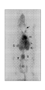
Figure 12 shows the maximum intensity projection (MIP) image of the
whole body distribution of 68Ga-PEP08296 in a healthy rat, summed during
1.5 h of data collection immediately following intravenous injection (tail
vein).
Circulating radioactivity in the major vessels (e.g. the jugular (long arrow)
and
femoral (short arrow)), the heart (H), liver (L), spleen (S), kidney (K) and
bladder (B) are readily delineated.
Figure 13 shows a gel filtration chromatogram of PEP07986 (SEQ ID
NO:163) injected at a concentration of 42 mg/ml, black solid line. A
chromatogram of ovalbumin (Mw 43 kDa) injected at a concentration of 5
mg/ml, gray broken line, is included for comparison, confirming that the peak
for PEP07986 is not an aggregate, which would have been expected in the
void volume eluted at an earlier time point than ovalbumin.
The invention will now be illustrated further through the non-limiting
description of experiments conducted in accordance therewith. Unless
otherwise specified, conventional chemistry and molecular biology methods
were used throughout.
CA 02804002 2012-12-27
WO 2012/004384 PCT/EP2011/061623
28
Examples
Example 1:
Cloning, expression, purification and characterization of albumin binding
polypeptides
In this example, ten different albumin binding polypeptides, PEP07913
(SEQ ID NO:153), PEP07912 (SEQ ID NO:156), PEP07914 (SEQ ID
NO:158), PEP07968 (DOTA-conjugated PEP07911, SEQ ID NO:159),
PEP06923 (SEQ ID NO:154), PEP07271 (SEQ ID NO:155), PEP07554 (SEQ
ID NO:156), PEP07844 (SEQ ID NO:161), PEP07986 (SEQ ID NO:163) and
PEP08296 (DOTA-conjugated PEP08185, SEQ ID NO:148), the amino acid
sequences of which are set out in Figure 1 and in the appended sequence
listing, were cloned, purified and characterized.
Material and methods
Cloning of albumin binding polypeptide variants
Mutations in G148-GA3 were generated using site directed
mutagenesis with the appropriate oligonucleotides to obtain the desired
albumin binding polypeptide variants. The gene fragments were amplified by
PCR with primers adding specific endonuclease sites as well as an N-terminal
MGSS sequence preceding the albumin binding polypeptide variants. The
fragments were cleaved with Ndel and Notl, purified and ligated to a cloning
vector, the plasmid pAY02556 (containing an origin of replication from
pBR322, a kanamycin resistance gene and a T7 promoter for expression of
the gene of interest), restricted with the same enzymes. Ligations were
transformed to electrocompetent E. coli TOP10 cells. The transformed cells
were spread on TBAB plates (30 g/I tryptose blood agar base) supplemented
with 50 pg/mlof kanamycin, followed by incubation at 37 00 overnight. The
colonies were screened using PCR and sequencing of amplified fragments
was performed using the biotinylated oligonucleotide and a Big Dye
Terminator v3.1 Cycle Sequencing Kit (Applied Biosystems), used in
accordance with the manufacturer's protocol. The sequencing reactions were
purified by binding to magnetic streptavidin coated beads using a Magnatrix
8000 (NorDiag AB), and analyzed on ABI PRISM 3100 Genetic Analyzer
(PE Applied Biosystems). All albumin binding polypeptide variants were
subcloned as monomers and the constructs encoded by the expression
CA 02804002 2012-12-27
WO 2012/004384 PCT/EP2011/061623
29
vectors were MGSS-[PP###], where PP### corresponds to the amino acid
residues constituting the sequence of the albumin binding polypeptide.
In addition, the gene fragments of G148-GA3, PP007 (SEQ ID NO:7),
PP013 (SEQ ID NO:13) and PP037 (SEQ ID NO:37) were amplified by PCR
with primers adding specific endonuclease sites as well as a hexahistidin
sequence, a TEV protease site and a glycine residue before the amino acid
residues constituting the sequence of the albumin binding polypeptide. The
polypeptides PEP07913 (SEQ ID NO:153), PEP07912 (SEQ ID NO:157),
PEP07914 (SEQ ID NO:158) and PEP07968 (SEQ ID NO:159) correspond to
the albumin binding polypeptides G148-GA3, PP007 (SEQ ID NO:7), PP013
(SEQ ID NO:13) and PP037 (SEQ ID NO:37) with glycine residues added.
The fragments were cleaved with Xbal and Notl, purified and ligated to a
cloning vector, the plasmid pAY02512 (containing an origin of replication from
pBR322, a kanamycin resistance gene and a T7 promoter for expression of
the gene of interest. The cloning site is preceded by a sequence encoding a
peptide containing a hexahistidine tag followed by a cleavage site for the TEV
protease), restricted with the same enzymes. Ligation, transformation and
sequence verification were performed as described above. The four albumin
binding polypeptide variants G148-GA3, PP007, PP013 and PP037 were
subcloned as monomers and the constructs encoded by the expression
vectors were MGSSHHHHHHLQSSGVDLGTENLYFQG-[PP###].
The expression vector encoding MGSSHHHHHHLQSSGVDLGTENLY-
FQG-[PP013] was further modified by site directed mutagenesis using
oligonucleotides, resulting in the insertion of a serine residue before the
amino acid residues constituting the sequence of the albumin binding
polypeptide, to obtain the construct MGSSHHHHHHLQSSGVDLGTENLYFQ-
GS-[PP013]. This construct was further modified by 1) site directed
mutagenesis to replace the serine residue at position 14 (within PP013) with a
cysteine residue, generating MGSSHHHHHHLQSSGVDLGTENLYFQGS-
[PPM], and 2) addition of a glycine residue C-terminally, generating
MGSSHHHHHHLOSSGVDLGTENLYFOGS-[PP049]-G. The addition of
glycine C-terminally was accomplished by PCR amplification with primers
including nucleotides encoding the glycine residue and specific endonuclease
sites. The fragment was cleaved with Xbal and Notl, purified and ligated to a
cloning vector, the plasmid pAY02641 (containing an origin of replication from
pBR322, a kanamycin resistance gene and a T7 promoter for expression of
the gene of interest), restricted with the same enzymes. Ligation,
CA 02804002 2012-12-27
WO 2012/004384
PCT/EP2011/061623
transformation and sequence verification were performed as described
above.
Protein expression
5 The albumin binding polypeptide variants were expressed in E. coli
BL21 (DE3) either with an N-terminal MGSS-extension or with an N-terminal
His6-tag followed by a TEV-protease recognition site and a glycine residue. A
colony of each albumin binding polypeptide variant was used to inoculate 4 ml
TSB+YE medium supplemented with kanamycin to a concentration of 50
10 pg/ml. The cultures were grown at 37 C for approximately 5 hours. 3 ml
from
each of the cultures was used to inoculate 800 ml TSB+YE supplemented
with kanamycin to a concentration of 50 pg/ml in parallel bio reactors (Greta
system, Belach Bioteknik AB). The cultivations were performed at 37 C, with
aeration at 800 ml/minute and an agitation profile to keep dissolved oxygen
15 levels above 30%, to an 0D600 of 2, which was followed by addition of
IPTG
to a final concentration of 0.5 mM. Cultivation was continued for five hours
after which the cultivation was cooled to 10 C, aeration was stopped and
agitation lowered to 300 rpm. Cell pellets were harvested by centrifugation
(15600 x g, 4 C, 20 minutes) and stored at -20 C until purification.
Purification of albumin binding polypeptide variants with a His6-tag and a TEV-
protease site
Frozen cell pellets harboring soluble hexahistidine-tagged polypeptides
PEP07913 (SEQ ID NO:153), PEP07912 (SEQ ID NO:156), PEP07914 (SEQ
ID NO:158), PEP07968 (SEQ ID NO:159), PEP07986 (SEQ ID NO:163) and
PEP08185 (SEQ ID NO:148) were suspended in 35 ml binding buffer (20 mM
sodium phosphate, 0.5 M NaCI, 20 mM imidazole, pH 7.4) with an addition of
1000 U Benzonase (1.01654.001, Merck) and disrupted by ultrasonication.
For each of the polypeptides, the ultrasonicated suspension was clarified by
centrifugation (1 h, 37000 x g, 4 C) and the supernatant was loaded onto a
His GraviTrapTm column (11-0033-99, GE Healthcare). The column was
washed with 10 ml washing buffer (20 mM sodium phosphate, 0.5 M NaCI, 60
mM imidazole, pH 7.4), before eluting the polypeptide with 3 ml elution buffer
(20 mM sodium phosphate, 0.5 M NaCI, 0.5 M imidazole, pH 7.4). The buffer
was exchanged to a cleavage buffer (50 mM Tris-HCI, 150 mM NaCI, pH 8)
using PD-10 desalting column (17-0851-01, GE Healthcare). The amount of
polypeptide product was determined by measuring the absorbance at 280 nm
CA 02804002 2012-12-27
WO 2012/004384 PCT/EP2011/061623
31
before adding DTT to a final concentration of 5 mM. His6-tagged TEV
protease was added to the cleavage buffer at a 1:10 mass ratio relative to the
polypeptide product. The cleavage was performed over night under slow
mixing at 4 'C. Imidazole was added to the cleavage mix, to a concentration
of 20 mM, before loading the mix onto a His GraviTrapTm column (11-0033-
99, GE Healthcare) for removing cleaved His6-tags, His6-tagged TEV
protease and His6-tagged uncleaved product.
For each variant, the flow-through, containing the albumin binding
polypeptide variant, was further purified by reversed phase chromatography
(RPC), as follows. The flow-through fraction was loaded on 1 ml Resource 15
RPC column (GE Healthcare), previously equilibrated with RPC A Buffer
(0.1 `)/0 TFA in water). After column wash with 10 column volumes (CV) RPC
A Buffer, bound polypeptides were eluted with a linear gradient of 0-50 (:)/0
RPC B Buffer (0.1 % TEA in acetonitrile) during 10 CV. The flow rate was 2
ml/min and the absorbance at 280 nm was monitored. Fractions containing
albumin binding polypeptide variant were identified by SOS-PAGE analysis
and pooled.
The RPC-purified albumin binding polypeptide variants were further
purified by gel filtration on 120 ml Superdex 75 (GE Healthcare) packed in an
XK16 column (GE Healthcare). The running buffer was 1xPBS, and the flow
rate 2 ml/min. Fractions containing pure albumin binding polypeptide variant
were pooled and concentrated to approximately 1.3 mg/ml. Finally, the
concentrate was purified from trace amounts of remaining endotoxins by
using 1 ml columns of AffinityPak Detoxi-Gel Endotoxin removing gel (Pierce,
prod#20344), according to the manufacture's recommendations.
The albumin binding polypeptide variants PEP07911 and PEP08185
were conjugated with Mal-DOTA before the RPC-purification step, as follows.
The buffer of the flow-through fraction from the IMAC-FT purification step was
exchanged to 0.2 M NaAc, pH 5.5, using a disposable PD-10 desalting
column (GE Healthcare). Maleimido-mono-amide-DOTA (Macrocyclics, cat.
no. B-272) was added at 5-fold molar excess and incubated for 60 minutes at
30 C under continuous shaking. The resulting polypeptide were denoted
PEP07968 and PEP08296, respectively.
Purification of albumin binding polypeptide-variants without His6-tag
Frozen cell pellets harboring soluble albumin binding polypeptide
variants PEP06923 (SEQ ID NO:154), PEP07271 (SEQ ID NO:155),
CA 02804002 2012-12-27
WO 2012/004384 PCT/EP2011/061623
32
PEP07554 (SEQ ID NO:156) and PEP07844 (SEQ ID NO:161) were
suspended in 20 mM Tris-HCI, pH 8 and disrupted by ultrasonication. For
each of the polypeptide variants, the ultrasonicated suspension was clarified
by centrifugation (30 min, 32000 x g, 4 C) and the supernatant was loaded
onto a HSA-Sepharose column (GE Healthcare). After washing with TST-
buffer (25 mM Tris-HCI, 1 mM EDTA, 200 mM NaCI, 0.05% Tween 20, pH
8.0), followed by 5 mM NH4Ac, pH 5.5, bound albumin binding polypeptide
variant was eluted with 0.5 M HAc, pH 3.2.
The albumin binding polypeptide variants were further purified by
reversed phase chromatography (RPC), as follows. For each of the variants,
the eluate from the HSA-affinity purification step was loaded on 1 ml
Resource 15 RPC column (GE Healthcare), previously equilibrated with RPC
A Buffer (0.1 A TFA in water). After column wash with 10 CV RPC A Buffer,
bound polypeptides were eluted with a linear gradient of 0-50 % RPC B Buffer
(0.1 `)/0 TFA in acetonitrile) during 10 CV. The flow rate was 2 ml/min and
the
absorbance at 280 nm was monitored. Fractions containing pure albumin
binding polypeptide variants were identified by SDS-PAGE analysis and
pooled. Finally, the buffer was exchanged to 1xPBS (2.68 mM KCI, 137 mM
NaCI, 1.47 mM KH2PO4, 8.1 mM Na2HPO4, pH 7.4) using a disposable PD-10
desalting column (GE Healthcare).
Characterization of purified albumin binding polypeptide-variants
The concentration was assessed by measuring the absorbance at 280
nm using a NanoDrop ND-1000 Spectrophotometer. The proteins were
further analyzed with SDS-PAGE and LC-MS.
For the SDS-PAGE analysis, approximately 10 pg of each albumin
binding polypeptide variant was mixed with NuPAGE LDS Sample Buffer
(Invitrogen), incubated at 70 C for 15 min and loaded onto NuPAGE 4-12 %
Bis-Tris Gels (Invitrogen). The gels were run with NuPAGE MES SDS
Running Buffer (Invitrogen) in an XCell II SureLock Electrophoresis Cell
(Novex) employing the Sharp Prestained Standard (Invitrogen) as molecular
weight marker and using PhastGel BlueR (GE Healthcare) for staining.
To verify the identity of the albumin binding polypeptide variants,
LC/MS analyses were performed using an Agilent 1100 LC/MSD system,
equipped with API-ESI and a single quadruple mass analyzer. Approximately
10 pg of each of the purified albumin binding polypeptide variants was loaded
on a Zorbax 300SB-C8 Narrow-Bore column (2.1 x 150 mm, 3.5 pm, Agilent
CA 02804002 2012-12-27
WO 2012/004384 PCT/EP2011/061623
33
Technologies) at a flow-rate of 0.5 ml/min. Polypeptides were eluted using a
linear gradient of 10-70% solution B for 15 min at 0.5 ml/min. The separation
was performed at 30 C. The ion signal and the absorbance at 280 and 220
nm were monitored. The molecular weights of the purified albumin binding
polypeptide variants were confirmed by MS.
Results
The expression levels of the albumin binding polypeptide variants were
10-30 mg product/g cell pellet, as estimated from SDS-PAGE analysis.
For all variants, the purity, as determined by SOS-PAGE analysis,
exceeded 95 % and the LC/MS analysis verified the correct molecular
weights. After purification, between 1 and 8 mg of pure polypeptide was
obtained for each of the ten albumin binding polypeptide variants.
Example 2:
Affinity determination for albumin binding polypeptides
In this example, PEP06923 (SEQ ID NO:154), PEP07271 (SEQ ID
NO:155), PEP07844 (SEQ ID NO:161), PEP07912 (SEQ ID NO:157),
PEP07913 (SEQ ID NO:153), PEP07914 (SEQ ID NO:158) and PEP07968,
(DOTA-conjugated PEP07911, SEQ ID NO:159), synthesized or expressed
and purified in Example 1 were characterized for affinity to human serum
albumin (HSA) using a Biacore instrument. PEP07913 corresponds to the
amino acid sequence of G148-GA3 with addition of a N-terminal glycine
residue, whereas PEP07271, PEP07844, PEP07912, PEP07914 and
PEP07968 correspond to the albumin binding polypeptides of PP001 (SEQ ID
NO:1), PP043 (SEQ ID NO:43), PP007 (SEQ ID NO:7), PP013 (SEQ ID
NO:13) and PP037 (SEQ ID NO:37) with different N-terminal amino acid
additions.
Material and methods
Biosensor analysis on a Biacore2000 instrument (GE Healthcare) was
performed with HSA (Albucult , Novozymes), immobilized by amine coupling
onto the carboxylated dextran layer of the surfaces of CM-5 chips (research
grade; GE Healthcare) according to the manufacturer's recommendations.
Surface 1 of the chip was activated and deactivated and used as a reference
CA 02804002 2012-12-27
WO 2012/004384
PCT/EP2011/061623
34
cell (blank surface) during injections, whereas surface 2 comprised HSA
immobilized to 731 resonance units (RU) and surface 4 comprised HSA
immobilized to 955 RU. The purified albumin binding polypeptide variants
were diluted in running buffer HBS-EP (GE Healthcare) to 2.5 nM, 10 nM and
40 nM, and injected at a constant flow-rate of 50 pl/min for 5 minutes,
followed by injection of HBS-EP for 60 minutes. The surfaces were
regenerated with one injection of 25 pl HCI, 10 mM. The affinity
measurements were performed in two sets; in the first set HBS-EP,
PEP06923, PEP07271, PEP07912, PEP07913, PEP07914 and PEP07968
were injected (chip surface 2), and in the second set HBS-EP, PEP06923,
PEP07844, PEP07912 and PEP07914 were injected (chip surface 4).
PEP06923 was injected twice in each run as a control. The results were
analyzed with a BiaEvaluation software (GE Healthcare). Curves of the blank
surface were subtracted from the curves of the ligand surfaces.
Results
The Biacore 2000 instrument has a technical limitation, hindering
measurements of very high affinity. Hence, the purpose of the Biacore study
was not to determine the exact kinetic parameters of the albumin binding
polypeptide variants' affinity for HSA. However, the results provide a
quantitative estimation of the relative affinities of these polypeptides for
albumin. After subtraction of reference surface and buffer injection, curves
were fitted to a 1:1 (Langmuir) binding model using BlAevaluation software
with correction for mass transfer and with RUmax set as a local parameter.
Curves are shown in Figure 2. The relative KID, ka (Icon) and kd (koff) values
were estimated and are presented in the Tables below.
Table 1:
Kinetic parameters (ka, kd and KD) of albumin binding polypeptides to HSA, 1st
set
ka (M51) kd (S-1) KD (M)
PEP07913 5.7 x 10⁵ 9.3 x 10⁻⁴ 1.6 x 10⁻⁹
PEP06923 (1) 2.9x 10⁷ 2.9x 10⁻⁵ 9.9x 10⁻¹³
PEP06923 (2) 2.6x 10⁷ 2.8x 10⁻⁵ 1.1 x 10⁻¹²
PEP07271 3.9 x 10⁶ 2.9 x 10⁻⁵ 7.5 x 10⁻¹²
PEP07912 4.6 x 10⁶ 2.8 x 10⁻⁵ 6.2 x 10⁻¹²
PEP07914 3.5 x 10⁶ 2.5 x 10⁻⁵ 7.2 x 10⁻¹²
PEP07968 3.0 x 10⁶ 2.7 x 10⁻⁵ 9.0 x 10⁻¹²
CA 02804002 2012-12-27
WO 2012/004384
PCT/EP2011/061623
Table 2:
Kinetic parameters (ka, kd and KD) of albumin binding polypeptides to HSA, 2nd
set
ka (MS-1) kd (S-1) KD (M)
PEP06923 (1) 2.0 x 10⁷ 2.6x 10⁻⁶ 1.3x 10⁻¹²
PEP06923 (2) 2.1 x 10⁷ 2.5x 10⁻⁶ 1.2x 10⁻¹²
PEP07912 5.4 x 10⁶ 2.8 x 10⁻⁶ 5.2 x 10⁻¹²
PEP07914 3.8 X 10⁶ 2.6 X 10⁻⁶ 6.9 X 10⁻¹²
PEP07844 5.4 x 10⁶ 2.3 x 10⁻⁶ 4.4 x 10⁻¹²
As shown in Tables 1 and 2, PEP07271 (SEQ ID NO:155), PEP07844
(SEQ ID NO:161), PEP07912 (SEQ ID NO:157), PEP07914 (SEQ ID
5 NO:158) and PEP07968 (DOTA-conjugated PEP07911 , SEQ ID NO:159) all
seem to have approximately the same affinity for HSA, widely exceeding the
affinity of the parent G148-GA3 (PEP07913; SEQ ID NO:153). The HSA
affinity of these polypeptides is slightly lower compared to PEP06923 (SEQ
ID NO:154), despite similar off-rate.
Example 3:
Determination of melting temperature (Tm) for albumin binding polypeptides
In this example, the albumin binding polypeptide variants PEP07913
(SEQ ID NO:153), PEP06923 (SEQ ID NO:154), PEP07271 (SEQ ID
NO:155), PEP07554 (SEQ ID NO:156), PEP07912 (SEQ ID NO:157),
PEP07914 (SEQ ID NO:158), PEP07968 (DOTA-conjugated PEP07911 ,
SEQ ID NO:159), PEP07844 (SEQ ID NO:161) and PEP07986 (SEQ ID
NO:163), expressed and purified as described in Example 1, and the albumin
polypeptide variant PEP07975 (DOTA-conjugated PEP07834, SEQ ID
NO:160), produced as described in Example 5, were analyzed by CD
analysis. PEP07913 corresponds to the sequence of G148-GA3 having an N-
terminal glycine residue, PEP06923 is an engineered high affinity derivative
previously described by Jonsson et al, supra, whereas PEP07271,
PEP07554, PEP07912, PEP07914, PEP07968, PEP07844 and PEP07975
are examples of the albumin binding polypeptides of PP001 (SEQ ID NO:1),
PP007 (SEQ ID NO:7), PP013 (SEQ ID NO:13), PP037 (SEQ ID NO:37) and
PP043 (SEQ ID NO:43) having different N-terminal amino acid additions
according to the present disclosure.
CA 02804002 2012-12-27
WO 2012/004384 PCT/EP2011/061623
36
Material and methods
Purified albumin binding polypeptide variants were diluted in 1xPBS, to
final concentrations between 0.4 and 0.5 mg/ml. Circular dichroism (CD)
analysis was performed on a Jasco J-810 spectropolarimeter in a cell with an
optical path-length of 1 mm. In the variable temperature measurements, the
absorbance was measured at 221 nm from 20 C to 90 C, with a
temperature slope of 5 C/min.
Results
The melting temperatures (Tm) of the different albumin binding
polypeptide variants were calculated by determining the midpoint of the
transition in the CD vs. temperature plot. The results are summarized in Table
3 below.
Table 3. Determined Tm values of tested albumin binding polypeptide variants
N-terminal
Variant SEQ ID NO:# sequence3 Tm ( C)
PEP07913 SEQ ID NO:153 GL 61
PEP06923 SEQ ID NO:154 GSSL 57
PEP07271 SEQ ID NO:155 GSSL 65
PEP07554 SEQ ID NO:156 GSSL 58
PEP07912 SEQ ID NO:157 GL 53
PEP07914 SEQ ID NO:158 GL 59
PEP07968 SEQ ID NO:1591 GL 53
PEP07975 SEQ ID NO:1601' 2 AL 50
PEP07844 SEQ ID NO:161 GSSL 65
PEP07986 SEQ ID NO:163 GSL 61
11The peptide is conjugated with maleimide-DOTA at the cysteine
2) The peptide is amidated at the C-terminus
Leucine (underlined) is the residue in position 1 of the amino acid sequence
of the
albumin binding polypeptide as defined in the first aspect of the present
disclosure
The polypeptide PEP07968 is identical to PEP07912, except for the
former having a cysteine residue in position 14 conjugated with maleimide
DOTA, and the latter a serine residue. Thus, the DOTA modification should
not affect the melting temperature. Also PEP07975 is conjugated with DOTA
using C14, and is identical to PEP07968 except for the C-terminal amide
(resulting from the peptide synthesis in Example 5) and for having an N-
81543849
37
terminal alanine instead of a glycine. Furthermore, comparing PEP07912 and
PEP07554
reveals that an N-terminal serine gives a higher melting temperature than a
glycine in the
same position (5 C difference in Tm). Thus, all albumin binding polypeptide
variants
according to the present disclosure show Tm above 55 C, except PEP07912 and
DOTA-conjugated variants. Taking into consideration the importance of the N-
terminal
portion, all the tested albumin binding polypeptides are superior to the prior
art derivative of
Jonsson et al, i.e. PEP06923.
Example 4:
Serum response analysis
The percentage of human serum containing IgG, capable of binding to a set of
albumin binding polypeptides as disclosed herein was analyzed by ELISA. In
total,
149 serum samples corresponding to 127 individuals were screened.
Material and methods
ELISA plates (96-well, half area plates (Costar, cat. No. 3690)) were coated
with
50 p1/well of Albuculte (Novozymes) diluted to 8 pg/ml in coating buffer
(SigmaTM, cat.
No. 3041). The plates were coated overnight for three days at 4 C. On the day
of analysis,
the plates were washed twice with tap water and blocked for 2 hours with 100
pl of phosphate
buffered saline (PBS) containing 0.05 % casein (PBSC). The plates were emptied
and
50 p1/well of the albumin binding polypeptides PEP07913 (SEQ ID NO:153),
PEP06923
(SEQ ID NO:154), PEP07271 (SEQ ID NO:155), PEP07912 (SEQ ID NO:157), PEP07554
(SEQ ID NO:156), PEP07914 (SEQ ID NO:158), PEP07968 (DOTA-conjugated PEP07911,
SEQ ID NO:159) and PEP07844 (SEQ ID NO:161), diluted to 2 pg/ml in PBSC were
added
according to a pre-made plate layout. After incubation for two hours at room
temperature (RT),
the plates were washed in PBSC four times using an automated ELISA washer. The
149
serum samples from 129 individuals were diluted 50 times in PBSC by adding 24
pl serum to
1174 pl PBSC. 50 pl of the diluted sera was added per well according to the
pre-made plate
layout. Each serum sample was tested as a singlet. Positive and negative
controls were
included on each plate and for each albumin binding polypeptide. Albumin
binding antibodies
CA 2804002 2017-09-08
CA 02804002 2012-12-27
WO 2012/004384 PCT/EP2011/061623
38
(50 pl, 0.5 p1/ml immunoglobulin solution prepared in house from sera from
primates immunized with PEP06923) was added as a positive control and 50
pl PBSC was used as a negative control. The plates were incubated for one
hour at RT and subsequently washed four times in PBSC using an automated
ELISA washer. The bound IgG was detected with 50 p1/well of anti-human
IgG (Southern Biotech, cat no 2040-05) diluted 10 000 times in PBSC. After
washing four times in PBSC using an automated ELISA washer, 50 p1/well of
substrate was added (Pierce cat. No. 34021). The reaction was stopped after
10-15 minutes by the addition of 50 pl H2504 to each well, prior to measuring
the absorbance using a multi-well plate reader (Victor3, Perkin Elmer).
Results
Of the 149 sera screened for IgG binding to the albumin binding
polypeptides, 23 were negative for all eight polypeptides (OD-value <0.1),
i.e.
showed no IgG bound to the polypeptides. The analysis was performed with
the 126 sera that were positive for one or more albumin binding polypeptides.
The average absorbance was calculated (Figure 3A) and the percentage of
sera with OD-values values either < 0.15 (Figure 3B) or > 1.0 (Figure 3C).
The highest average OD-value and the highest percentage of serum with IgG
binding were obtained with PEP07913 (SEQ ID NO:153), PEP06923 (SEQ ID
NO:154) and PEP07844 (SEQ ID NO:161), whereas least reactivity was
found against PEP07968 (DOTA-conjugated PEP07911, SEQ ID NO:159),
PEP07914 (SEQ ID NO:158) and PEP07954 (SEQ ID NO:156).
Thus, the most reactive albumin binding polypeptides were the
parental G148-GA3 (PEP07913, SEQ ID NO:153) and the previously affinity
improved derivative (PEP06923, SEQ ID NO:154), having helix 1 retained
from G148-GA3. The third of the more reactive polypeptides (PEP07844,
SEQ ID NO:161) contains the original lysine in position 14 in helix 1. This
residue is intended for conjugation, and will therefore not be exposed in the
final context. The identical albumin binding polypeptide variant, except for
having an alanine in position 14 (PEP07554, SEQ ID NO:156), is one of the
least reactive.
CA 02804002 2012-12-27
WO 2012/004384
PCT/EP2011/061623
39
Example 5:
Chemical synthesis of a DOTA-conjugated albumin binding polypeptide
Material and methods
The albumin binding polypeptide PEP07834 (SEQ ID NO:160) was
synthesized by solid phase peptide synthesis (SPPS, as described by Quibell,
M. & Johnson, T., in Fmoc Solid Phase Peptide Synthesis-A Practical
Approach, W.C. Chan, P.D. White Eds, Oxford University Press 2000, 115-
135) in a 433 A Peptide Synthesizer reactor (Applied Biosystems, Foster City,
CA) on a 0.1 mmol scale, i.e. with a theoretical possible yield of 0.1 mmol
peptide, using standard Fmoc chemistry. An acid-labile Fmoc amide resin
was used as solid support throughout the synthesis (Rink Amide MBHA Resin
LL (100-200 mesh), loading 0.39 mmol amide/g resin (Novabiochem)).
47 amino acid residues according to the sequence below were coupled
to the amide resin by acylation reactions in the reactor for 10 minutes at
room
temperature (RI) and mixing. The acylation reactions were performed with a
ten-fold molecular excess of Fmoc protected amino acids in NMP
(N-methylpyrrolidone, Merck), activated with 1 eq of 2-(1H-benzotriazole-1-
y1)-1,1,3,3-tetramethylaminium hexafluorophosphate (HBTU, IRIS Biotech), 1
eq of 1-hydroxybenzotriazole (HOBt, IRIS Biotech) and 2 eq of
diisopropylethylamine (DIEA, Applied Biosystems). In addition, all reactive
amino acid side chains were protected with standard side chain protection
groups (tert-butyl (tBu) for Asp, Glu, Ser, Thr and Tyr, tert-butyloxycarbonyl
(Boc) for Lys, 2,2,4,6,7-pentamethyldihydrobenzofuran-5-sulfonyl (Pbf) for
Arg, and trityl (Trt) for Asn and Cys) prior to activation and coupling. In
order
to diminish the amount of incomplete couplings leading to truncated peptides,
a minor amount of selected amino acid residues were subjected to coupling
by acylation twice, without Fmoc deprotection as described below between
the first and second coupling. The amino acid sequence of the synthesized
albumin binding polypeptide PEP07834 was
ALASAKEAAN AELDCYGVSD FYKRLIDKAK TVEGVEALKD AILAALP-N H2
(SEQ ID NO:160-NH2).
The underlined amino acid residues were double coupled. Any remaining
unreacted amino groups on the resin bound peptides were capped with acetic
anhydride (0.5 M acetic anhydride (AlfaAesar), 0.125 M DIEA, 0.015 M HOBt
CA 02804002 2012-12-27
WO 2012/004384 PCT/EP2011/061623
in NMP) for 5 min. Following every coupling, deprotection of the N-terminal
Fmoc group on the resin bound peptides were performed by treatment with
20% piperidine (Sigma-Aldrich) in NMP for 10 min.
After completed synthesis, the peptides were cleaved from the solid
5 support and simultaneously the side chain protection groups were cleaved
off
by treatment with TFA/EDT/H20/TIS (94:2.5:2.5:1) (TFA: trifluoroacetic acid
(Apollo), EDT: 1,2-ethanedithiol (Aldrich), TIS: triisopropylsilane (Aldrich))
at
RT for 2 h with occasional mixing. After TFA treatment, the peptides were
extracted three times using 20 % acetonitrile (Merck) in water and tert-butyl
10 methyl ether (Merck). The aqueous phases were combined, filtered and
lyophilized.
The crude peptides were analyzed and purified by semi-preparative
RP-HPLC (Reprosil GOLD 018 300, 250*10 mm, 5 pm particle size) and a
gradient of 32-55% B (A: 0.1 % TFA-H20; B: 0.1 % TFA-CH3CN) during 25
15 min at a flow rate of 2.5 ml min-1, followed by lyophilization.
The synthetic yield was determined by calculation of the integrated
areas under the peaks from the 220 nm signal from the crude analysis on RP-
HPLC. The correct molecular weight was verified using liquid chromatography
electrospray ionization mass spectrometry (LC-ESI-MS) on a 6520 Accurate
20 Mass Q-TOF LC/MS (Agilent Technologies). The purity of the product was
verified using RP-HPLC (Reprosil GOLD 018 300, 250*4.6 mm, 3 pm particle
size) using a gradient of 35-55 % B over 25 min at a flow rate of 1.0 ml
DOTA conjugation
25 3 mg of PEP07834-amide (SEQ ID NO:160-amide) was reduced with
20 mM DTT at 40 C for 30 minutes. Excess DTT was removed by buffer
exchange on a PD-10 column (GE Healthcare) to 0.2 M ammonium acetate,
pH 5.5. The coupling was performed with a 5-fold molar excess of chelator,
maleimido-mono-amide-DOTA (Macrocyclics, Cat. No. B-272) solution in
30 water (1 mg/ml). The mixture was incubated for 1 hour at 30 C under
continuous shaking. Purification from non-conjugated chelators was made on
a semi-preparative RPC column (Zorbax 300SB 018, 9.4x250 mm, 5 pm).The
coupling degree of the purified material was analyzed by HPLC-MS on a
Zorbax 300SB C8 150 x 2.1 mm, 3.5 pm analytical column. Only nnaleinnide-
35 DOTA-conjugated PEP07834, denoted PEP07975, was detected by the
method.
CA 02804002 2012-12-27
WO 2012/004384 PCT/EP2011/061623
41
Results
Based on the elution profile of the crude material, the synthetic yield of
the albumin binding polypeptide PEP07834-amide (SEQ ID NO:160-amide)
was determined to be 8 %. The found molecular weight was 4952.9 Da, which
is in good agreement with the theoretical molecular weight calculated to
4952.6 Da. When analyzing the purified product, approximately 10-15 % of
the protein was found to be a disulfide linked homodinner (Figure 4 and 5).
The binding activity of the DOTA-conjugated peptide (PEP07975) was
confirmed as described in Example 2 (data not shown), and the melting
temperature determined as described in Example 3.
Example 6:
lmmunogenicity testing of albumin binding polypeptides
PEP07913 (SEQ ID NO:153), PEP07912 (SEQ ID NO:157),
PEP07914 (SEQ ID NO:158), and PEP07968 (DOTA-conjugated PEP07911,
SEQ ID NO:159) were screened for their ability to induce T cell proliferation
in
peripheral blood mononuclear cells (PBMC) from 52 human Caucasian
individuals (obtained from CRI-Labo Medische Analyse, Gent, Belgium).
PEP07913 corresponds to the sequence of G148-GA3 having an N-terminal
glycine residue, whereas PEP07912, PEP07914 and PEP07968 are
examples of the albumin binding polypeptides of PP007 (SEQ ID NO:7),
PP013 (SEQ ID NO:13) and PP037 (SEQ ID NO:37) haying different N-
terminal amino acid additions according to the present disclosure.
Materials and methods
PBMCs, prepared according to standard cell biological methods, were
added to a tissue culture (TC) treated 96-well round bottom plate (Falcon) in
an amount of 300 000 PBMCs/ well. The cells were stimulated by addition of
100 p1/well of albumin binding polypeptides PEP07913, PEP07912,
PEP07914 and PEP07968 in AIMV medium (Invitrogen) additionally
containing 900 pg/nnl (3-fold molar excess) of recombinant human albumin
(Albucult , Novozymes). This corresponded to a final concentration of
albumin binding polypeptide of 30 pg/ml. The stimulation was done in eight-
plicates, i.e. the same albumin binding polypeptide were added to eight wells
in identical amounts and under the same conditions. In positive control wells,
CA 02804002 2012-12-27
WO 2012/004384 PCT/EP2011/061623
42
the cells were stimulated with either 30 pg/ml Keyhole Limpet Hemocyanin
(KLH, Calbiochem) or 30 pg/ml tetanus toxoid (TT, Statens Serum Institut). In
negative control wells, only AIMV medium with or without 900 pg/ml of
albumin were added.
Cell proliferation was assessed after seven days of culturing using
Alexa Fluor 488 Click-iT EdU flow cytometry assay kit (Invitrogen). 1 pM/well
of EdU incorporation marker was added on day six. On day seven, cells were
washed, dissociated from the plate, washed again and stained for 30 minutes
with anti-CD3-PerCP reagent (Becton Dickinson) and anti-CD4-Alexa647
reagent (Becton Dickinson). Following staining, the cells were washed, fixed
(BD cellfix, BD biosciences), permeabilized (using saponin) and stained for
EdU by addition of Click-iT reagent according to the manufacturer's protocol
(Invitrogen). After completed staining, cells were washed again and analyzed
using flow cytometry (FACSCantoll, BD Biosciences). To assess the number
of proliferating cells, a fixed number of fluospheres (Invitrogen) was added
to
each well before analysis. All staining procedures and washes were
performed directly in the 96-well plate.
The raw FACSCantoll data were gated hierarchically on CD3+ CD4+ T
cells and the number of gated cells as well as their fluorescence intensity of
EdU-Alexa Flour 488 incorporation marker were recorded. The mean values
of the number of proliferating cells/eight-plicate of protein treated wells
were
compared to the positive and negative controls and the resulting ratios,
described as stimulation indices (SI), were calculated. Based on the SI and
the variation between replicates, threshold SI-values were set to 2.0 and 0.5
for stimulation and inhibition, respectively.
Results
The albumin binding polypeptides PEP07913, PEP07912, PEP07914
and PEP07968 were assessed for their immunogenic potential in the
presence of 3-fold excess of recombinant human albumin in a target human
population using an in vitro PBMC proliferation assay. Compared to the
albumin control, PEP07913 induced CD3+CD4+ T cells proliferation in 6 of 52
donors, PEP07912 in 5 of 52 donors and PEP07914 and PEP07968 in 1 of
52 donors (Figure 6A).
The mean stimulation index (SI) for all 52 donors was not significantly
different for PEP07914 and PEP07968 compared to the negative control
CA 02804002 2012-12-27
WO 2012/004384 PCT/EP2011/061623
43
containing recombinant human albumin (p = 0.79 and 0.48 respectively,
Figure 6B). The SI for PEP07913 was significantly higher (p = 0.002) whereas
the SI for PEP07912 was higher but not significant (p=0.03, Figure 6B).
As compared to buffer only, the number of responding individuals was
10 for PEP07912, 7 for PEP07912, 2 for PEP07914, 1 for PEP07968, 2 for
recombinant human albumin, and 49 and 51 for the two positive controls TT
and KLH, respectively (Figure 6C). The albumin binding polypeptides were
ranked according to their immunogenicity in the following order: PEP07913 >
PEP07912 > PEP07914 > PEP07968. Both PEP07914 and PEP07968 were
defined as non-immunogenic. The above results thus demonstrate that the
immunogenic potential of the albumin binding polypeptides of the present
disclosure is low, as compared to the positive controls.
Example 7:
Albumin binding polypeptides' affinity to albumin from different species
In this example, PEP06923 (SEQ ID NO:154), PEP07986 (SEQ ID
NO:163) and PEP08296, (DOTA-conjugated PEP08185, SEQ ID NO:148),
expressed and purified as described in Example 1, were characterized for
affinity to albumin from human (HSA), cynomolgus monkey (CSA), rat (RSA),
mouse (MSA) and dog (DSA) using a Biacore instrument.
Material and methods
Biosensor analysis on a Biacore2000 instrument (GE Healthcare) was
performed with HSA (Albucult , Novozymes), CSA (purified in-house from
cynonnolgus serum), RSA (Sigma-Aldrich, Cat. No. A6272), MSA (Sigma-
Aldrich, Cat. No. A3559) and DSA (MP Biomedicals, Cat. No. 55925),
immobilized by amine coupling onto the carboxylated dextran layer of the
surfaces of CM-5 chips (research grade; GE Healthcare) according to the
manufacturer's recommendations.
On chip 1, surface 1 was activated and deactivated and used as a
reference cell (blank surface) during injections, whereas surface 2 comprised
HSA immobilized to 1130 resonance units (RU), surface 3 comprised CSA
immobilized to 1046 RU, surface 4 comprised RSA immobilized to 831 RU.
On chip 2, surface 1 was used as blank surface, whereas surface 3
CA 02804002 2012-12-27
WO 2012/004384 PCT/EP2011/061623
44
comprised MSA immobilized to 858 RU. On chip 3, surface 1 was used as
blank surface, whereas surface 2 comprised DSA immobilized to 1053 RU.
For analysis of affinity for HSA, CSA, and RSA (chip 1), the purified
albumin binding polypeptide variants were diluted in running buffer HBS-EP
(GE Healthcare) to 40 nM, 10 nM and 2.5 nM; for analysis of affinity for MSA
(chip 2) the albumin binding polypeptide variants were diluted to 1280 nM,
640 nM, 160 nM and 40 nM and for analysis of affinity for DSA (chip 3)
albumin binding polypeptide variants were diluted to 1280 nM, 640 nM, 160
nM, 40 nM and 10 nM. The albumin binding polypeptides were injected at a
constant flow-rate of 50 pl/min for 5 minutes, followed by injection of HBS-EP
for 60 minutes. The surfaces were regenerated with one injection of 25 pl
HCI, 10 mM. All samples were run in duplicates.
The results were analyzed with a BlAevaluation software (GE
Healthcare). Curves of the blank surface were subtracted from the curves of
the ligand surfaces.
Results
The Biacore 2000 instrument has a technical limitation, hindering
measurements of very high affinity. Hence, the purpose of the Biacore study
was not to determine the exact kinetic parameters of the albumin binding
polypeptide variants' affinity for HSA, CSA, RSA, MSA and DSA respectively.
However, the results provide a quantitative estimation of the relative
affinities
of the enclosed polypeptides for albumin from these different species. After
subtraction of reference surface and buffer injection, curves were fitted to a
1:1 (Langmuir) binding model using BlAevaluation software with correction for
mass transfer and with RUmax set as a local parameter. Representative
binding curves are shown in Figure 7.
PEP07986 and PEP08296 (DOTA-conjugated PEP08185) bind with
high affinity (KD in the range from below picomolar to below nanomolar) to
human serum albumin as well as to albumin from the frequent preclinical
model species rat, cynomolgus monkey, mouse and dog. The relative
affinities for the different species can be ranked as RSA HSA/CSA >
MSA/DSA, i.e. the KD values ranked as KD-RSA KD-HSPIKD-CSA < KD-MSA/KD-DSA.
The affinities in terms of KD values are the same or slightly lower (but in
the
same order of magnitude) as the affinity obtained for PEP06923 (non-
inventive polypeptide).
CA 02804002 2012-12-27
WO 2012/004384 PCT/EP2011/061623
Example 8:
In vitro activity of protein Z variants fused to an albumin binding
polypeptide
In this example, polypeptides comprising cytokine-specific protein Z
5 (derivative of domain B of staphylococcal protein A) variants genetically
fused
to the albumin binding polypeptide variant PP013 (SEQ ID NO:13) were
tested for their functionality, this being to block cytokine-induced
proliferation
of TF-1 cells in the presence of human serum albumin. Proliferation of TF-1
cells is dependent of the presence of any of several different types of
10 cytokines and the proliferative response can be inhibited by blocking
reagents
such as the corresponding cytokine-specific protein Z variant. PP013 fused to
a protein Z variant with specificity for an irrelevant protein was used as
negative control.
15 Materials and methods
Cloning of Z¨ PP013 fusion proteins
Gene fragments of protein Z variants with specificity for cytokine X or Y
respectively, or for an irrelevant protein (negative control), were amplified
by
PCR using primers adding Pstl and Accl specific endonuclease sites. The
20 fragments were cleaved with Pstl and Accl, purified and ligated into an
expression vector, the plasmid pAY02747, restricted with the same enzymes.
pAY02747 contains an origin of replication from pBR322, a kanamycin
resistance gene and a T7 promoter for expression of the gene of interest. The
cloning site is preceded by a sequence encoding the amino acids MGSSLQ
25 and succeded by a sequence encoding VDSS-PP013, where PP013 is the
disclosed albumin binding polypeptide with SEQ ID NO:13. Ligation,
transformation and sequence verification were performed as described
above. The encoded proteins were:
30 1) MGSSLQ-Zx-VDSS-PP013 (denoted Zx-PP013)
2) MGSSLQ-Zy-VDSS-PP013 (denoted Zy-PP013)
3) MGSSLQ-Zneg-VDSS-PP013 (denoted Zneg-PP013)
Protein expression
35 Zx-PP013, Zy-PP013 and Zneg-PP013 were expressed in E. coli BL21
(DE3) cells. Colonies from the transformations of each fusion variant were
used to inoculate starter cultures of 50 ml TSB+YE medium supplemented
CA 02804002 2012-12-27
WO 2012/004384 PCT/EP2011/061623
46
with kanamycin to a concentration of 50 pg/ml. The cultures were grown at
37 C over night with agitation, 100 rpm. The starter cultures were then used
to inoculate 900 ml TSB+YE medium supplemented with kanamycin to a
concentration of 50 pg/ml. The cultures were grown for approximately 1.5 h to
an 0D600 of >1.1, upon which IPTG was added to a final concentration of 0.2
mM. Cultivation was continued for five hours. Cell pellets were harvested by
centrifugation (15600 g, 4 C, 20 minutes) and stored at -20 C until
purification.
Protein purification
Frozen cell pellets harboring soluble fusion protein variants Zx-PP013,
Zy-PP013 and Zneg-PP013 were resuspended in 50 mM Tris-HCI, 150 mM
NaCI, pH 8 and 1000 U Benzonase (Merck Cat. No. 1.01654.0001) was
added. The cells were disrupted by ultrasonication and for each of the fusion
protein variants, the ultrasonicated suspension was clarified by
centrifugation
(15 min, 37000 g, 4 C). 20x TST-buffer (20x [25 mM Tris-HCI, 1 mM EDTA,
200 mM NaCI, 0.05% Tween 20, pH 8.0]) was added at a volume resulting in
1 x TST buffer in the clarified suspension. Each sample of fusion protein
variant was loaded onto a HSA-Sepharose column (GE Healthcare). After
washing with TST-buffer, followed by 5 mM NH4Ac, pH 5.5, bound fusion
protein variant was eluted with 0.5 M HAc, pH 2.5.
The fusion protein variants were further purified by reversed phase
chromatography (RPC), as follows. For each of the variants, the eluate from
the HSA-affinity purification step was loaded on a 1 ml Resource 15 RPC
column (GE Healthcare) previously equilibrated with RPC A Buffer (0.1 A
TFA in water). After column wash with 10 CV RPC A Buffer and 5 CV of RPC
B Buffer (0.1 A TFA in acetonitrile), bound fusion proteins were eluted with
a
linear gradient of 10-50% RPC B Buffer over 20 CV. The flow rate was 2
ml/min and the absorbance at 280 nm was monitored. Fractions containing
pure fusion protein variants were identified by SDS-PAGE analysis and
pooled. Finally, the buffer was exchanged to 1xPBS (2.68 mM KCI, 137 mM
NaCI, 1.47 mM KH2PO4, 8.1 mM Na2HPO4, pH 7.4) using a disposable P0-10
desalting column (GE Healthcare). To verify the identity of the fusion protein
variants, SDS-PAGE and LC/MS analyses were performed as described in
Example 1.
CA 02804002 2012-12-27
WO 2012/004384 PCT/EP2011/061623
47
In vitro cell assay of Z- PP013 fusion proteins
The cell line TF-1 (CLS Cat. No. 300434) was propagated as
recommended by the provider in RPM! 1640 medium + 10% fetal calf serum
(Gibco) with the addition of 2 ng/ml of rhGM-CSF (Miltenyi). At the day of
experiment, the cells were washed in RPMI 1640 medium + 10% fetal calf
serum to remove GM-CSF.
The ability of Zx-PP013 and Zy-PP013 to block cytokine induced
proliferation was analyzed by mixing the molecules Zx-PP013, Zy-PP013 and
Zõg-PP013 with cytokines X and Y respectively, and with a five times molar
excess of HSA (Albucult0, Novozymes). The molecules were titrated in a 2-
fold dilution series with a fixed concentration of cytokine (4.9 pM) and a
five
times molar excess of HSA. The titration was performed in 96-well plates in a
volume of 100 pl. 25 000 cells were added per well (100 pl) and plates were
incubated at 37 C, 5% CO2 for three days. To measure the proliferation, 19
pl of CCK-8 cell proliferation reagent (Sigma) diluted two times in RPM! 1640
medium + 10% fetal calf serum, was added per well. The color reaction was
monitored after 4 hours using 96-well plate reader (Victor3; Perkin Elmer).
Results
As shown in Figure 8, both Zx-PP013 and Zy-PP013 inhibited the
respective cytokine induced proliferation in the presence of HSA whereas
Zneg-PP013, the negative control, did not affect proliferation of TF-1. Thus,
the
experiment shows that the function of the Z molecules was retained when
incorporated into a fusion protein containing the albumin binding polypeptide,
and also when the fusion proteins were bound to albumin.
Example 9:
Long-term stability of an albumin binding Qolypeptide
In this example, the stability of PEP07986 (SEQ ID NO:163),
expressed and purified as described in Example 1, was investigated after
storage at 4, 25, and 40 C for up to three months. The status of the
polypeptide after storage was investigated by measuring its binding to HSA
using a Biacore instrument.
CA 02804002 2012-12-27
WO 2012/004384 PCT/EP2011/061623
48
Material and methods
Lyophilized PEP07986 was dissolved in sterile NaPi buffer (20 mM
sodium phosphate, 150 mM sodium chloride, pH 7.2) at a concentration of 2
mg/ml. A reference sample (time = 0) was removed and stored at -80 C.
Aliquots of 105 pl were stored in sterile screw-cap eppendorf tubes sealed
with parafilm at 4, 25, and 40 'C. After one week, two weeks, one month and
three months, a sample stored at each temperature was cooled to 4 C,
centrifuged for 5 min at 13000 rpm and then stored at -80 `DC awaiting
Biosensor analysis.
Biosensor analysis was performed essentially as described in Example
2 but with HSA (Albucult0, Novozymes), immobilized to 704 resonance units
(RU) and the albumin binding polypeptide variant was diluted to 10 nM and
injected at a constant flow-rate of 20 pl/min for 10 minutes, followed by
injection of HBS-EP for 10 minutes.
Results
The binding to HSA of PEP07986 (SEQ ID NO:163) was retained after
storage at 4, 25, and 40 C for at least three months. The maximum binding
responses to HSA obtained for PEP07986 stored at the various conditions
are shown in Figure 9.
Example 10:
Stability of an albumin binding polypeptide under extreme conditions
In this example, biosensor and circular dichroism (CD) analysis of the
albumin binding polypeptide PEP08296 (DOTA-conjugated PEP08185, SEQ
ID NO:148) after heat treatment (90 C) in low pH (-4.0) buffer is described.
Since such extreme reaction conditions have to be used for example for 68Ga
labeling of DOTA-modified proteins, the influence of high heat and low pH
treatment on the structural identity of the polypeptide and its capacity to
bind
HSA was investigated by measuring the melting temperature (Tm), refolding
properties and binding to HSA.
CA 02804002 2012-12-27
WO 2012/004384 PCT/EP2011/061623
49
Material and methods
Biosensor analysis of heat stability
Biosensor analysis on a Biacore 2000 instrument (GE Healthcare) was
performed with HSA (Albucult , Novozymes) immobilized by amine coupling
onto the carboxylated dextran layer of the surfaces of CM-5 chip (research
grade; GE Healthcare) according to the manufacturer's recommendations.
Surface 1 of the chip was activated and deactivated and used as a reference
cell (blank surface) during injections, whereas surface 2 comprised HSA
immobilized to 724 resonance units (RU). PEP08296 (50 pl, 100 pg) in a 15
ml Falcon tube was diluted with 450 pl 0.2 M sodium acetate (NaAc) pH 5.5 to
a final peptide concentration of 0.2 mg/rrIL. After addition of 1.5 ml 0,05 M
HCI
(resembling the conditions and volume used for eluting a 68Ge/68Ga
generator) the sample was incubated for 10 minutes at 90 C or RT (control)
and then transferred to RT. 6 ml 0.1 M sodium citrate was added to neutralize
the pH. The heat treated PEP08296 (0.8 and 4 nM) was injected at a constant
flow-rate of 50 pl/min for 5 minutes, followed by dissociation in HBS-EP for
15
minutes. The surfaces were regenerated with one injection of 25 p110 mM
HCI. The results were analyzed with BlAevaluation software (GE Healthcare).
Curves of the blank surface were subtracted from the curves of the ligand
surfaces.
Determination of the melting temperature (Tm)
PEP08296 was dissolved in PBS to a final concentration of 0.5 mg/ml.
PBS with a pH of approximately 4.0 was prepared by adding 9.5 p1100 mM
HCI to 100 pl PBS. Circular dichroism (CD) analysis was performed as
described in Example 3.
CD analysis of heat stability
To investigate structural reversibility of PEP08296 after heat treatment,
two CD spectra between 195 and 250 were recorded per sample at 20 C.
After the first spectrum, a VIM cycle with heating to 90 C was run as
described above followed by collection of the second CD spectrum between
195 and 250 nm at 20 C. In addition, PEP08296 was incubated in PBS pH
4.0 buffer or PBS pH 7.2 buffer for 12 minutes at 90 C in a thermomixer (500
rpm, interval mixing 10 son, 30 s off). After incubation, the samples were
cooled on ice followed by centrifugation at 13000 rpm for 1 minute, and a CD
spectrum between 195 and 250 nnn was recorded at 20 C.
CA 02804002 2012-12-27
WO 2012/004384 PCT/EP2011/061623
Results
Biosensor analysis was used to investigate if heat treatment in
combination with low pH, i.e. common conditions needed for 68Ga-labeling of
5 polypeptide, would affect the capacity of PEP08296 to bind to HSA. Figure
10
shows the result of this binding analysis performed with a Biacore 2000
instrument. Two different concentrations of PEP08296, 0.8 nM and 4 nM,
were injected over a surface with 724 RU of immobilized human serum
albumin. Heat treatment for 10 min at 90 C, pH 4.0, slightly reduced the
10 binding capacity of PEP08296 to HSA, indicating a potential structural
change
of the molecule.
CD was used to further investigate the potential structural change of
the molecule. Similar CD spectra before and after heating would prove a
sample to be structurally reversible. In the first experiment ,the samples
were
15 heated with a temperature gradient from 20 C to 90 C. The CD spectra
before and after heat treatment were similar in the Tm determination
experiment with the typical minima at 207 and 221 nm indicating a-helicity,
i.e. short time heating to 90 C in either pH 4 or pH 7.2 buffer had no effect
on
the structure of PEP08296.
20 However, pretreatment of PEP08296 for 12 minutes at 90 00 showed a
slightly reduced alpha helix content of PEP08296 if incubated at pH 4.0, but
no change in alpha helix content if incubated at pH 7.2. Typical overlays of
two CD spectra before and after heating are shown in Figure 11.
25 The results from the melting temperature (Tm) determination are
summarized in Table 4.
Table 4. Tm of PEP08296
Designation Tm ( C)
PEP08296 at pH 7.2 59
PEP08296 at pH 4.0 62
CA 02804002 2012-12-27
WO 2012/004384 PCT/EP2011/061623
51
Example 11:
Blood pool imaging using a 68Ga-labeled albumin binding polypeptide
In the experiments making up this example, whole body distribution of
68Ga-labeled PEP08296 (DOTA-conjugated PEP08185, SEQ ID NO:148) in
rats was followed by dynamic imaging over 1.5 hours. Due to the strong
association between the labeled polypeptide and serum albumin, the labeled
polypeptide can be used for example to study blood pool and tissue
permeability.
Material and methods
68Ga-labeling of PEP08296
68Ga was eluted as 68GaCI3from the 68Ge/68Ga generator (Obninsk,
Russia) with 0.1 M HCI, converted to 68GaC14- with concentrated HCI, trapped
on an anionic exchange column (Chromafix-HCO3) and subsequently eluted
with 18 MO water, as previously described (Velikyan et al (2008), Nucl Med
Biol 35:529-536).
The labeling was performed essentially as described in Tolmachev et
al. (EJNMMI 37:1356-1367, 2010). The concentrated 68Ga-eluate (150-200
pl) was added to PEP08296 00 pg in 0.2 M sodium acetate buffer pH 5.5)
and the pH was adjusted to 3.5-4 using sodium acetate (1.25 M) or HCI
(0.1 M). The labeling mixture was incubated at 90 C for 15 min before
cooling, and the labeled protein was isolated by size exclusion purification
on
a NAP-5 column eluted with physiologically buffered saline.
The radiochemical purity and identity of the 68Ga-labeled protein was
assessed by radio-HPLC using UV (210 nm) and radioactivity detectors in
series and a Superdex Peptide 10/300 GL column (GE Healthcare) eluted
with physiologically buffered saline.
Small animal PET
A rat (277 g) was anesthetized with isoflurane (initially 5 %, then 2 %
blended with 7:3 air/02), controlled by an E-Z vaporizer using Microflex non-
rebreather masks from Euthanex Corporation, and was kept on a heating pad
(37 C) while lying within a microPET Focus120 system (Siemens, CTI
Concorde Microsystems). 68Ga-PEP08296, 33 MBq, was dispensed in a
syringe, diluted with saline to 0.5 ml and injected via the tail vein. Data
were
acquired from the whole body by moving the bed in a constant bed motion
CA 02804002 2012-12-27
WO 2012/004384 PCT/EP2011/061623
52
protocol for 1.5 h. Data were processed with MicroPET Manager and
corrected for randoms, dead time and decay. Images were reconstructed by
standard 2D filtered back projection using a ramp filter and evaluated using
Inveon Research Workplace (Siemens Medical Solutions) software.
Results
Basic distribution patterns (Figure 12) for PEP08296 were very similar
to that of albumin labeled with radioisotopes such as 68Ga-DOTA, 64Cu-DOTA
and 11C (see e.g. Hoffend et al (2005), Nucl Med Biol 32:287-292 and Lu eta!
(2008), 11-11C]Butanol and [Methyl-11C]Albumin for Blood Flow and Blood
Pool Imaging", poster at the Xlth Turku PET Symposium, 24-27 May 2008). In
brief, high radioactivity concentrations were observed in major blood vessels
throughout the scan. Organs with large blood volumes (liver, spleen and
kidney) were also clearly delineated, as was the cardiac blood pool
radioactivity. Radioactivity in the urinary bladder increased during the
observation period, this observation of renal elimination being consistent
with
previous observations with labeled albumin-based tracers and with that of the
metabolism of albumin itself.
The general distribution pattern of radioactivity and very slow plasma
clearance after intravenous injection of 68Ga-PEP08296 is consistent with its
expected very rapid and strong binding to albumin. These results therefore
support further applications of the radiotracer as an in vivo blood pool
imaging
agent for use with positron emission tomography studies of tissue
permeability, both during the development of disease and during therapeutic
intervention.
CA 02804002 2012-12-27
WO 2012/004384 PCT/EP2011/061623
53
Example 12:
Solubility of an albumin binding golyceotide
The solubility of PEP07986 (SEQ ID NO:163) in physiological buffer
was investigated by consecutive concentrations of the sample using
ultrafiltration, followed by concentration measurement and investigation of
aggregation status. Concentrations determined by direct absorbance readings
at 280 nm were consistent with concentrations determined by gel filtration,
showing a solubility of more than 42 mg/ml with no aggregates detected.
Material and methods
Lyophilized PEP07986 was dissolved in NaPi buffer (20 nnM sodium
phosphate, 150 mM sodium chloride, pH 7.2) at a concentration of 3 mg/ml.
Amicon Ultra centrifugal filter units, cut off of 3 kDa, (Millipore, Cat. No.
UFC800324) were prerinsed with 2 ml NaPi buffer by centrifugation at 4000 g
for 20 min in a swinging bucket rotor centrifuge (Multifuge, Heraeus). 1620 pl
of 3 mg/ml PEP07986 was applied to a first centrifugal filter unit and
centrifugation was performed at 4000 g, 20 C, for 7 min. A 25 pl sample was
removed (UF sample 1) for further analysis and the rest of the sample was
transferred to a second centrifugal filter unit. The centrifugation and sample
removal were repeated three times with spinning times of 8, 9 and 20 min
respectively (UF sample 2, 3 and 4 respectively). Absorbance readings were
performed using a NanoDrope ND-1000 Spectrophotometer and by diluting
UF samples 1-4 in NaPi buffer 2, 4, 6 and 12 times respectively. The
concentrations were calculated using the extinction coefficient 1 Abs 280 =
1.955 mg/ml. Gel filtration was performed on a 1100 HPLC system (Agilent
Technologies) using a Superdex75 10/300 GL column (GE Healthcare) which
had been equilibrated in NaPi buffer. 10 pl of each UF sample were applied to
the column; NaPi buffer was used as running buffer and the flow rate was 0.5
ml/min. A chromatogram of the molecular weight standard ovalbumin (GE
Healthcare), injected at a concentration of 5 mg/ml was collected as well.
Concentrations were determined by integrating the area under the curve.
81543849
54
Results
Concentrations determined by direct absorbance readings at 280 nm and
concentrations determined by gel filtration are shown in Table 5. The
solubility of
PEP07986 (SEQ ID NO:163) is at least 42 mg/ml in physiological buffer (20 mM
sodium
phosphate, 150 mM sodium chloride, pH 7.2). No aggregates were detected by gel
filtration, as shown by Figure 13.
Table 5. Concentrations determined after consecutive concentration of PEP07986
(SEQ ID NO:163)
Sample Concentrations (mg/m1) determined by
Spectrophotometer Gel filtration
UF2 12.1 12.4
UF3 22.2 22.1
UF4 42.7 42.6
SEQUENCE LISTING IN ELECTRONIC FORM
In accordance with Section 111(1) of the Patent Rules, this description
contains a sequence
listing in electronic form in ASCII text format (file: 22819-643 Seq 14-12-12
v1.txt).
A copy of the sequence listing in electronic form is available from the
Canadian Intellectual
Property Office.
Date Recue/Date Received 2020-06-26