Note: Descriptions are shown in the official language in which they were submitted.
CA 2804595 2017-05-31
DESCRIPTION
ENDOTHELIAL CELL PRODUCTION BY PROGRAMMING
BACKGROUND OF THE INVENTION
[0001] This application claims priority to U.S. Provisional Application No.
61/362,085 filed on July 7,2010.
1. Field of the Invention
[0002] The present invention relates generally to the field of molecular
biology, stem
cells, and differentiated cells. More particularly, it concerns programming of
somatic cells
and undifferentiated cells toward specific cell lineages, particularly
endothelial cells and
precursors of endothelial cells, such as endothelial progenitor cells.
2. Description of Related Art
[00031 Endothelial cells and precursors of endothelial cells have many
potential
therapeutic uses, including treatment of tissue ischemia¨e.g., as occurs in
atherosclerosis,
myocardial infarction, and limb ischemia¨repair of injured blood vessels, and
bioengineering of grafts. Preliminary studies have shown that transplantation
of endothelial
progenitor cells (EPCs) may be useful in treating ischemia in patients with
myocardial
infarction or limb ischemia (Dzau et al., 2005). However, the clinical
usefulness of EPCs
obtained from patients is limited because patients in need of endothelial cell
therapies often
produce too few EPCs or EPCs that are functionally deficient.
[0004] In addition to such clinical applications, endothelial cells are in
high demand
for use in screening compounds and drugs for vascular toxicity, vascular
permeability, and
anti-cancer activity. However, primary endothelial cells have a finite
proliferative potential
due to their age, donor, and organ-type specific variations, all of which
limit the ability to
standardize endothelial cell culture protocols and to expand these cells in
sufficient numbers
for drug-screening purposes.
[0005] Endothelial cells may also be obtained from human embryonic stem cells
(ESCs) or induced pluripotcnt stem cells (iPSCs), both of which are capable of
unlimited
proliferation in vivo and retain their potential to differentiate into all
somatic cell types.
1
CA 02804595 2013-01-07
WO 2012/006440 PCT/US2011/043218
Differentiation of human ESCs or iPSCs into cells of endothelial lineage in
vitro recapitulates
normal in vivo development and includes stages of mesoderm induction and
specification of
angiogenic mesodermal precursors. The process requires the addition of
specific inductive
factors. Endothelial cells derived from human ESCs or iPSCs are functional in
in vitro assays
and capable of transplantation in vivo (Li et al., 2009). However,
differentiation of
endothelial cells from human ESCs or iPSCs is an inefficient process.
[0006] Therefore, there is a need for efficient production of endothelial
cells and
endothelial cell precursors for therapeutic and research uses.
SUMMARY OF THE INVENTION
[0007] The present invention overcomes a major deficiency in the art in
providing
endothelial cells and precursors of endothelial cells by forward programming
or
transdifferentiation to provide an unlimited supply of endothelial cells or
precursors of
endothelial cells. The methods may be particularly useful in providing an
unlimited supply
of patient-specific endothelial cells.
[0008] Methods disclosed herein provide endothelial cells or endothelial
precursor
cells by programming a variety of cell types. In certain aspects, programming
methods
include culturing pluripotent stem cells or somatic cells under conditions
that increase the
expression level of one or more genes that, when expressed alone or in
combination with
other programming factor genes, are capable of promoting programming to the
endothelial
lineage. Such genes are termed "endothelial programming factor genes."
Endothelial
programming factor genes useful in the invention may include any genes that,
alone or in
combination, directly impose endothelial fate upon non-endothelial cells and
may include
transcription factor genes or other genes that are important in endothelial
cell differentiation
or function. The process of programming alters the type of progeny a cell can
produce and
includes the distinct processes of forward programming and
transdifferentiation. In some
embodiments, forward programming of multipotent cells or pluripotent cells
provides
endothelial cells or endothelial precursor cells. In other embodiments,
transdifferentiation of
non-endothelial somatic cells provides endothelial cells or endothelial
precursor cells. In
certain aspects, programming may comprise increasing the expression level of a
sufficient
number of endothelial programming factor genes to cause forward programming or
transdifferentiation of non-endothelial cells to endothelial precursor cells
or endothelial cells.
Sources of cells suitable for endothelial programming may include any stem
cells or non-
2
CA 02804595 2013-01-07
WO 2012/006440 PCT/US2011/043218
endothelial cell somatic cells. For example, the stem cells may be pluripotent
stem cells or
any non-pluripotent stem cells. As used herein, a "pluripotent cell" or
"pluripotent stem cell"
is a cell that has the capacity to differentiate into essentially any fetal or
adult cell type.
Exemplary types of pluripotent stem cells may include, but are not limited to,
embryonic
stem cells and induced pluripotent stem cells (or iPS cells). Such a
pluripotent stem cell may
be a mammalian pluripotent stem cell. In certain embodiments, the pluripotent
stem cell is a
human pluripotent stem cell. Sources of cells suitable for programming of
endothelial
precursors or endothelial cells by transdifferentiation may include any non-
endothelial
somatic cells. Such somatic cells may be any cells forming the body of an
organism. In a
particular aspect, the somatic cells may be immortalized to provide an
unlimited supply of
cells, for example, by increasing the level of telomerase reverse
transcriptase (TERT). For
example, the level of TERT can be increased by increasing the transcription of
TERT from
the endogenous gene, or by introducing a transgene through any gene delivery
method or
system.
[0009] Pluripotent stem cells useful in the invention may be induced
pluripotent stem
cells, embryonic stem cells, or pluripotent stem cells derived by nuclear
transfer or cell
fusion. The stem cells may also include multipotent stem cells, oligopotent
stem cells, or
unipotent stem cells. The stem cells may also include fetal stem cells or
adult stem cells, such
as hematopoietic stem cells, mesenchymal stem cells, neural stem cells,
epithelial stem cells,
or skin stem cells. In certain aspects, the stem cells may be isolated from
umbilical tissue,
placenta, amniotic fluid, chorion villi, blastocysts, bone marrow, adipose
tissue, brain,
peripheral blood, cord blood, menstrual blood, blood vessels, skeletal muscle,
skin or liver.
[0010] A "progenitor cell" or "precursor cell" refers to a lineage-committed
cell
derived from a pluripotent stem cell. Thus, progenitor cells or precursor
cells are more
differentiated than pluripotent stem cells, but still have the capacity to
differentiate into more
than one type of cell. Endothelial cells provided by methods disclosed here
may be mature
endothelial cells. In other embodiments, the disclosed methods provide
endothelial
progenitor cells or endothelial precursor cells. Such cells are more
differentiated than
pluripotent stem cells but are capable of differentiating into endothelial
cells or into other
types of cells. In some aspects, the disclosed methods provide
hematoendothelial (or
hemangioblast) progenitor cells, which are capable of differentiating into
hematopoietic cells
or endothelial cells. In yet other embodiments, methods are provided for
differentiating
3
CA 02804595 2013-01-07
WO 2012/006440 PCT/US2011/043218
endothelial progenitor cells or endothelial precursor cells into endothelial
cells by forward
programming.
100111 In certain embodiments, endothelial cells or endothelial precursor
cells are
provided by forward programming of pluripotent stem cells or
transdifferentiation of somatic
cells. Such a method may comprise providing the endothelial cells or
endothelial precursor
cells by culturing the pluripotent stem cells or somatic cells under
conditions to increase the
expression level of one or more endothelial programming factor genes capable
of causing
forward programming of the pluripotent stem cells or transdifferentiation of
the somatic cells
into endothelial cells or endothelial precursor cells, thereby forward
programming the
.. pluripotent stem cells or transdifferentiating the somatic cells into
endothelial cells or
endothelial precursor cells.
[0012] As a skilled artisan would understand, methods for increasing the
expression
of the endothelial programming factor genes in the cells to be programmed may
include any
method known in the art, for example, by induction of expression of one or
more expression
cassettes previously introduced into the cells, or by introduction of nucleic
acids such as
DNA or RNA, polypeptides, or small molecules to the cells. Increasing the
expression of
certain endogenous but transcriptionally repressed programming factor genes
may also
comprise reversing the silencing or inhibitory effect on the expression of
these programming
factor genes by regulating the upstream transcription factor expression or
epigenetic
modulation.
[0013] In certain aspects, endothelial cells or endothelial precursor cells
are provided
by forward programming of pluripotent stem cells. Such pluripotent stem cells
may be
induced pluripotent stem cells. In other aspects, endothelial cells or
endothelial precursor
cells are provided by transdifferentiation of somatic cells. In some
embodiments, the somatic
cells are human somatic cells such as skin fibroblasts, adipose tissue-derived
cells,
keratinocytes, or blood cells. Somatic cells useful for
transdifferentiation may be
immortalized somatic cells. In a particular aspect, the somatic cells may be
immortalized to
provide an unlimited supply of cells, for example, by increasing the level of
telomerase
reverse transcriptase (TERT). For example, the level of TERT can be increased
by increasing
the transcription of TERT from the endogenous gene, or by introducing a
transgene through
any gene delivery method or system.
4
CA 02804595 2013-01-07
WO 2012/006440 PCT/US2011/043218
[0014] Endothelial cells or endothelial precursor cells may be provided by
forward
programming of pluripotent stem cells or transdifferentiation of somatic cells
that comprise at
least one exogenous expression cassette. The expression cassette may comprise
one or more
endothelial programming factor genes. In some aspects, pluripotent stem cells
or somatic
cells are contacted with one or more such endothelial programming factors
comprising gene
products of the one or more endothelial programming factor genes in an amount
sufficient to
cause forward programming of the pluripotent cells or transdifferentiation of
the somatic cells
into endothelial cells or endothelial precursor cells. In some embodiments,
the one or more
gene products are polypeptide products of one or more endothelial programming
factor
genes. In certain aspects, the one or more endothelial programming factors
include a protein
transduction domain to facilitate intracellular entry of polypeptides of the
endothelial
programming factor genes. Such protein transduction domains are well known in
the art,
such as an HIV TAT protein transduction domain, HSV VP22 protein transduction
domain,
Drosophila Antennapedia homeodomain ,or variants thereof. In other
embodiments, the one
or more gene products are RNA transcripts of one or more endothelial
programming factor
genes.
[0015] Endothelial programming factor genes useful in the invention may
include any
genes that, alone or in combination, directly impose endothelial fate upon non-
endothelial
cells, especially transcription factor genes or genes that are important in
endothelial cell
differentiation or endothelial cell function when expressed in cells.
Endothelial cell
programming factor genes include, but are not limited to v-ets
erythroblastosis virus E26
oncogene homolog (avian) (ERG), v-ets erythroblastosis virus E26 oncogene
homolog 1
(avian) (ETS1), v-ets erythroblastosis virus E26 oncogene homolog 2 (avian)
(ETS2), ELF-1,
ELF-4, FLI-1, TEL, ETV2 (ets variant 2, ER71, or Etsrp71), TALI (SCL), GATA2,
or the
Forkhead (FOX) transcription factors (e.g., FoxC, FoxF, FoxH, and Fox0
families). For
example, one, two, three, four, five, six, seven, eight, nine, ten, or more of
these exemplary
genes, isoforms of such genes, or variants thereof may be used in certain
aspects of the
invention. Many of these genes have different isoforms, which may have similar
functions
and thus are contemplated for use in certain aspects of the invention.
[0016] In particular aspects, the endothelial programming factor gene is ERG.
In
certain embodiments, the endothelial programming factor gene is ERG isoform 3
(ERG-3);
however, the programming factor gene may be any isoform of ERG, including ERG
isoform
5
CA 02804595 2013-01-07
WO 2012/006440 PCT/US2011/043218
1 (ERG-1), ERG isofoim 2 (ERG-2), and ERG isoform 4 (ERG-4). In yet other
particular
embodiments, the endothelial programming factor gene is ETV2.
[0017] "Forward programming," as used herein, refers to a process having
essentially
no requirement to culture cells through intermediate cellular stages using
culture conditions
that are adapted for each such stage and/or, optionally, having no need to add
different
growth factors during different time points between the starting cell source
and the desired
end cell product, e.g., endothelial cells or endothelial cell precursors, as
exemplified in the
upper part of FIG. 1. On the other hand, the bottom part of FIG. 1
demonstrates various
developmental stages present in a step-wise differentiation process and the
need to add
different growth factors at different times during the process, which involves
more labor,
time, and expense than methods described in certain aspects of the current
invention.
Therefore, the methods of forward programming in certain aspects of the
present invention
are advantageous by avoiding the need to add different growth factors at
different stages of
programming or differentiation to improve efficiency.
[0018] In certain aspects, the cells for endothelial cell or endothelial
precursor
programming, such as, for example, pluripotent stem cells or somatic cells,
comprise at least
one exogenous expression cassette, wherein the expression cassette comprises
one or more
endothelial programming factor genes. One or more expression cassettes may
drive
expression of one or more endothelial programming factor genes in an amount
sufficient to
cause forward programming of pluripotent cells into endothelial cells or
transdifferentiation
of somatic cells into endothelial cells. In certain embodiments, one or more
expression
cassettes drive expression of v-ets erythroblastosis virus E26 oncogene
homolog (avian)
(ERG). In other certain aspects, one or more expression cassettes may drive
expression of
ETV2. Alternatively, the expression of one or more endothelial programming
factor genes
may be increased without the use of an expression cassette.
[0019] In methods utilizing one or more exogenous expression cassettes, such
an
expression cassette may include an externally inducible transcriptional
regulatory element for
inducible expression of one or more endothelial programming factor genes. For
example, an
exogenous expression cassette useful in the invention may contain an inducible
promoter,
such as a promoter that includes a tetracycline response element. In some
embodiments, the
exogenous expression cassette is comprised in a gene delivery system. For
example, such a
gene delivery system may be a transposon system, a viral gene delivery system,
or an
6
CA 02804595 2013-01-07
WO 2012/006440 PCT/US2011/043218
episomal gene delivery system. A viral gene delivery system useful in the
invention may be
an RNA-based or DNA-based viral vector. An episomal gene delivery system
useful in the
invention may be a plasmid, an Epstein-Barr virus (EBV)-based episomal vector,
a yeast-
based vector, an adenovirus-based vector, a simian virus 40 (SV40)-based
episomal vector, a
bovine papilloma virus (BPV)-based vector, or the like. In certain aspects, an
expression
cassette for use in forward programming or transdifferentiation may include an
endothelial-
specific transcriptional regulatory element operably linked to a reporter
gene.
[0020] In certain methods, cells for endothelial cell programming, such as
pluripotent
stem cells, are contacted with one or more endothelial programming factors in
an amount
.. sufficient to cause forward programming of the cells into endothelial
cells. Endothelial
programming factors include endothelial programming factor genes, products of
such genes,
or fragments of products of such genes. Endothelial programming factors may be
gene
products of one or more endothelial programming factor genes. For example, the
one or
more gene products may be polypeptides of one or more endothelial programming
factor
genes or fragments of polypeptides of one or more endothelial programming
factor genes. In
particular embodiments, an endothelial programming factor is a product of the
ERG gene
(including any isoform thereof), ETSI gene, ETS2 gene, ELF-1 gene, ELF-4 gene,
FLI-1
gene, TEL gene, ETV2 (ER71 or Etsrp71) gene, TALI (SCL) gene, GATA2 gene, or a
Forkhead (FOX) transcription factor gene (e.g., a member of the FoxC, FoxF,
FoxH, or Fox
family).
[0021] In some embodiments, methods of providing endothelial cells or
endothelial
precursor cells by forward programming of pluripotent stem cells or
transdifferentiation of
somatic cells are provided wherein the pluripotent stem cells, somatic cells,
or progeny cells
of pluripotent stem cells or somatic cells contain a reporter expression
cassette. Such an
expression cassette may comprise an endothelial programming factor gene. In
certain
embodiments, such an expression cassette may comprise an endothelial cell-
specific
transcriptional regulatory element operably linked to a reporter gene. In
particular
embodiments, an endothelial cell-specific promoter may be operably linked to a
reporter. For
example, the promoter of FLT-1, von Willebrand factor (vWF), or TIE1 may be
operably
linked to a reporter in an expression cassette in some embodiments.
[0022] Endothelial cells or endothelial precursor cells generated by any of
the
methods provided here may have one or more characteristics of endothelial
cells. For
7
CA 2804595 2017-05-31
example, such endothelial cells may express one or more endothelial cell
markers.
Endothelial cell markers include, but are not limited to, VE-cadherin (CD144),
ACE
(angiotensin-converting enzyme) (CDI43), BNH9/BNF13, CD31, CD34, CD54 (ICAM-
),
CD62E (E-Selectin), CD105 (Endoglin), CD146, Endocan (also called ESM-I),
Endoglyx-1,
Endomucin, Eotaxin-3, EPAS I (Endothelial PAS domain protein 1), Factor VIII
related
antigen, ELI-1, Flk-1 (KDR, VEGFR-2), FLT-I (VEGFR-1), GATA2, GBP-1 (guanylate-
binding protein-1), GRO-alpha. HEX, ICAM-2 (intercellular adhesion molecule
2), LM02,
LYVE-1, MRB (magic roundabout), Nucleolin, PAL-E (pathologische anatomie
Leiden-
endothelium), RTKs, sVCAM-1, TALI, TEM1 (Tumor endothelial marker 1). TEM5
(Tumor
endothelial marker 5), TEM7 (Tumor endothelial marker 7). Thrombomodul in (TM,
CD141),
VCAM-1 (vascular cell adhesion molecule-1) (CD106), VEGF (Vascular endothelial
growth
factor), vWF (von Willebrand factor, also called Factor VIII), ZO-1,
endothelial cell-selective
adhesion molecule (ESAM), CD102, CD93, CD184, CD304, and DLL4. In particular
embodiments, an endothelial cell marker useful in the invention is one or more
of CD144,
CD31, CD34, ESAM, CD102, CD143, CD93, CD184, CD105, CD146, von Willebrand
factor, ZO-1, CD304, and DLL4. In some embodiments, the endothelial cells
produced by
forward programming or transdifferentiation do not express certain markers or
exhibit
decreased expression of certain markers, such as markers of mesenchymal cells
(e.g.,
CD140a, CD140b), markers of hematopoietic cells (e.g., CD43, CD4.5, CD235a, or
CD41a)
or markers of human pluripotent stem cells (e.g., TRA1-60).
[0023] Other characteristics of endothelial cells useful in the invention are
functional
characteristics of endothelial cells. For example, one such functional
characteristic is the
ability to take up acetylated low density lipoprotein (ac-LDL). Yet another
functional
characteristic of endothelial cells is the ability to form tube-like
structures in a three
dimensional matrix, such as MatrigelTM. An additional functional
characteristic of
endothelial cells is barrier function. Another characteristic of endothelial
cells useful in the
invention is the ability to respond to one or more pro-inflammatory stimuli
(e.g., TNF, and
IL-1) by upregulating the expression of cell-adhesion molecules (e.g., CD54
(ICAM-1),
CD 106, and CD62E). Yet another characteristic of endothelial cells useful in
the invention is
the expression of tight junction proteins (e.g., Claudin 5 and ZO-1). Other
additional
characteristics of endothelial cells useful in the invention are morphological
features, such as
a flattened (or squamous) appearance and a large, central nucleus.
8
CA 02804595 2013-01-07
WO 2012/006440 PCT/US2011/043218
[0024] In certain embodiments, methods may further include one or more steps
that
select or enrich for endothelial cells. For example, the selected or enriched
endothelial cells
may express a reporter gene that is operably linked to an endothelial cell-
specific
transcriptional regulatory element. In other embodiments, the selected or
enriched
endothelial cells may exhibit one or more endothelial cell characteristics.
For example, the
selected or enriched endothelial cells may express one or more endothelial
cell markers,
exhibit one or more functional characteristics of endothelial cells, or
exhibit one or more
morphological characteristics of endothelial cells.
[0025] In certain embodiments, pluripotent stem cells used in methods
disclosed here
are cultured in a medium that contains one or more growth factors. For
example, the medium
may contain basic FGF, VEGF, or both. Such culturing may be prior to, during,
or after the
increased expression of endothelial programming factors.
[0026] Endothelial cells provided by methods disclosed herein may be provided
at
least, about or up to 3, 4, 5, 6, 7, 8, 9, 10, 11, 12, 13, 14, 15, 16, 17, 18,
19, 20 days (or any
range derivable therein) after the increased expression or culturing in the
presence or absence
of growth factors. In some particular methods, the provided endothelial cells
are obtained
after up to 10 days of the increased expression of one or more endothelial
programming
factor genes. In other embodiments, the provided endothelial cells are
obtained after up to 4
days of the increased expression.
[0027] In certain aspects, the methods include one or more additional steps
wherein
cell groupings are dispersed into essentially individual cells. The dispersing
may be
performed, for example, at least about 24 hours after the increased
expression. In some
embodiments, the dispersing is performed at least 1, 2, 3, 4, or more days
after the increased
expression. The methods may also include one or more steps wherein the
essentially
individual cells are dispersed onto a surface coated with a matrix component.
For example,
the surface may be coated with fibronectin, gelatin, collagen, poly-d-lysine,
matrigel, or an
RGD peptide. Cells plated onto a surface coated with a matrix component may be
cultured.
In some embodiments, cells plated onto a surface coated with a matrix
component are
cultured for at least about 12 hours. After the culturing, unattached cells
may be removed,
and the attached cells may be further cultured. For example, the attached
cells may be further
cultured for at least two days.
9
CA 02804595 2013-01-07
WO 2012/006440 PCT/US2011/043218
[0028] Dispersing of cell groupings may be performed by mechanical or
enzymatic
means. For example, the cells may be dispersed by treatment with an effective
amount of one
or more enzymes, such as trypsin or trypLE, or a mixture of enzymes such as
Accutase .
Dispersed cells may be cultured in a medium comprising one or more growth
factors. For
example, the dispersed cells may be cultured in a medium that contains basic
FGF, VEGF, or
both.
[0029] Also provided are methods of providing endothelial progenitor cells by
forward programming of pluripotent stem cells or transdifferentiation of
somatic cells. In
such methods, the endothelial progenitor cells may be provided by culturing
pluripotent stem
cells or somatic cells under conditions to increase the expression level of
one or more
endothelial programming factor genes capable of causing forward programming of
the
pluripotent cells or transdifferentiation of the somatic cells into
endothelial progenitor cells,
thereby forward programming the pluripotent stem cells into endothelial
progenitor cells or
transdifferentiating the somatic cells into endothelial progenitor cells.
[0030] Methods of providing arterial endothelial cells are also provided. In
some
aspects, the method includes increasing the expression of one or more
endothelial
programming factors such as, for example ERG or ETV2. In certain embodiments,
the
arterial endothelial cells express one or more arterial endothelial cells
markers such as, for
example, CD304, CD184, or DLL4.
[0031] In certain aspects, methods of providing hemogenic endothelial cells
are
provided. In some aspects, the hemogenic endothelial cells are provided by
increasing
expression of an endothelial programming factor, such as, for example, ETV2.
Such
hemogenic endothelial cells may be used to generate hematopoietic cells when
cultured in a
hematopoietic culture medium. The hematopoietic culture medium may be any
medium
suitable for generating hematopoietic cells. For example, the hematopoietic
culture medium
may include one or more components selected from the group consisting of ESFM,
StemLine
HSC medium (Sigma), fibroblast growth factor (FGF), vascular endothelial
growth factor
(VEGF), stem cell factor (SCF), thrombopoietin (TPO), interleukin-3 (IL-3),
and interleukin-
6 (IL-6). In particular embodiments, the hematopoietic culture medium includes
ESFM,
StemLine HSC medium (Sigma), FGF, VEGF, SCF, TPO, IL-3, and IL-6. The
generated
hematopoietic cells may comprise one or more hematopoietic cell markers
selected from the
CA 02804595 2013-01-07
WO 2012/006440 PCT/US2011/043218
group consisting of CD43, CD45, CD235a, and CD41a. In certain aspects, the
hematopoietic
cells are CD43+, CD45+, CD235a+ and/or CD41a+.
[0032] In other aspects, methods of providing mesenchymogenic endothelial
cells are
provided. In some aspects, the mesenchymogenic endothelial cells are provided
by
increasing expression of an endothelial programming factor, such as ERG or
ETV2. In
particular embodiments, the mesenchymogenic endothelial cells are provided by
increasing
expression of ERG. Such mesenchymogenic endothelial cells may be used to
generate
mesenchymal cells when cultured in a mesenchymal culture medium. The
mesenchymal
culture medium may be any medium suitable for generating mesenchymal cells.
For
example, the mesenchymal culture medium may include one or more components
selected
from the group consisting of FGF and a TGF-beta inhibitor such as, for
example, A83-01. In
particular embodiments, the mesenchymal culture medium includes FGF and A83-
01. The
generated mesenchymal cells may comprise one or more mesenchymal cell markers
selected
from the group consisting of CD73 and CD105. In some aspects, the generated
mesenchymal
cells are CD31-CD73+CD105+.
[0033] The endothelial cells, endothelial progenitor cells, or precursors of
endothelial
cells provided herein may be used in any methods and applications currently
known in the art
for endothelial cells, such as clinical or screening applications. For
example, the invention
provides methods of assessing a compound for an effect on an endothelial cell.
In such
methods, an endothelial cell, which may be provided by any method disclosed
here, may be
contacted with a compound, and the effect of the compound on the endothelial
cell may be
assayed. For example, a pharmacological or toxicological effect on the
endothelial cell may
be assayed. In certain embodiments, endothelial cells of the invention are
used to assess drug
vascular toxicity or vascular permeability. In other embodiments, endothelial
cells of the
invention are used for development of anti-cancer drugs. Arterial endothelial
cells may be
used to study diseases such as thrombosis, atherosclerosis, and hypertension.
[0034] In some aspects, methods of treating a subject are provided. For
example, the
subject may have, or is at risk for, a cardiovascular disease or a
cardiovascular injury. In
some embodiments, the subject has, or is at risk for, ischemia. In yet other
embodiments, the
subject has a tissue injury or is in need of a tissue graft. In certain
aspects, any such subject is
treated by administering to the subject a therapeutically effective amount of
endothelial cells
or endothelial progenitor cells that are provided by any method disclosed
herein. In addition,
11
CA 02804595 2013-01-07
WO 2012/006440 PCT/US2011/043218
in some embodiments, endothelial cells provided by methods of the invention
may be used to
bioengineer a tissue graft that is administered to a patient in need of such
therapy. In some
embodiments, arterial endothelial cells are used in methods of treatment, such
as in methods
of treating arterial insults, injuries, or diseases.
[0035] In certain embodiments, the invention is directed to an endothelial
cell or
endothelial precursor cell. Such an endothelial cell or endothelial precursor
may be provided
by a process in accordance with any of the methods disclosed herein. In other
certain
embodiments, the invention is directed to an endothelial progenitor cell or
endothelial
precursor cell. Such endothelial progenitor cells or precursor cells may be
provided by a
.. process in accordance with any of the methods disclosed herein.
[0036] In yet other embodiments, a cell population is provided. Such a cell
population may comprise pluripotent stem cells, somatic cells, endothelial
cells, endothelial
progenitor cells, other precursors of endothelial cells, stem cells, or
progeny of any of these.
For example, the cell population may consist of endothelial cells, wherein at
least 1, 5, 10, 15,
.. 20, 25, 30, 35, 40, 45, 50, 60, 70, 80, 90, 99% or more of the endothelial
cells, or any range
derivable therein, carry an exogenous expression cassette that includes one or
more
endothelial programming factor genes. In particular embodiments, 80% of the
endothelial
cells carry an exogenous expression cassette that includes one or more
endothelial
programming factor genes. In other aspects, the cell population may consist of
endothelial
progenitor cells, wherein at least 80% of the endothelial progenitor cells
carry an exogenous
expression cassette that includes one or more endothelial programming factor
genes. In yet
other aspects, a cell population is provided that contains pluripotent stem
cells or somatic
cells where 15, 20, 25, 30, 35, 40, 45, 50, 60, 70, 80, 90, 99% or more of the
cells, or any
range derivable therein, carry an exogenous expression cassette that includes
one or more
.. endothelial programming factor genes. For example, the endothelial
programming factor
gene may be ERG or ETV2.
[0037] Also provided is a composition comprising a cell population comprising
two
cell types, i.e., the cells to be programmed to endothelial cells and
endothelial cells, and
essentially free of other intermediate cell types. For example, such a cell
population may
have two cell types including stem cells and endothelial cells, but
essentially free of other cell
types in the intermediate developmental stages along the endothelial cell
differentiation
process. In particular, a composition comprising a cell population consisting
of stem cells and
12
CA 02804595 2013-01-07
WO 2012/006440 PCT/US2011/043218
endothelial cells may be provided. The stem cells may be particularly
pluripotent stem cells,
e.g., induced pluripotent stem cells. Endothelial cells may be at least,
about, or up to 1, 5, 10,
15, 20, 25, 30, 35, 40, 45, 50, 60, 70, 80, 90, 99% of the cell population, or
any range
derivable therein.
[0038] In certain embodiments, endothelial cells are provided by forward
programming of endothelial progenitor cells. For example, the endothelial
progenitor cells
may be cultured under conditions to increase the expression level of one or
more endothelial
programming factor genes, such as those described herein, capable of causing
forward
programming of the endothelial progenitor cells into endothelial cells,
thereby forward
programming the progenitor cells into endothelial cells. In other embodiments,
endothelial
cells are provided by transdifferentiation of non-endothelial immortalized
somatic cells. For
example, the non-endothelial immortalized somatic cells may be cultured under
conditions to
increase the expression level of one or more endothelial programming factor
genes, such as
those described herein, capable of causing transdifferentiation of the somatic
cells to
endothelial cells, thereby transdifferentiating the somatic cells into
endothelial cells.
[0039] Embodiments discussed in the context of methods and/or compositions of
the
invention may be employed with respect to any other method or composition
described
herein. Thus, an embodiment pertaining to one method or composition may be
applied to
other methods and compositions of the invention as well.
[0040] As used herein the terms "encode" or "encoding" with reference to a
nucleic
acid are used to make the invention readily understandable by the skilled
artisan however
these terms may be used interchangeably with "comprise" or "comprising"
respectively.
[0041] As used herein the specification, "a" or "an" may mean one or more. As
used
herein in the claim(s), when used in conjunction with the word "comprising,"
the words "a"
or "an" may mean one or more than one.
[0042] The use of the term "or" in the claims is used to mean "and/or" unless
explicitly indicated to refer to alternatives only or the alternatives are
mutually exclusive,
although the disclosure supports a definition that refers to only alternatives
and "and/or." As
used herein "another" may mean at least a second or more.
13
CA 02804595 2013-01-07
WO 2012/006440 PCT/US2011/043218
[0043] Throughout this application, the term "about" is used to indicate that
a value
includes the inherent variation of error for the device, the method being
employed to
determine the value, or the variation that exists among the study subjects.
[0044] Other objects, features and advantages of the present invention will
become
apparent from the following detailed description. It should be understood,
however, that the
detailed description and the specific examples, while indicating preferred
embodiments of the
invention, are given by way of illustration only, since various changes and
modifications
within the spirit and scope of the invention will become apparent to those
skilled in the art
from this detailed description.
BRIEF DESCRIPTION OF THE DRAWINGS
[0045] The following drawings form part of the present specification and are
included
to further demonstrate certain aspects of the present invention. The invention
may be better
understood by reference to one or more of these drawings in combination with
the detailed
description of specific embodiments presented herein.
[0046] FIG. 1. Alternative approaches for endothelial cell differentiation
from
human ESCs/iPSCs. ECs can be efficiently induced from human ESCs/iPSCs via
expression
of appropriate transgene(s) (top box), bypassing most, if not all,
developmental stages
observed during normal differentiation (bottom box).
[0047] FIG. 2. The strategy employed for identifying transgenes that directly
convert
human ESC/iPSCs to endothelial cells. Human ESCs/iPSCs were engineered to
constitutively express rtTET protein for inducible gene expression. Transgenes
under the
control of the inducible promoter Ptight are introduced into the engineered
hESCs/iPSCs by
electroporation. Upon doxycycline (Dox) addition, transgcne expression is
induced, and EC
differentiation is monitored by the characteristic EC morphology, along with
expression of
EC markers (CD31, CD144 (VE-cadherin)) by flow cytometry.
[0048] FIG. 3. The establishment of human ESC/iPSC inducible lines for
endothelial
cell differentiation. The human Rosa26 locus on chromosome 3 was selected to
allow the
expression of rtTET, while minimizing the chromosome location-dependent
silencing effect.
First, the LoxP recombination sites (LOX71 and L0X2272) were introduced into a
site
between exon 1 and exon 2 of the human ROSA 26 gene via homologous
recombination.
14
CA 02804595 2013-01-07
WO 2012/006440 PCT/US2011/043218
The targeting construct (KI construct) used the phosphoglycerate kinase
promoter (PGK)-
driven expression of diphtheria toxin A fragment gene (DTA) for negative
selection, and
contains a ¨ 2.0 kb 5' arm and a 4.5 kb 3' arm. A splicing acceptor signal
from human BCL2
gene (SA) was placed in front of LOX71 site to allow the expression of
selection markers
from the endogenous human ROSA26 promoter. The coding region for thymidine
kinase
(TK) was included to enable negative selection against incorrect Cre/LoxP
recombination
events at step 2 using ganciclovir. The neomycin phosphotransferase (Neo) was
used for
positive selection during homologous recombination (step 1). The foot-and-
mouth disease
virus peptide (2A) was used to co-express the TK and Neo genes from the
endogenous
human ROSA26 promoter. BGHpA: polyadenylation signal derived from bovine
growth
hormone gene. The homologous recombination yielded parental human ESC/iPSC
lines for
efficient cassette exchange via Cre/LoxP recombination. To establish inducible
cell lines for
EC differentiation, rtTET driven by the constitutively active eukaryotic
elongation factor la
promoter (pEF) was introduced into the Rosa 26 locus by lipid-mediated
cotransfection of
the recombination mediated cassette exchange (RMCE) vector and a Cre-
expressing plasmid.
The puromycin N-acetyl-transferase (Puro) was used to select for recombination
events. The
correctly recombined inducible cells are resistant to puromycin (Puro+) and
ganciclovir (TK-
), and sensitive to geneticin selection (Neo-).
[0049] FIGS. 4A, 4B. Confirmation of Tet-On inducible gene expression in human
H1 ESC inducible lines. FIG. 4A. A two-vector PiggyBac stable gene expression
system;
Ptight is an rtTET-responsive inducible promoter; pEF is the eukaryotic
elongation factor la
promoter; hPBase is the coding region for the PiggyBac transposase with codons
optimized
for expression in human cells. FIG. 4B. Flow cytometric analysis of EGFP
expression in
human ESC inducilile lines after 4 days induction with or without Doxycycline
(1 ttg/mL).
Gray lines: Human ESC inducible lines with transfection of the EGFP vector;
Black lines:
Human ESC Rh I lines with stable PiggyBac transposon integration after 4 days
induction with
or without Doxycycline.
[0050] FIG. 5. Bright-field images of direct endothelial cell (EC) induction
from
human ESC inducible lines via ERG expression. ERG-3 was cloned into the
PiggyBac
.. vector (Fig. 4A) under the control of the Ptight promoter and introduced
into the human ESC
inducible line by electroporation, along with an hPBase-expressing vector.
Transfected cells
were cultured in TeSR medium on matrigel in the presence of geneticin (100
1..ig/m1) for
CA 02804595 2013-01-07
WO 2012/006440 PCT/US2011/043218
selection of transforrnants having stable genomic transgene integration.
Doxycycline (0.2
g/ml) was added to induce ERG-3 expression, and the TeSR was replaced with
endothelial
serum-free medium (ESFM; Invitrogen) supplemented with 10 ng/ml basic FGF and
20
ng/ml VEGF (both from Peprotech). Differentiated cells acquire the EC
morphology on day
2-3 of ERG induction. Although ERG-3 expression was used in these experiments
to provide
the results shown here, similar results were obtained with the other ERG
isoforms including
ERG isoform 1, ERG isoform 2, and ERG isoform 4 (data not shown).
[0051] FIG. 6. Bright-field images of forward programming of ECs from human
ESC inducible lines via ETV2 expression. ETV2 was cloned into the PiggyBac
vector (Fig.
4A) under the control of the Ptight promoter and then introduced into the
human ESC
inducible line by electroporation along with the hPBase-expressing vector.
Transfected cells
were cultured in TeSR medium on matrigel in the presence of geneticin (100
g/ml) for
selection of transformants having stable genomic transgene integration.
Doxycycline (0.2
gimp was added to induce ETV2 expression, and the TeSR was replaced with
endothelial
serum-free medium (ESFM; lnvitrogen) supplemented with 10 ng/ml basic FGF and
20
ng/ml VEGF (both from Peprotech). Differentiated cells acquire EC morphology
on day 2-3
of ETV2 induction.
[0052] FIG. 7. Flow cytometric expression analysis of the human pluripotent
stem
cell-specific marker TRA-1-60 and the EC markers (CD144/VE-cadherin and CD31)
during
ERG-induced EC differentiation from human ESCs. The ERG-induced differentiated
cells
up-regulated the expression of the EC markers (CD144 and CD31), while down-
regulating
the expression of the human pluripotent stem cell marker TRA-1-60.
[0053] FIG. 8. Flow cytometric expression analysis of the human pluripotent
stem
cell-specific marker TRA-1-60 and the EC markers (CD144/VE-cadherin and CD31)
during
ETV2-induced EC differentiation from human ESCs. The ETV2-induced
differentiated cells
up-regulated the expression of the EC markers (CD144 and CD31), while down-
regulated the
expression of the human pluripotent stem cell marker TRA-1-60.
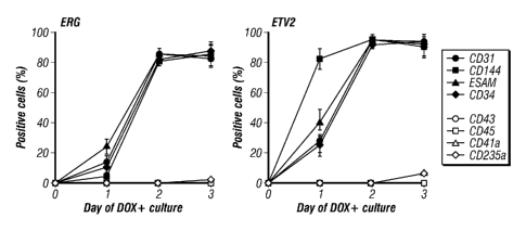
[0054] FIG. 9. Kinetic analysis of the expression of EC markers (CD31, CD144,
ESAM, CD34) and hematopoietic markers (CD43, CD45, CD41a, CD235a) in ERG- and
ETV2-induced hESC cultures.
16
CA 2804595 2017-05-31
[0055] FIG. 10. Bright-field images of established ECs obtained from human
ESCs
either through normal differentiation (EEC) or via the expression of ERG (ERG-
EC) or
ETV2 (ETV2-EC). Cell cultures on day 3 of induction were dissociated into
single-cell
suspension by Accutasc treatment (Invitrogen) and plated on gelatin-coated
plastic in ESFM
supplemented with 10 ng/ml basic FGF. After 2 hours of plating, medium
containing non-
adherent cells was removed and attached cells were cultured in ESFM
supplemented with 10
ng/ml basic FGF and 5 ug/m1 human fibronectin (Invitrogen). The morphology of
ERG-ECs
and ETV2-ECs was highly similar to that of HUVEC and FECs.
[0056] FIG 11. Flow cytometric expression analysis of EC and hematopoietic
markers. The expression of all three markers for arterial ECs (CD304/NRP1,
CD184/CXCR4
and DLL4) in ERG-ECs and ETV2-ECs might suggest an arterial fate of these
induced ECs,
different from HUVEC and EECs.
[0057] FIG. 12. Immunofluorescence analysis of ERG-ECs and ETV2-ECs: DAPI
was used to counterstain the nuclei. Although the staining was generally
weaker than in
HUVEC, vWF was clearly expressed in both ERG-ECs and ETV2-ECs.
[0058] FIG. 13. Flow cytometric and Immunofluorescence analysis of tight
junction
proteins (Claudin 5 and ZO-1) expression in ERG-ECs and ETV2-ECs.
[0059] FIG. 14. Ac-LDL incorporation by ERG-ECs and ETV2-ECs. ECs were
incubated with AeLDL-Dil conjugate (Invitrogen, 2 g/ml) for 4 hours at 37 C,
followed by
incubation with 0.5 ug/m1 Hoechst 33258 for 5 minutes for counterstaining. For
flow
cytometric analysis, the Ac-LDL-treated cultures were cultured in fresh medium
overnight
prior to Accutase dissociation. Non-treated ECs were used as control.
[0060] FIG. 15. Barrier function analysis of ERG-ECs and ETV2-ECs by measuring
their transendothelial resistance (TER) using the ECIS Zeinstrument (Applied
Biophysics).
The upper line shows the baseline TER, while the lower line shows the
disruption and
recovery of barrier function in response to thrombin (0.5 U/ml). The ERG-ECs
showed
similar kinetics in barrier function recovery as compared to HUVEC, while the
ETV2-ECs
were slower, suggesting that the ERG-ECs and ETV2-ECs are different.
17
CA 02804595 2013-01-07
WO 2012/006440 PCT/US2011/043218
[0061] FIG. 16. Tube formation by ERG-induced ECs. ERG-ECs were plated on the
solidified matrigel at 25000 cells/cm2 in ESFM supplemented with 40 ng/ml VEGF
and
incubated 12 hours.
[0062] FIG. 17. Inflammatory responses of ERG-ECs and ETV2-ECs by increased
expression of CD54, CD62E and CD106 activation markers in response to TNF
treatment.
EC cultures were treated with 25 ng/ml TNF for 24 hours and analyzed by flow
cytometry.
[0063] FIG. 18. Hemogenic function of ETV2-ECs. ETV2 and ERG induction was
performed in medium containing 50% ESFM, 50% StemLine HSC medium (Sigma), 10
ng/ml FGF, 5 ng/ml VEGF, 50 ng/ml SCF, 20 ng/ml SCF, 10 ng/nl TPO, 10 ng/ml
IL3 and
20 ng/ml IL6. Hematopoietic cells defined by CD31+CD43+ phenotype were
detected in
ETV2, but not in ERG-induced cultures. The majority of the hematopoietic cells
in the day 9
ETV2 culture were also CD235a/CD41a-CD45+, suggesting definitive
hematopoiesis.
[0064] FIG. 19. Mesenchymogenic potential of ERG-ECs. ERG-ECs were cultured
in ESFM containing 10 ng/ml FGF2 and additionally supplemented either with 20
ng/ml
VEGF or with 1 1.trn A83-01 (TGFO inhibitor). Gradual transition of ERG-EC to
mesenchymal cells defined by CD31-CD73+CD105+ phenotype was observed in
cultures
containing FGF+A83-01, but not FGF+VEGF. Although ETV2-EC cells undergo a
similar
mesenchymal transition, efficiency was lower than in ERG-EC.
DESCRIPTION OF ILLUSTRATIVE EMBODIMENTS
[0065] Endothelial cells comprise the lining of the blood vessels and are
important for
a variety of processes in the body. For example, endothelial cells play roles
in angiogenesis,
regulation of blood pressure, blood clotting, inflammation, and filtration.
Endothelial cells
are a heterogeneous group of cells and may have a variety of characteristics
depending upon
vessel size, specification to a specific organ, and morphology. Some
characteristics of
endothelial cells include expression of CD31, CD105 (endoglin), and Willebrand
factor (also
called Factor VIII), as well as the ability to take up acetylated low density
lipoprotein (ac-
LDL).
18
CA 02804595 2013-01-07
WO 2012/006440 PCT/US2011/043218
[0066] The present invention overcomes several major problems with current
technologies by providing methods and compositions for endothelial cell
production by
forward programming or transdifferentiation. In contrast to previous methods
using step-
wise differentiation protocols, certain aspects of these methods increase the
level of
.. endothelial programming transcription factors in non-endothelial cells to
provide endothelial
cells by forward programming or transdifferentiation. Extra steps, such as
adding different
growth factors during various intermediate developmental stages may be
unnecessary in
certain aspects of the present methods. Therefore, certain aspects of the
present methods may
be more time- and cost-efficient and may enable manufacture of endothelial
cells or
endothelial progenitor cells for therapeutics from a renewable source, such
as, for example,
stem cells or somatic cells. Further embodiments and advantages of the
invention are
described below.
I. Definitions
[0067] "Programming" is a process that alters the type of progeny a cell can
produce.
For example, a cell has been programmed when it has been altered so that it
can form
progeny of at least one new cell type, either in culture or in vivo, as
compared to what it
would have been able to form under the same conditions without programming.
This means
that after sufficient proliferation, a measurable proportion of progeny having
phenotypic
characteristics of the new cell type are observed, if essentially no such
progeny could form
before programming; alternatively, the proportion having characteristics of
the new cell type
is measurably more than before programming. This process includes
differentiation,
dedifferentiation and transdifferentiation. "Differentiation" is the process
by which a less
specialized cell becomes a more specialized cell type. "Dedifferentiation" is
a cellular process
in which a partially or terminally differentiated cell reverts to an earlier
developmental stage,
such as pluripotency or multipotency. "Transdifferentiation" is a process of
transforming one
differentiated cell type into another differentiated cell type. Under certain
conditions, the
proportion of progeny with characteristics of the new cell type may be at
least about 1%, 5%,
25% or more in order of increasing preference.
[0068] The term "endothelial programming factor" is a gene that, when
expressed
alone or in combination with another programming factor gene, is capable of
causing direct
differentiation of pluripotent cells or non-endothelial somatic cells into
endothelial cells or
endothelial precursor cells.
19
CA 02804595 2013-01-07
WO 2012/006440 PCT/US2011/043218
[0069] The term "forward programming" refers to the programming of a
multipotent
or pluripotent cell, as opposed to a differentiated somatic cell that has no
pluripotency, by the
provision of one or more specific lineage-determining genes or gene products
to the
multipotent or pluripotent cell. For example, forward programming may describe
the process
of programming ESCs or iPSCs to endothelial cells, endothelial precursor
cells, other
precursor cells, or other differentiated somatic cells.
[0070] The term "exogenous," when used in relation to a protein, gene, nucleic
acid,
or polynucleotide in a cell or organism refers to a protein, gene, nucleic
acid, or
polynucleotide that has been introduced into the cell or organism by
artificial or natural
means; or in relation to a cell, refers to a cell that was isolated and
subsequently introduced to
other cells or to an organism by artificial or natural means. An exogenous
nucleic acid may
be from a different organism or cell, or it may be one or more additional
copies of a nucleic
acid that occurs naturally within the organism or cell. An exogenous cell may
be from a
different organism, or it may be from the same organism. By way of a non-
limiting example,
an exogenous nucleic acid is one that is in a chromosomal location different
from that of
natural cells, or is otherwise flanked by a different nucleic acid sequence
than that found in
nature. An exogenous nucleic acid may also be extra-chromosomal, such as an
episomal
vector.
[0071] By "expression construct" or "expression cassette" is meant a nucleic
acid
molecule that is capable of directing transcription. An expression construct
includes, at a
minimum, one or more transcriptional control elements (such as promoters,
enhancers or a
structure functionally equivalent thereof) that direct gene expression in one
or more desired
cell types, tissues or organs. Additional elements, such as a transcription
termination signal,
may also be included.
[0072] A "vector" or "construct" (sometimes referred to as a gene delivery
system or
gene transfer "vehicle") refers to a macromolecule or complex of molecules
comprising a
polynucleotide to be delivered to a host cell, either in vitro or in vivo.
[0073] A "plasmid," a common type of a vector, is an extra-chromosomal DNA
molecule separate from the chromosomal DNA that is capable of replicating
independently of
the chromosomal DNA. In certain cases, it is circular and double-stranded.
CA 02804595 2013-01-07
WO 2012/006440 PCT/US2011/043218
[0074] An "origin of replication" ("ori") or "replication origin" is a DNA
sequence,
e.g., in a lymphotrophic herpes virus, that when present in a plasmid in a
cell is capable of
maintaining linked sequences in the plasmid, and/or a site at or near where
DNA synthesis
initiates. An on for EBV includes FR sequences (20 imperfect copies of a 30 bp
repeat), and
preferably DS sequences; however, other sites in EBV bind EBNA-1, e.g., Rep*
sequences
can substitute for DS as an origin of replication (Kirshmaier and Sugden,
1998). Thus, a
replication origin of EBV includes FR, DS or Rep* sequences or any
functionally equivalent
sequences through nucleic acid modifications or synthetic combination derived
therefrom.
For example, the present invention may also use genetically engineered
replication origin of
EBV, such as by insertion or mutation of individual elements, as specifically
described in
Lindner, et. al., 2008.
[0075] The term "corresponds to" is used herein to mean that a polynucleotide
sequence is homologous (i.e., is identical, not strictly evolutionarily
related) to all or a portion
of a reference polynucleotide sequence, or that a polypeptide sequence is
identical to a
reference polypeptide sequence. In contradistinction, the term "complementary
to" is used
herein to mean that the complementary sequence is homologous to all or a
portion of a
reference polynucleotide sequence. For illustration, the nucleotide sequence
"TATAC"
corresponds to a reference sequence "TATAC" and is complementary to a
reference sequence
"GTATA."
[0076] A "gene," "polynucleotide," "coding region," "sequence," "segment,"
"fragment," or "transgene" that "encodes" a particular protein, is a nucleic
acid molecule that
is transcribed and optionally also translated into a gene product, e.g., a
polypeptide, in vitro
or in vivo when placed under the control of appropriate regulatory sequences.
The coding
region may be present in either a cDNA, genomic DNA, or RNA form. When present
in a
DNA form, the nucleic acid molecule may be single-stranded (i.e., the sense
strand) or
double-stranded. The boundaries of a coding region are determined by a start
codon at the 5'
(amino) terminus and a translation stop codon at the 3' (carboxy) terminus. A
gene can
include, but is not limited to, cDNA from prokaryotic or eukaryotic mRNA,
genomic DNA
sequences from prokaryotic or eukaryotic DNA, and synthetic DNA sequences. A
transcription termination sequence will usually be located 3' to the gene
sequence.
[0077] The term "control elements" refers collectively to promoter regions,
polyadenylation signals, transcription termination sequences, upstream
regulatory domains,
21
CA 02804595 2013-01-07
WO 2012/006440 PCT/US2011/043218
origins of replication, internal ribosome entry sites (IRES), enhancers,
splice junctions, and
the like, which collectively provide for the replication, transcription, post-
transcriptional
processing, and translation of a coding sequence in a recipient cell. Not all
of these control
elements need be present so long as the selected coding sequence is capable of
being
replicated, transcribed, and translated in an appropriate host cell.
[0078] The term "promoter" is used herein in its ordinary sense to refer to a
nucleotide region comprising a DNA regulatory sequence, wherein the regulatory
sequence is
derived from a gene that is capable of binding RNA polymerase and initiating
transcription of
a downstream (3' direction) coding sequence.
[0079] By "enhancer" is meant a nucleic acid sequence that, when positioned
proximate to a promoter, confers increased transcription activity relative to
the transcription
activity resulting from the promoter in the absence of the enhancer domain.
[0080] By "operably linked" with reference to nucleic acid molecules is meant
that
two or more nucleic acid molecules (e.g., a nucleic acid molecule to be
transcribed, a
promoter, and an enhancer element) are connected in such a way as to permit
transcription of
the nucleic acid molecule. "Operably linked" with reference to peptide and/or
polypeptide
molecules means that two or more peptide and/or polypeptide molecules are
connected in
such a way as to yield a single polypeptide chain, i.e., a fusion polypeptide,
having at least
one property of each peptide and/or polypeptide component of the fusion. The
fusion
polypeptide is preferably chimeric, i.e., composed of heterologous molecules.
[0081] "Homology" refers to the percent of identity between two
polynucleotides or
two polypeptides. The correspondence between one sequence and another can be
determined
by techniques known in the art. For example, homology can be determined by a
direct
comparison of the sequence information between two polypeptide molecules by
aligning the
sequence information and using readily available computer programs.
Alternatively,
homology can be determined by hybridization of pol3mucleotides under
conditions that
promote the formation of stable duplexes between homologous regions, followed
by
digestion with single strand-specific nuclease(s), and size determination of
the digested
fragments. Two DNA, or two polypeptide, sequences are "substantially
homologous" to each
other when at least about 80%, preferably at least about 90%, and most
preferably at least
22
CA 02804595 2013-01-07
WO 2012/006440 PCT/US2011/043218
about 95% of the nucleotides, or amino acids, respectively match over a
defined length of the
molecules, as determined using the methods above.
[0082] The term "cell" is herein used in its broadest sense in the art and
refers to a
living body that is a structural unit of tissue of a multicellular organism,
is surrounded by a
membrane structure that isolates it from the outside, has the capability of
self-replicating, and
has genetic information and a mechanism for expressing it. Cells used herein
may be
naturally-occurring cells or artificially modified cells (e.g., fusion cells,
genetically modified
cells, etc.).
[0083] As used herein, the term "stem cell" refers to a cell capable of giving
rising to
.. at least one type of a more specialized cell. A stem cells has the ability
to self-renew, i.e., to
go through numerous cycles of cell division while maintaining the
undifferentiated state, and
has potency, i.e., the capacity to differentiate into specialized cell types.
Typically, stem cells
can regenerate an injured tissue. Stem cells herein may be, but are not
limited to, embryonic
stem (ES) cells, induced pluripotent stem cells, or tissue stem cells (also
called tissue-specific
stem cells, or somatic stem cells). Any artificially produced cell having the
above-described
abilities (e.g., fusion cells, reprogrammed cells, or the like used herein)
may be a stem cell.
[0084] "Embryonic stem (ES) cells" are pluripotent stem cells derived from
early
embryos. An ES cell was first established in 1981, which has also been applied
to production
of knockout mice since 1989. In 1998, a human ES cell was established, which
is currently
becoming available for regenerative medicine.
[0085] Unlike ES cells, tissue stem cells have a limited differentiation
potential.
Tissue stern cells are present at particular locations in tissues and have an
undifferentiated
intracellular structure. Therefore, the pluripotency of tissue stem cells is
typically low. Tissue
stem cells have a higher nucleus/cytoplasm ratio and have few intracellular
organelles. Most
tissue stem cells have low pluripotency, a long cell cycle, and proliferative
ability beyond the
life of the individual. Tissue stem cells are separated into categories, based
on the sites from
which the cells are derived, such as the dermal system, the digestive system,
the bone marrow
system, the nervous system, and the like. Tissue stem cells in the dermal
system include
epidermal stem cells, hair follicle stem cells, and the like. Tissue stem
cells in the digestive
system include pancreatic (common) stem cells, liver stem cells, and the like.
Tissue stem
cells in the bone marrow system include hematopoietic stem cells, mesenchymal
stem cells,
23
CA 02804595 2013-01-07
WO 2012/006440 PCT/US2011/043218
and the like. Tissue stem cells in the nervous system include neural stem
cells, retinal stem
cells, and the like.
[0086] "Induced pluripotent stem cells," commonly abbreviated as iPS cells or
iPSCs,
refer to a type of pluripotent stem cell artificially prepared from a non-
pluripotent cell,
typically an adult somatic cell, or terminally differentiated cell, such as a
fibroblast, a
hematopoietic cell, a myocyte, a neuron, an epidermal cell, or the like, by
inserting certain
genes, referred to as reprogramming factors.
[0087] "Pluripotency" refers to a stem cell that has the potential to
differentiate into
all cells constituting one or more tissues or organs, or preferably, any of
the three germ
layers: endoderm (interior stomach lining, gastrointestinal tract, the lungs),
mesoderm
(muscle, bone, blood, urogenital), or ectoderm (epidermal tissues and nervous
system).
"Pluripotent stem cells" used herein refer to cells that can differentiate
into cells derived from
any of the three germ layers, for example, direct descendants of totipotent
cells or induced
pluripotent cells.
[0088] As used herein "totipotent stem cells" refers to cells having the
ability to
differentiate into all cells constituting an organism, such as cells that are
produced from the
fusion of an egg and sperm cell. Cells produced by the first few divisions of
the fertilized egg
are also totipotent. These cells can differentiate into embryonic and
extraembryonic cell
types. Pluripotent stem cells can give rise to any fetal or adult cell type.
However, alone they
cannot develop into a fetal or adult animal because they lack the potential to
contribute to
extraembryonic tissue, such as the placenta.
[0089] In contrast, many progenitor cells are multipotcnt stem cells, i.e.,
they are
capable of differentiating into a limited number of cell fates. Multipotent
progenitor cells can
give rise to several other cell types, but those types are limited in number.
An example of a
___________________________________________________________________
multipotent stem cell is a hematopoietic cell a blood stem cell that can
develop into
several types of blood cells, but cannot develop into brain cells or other
types of cells. At the
end of the long series of cell divisions that form the embryo are cells that
are terminally
differentiated, or that are considered to be permanently committed to a
specific function.
[0090] As used herein, the term "somatic cell" refers to any cell other than a
germ
cell, such as an egg, a sperm, or the like, that does not directly transfer
its DNA to the next
24
CA 02804595 2013-01-07
WO 2012/006440 PCT/US2011/043218
generation. Typically, somatic cells have limited or no pluripotency. Somatic
cells used
herein may be naturally-occun-ing or genetically modified.
[0091] Cells are "substantially free" of certain undesired cell types, as used
herein,
when they have less that 10% of the undesired cell types, and are "essentially
free" of certain
.. cell types when they have less than 1% of the undesired cell types.
However, even more
desirable are cell populations wherein less than 0.5% or less than 0.1% of the
total cell
population comprise the undesired cell types. Thus, cell populations wherein
less than 0.1%
to 1% (including all intermediate percentages) of the cells of the population
comprise
undesirable cell types are essentially free of these cell types. A medium is
"essentially free"
.. of certain reagents, as used herein, when there is no external addition of
such agents. More
preferably, these agents are absent or present at an undetectable amount.
11. Cells involved in endothelial cell programming
[0092] In certain embodiments of the invention, there are disclosed methods
and
compositions for providing endothelial cells by forward programming of cells
that are not
.. endothelial cells. There may be also provided cells that comprise exogenous
expression
cassettes including one or more endothelial programming factor genes and/or
reporter
expression cassettes specific for endothelial cell identification. In some
embodiments, the
cells may be stem cells, including but not limited to, embryonic stem cells,
fetal stem cells, or
adult stem cells. In further embodiments, the cells may be any somatic cells.
B. Stem Cells
[0093] Stem cells are cells found in most, if not all, multi-cellular
organisms. They
are characterized by the ability to renew themselves through mitotic cell
division and the
ability to differentiate into a diverse range of specialized cell types. The
two broad types of
mammalian stem cells are: embryonic stem cells that are found in blastocysts,
and adult stem
cells that are found in adult tissues. In a developing embryo, stem cells can
differentiate into
all of the specialized embryonic tissues. In adult organisms, stem cells and
progenitor cells
act as a repair system for the body, replenishing specialized cells, and also
maintain the
normal turnover of regenerative organs, such as blood, skin or intestinal
tissues.
[0094] Human embryonic stem cells (ESCs) and induced pluripotent stem cells
(iPSCs) are capable of long-term proliferation in vitro, while retaining the
potential to
differentiate into all cell types of the body, including endothelial cells.
Thus these cells could
CA 02804595 2013-01-07
WO 2012/006440 PCT/US2011/043218
potentially provide an unlimited supply of patient-specific functional
endothelial cells for
both drug development and therapeutic uses. The differentiation of human
ESCs/iPSCs to
endothelial cells in vitro recapitulates normal in vivo development; i.e. they
undergo the
normal sequential developmental stages including mesoderm differentiation and
angiogenic
specification (FIG. 1). That sequential developmental process requires the
addition of
different growth factors at different stages of differentiation. Certain
aspects of the invention
provide fully functional endothelial cells by forward programming from human
ESCs/iPSCs
via expression of a combination of transcription factors important for
endothelial cell
differentiation/function, similar to the generation of iPSCs, bypassing most,
if not all, normal
developmental stages (FIG. 1). This approach may be more time- and cost-
efficient, and
generate endothelial cells with functions highly similar, if not identical, to
human primary
adult endothelial cells. In addition, human ESC/iPSCs, with their unlimited
proliferation
ability, have a unique advantage over somatic cells as the starting cell
population for
endothelial cell differentiation.
2. Embryonic stem cells
[0095] Embryonic stem cell lines (ES cell lines) are cultures of cells derived
from the
epiblast tissue of the inner cell mass (ICM) of a blastocyst or earlier morula
stage embryos. A
blastocyst is an early stage embryo¨approximately four to five days old in
humans and
consisting of 50-150 cells. ES cells are pluripotent and give rise during
development to all
derivatives of the three primary germ layers: ectoderm, endoderm and mesoderm.
In other
words, they can develop into each of the more than 200 cell types of the adult
body when
given sufficient and necessary stimulation for a specific cell type. They do
not contribute to
the extra-embryonic membranes or the placenta.
[0096] Nearly all research to date has taken place using mouse embryonic stem
cells
(mES) or human embryonic stem cells (hES). Both have the essential stem cell
characteristics, yet they require very different environments in order to
maintain an
undifferentiated state. Mouse ES cells may be grown on a layer of gelatin and
require the
presence of Leukemia Inhibitory Factor (LIF). Human ES cells could be grown on
a feeder
layer of mouse embryonic fibroblasts (MEFs) and often require the presence of
basic
Fibroblast Growth Factor (bFGF or FGF-2). Without optimal culture conditions
or genetic
manipulation (Chambers et al., 2003), embryonic stem cells will rapidly
differentiate.
26
CA 02804595 2013-01-07
WO 2012/006440 PCT/US2011/043218
[0097] A human embryonic stem cell may also be defined by the presence of
several
transcription factors and cell surface proteins. The transcription factors Oct-
4, Nanog, and
Sox-2 form the core regulatory network that ensures the suppression of genes
that lead to
differentiation and the maintenance of pluripotency (Boyer et al., 2005). The
cell surface
antigens most commonly used to identify hES cells include the glycolipids
SSEA3 and
SSEA4 and the keratan sulfate antigens Tra-1-60 and Tra-1-81.
[0098] Methods for obtaining mouse ES cells are well known. In one method, a
preimplantati on blastocyst from the 129 strain of mice is treated with mouse
antiserum to
remove the trophoectoderm, and the inner cell mass is cultured on a feeder
cell layer of
chemically inactivated mouse embryonic fibroblasts in medium containing fetal
calf serum.
Colonies of undifferentiated ES cells that develop are subcultured on mouse
embryonic
fibroblast feeder layers in the presence of fetal calf serum to produce
populations of ES cells.
In some methods, mouse ES cells can be grown in the absence of a feeder layer
by adding the
cytokine leukemia inhibitory factor (LIF) to serum-containing culture medium
(Smith, 2000).
In other methods, mouse ES cells can be grown in serum-free medium in the
presence of
bone morpho genetic protein and LIP (Ying et al., 2003).
[0099] Human ES cells can be obtained from blastocysts using previously
described
methods (Thomson et al., 1995; Thomson et al., 1998; Thomson and Marshall,
1998;
Reubinoff et al, 2000.) In one method, day-5 human blastocysts are exposed to
rabbit anti-
human spleen cell antiserum, then exposed to a 1:5 dilution of Guinea pig
complement to lyse
trophectoderm cells. After removing the lysed trophectodeim cells from the
intact inner cell
mass, the inner cell mass is cultured on a feeder layer of gamma-inactivated
mouse
embryonic fibroblasts and in the presence of fetal bovine serum. After 9 to 15
days, clumps
of cells derived from the inner cell mass can be chemically (i.e. exposed to
trypsin) or
mechanically dissociated and replated in fresh medium containing fetal bovine
serum and a
feeder layer of mouse embryonic fibroblasts. Upon further proliferation,
colonies having
undifferentiated morphology are selected by micropipette, mechanically
dissociated into
clumps, and replated (see U.S. Patent No. 6,833,269). ES-like morphology is
characterized
as compact colonies with apparently high nucleus to cytoplasm ratio and
prominent nucleoli.
Resulting ES cells can be routinely passaged by brief trypsinization or by
selection of
individual colonies by micropipettc. In some methods, human ES cells can be
grown without
serum by culturing the ES cells on a feeder layer of fibroblasts in the
presence of basic
27
CA 2804595 2017-05-31
fibroblast growth factor (Amit et al., 2000). In other methods, human ES cells
can be grown
without a feeder cell layer by culturing the cells on a protein matrix such as
MatrigelTM or
laminin in the presence of "conditioned" medium containing basic fibroblast
growth factor
(Xu etal., 2001). The medium is previously conditioned by coculturing with
fibroblasts.
[0100] Methods for the isolation of rhesus monkey and common marmoset ES cells
are also known (Thomson, and Marshall, 1998; Thomson etal., 1995; Thomson and
Odorico,
2000).
[0101] Another source of ES cells are established ES cell lines. Various mouse
cell
lines and human ES cell lines are known and conditions for their growth and
propagation
have been defined. For example, the mouse CGR8 cell line was established from
the inner
cell mass of mouse strain 129 embryos, and cultures of CGR8 cells can be grown
in the
presence of LIF without feeder layers. As a further example, human ES cell
lines H1, H7,
H9, H13 and H14 were established by Thompson et al. In addition, subclones
H9.1 and H9.2
of the H9 line have been developed. It is anticipated that virtually any ES or
stem cell line
known in the art may be used with the present invention, such as, e.g., those
described in Yu
and Thompson, 2008.
[0102] The source of ES cells for use in connection with the present invention
can be
a blastocyst, cells derived from culturing the inner cell mass of a
blastocyst, or cells obtained
from cultures of established cell lines. Thus, as used herein, the term "ES
cells" can refer to
inner cell mass cells of a blastocyst, ES cells obtained from cultures of
inner mass cells, and
ES cells obtained from cultures of ES cell lines.
3. Induced pluripotent stem cells
[0103] Induced pluripotent stem (iPS) cells are cells that have the
characteristics of
ES cells but are obtained by the reprogramming of differentiated somatic
cells. Induced
pluripotent stem cells have been obtained by various methods. In one method,
adult human
dermal fibroblasts are transfected with transcription factors 0ct4, Sox2, c-
Myc and Klf4
using retroviral transduction (Takahashi et al., 2007). The transfected cells
are plated on
SNL feeder cells (a mouse cell fibroblast cell line that produces LIF) in
medium
supplemented with basic fibroblast growth factor (bFGF). After approximately
25 days,
colonies resembling human ES cell colonies appear in culture. The ES cell-like
colonies are
picked and expanded on feeder cells in the presence of bFGE
28
CA 02804595 2013-01-07
WO 2012/006440 PCT/US2011/043218
[0104] Based on cell characteristics, cells of the ES cell-like colonies are
induced
pluripotent stem cells. The induced pluripotent stem cells are morphologically
similar to
human ES cells, and express various human ES cell markers. Also, when grown
under
conditions that are known to result in differentiation of human ES cells, the
induced
pluripotent stem cells differentiate accordingly. For example, the induced
pluripotent stem
cells can differentiate into cells having endothelial cell structures and
endothelial cell
markers. It is anticipated that virtually any iPS cells or cell lines may be
used with the
present invention, including, e.g., those described in Yu and Thompson, 2008.
[0105] In another method, human fetal or newborn fibroblasts are transfected
with
four genes, 0ct4, Sox2, Nanog and Lin28 using lentivirus transduction (Yu et
al., 2007). At
12-20 days post infection, colonies with human ES cell morphology become
visible. The
colonies are picked and expanded. The induced pluripotent stem cells making up
the
colonies are morphologically similar to human ES cells, express various human
ES cell
markers, and form teratomas having neural tissue, cartilage, and gut
epithelium after injection
into mice.
[0106] Methods of preparing induced pluripotent stem cells from mouse are also
known (Takahashi and Yamanaka, 2006). Induction of iPS cells typically require
the
expression of or exposure to at least one member from Sox family and at least
one member
from Oct family. Sox and Oct are thought to be central to the transcriptional
regulatory
hierarchy that specifies ES cell identity. For example, Sox may be Sox-1, Sox-
2, Sox-3, Sox-
15, or Sox-18; Oct may be Oct-4. Additional factors may increase the
reprogramming
efficiency, like Nanog, Lin28, K1f4, or c-Myc; specific sets of reprogramming
factors may be
a set comprising Sox-2, Oct-4, Nanog and, optionally, Lin-28; or comprising
Sox-2, 0ct4,
Klf and, optionally, c-Myc.
[0107] iPS cells, like ES cells, have characteristic antigens that can be
identified or
confirmed by immunohistochemistry or flow cytometry, using antibodies for SSEA-
1, SSEA-
3 and SSEA-4 (Developmental Studies Hybridoma Bank, National Institute of
Child Health
and Human Development, Bethesda Md.), and TRA-1-60 and TRA-1-81 (Andrews et
al.,
1987). Pluripotency of embryonic stem cells can be confirmed by injecting
approximately
0.5-10 X 106 cells into the rear leg muscles of 8-12 week old male SCID mice.
Teratomas
develop that demonstrate at least one cell type of each of the three germ
layers.
29
CA 2804595 2017-05-31
[0108] In certain aspects of the present invention, iPS cells are made from
reprogramming somatic cells using reprogramming factors comprising an Oct
family member
and a Sox family member, such as 0ct4 and Sox2 in combination with Klf or
Nanog as
described above. The somatic cell for reprogramming may be any somatic cell
that can be
induced to pluripotency, such as a fibroblast, a keratinocyte, a hematopoietic
cell, a
mesenchyrnal cell, a liver cell, a stomach cell, or a (3 cell. In a certain
aspect, T cells may also
be used as source of somatic cells for reprogramming (see U.S. Application No.
61/184,546).
I 0 101091
Reprogramming factors may be expressed from expression cassettes
comprised in one or more vectors, such as an integrating vector or an episomal
vector, e.g.,
an EBV element-based system (see U.S. Application No. 61/058,858; Yu et al.,
2009). In a
further aspect, reprogramming proteins could be introduced directly into
somatic cells by
protein transduction (see U.S. Application No. 61/172,079).
4. Embryonic Stem Cells Derived by Somatic Cell Nuclear Transfer
[01101 Pluripotent stem cells can be prepared by means of somatic cell nuclear
transfer, in which a donor nucleus is transferred into a spindle-free oocyte.
Stern cells
produced by nuclear transfer are genetically identical to the donor nuclei. In
one method,
donor fibroblast nuclei from skin fibroblasts of a rhesus macaque are
introduced into the
cytoplasm of spindle-free, mature metaphase II rhesus macaque ooctyes by
electrofusion
(Byrne et al., 2007). The fused oocytes are activated by exposure to
ionomycin, then
incubated until the blastocyst stage. The inner cell mass of selected
blastocysts are then
cultured to produce embryonic stem cell lines. The embryonic stem cell lines
show normal
ES cell morphology, express various ES cell markers, and differentiate into
multiple cell
types both in vitro and in vivo. As used herein, the term "ES cells" refers to
embryonic stem
cells derived from embryos containing fertilized nuclei. ES cells are
distinguished from
embryonic stem cells produced by nuclear transfer, which are referred to as
"embryonic stem
cells derived by somatic cell nuclear transfer."
5. Other stem cells
[0111] Fetal stem cells are cells with self-renewal capability and pluripotcnt
differentiation potential. They can be isolated and expanded from fetal
cytotrophoblast cells
CA 2804595 2017-05-31
(European Patent EP0412700) and chorionic villi, amniotic fluid and the
placenta
(Vv'0/2003/042405). Cell surface markers of fetal stem cells include CDI17/c-
kif-, SSEA3+,
SSEA4'l and SSEA1-.
[01121 Somatic stem cells have been identified in most organ tissues. The best
characterized is the hematopoietic stem cell. This is a mesoderm-derived cell
that has been
purified based on cell surface markers and functional characteristics. The
hematopoietic stem
cell, isolated from bone marrow, blood, cord blood, fetal liver and yolk sac,
is the progenitor
cell that reinitiates hematopoiesis for the life of a recipient and generates
multiple
hematopoietic lineages (see U.S. Pat, No. 5,635,387; 5,460,964; 5,677,136;
5,750,397;
5,759,793; 5,681,599; 5,716,827; Hill et al., 1996). When transplanted into
lethally irradiated
animals or humans, hematopoietic stem cells can repopulate the erythroid,
neutrophil-
macrophage, megakaryocyte and lymphoid hematopoietic cell pool. In vitro,
hematopoietic
stem cells can be induced to undergo at least some self-renewing cell
divisions and can be
induced to differentiate to the same lineages as is seen in vivo. Therefore,
this cell fulfills the
criteria of a stem cell.
101131 The next best characterized is the mesenchymal stem cells (MSC),
originally
derived from the embryonic mesoderm and isolated from adult bone marrow, can
differentiate to form muscle, bone, cartilage, fat, marrow stroma, and tendon.
During
embryogenesis, the mesoderm develops into limb-bud mesoderm, tissue that
generates bone,
cartilage, fat, skeletal muscle and possibly endothelium. Mesoderm also
differentiates to
visceral mesoderm, which can give rise to cardiac muscle, smooth muscle, or
blood islands
consisting of endothelium and hematopoietic progenitor cells. Primitive
mesodermal or
mesenchymal stem cells, therefore, could provide a source for a number of cell
and tissue
types. A number of mesenchymal stem cells have been isolated (see, for
example, U.S. Pat.
No. 5,486,359; 5,827,735; 5,811,094; 5,736,396; U.S. Pat. No. 5,837,539;
5,837,670;
5,827,740; Jaiswal etal., 1997; Cassiede et al., 1996; Johnstone et al., 1998;
Yoo et al., 1998;
Gronthos, 1994; Makino et al., 1999).
Of the many mesenchymal stem cells that have been described, all have
demonstrated limited differentiation to form only those differentiated cells
generally
considered to be of mesenchymal origin. To date, the most multipotent
mesenchymal stem
31
CA 2804595 2017-05-31
cell expresses the SH2 SH4' CD29+ CD44+ CD71+ CD90+ CD106' CD120a+ CD124+
CD14- CD34- CD45- phenotype.
[0114] Other stem cells have been identified, including gastrointestinal stem
cells,
epidermal stem cells, neural and hepatic stem cells, also termed oval cells
(Potten, 1998;
.. Watt, 1997; Alison eta!, 1998).
[0115] In some embodiments, the stem cells useful for methods described herein
include, but are not limited to, embryonic stem cells, induced plurpotent stem
cells,
mesenchymal stem cells, bone-marrow derived stem cells, hematopoietic stem
cells,
chrondrocyte progenitor cells, epidermal stem cells, gastrointestinal stem
cells, neural stem
cells, hepatic stem cells, adipose-derived mesenchymal stem cells, pancreatic
progenitor
cells, hair follicular stem cells, endothelial progenitor cells, and smooth
muscle progenitor
cells.
[0116] In some embodiments, the stem cells used for methods described herein
are
isolated from umbilical cord, placenta, amniotic fluid, chorion villi,
blastocysts, bone
marrow, adipose tissue, brain, peripheral blood, the gastrointestinal tract,
cord blood, blood
vessels, skeletal muscle, skin, liver, and menstrual blood. Stem cells
prepared in the
menstrual blood are called endometrial regenerative cells (available from
Medistem, Inc.).
[0117] One ordinarily skilled in the art can locate, isolate, and expand such
stem cells.
The detailed procedures for the isolation of human stem cells from various
sources are
described in Current Protocols in Stem Cell Biology (2007). Alternatively,
commercial kits
and isolation systems can be used¨e.g., the BD FACSAria cell sorting system,
BD IMag
magnetic cell separation system, and BD IMag mouse hematopoietic progenitor
cell
enrichment set from BD Biosciences. Methods of isolating and culturing stem
cells from
various sources arc also described in 5,486,359, 6,991,897, 7.015,037,
7,422,736, 7,410,798,
7,410,773, 7,399,632.
C. Somatic cells
[0118] In certain aspects of the invention, there may also be provided methods
of
transdifferentiation. i.e., the direct conversion of one somatic cell type
into another, e.g.,
deriving endothelial cells from other somatic cells. However, the human
somatic cells may
32
CA 02804595 2013-01-07
WO 2012/006440 PCT/US2011/043218
be limited in supply, especially those from living donors. In certain aspects,
to provide an
unlimited supply of starting cells for programming, somatic cells may be
immortalized by
introduction of immortalizing genes or proteins, such as hTERT or oncogenes.
The
immortalization of cells may be reversible (e.g., using removable expression
cassettes) or
inducible (e.g., using inducible promoters).
[0119] Somatic cells in certain aspects of the invention may be primary cells
(non-
immortalized cells), such as those freshly isolated from an animal, or may be
derived from a
cell line (immortalized cells). The cells may be maintained in cell culture
following their
isolation from a subject. In certain embodiments, the cells are passaged once
or more than
once (e.g., between 2-5, 5-10, 10-20, 20-50, 50-100 times, or more) prior to
their use in a
method of the invention. In some embodiments the cells will have been passaged
no more
than 1, 2, 5, 10, 20, or 50 times prior to their use in a method of the
invention. They may be
frozen, thawed, etc.
[0120] The somatic cells used or described herein may be native somatic cells,
or
engineered somatic cells, i.e., somatic cells which have been genetically
altered. Somatic
cells of the present invention are typically mammalian cells, such as, for
example, human
cells, primate cells or mouse cells. They may be obtained by well-known
methods and can be
obtained from any organ or tissue containing live somatic cells, e.g., blood,
bone marrow,
skin, lung, pancreas, liver, stomach, intestine, heart, reproductive organs,
bladder, kidney,
urethra and other urinary organs, etc.
[0121] Mammalian somatic cells useful in the present invention include, but
are not
limited to, Sertoli cells, endothelial cells, granulosa cells, neurons,
pancreatic islet cells,
epidermal cells, epithelial cells, hepatocytes, hair follicle cells,
keratinocytes, hematopoietic
cells, melanocytes, chondrocytes, lymphocytes (B and T lymphocytes),
erythrocytes,
macrophages, monocytes, mononuclear cells, cardiac muscle cells, and other
muscle cells,
etc.
[0122] In some embodiments, cells are selected based on their expression of an
endogenous marker known to be expressed only or primarily in a desired cell
type. For
example, vimentin is a fibroblast marker. Other useful markers include various
keratins, cell
adhesion molecules such as cadherins, fibronectin, CD molecules, etc. The
population of
somatic cells may have an average cell cycle time of between 18 and 96 hours,
e.g., between
33
CA 02804595 2013-01-07
WO 2012/006440 PCT/US2011/043218
24-48 hours, between 48-72 hours, etc. In some embodiments, at least 90%, 95%,
98%, 99%,
or more of the cells would be expected to divide within a predetermined time
such as 24, 48,
72, or 96 hours.
[0123] Methods described herein may be used to program one or more somatic
cells,
e.g., colonies or populations of somatic cells into endothelial cells. In some
embodiments, a
population of cells of the present invention is substantially uniform in that
at least 90% of the
cells display a phenotype or characteristic of interest. In some embodiments
at least 95%,
96%, 97%, 98%, 99%, 99.5%, 99.8%, 99.9, 99.95% or more of the cells display a
phenotype
or characteristic of interest. In certain embodiments of the invention the
somatic cells have
the capacity to divide, i.e., the somatic cells are not post-mitotic.
[0124] Somatic cells may be partially or completely differentiated.
Differentiation is
the process by which a less specialized cell becomes a more specialized cell
type. Cell
differentiation can involve changes in the size, shape, polarity, metabolic
activity, gene
expression and/or responsiveness to signals of the cell. For example,
hematopoietic stem cells
differentiate to give rise to all the blood cell types including myeloid
(monocytes and
macrophages, neutrophils, basophils, eosinophils, erythrocytes,
megakaryocytes/platelets,
dendritic cells) and lymphoid lineages (T-cells, B-cells, NK-cells). During
progression along
the path of differentiation, the ultimate fate of a cell becomes more fixed.
As described
herein, both partially differentiated somatic cells and fully differentiated
somatic cells can be
programmed as described herein to produce desired cell types such as
endothelial cells.
III. Endothelial programming factors
[0125] Certain aspects of the invention provide endothelial programming
factors for
endothelial programming. The endothelial cells could be produced directly from
other cell
sources by increasing the level of endothelial programming factors in cells.
The numerous
functions of endothelial cells could be controlled at the transcriptional
level by the concerted
actions of a limited number of endothelial cell-enriched transcription
factors. Any
transcription factors important for endothelial cell differentiation or
function may be used
herein, like endothelial cell-enriched transcription factors, particularly the
genes thereof listed
in this section. The inventors also contemplate that all the isoforms and
variants of the genes
listed in this section are included in this invention, and non-limiting
examples of accession
numbers for certain isoforms or variants are provided.
34
CA 02804595 2013-01-07
WO 2012/006440 PCT/US2011/043218
[0126] For example, by effecting expression of a combination of transcription
factors
disclosed herein, the differentiation into endothelial cells from pluripotent
stem cells may
bypass most, if not all, normal developmental stages.
[0127] In certain embodiments, the endothelial programming factor is ERG,
which is
also known as: transcriptional regulator ERG, ets-related transforming protein
ERG,
TMPRSS2-ERG prostate cancer specific, v-ets erythroblastosis virus E26
oncogene like, v-
ets avian erythroblastosis virus E26 oncogene related, transforming protein
ERG. In some
embodiments, the endothelial programming factor may be an isoform of ERG, such
as ERG-
1 (accession number: NM 182918.3; GI:209954798) (SEQ ID NOS:13 and 14), ERG-2
(SEQ ID NOS:9 and 10), ERG isoform 3 (SEQ ED NOS:11 and 12), or ERG-4
(accession
number: NM 001136155.1; GI:209954807) (SEQ ID NOS:15 and 16). In particular
embodiments, the endothelial programming factor is ERG-3. In
other particular
embodiments, the endothelial programming factor is ETV2 (also called ER71,
ETSRP71)
(NCBI Accession No. NM 014209, Version NM_014209.2, GI: 153791177) (SEQ ID
NOS:17 and 18).
[0128] In other embodiments, the one or more endothelial programming factors
is v-
ets erythroblastosis virus E26 oncogene homolog 1 (avian) (ETS1) isoform 1
(NCBI
Accession No. NM 001143820, Version NM 001143820.1, GI:219689117), ETS1
isoform 3
(NCBI Accession No. NM 001162422, Version NM_001162422.1, GI:241666445), ETS1
isoform 2 (NCBI Accession No. NM 005238, Version NM 005238.3, GI:219689116), V-
cts
erythroblastosis virus E26 oncogene homolog 2 (avian) (ETS2) (NCBT Accession
No.
NM 005239, Version NM 005239.4, GI:56119171), E74-like factor 1 (ELF-1)
isoform b
(NCBI Accession No. NM 001145353, Version NM _001145353.1, GI: 223941928), ELF-
1
isoform a (NCBI Accession No. NM 172373, Version NM 172373.3, GI: 223941931),
ELK-4 isoform a (NCBI Accession No. NM 001973, Version NM_001973.2, GI:
41872447), ELK-4 isoform b (NCBI Accession No. NM 021795, Version NM 021795.2,
GI: 41872461), friend leukemia virus integration 1 (FLI-1) isoform 2 (NCBI
Accession No.
NM 001167681, Version NM 001167681.1, GI: 264681553), FLI-1 isoform 1 (NCBI
Accession No. NM 002017, Version NM 002017.3, GI: 194018460), ETV6 (also
called
TEL or TEL1) (NCBI Accession No. NM 001987, Version NM 001987.4, GI:
153267458),
T-cell acute lymphocytic leukemia 1 (TAL1, also called SCL) (NCBI Accession
No.
NM 003189, Version NM 003189.2, GI: 197927279), GATA binding protein 2 (GATA2)
CA 2804595 2017-05-31
isoform 1 variant 1 (NCBI Accession No. NM 001145661, Version NM 001145661.1.
GI:
224611698), GATA2 isoform 1 variant 2 (NCB! Accession No. NM 032638, Version
NM 032638.4, GI: 224611697), GATA2 isoform 2 (NCBI Accession No. NM_001145662,
Version NM 001145662.1, GI: 224611700), or a Forkhead (FOX) transcription
factors (e.g.,
a member of the FoxC, FoxF, FoxH, or Fox0 family).
[0129] In yet other embodiments, the one or more endothelial programming
factors is
BMP-4, which is important for the modulation of the proliferative and
differentiative
potential of hematopoietic progenitor cells (Bhardwaj et al., 2001; Bhatia et
al., 1999;
Chadwick 2003). Additionally, BMP-4 can modulate early hematopoietic cell
development
in human fetal, neonatal, and adult hematopoietic progenitor cells (Davidson
and Zon, 2000;
Huber el al., 1998; Marshall et al., 2000). For example, BMP-4 can regulate
the proliferation
and differentiation of highly purified primitive human hematopoietic cells
from adult and
neonatal sources (Bhatia et al., 1999), and BMP-4 can promote hematopoietic
differentiation
in human embryonic stem cells (Chadwick, 2003). BMP-4 can also promote
differentiation
of endothelial cells from endothelial progenitor cells (Wang et al., 2007).
[0130] In further embodiments, the one or more endothelial programming factors
is
vascular endothelial growth factor (VEGF), which is an important signaling
protein that is
involved in formation of the embryonic circulatory system and angiogenesis.
VEGF can
affect a variety of cell types including vascular endothelium and other cell
types (e.g,
neurons, cancer cells, kidney epithelial cells ). In vitro, VEGF can stimulate
endothelial cell
mitogenesis and cell migration. VEGF function has also been shown to be
important in a
variety of disease states including cancer, diabetes, autoimmune diseases, and
ocular vascular
diseases.
[0131] In still further embodiments, the one or more endothelial programming
factors
may be Vezfl/DB1, endothelial PAS domain-containing protein 1 (EPAS1), FOX03a,
hypoxia-inducible transcription factor-2, FoxF1, FoxH1, FoxCl. FoxC2, Kruppel-
like factor
2, or Kruppel-like factor 6.
[0132] Forward programming to provide endothelial cells may be accomplished by
increasing the expression of any one or more of the endothelial cell factors
described in this
section.
36
CA 2804595 2017-05-31
IV. Delivery of gene or gene products
[0133] In certain embodiments, vectors for delivery of nucleic acids encoding
endothelial programming or differentiation factors may be constructed to
express those
factors in cells. Details of components of such vectors and delivery methods
are disclosed
below. In addition, protein transduction compositions or methods may be used
to effect
expression of the endothelial programming factors.
[0134] In a further aspect, the following systems and methods may also be used
in
delivery of a reporter expression cassette for identification of desired cell
types, such as
endothelial cells. In particular, an endothelial cell-specific regulatory
element may be used to
drive expression of a reporter gene. Therefore endothelial cells derived from
programming
may be characterized, selected or enriched via use of the reporter.
B. Nucleic acid delivery systems
[0135] One of skill in the art would be well-equipped to construct a vector
through
standard recombinant techniques (see, for example, Sambrook et al., 2001 and
Ausubel et at,
1996). Vectors include but are not limited to, plasmids, cosmids, viruses
(bacteriophage,
animal viruses, and plant viruses), and artificial chromosomes (e.g., YACs),
such as retroviral
vectors (e.g. derived from Moloney murine leukemia virus vectors (MoMLV),
MSCV, SFFV,
MPSV, SNV etc), lentiviral vectors (e.g. derived from HIV-1, HIV-2, SIV, BIV,
FIV etc.),
adenoviral (Ad) vectors including replication competent, replication deficient
and gutless
forms thereof, adeno-associated viral (AAV) vectors, simian virus 40 (SV-40)
vectors, bovine
papilloma virus vectors, Epstein-Barr virus vectors, herpes virus vectors.
vaccinia virus
vectors, Harvey murine sarcoma virus vectors, murine mammary tumor virus
vectors, Rous
sarcoma virus vectors.
2. Viral Vectors
[0136] In generating recombinant viral vectors, non-essential genes are
typically
replaced with a gene or coding sequence for a heterologous (or non-native)
protein. A viral
vector is a kind of expression construct that utilizes viral sequences to
introduce nucleic acid
and possibly proteins into a cell. The ability of certain viruses to infect
cells or enter cells via
receptor-mediated endocytosis, and to integrate into host cell genomes and
express viral
genes stably and efficiently have made them attractive candidates for the
transfer of foreign
nucleic acids into cells (e.g., mammalian cells). Non-limiting examples of
virus vectors that
37
CA 2804595 2017-05-31
may be used to deliver a nucleic acid of certain aspects of the present
invention are described
below.
[0137] Retroviruses have promise as gene delivery vectors due to their ability
to
integrate their genes into the host genome, transfer a large amount of foreign
genetic material,
infect a broad spectrum of species and cell types, and be packaged in special
cell-lines
(Miller, 1992).
[0138] In order to construct a retroviral vector, a nucleic acid is inserted
into the viral
genome in place of certain viral sequences to produce a virus that is
replication-defective. In
order to produce virions, a packaging cell line containing the gag. pol, and
env genes¨but
without the LTR and packaging components¨is constructed (Mann et al., 1983).
When a
recombinant plasmid containing a cDNA, together with the retroviral LTR and
packaging
sequences, is introduced into a special cell line (e.g., by calcium phosphate
precipitation), the
packaging sequence allows the RNA transcript of the recombinant plasmid to be
packaged
into viral particles, which are then secreted into the culture medium (Nicolas
and Rubenstein,
1988; Temin, 1986; Mann et al., 1983). The medium containing the recombinant
retroviruses
is then collected, optionally concentrated, and used for gene transfer.
Retroviral vectors are
able to infect a broad variety of cell types. However, integration and stable
expression
require the division of host cells (Paskind et al., 1975).
[01391 Lentiviruses are complex retroviruses, which, in addition to the common
retroviral genes gag, pol, and env, contain other genes with regulatory or
structural function.
Lentiviral vectors are well known in the art (see, for example, Naldini etal.,
1996; Zufferey
etal., 1997; Blomer et al., 1997; U.S. Patents 6,013,516 and 5,994,136).
[0140] Recombinant lentiviral vectors are capable of infecting non-dividing
cells and
can be used for both in vivo and ex vivo gene transfer and expression of
nucleic acid
sequences. For example, recombinant lentivirus capable of infecting a non-
dividing cell
wherein a suitable host cell is transfected with two or more vectors carrying
the packaging
functions, namely gag, pol and env, as well as rev and tat¨is described in
U.S. Patent
5,994,136.
38
CA 2804595 2017-05-31
3. Episomal Vectors
[0141] The use of plasmid- or liposome-based extra-chromosomal (i.e.,
episomal)
vectors may be also provided in certain aspects of the invention. Such
episomal vectors may
include, e.g., oriP-based vectors, and/or vectors encoding a derivative of
EBNA-1. These
vectors may permit large fragments of DNA to be introduced unto a cell and
maintained
extra-chromosomally, replicated once per cell cycle, partitioned to daughter
cells efficiently,
and elicit substantially no immune response.
[0142] In particular, EBNA-1, the only viral protein required for the
replication of the
oriP-based expression vector, does not elicit a cellular immune response
because it has
developed an efficient mechanism to bypass the processing required for
presentation of its
antigens on MHC class I molecules (Levitskaya et al., 1997). Further, EBNA-1
can act in
trans to enhance expression of the cloned gene. inducing expression of a
cloned gene up to
100-fold in some cell lines (Langle-Rouault et al., 1998; Evans et al.. 1997).
Finally, the
manufacture of such oriP-based expression vectors is inexpensive.
[0143] Other extra-chromosomal vectors include other lymphotrophic herpes
virus-
based vectors. Lymphotrophic herpes virus is a herpes virus that replicates in
a lymphoblast
(e.g., a human B lymphoblast) and becomes a plasmid for a part of its natural
life-cycle.
Herpes simplex virus (11SV) is not a -lymphotrophic" herpes virus. Exemplary
lymphotrophic herpes viruses include, but are not limited to EBV, Kaposi's
sarcoma herpes
virus (KSHV); Herpes virus saimiri (HS) and Marek's disease virus (MDV). Other
sources of
episome-based vectors are also contemplated, such as yeast ARS, adenovirus,
SV40, or BPV.
[0144] One of skill in the art would be well-equipped to construct a vector
through
standard recombinant techniques (see, for example, Maniatis et al., 1988 and
Ausubel el al.,
1994).
[0145] Vectors can also comprise other components or functionalities that
further
modulate gene delivery and/or gene expression, or that otherwise provide
beneficial
properties to the targeted cells. Such other components include, for example,
components that
influence binding or targeting to cells (including components that mediate
cell-type or tissue-
specific binding); components that influence uptake of the vector nucleic acid
by the cell;
components that influence localization of the polynucleotide within the cell
after uptake (such
39
CA 02804595 2013-01-07
WO 2012/006440 PCT/US2011/043218
as agents mediating nuclear localization); and components that influence
expression of the
polynucleotide.
[0146] Such components also may include markers, such as detectable and/or
selection markers that can be used to detect or select for cells that have
taken up and are
expressing the nucleic acid delivered by the vector. Such components can be
provided as a
natural feature of the vector (such as the use of certain viral vectors that
have components or
functionalities mediating binding and uptake), or vectors can be modified to
provide such
functionalities. A large variety of such vectors are known in the art and are
generally
available. When a vector is maintained in a host cell, the vector can either
be stably replicated
by the cells during mitosis as an autonomous structure, incorporated within
the genome of the
host cell, or maintained in the host cell's nucleus or cytoplasm.
4. Transposon-based system
[0147] According to a particular embodiment the introduction of nucleic acids
may
use a transposon - transposase system. The used transposon - transposase
system could be the
well known Sleeping Beauty, the Frog Prince transposon - transposase system
(for a
description of the latter, see, e.g., EP1507865), or the TTAA-specific
transposon piggyBac
system.
[0148] Transposons are sequences of DNA that can move around to different
positions within the genome of a single cell, a process called transposition.
In the process,
they can cause mutations and change the amount of DNA in the genome.
Transposons were
also once called jumping genes, and are examples of mobile genetic elements.
[0149] There are a variety of mobile genetic elements, and they can be grouped
based
on their mechanism of transposition. Class I mobile genetic elements, or
retrotransposons,
copy themselves by first being transcribed to RNA, then reverse transcribed
back to DNA by
reverse transcriptase, and then being inserted at another position in the
genome. Class II
mobile genetic elements move directly from one position to another using a
transposase to
"cut and paste" them within the genome.
5. mRNA Delivery
[0150] One of skill in the art would be well-equipped to deliver to a cell any
mRNA
useful in the invention. For example, such techniques are provided in Yamamoto
et al., 2009.
CA 02804595 2013-01-07
WO 2012/006440 PCT/US2011/043218
6. Homologous Recombination
[0151] n certain aspects of the invention, nucleic acid molecules can be
introduced
into cells in a specific manner for genome engineering, for example, via
homologous
recombination. As discussed above, some approaches to express genes in cells
involve the
use of viral vectors or transgenes that integrate randomly in the genome.
These approaches,
however, have the drawback of integration occurring either at sites that are
unable to
effectively mediate expression from the integrated nucleic or that result in
the disruption of
native genes. Problems associated with random integration could be partially
overcome by
homologous recombination to a specific locus in the target genome, e.g.,
Rosa26 locus.
[0152] Homologous recombination (HR), also known as general recombination, is
a
type of genetic recombination used in all forms of life in which nucleotide
sequences are
exchanged between two similar or identical strands of DNA. The technique has
been the
standard method for genome engineering in mammalian cells since the mid 1980s.
The
process involves several steps of physical breaking and the eventual rejoining
of DNA. This
process is most widely used to repair potentially lethal double-strand breaks
in DNA. In
addition, homologous recombination produces new combinations of DNA sequences
during
meiosis, the process by which eukaryotes make germ cells like sperm and ova.
These new
combinations of DNA represent genetic variation in offspring which allow
populations to
evolutionarily adapt to changing environmental conditions over time.
Homologous
recombination is also used in horizontal gene transfer to exchange genetic
material between
different strains and species of bacteria and viruses. Homologous
recombination is also used
as a technique in molecular biology for introducing genetic changes into
target organisms.
[0153] Homologous recombination can be used as targeted genome modification.
The
efficiency of standard HR in mammalian cells is only 10-6 to 10-9 of cells
treated (Capecchi,
1990). The use of meganucleases, or homing endonucleases, such as I-SceI have
been used to
increase the efficiency of HR. Both natural meganucleases as well as
engineered
meganucleases with modified targeting specificities have been utilized to
increase HR
efficiency (Pingoud and Silva, 2007; Chevalier et al., 2002).
[0154] On the path toward increasing the efficiency of HR has been to engineer
chimeric endonucleases with programmable DNA specificity domains (Silva et
al., 2011).
Zinc-finger nucleases (ZFN) are one example of such a chimeric molecule in
which Zinc-
41
CA 02804595 2013-01-07
WO 2012/006440 PCT/US2011/043218
finger DNA binding domains are fused with the catalytic domain of a Type IIS
restriction
endonuclease such as FokI (as reviewed in Durai et al., 2005;
PCT/US2004/030606).
[0155] Another class of such specificity molecules includes Transcription
Activator
Like Effector (TALE) DNA binding domains fused to the catalytic domain of a
Type IIS
restriction endonuclease such as FokI (Miller et al., 2011;
PCT/I132010/000154). TALENs
can be designed for site-specific genome modification at virtually any given
site of interest
(Cermak etal., 2011; Christian etal., 2010; Li etal., 2011; Miller et al.,
2011; Weber et al.,
2011; Zhang et al., 2011). The site-specific DNA binding domain is expressed
as a fusion
protein with a DNA cleavage enzyme such as Fok I. The DNA binding domain is a
scaffold
of repeating amino acids; linking each of the repeats are two variable amino
acids that bind to
a single nucleotide in the DNA. For example, Asn-Asn binds guanosine, Asn-Ile
binds
adenosine, Asn-Gly bind thyrnidine, and His-Asp binds Cytosine. These two
amino acids are
known as the Repeat Variable Diresidue or RVD. There are many different RVD's
and they
can be engineered into the TAL Effector/Fokl protein construct to create a
specific TALEN.
The RNA encoding the recombinant TALEN can then be purified and transfected
into a cell
for site-specific genome modification. Once the TALEN introduces the double
strand DNA
break, the DNA can be modified by non-homologous end joining (NHEJ) or by
homologous
directed repair (HDR). This allows DNA mutagenesis, deletions, or additions
depending on
what additional sequences are present during the DNA repair.
C. Regulatory Elements:
[0156] Eukaryotic expression cassettes included in vectors useful in the
invention
preferably contain (in a 5'-to-3' direction) a eukaryotic transcriptional
promoter operably
linked to a protein-coding sequence, splice signals including intervening
sequences, and a
transcriptional termination/polyadenylation sequence.
2. Promoter/Enhancers
[0157] A "promoter" is a control sequence that is a region of a nucleic acid
sequence
at which initiation and rate of transcription are controlled. It may contain
genetic elements at
which regulatory proteins and molecules may bind, such as RNA polymerase and
other
transcription factors, to initiate the specific transcription of a nucleic
acid sequence. The
phrases "operatively positioned," "operatively linked," "under control," and
"under
transcriptional control" mean that a promoter is in a correct functional
location and/or
42
CA 02804595 2013-01-07
WO 2012/006440 PCT/US2011/043218
orientation in relation to a nucleic acid sequence to control transcriptional
initiation and/or
expression of that sequence.
[0158] A promoter generally comprises a sequence that functions to position
the start
site for RNA synthesis. The best known example of this is the TATA box, but in
some
promoters lacking a TATA box, such as, for example, the promoter for the
mammalian
terminal deoxynucleotidyl transferase gene and the promoter for the SV40 late
genes, a
discrete element overlying the start site itself helps to fix the place of
initiation. Additional
promoter elements regulate the frequency of transcriptional initiation.
Typically, these are
located in the region 30-110 bp upstream of the start site, although a number
of promoters
have been shown to contain functional elements downstream of the start site as
well. To
bring a coding sequence "under the control of' a promoter, one positions the
5' end of the
transcription initiation site of the transcriptional reading frame
"downstream" of (i.e., 3' of)
the chosen promoter. The "upstream" promoter stimulates transcription of the
DNA and
promotes expression of the encoded RNA.
[0159] The spacing between promoter elements frequently is flexible, so that
promoter function is preserved when elements are inverted or moved relative to
one another.
In the tk promoter, the spacing between promoter elements can be increased to
50 bp apart
before activity begins to decline. Depending on the promoter, it appears that
individual
elements can function either cooperatively or independently to activate
transcription. A
promoter may or may not be used in conjunction with an "enhancer," which
refers to a cis-
acting regulatory sequence involved in the transcriptional activation of a
nucleic acid
sequence.
[0160] A promoter may be one naturally associated with a nucleic acid
sequence, as
may be obtained by isolating the 5' non-coding sequences located upstream of
the coding
segment and/or exon. Such a promoter can be referred to as "endogenous."
Similarly, an
enhancer may be one naturally associated with a nucleic acid sequence, located
either
downstream or upstream of that sequence. Alternatively, certain advantages
will be gained
by positioning the coding nucleic acid segment under the control of a
recombinant or
heterologous promoter, which refers to a promoter that is not normally
associated with a
nucleic acid sequence in its natural environment. A recombinant or
heterologous enhancer
refers also to an enhancer not normally associated with a nucleic acid
sequence in its natural
environment. Such promoters or enhancers may include promoters or enhancers of
other
43
CA 2804595 2017-05-31
genes, and promoters or enhancers isolated from any other virus, or
prokaryotic or eukaryotic
cell, and promoters or enhancers not "naturally occurring," i.e., containing
different elements
of different transcriptional regulatory regions, and/or mutations that alter
expression. For
example, promoters that are most commonly used in recombinant DNA construction
include
the 13-lactamase (penicillinase), lactose and tryptophan (trp) promoter
systems. In addition to
producing nucleic acid sequences of promoters and enhancers synthetically,
sequences may
be produced using recombinant cloning and/or nucleic acid amplification
technology,
including PCRTM, in connection with the compositions disclosed herein (see
U.S. Patent Nos.
4,683,202 and 5,928,906). Furthermore, it is contemplated that the control
sequences that
direct transcription and/or expression of sequences within non-nuclear
organelles such as
mitochondria, chloroplasts, and the like, can be employed as well.
[0161] Naturally, it will be important to employ a promoter and/or enhancer
that
effectively directs the expression of the DNA segment in the organelle, cell
type, tissue,
organ, or organism chosen for expression. Those of skill in the art of
molecular biology
generally know the use of promoters, enhancers, and cell type combinations for
protein
expression, (see, for example Sambrook et al. 1989). The promoters employed
may be
constitutive, tissue-specific, inducible, and/or useful under the appropriate
conditions to
direct high level expression of the introduced DNA segment, such as is
advantageous in the
large-scale production of recombinant proteins and/or peptides. The promoter
may be
heterologous or endogenous.
[0162] Additionally any promoter/enhancer combination (as per, for example,
the
Eukaryotic Promoter Data Base EPDB, through world wide web at epd.isb-sib.ch/)
could also
be used to drive expression. Use of a T3, T7 or SP6 cytoplasmic expression
system is
another possible embodiment. Eukaryotie cells can support cytoplasmic
transcription from
certain bacterial promoters if the appropriate bacterial polymerase is
provided, either as part
of the delivery complex or as an additional genetic expression construct.
[0163] Non-limiting examples of promoters include early or late viral
promoters, such
as, SV40 early or late promoters, cytomegalovirus (CMV) immediate early
promoters, Rous
Sarcoma Virus (RSV) early promoters; eukaryotic cell promoters, such as, e.
g., beta actin
promoter (Ng, 1989; Quitsche et al., 1989), GADPH promoter (Alexander et al.,
1988,
Ercolani et al., 1988), metallothionein promoter (Karin et al.,1989; Richards
et al., 1984);
44
CA 02804595 2013-01-07
WO 2012/006440 PCT/US2011/043218
and concatenated response element promoters, such as cyclic AMP response
element
promoters (cre), serum response element promoter (sre), phorbol ester promoter
(TPA) and
response element promoters (tre) near a minimal TATA box. It is also possible
to use human
growth hormone promoter sequences (e.g., the human growth hormone minimal
promoter
described at Genbank, accession no. X05244, nucleotide 283-341) or a mouse
mammary
tumor promoter (available from the ATCC, Cat. No. ATCC 45007). A specific
example
could be a phosphoglycerate kinase (PGK) promoter.
[0164] Endothelial cells can be readily identified and purified based on cell-
surface
antigen expression of, for example, CD31 and/or VE-cadherin. In some
embodiments,
endothelial cells produced by forward programming may be identified based on
expression of
a reporter gene. To increase both specificity and activity, the use of cis-
acting regulatory
elements has been contemplated. For example, an endothelial cell-specific
promoter may be
used. Many endothelial cell-specific promoters are known in the art. (See,
e.g., DeVal and
Black, 2009). Examples include, but are not limited to a promoter of Mef2c,
Flkl, Tall,
endoglin, LM02, Fiji, Tie2, Tiel, Fltl, Gata2, Proxl, ECE1, FLT4, PDGFR-beta,
FOXP1,
NRP1, NOTCH4, LYL1, EPCR, von Willebrand factor, factor VIII¨related antigen,
CD31/PECAM-1, angioten sin-converting enzyme, vascular endothelial cadherin
(Cdh5),
CD34, CD102/ICAM-2, CD51/61 (vitronectin receptor), CD105/endoglin, CD36,
CD73/VAP-2, or Sca-1.
[0165] In certain aspects, methods of the invention also concern enhancer
sequences,
i.e. nucleic acid sequences that increase a promoter's activity and that have
the potential to
act in cis, and regardless of their orientation, even over relatively long
distances (up to
several kilobases away from the target promoter). However, enhancer function
is not
necessarily restricted to such long distances as they may also function in
close proximity to a
given promoter.
[0166] Many endothelial cell promoter and enhancer sequences have been
identified,
and may be useful in methods of the invention. See, e.g., U.S. Patent App.
20100081193;
DeVal and Black, 2009; Liu et al. 1995; Collins et al. 1995; Schlaeger et al.,
1997.
3. Initiation Signals and Internal Ribosome Binding Sites
[0167] A specific initiation signal also may be used for efficient translation
of coding
sequences. These signals include the ATG initiation codon or adjacent
sequences.
CA 2804595 2017-05-31
Exogenous translational control signals, including the ATG initiation codon,
may need to be
provided. One of ordinary skill in the art would readily be capable of
determining this and
providing the necessaq signals. It is well known that the initiation codon
must be "in-frame"
with the reading frame of the desired coding sequence to ensure translation of
the entire
insert. The exogenous translational control signals and initiation codons can
be either natural
or synthetic. The efficiency of expression may be enhanced by the inclusion of
appropriate
transcription enhancer elements.
[0168] In certain embodiments of the invention, the use of internal ribosome
entry
sites (IRES) elements are used to create multigene, or polycistronic,
messages. [RES
elements are able to bypass the ribosome scanning model of 5' methylated Cap
dependent
translation and begin translation at internal sites (Pelletier and Sonenberg,
1988). IRES
elements from two members of the picornavirus family (polio and
encephalomyocarditis)
have been described (Pelletier and Sonenberg, 1988), as well an IRES from a
mammalian
message (Macejak and Sarnow, 1991). IRES elements can be linked to
heterologous open
reading frames. Multiple open reading frames can he transcribed together, each
separated by
an IRES, creating polycistronic messages. By virtue of the IRES element, each
open reading
frame is accessible to ribosomes for efficient translation. Multiple genes can
be efficiently
expressed using a single promoter/enhancer to transcribe a single message (see
U.S. Patent
Nos. 5,925,565 and 5,935,819).
4. Origins of Replication
[0169] In order to propagate a vector in a host cell, it may contain one or
more origins
of replication sites (often termed "on"), for example, a nucleic acid sequence
corresponding
to oriP of EBV as described above or a genetically engineered oriP with a
similar or elevated
function in programming, which is a specific nucleic acid sequence at which
replication is
initiated. Alternatively a replication origin of other extra-chromosomally
replicating virus as
described above or an autonomously replicating sequence (ARS) can be employed.
5. Selection and Screenable Markers
[0170] In certain embodiments of the invention, cells containing a nucleic
acid
construct of the present invention may be identified in vitro or in vivo by
including a marker
in the expression vector. Such markers would confer an identifiable change to
the cell
permitting easy identification of cells containing the expression vector.
Generally, a selection
46
CA 2804595 2017-05-31
marker is one that confers a property that allows for selection. A positive
selection marker is
one in which the presence of the marker allows for its selection, while a
negative selection
marker is one in which its presence prevents its selection. An example of a
positive selection
marker is a drug resistance marker.
[0171] Usually the inclusion of a drug selection marker aids in the cloning
and
identification of transformants, for example, genes that confer resistance to
neomycin,
puromycin, hygromycin, DHFR, GPT, zeocin and histidinol are useful selection
markers. In
addition to markers conferring a phenotype that allows for the discrimination
of
transformants based on the implementation of conditions, other types of
markers including
screenable markers such as GFP, whose basis is colorimetric analysis, are also
contemplated.
Alternatively, screenable enzymes as negative selection markers such as herpes
simplex virus
thymidine kinase WO or chloramphenicol acetyltransferase (CAT) may be
utilized. One of
skill in the art would also know how to employ immunologic markers, possibly
in
conjunction with FACS analysis. The marker used is not believed to be
important, so long as
it is capable of being expressed simultaneously with the nucleic acid encoding
a gene
product. Further examples of selection and screenable markers are well known
to one of skill
in the art. One feature of the present invention includes using selection and
screenable
markers to select for endothelial cells after the programming factors have
effected a desired
programming change in those cells.
D. Nucleic acid Delivery
[0172] Introduction of a nucleic acid, such as DNA or RNA, into cells to be
programmed with the current invention may use any suitable methods for nucleic
acid
delivery for transformation of a cell, as described herein or as would be
known to one of
ordinary skill in the art. Such methods include, but are not limited to,
direct delivery of DNA
such as by ex vivo transfection (Wilson et al., 1989, Nabel et al, 1989), by
injection (U.S.
Patent Nos. 5,994,624, 5,981,274, 5,945,100, 5,780,448, 5,736,524, 5,702,932,
5,656,610,
5,589,466 and 5,580,859), including microinjection (Harland and Weintraub,
1985; U.S.
Patent No. 5,789,215); by electroporation (U.S. Patent No. 5,384,253; Tur-
Kaspa et al., 1986;
Potter et al., 1984); by calcium phosphate precipitation (Graham and Van Der
Eb, 1973;
Chen and Okayama, 1987; Rippe et al., 1990); by using DEAE-dextran followed by
polyethylene glycol (Gopal, 1985); by direct sonic loading (Fechheimer et al.,
1987); by
47
CA 2804595 2017-05-31
liposome mediated transfection (Nicolau and Sene, 1982;
Fraley et al., 1979;
Nicolau et al., 1987; Wong et al., 1980; Kaneda et al., 1989; Kato et al.,
1991) and receptor-
mediated transfection (Wu and Wu, 1987; Wu and Wu, 1988); by microprojectile
bombardment (PCT Application Nos. WO 94/09699 and 95/06128; U.S. Patent Nos.
5,610,042; 5,322,783 5,563,055, 5,550,318, 5,538,877 and 5,538,880); by
agitation with
silicon carbide fibers (Kaeppler et al., 1990; U.S. Patent Nos. 5,302,523 and
5,464,765); by
Agrobacterium-mediated transformation (U.S. Patent Nos. 5,591,616 and
5,563,055); by
desiccation/inhibition-mediated DNA uptake (Potrykus et al., 1985), and any
combination of
such methods. Through the application of techniques such as these,
organelle(s), cell(s),
tissue(s) or organism(s) may be stably or transiently transformed.
2. Liposome-Mediated Transfection
[0173] In a certain embodiment of the invention, a nucleic acid may be
entrapped in a
lipid complex such as, for example, a liposome. Liposomes are vesicular
structures
characterized by a phospholipid bilayer membrane and an inner aqueous medium.
Multilamellarliposomes have multiple lipid layers separated by aqueous medium.
They form
spontaneously when phospholipids are suspended in an excess of aqueous
solution. The lipid
components undergo self-rearrangement before the formation of closed
structures and entrap
water and dissolved solutes between the lipid bilayers (Ghosh and Bachhawat,
1991). Also
contemplated is a nucleic acid complexed with Lipofectamine (Gibco BRL) or
Superfect
(Qiagen). The amount of liposomes used may vary based upon the nature of the
liposome as
well as the cell used, for example, about 5 to about 20 ug vector DNA per I to
10 million of
cells may be contemplated.
[0174] Liposome-mediated nucleic acid delivery and expression of foreign DNA
in vitro has been very successful (Nicolau and Sene, 1982; Fraley et al.,
1979;
Nicolau etal., 1987). The feasibility of liposome-mediated delivery and
expression of
foreign DNA in cultured chick embryo, HeLa and hepatoma cells has also been
demonstrated
(Wong et al., 1980).
[0175] In certain embodiments of the invention, a liposome may be complexed
with a
hemagglutinating virus (HVJ). This has been shown to facilitate fusion with
the cell
48
CA 02804595 2013-01-07
WO 2012/006440 PCT/US2011/043218
membrane and promote cell entry of liposome-encapsulated DNA (Kaneda et al.,
1989). In
other embodiments, a liposome may be complexed or employed in conjunction with
nuclear
non-histone chromosomal proteins (HMG-1) (Kato et al., 1991). In yet further
embodiments,
a liposome may be complexed or employed in conjunction with both HVJ and HMG-
1. In
other embodiments, a delivery vehicle may comprise a ligand and a liposome.
3. Electroporation
[0176] In certain embodiments of the present invention, a nucleic acid is
introduced
into an organelle, a cell, a tissue or an organism via electroporation.
Electroporation involves
the exposure of a suspension of cells and DNA to a high-voltage electric
discharge.
Recipient cells can be made more susceptible to transformation by mechanical
wounding.
Also the amount of vectors used may vary upon the nature of the cells used,
for example,
about 5 to about 20 ug vector DNA per 1 to 10 million of cells may be
contemplated.
[0177] Transfection of eukaryotic cells using electroporation has been quite
successful. Mouse pre-B lymphocytes have been transfected with human kappa-
immunoglobulin genes (Potter et at., 1984), and rat hepatocytes have been
transfected with
the chloramphenicol acetyltransferase gene (Tur-Kaspa et al., 1986) in this
manner.
[0178] One type of electroporation is nucleofection, in which nucleic acid is
transferred to a cell through the use of a device called a Nucleofector and in
combination with
cell specific reagents (such as the Amaxa system; Lonza Cologne AG).
4. Calcium Phosphate
[0179] In other embodiments of the present invention, a nucleic acid is
introduced to
the cells using calcium phosphate precipitation. Human KB cells have been
transfected with
adenovirus 5 DNA (Graham and Van Der Eb, 1973) using this technique. Also in
this
manner, mouse L(A9), mouse C127, CHO, CV-1, BHK, NIH3T3 and HeLa cells were
transfected with a neomycin marker gene (Chen and Okayama, 1987), and rat
hepatocytes
were transfected with a variety of marker genes (Rippe et al., 1990).
5. DEAE-Dextran
[0180] In another embodiment, a nucleic acid is delivered into a cell using
DEAE-
dextran followed by polyethylene glycol. In this manner, reporter plasmids
were introduced
into mouse myeloma and erythroleukemia cells (Gopal, 1985).
49
CA 2804595 2017-05-31
6. Sonication Loading
[0181] Additional embodiments of the present invention include the
introduction of a
nucleic acid by direct sonic loading. LTK-fibroblasts have been transfected
with the
thymidine kinase gene by sonication loading (Fechheimer et al., 1987).
7. Microprojectile Bombardment
[0182] Microprojectile bombardment techniques can be used to introduce a
nucleic
acid into at least one, organelle, cell, tissue or organism (U.S. Patent No.
5,550,318; U.S.
Patent No. 5,538,880; U.S. Patent No. 5,610,042; and PCT Application WO
94/09699). This
method depends on the ability to accelerate DNA-coated microprojectiles to a
high velocity
allowing them to pierce cell membranes and enter cells without killing them
(Klein of al.,
1987). There are a wide variety of microprojectile bombardment techniques
known in the art,
many of which are applicable to the invention.
101831 In this microprojectile bombardment, one or more particles may be
coated
with at least one nucleic acid and delivered into cells by a propelling force.
Several devices
for accelerating small particles have been developed. One such device relies
on a high
voltage discharge to generate an electrical current, which in turn provides
the motive force
(Yang et al., 1990). The microprojectiles used have consisted of biologically
inert substances
such as tungsten or gold particles or beads. Exemplary particles include those
comprised of
tungsten, platinum, and preferably, gold. It is contemplated that in some
instances DNA
precipitation onto metal particles would not be necessary for DNA delivery to
a recipient cell
using microprojectile bombardment. However, it is contemplated that particles
may contain
DNA rather than be coated with DNA. DNA-coated particles may increase the
level of DNA
delivery via particle bombardment but are not, in and of themselves,
necessary.
[0184] For the bombardment, cells in suspension are concentrated on filters or
solid
culture medium. Alternatively, immature embryos or other target cells may be
arranged on
solid culture medium. The cells to be bombarded are positioned at an
appropriate distance
below the macroprojectile stopping plate.
CA 02804595 2013-01-07
WO 2012/006440 PCT/US2011/043218
E. Protein Transduction
[0185] In certain aspects of the present invention, the cells to be programmed
into
endothelial cells may be contacted with endothelial programming factors
comprising
polypeptides of endothelial cell transcription factor genes at a sufficient
amount for forward
programming. Protein transduction has been used as a method for enhancing the
delivery of
macromolecules into cells. Protein transduction domains may be used to
introduce
endothelial programming polypeptides or functional fragments thereof directly
into cells.
Research by many groups has shown that a region of the TAT protein which is
derived from
the HIV Tat protein can be fused to a target protein allowing the entry of the
target protein
into the cell. A particular exemplary protein sequence of this domain is
RKKRRQRRR (SEQ
ID NO:1) where R encodes Arginine, K encodes Lysine and Q encodes Glutamine.
This
sequence has been shown to enable the entry of a protein fusion both as an N-
terminal or C-
terminal fusion. The mechanism of TAT mediated entry is thought to be by
macropinocytosis
(Gump and Dowdy).
[0186] A "protein transduction domain" or "PTD" is an amino acid sequence that
can
cross a biological membrane, particularly a cell membrane. When attached to a
heterologous
polypeptide, a PTD can enhance the translocation of the heterologous
polypeptide across a
biological membrane. The PTD is typically covalently attached (e.g., by a
peptide bond) to
the heterologous DNA binding domain. For example, the PTD and the heterologous
DNA
binding domain can be encoded by a single nucleic acid, e.g., in a common open
reading
frame or in one or more exons of a common gene. An exemplary PTD can include
between
10-30 amino acids and may form an amphipathic helix. Many PTDs are basic in
character.
For example, a basic PTD can include at least 4, 5, 6 or 8 basic residues
(e.g., arginine or
lysine). A PTD may be able to enhance the translocation of a polypeptide into
a cell that lacks
a cell wall or a cell from a particular species, e.g., a mammalian cell, such
as a human,
simian, murine, bovine, equine, feline, or ovine cell.
[0187] A PTD can be linked to an artificial transcription factor, for example,
using a
flexible linker. Flexible linkers can include one or more glycine residues to
allow for free
rotation. For example, the PTD can be spaced from a DNA binding domain of the
transcription factor by at least 10, 20, or 50 amino acids. A PTD can be
located N- or C-
terminal relative to a DNA binding domain. Being located N- or C-terminal to a
particular
domain does not require being adjacent to that particular domain. For example,
a PTD N-
51
CA 02804595 2013-01-07
WO 2012/006440 PCT/US2011/043218
terminal to a DNA binding domain can be separated from the DNA binding domain
by a
spacer and/or other types of domains. A PTD can be chemically synthesized then
conjugated
chemically to a separately prepared DNA binding domain with or without a
linker peptide.
An artificial transcription factor can also include a plurality of PTDs, e.g.,
a plurality of
different PTDs or at least two copies of one PTD.
101881 Several proteins and small peptides have the ability to transduce or
travel
through biological membranes independent of classical receptor- or endocytosis-
mediated
pathways. Examples of these proteins include the HIV-1 TAT protein, the herpes
simplex
virus 1 (HSV-1) DNA-binding protein VP22, and the Drosophila Antennapedia
(Antp)
homeotic transcription factor. The small protein transduction domains (PTDs)
from these
proteins can be fused to other macromolecules, peptides, or proteins to
successfully transport
them into a cell. Sequence alignments of the transduction domains from these
proteins show a
high basic amino acid content (Lys and Arg) which may facilitate interaction
of these regions
with negatively charged lipids in the membrane. Secondary structure analyses
show no
consistent structure between all three domains.
[0189] The advantages of using fusions of these transduction domains is that
protein
entry is rapid, concentration-dependent, and appears to work with difficult
cell types.
[0190] The Tat protein from human immunodeficiency virus type I (HIV-1) has
the
remarkable capacity to enter cells when added exogenously (Frankel and Pabo,
1988; Mann
and Frankel, 1991; Fawell et al., 1994). A particular example of a Tat PTD may
include
residues 47-57 of the human immunodeficiency virus Tat protein: YGRKKRRQRRR
(SEQ
ID NO:2). This peptide sequence is referred to as "TAT" herein. This peptide
has been shown
to successfully mediate the introduction of heterologous peptides and proteins
in excess of
100 kDa into mammalian cells in vitro and in vivo (Ho et al., 2001). Schwarze
et al. showed
that when the 120 kDa 0-galactosidase protein fused with TAT was injected into
mouse
intraperitoneally, the fusion proteins were found in all types of cells and
tissues even
including brain, which has been thought to be difficult because of the blood-
brain-barrier
(Schwarze etal., 1999).
[0191] The antennapedia homeodomain also includes a peptide that is a PTD
(Derossi
et al., 1994). This peptide, also referred to as "Penetratin", includes the
amino acid sequence:
AKIWFQNRRMKWKKENN (SEQ ID NO:3).
52
CA 02804595 2013-01-07
WO 2012/006440 PCT/US2011/043218
[0192] The HSV VP22 protein also includes a PTD. This PTD is located at the
VP22
C-tenninal 34 amino acid residues: DAATATRGRSAASRPTERPRAPARSASRPRRPVE
(SEQ ID NO:4). See, e.g., Elliott and O'Hare (1997) and U.S. Pat. No.
6,184,038.
[0193] In one embodiment, the PTD is obtained from a human or other mammalian
protein. Exemplary mammalian PTDs are described in WO 03/059940 (human SIM-2)
and
WO 03/059941 (Mph). In certain embodiments, the PTD could be a synthetic PTD.
The
minimal Tat PTD (aa 47-57) was modified to optimize protein transduction
potential (Ho et
al., 2001). A FITC coupled with series of synthetic PTDs was tested with
cultured T
lymphocytes. Some synthetic PTDs showed enhanced protein transduction compared
to Tat
PTD. These PTD include: YARKARRQARR (SEQ ID NO:5); YARAARRAARR (SEQ ID
NO:6); YARAARRAARA (SEQ ID NO:7); YARAAARQARA (SEQ ID NO:8). Especially,
the FITC conjugated with synthetic PTD YARAAARQARA (SEQ ID NO:8); showed
enhanced uptake by whole blood cells when the mice were i.p. injected.
[0194] The poly-arginine peptides composed of about 6-12 arginine residues
also can
mediate protein transduction in some cases. For additional information about
poly-arginine,
see, e.g., Rothbard et al. (2000); Wender et al. (2000).
[0195] For additional information about PTDs, see also U.S. 2003/0082561; U.S.
2002/0102265; U.S. 2003/0040038; Schwarze et al. (1999); Derossi et al.
(1996); Hancock et
al. (1991); Buss et al. (1988); Dcrossi et al. (1998); Lindgren et al. (2000);
Kilic et al.
(2003); Asoh et al. (2002); and Tanaka et al. (2003).
[0196] In addition to PTDs, cellular uptake signals can be used. Such signals
include
amino acid sequences that are specifically recognized by cellular receptors or
other surface
proteins. Interaction between the cellular uptake signal and the cell causes
internalization of
the artificial transcription factor that includes the cellular uptake signal.
Some PTDs may also
function by interaction with cellular receptors or other surface proteins.
[0197] A number of assays are available to determine if an amino acid sequence
can
function as a PTD. For example, the amino acid sequence can be fused to a
reporter protein
such as 0-galactosidase to form a fusion protein. This fusion protein is
contacted with culture
cells. The cells are washed and then assayed for reporter activity. Another
assay detects the
presence of a fusion protein that includes the amino acid sequence in question
and another
detectable sequence, e.g., an epitope tag. This fusion protein is contacted
with culture cells.
53
CA 02804595 2013-01-07
WO 2012/006440 PCT/US2011/043218
The cells are washed and then analyzed by Western or immunofluorescence to
detect
presence of the detectable sequence in cells. Still other assays can be used
to detect
transcriptional regulatory activity of a fusion protein that includes the
putative PTD, a DNA
binding domain, and optionally an effector domain. For example, cells
contacted with such
fusion proteins can be assayed for the presence or amount of mRNA or protein,
e.g., using
microarrays, mass spectroscopy, and high-throughput techniques.
V. Cell culturing
[0198] Generally, cells of the present invention are cultured in a culture
medium,
which is a nutrient-rich buffered solution capable of sustaining cell growth.
[0199] Culture media suitable for isolating, expanding and differentiating
stem cells
into endothelial cells according to the method described herein include but
not limited to high
glucose Dulbecco's Modified Eagle's Medium (DMEM), DMEM/F-15, Liebovitz L-15,
RPMI 1640, Iscove's modified Dubelcco's media (IMDM), and Opti-MEM SFM
(Invitrogen
Inc.). Chemically Defined Medium comprises a minimum essential medium such as
Iscove's
Modified Dulbecco's Medium (IMDM) (Gibco), supplemented with human serum
albumin,
human Ex Cyte lipoprotein, transferrin, insulin, vitamins, essential and non
essential amino
acids, sodium pyruvate, glutamine and a mitogen is also suitable. As used
herein, a mitogen
refers to an agent that stimulates division of a cell. An agent can be a
chemical, usually some
form of a protein that encourages a cell to commence cell division, triggering
mitosis. In one
embodiment, serum free media such as those described in U.S. Ser. No.
08/464,599 and
W096/39487, and the "complete media" as described in U.S. Pat. No. 5,486,359
are
contemplated for use with the method described herein. In some embodiments,
the culture
medium is supplemented with 10% Fetal Bovine Serum (FBS), human autologous
serum,
human AB serum or platelet rich plasma supplemented with heparin (2U/mL). Cell
cultures
may be maintained in a CO2 atmosphere, e.g., 5% to 12%, to maintain pH of the
culture fluid,
incubated at 37 C in a humid atmosphere and passaged to maintain a confluence
below 85%.
[0200] Pluripotent stem cells to be differentiated into endothelial cells may
be
cultured in a medium sufficient to maintain the pluripotency. Culturing of
induced pluripotent
stem (iPS) cells generated in certain aspects of this invention can use
various medium and
techniques developed to culture primate pluripotent stem cells, more
specially, embryonic
stem cells, as described in U.S. Pat. App. 20070238170 and U.S. Pat. App.
20030211603. For
example, like human embryonic stem (hES) cells, iPS cells can be maintained in
80%
54
CA 02804595 2013-01-07
WO 2012/006440 PCT/US2011/043218
DMEM (Gibco #10829-018 or #11965-092), 20% defined fetal bovine serum (FBS)
not heat
inactivated, 1% non-essential amino acids, 1 mM L-glutamine, and 0.1 mM .beta.-
mercaptoethanol. Alternatively, ES cells can be maintained in serum-free
medium, made with
80% Knock-Out DMEM (Gibco #10829-018), 20% serum replacement (Gibco #10828-
028),
1% non-essential amino acids, 1 mM L-glutamine, and 0.1 mM .beta.-
mercaptoethanol. Just
before use, human bFGF may be added to a final concentration of .about 4 ng/mL
(WO
99/20741).
[0201] Endothelial cells of this invention can be made by culturing
pluripotent stem
cells or other non-endothelial cells in a medium under conditions that
increase the
intracellular level of endothelial programming factors to be sufficient to
promote
programming of the cells into endothelial cells. The medium may also contain
one or more
endothelial cell differentiation and maturation agents, like various kinds of
growth factors.
However, by increasing the intracellular level of endothelial programming
transcription
factors, aspects of the present invention bypass most stages toward mature
endothelial cells
without the need to change the medium for each of the stages. Therefore, in
view of the
advantages provided by the present invention, in particular aspects, the
medium for culturing
cells under endothelial programming may be essentially free of one or more of
the endothelial
cell differentiation and maturation agents, or may not undergo serial change
with media
containing different combination of such agents.
[0202] These agents may either help induce cells to commit to a more mature
phenotype¨or preferentially promote survival of the mature cells¨or have a
combination of
both these effects. Endothelial cell differentiation and maturation agents
illustrated in this
disclosure may include soluble growth factors (peptide hormones, cytokines,
ligand-receptor
complexes, and other compounds) that are capable of promoting the growth of
cells of the
.. endothelial cell lineage. Non-limiting examples of such agents include but
are not limited to
endothelial growth factors such as basic FGF (bFGF), BMP-4, and VEGF, or
isoforms or
variants thereof.
VI. Endothelial cell characteristics
[0203] Cells can be characterized according to a number of phenotypic
criteria. The
criteria include but are not limited to the detection or quantitation of
expressed cell markers,
enzymatic activity, and the characterization of morphological features and
intercellular
signaling. In other aspects, cells to be programmed may comprise reporter gene
expression
CA 02804595 2013-01-07
WO 2012/006440 PCT/US2011/043218
cassette comprising tissue- or cell-specific transcriptional regulatory
element, like endothelial
cell-specific promoters for endothelial cell identification.
[0204] Endothelial cells embodied in certain aspects of this invention have
morphological features characteristic of endothelial cells in the nature. The
features are
readily appreciated by those skilled in evaluating such things, and include a
squamous
appearance and a large central nucleus. One or more such features present in a
single cell are
consistent with the cell being a member of the endothelial cell lineage.
Unbiased
determination of whether cells have morphologic features characteristic of
endothelial cells
can be made by coding micrographs of programming progeny cells, adult or fetal
endothelial
cells, and one or more negative control cells, such as a fibroblast, or RPE
(Retinal pigment
epithelial) cells¨then evaluating the micrographs in a blinded fashion, and
breaking the code
to determine if the endothelial cells from programming arc accurately
identified.
[0205] Cells of this invention can also be characterized according to whether
they
express phenotypic markers characteristic of cells of the endothelial cell
lineage. Non-
limiting examples of cell markers useful in distinguishing endothelial cells
include: 7B4
antigen, ACE (angiotensin-converting enzyme), BNH9/BNF13, CD31, CD34, CD54
(ICAM-
1), CD62P (p-Selectin GMP140), CD105 (Endoglin), CD144, CD146, Endocan (also
called
ESM-1), Endoglin (CD105), Endoglyx-1, Endomuci, Eotaxin-3, EPAS1 (Endothelial
PAS
domain protein 1), Factor VIII related antigen, FLI-1, Flk-1 (VEGFR-2), Flt-1
(VEGFR-1),
GATA2, GBP-1 (guanylate-binding protein-1), GRO-alpha, Hex, ICAM-2
(intercellular
adhesion molecule 2), LM02, LYVE-1, MRB (magic roundabout), Nucleolin, PAL-E
(pathologische anatomie Leiden-endothelium), RTKs, sVCAM-1, TAL1, TEM1 (Tumor
endothelial marker 1), TEM5 (Tumor endothelial marker 5), TEM7 (Tumor
endothelial
marker 7), Thrombomodulin (TM, CD141), VCAM-1 (vascular cell adhesion molecule-
1)
(CD106), VE-cadherin (CD144), and vWF (von Willebrand factor, also called
Factor VIII).
[0206] Assessment of the level of expression of such markers can be determined
in
comparison with other cells. Positive controls for the markers of mature
endothelial cells
include adult endothelial cells of the species of interest, and established
endothelial cell lines.
The reader is cautioned that permanent cell lines or long-term endothelial
cells cultures may
be metabolically altered, and fail to express certain characteristics of
primary endothelial
cells. Negative controls include cells of a separate lineage, such as an adult
fibroblast cell
line, or retinal pigment epithelial (RPE) cells. Undifferentiated stem cells
are positive for
56
CA 02804595 2013-01-07
WO 2012/006440 PCT/US2011/043218
some of the markers listed above, but negative for markers of mature
endothelial cells, as
illustrated in the examples below.
102071 Tissue-specific (e.g., endothelial cell-specific) protein and
oligosaccharide
determinants listed in this disclosure can be detected using any suitable
immunological
technique¨such as flow immunocytochemistry for cell-surface markers,
immunohistochemistry (for example, of fixed cells or tissue sections) for
intracellular or cell-
surface markers, Western blot analysis of cellular extracts, and enzyme-linked
immunoassay,
for cellular extracts or products secreted into the medium. Expression of an
antigen by a cell
is said to be "antibody-detectable" if a significantly detectable amount of
antibody will bind
to the antigen in a standard immunocytochemistry or flow cytometry assay,
optionally after
fixation of the cells, and optionally using a labeled secondary antibody or
other conjugate
(such as a biotin-avidin conjugate) to amplify labeling.
[0208] The expression of tissue-specific (e.g., endothelial cell-specific)
markers can
also be detected at the mRNA level by Northern blot analysis, dot-blot
hybridization analysis,
or by real time polymerase chain reaction (RT-PCR) using sequence-specific
primers in
standard amplification methods (U.S. Pat. No. 5,843,780). Sequence data for
the particular
markers listed in this disclosure can be obtained from public databases such
as GenBank.
Expression at the mRNA level is said to be "detectable" according to one of
the assays
described in this disclosure if the performance of the assay on cell samples
according to
standard procedures in a typical controlled experiment results in clearly
discernable
hybridization or amplification product within a standard time window. Unless
otherwise
required, expression of a particular marker is indicated if the corresponding
mRNA is
detectable by RT-PCR. Expression of tissue-specific markers as detected at the
protein or
mRNA level is considered positive if the level is at least 2-fold, and
preferably more than 10-
or 50-fold above that of a control cell, such as an undifferentiated
pluripotent stem cell, a
fibroblast, or other unrelated cell type.
[0209] Cells can also be characterized according to whether they display
enzymatic
activity that is characteristic of cells of the endothelial lineage. For
example, assays that
detect uptake of acetylated low density lipoprotein, bradykinin degradation,
angiotensin I
conversion, or nitric oxide production may be useful. See, e.g., Voyta et al.,
1984; King, et
al., 1989; Graf et al., 1992; Ming et al., 2002. In other embodiments, cells
of the invention
are assayed for the ability to form tube-like structures or to respond to pro-
inflammatory
57
CA 02804595 2013-01-07
WO 2012/006440 PCT/US2011/043218
stimuli (e.g., TNF and/or IL-1) by upregulating the expression of one or more
cell-adhesion
molecules such (e.g., CD54 and/or CD62E). See, e.g., Chalupowicz et al., 1995.
[0210] The skilled reader will readily appreciate that an advantage of
programming-
derived endothelial cells is that they will be essentially free of other cell
types that typically
contaminate primary endothelial cell cultures isolated from adult or fetal
tissue, such as
fibroblasts, immune cells, pericytes, Kupffer cells, and other stromal cells.
Programming-
derived endothelial cells can be characterized as essentially free of some or
all of contaminant
cell types if less than 0.1% (preferably less than 100 or 10 ppm) bear markers
or other
features of the undesired cell type, as determined by immunostaining and
fluorescence-
activated quantitation, or other appropriate techniques. Moreover, programming-
derived
endothelial cells may be free or essentially free of mesenchymal cells or
hematopoietic cells.
[0211] Endothelial cells provided by programming according to this invention
can
have a number of the features of the stage of cell they are intended to
represent. The more of
these features that are present in a particular cell, the more it can be
characterized as a cell of
the endothelial cell lineage. Cells having at least 2, 3, 5, 7, or 9 of these
features are
increasingly more preferred. In reference to a particular cell population as
may be present in a
culture vessel or a preparation for administration, uniformity between cells
in the expression
of these features is often advantageous. In this circumstance, populations in
which at least
about 40%, 60%, 80%, 90%, 95%, or 98% of the cells have the desired features
are
increasingly more preferred.
VII. Use of endothelial cells
[0212] The endothelial cells provided by methods and compositions of certain
aspects
of the invention can be used in a variety of applications. These include but
are not limited to
transplantation or implantation of the endothelial cells in vivo; screening
cytotoxic
compounds, carcinogens, mutagens growth/regulatory factors, pharmaceutical
compounds,
etc., in vitro; elucidating the mechanism of cardiovascular diseases and
injuries; studying the
mechanism by which drugs and/or growth factors operate; diagnosing and
monitoring cancer
in a patient; gene therapy; and the production of biologically active
products, to name but a
few.
58
CA 02804595 2013-01-07
WO 2012/006440 PCT/US2011/043218
B. Test compound screening
[0213] Programming-derived endothelial cells of this invention can be used to
screen
for factors (such as solvents, small molecule drugs, peptides, and
polynucleotides) or
environmental conditions (such as culture conditions or manipulation) that
affect the
characteristics of endothelial cells provided herein.
[0214] In some applications, stem cells (differentiated or undifferentiated)
are used to
screen factors that promote maturation of cells along the endothelial cell
lineage, or promote
proliferation and maintenance of such cells in long-term culture. For example,
candidate
endothelial cell maturation factors or growth factors are tested by adding
them to stem cells
in different wells, and then determining any phenotypic change that results,
according to
desirable criteria for further culture and use of the cells.
[0215] Particular screening applications of this invention relate to the
testing of
pharmaceutical compounds in drug research. The reader is referred generally to
the standard
textbook In vitro Methods in Pharmaceutical Research, Academic Press, 1997,
and U.S. Pat.
No. 5,030,015). In certain aspects of this invention, cells programmed to the
endothelial
lineage play the role of test cells for standard drug screening and toxicity
assays, as have been
previously performed on endothelial cell lines or primary endothelial cells in
short-term
culture. Assessment of the activity of candidate pharmaceutical compounds
generally
involves combining the endothelial cells provided in certain aspects of this
invention with the
candidate compound, determining any change in the morphology, marker
phenotype, or
metabolic activity of the cells that is attributable to the compound (compared
with untreated
cells or cells treated with an inert compound), and then correlating the
effect of the compound
with the observed change. The screening may be done either because the
compound is
designed to have a pharmacological effect on endothelial cells, or because a
compound
designed to have effects elsewhere may have unintended endothelial cell side
effects. Two or
more drugs can be tested in combination (by combining with the cells either
simultaneously
or sequentially), to detect possible drug-drug interaction effects.
[0216] In some applications, compounds are screened for toxicity to
endothelial cells.
See, e.g., Kuzuya et al., 2001. In other applications, endothelial cells
derived from the
programming methods disclosed herein are used to test the vascular
permeability of a
compound.
59
CA 02804595 2013-01-07
WO 2012/006440 PCT/US2011/043218
C. Endothelial cell therapy and transplantation
[0217] This invention also provides for the use of endothelial cells provided
herein to
restore a degree of liver function to a subject needing such therapy, perhaps
due to a
cardiovascular disease, cardiovascular injury, or tissue injury. For example,
endothelial cells
and endothelial progenitor cells derived by methods disclosed here may be used
to treat
vascular diseases, cardiovascular diseases , ischemic diseases, vascular or
other tissue injury
(such as, e.g., by engineering of grafts), or hypertension, as disclosed in,
for example, Dzau et
al., 2005 and Li et al., 2009.
[0218] To determine the suitability of endothelial cells provided herein for
therapeutic
applications, the cells can first be tested in a suitable animal model. At one
level, cells are
assessed for their ability to survive and maintain their phenotype in vivo.
Endothelial cells
provided herein are administered to immunodeficient animals (such as SCID
mice, or animals
rendered immunodeficient chemically or by irradiation) at a site amenable for
further
observation, such as under the kidney capsule, into the spleen, or into a
liver lobule. Tissues
are harvested after a period of a few days to several weeks or more, and
assessed as to
whether starting cell types such as pluripotent stem cells are still present.
This can be
performed by providing the administered cells with a detectable label (such as
green
fluorescent protein, or 0-galactosidase); or by measuring a constitutive
marker specific for the
administered cells. Where endothelial cells provided herein are being tested
in a rodent
model, the presence and phenotype of the administered cells can be assessed by
immunohistochemistry or ELISA using human-specific antibody, or by RT-PCR
analysis
using primers and hybridization conditions that cause amplification to be
specific for human
polynucleotide sequences. Suitable markers for assessing gene expression at
the mRNA or
protein level are provided in elsewhere in this disclosure.
[0219] Endothelial cells and endothelial progenitor cells provided by methods
of the
invention may be tested in various animal models for their ability to treat
cardiovascular
diseases, vascular disease, vascular injuries, tissue injuries, and the like.
Various such animal
models that may find use in certain aspects of the present invention are
discussed in, for
example, Dzau et al., 2005 and Li et al., 2009.
[0220] Endothelial cells and endothelial progenitor cells provided in certain
aspects of
this invention that demonstrate desirable functional characteristics according
to their profile
of enzymes, or efficacy in animal models, may also be suitable for direct
administration to
CA 02804595 2013-01-07
WO 2012/006440 PCT/US2011/043218
human subjects in need thereof For purposes of hemostasis, the cells can be
administered at
any site that has adequate access to the circulation. Endothelial cells may
also be delivered at
a site of injury or disease.
[0221] The endothelial cells or endothelial precursors provided in certain
aspects of
this invention can be used for therapy of any subject in need thereof. Human
conditions that
may be appropriate for such therapy include cardiovascular disease, vascular
disease,
ischemia, vascular injury, tissue injury, diabetes, coronary artery disease,
atherosclerosis,
peripheral artery disease, aneurysm, or hypertension. For human therapy, the
dose is
generally between about 109 and 1012 cells, and typically between about 5x10⁹
and 5x101
cells, making adjustments for the body weight of the subject, nature and
severity of the
affliction, and the replicative capacity of the administered cells. The
ultimate responsibility
for determining the mode of treatment and the appropriate dose lies with the
managing
clinician.
[0222] Certain aspects of the invention include endothelial cells or
endothelial
progenitor cells provided herein that form part of a bioengineered tissue
graft. Such a tissue
graft may be a heart tissue graft (see, e.g., U.S. Patent App. 20080199843), a
vascularized
tissue graft (see, e.g., U.S. Patent App. 20070299508), or any other tissue
graft known in the
art (see, e.g., U.S. Patent App. Nos. 20080063627; 20070184122; 20070141037;
20100145444; 20090324683; 20090149569; 20070122388).
D. Distribution for Commercial, Therapeutic, and Research Purposes
[0223] For purposes of manufacture, distribution, and use, the endothelial
lineage
cells of this invention are typically supplied in the form of a cell culture
or suspension in an
isotonic excipient or culture medium, optionally frozen to facilitate
transportation or storage.
[0224] This invention also includes different reagent systems, comprising a
set or
combination of cells that exist at any time during manufacture, distribution,
or use. The cell
sets comprise any combination of two or more cell populations described in
this disclosure,
exemplified but not limited to programming-derived cells (endothelial lineage
cells, their
precursors and subtypes), in combination with undifferentiated stem cells,
somatic cell-
derived endothelial cells, or other differentiated cell types. The cell
populations in the set
sometimes share the same genome or a genetically modified form thereof. Each
cell type in
the set may be packaged together, or in separate containers in the same
facility, or at different
61
CA 02804595 2013-01-07
WO 2012/006440 PCT/US2011/043218
locations, at the same or different times, under control of the same entity or
different entities
sharing a business relationship.
VIII. Cells and methods for testing candidate genes in programming
[0225] The ability of a particular candidate gene or a combination of
candidate genes
to act as programming factors for a specific cell type, such as endothelial
cells, can be tested
using the methods and cells provided in this disclosure. Efficacy of
particular candidate genes
or combinations of candidate genes in programming can be assessed by their
effect on cell
morphology, marker expression, enzymatic activity, proliferative capacity, or
other features
of interest, which is then determined in comparison with parallel cultures
that did not include
the candidate genes or combinations. Candidate genes may be transcription
factors important
for differentiation into desired cell types or for function of the desired
cell types.
[0226] In certain embodiments, starting cells, such as pluripotent stem cells,
comprising at least one expression cassette for expression of a candidate gene
or a
combination of candidate genes may be provided. The expression cassette may
comprise an
externally controllable transcriptional regulatory element, such as an
inducible promoter. The
activity of these promoters may be induced by the presence or absence of
biotic or abiotic
factors. Inducible promoters are a very powerful tool in genetic engineering
because the
expression of genes operably linked to them can be turned on or off at certain
stages of
development of an organism or in a particular tissue. Tet-On and Tet-Off
inducible gene
expression systems based on the essential regulatory components of the E. coli
tetracycline-
resistance operon may be used. Once established in the starting cells, the
inducer doxycycline
(Dox, a tetracycline derivative) could controls the expression system in a
dose-dependent
manner, allowing to precisely modulate the expression levels of candidate
genes.
[0227] To aid identification of desired cell types, the starting cells may
further
comprise a cell-specific or tissue-specific reporter expression cassette. The
reporter
expression cassette may comprise a reporter gene operably linked to a
transcriptional
regulatory element specific for the desired cell types. For example, the
reporter expression
cassette may comprise a endothelial cell-specific promoter for endothelial
cell production,
isolation, selection, or enrichment. The reporter gene may be any selectable
or screenable
marker gene known in the art and exemplified in the preceding disclosure.
62
CA 02804595 2013-01-07
WO 2012/006440 PCT/US2011/043218
IX. Examples
[0228] The following examples are included to demonstrate preferred
embodiments
of the invention. It should be appreciated by those of skill in the art that
the techniques
disclosed in the examples which follow represent techniques discovered by the
inventors to
function well in the practice of the invention, and thus can be considered to
constitute
preferred modes for its practice. However, those of skill in the art should,
in light of the
present disclosure, appreciate that many changes can be made in the specific
embodiments
which are disclosed and still obtain a like or similar result without
departing from the spirit
and scope of the invention.
Example 1 - Forward programming into endothelial cells
[0229] Alternative approaches for endothelial cell differentiation from human
ESC/iPSCs are shown in FIG. 1. Endothelial cells can likely be efficiently
induced from
human ESC/iPSCs via expression of an appropriate transgene or transgene
combination (top
box), bypassing most, if not all, developmental stages required during normal
differentiation
(bottom box).
[0230] The strategy employed for identifying transgenes that could directly
convert
human ESC/iPSCs to mature endothelial cells (FIG. 2). Human ESC/iPSCs were
engineered
to constitutively express rtTET protein for inducible gene expression.
Transgenes under the
control of the inducible promoter Ptight were introduced into the engineered
hESC/iPSCs by
electroporation. Upon doxycycline (Dox) addition, transgene expression is
induced, and EC
differentiation is monitored by the characteristic EC morphology along with
expression of
definitive EC markers (e.g., CD31, CD144 (VE-cadherin)) by flow cytometry.
Endothelial
cells thus programmed are purified for in vitro and in vivo functional assays.
[0231] The establishment of human ESC/iPSC inducible (PIT) lines for
endothelial
cell differentiation (FIG. 3). The human Rosa26 locus on chromosome 3 was
selected to
allow the expression of rtTET, while minimizing the chromosome location-
dependent
silencing effect. First, the LoxP recombination sites (LOX71 and L0X2272) were
introduced
into a site between exon 1 and exon 2 of the human ROSA 26 gene via homologous
recombination. The targeting construct (KI construct) used the
phosphoglycerate kinase
promoter (PGK)-driven expression of diphtheria toxin A fragment gene (DTA) for
negative
selection, and contains a ¨ 2.0 kb 5' arm and a 4.5 kb 3' arm. A splicing
acceptor signal from
63
CA 02804595 2013-01-07
WO 2012/006440 PCT/US2011/043218
the human BCL2 gene (SA) was placed in front of the LOX71 site to allow the
expression of
selection markers from the endogenous human ROSA26 promoter. The coding region
for
thymidine kinase (TK) was included to enable negative selection against
incorrect Cre/LoxP
recombination events at step 2 using ganciclovir. The neomycin
phosphotransferase (Neo)
was used for positive selection during homologous recombination (step 1). The
foot-and-
mouth disease virus peptide (2A) was used to co-express the TK and Neo genes
from the
endogenous human ROSA26 promoter. BGHpA is a polyadenylation signal derived
from
bovine growth hormone gene. The homologous recombination yielded parental
human
ESC/iPSC lines for efficient cassette exchange via Cre/LoxP recombination.
[0232] To establish inducible cell lines for endothelial cell differentiation,
rtTET
driven by the constitutively active eukaryotic elongation factor 1 a promoter
(pEF) was
introduced into the Rosa 26 locus by lipid-mediated cotransfection of the
recombination
mediated cassette exchange (RMCE) vector and a Cre-expressing plasmid. The
puromycin
N-acetyl-transferase (Puro) was used to select for recombination events. The
correctly
recombined inducible cells are resistant to puromycin (Puro+) and ganciclovir
(TK-), and
sensitive to geneticin selection (Neo-).
[0233] Confirmation of the Tet-On inducible gene expression in human H1 ESC
inducible lines (FIG. 4). FIG. 4A shows a two-vector PiggyBac stable gene
expression
system. Ptight is an rtTET-responsive inducible promoter; pEF is the
eukaryotic elongation
factor la promoter; and hPBase is the coding region for the PiggyBac
transposase with
codons optimized for expression in human cells.
[0234] EGFP driven by the Ptight promoter was introduced into human ESC
inducible lines using Fugene HD-mediated transfection of both shown in FIG.
4A. Human
ESCs with stable PiggyBac transposon integration were selected with geneticin
(100 i.tg/mL).
The cells were observed after 2 days induction with or without Doxycycline (at
1 iig/mL),
and EGFP expression in the Doxycycline-induced cells was confirmed
microscopically.
FIG. 4B shows flow cytometric analysis of EGFP expression in human ESC
inducible lines
after 4 days induction with or without Doxycycline (1 g/mL). Gray lines are
Human ESC
inducible lines with transfection of the EGFP vector; black lines are Human
ESC Rh I lines
with stable PiggyBac transposon integration after 4 days induction with or
without
Doxycycline.
64
CA 02804595 2013-01-07
WO 2012/006440 PCT/US2011/043218
[0235] Forward programming of endothelial cells (ECs) from human embryonic
stem
cell (ESC) inducible lines through ERG-3 expression (FIG. 5). ERG-3 was cloned
into the
PiggyBac vector (FIG. 4A) under the control of the Ptight promoter and
introduced into the
human ESC inducible line along with an hPBase-expressing vector by
electroporation.
Transfected cells were cultured in TeSR medium on matrigel in the presence of
geneticin
(100 1.1g/m1) for selection of transformants having stable genomic transgene
integration.
Doxycycline (0.2 ug/m1) was added to induce ERG expression, and the TeSR was
replaced
with endothelial serum-free medium (ESFM; Invitrogen) supplemented with 10
ng/ml basic
FGF and 20 ng/ml VEGF (both from Pcprotech). Differentiated cells acquire the
EC
morphology on day 2-3 of ERG induction. As shown in FIG. 5, bright-field
images of
forward programmed ECs showed EC morphology.
[0236] Although ERG-3 was selected for these experiments, the other ERG
isoforms
(including ERG-1, ERG-2, and ERG-4) provided similar results. Thus, although
ERG
isoform 3 was selected for the experiments because it was consistently the
more efficient
isoform, all other isoforms can be used as well because all isoforms provide
endothelial cells
by forward programming.
[0237] Forward programming of endothelial cells (ECs) from human embryonic
stem
cell (ESC) inducible lines through ETV2 expression. ETV2 was cloned into the
PiggyBac
vector (FIG. 4A) under the control of the Ptight promoter and then introduced
into the human
ESC inducible line by electroporation along with the hPBase-expressing vector.
Transfected
cells were cultured in TeSR medium on matrigel in the presence of geneticin
(100 pig/m1) for
selection of transformants having stable genomic transgene integration.
Doxycycline (0.2
jig/m1) was added to induce ETV2 expression, and the TeSR was replaced with
endothelial
serum-free medium (ESFM; Invitrogen) supplemented with 10 ng/ml basic FGF and
20
ng/ml VEGF (both from Peprotech). Differentiated cells acquire EC morphology
on day 2-3
of ETV2 induction. As can be seen in FIG. 6, bright-field images of forward
programmed
ECs showed EC morphology.
[0238] Flow cytometric expression analysis of markers in forward programmed
ECs.
The ERG-3-induced differentiated cells up-regulated the expression of the EC
markers
(CD144 and CD31), while down-regulating the expression of the human
pluripotent stem cell
marker TRA-1-60 (FIG. 7). The ETV2-induced differentiated cells up-regulated
the
expression of the EC markers (CD144 and CD31), while down-regulated the
expression of
CA 02804595 2013-01-07
WO 2012/006440 PCT/US2011/043218
the human pluripotent stem cell marker TRA-1-60. (FIG. 8). Over time ERG-3-
induced ECs
and ETV2-induced ECs increased expression of EC markers (CD31, CD144, ESAM,
CD34)
but not hematopoietic markers (CD43, CD45, CD41a, CD235a) (FIG. 9).
[0239] Comparison of forward programming to normal differentiation. Cell
cultures
on day 3 of induction were dissociated into single-cell suspension by Accutase
treatment
(Invitrogen) and plated on gelatin-coated plastic in ESFM supplemented with 10
ng/ml basic
FGF. After 2 hours of plating, medium containing non-adherent cells was
removed and
attached cells were cultured in ESFM supplemented with 10 ng/ml basic FGF and
5 ug/m1
human fibronectin (Invitrogen). The morphology of ERG-3-ECs and ETV2-ECs was
highly
similar to that of HUVEC and EECs (FIG. 10).
[0240] Flow cytometric analysis of arterial EC markers. The expression of all
three
markers for arterial ECs (CD304/NRP1, CD184 and DLL4) in ERG-3-ECs and ETV2-
ECs
suggests an arterial fate of these induced ECs, different from HUVEC and EECs
(FIG. 11).
[0241] Analysis of markers found in HUVEC. ERG-3-ECs and ETV2-ECs show a
similar staining pattern for CD144, vWF, and eNOS as compared to HUVEC (FIG.
12).
DAPI was used to counterstain the nuclei. Although the staining was generally
weaker than
in HUVEC, vWF was clearly expressed in both ERG-3-ECs and ETV2-ECs.
[0242] Analysis of proteins found in tight junctions. Tight junction proteins
Claudin
5 and ZO-1 are expressed in ERG-3-ECs and ETV2-ECs (FIG. 13).
[0243] Forward programmed ECs exhibit EC functional characteristics. Ac-LDL
was
incorporated by ERG-3-ECs and ETV2-ECs (FIG. 14). ECs were incubated with
AcLDL-
Dil conjugate (Invitrogen, 2 ug/m1) for 4 hours at 37 C, followed by
incubation with 0.5
ug/m1 Hoechst 33258 for 5 minutes for counterstaining. For flow cytometric
analysis, the
Ac-LDL-treated cultures were cultured in fresh medium overnight prior to
accutase
dissociation. Non-treated ECs were used as control. A barrier function test
revealed that
ERG-3-ECs are similar to HUVEC (FIG. 15). In particular, ERG-3-ECs showed
similar
kinetics in barrier function recovery as compared to HUVEC, while the ETV2-ECs
were
slower, suggesting that the ERG-3-ECs and ETV2-ECs are different with regard
to barrier
function. ERG-3-ECs were also similar to HUVEC in their ability to form tubes
when plated
on solidified matrigel (FIG. 16).
66
CA 02804595 2013-01-07
WO 2012/006440 PCT/US2011/043218
[0244] Hemogenic function of forward programmed ECs. ETV2 and ERG-3
induction was performed in medium containing 50% ESFM, 50% StemLine HSC medium
(Sigma), 10 ng/ml FGF, 5 ng/ml VEGF, 50 ng/ml SCF, 20 ng/ml SCF, 10 ng/nl TPO,
10
ng/ml IL3 and 20 ng/ml IL6. Hematopoietic cells (defined by CD31+CD43+
phenotype)
were detected in ETV2, but not in ERG-3-induced cultures (FIG. 18). The
majority of the
hematopoietic cells in the day 9 ETV2 culture were also CD235a/CD41a-CD45+,
suggesting
definitive hcmatopoiesis.
[0245] Mesenchymogenic potential of forward programmed ECs. ERG-3-ECs were
cultured in ESFM containing 10 ng/ml FGF2 and additionally supplemented either
with 20
ng/ml VEGF or with 1 pm A83-01 (TGF13 inhibitor). Gradual transition of ERG-3-
EC to
mesenchymal cells (defined by CD31-CD73+CD105+ phenotype) was observed in
cultures
containing FGF+A83-01, but not FGF+VEGF (FIG. 19). Although ETV2-EC cells
undergo a
similar mesenchymal transition, efficiency was lower than in ERG-3-EC.
Example 2 ¨ Transdifferentiation into endothelial cells or endothelial
precursor
cells
[0246] Similar to forward programming, endothelial cells or endothelial
precursors
may also be obtained via transdiffcrentiation from human somatic cells such as
skin
fibroblasts, adipose tissue-derived cells, keratinocytes, and blood cells. To
identify genes that
can convert somatic cells to endothelial cells or endothelial precursor cells,
a lentiviral
transgene delivery system will be used for the inducible expression of
candidate genes (called
the TET-ON system). Briefly, the cytomegalovirus (CMV) promoter will be used
to drive
the expression of the rtTET protein, and the candidate genes will be placed
under the control
of the rtTET-responsive inducible promoter (called Ptight). Both the rtTET and
transgene-
expressing lentivirus will be used to cotransduce cells. Doxycycline (0.2 ¨ 1
ug/mL) will be
added to the transduced cells to induce transgene expression, and the cell
culture medium will
be replaced with endothelial cell culture medium to support programming.
Alternatively, the
piggyBac vector system (rather than a lentiviral delivery system) may be used
for the
inducible expression of candidate genes.
[0247] The confirmation of endothelial cells or endothelial precursors will be
carried
out similarly to forward programming from hESC/iPSCs and may include
morphological
characteristics, cell-surface marker expression, and functional
characteristics. Genes
67
CA 02804595 2013-01-07
WO 2012/006440 PCT/US2011/043218
identified from forward programming from hESC/iPSCs, such as ERG and ETV2 are
strong
candidates for use in the transdifferentiation of human somatic cells to
endothelial cells or
endothelial precursors, although additional programming genes (e.g., iPSC
reprogramming
genes, such as OCT4) may be needed to achieve optimal programming efficiency
by
destabilizing the established differentiated state in the somatic cells.
* * *
102481 All of the methods disclosed and claimed herein can be made and
executed
without undue experimentation in light of the present disclosure. While the
compositions and
methods of this invention have been described in terms of preferred
embodiments, it will be
apparent to those of skill in the art that variations may be applied to the
methods and in the
steps or in the sequence of steps of the method described herein without
departing from the
concept, spirit and scope of the invention. More specifically, it will be
apparent that certain
agents which are both chemically and physiologically related may be
substituted for the
agents described herein while the same or similar results would be achieved.
All such similar
substitutes and modifications apparent to those skilled in the art are deemed
to be within the
spirit, scope and concept of the invention as defined by the appended claims.
68
CA 2804595 2017-05-31
REFERENCES
The following references provide exemplary procedural or other details
supplementary to those set forth herein.
U.S. Patent 4,683,202
U.S. Patent 5,030,015
U.S. Patent 5,302,523
U.S. Patent 5,322,783
U.S. Patent 5,384,253
U.S. Patent 5,460,964
U.S. Patent 5,464,765
U.S. Patent 5,486,359
U.S. Patent 5,486,359
U.S. Patent 5,538,877
U.S. Patent 5,538,880
U.S. Patent 5,550,318
U.S. Patent 5,563,055
U.S. Patent 5,580,859
U.S. Patent 5,589,466
U.S. Patent 5,591,616
U.S. Patent 5,610,042
U.S. Patent 5,635,387
U.S. Patent 5,656,610
U.S. Patent 5,677,136
U.S. Patent 5,681,599
U.S. Patent 5,702,932
U.S. Patent 5,716,827
U.S. Patent 5,736,396
U.S. Patent 5,736,524
U.S. Patent 5,750,397
U.S. Patent 5,759,793
U.S. Patent 5,780,448
69
CA 02804595 2013-01-07
WO 2012/006440
PCT/US2011/043218
U.S. Patent 5,789,215
U.S. Patent 5,811,094
U.S. Patent 5,827,735
U.S. Patent 5,827,740
U.S. Patent 5,837,539
U.S. Patent 5,837,670
U.S. Patent 5,843,780
U.S. Patent 5,925,565
U.S. Patent 5,928,906
U.S. Patent 5,935,819
U.S. Patent 5,945,100
U.S. Patent 5,981,274
U.S. Patent 5,994,136
U.S. Patent 5,994,624
U.S. Patent 6,013,516
U.S. Patent 6,184,038
U.S. Patent 6,833,269
U.S. Patent 6,991,897
U.S. Patent 7,015,037
U.S. Patent 7,399,632
U.S. Patent 7,410,773
U.S. Patent 7,410,798
U.S. Patent 7,422,736
U.S. Appin. 08/464,599
U.S. Appin. 61/058,858
U.S. Appin. 61/172,079
U.S. Appin. 61/184,546
U.S. Patent Publn, 2002/0102265
U.S. Patent Publn. 2003/0040038
U.S. Patent Publn. 20030211603
U.S. Patent Publn. 20070122388
U.S. Patent Publn. 20070141037
U.S. Patent Publn. 20070184122
U.S. Patent Publn. 20070238170
CA 02804595 2013-01-07
WO 2012/006440 PCT/US2011/043218
U.S. Patent PubIn. 20070299508
U.S. Patent PubIn. 20080063627
U.S. Patent PubIn. 20080199843
U.S. Patent PubIn. 20090149569
U.S. Patent PubIn. 20090324683
U.S. Patent Publn. 20100145444
U.S. Patent Publn. 2003/0082561
U.S. Patent Publn. 20100081193
Alexander et al., Proc. Nat. Acad. Sci. USA, 85:5092-5096,1988.
Alison eta!, HepatoL, 29:678-83, 1998.
Amit et al., Dev. Bio., 227:271-278, 2000.
Andrews et al., In: Teratocarcinomas and Embryonic Stem Cells, Robertson
(Ed.), Mt Press,
207-246,1987.
Asoh etal., Proc. Natl. Acad. Sci. USA, 99(26):17107-12, 2002.
Ausubel et al., Current Protocols in Molecular Biology, Greene Publ. Assoc.
Inc. & John
Wiley & Sons, Inc., MA, 1996.
Ausubel et al., In: Current Protocols in Molecular Biology, John, Wiley &
Sons, Inc, New
York, 1994.
Bhardwaj etal., Nat. Immunol., 2:172-180, 2001.
Bhatia et al., I Exp. Med., 189:1139-1148, 1999.
Blomer et al., I Virol., 71(9):6641-6649, 1997.
Boyer et al., Cell, 122(6):947-56, 2005.
Buss et al., MoL Cell. Biol., 8:3960-3963, 1988.
Byrne etal., Nature, 450(7169):497-502, 2007.
Cassiede etal., I Bone Miner. Res., 11(9):1264-1273, 1996.
Chadwick etal.., Blood, 102(3):906-915, 2003.
Chalupowicz et al., I Cell Biol., 130(1):207-215, 1995.
Chambers et al., Cell, 113(5):643-55, 2003.
Chen and Okayama, MoL Cell Biol., 7(8):2745-2752, 1987.
Collins etal., FASEB J., 9:899-909, 1995.
Current Protocols in Stem Cell Biology, Bhatia et. al. (Ed.), John Wiley and
Sons, Inc., 2007.
Davidson and Zon, Curr. Top Dev. Biol., 50:45-60, 2000.
Derossi etal., J. Bio. Chem., 269:10444-10450, 1994.
Derossi et al., I Biol. Chem., 271:18188, 1996.
71
CA 02804595 2013-01-07
WO 2012/006440 PCT/US2011/043218
Derossi et at., Trends in Cell Biol., 8:84-87, 1998.
DeVal et al., Developmental Cell, 16(2):180-195, 2009.
Dzau et al., Hypertension, 46:7-18, 2005.
Elliott and O'Hare, Cell, 88:223-234, 1997.
EP 1507865
EP0412700
Ercolani et al., J Biol. Chem., 263:15335-15341,1988.
Evans, et al., In: Cancer Principles and Practice of Oncology, Devita et al.
(Eds.), Lippincot-
Raven, NY, 1054-1087, 1997.
Fawell et al., Proc. Na/i. Acad. Sci. USA, 91:664-668, 1994.
Fechhcimer c/at., Proc NatL Acad. Sci. USA, 84:8463-8467, 1987.
Fraley et al., Proc. Natl. Acad. Sci. USA, 76:3348-3352, 1979.
Frankel and Pabo, Cell, 55(6):1189-1193, 1988.
Ghosh and Bachhawat, In: Liver Diseases, Targeted Diagnosis and Therapy Using
Specific
Receptors and Ligands, Wu etal. (Eds.), Marcel Dekker, NY, 87-104, 1991.
Gopal, Mol. Cell Biol., 5:1188-1190, 1985.
Graf et at., .1 Cardiovas. Pharrn., 20(Suppl. 9):S16-820, 1992.
Graham and Van Der Eb, Virology, 52:456-467, 1973.
Gronthos, Blood, 84(12):41644173, 1994.
Hancock et at., EMBO J., 10:4033-4039, 1991.
Harland and Weintraub, J. Cell Biol., 101(3):1094-1099, 1985.
Hill et at., Exp. Hematol., 24(8):936-943, 1996.
Ho et at., Cancer Res., 61(2):474-7, 2001.
Huber et al., Blood, 92: 4128-4137, 1998.
In vitro Methods in Pharmaceutical Research, Academic Press, 1997.
Jaiswal et al., .1 Cell Biochem., 64(2):295-312, 1997.
Johnstone et al., 238(1):265-272, 1998.
Kaeppler et al., Plant Cell Rep., 8:415-418, 1990.
Kaneda etal., Science, 243:375-378, 1989.
Karin et al. Cell, 36: 371-379,1989.
Kato et al, J. Biol. Chem., 266:3361-3364, 1991.
Kilic et al., Stroke, 34:1304-10, 2003.
King, et al., Am. J. PhysioL Cell Physiol., 256:C1231-C1238, 1989.
Kirchmaier and Sugden, J. Virol., 72(6):4657-4666, 1998.
72
CA 02804595 2013-01-07
WO 2012/006440 PCT/US2011/043218
Klein etal., Nature, 327:70-73, 1987.
Kuzuya etal., Arterioscl., Thromb. Vascular Biol., 21:765, 2001.
Langle-Rouault etal., J. Virol., 72(7):6181-6185, 1998.
Levitskaya et al., Proc. Natl. Acad. Sci. USA, 94(23):12616-12621, 1997.
Li etal., J. Cell Biochem., 106:194-199, 2009.
Lindgren etal., Trends in Pharmacol. Sci., 21:99-103, 2000.
Lindner et. al., J Virol., 82(12):5693-702, 2008.
Liu et al., Circulation Res., 77:638-643, 1995.
Macejak and Sarnow, Nature, 353:90-94, 1991.
Makin etal., J. Clin. Invest., 103(5):697-705, 1999.
Maniatis, et al., Molecular Cloning, A Laboratory Manual, Cold Spring Harbor
Press, Cold
Spring Harbor, N.Y., 1988.
Mann and Frankel, EMBO J., 10:1733-1739, 1991.
Mann et al., Cell, 33:153-159, 1983.
Manno et al., Nat. Med., 12(3):342-7, 2006.
Marshall etal., Blood, 96:1591-1593, 2000.
McLaughlin etal., Blood, 98:3332-3339, 2001.
Miller et al., Am. J. Clin. Oncol., 15(3):216-221, 1992.
Ming et al., Molec. Cell. Biol., 22(24):8467-8477, 2002.
Nabel et al., Science, 244(4910):1342-1344, 1989.
Naldini et al., Science, 272(5259):263-267, 1996.
Ng, Nuc. Acid Res., 17:601-615,1989.
Nicolas and Rubenstein, In: Vectors: A survey of molecular cloning vectors and
their uses,
Rodriguez and Denhardt, eds., Stoneham: Butterworth, pp. 494-513, 1988.
Nicolau and Sene, Biochim. Biophys. Acta, 721:185-190, 1982.
Nicolau etal., Methods Enzymol., 149:157-176, 1987.
Nikolova-Krstevski et al. BMC Developmental Biology, 9:72, 2009.
Paskind etal., Virology, 67:242-248, 1975.
PCT Appin. WO 03/059940
PCT Appin. WO 03/059941
PCT Appin. WO 94/09699
PCT Appin. WO 94/09699
PCT Appin. WO 95/06128
PCT Appin. WO 96/39487
73
CA 02804595 2013-01-07
WO 2012/006440 PCT/US2011/043218
PCT Appin. WO 99/20741
PCT Appin. WO/2003/042405
Pelletier and Sonenberg, Nature, 334:320-325, 1988.
Potrykus et al., Mol. Gen. Genet., 199(2):169-177, 1985.
Pottcn, Philos. Trans. R Soc. Lond. B Biol. Sci., 353:821-30, 1998.
Potter et al., Proc. Natl. Acad. Sci. USA, 81:7161-7165, 1984.
Quitschc et al., J. Biol. Chem., 264:9539-9545,1989.
Reubinoff et al., Nat. Biotechnol., 18:3993404, 2000.
Richards etal., Cell, 37: 263-272,1984.
Rippe, et al., MoL Cell Biol., 10:689-695, 1990.
Rothbard et al., Nat. Med., 6(11):1253-7, 2000.
Sambrook and Russell, Molecular Cloning: A Laboratory Manual, 3rd Ed. Cold
Spring
Harbor Lab. Press, 2001.
Sambrook et al., In:Molecular Cloning: A Laboratory Manual, Vol. 1, Cold
Spring Harbor
Laboratory Press, Cold Spring Harbor, NY, (7)7:19-17.29, 1989.
Schlaeger et al., Proc. Natl. Acad. Sci. USA, 94(7):3058-3063, 1997.
Schwarze etal., Science, 285(5433):1466-7, 1999.
Schwarze etal., Science, 285:1569-1572, 1999.
Smith, In: Origins and Properties of Mouse Embryonic Stem Cells, Annu. Rev.
Cell. Dev.
Biol., 2000.
Takahashi and Yamanaka, Cell, 126:663-676, 2006.
Takahashi et al., Cell, 126(4):663-76, 2007.
Takahashi etal., Cell, 131:861-872, 2007.
Tanaka et al., J. Immunol., 170(3):1291-8, 2003.
Temin, In: Gene Transfer, Kucherlapati (Ed.), NY, Plenum Press, 149-188, 1986.
Thomson and Marshall, Curr. Top. Dev. Biol., 38:133-165, 1998.
Thomson and Odorico, J. Trends. BiotechnoL, 18:53-57, 2000.
Thomson et al. Proc. Natl. Acad. Scie. USA, 92:7844-7848, 1995.
Thomson etal., Science, 282:1145, 1998.
Tur-Kaspa et al., MoL Cell Biol., 6:716-718, 1986.
Vlaeminck-Guillern etal. Mechanisms of Development, 91: 331-335, 2009.
Voyta et at., 1 Cell Biol., 99:2034-2040, 1984.
Wang et al., Nature Biotech., 25(3):317-318, 2007.
74
CA 02804595 2013-01-07
WO 2012/006440
PCT/US2011/043218
Watt, Philos. Trans. R. Soc. Lond. B. Biol. Sci., 353:831, 1997.
Wender et al., Proc. Natl. Acad. Sci. USA, 97(24):13003-8, 2000.
Wilson et al., Science, 244:1344-1346, 1989.
Wong et al., Gene, 10:87-94, 1980.
Wu and Wu, Biochemistry, 27: 887-892, 1988.
Wu and Wu, J. Biol. Chem., 262:4429-4432, 1987.
Xu et al., Nat. BiotechnoL, 19:971-974, 2001.
Yamamoto et al., Eur. J. Pharmaceutics and Biopharmaceutics, 71(3):484-89,
2009.
Yang and Russell, Proc. Natl. Acad. Sci. USA, 87:4144-4148, 1990.
Ying etal., Cell, 115:281-292, 2003.
Yoo et al., 1 Bone Joint Sure. Am., 80(12):1745-1757, 1998.
Yu and Thompson, Genes Dev., 22(15):1987-97, 2008.
Yu et al., Science, 318:1917-1920, 2007.
Yu et al., Science, 324(5928):797-801, 2009.
Zufferey et al., Nat. Biotechnol., 15(9):871-875, 1997.