Note: Descriptions are shown in the official language in which they were submitted.
ENVIRONMENTALLY SENSITIVE COMPOSITION COMPRISING A
pH TRIGGERED PEPTIDE AND USES THEREOF
FIELD OF THE INVENTION
The invention relates to compositions and methods for delivery of molecules to
cells
and cell membrane insertion.
BACKGROUND OF THE INVENTION
Despite many advances in the field of cancer diagnosis and treatment, a
reliable
method of identifying and treating cancer cells while sparing non-cancerous
cells has been
elusive. One of the limitations is the heterogeneity of human cancers. It has
therefore been
problematic to rely on any single tumor biomarker even for one type of cancer.
Detection of
tumor acidity may be an alternative strategy for targeting tumor cells. A pH-
sensitive
polypeptide with a predominantly hydrophobic sequence long enough to span a
membrane
lipid bilayer as a transmembrane helix and two flanking sequences (FS) has
been described
(WO 2006/078816 A2). Selective and efficient targeting and delivery of
therapeutic agents
to tumor cells remains a challenge.
SUMMARY OF THE INVENTION
The compositions and methods described herein solve this problem with improved
environmentally sensitive membrane binding polypeptides with improved
insertion kinetics
balanced with solubility. The invention is based on the discovery that certain
changes to a
pH-sensitive membrane peptide (pHLIP), e.g.,
AAEQNPIYW ARYADWLI- ____ ITPLILLDLALLVDADEGTCG (SEQ ID NO:1), dramatically
affect the performance of the polypeptide in clinical situations. For example,
alterations in
the peptide lead to faster or slower insertion into lipid bilayer structures,
e.g., cell
membranes. Moreover, the definition of critical amino acids comprising a
nominal
CA 2805387 2017-11-06
CA 02805387 2013-01-14
WO 2012/047354
PCT/US2011/043928
membrane insertion sequence (eight amino acids) has lead to improved
performance and
design of customized constructs for both diagnostic and therapeutic
applications. Variations
in amino acid sequence of the membrane sequence lead to classification of
pHLIP peptides
into (I) fast-inserting and (II) slow-inserting classes.
Accordingly, an environmentally sensitive composition comprises a pH triggered
peptide with a membrane sequence that comprises at least 8 amino acids.
Preferably, the
length of the peptide does not exceed 50 amino acids (excluding the cargo
moiety). Thus, the
environmentally sensitive composition is characterized by pH-dependent
membrane-binding
or membrane-inserting activity. A membrane sequence is an amino acid sequence
of a
peptide that associates with or inserts into a lipid bilayer. For example, the
membrane
sequence of the peptide spans a cell membrane structure. The membrane sequence
mediates
translocation of a composition (e.g., cargo compounds) that is attached to,
e.g., conjugated to,
the membrane sequence. The peptide component of the composition (e.g.,
membrane
sequence) is monomeric and non-pore forming, i.e., a peptide comprising the
membrane
sequence does not assemble into a multimeric pore or channel structure in a
lipid bilayer or
cell membrane. For example, insertion of the membrane sequence of the
composition into a
lipid membrane does not cause calcium release out of lipid vescicles and does
not cause
hemoglobin leakage out of red blood cells.
The membrane sequence comprises greater than 8 and less than 50 residues.
Preferably, the range is 13-25 residues. At least 6 of the 8 amino acids of
the insertion
sequence are non-polar; the 6 non-polar amino acids of the membrane sequence
are
contiguous. At least one of the 8 amino acids of the insertion sequence is
protonatable. The
protonatable amino acid is located within 10 amino acids (e.g., within 2, 3,
4, 5, 6, 7, 8, or 9
residues) of the non-polar amino acids (not-immediately contiguous to a non-
polar amino
acid). The peptide comprises naturally-occuring amino acids, non-naturally
occurring amino
acids, amino acids that are DNA-encoded as well as those that are not encoded
by DNA or
RNA. The peptide includes L-amino acids as well as D-amino acids.
The peptide has a higher affinity for a membrane lipid bilayer at pH compared
to that
at pH8. For example, the affinity is at least 5 times higher at pH5.0 than at
pH 8Ø In some
embodiments, the the affinity is at least 10 times higher at pH5.0 than at pH
8Ø Preferably,
the composition does not comprise the amino acid sequence of SEQ ID NO: 1. A a
non-polar
amino acid is defined as one having a solvation energy >0.5 kcal/mol. The
values of
2
CA 02805387 2013-01-14
WO 2012/047354
PCT/US2011/043928
solvation energy (AGx`') for 20 natural amino acids are known, e.g., as
determined by
Wimley WC, Creamer TP & White SH (1996). Biochemistry 35, 5109-5124. Values
for
solvation energy are provided below in Table 3. Coded amino acids and
exemplary non-
coded amino acids are provided below in Table 4.
The composition further comprises a single flanking domain at an N-terminus or
at a
C-terminus of the membrane insertion sequence. In another embodiment, the
composition
comprises two flanking domains on either side of the insertion sequence: a
first flanking
domain at C-terminus of the insertion sequence and a second flanking domain at
the N-
terminus of the insertion sequence. For example, the composition comprises a
polypeptide
comprising the amino acid sequence selected from the group consisting of SEQ
ID NO:21-
51. Numerous examples of environmentally sensitive membrane-binding/membrane-
inserting peptides are shown in Tables 1-2. pHLIP peptide may be classified by
attributes of
the flanking domains: (I) Cys present solely in the amino-terminal flanking
region; (II) Cys
present solely in the carboxy-terminal flanking region; (III) Cys present in
both the amino-
terminal flanking region and in the carboxy-terminal flanking region. The
cysteine residues
serves as points of conjugation of cargo, e.g,. using S-S (thiol) linkage.
Other means of
linking cargo to the pHLIP peptide include an ester linkage. Ester linkages
are particularly
useful in humans, the cells of which contain esterases in the cytoplasm to
liberate the cargo
inside the cells. This.system is less useful in the mouse or other rodents,
which species are
characterized by a high level of esterases in the blood (thereby leading to
premature release
of cargo molecules).
The peptide constructs of the composition are useful for medical applications,
e.g.,
therapeutic, diagnostic, prophylactic, imaging, gene regulation, or as
research reagents/tools,
e.g., to evaluate cell function regulation, apoptotis, or other cell
activities. For such
applications, the composition further comprises a moiety attached to one (or
both) of the
flanking domains. Exemplary moieties include dyes or other detectable labels
and cytotoxic
agents. For example, pi-ILIP peptides translocate cell impermeable cargo
molecules, such as
nanoparticles, organic dyes, peptides, peptide nucleic acids and toxins,
across the plasma
membrane into the cytoplasm of tumor cells. pHLIP itself is non-toxic.
Additional examples
of cargo molecules are magnetic resonance (MR), positron emission tomography
(PET),
single photon emission computed tomography (SPECT), fluorescence imaging
agents, natural
toxins, DNA intercalators, peptide nucleic acids (PNA), morpholino (e.g.,
morpholino
3
CA 02805387 2013-01-14
WO 2012/047354
PCT/US2011/043928
oligomers), peptides, and naturally-occurring or synthetic drug molecules.
Other examples
therapeutic or diagnostic moieties or cargo compounds include radiation-
enhancing or
radiation-sensitizing compounds such as nanogold particles to enhance imaging
or cell
destruction, e.g., tumor cell killing, by radiation or boron-containing
compounds such as
Disodium mercapto-closo-dodecaborate (BSH) for boron neutron capture therapy
(BNCT)
that kills labeled target cells while sparing unlabeled non-target (non-
diseased) cells. For
imaging or other applications for which detection is desired, one or more
atoms are optionally
replaced by radioactive isotopes. For example, one or more of the amino acid
side chains are
chemically modified to render them radioactive or detectable by probing
radiation.
The moiety is attached to the flanking region via linkage such as a thiol
linkage or
ester linkage. Other types of linkages, chemical bonds, or binding
associations are also used.
Exemplary linkages or associations are mediated by disulfide, and/or a peptide
with a protein
binding motif, and/or a protein kinase consensus sequence, and/or a protein
phosphatase
consensus sequence, and/or a protease-reactive sequence, and/or a peptidase-
reactive
sequence, and/or a transferase-reactive sequence, and/or a hydrolase-reactive
sequence,
and/or an isomerase-reactive sequence, and/or a ligase-reactive sequence,
and/or an
extracellular metalloprotease-reactive sequence, and/or a lysosomal protease-
reactive
sequence, and/or a beta-lactamase-reactive sequence, and/or an oxidoreductase-
reactive
sequence, and/or an esterase-reactive sequence, and/or a glycosidase-reactive
sequence,
and/or a nuclease-reactive sequence.
One use of the environmentally-sensitive compositions is to shuttle molecules
across
a cell membrane. For example, the composition is used as an agent to deliver a
functional
moiety across cell membranes to cells in a diseased tissue with a naturally
acidic extracellular
environment or in a tissue with an artificially induced acidic extracellular
environment
relative to normal physiological pH. Many diseased tissues are characterized
by an acidic
microenvironment. However, acidity in tumors or non-tumor target tissues is
optionally
induced by co-injection of glucose or a diluted solution of acid at the tissue
site at which
therapy using the compositions is desired. For example, an acidifying
composition (e.g.,
glucose or dilute acid) is administered, e.g., injected subcutaneously, before
delivery of the
pH sensitive compositions (30 s, 1 min., 5 min., 10 min., 30 min., 1 hr., 2
hrs, 6 hrs, 12 hrs,
24 hrs, 48 hrs, or more prior to administration of the environmentally
sensitive composition
to the target tissue site). Alternatively, the tissue acidifying agent and the
pHLIP composition
4
CA 02805387 2013-01-14
WO 2012/047354
PCT/US2011/043928
are co-administered. For example, the diseased tissue is selected from the
group consisting of
cancer, inflammation/inflamed tissue, ischemia/ischemic tissue, tissue
affected by stroke,
arthritis, infection with a microorganism (e.g., a bacteria, virus, or
fungus), or atherosclerotic
plaques. The compositions are also useful to deliver a functional moiety to
cell surfaces in a
diseased tissue with a naturally acidic extracellular environment or in a
tissue with an
artificially induced acidic extracellular environment relative to normal
physiological pH.
Administration of a neutralizing agent to an acidic site, e.g., a bicarbonate
solution, is used to
reduce pHLIP binding/insertion and pHLIP labeling or targeting of cells at
that site.
A subclass of environmentally-sensitive peptide compositions is characterized
by
relatively fast membrane insertion. For example, the compositions are
comprises a rate of
membrane insertion is at least 10 times faster compared to that of SEQ ID NO:
1. In some
case, the compositions, e.g., variants of SEQ ID NO: I, insert into the
membrane at least 25
times, 50 times, or 100 times faster compared to that of SEQ ID NO: I.
As is described above, the compositions are used in a clinical setting for
diagnostic
and therapeutic applications in humans as well as animals (e.g., companion
animals such as
dogs and cats as well as livestock such as horses, cattle, goats, sheep,
llamas). A diagnostic
conjugate comprises the environmentally-sensitive composition and a
pharmaceutically-
acceptable detectable marker linked thereto. Exemplary detectable markers
include a
fluorescent dye, and MR, PET, SPECT, and other imaging agents. Such conjugates
are used
in a variety of clinical diagnostic methods, including real-time image-guided
therapeutic
interventions. For example, a method of guiding surgical tumor excision is
carried out by
administering to an anatomical site comprising a tumor the conjugate to an
anatomical site
described above, removing a primary tumor from the site, and detecting
residual tumor cells
by virtue of binding of-the conjugate to residual tumor cells.
The compositions are administered to the body for diagnostic and therapeutic
use
using methods known in the art. For example, the methods are carried out by
infusing into a
vascular lumen, e.g., intravenously, via a jugular vein, peripheral vein or
the perivascular
space. In some embodiments, the composition is infused into the lungs of said
mammal, e.g.,
as an aerosol or lavage. In other embodiments, the composition of the
invention is
administered by injection, e.g., into an anatomical region of interest such as
a tumor site or
site of another pathological condition or suspected pathological condition. In
various
embodiments, the injection can be into the peritoneal cavity of the mammal,
subdermally ,or
CA 02805387 2013-01-14
WO 2012/047354
PCT/US2011/043928
subcutaneously. The compositions can also be administered transderrnally.
Solutions
containing the imaging conjugates or therapeutic conjugates are administered
intravenously,
by lavage of the area (e.g., peritoneal tissue or lung tissue), topically,
transgermally, by
inhalation, or by injection (e.g., directly into a tumor or tumor border
area). For example, 1 ¨
50 mg in 100 mL is used for lavage and 0.1 ¨ 100 mg/kg is used for other
routes of
administration.
In addition to image-guided therapies, the compositions are useful to diagnose
or
measure the severity of a pathological condition. For example, a method of
determining the
aggressiveness of a primary tumor is carried out by contacting the tumor with
the
environmentally-sensitive composition, and an increased level of binding of
the composition
compared to a control level of binding indicates an increased risk of
metastasis from primary
tumor. Thus, the compositions aid the physician in determining a prognosis for
disease
progression and appropriately tailoring therapy based on the severity or
aggressiveness of the
disease.
Therapeutic uses involve delivery of a composition to diseased (or
artificially
acidified tissue) for clinical benefit. Thus, a therapeutic conjugate
comprises an
environmentally-sensitive composition that includes a therapeutic cargo. In
some cases, the
conjugate comprises a first cargo comprising a cytotoxic agent and a second
cargo
comprising a hydrophobicity-balancing moiety. The aggregate (environmentally-
sensitive
peptide construct and cargo is characterized by LogP of cargoes together in
range of 0 to -3.
Thus if a cargo is very polar with LogP <-3, it is combined with a hydrophobic
cargo of
LogP>0, thereby leading to a balanced polarity. One example of such a
balancing strategy iss
pHLIP-KC, where phalloidin (LogP =-1.5) is attached to the C-terminus together
with
Rhodamine (hydrophobic). The resulting total LogP is then the same or similar
to logP of
phalloidin-rhodamine, which is -0.05. This balancing strategy is particularly
useful for
delivery of polar drugs to target cells. Other exemplary cytotoxic agents
include phallo and
amanitin toxins as well as DNA intercalators.
A method of preferentially inhibiting proliferation of tumor cells is carried
out by
administering to a subject suffering from or at risk of developing a tumor the
therapeutic
conjugate compositions described above to the subject. Tumor cells are
preferentially
inhibited compared to normal non-tumor cells. The pHLIP delivery system, e.g.,
exemplified
by the therapeutic conjugates, are therefore used in a method of manufacturing
a
6
=
pharmaceutical composition or medicament for treatment of tissues
characterized by disease
or an acid microenvironment.
The compositions and elements of the compositions (e.g., peptides, moieties,
and
other components of the compositions) described herein are purified. For
example, purified
naturally-occurring, synthetically produced, or recombinant compounds, e.g.,
polypeptides,
nucleic acids, small molecules, or other agents, are separated from compounds
with which
they exist in nature. Purified compounds are at least 60% by weight (dry
weight) the
compound of interest. Preferably, the preparation is at least 75%, more
preferably at least
90%, and most preferably at least 99% or 100%, by weight the compound of
interest. Purity
is measured by any appropriate standard method, for example, by column
chromatography,
polyacrylamide gel electrophoresis, or HPLC analysis.
The transitional term "comprising," which is synonymous with "including,"
"containing," or "characterized by," is inclusive or open-ended and does not
exclude
additional, unrecited elements or method steps. By contrast, the transitional
phrase
"consisting of" excludes any element, step, or ingredient not specified in the
claim. The
transitional phrase "consisting essentially of' limits the scope of a claim to
the specified
materials or steps "and those that do not materially affect the basic and
novel
characteristic(s)" of the claimed invention.
The details of one or more embodiments of the invention are set forth in the
accompanying description below. Although any methods and materials similar or
equivalent
to those described herein can be used in the practice or testing of the
present invention, the
preferred methods and materials are now described. Other features, objects,
and advantages
of the invention will be apparent from the description. In the specification
and the appended
claims, the singular forms also include the plural unless the context clearly
dictates otherwise.
Unless defined otherwise, all technical and scientific terms used herein have
the same
meaning as commonly understood by one of ordinary skill in the art to which
this invention
belongs. In the case of conflict, the present Specification, including
definitions, will control.
In addition, the materials, methods, and examples are illustrative only and
not intended to be
limiting.
7
CA 2805387 2017-11-06
CA 02805387 2013-01-14
WO 2012/047354
PCT/US2011/043928
BRIEF DESCRIPTION OF THE DRAWINGS
Figure 1 is a diagram of the sequence of a pHLIP peptide (SEQ ID NO:2) showing
various domains of the polypeptide.
Figure 2 is a diagram showing the topology of the protein in three different
states. The
three major states of pHLIP at a concentration of < 30 i.tg/mL are
illustrated: unstructured and
soluble in water at pH >7 (state I), unstructured and bound to the surface of
a lipid bilayer at
the same pH and at a lipid:peptide molar ratio > 100 (state II), and inserted
across the bilayer
as an a-helix at low pH (state III).
Figure 3 is a schematic representation showing the dual delivery capabilities
of
pHLIP. a) tethering of cargo molecules to the surface of cells with low
extracellular pH and
b) translocation of cell-impermeable polar cargo molecules across the membrane
lipid
bilayer. State I corresponds to the peptide in solution at normal and basic
pHs. By addition
of vesicles, the unstructured peptide is adsorbed on the membrane surface,
raising the local
concentration (State II). A drop of pH leads to the protonation of Asp
residues, increasing
peptide hydrophobicity, and resulting in the insertion and formation of a
transmembrane
alpha-helix (State III). Lipids interacting with the peptide directly are
marked with blue head
groups, lipids influenced by the interaction but not interacting with the
peptide directly have
cyan head groups, and lipids that are not involved in the interaction with
pHLIP have yellow
head groups. (chemistry-today.teknoscience.com).
Figures 4a-d are a series of photographs showing targeting of tumors by
fluorescent
pHLIP as demonstrated by whole-body fluorescence imaging. a) NIR (Alexa750-
pHLIP)
fluorescence (yellow/red) and overlay of light (photo) and GFP (green)
fluorescence images
of mouse bearing tumor established by subcutaneous injection of GFP-expressing
HeLa
cancer cells in the right flank. Alexa750-pHLIP was given as a single iv
injection and
imaging was performed 72 hours post-injection. b) NIR fluorescence and overlay
of light
(photo) and GFP fluorescence images of a mouse tumor site are presented in (a)
with skin
removed from the tumor site (yellow color presents higher level of intensity
than red color).
The figure demonstrates that fluorescent pHLIP marks the tumor boundary with
high
precision. c) Fluorescent pHLIP can distinguish between metastatic (M4A4) and
non-
metastatic (NM2C5) tumors by better targeting of the more aggressive tumor
phenotype.
Light (photo), GFP and NIR (Alexa750-pHLIP) fluorescence images of mice
bearing
metastatic and non-metastatic tumors are presented. NIR fluorescence is given
in rainbow
8
CA 02805387 2013-01-14
WO 2012/047354
PCT/US2011/043928
presentation. d) Fluorescent pHLIP can target millimeter-size tumor spots
identified by GFP
fluorescence.
Figures 5a-c are a series of bar graphs showing contrast index over time.
Contrast
index (CI) calculated for various fluorescent constructs and for different
tumor models (see
methods section for CI calculations). a) Alexa750-pHLIP construct targets
tumors slightly
better than Cy5.5-pHLIP (see explanation in the text). b) Targeting of tumors
(established by
subcutaneous injection of HeLa cancer cells) by fluorescent pHLIP (Cy5.5-
pHLIP) was
enhanced by co-injection intraperitoneally of 200 I of 25% solution of
glucose. The non-
inserting control peptide Cy5.5-K-pHLIP demonstrates significantly low tumor
targeting,
which does not change much with time. c) Targeting of a metastatic (M4A4)
tumor with
Alexa750-pHLIP was higher than of a non-metastatic (NM2C5) tumor. Mean
fluorescence
was calculated by using the Kodak image software. Data presented as Mean SD,
*=p<0.05
using two tailed t-test.
Figs 6a-c are a series of photographs showing fluorescent pHLIP targeting of
metastatic lesions in lungs. A primary tumor was established by subcutaneous
injection of
M4A4 cancer cells, and the tumor was grown until it gave lung metastases.
Then, the
primary tumor was removed and Alexa750-pHLIP was given as a single iv
injection. One
day after injection, the animal was euthanized, the chest was opened, and
whole-body
imaging was carried out. a) Whole-body GFP and NIR (Alexa750-pHLIP)
fluorescent
images are shown. b) Targeting of millimeter-size metastatic lesion in ribs by
fluorescent
pHLIP is evident. The ruler is in millimeters. c) The magnified GFP and Alexa
images of
millimeter-size metastatic lesion in ribs shown on (b) with tumor margins
calculated by using
the EdgeFinder program. Contours of GFP and NIR fluorescence shown in red and
light
blue, respectively, coincide with sub-millimeter precession.
Figures 7a-d are a series of photographs showing that fluorescent pHLIP
targets
metastatic nodules in lungs and is distributed in the extracellular space and
cellular
membranes of the tumor cells. Metastases were established by i.v. injection of
M4A4 cancer
cells. Alexa750-pHLIP was given as a single iv injection. One day after
injection, the chest
was opened and imaging was carried out. a) Whole-body GFP, NIR (Alexa750-
pHLIP)
fluorescent and light (photo) images are shown. b) Co-localization of GFP and
NIR
fluorescence is shown on the excised lungs. c) A metastatic lesion analyzed
under the
fluorescence microscope at 10x magnification demonstrates co-localization of
GFP and NIR
9
CA 02805387 2013-01-14
WO 2012/047354
PCT/US2011/043928
emission. d) A detailed analysis of NIR (Alexa750-pHLIP) fluorescence
distribution was
carried at 100x magnification. It is clearly seen that NIR fluorescence is
distributed in the
extracellular space with staining of the cellular membrane, which confirms the
mechanism of
pHLIP action.
Fig 8 is a diagram of structures of phalloidin and derivatives thereof. For
phalloidin-
TRITC 4, a star (*) denotes a carbon center of mixed or unspecific
stereochemistry.
Structures of pHLIP delivery constructs (constructs 5 and 6) are described in
greater detail in
Example 2.
Figures 9a-f are are a series of bar graphs showing inhibition of cell
proliferation after
contact with a pHLIP construct. (a) Phalloidin delivery construct pHLIP-
K(rho)C(aph)
inhibits HeLa cells proliferation in a pH-dependent fashion. HeLa cells in 96-
well plates (-
4,000 cells per well) were incubated with 1, 2, or 4 pM of pHLIP-K(rho)C(aph)
for 3 h at pH
6.2 (black bars) or 7.4 (grey). After 4 days of growth, the number of
proliferated cells was
estimated using the MTS tetrazolium reagent (with OD 490 nm as read-out). All
OD 490 nm
readings are normalized to the DMSO control (0 pM, pH 7.4) as 100%, which is ¨
60,000 to
70,000 cells per well. Errors of the means were estimated at the 95%
confidence level using
the two-tailed Student's T distribution coefficient (n = 12 except n = 4 for 4
pM at pH 7.4,
see Supporting Information for more details). (b) Inhibition of JC
proliferation by pHLIP-
K(rho)-C(aph) at pH 6.1 (n = 4 except n = 8 for 0 pM data). A two-tailed
Student's T-test
with unequal variance (heteroscedastic) was carried out for the comparison of
0 pM and 2
pM pH 6.1 data sets (***: p-value = 0.00071). (c) Inhibition of M4A4
proliferation by
pHLIP-K(rho)-C(aph) at pH 6.2 (n = 4 except n = 8 for 0 pM data). Two pairs of
pH 6.2 data
sets were compared: 0 pM vs. 2 pM (***: p-value = 0.00063) and 0 pM vs. 4 pM
(***: p-
value = 0.00015). (d) HeLa cells were treated with pHLIP-K-C(aph) (n = 4), and
the anti-
proliferative effect was not observed. (e) pHLIP-C(aph) does not inhibit JC
proliferation (n =
4 except n = 8 for 0 pM). (f) Phalloidin alone does not inhibit M4A4
proliferation (n = 4
except n = 8 for 0 pM).
Figures 10A-D are a series of photomicrographs showing cell morphology
following
contact with pHLIP constructs. Following incubation with pHLIP-K(rho)C(aph) (4
pM, 3 h)
at pH 7, HeLa cells rounded and dissociated quickly after trypsinization:
compare phase
contrast image C taken before trypsinization with image D of the same view
taken 5 min after
CA 02805387 2013-01-14
WO 2012/047354
PCT/US2011/043928
addition of trypsinfEDTA. In contrast, HeLa cells treated with pHLIP-
K(rho)C(aph) at pH
6.1 (also 4 M, 3h) resisted to contract¨a sign of cytoskeleton rigidification,
evident from
images taken before (A) and 5 min after (B) the addition of trypsin/EDTA
solution. (E)
M4A4 cells also did not round-up when trypsinized after treatment with pHLIP-
K(rho)C(aph)
at pH 6.1-6.2. All trypsinizations were carried out at room temperature in PBS
(at pH 7.4).
The images were taken at the epi-fluorescence inverted microscope (Olympus
IX71) at 20x
magnification.
Figures 11A-F are photmicrographs showing nuclei of cells treated with pHLIP
constructs. HeLa and M4A4 cells were treated with pHLIP-K(rho)C(aph) at 4 pM,
pH 6.2
for 3 h. After 2-3 days of growth, a subpopulation of the treated cells became
multinucleated.
(A) DAPI fluorescence image (artificial blue color) of a M4A4 cell with four
nuclei (DAPI
selectively stains the nucleus); (B) Phase contrast image of the same
multinucleated M4A4
cell; (C) Overlay of images A and B; (D) DAPI fluorescence image of a HeLa
cell with four
nuclei; (E) Phase contrast image of the same HeLa cell, showing an unusually
large volume
of cytoplasm; (F) Overlay of D and E. The images were taken at the epi-
fluorescence
inverted microscope (Olympus IX71) at 100x magnification.
Figures 12a-f are line graphs showing the results of biophysical studies of
pHLIP-
K(rho)C(aph) and pHLIP-C(aph) in the presence of POPC liposomes. (a) Trp
fluorescence
spectra of pHLIP-C(aph) and (b) pHLIP-K(rho)C(aph) at different pHs are shown.
Apparent
pKa of insertion into POPC bilayer for pHLIP-C(aph) (c) and pHLIP-K(rho)C(aph)
(d) were
calculated from the pH-dependences of the position of maximum of fluorescence
spectra
fitted by the Henderson-Hasselbalch equation (see Supporting Information).
Kinetics of
pHLIP-C(aph) (e) and pHLIP-K(rho)C(aph) (f) insertion into lipid bilayer were
monitored
by changes of fluorescence intensity at 330 nm where the pH was droped from
from 8 to 5.9.
(data points for the first 35 sec are missing due to the time required to mix
the sample and
then initiate acquisition).
Figure 13 is a bar graph showing that native pHLIP (without cargo) does not
inhibit
cancer cell proliferation. These results confirm that pHLIP insertion in
itself is benign to
cells. HeLa cells were treated with pHLIP at pH - 6.2 as described for pHLIP-
K(rho)-C(aph)
experiments (n = 4).
CA 02805387 2013-01-14
WO 2012/047354
PCT/US2011/043928
Figure 14 is a line graph showing the circular dichroism spectra of nanogold-
pHLIP in
buffer pH 8.0 (state I), and in the presence of POPC liposomes at pH 8.0
(state II) and pH 4.0
(state III).
Figure 15 is a series of photomicrographs showing the cellular uptake of gold-
pHLIP
and gold nanoparticles. The images a-g and d-h were taken with x10 and x40
objectives,
respectively.
Figure 16 is a series of bar charts showing ICP-MS analysis of the amount of
gold in
the excised tissues. The detail values are given in the accompanying table.
Figure 17 is a series of photomicrographs showing the accumulation of gold-
pHLIP
and gold nanoparticles in tumor, kidney and liver. The slices indicated by *
were not treated
with silver enhancement solution.
Figure 18 is a series of photomicrographs (x10) of gold nanoparticles in
tumor,
kidney and liver sections after silver staining.
Figure 19 is a series of photomicrographs showing the distributions of gold-
pHLIP
enhanced by silver in tumor, kidney and liver. Slices were visualized under an
inverted
optical microscope with x100 objective. The nuclei were stained with DAN (blue
color).
Bright field (a, d, g) and fluorescent (b, e, h) images of the same sections
and their overlays
(c, f i) of tumor, kidney and liver slices are presented.
Figure 20 is a series of photographs showing T1 values for cross-section
slices obtained
in the result of the MRI on mouse before (pre pHLIP) and 24 hours after (24h
post pHLIP)
Gd-DOTA-pHLIP administration are presented in gray and rainbow scales. Tumor
is
indicated by arrow. There was no change at 3 hours (data not shown), but there
was a
significant change at 24 hours. TI in the bladder has gone way down,
indicating extraction in
progress. In the 24h case, there is 25% decrease in average TI for tumor
tissue while no
changes in other tissues.
Figure 21 is a series of line graphs demonstrating the three states and pH-
dependent
insertion into membrane for pHLIP-2 and -1 variants. Three states of the pHLIP-
2 and -1
variants monitored by the changes of the steady-state tryptophan fluorescence
(a, d) and CD
(b, e) spectroscopic signals are presented (state I corresponds to the peptide
in solution at
pH8; state II corresponds to the peptide in presence of POPC liposomes at pH8;
state III
corresponds to the peptide with POPC, when pH was dropped from 8 to 3.6 by
addition of
aliquot of HC1). OCD signals (green lines on the b, e) demonstrate
transmembrane
12
CA 02805387 2013-01-14
WO 2012/047354
PCT/US2011/043928
orientation of the helices at low pH. The pH-dependent insertion into the
lipid bilayer of
membrane for the pHLIP-2 and -1 is shown on c and f, respectively.
Figure 22 is a series of line graphs demonstrating the three states of single-
Tip pHLIP
variants. Three states of the single-Trp pFILIP variants (pHLIP-W1, -W2, -W3)
monitored by
the changes of the steady-state tryptophan fluorescence (a, c, e) and CD (b,
d, f)
spectroscopic signals are presented. OCD signals (green lines on the b, d, f)
demonstrate
transmembrane orientation of the helices at low pH.
Figure 23 is a series of line graphs showing insertion and folding of pHLIP-4,
-2 and-
variants at different temperatures and Arrhenius plot. Kinetics of the
fluorescence changes
for the pHLIP-4, -2, -I (a, b, c) recorded at various temperatures are
presented. The fitting
curves are colored in red. Arrhenius plots (d) are shown for the second and
third rates of the
pHLIP-2, -1 and 4. The data were fitted by the Arrhenius equation (7).
Figure 24 is a series of line graphs illustrating insertion and folding of
pHLIP-4, -2
and -1 variants at different pHs. Kinetics of the fluorescence and CD changes
recorded at
different pH jump transitions (pH 8 - 6 - blue line; pH 8 - 5 green line; and
pH 8- 3.6 black
line) for pHLIP-1 (a), pHLIP-2 short time scale (b) and long timescale (e-f),
pHLIP-4 short
time scale (c) and long timescale (g-h) are presented. The representative
kinetic of the CD
changes for the pH8-3.6 transition is shown (d) (similar signal was obtained
for all pHLIP
variants). All fitting curves are colored in red.
Figure 25 is a series of line graphs showing "Kink" on the fluorescence and CD
kinetic curves. The CD (blue line) and fluorescence (red line) signal changes
at the pH8-6
transition for the pHLIP-4 variant are shown.
Figure 26 is a series of line graphs showing exit and unfolding of pHLIP-4, -2
and -1
variants at different pHs. Kinetics of the fluorescence and CD changes
recorded at different
pH jump transitions (pH 3.6- 6- blue line; pH 3.6- 7 - green line; and pH 3.6-
8 - black -
line) for pHLIP-1 (a), pHLIP-2 short time scale (b) and long timescale (e-f),
pHLIP-4 short
time scale (c) and long timescale (g-h) are presented. The representative
kinetic of the CD
changes for the pH3.6-8 transition is shown (d) (similar signal was obtained
for all pHLIP
variants). All fitting curves are colored in red.
Figure 27 is a series of line graphs demonstrating insertion/exit of single-
Tip pHLIP
variants at different pHs. Kinetics of the fluorescence changes recorded at
different pH jump
transitions for pHLIP-W1 (black line), pHLIP-W2 (green line), and pHLIP-W3
(blue line) at
13
CA 02805387 2013-01-14
WO 2012/047354
PCT/US2011/043928
pH 8 - 3.6 (a), at pH 8 - 6 (b), at pH3.6 - 8 (c) and pH 3.6 - 6 (d)
transitions are presented.
All fitting curves are colored in red.
Figure 28 is a schematic illustrating a model of membrane-associate folding
and
unfolding for pHLIP-4. The schematic presentation of insertion/folding and
exit/unfolding of
the pHLIP-4 in a result of pH jumps from 8 to 3.6 and vice versa (a) and
intermediate pH
jumps from 8 to 6 and from pH3.6 to pH8 (b). Letter "W" indicated approximate
positions of
Trp residues in the single-Trp pHLIP-4 variants. Circles represent approximate
position of
the protonatable carboxyl groups of Asp, Glu and C-terminus. Membrane
distortion is shown
. by lipids with darker headgroups.
Figure 29 is a schematic showing a model of membrane-associate folding and
unfolding for pHLIP-2/pHLIP-1 variants. The schematic presentation of
insertion/folding
and exit/unfolding of the pHLIP-2 and -1 in a result of pH jumps from 8 to 3.6
and vice
versa. Circles represent approximate position of the protonatable carboxyl
groups of Asp,
Glu and C-terminus. Membrane distortion is shown by lipids with darker
headgroups.
Figure 30 is a series of line graphs showing the three states monitored by the
changes
of fluorescence for pHLIP-cargo constructs. Three states of the pHLIP-4, -2
and -2E with
biotin and biotingPeg cargoes monitored by the changes of the steady-state
peptide
fluorescence are presented (state I corresponds to the peptide-cargo in
solution at pH8; state
II corresponds to the peptide-cargo in presence of POPC liposornes at pH8;
state III
corresponds to the peptide-cargo with POPC, when pH was dropped from 8 to 3.6
by addition
of aliquot of HCl).
Figure 3 I is a series of line graphs demonstrating the three states monitored
by the
changes of CD for piLIP-cargo constructs. Three states of the pHLIP-4, -2 and -
2E with
biotin and biotingPeg cargoes monitored by the changes of the steady-state
peptide CD are
presented.
Figure 32 is a series of line graphs illustrating the pH-dependent insertion
into lipid
bilayer of membrane of the pHLIP-2-bt (a) and the pHLIP-2E-bt (b) is shown.
The pKa of
the transitions were found by the fitting of the curves with the
Henderson¨Hasselbalch
equation. The fitting curves are colored in red.
Figure 33 is a series of line graphs showing the insertion into membrane of
the
pHLIP-4 and -2 without and with biotin cargo attached to the C-terminus.
Insertion of the
14
CA 02805387 2013-01-14
WO 2012/047354
PCT/US2011/043928
pHLIP-4-bt and pHLIP-2-bt is about 20 and 4 times slower than the insertion of
the pHLIP-4
and pHLIP-2 with no cargo, respectively.
Figure 34 is a series of line graphs demonstrating the insertion into membrane
of the
pHLIP-2E, -2E-bt and pHLIP-2E-btPeg at different temperatures, the Arrhenius
plot.
Kinetics of the fluorescence changes for the pHLIP-2E, -2E-bt, -2E-btPeg
recorded at various
temperatures are presented. The Arrhenius plots are shown on d. The data were
fitted by the
Arrhenius equation (5). The fitting curves are colored in red.
Figure 35 is a schematic illustrating a model of cargo translocation across a
bilayer.
The schematic presentation of the pHLIP-2E insertion into bilayer (a) and
cargo translocation
across a bilayer (b) in a result of pH jump from 8 to 3.6. Circles represent
approximate
position of the protonatable carboxyl groups. Membrane distortion is shown by
lipids with
darker headgroups.
Figure 36 is a line graph showing sedimentation velocity of the different
peptide
variants. Apparent sedimentation coefficient distribution derived from
sedimentation velocity
profiles of the peptides in 5 mM phosphate buffer, pH 8, at 7 MM.
Figure 37 is a series of line graphs showing fluorescence spectra in buffer
and POPC
vesicles. Emission spectra of each variant were recorded under the following
conditions:
buffer at pH 7.5 (black lines), POPC at neutral pH (blue lines), and POPC pH 4
(red lines).
The pH values for the different POPC samples at neutral pH were selected
according to the
midpoint and slope of the transitions shown in Fig. 41: wt, pH 7.5; D3a, pH
7.5; D3b, pH 7.1;
D2, pH 6.5; DI, pH 6.2; DO, pH 8. Peptide concentration was 1.5 M, and the
lipid
concentration 375 LiM. Fluorescence intensity is given in arbitrary units (A.
U.).
Figure 38 is a series of line graphs showing circular dichroism in buffer and
POPC
vesicles. Far-UV CD spectra were recorded for all variants under different
conditions: buffer
pH 7.5 (black lines), POPC pH 7.4 (blue lines), and POPC pH 4 (red lines). The
reversibility
of the insertion process was studied by raising the pH of samples at pH 4
(dashed blue line) to
7.4.. Reversibility for DO was not studied, as the ellipticity changes between
the states at pH
7.5 and 4 were negligible. In all samples, final peptide and lipid
concentrations were 5 1AM
and 1.5 mM, respectively.
Figure 39 is a series of line graphs showing oriented circular dichroism. OCD
spectra
of D2, DI and DO measured on POPC supported bilayers at neutral (blue lines)
and acid (red
CA 02805387 2013-01-14
WO 2012/047354
PCT/US2011/043928
lines) pHs. The OCD spectrum of D2 at pH 1.9 was also recorded (purple line).
The
experimental spectra are corrected for the lipid background.
Figure 40 is a box plot showing the quantification of the membrane insertion
(biotin
translocation) and reversibility. Data corresponding to the biotin
translocation assay (open
squares) and CD (black symbols) were plotted against the number of Asp
residues in the TM
and C-terminal regions. (A) Degree of normalized biotin translocation (open
squares). For
data normalization, the translocation level of wt pHLIP labeled with biotin at
the C- and N-
terminus were used as 100 % and 0 %, respectively. Results from D3a and D3b
are not
shown for the biotin translocation assay, as the biotin labeling for these
peptides affected the
interaction with lipids (data not shown). No adverse effects of the labeling
were observed for
the rest of the peptides tested. The averages and standard deviations are
shown. (B) The
percentage of reversibility of biotin translocation of the samples used in (A)
is shown (open
squares). For CD experiments (Fig. 40), the degree of reversibility was
determined
monitoring the relative changes in ellipticity at 222 nm (black symbols). The
averages and
the standard deviations are shown. Data corresponding to D3b appears as a
triangle, while the
rest of the CD data appear as circles. All data points were used for a linear
fitting (R2 = 0.95).
Figure 41 is a series of line graphs showing fluorescence spectral maximum
changes
upon PH titration. The pH-controlled transitions of the peptides in POPC were
followed by
monitoring the variations in the spectral maxima. The experimental data for
the different
peptides were fitted to Equation 1 (black lines). Representative experiments
are shown.
Figure 42 is a series of dot plots showing the parameters obtained from the
fitting of
the fluorescence transitions. The pKa (A) and m parameter (B) values
obtained from the
fitting of the data in Fig. 41 to Equation 1 are shown in black symbols. Data
from the D3b
variant is shown as triangles (to maintain the representation as in Fig. 40).
The line
corresponds to the fitting of all data points (R2 =0.93). Averages and
standard deviations are
shown.
Figure 43 is a line graph showing fluorescence of D2 in presence of POPC at
various
pHs.
Figure 44 is a line graph demonstrating leakeage of encapsulated calcein. The
release
of calcein encapsulated in large unilamellar POPC liposomes was measured
following the
fluorescence at 515 nm in the presence of different concentrations of
peptides. The level of
100% disruption of liposomes was determined after adition of 0.05% Triton X-
100.
16
CA 02805387 2013-01-14
WO 2012/047354
PCT/US2011/043928
Figure 45 is a series of line graphs and dot plots showing the fluorescence of
wt and
D2 at low pHs. The usual range of pHs was extended to lower values to study
the protonation
state of His residues. D2 was employed as an example of peptide containing two
His
residues. Upper panels: Emission spectra in POPC liposomes at pH 2.2, 3.3 and
6.3. Lower
panels: the fluorescence intensity and center of mass were calculated for the
complete pH
range studied for D2 and wt pHLIP.
Figure 46 is a series of line graphs showing fluorescence studies of the
reversibility of
the membrane insertion for D2, Dl and DO. Spectra of the peptides in the
presence of POPC
at pH 4.1 (red lines) and 7.8 (straight blue lines). The pH of the samples at
pH 4.1 was
increased back to 7.8 (dashed blue lines) to study reversibility. For D2,
where acidification
caused TM helix formation occurs, the two blue lines have a good overlapping,
suggesting a
high degree of reversibility. For DI and DO, a TM helix is not formed in a pH-
dependent
fashion, and then the interpretation of the reversibility data is less
straightforward.
Figure 47 is a diagram and a series of line graphs showing that protein
unfolding leads
to H-type dimer release.
Figure 48 is a series of line graph showing that DTT treatment releases self-
quenching.
Figure 49 is a schematic showing that sedimentation ultracentrifugation is
employed
to examine the membrane insertion property of GFP-pHLIP fusion protein. The
fusion
protein is mixed with lipid vesicles, and the pH of te solution is
subsequently adjusted. The
resulting mixture is then laid on top of sucrose gradient and fractionated by
ultracentrifugation at 200,000 xg for 1 hour at 25 C. Fractions are collected
from the bottom
of the tube and analyzed for the presence of the fusion protein using
fluorescence
spectroscopy and dot blotting.
Figure 50 is a series of schematics, bar charts and blots showing that pHLIP
is
sufficient to mediate membrane insertion of GFP- fusion protein. The presence
of GFP in the
ultracentrifugation fractions is detected by monitoring GFP fluorescence or by
dot blotting
with anti-GFP antibody (middle panels). On the dot blot data, S represents the
starting
material laid on top of the sucrose layers, and the fraction numbers and their
positions in the
gradient are indicated (Ito 8). The distribution of GFP-pHLIP and GFP in the
fractions for
the GFP-pHLIP/Iiposome and GFP/liposome samples at pH 8.0 and 5.0 is depicted,
respectively, on the left and right of the data. Lipid vesicles can be
detected by dot blotting
17
CA 02805387 2013-01-14
WO 2012/047354
PCT/US2011/043928
with Streptavidin-HRP conjugate because of incorporation of biotinyl-cap PE in
liposome
preparation. By combining the GFP fluorescence data and the dot blot analysis,
Co-
. localization of GFP-pHLIP with lipid vesicles is observed at pH 5.0, but at
pH 8.0 co-
localization is not ideal, perhaps because pHLIP interacts weakly with the
membrane or
present as membrane free molecules. By comparison, GFP alone does not co-
localize with
liposome either at pH 5.0 or 8Ø Together, these data support the notion that
pHLIP is
sufficient drive membrane insertion of the GFP-pHLIP fusion construct.
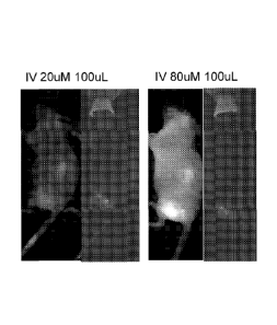
Figure 51 is a photomicrograph showing GFP fluorescent images of two tumors
cut in
half after 24 hours after iv (tail vein) injection of 200uL of 33 uM of GFP-
pHLIP. Tumors
were implanted by subcutaneous injections of human cervical cancer cells
(HeLa) into right
flank of athymic nude mice.
Figure 52 is a photomicrograph showing GFP fluorescent images of tumor and
kidney
cut in half after 24 hours after iv (tail vein) injection of 200uL of 33 uM of
GFP-pHLIP.
Tumor uptake of GFP-pHLIP is higher than kidney uptake. The average
fluorescence signal
in liver, kidney and tumor is 147.0 5.7, 201.5 12.0, 388.5 10.8, respectively,
which shows
that tumor uptake is higher than kidney and liver uptake.
Figure 53 is a series of line graphs showing the results of kinetics
experiments
performed with pHLIP-4, pHLIP-2, and pHLIP-1.
Figure 54 is a line graph showing the effect of biotin on peptide insertion
into the
membrane.
Figure 55 is a series of line graphs showing the effect of replacement of Asp
residues
with Glu in pHLIP variants. Fluorescence (a) and CD (b) spectra of three
states of pHLIP-2
and pHLIP-2E. Black ¨ State 1, Blue ¨ State II, Red ¨ State III.
Figure 56 is a series of line graphs showing the pH-dependences of insertion
into
membrane of pHLIP-2 and pHLIP-2E.
Figure 57 is a line graph showing PNA translocation by pHLIP peptides.
Figure 58 is a line graph showing the Effect of wt pHLIP-SMPT-amanitin on
cancer
cells: pH-dependent cell death.
Figure 59 is a line graph showing the electric Cell-substrate Impedance
Sensing
tECIS) assay: Kinetics of induction of cell death by pHLIP-SMPT-amanitin.
18
CA 02805387 2013-01-14
WO 2012/047354
PCT/US2011/043928
Figure 60 is a series of images showing pHLIP labeled with Alexa750
(covalently
attached to the N-terminus) and amanitin (attached by SMPT via S-S bond to the
C-terminus)
administered to the tumors of mice.
Figure 61 is a series of bar charts showing tumor/organ ratios calculated at 4
& 24 hs
post-injection.
Figure 62 is a bar chart showing normalized mean fluorescence of tumor and
organs
at 4 hours after injection of Var7 pHLIP.
Figure 63 is a bar chart showing normalized mean fluorescence of tumor and
organs
at 24 hours after injection of Var7 pHLIP.
Figure 64 is a bar chart showing Normalized mean fluorescence of tumor and
organs
at 4 hours after injection of Var3 pHLIP.
Figure 65 is a bar chart showing normalized mean fluorescence of tumor and
organs
at 24 hours after injection of Var3 pHLIP.
DETAILED DESCRIPTION OF THE INVENTION
The invention features diagnostic or therapeutic agents comprising improved
pHLIP
constructs that selectively deliver compositions to a diseased tissue compared
to non-diseased
tissue, thereby significantly improving diagnosis and treatment. A class of
delivery vehicles
based on pH-sensitive, water soluble membrane peptides, pHLIPs, that target
cells located in
an acidic microenvironment found in many diseased tissues, including tumors,
was
developed. Specific targeting by pHLIPs is achieved as a result of helix
formation and
membrane insertion. In contrast to the earlier technologies based on cell-
penetrating peptides
(CPPs), pHLIPs act as monomeric membrane-inserting peptides that translocate
one terminus
across a membrane into the cytoplasm, while the other terminus remains in the
extracellular
space, locating the peptide in the membrane lipid bilayer. pHLIP peptides
insert into a lipid
bilayer membrane at low pH but not at high pH (<7.0). Once inserted into the
membrane,
they can exit the membrane under conditions of high pH (e.g., a change in pH),
exiting from
the same side from which they entered. pHLIP peptides do not traverse the
membrane and
emerge in their entirety on the inside of the cell. Therefore, pHLIP has a
dual delivery
capability: it can tether cargo molecules or nanoparticles to the surfaces of
cells in diseased
tissues and/or it can move a cell-impermeable cargo molecule across the
membrane into the
cytoplasm. The source of energy for moving polar molecules attached to pHLIP
through the
19
CA 02805387 2013-01-14
WO 2012/047354 .
PCT/US2011/043928
hydrophobic layer of a membrane bilayer is the membrane-associated folding of
the
polypeptide. A drop in pH leads to the protonation of negatively charged
residues (Asp or
Glu), which enhances peptide hydrophobicity, increasing the affinity of the
peptide for the
lipid bilayer and triggering peptide folding and subsequent membrane
insertion. The process
is accompanied by the release of energy that is utilized to move cell-
impermeable cargo
across a membrane. pHLIP acts as a monomer in the following diagnostic and
therapeutic
applications: targeted therapy - selective delivery of therapeutic and imaging
agents to
diseased tissue, thereby increasing the effective concentration of these
agents and reducing
their accumulation in healthy tissue; improved route of drug administration:
agents with
improved pharmacokinetic properties of a drug; locally activated therapy -
activation of a
targeted therapeutic agent by local microenvironment of diseased tissue; fine
specificity -
cell-impermeable molecules translocated into cells only in diseased tissue
while not affecting
healthy cells; and multi-functionality - simultaneous targeted delivery of a
therapeutic agent
and an imaging probe to monitor drug distribution.
The compositions described herein are characterized by much higher efficacy
and/or
significantly reduced side effects compared to other cell-penetrating
contructs/carriers. Such
improvements are especially important for cancer treatment, since the majority
of anti-cancer
drugs are poisons that damage normal cells. Other diseased tissues are treated
using the same
compositions.
The challenge of selective delivery to tumors or other tissues characterized
by a pH
lower that physiological pH has been answered by the p1-LIP peptides and
constructs
described herein. Disease-specific delivery coupled with local activation
allows i)
accumulating and, therefore, increasing the effective concentration of
therapeutic or
diagnostic agents in a diseased area and ii) reducing the side effects
associated with treatment
by reducing the targeting of normal cells. Local activation further improves
the protection of
normal tissue.
Tissue acidosis
Hypoxia and acidosis are physiological markers of many diseased processes such
as a
cancer (Stubbs et al., 2000, Mol. Med. Today, 6, 15; Helmlinger et al., 2002,
Clin. Cancer
Res. 8, 1284; Izumi et al. 2003, Cancer Treat. Reviews. 29, 541); an
infarction (Graham et
al., 2004, J Exp Biol., 207, 3189; Yao and Haddad, 2004, Cell Calcium., 36,
247; Yamamoto
and Ehara, 2005, Am J Physiol Heart Circ Physiol., in press); a stroke
(Rehncrona 1985, Ann.
CA 02805387 2013-01-14
WO 2012/047354
PCT/US2011/043928
Emerg. Med. 14, 770; Siesjo et al., 1996, Adv. Neurol. 71, 209; Ying et al.,
1999, J.
Neurochem. 73, 1549); an atherosclerotic lesion (Leake 1997, Atherosclerosis,
129, 149); a
trauma (Mikhail, 1999, AACN Clin Issues, 10, 85; Clausen et al., 2005, J
Neurosurg, 103,
597); an inflammation (Kalantar-Zadeh et al., 2004, Semin Dial, 17, 455); an
infection
(Holloway et al., 1995; Exp Parasitol., 80, 624; Headley, 2003, Am Fam
Physician., 68, 323).
The compositions are useful for pH-selective delivery of molecules to diseased
tissue, e.g.,
tumors.
The most important limitation of specific cancer cell receptor targeting is
the
heterogeneity of human cancers. Recent studies of gene expression in cancer
cells indicate
that a number of genes are up- and down-regulated, and that cells in a tumor
are
heterogeneous. It is therefore problematic to rely on any single tumor
biomarker even for one
type of cancer. Using tumor acidity may be an alternative, since it is well
established that
salient features of the microenvironment of solid tum9rs include hypoxia and
extracellular
acidity. These factors contribute to the selection of the cancerous phenotype,
and also to the
progression from benign to malignant tumors. Acidosis is associated with tumor
development both at very early and at advanced stages. Rapidly proliferating
cancer cells
become partially anaerobic, leading to the elevation of glycolysis in response
to hypoxia
(Pasteur effect). Hypoxia and acidity are partly a result of the chaotic and
heterogeneous
microvasculature structure of solid tumors, where the oxygen concentration
decreases with
distance from a capillary. Hypoxia and low blood supply are involved in cancer
progression,
but they are not the only mechanism responsible for the development of an
acidic
environment within solid tumors. A hallmark of malignant cancers is an
elevated glucose
uptake even under normal oxygen conditions, known as "aerobic glycolysis" or
the Warburg
effect. Cells exhibiting a Warburg effect catabolize glucose at a high rate.
The consequence of glycolytic metabolism in any tissue is the formation of fr,
which
must be removed from the cell if the internal milieu is to maintain its normal
pH, because
many cellular processes have a narrow pH optimum. Four major types of
intracellular pH
(pHi) regulatory mechanisms have been identified in tumor cells: Neal+
exchangers,
bicarbonate transporters, proton-lactate symporters and proton pumps. These
transmembrane
proteins are ion pumps or ion exchangers that pump protons across the plasma
membrane
from the cytoplasm to the opposite site of the membrane, the extracellular
space or the lumen
of various organelles. A consequence of the activity of ion pumps is an
enhanced pH
21
CA 02805387 2013-01-14
WO 2012/047354
PCT/US2011/043928
gradient across the plasma membrane of cancer cells in comparison with normal
cells, and a
lower pH in the extracellular milieu.
Usually, exposure to an acidic environment results in cell death, however
cancer cells
adapt through resistance to apoptosis and up-regulation of membrane ion
channels in order to
maintain intracellular pH in the range of normality. Indeed, the unfavorable
environment
may favor ttimor cell survival in acidic conditions via selection of cells
that are resistant to
acid-induced cell toxicity and hypoxia-induced, p53-dependent apoptosis, and
promote
invasiveness by killing normal tissue cells. Malignant tumor cells not only
survive better in
acidic environments, but they also demonstrate phagocytotic and cannibalistic
behavior.
Extracellular acidification promotes cancer invasion and metastasis by
increased secretion
and activation of proteases, matrix metalloproteinases, bone morphogenetic
protein-1-type
metalloproteinases, tissue serine proteases, and adamalysin-related membrane
proteases.
Enhanced mutation rates, chromosomal instability, and spontaneous
transformation are
associated with acidity. Hypoxia and acidity also cause resistance to
radiotherapies and
chemotherapies, and promote the expression of the human multi-drug-resistance
protein.
Tumor acidity is an alternative targeting strategy to specific molecular
biomarkers for
tumor targeting and detection and is also useful for monitoring therapy
outcomes. For
example, the level of extracellular pH is related to the overall survival of
canines with
spontaneous sarcomas. Thus, the pH was predictive of a clinical outcome. The
advantages
of targeting acidity include its generality and the absence of tumor
heterogeneity issues.
Hydrophobicity and drug development
If the target of a therapeutic is cytoplasmic, the selective delivery of
therapeutics to a
tumor is not enough to improve treatment; the strategy must also enable the
agent to cross the
hydrophobic barrier of a cell membrane and release its payload inside cells.
The two major
mechanisms for the translocation of molecules and nanoparticles across the
membrane are
passive diffusion and endocytosis. Neither is specific for cancer cells, so
each would
promote translocation of therapeutics across the membranes of cells in both
diseased and
healthy tissues. In conventional drug design and discovery the Lipinski rules
of five are
widely used to guide molecular designs. The rules postulate that a successful
drug should be
hydrophobic and small in order to traverse membranes and reach cytoplasmic
targets (e.g. the
. logarithm of the octanol-water partition coefficient LogPo/w is -0.4 to +5.6
and the MW is
160 to 480 g.mo1-1). Drugs designed in this way will indiscriminately enter
all cells they
22
CA 02805387 2013-01-14
WO 2012/047354
PCT/US2011/043928
encounter, and are also likely to be substrates for efflux pumps that reduce
their efficacy. It is
important to note that the majority of inhibitors found for biological targets
located inside a
cell are molecules that cannot cross a membrane. Another large class of cell-
impermeable
functional molecules comprises gene regulation agents such as DNA, siRNA, and
PNA
(peptide nucleic acid. Gene-targeted therapies also involve passage through
the cell
membrane, which appears to be a general problem associated with that approach.
Cell-penetrating peptides have been used for the delivery of liposomes,
nanoparticles,
adenoviruses, and a variety of biological molecules into cells. Among these
peptides are
TAT, antennapedia, arginine-rich and others. In contrast to the pHLIP peptides
described
herein, these peptides enter the cell via "endocytic pathway. When taken up by
endocytosis,
molecules or nanoparticles are trapped in the lysosome compartment and need to
be released
into the cytoplasm.
Selective delivery and advantages of environmentally-sensitive conjugates
Diagnostic and treatment would be improved dramatically by improving the
selective
delivery of imaging and therapeutic agents to diseased tissue. Traditionally,
receptors and
enzymes overexpressed in cancer cells are considered as cancer biomarkers.
They are
indicators of the change in physiologic state during a disease process
(Srinivas et al., 2001,
2002; Hanke et al., 2004; Janssens et al., 2004; Kennedy and Hirsch, 2004).
There has been a
great deal of research into the development of peptides and antibody fragments
directed
toward cell surface receptors (Goldsmith, 1997; Freimark et al., 2007).
Initially, monoclonal
antibodies were the most promising candidates for specific targeting
strategies. However,
because of problems associated with their specificity and high molecular
weight, clinically
successful developments were difficult. Only over the last few years has
advanced antibody
engineering technology enabled therapeutic concepts based on antibodies and
conjugates
thereof to successfully enter clinical practice (Carter, 2001; Payne, 2003).
Antibodies and
their fragments have been used to map the expression or overexpression of
tumor-related
proteins, such as prostate-specific membrane antigen (Polascik et al., 1999),
human
epidermal growth factor receptor-2 (HER2) (Moasser, 2007); carcinoembryonic
antigen
(Hughes et al, 1997; Lu et al., 2007), TAG-72 (Muxi et al. 1999), Ep-CAM
(Breitz et al.,
1997; de Bono et al., 2004) and others. However, a number of complications
still vex
development of antibody applications, such as purity, immungenicity, slow
diffusion in
23
CA 02805387 2013-01-14
WO 2012/047354
PCT/US2011/043928
tissues, plasma clearance, and production difficulties (Blattler and Chari,
2001). An
attractive direction is the development of low molecular weight peptides for
rapid tumor
targeting. In contrast to antibodies, peptides can be easily synthesized,
modified and
stabilized to obtain optimized pharmacokinetic parameters (Lister-James et
al.: 1997;
Signore, 2001). Usually they are not immunogenic and have high receptor
affinity. The most
developed and widely accepted are somatastatin analogs introduced to visualize
various
somatostatin receptor-positive tumors (Buchsbaum, 2004; Krenning et al.,
2004).
Furthermore, a number of other peptides targeting various receptors expressed
in cancer cells
have recently been tested for tumor detection (Signore, 2001; Ma et at.,
2007).
An important limitation of approaches based on targeting specific cancer cell
receptors is the variability of cells in human cancers (Jeffrey et al., 2005).
Recent studies of
gene expression in cancer cells indicate that a number of genes are up- and
down-regulated,
so that cell surfaces in a tumor are heterogeneous. It is therefore
problematic to rely on any
single tumor biomarker even for one type of cancer (Bild et al., 2006). On the
other hand,
tumor acidity, which is a feature of most solid tumors, is a reliable cancer
biomarker that is
exploited by the compositions and methods described herein.
Several nano sized systems with pH-sensitive properties have been developed,
among
them are polymers, dendrimers, micelles, liposomes, and hydrogel nanoparticles
(Blume and
Cevc, 1990; Kobayashi et al, 2001; Lian and Ho, 2001; Portney and Ozkan, 2006;
and see
review by Ganta et al., 2008). The main feature of these nanocarriers is their
ability to
release encapsulated therapeutic and/or imaging agents in response to changes
in pH Bulmus
et al., 2003; Murthy et al., 2003; Na et al., 2003; Tomlinson et al., 2003;
Kamada et al., 2004;
Ulbrich et al., 2004; Simoes et al., 2004; Stayton et al., 2005; Henry et al.,
2006; Devalapally
et al., 2007). However, most such pH-sensitive carriers are used to enable
drug release in the
environment of endosomes and/or lysosomes after cellular uptake of the
conjugates by
endocytosis. A significant advantage of the compositions and methods described
is that they
do not rely on or involve endocytosis. An additional advantage is that little
or no
immunogenicity is associated with the compositions.
pHLIP peptide is monomeric
pHLIP peptides, e.g., (SEQ ID NO:2, shown in Figure 1) are a water-soluble
polypeptides based on the the bacteriorhodopsin C helix, which was found to
insert across a
membrane to form a stable transmembrane alpha helix. Peptide folding and
membrane
24
CA 02805387 2013-01-14
WO 2012/047354
PCT/US2011/043928
insertion are driven by a drop of pH from neutral or high (>7.4) to slightly
acidic (7.0-6.5 and
less) pHs. The apparent pK of insertion was found to be 6Ø pHLIP is a
monomer in each of
its three major states: unstructured and soluble in water (state I) at neutral
pH, unstructured
and bound to the surface of a membrane at neutral pH (state II), and inserted
across the
membrane as an a-helix at low pH (state III) (Figure 2). In contrast, all pore
forming
peptides, first form aggregates on the membrane surface and then "fall" into
membrane and
form pores. Thus, an additional advantage of the environmentally-sensitive
compositions is
their monomeric nature, e.g., they do not require assembly into a multimeric
suprastructure
like pore formers.
State II pHLIP peptides are particularly well suited for imaging uses, and
State III
pHLIP peptides are ideally suited for delivery of cargo molecules, e.g.,
toxins, across the cell
membrane and into the cytoplasm of cells. State III pHLIP peptides are
typically short
peptides. Within the state III class of pHLIP peptides, binding to the cell
membrane becomes
stronger as the pH goes down. However, the pHLIP peptides do not move entirely
across the
cell membrane to emerge on the other (inside) of the cell (i.e., the
cytoplasm). Rather the
membrane sequence of the peptide remains lodged in the cell membrane, unless
and until the
local pH is raised, e.g., above 7. Under high pH conditions, the pHLIP peptide
may exit the
membrane, but only in the direction from which it came.
Toxicity
Toxicity is one of the most critical issues in the selection of any delivery
agent. For
example, the use of pore-forming membrane peptides as delivery agents is
complicated by the
toxicity associated with the formation of pores in cellular membranes in vivo.
By contrast,
the interaction of pHLIP with liposomes and cellular membranes at both neutral
and low pHs
does not lead to membrane leakage, and no cellular toxicity was seen over a
range of peptide
concentrations. Also, mice receiving a high dose (about 5 mg/kg) of peptide
did not show
any adverse effects within two months after intravenous peptide
administration.
Selectivity of targeting
The pH-dependent interaction of pHLIP with membranes allows selectivity in the
targeting of acidic diseased tissue. As noted above, acidity and hypoxia are
considered as
universal cancer biomarkers, and pHLIP is used as an acidity-targeting probe.
Besides
cancer, many other pathological states, such as inflammation, ischemia,
stroke, arthritis and
others are characterized by acidity in the extracellular space, which may
broaden the potential
CA 02805387 2013-01-14
WO 2012/047354
PCT/US2011/043928
applications of pHLIP. In vivo fluorescence imaging in mice and rats
demonstrated that
pHLIP can target acidic tissues, such as kidneys, tumors of various sizes and
origins, and the
site of experimentally induced inflammatory arthritis. In addition to
fluorescence imaging,
PET (positron emission tomography) imaging of the acidic environment in human
prostate
tumors was performed using 64Cu-DOTA conjugated to pHLIP. PET studies
demonstrated
that the construct avidly accumulated in LNCaP and PC-3 tumors and that tumor
uptake
correlates with the differences in the bulk extracellular pH (pHe) measured by
MR
spectroscopy. Feeding animals with bicarbonated water, which increases tissue
pH, results in
a reduction of tumor targeting by pHLIP.
Molecular mechanism of pH-dependent membrane insertion of pHLIP
The putative transmembrane (TM) part of pHLIP peptide contains two Asp
residues
(Figure 1). At neutral pH these charged residues enhance peptide solubility
and serve as
anchors keeping the peptide at the surface of membrane, thereby preventing
pHLIP
partitioning into the hydrophobic membrane bilayer. A reduction of pH induces
protonation
of Asp residues, and as a result, the overall hydrophobicity of the peptide
increases,
enhancing the affinity of the peptide for the lipid bilayer core and
triggering peptide folding
and insertion. The replacement of the key Asp residues in by Lys, Ala or Asn
leads to the
loss of peptide of pH-dependent membrane insertion, as measured in liposomes,
red blood
cells and confirmed by in vivo fluorescence imaging. The K-pHLIP peptide,
where the two
Asp residues in the putative transmembrane region are replaced with Lys
residues, does not
demonstrate tumor targeting. The Ala substitutions give a peptide that
aggregates in solution,
while the Lys and Asn substitutions give peptides that are too polar to insert
either at neutral
or low pH. The replacement of one of the Asp residues in the TM part of the
peptide by a
Glu residue results in a shift of pH of membrane insertion from 6.0 to 6.5.
Replacement of
both Asp residues by Glu results in enhancement of peptide aggregation and
formation of
elements of secondary structure on the bilayer surface at neutral pH (see
Tables 1 and 2).
However, aggregation in solution is often concentration specific and
reversible, i.e., a pHLIP
peptide may exhibit aggregation in solution in vitro but the aggregation is
reversible after
administration to the subject due to dilution.
Data obtained on model systems (liposomes), cultured cells and mice confirmed
that
the mechanism of membrane entry of pHLIP is not mediated by endocytosis,
interactions
with cell receptors or pore formation; rather, the mechanism is the formation
of a helix across
26
CA 02805387 2013-01-14
WO 2012/047354
PCT/US2011/043928
the lipid bilayer, triggered by the increase of peptide hydrophobicity due to
the protonation of
negatively charged residues induced by low pH.
Solubility and stability of pHLIP in blood
Poor solubility due to aggregation is a typical property of membrane peptides,
which
has complicated studies and applications. pHLIP, as any membrane peptide, also
has a
tendency to aggregate, especially at high concentrations and/or low pH.
However, in aqueous
solution at neutral pH pHLIP exists as a monomer at concentrations less than
30 ii.g/mL (-7.0
04), as studied by fluorescence and CD spectroscopy measurements, size
exclusion
chromatography coupled with "on-line" laser light scattering, ultraviolet and
refractive index
detection (SEC-LS/UV/RI) and analytical ultracentrifugation experiments. When
the
solubility of the peptide is compromised as a result of mutations, the
affinity of the peptide
for a membrane and its overall conformational properties change. Thus, studies
were
undertaken to design pHLIP peptides that are optimized for clinical diagnostic
and
therapeutic use.
The oligomeric state of the peptide on the surface of a membrane (state II)
and
inserted into the lipid bilayer (state III) were evaluated by FRET performed
with two
different donor-acceptor probes attached to the N-terminus of the peptide. The
data
demonstrate that, at low concentrations, the peptide is monomeric in both
states II and III.
Peptide interactions with proteins, especially plasma proteins, and membranes
determine the pharmacokinetics of the peptide at neutral pH. pHLIP
demonstrates prolonged
circulation in the blood (several hours), which is consistent with its ability
to bind weakly to
membrane surfaces at neutral and high pH, preventing the rapid clearance by
the kidney
expected for a small, soluble peptide. pHLIP binding to membranes is driven by
hydrophobic
interactions. If the peptide sequence were made more hydrophobic, tighter
binding to red
blood cells and epithelial cells and more aggregation in solution, and slower
clearance and
reduced bioavailability would occur. Making the peptide less hydrophobic
accelerates
clearance and prevents the peptide from finding its targets. Therefore, fine
tuning of the
solubility is an important property to optimize pHLIP performance in vivo.
Another important property is the stability of peptides in the blood, since
proteases in
the serum can degrade peptides consisting of L-amino acids within minutes.
While
polypeptides made from D-amino acids are much more stable, they are often
unsuitable for
specific receptor binding applications as a consequence of their altered
chirality. Since the
27
CA 02805387 2013-01-14
WO 2012/047354
PCT/US2011/043928
mechanism of pHLIP involves relatively nonspecific interactions with a fluid
lipid bilayer,
pHLIP peptides composed of L- or D-amino acids demonstrate the same
biophysical and
tumor targeting properties. This observation adds to the evidence that the
pHLIP targeting
does not require any specific molecular binding event. The only conspicuous
difference is
that D-pHLIPs form left-handed helices across membranes rather than the right-
handed
. helices formed by L-pHLIPs.
Topology of membrane insertion
The topology of pHLIP insertion was probed using an NBD-dithionite quenching
assay, and then confirmed in experiments on cultured cells. NOB (4-ctiloro-7-
nitrobenz-2-
oxa-1,3-diazole) and IANBD (N,Ar-dimethyl-N-(iodoacety1)-N'-(7-nitrobenz-2-oxa-
1,3-
diazol-4-ypethylenediarnine) were covalently attached to the N- and the C-
terminus of
pHLIP peptides, respectively. Fluorescently labeled peptides were inserted
into the lipid
bilayer at low pH, and changes of fluorescence signal of NOB were measured
after
quenching by dithionite, a membrane-impermeable agent that abolishes NBD
fluorescence if
it contacts the dye. The data clearly indicate that the N-terminus of pHLIP
stays outside of
the bilayer, while the C-terminus inserts across the lipid bilayer at low pH.
Cargo translocation by pHL1P insertion
The partition of pHLIP into the outer leaflet of lipid bilayer at neutral pH
and the
folding/insertion at low pH are accompanied by the release of energy.
Fluorescence
spectroscopy, isothermal titration calorimetry and acid titration calorimetry
were used to
study the interactions of pHLIP with a POPC lipid bilayer and to calculate the
transition
energies between states. The Gibbs Free Energy of binding to a POPC surface
(state I - state
II transition) at 37 C is about -7 kcal/mol near neutral pH and the additional
free energy of
insertion and folding across a lipid bilayer at low pH (state II - state III
transition) is nearly -2
kcal/mol. The energy difference between state II and state III mediates the
movement of
cargo across the hydrophobic bilayer of membrane. By knowing this energy, it
is possible to
estimate the polarity of cargo molecules that can be translocated across a
membrane by
pHLIP: 2 kcal/mol might be enough to translocate molecules with LogPaw - -1.4
[AG = -RT
, where RT -0.6 kcal/mol at room temperature]. Thus, membrane-impermeable
cargo
molecules with LogP,õ in range of -0.5 to -3 are translocated (with
probability proportional
to the LogP,Aõ) across a membrane. If the cargo is released, as in delivery to
a cell using a
28
CA 02805387 2013-01-14
WO 2012/047354
PCT/US2011/043928
link that is broken in the cytoplasm, the progressive mass action will exploit
even a weakly
favorable energy balance to accumulate delivered cargo in a cell.
Kinetics of pHLIP insertion into membrane
While the equilibrium thermodynamics favor binding and insertion of pHLIP,
slow
kinetics could be limiting for in vivo use, since blood flow is very fast.
Kinetic studies of
pHLIP folding and insertion across a POPC lipid bilayer triggered by a pH drop
from 8.0 to
4.0 indicate that insertion takes 100 sec, with a rapid (0.1 sec) interfacial
helix formation
followed by insertion to give a transmembrane helix. In the case of a pH drop
to 6.0 the
insertion is slower, about 300 sec. However, data obtained on various pHLIP
variants show
that the process of insertion can be accelerated by 10-20 times and up to 100
times if acidic
residues are removed from the C-terminus of the peptide.
Dual delivery capability of pHLIP
pHLIP, in contrast to other cell-penetrating peptides, stays in the cellular
membrane
after insertion, translocating one end into cytoplasm and leaving the other
end in the
extracellular space. Therefore, the peptide possesses dual delivery
capabilities: it can tether
cargo molecules to the cell surface and/or it can inject and release cell-
impermeable cargo
molecules into the cytoplasm (Figure 3). In the first scenario, a cargo
molecule is attached to
the pHLIP N-terminus. External cargo of wide range of polarity and size is
reliably
transported. One of the applications is to deliver imaging probes to acidic
tissue, where they
will be stably tethered to the surfaces of cells. pHLIP sequences deliver and
tether various
nanoparticles to the surface of cancer cells. The second delivery capability
of pHLIP is based
on conjugation of cargo molecules to the C-terminus via a bond that is cleaved
in the
environment of the cytoplasm, such as a disulfide. Since the energy released
during peptide
folding and insertion across a membrane is limited, and since strongly polar
molecules will
reach equilibrium slowly, there may be a limit on cargo polarity and on size.
Preferably
polarity is in range of 0.5< LogP <-3. Cargo size is preferably less than 100
kDa. For
example, cargo size is in the range of 0.1- 50 IdDa, 1-25 kDa, and 1-10 kDa.
The data
indicated that cargo with a molecular mass of 5kDa is effectively
translocated/delivered
across a membrane by pHLIP, even taking into account the mass action effect
discussed
above.
pHLIP-mediated translocation of cell-impermeable functional cargos
29
CA 02805387 2013-01-14
WO 2012/047354
PCT/US2011/043928
pHLIP peptides translocate cargo molecules attached to their C-terminus.
Translocation is selective for low pH, and various types of cargo molecules
attached by
disulfides are subsequently released in the cytoplasm, including various
fluorescent dyes,
synthetic cyclic peptides, toxins and peptide nucleic acids (Reshetnyak et
al., 2006, PNAS
103:6460-465.). Cell-impermeable fluorescently-labeled toxin, phalloidin-
rhodamine,
conjugated to the C-terminus of pliLIP via an S-S- bond, is moved across the
membrane in a
pH-dependent manner. The pH-dependent translocation of the fluorescent
phalloidin by the
peptide was confirmed by fluorescence microscopy and fluorescence activated
cell sorting. If
phalloidin-rhodamine enters a cell, it binds tightly to actin filaments at
nanomolar
concentration (1(0= 40 nM) and strongly inhibits their depolymerization. Actin
filaments
stained with fluorescent phalloidin have an unmistakable filamentous pattern,
distinct from
the appearance of other cellular structures, organelles or membrane staining.
The phalloidin
translocated into the cytoplasm of live cells inhibits the proliferation,
contractility, migration
and division of cells. A long term effect of phalloidin is the formation of
multinucleated
cells, since nuclei can divide in treated cells, but the cell itself cannot.
This process leads to
the formation of multiple nuclei in one cell and eventual cell death.
Another example is the translocation of a class of cell-impermeable functional
cargo-
molecule, peptide nucleic acids (PNA), by pHLIP. PNAs can base pair
specifically to target
nucleic acid sequences, but lack the highly charged backbone of biological
nucleic acids, and
are therefore candidates for pHLIP delivery. In vitro studies show that PNA
can inhibit both
transcription and translation of genes to which it has been targeted, which
suggests use of
PNA in antigene and antisense therapy(Nelson, PE, 2005, Q. Rev. Biophys.
38:345-350).
However, a major obstacle has been the delivery of PNA (as well as RNA or ODN)
across
membranes into cells. pHLIP has been shown to translocate a fluorescence-
labeled 12 base
PNA into cells. Treatment of cells with PNA-rhodamine alone did not give
fluorescent
staining of cells at pH 6.5 or 7.4, but fluorescence was observed in cells
when the PNA was
linked to the C-terminus of pHLIP via a disulfide and added at pH 6.5. The
labeled cells
were alive by using the dead cell marker SYTOX-Green. Additional in vivo
studies showed
that pHLIP delivered an18-base PNA (MW 4.7 kDa) to a mouse tumor, translocated
PNA
across membranes, and activated luciferase expression in a result of splicing
correction.
pHLIP represents a new class of delivery agents
CA 02805387 2013-01-14
WO 2012/047354
PCT/US2011/043928
Numerous variants of the parent pHLIP sequence (SEQ ID NO:1) have been made.
The base refernence sequence has three main blocks (Figure 1). The middle part
is a
moderately polar sequence that contains protonatable residues, and is the
environment-
sensitive, membrane inserting part of the peptide. The transmembrane helix can
be as short
as 15 residues. The membrane-inserting sequences is at least 8 amino acids,
e.g., it ranges in
size from 13 to 25 amino acids. The other two blocks are the two flanking
sequences - a first
flanking sequence on the C-terminal side and a second flanking sequence on the
N-terminal
side. The role of the flanking sequences is to modulate the peptide
solubility, but may also
include functional motifs; e.g., protease cleavage or receptor binding
sequences, or amino
acids that can undergo phosphorylation in the cytoplasm. There are several
restrictions
applied to the flanking sequence that inserts across the membrane: i) it
should not contain
many charged residues (especially positive charges); ii) it should not be
long; iii) the speed
and cooperativity of peptide insertion into membrane depends on the number of
Asp or Glu
residues present in this sequence.
There is no specific restriction to the flanking sequence that stays outside
of
membrane other than its role in peptide solubility; however a danger is that
dramatic changes
of peptide pharmacokinetics might result from extreme variations. Thus,
numerous
sequences have been made and tested. The results of testing led to the
definition of key
elements of pHLIP sdquences that make them suitable for diagnostic and
therapeutic
applications. The membrane-inserting sequence of pHLIP does not contain Cys or
Lys
residues. Any of these residues or both could be placed at the N- or C-
terminus of the
peptide for the purpose of cargo conjugation to pHLIP (NHS and maleimide click
chemistry
is developed very well and widely used for conjugation purposes).
pHLIP sequences
Tables 1-2 provide a summary of pHLIP sequences that have been made and
tested.
Table 1 includes long pHLIP sequences. The sequences of Table 1, if they
insert into a
membrane, go with their C-terminus across a membrane and leave N-terminus in
the
extracellular space. Some of the peptides lost main properties of pH-dependent
insertion into
membrane.
31
CA 02805387 2013-01-14
WO 2012/047354
PCT/US2011/043928
TABLE 1
Name Sequence
WT-1 GGEQNPIYWARYADWLFTTPLLLLDLALLVDADEGT SEQ. ID NO. 3
WT-2 AEQNPIYWARYADWLFTTPLLLLDLALLVDADEGT SEQ. ID NO. 4
WT-Cysl AAEQNPIYWARYADWLFTTPLLLLDLALLVDADEGTCG SEQ. ID NO. 5
WT-Cys2 Ac-AEQNPIYWARYADWLFTTPLLLLDLALLVDADEGCT SEQ ID NO: 274
WT-Cys3 GGEQNPIYWARYADWLFTTPLLLLDLALLVDADEGTCG SEQ. ID NO. 6
Cys-WT1 Ac-ACEQNPIYWARYADWLFTTPLLLLDLALLVDADEGTG SEQ. ID NO. 7
VarO-NT ACEQNPIYWARYADWLFTTPLLLLDLALLVDADEGT SEQ. ID NO. 8
Lys-WT1 AKEQNPIYWARYADWLFTTPLLLLDLALLVDADEGT SEQ. ID NO. 9
Lys-WT2 Ac-AKEQNPIYWARYADWLFTTPLLLLDLALLVDADEGTG SEQ ID NO: 275
WT-KC AAEQNPIYWARYADWLFTTPLLLLDLALLVDADEGTKCG SEQ. ID NO. 10
K-WT-C AKEQNPIYWARYADWLFTTPLLLLDLALLVDADECT SEQ. ID NO. 11
N-pHLIP ACEQNPIYWAI2YANWLFTTPLLUNLALLVDADEGTG SEQ. ID NO. 12
N-pHLIP-b ACEQNPIYWARYANWLFTTPLLLLNLALLVDADEGT SEQ ID NO: 276
K-pHLIP ACEQNPIYWARYAKWLFTTPLLLLKLALLVDADEGTG SEQ. ID NO. 13
NNQ GGEQNPIYWARYADWLFTTPLLLLDLALLVNANQGT SEQ. ID NO. 14
D25A AAEQNPIYWARYADWLFTTPLLLLALALLVDADEGT SEQ. ID NO. 15
D25A-KC Ac-AAEQNPIYWARYADWLFTTPLLLLELALLVDADEGTKCG SEQ ID NO: 277
D14A AREQNPIYWARYAAWLFTTPLLUDLALLVDADEGT SEQ. ID NO. 16
P20A AREQNPIYWARYADWLFTTALLLLDLALLVDADECT SEQ. ID NO. 17
025E AAEQNPIYWARYADWLFTTPLLLLELALLVDADEGT SEQ. ID NO. 18
D14E AAEQNPIYWARYAEWLFTTPLLLLDLALLVDADEGT SEQ. ID NO. 19
3D AAEQNPIIYWARYADWLFTDLPLLLLDLLALLVDADEGT SEQ. ID NO. 20
R11Q GEONPIYWAQYADWLFTTPLULDLALLVDADEGTCG SEQ. ID NO. 21
D25Up GGEQNPIYWARYADWLFTTPLLLDLLALLVDADEGTCG SEQ. ID NO. 22
D2500wn GGEQNPIYWARYADWLFTTPLLLLLDALLVDADEGTCG SEQ. ID NO. 23
32
CA 02805387 2013-01-14
WO 2012/047354
PCT/US2011/043928
D14Up GGEQNPIYWARYDAWLFTTPLLLLDLALLVDADEGTCG SEQ. ID NO. 24
D14Down GGEQNPIYWARYAWDLFTTPLLLLDLALLVDADEGTCG SEQ. ID NO. 25
P2OG AAEQNPIYWARYADWLFTTGLLLLDLALLVDADEGT SEQ. ID NO. 26
Hl-Cys DDDEDNPIYWARYADWLFTTPLLLLHGALLVDADECT SEQ. ID NO. 27
H1 DDDEDNPIYWARYADWLFTTPLLLLHGALLVDADET SEQ ID NO: 278
H2-Cys DDDEDNPIYWARYAHWLFTTPLLLLHGALLVDADEGCT SEQ. ID NO. 28
Cys-H2 CDDDEDNPIYWARYAHWLFTTPLLLLHGALLVDADET SEQ ID NO: 279
H2 DDDEDNPIYWARYAHWLFTTPLLLLHGALLVDADEGT SEQ ID NO: 280
H2N-Cys DDDEDNPIYWARYAHWLFTTPLLLLHGALLVNADECT SEQ. ID NO. 29
H2N DDDEDNPIYWARYAHWLFTTPLLLLHGALLVNADEGT SEQ ID NO: 281
H2N2-Cys DDDEDNPIYWARYAHWLFTTPLLLLHGALLVNANECT SEQ. ID NO. 30
H2N2 DDDEDNPIYWARYAHWLFTTPLLLLHGALLVNANEGT SEQ ID NO:282
la-Trp AEQNPIYWARYADFLFTTPLLLLDLALLVDADET SEQ. ID NO. 31
lb-Trp AEQNPIYFARYADWLFTTPLLLLDLALLVDADEGT SEQ. ID NO. 32
lc-Trp AEQNPIYFARYADFLFTTPLLLLDLALLWDADET SEQ. ID NO. 33
Fast-1 or Van l AKEDQNPYWARYADWLFTTPLLLLDLALLVDG SEQ. ID NO. 34
Var1-2D1D ACEDQNPYWARYADWLFTTPLLLLDLALLVDG SEQ. ID NO. 35
Fastl-Cys or Var1-2D1D-Cys AEDQNPYWARYADWLFTTPLLLLDLALLVDCG SEQ. ID NO. 36
Fastl-E-Cys or VarlE AEDQNPYWARYADWLFTTPLLLLELALLVECG SEQ. ID NO. 37 .
Fastl-E-Lys AKEDQNDPYWARYADWLFTTPLLLLDLALLVG SEQ ID NO: 283
Fast2 or Var2 AKEDQNPYWRAYADLFTPLTLLDLLALWDG SEQ. ID NO. 38
Fast2-E-Cys or Var2E AEDQNPYWARYADWLFTTPLLLLELALLVCG SEQ ID NO: 284
Var2-2D1D ACEDQNPYWRAYADLFTPLTLLDLLALWDG SEQ. ID NO. 39
Var3-3D ACDDQNPWRAYLDLLFPTDTLLLDLLW SEQ. ID NO. 40
Var3-3D-cys AKDDQNPWRAYLDLLFPTDTLLLDLLWC SEQ ID NO: 285
Var4-3E ACEEQNPWRAYLELLFPTETLLLELLw SEQ ID NO: 286
Var5-3Da ACDDQNPWARYLDWLFPTDTLLLDL SEQ ID NO: 287
33
CA 02805387 2013-01-14
WO 2012/047354
PCT/US2011/043928
Var6-3Db CDNNNPWRAYLDLLFPTDTLLLDW SEQ ID NO:
288
Var8-3Eb CEEQQPWAQYLELLFPTETLLLEW SEQ ID NO:
289
Var9-3Ec CEEQQPWRAYLELLFPTETLLLEW SEQ ID NO:
290
Var15-2N CDDDDDNPNYWARYANWLFTTPLLLLNGALLVEAEET SEQ ID NO:
291
Var16-2P CDDDDDNPNYWARYAPWLFTTPLLLLPGALLVEAEE SEQ ID NO:
292
Table 2
Name Sequence
Var14-Rev Ac-TEDADVLLALDLLLLPTTFLWDAYRAWYPNQECA-Am SEQ. ID NO.
41
Sh AEQNPIYWARYADWLFTTPL SEQ. ID NO.
42
Sh-Cys AEQNPIYWARYADWLFTTPCL SEQ. ID NO.
43
Cys-Sh ACEQNPIYWARYADWLFTTPL SEQ. ID NO.
44
Sh-lTrp AEQNPIYFARYADWLFTTPL SEQ. ID NO.
45
Sh-W2 AEQNPIYFARYADLLFPTTLAW SEQ ID NO:
293
Sh-W1 AEQNPIYWARYADLLFPTTLAF SEQ ID NO:
294
Sh-2W AEQNPIYWARYADLLFPTTLAW SEQ ID NO:
295
Sh-1D KEDQNPWARYADLLFPTTLAW SEQ. ID NO.
46
Sh-lDb KEDQNPWARYADLLFPTTLW SEQ ID NO:
296
Var12-1D ACEDQNPWARYADLLFPTTLAW SEQ. ID NO.
47
Var10-2D ACEDQNPWARYADWLFPTTLLLLD SEQ. ID NO.
48
Var13-1E ACEEQNPWARYAELLFPTTLAW SEQ. ID NO.
49
Var11-2E ACEEQNPWARYAEWLFPTTLLLLE SEQ. ID NO.
50
Var7-3E ACEEQNPWARYLEWLFPTETLLLEL SEQ. ID NO.
51
Var7-3Eb ACEEQNPQAEYAEWLFPTTLLLLE SEQ ID NO:
297
AAEEQNPWARYLEWLFPTETLLLEL
Var7-K SEQ ID NO:
298
AKEEQNPWARYLEWLFPTETLLLEL
Var7-A SEQ ID NO:
299
34
CA 02805387 2013-01-14
WO 2012/047354 PCT/US2011/043928
Ac means Acetylated N-terminus (peptide does not comprise free terminal NH4
group)
Am means Amidated C-terminus (peptide does not comprise a free terminal COOH
group)
In SEQ ID NO: 298 and 299, Cys was replaced by Ala and Lys, respectively, for
different chemical schemes of chelate conjugation.
Table 2 includes sequences termed short and medium pHLIP sequences. They all
insert
in membrane in a pH-dependent manner, while they do not have C-terminal
flanking sequence.
Double underline indicates residues (Cys or Lys), which are used to conjugate
pHLIPs with
cargo molecules. Translocation means translocation of cargo across membrane of
liposomes
and/or cells. Imaging means whole-body in vivo imaging on mice.
Biophysical studies were carried out with the following short pHLIP peptides:
AEQNPIYFARYADLLFPTTLAW (Short-W2) (SEQ ID NO: 218)
AEQNPIYWARYADLLF111-1LAF (Short-W1) (SEQ ID NO: 219)
AEQNPIYWARYADLLFPTTLAW (Short-2W) (SEQ ID NO: 220)
All peptides were directly dissolved in buffer (pH8) and used for experiments
using 50 nm
liposomes. The concentration of peptide was 5 M, ,and the POPC concentration
was 1mM.
POPC blank was subtracted for CD and FL. Circular dichroism (CD) and
fluorescence spectra
showed insertion into lipid bilayers and formation of 3/10 or mixture of alpha
and 3/10 helices at
low pH. These data indicate that the short pHLIP peptides interact with lipid
bilayers and cell
membranes in a pH-dependent manner.
Table 3 shows salvation free energies of naturally-occurring amino acids.
Table 3: Solvation Free Energies of the Side Chains (X) of the 20 Natural
Amino Acids in AcWL-X-LL and Ac-X-Arnide
mole fraction Flory-Huggins'
residue" charge A GZ'a Arkis AG" .GXG
Ala +0.65 +0.81 +0.42 +0.13 +0.69 +0.99
Arg -0.66 -0.47 -1.37 +1.44 +1.81
Asn +0.30 +0.32 -0.79 +1.06 +1.10
Asp 0 +0.72 +0.75 +1.33 +1.39
Asp -1 -2.49 -2.46 -2.46 (-L05) -3.50 -1.88 -
1.83
Cys +1.17 +1.39 +1.39 (+2.10)
+1.72 +2.14
Gln +0.38 +0,50 -0.30 +1.66 +I 90
Glu 0 +1.04 +1.17 +2 19 +2.44
CA 028 0538 7 2013-01-14
WO 2012/047354 PCT/US2011/043928
Via EFS
Date of Deposit: July 13, 2011
Glu -1 -2.480 -2.35 -2.35 (- -3.12 -1.33 -1.08
GLY* 0 0.87) 0" 0. 0" oh
His +1 -1.18 -0.96 +0.24 +0.68
His 0 +1.04 +1.27 +0.18 +0.16 +2.46 +2.90
Ile +2.27 +2.70 +2.46 +3.72 +4.56
Leu +2.40 +2.77 +2.30 +420 +4.92
Lys +1 -1.65 -1,39 -1.35 +0.17 +0.67
Met +1.82 +2.18 +1.68 +3.45 +4.14
Phe +2.86 +3.24 +2.44 +2.19 +4.96 +5.71
Pro +1.01 - +1.35 +0.67 +0.29 +1.59 +2.25
Ser +0.69 +0.74 -0.05 +0.78 +0.89
Thr +0.90 +1.08 +0.35 +1.58 +1.93
Trp +3.24 +3.62 +3.07 +2.52 +6.15 +6.88
Tyr +1.86 +2.21 +1.31 +4.08 +4.75
Val +1.61 +1.99 +1.66 +2.86 +3.61.
Residue solvation free energies of the 20 natural amino acids relative to
glycine calculated from the data in Table I. Free
energies were corrected for the occlusion of neighboring residue areas (see
text) and for the anomalous properties of glycine (see
text). Residue solvation free energies calculated with mole-fraction units.
Residue solvation free energy calculated with the Flory-
_
Huggins correction (Sharp et al.. 1991; De Young & Dill, 1990) (see Appendix).
Constituent molar volumes were taken from
Makhatadze et. al. (1990). 6 Residue solvation free energies for the X residue
in the context of a AcWL-X-LL peptide calculated
from the free energies in Table I using the virtual glycine (GLY*) as the
reference (see text). AUK.' = AGWIALL AG-wLG=LL
Aur,pAilhõõ where Ahõ,(X) = ATõp(WLXLL) - Axõ,(WI.XLL). These "corrected"
values account for X-dependent changes in the
nonpolar ASA of the host peptide. Values for Arg and Lys were calculated from
experimental free energies measured at pH 1
where the ionic interaction between the side chain and carboxyl group does not
occur. AGT is the best estimate of the solvation
energy of residues occluded by neighboring residues of moderate size. '
Residue solvation free energies for the X residue in the
context of a AcGG-X-GG peptide calculated from AG',,' and the data in Table I.
AG',"(."' = AGV' + 22.8AAx where AAx =
,4,,p(WLXLL) - Ax,p(GGXCG). This additional correction accounts for occlusion
of the guest residue by the host (see text). AG,0.0
is the best estimate of the solvation energy of the fully exposed
residue..1Modified Fauchere and PliAa (1983) solvation energies,
relative to Gly, for the transfer of acetyl amino acid amides from n-octanol
to unbuffered aqueous phase. In this modified scale,
the original values of FP for Asp, Glu. and Cys have been replaced by the
AC.A.G in the left-hand adjacent column (see text).
The original values of FP for Asp. Glu, and Cys are shown in parentheses. g
Residue solvation free energies relative, relative to
Gly, for the transfer of AcA-X-Ailitu tripeptides from n-octanol to buffer. pH
7.2. Data are those of Kim and Szoka (1992). "
Reference state is the experimentally determined Gly value rather than GLY*.
36
CA 02805387 2013-01-14
WO 2012/047354
PCT/US2011/043928
Tabled.
Coded and Non-Coded Amino Adds
no. abbrev names
1 Ala Alanine
2 Arg Arglnine
3 Asn Asparagines
4 Asp aspartic acid
Cys Cysteine
6 Gin Glutamine
7 Glu glutamic acid
8 Gly Glycine
9 His Histidine
Ile Isoleucine
11 Leu Leucine
12 Lys Lysine
13 Met Methionine
14 Phe Phenyl- alanine
Pro Proline
16 Ser Serine
17 Thr Threonine
18 Trp Tryptophan
19 Tyr Tyrosine
Val Valine
21 -Acpa Aminocap- rylic acid
22 Aecys (5)-2- aminoethyl-Lcysteine=HCI
23 Afa aminophenyla cetate
24 Aiba -aminoiso- bytyric acid
Aile Alloisoleucine
26 AIg L-allylglycine
CA 02805387 2013-01-14
WO 2012/047354
PCT/US2011/043928
27 Aba amlnobutyric acid
28 Aphe p- aminophenyla lanine
29 Bat -alanine
30 Brphe p- bromophenyla lanine
31 Cha cyclohexylala nine
32 Cit Citrulline
33 aala -chloroalanine
34 Cie Cycioleucine
35 aphe p- chiorophenyla lanine
36 Cya cysteic acid
37 Dab 2,4-diamino- butyric acid
38 Dap 2,3- diaminopropionic acid
39 Dhp 3,4-dehydro- proline
40 Dhphe 3,4-, dihydroxy-phenyl-
Alanine
41 Fphe p- fluorophenylal anine
42 Gaa D-glucose- aminic acid
43 Hag Homo- arginine
44 Hlys hydroxyl- lysine=FICI
45 Hnvl DL-hydrox- ynorvaline
46 Hog Homoglut- amine
47 'Hoph homophenylal anine
48 Has Homoserine
49 Hpr hydroxyl- proline
50 Iphe p-lodopheny- lalanine
51 Ise Isoserine
52 Mle -methyl- leucine
-53 Msmet DL- methionine-s-methylsulfo-niumchloride
-54 1Nala 3-(1- naphthyl)alani ne
55 2Nala 3-(2- naphthyl)alani ne
38
CA 02805387 2013-01-14
WO 2012/047354
PCT/US2011/043928
56 Nle norieucine (or 2-aminohexanoic acid)
57 Nmala N-methyl- alanine
58 Nva norvaline (or 2-
aminopentanoic acid)
59 Obser 0- benzylserine
60 Obtyr 0-benzyl- tyrosine
61 Oetyr 0- ethyltyrosine
62 Omser 0- methylserine
63 Omthr 0-methyt- hreonine
64 Omtyr 0-methyl- tyrosine
65 Orn Ornithine
66 Pen Penicillamlne
67 Pga pyroglutamic acid
68 Pip pipecolic acid
69 Sar Sarcosine
70 Tfa 3,3,3- trifluoroalanine
71 Thphe 6- hydroxydopa
72 Vig L-vinylglycine
73 Aaspa (-)-(2R)-2- amino-3-(2- aminoethylsuffonyl)propa noic acid
dihydrochloride
74 Ahdna (25)-2- amino-9- hydroxy-4,7- dioxanonanolc acid
75 Ahoha (25)-2- amino-6- hydroxy-4- oxahexanoic acid
76 Ahsopa (-)-(2R)-2- amino-3-(2- hydroxyethyls ulfonyl)propa noic acid
A "conservative amino acid substitution" is one in which the amino acid
residue is
replaced with an amino acid residue having a similar side chain. Families of
amino acid
residues having similar side chains have been defined in the art. These
families include amino
acids with basic side chains (e.g., lysine, arginine, histidine), acidic side
chains (e.g., aspartic
acid, glutamic acid), uncharged polar side chains (e.g., glycine, asparagine,
glutamine, serine,
threonine, tyrosine, cysteine), nonpolar side chains (e.g., alanine, valine,
leucine, isoleucine,
proline, phenylalanine, methionine, tryptophan), beta-branched side chains
(e.g., threonine,
valine, isoleucine) and aromatic side chains (e.g., tyrosine, phenylalanine,
tryptophan,
39
CA 02805387 2013-01-14
WO 2012/047354
PCT/US2011/043928
histidine). Thus, a residue in a pHLIP sequence (corresponding to a location
relative to SEQ
ID NO:1) is replaced with another amino acid residue from the same side chain
family.
Physical properties of the membrane influence the insertion of pHLIP
pHLIP interaction with a cell membrane or lipid bilayer is affected by
membrane lipid
composition. The cell membrane composition of normal cells is different from
that of
malignant cancer cells (or cells in tissue related to other pathological
states). Thus, pHLIP
binds differently to the plasma membrane of cells in a healthy compared to a
diseased area, or
interacts differently with lipid bilayer of membrane of various cellular
compartments.
The cell plasma membrane is a complex medium that contains a high variety of
lipid
types. Studies were undertaken to understand whether the properties of pHLIP
are maintained
for bilayers composed of different lipids, e.g., whether the physical
properties of the bilayer
can alter the insertion of pHLIP. The conformation and membrane insertion
properties of
pHLIP were tested for a series of phosphatydylcholine (PC) lipids (the most
abundant lipid
type in human cells) of different length (X:1-PC; where Xis the acyl chain
length, ranging
from 14 to 22 carbons, and :1 marks the presence of a single unsaturation per
acyl chain).
The results show that the secondary structure of pHLIP for state II is highly
sensitive
to the acyl chain length (the amount of secondary structure is higher for
lipids of longer acyl
chain), while the changes observed for state III are smaller. To study the
contribution of alpha
helix formation to this phenomenon, P2OG pHLIP mutant was used. P2OG pHLIP
contains a
higher level of secondary structure for both state II and HI, but reacts in a
similar fashion to
changes in the lipid length.
The insertion pKa was also observed to be greatly influenced by the acyl chain
length
for both wt and P2OG pHLIP. High pKa values were determined for short lipids,
which
decreased in a linear fashion till a minimum pKa value was obtained for 20:1-
PC. These
results show that the acyl chain length controls both the conformation of
state II and the
insertion pKa, and that a more helical peptide inserts into the membrane at
more
physiological pHs.
Cells (but not artificial vesicles) demonstrate a lock phenomenon in which
a
pHLIP peptide is biased to tight binding once it has associated with a cell
membrane. This
situation is due to the an environment of a pH of near 7 inside the cell
(cytoplasm) and an
acidic pH outside of a cell in a diseased tissue. Once a pHLIP peptide or
peptide portion of a
pHLIP conjugate is inserted into the cell membrane, it is "locked" or
"stapled" there due to
CA 02805387 2013-01-14
WO 2012/047354
PCT/US2011/043928
the pH differential. This phenomenon leads to a persistence effect; a C-
terminal flanking
group enhances the persistence of binding. The phenomenon does not occur with
artificial
vesicles, because the pH on the inside and the outside of such constructs is
the same or nearly
so. This pH lock effect is particularly important for membrane-inserting
peptides, e.g., tumor
targeting pHLIP peptides, as it mediates persistence or the ability of the
pHLIP peptide to
stay in the cancer cell.
The effects of cholesterol on pHLIP's biophysical properties.
As complement to the studies of pHLIP insertion into lipids of different acyl
chain
length, the effect of cholesterol was examined. Since cholesterol is an
essential component of
many biological membranes, it was relevant to examine pHLIP's biophysical
interactions
relation to cholesterol as a membrane component. Liposomes of both different
acyl chain
length and different cholesterol concentration were made. The results
indicated that
increasing cholesterol content lowers the pK of insertion for all lipid types
examined and
raises the tryptophan fluorescence lambda max for pHLIP's membrane surface-
bound state,
State II.
Unilaminar vesicles were made with POPC lipids or single monounsaturated
phosphatidylcholine lipids of 14, 16, 18, or 20 acyl chain lengths. Different
samples of these
lipids were made with 0, 10, 20, or 30 percent cholesterol. After the addition
of pHLIP, a
panel of samples was made with each sample adjusted to a different pH between
4.0 and 8Ø
The resulting tryptophan fluorescence curves generated from each sample in the
panel were
examined to determine pHLIP's insertion pK. pKa of insertion of pHLIP
decreased with the
addition of cholesterol.
After the addition of pHLIP to the same unilaminar vesicles as described above
(made
with POPC lipids or single monounsaturated phosphatidylcholine lipids of 14,
16, 18, or 20
acyl chain lengths with varying amounts (0, 10, 20, or 30 percent) of
cholesterol, a panel of
samples was created with each sample adjusted to a different pH in the range
between 7.0 and
7.5. The tryptophan fluorescence curves taken from each sample were fit using
PFAST to
determine the lambda max. State II Lambda Max was found to increase with
cholesterol for
all lipid types.
Redirecting the immune system towards cancer using pHLIP
Cancer cells are preferentially labeled with small molecules that recruit
antibodies
already present in the human bloodstream and redirect immune responses,
resulting in
41
CA 02805387 2013-01-14
WO 2012/047354
PCT/US2011/043928
targeted cell cytotoxicity. To achieve this goal, a small molecule such as 2,4-
dinitrophenyl
(DNP) group in conjugation with pHLIP is used as an antigen. pHLIP is
conjugated to the
immune active molecule (DNP), and DNP-pHLIP interaction with cells results in
a pH-
dependent cytotoxicity.
Anti-DNP antibodies are not only already found in the human bloodstream in a
high
percentage of the human population but also capable of redirecting immune
responses such as
complement dependent cytotoxicity (CDC) and antibody-dependent cell-mediated
cytotoxicity (ADCC). An exemplary class of small molecules containing DNP is
called
ARM-Ps (antibody-recruiting molecules targeting prostate cancer). These
molecules are
capable of enhancing antibody-mediated immune recognition of prostate cancer
cells.
(Murelli et al., 2009, J Am Chem Soc 131, 17090-17092).
Such DNP molecules conjugated to a N-terminal cysteine on pHLIP recruited anti-
DNP antibodies to cultured HeLa cells in a pH-dependent manner. Between two
and four
times more recruitment was observed at pH 6.2 than at pH 7.4. When the cells
are treated
with DNP in conjugation with the D25E pHLIP peptide (a variant with a pK of
insertion of
about 6.5), between 20 and 50 times more recruitment was seen at pH 6.2 than
at pH 7.4. In
each case, pHLIP alone was not capable of significantly recruiting anti-DNP
antibodies to the
surface of cells. Similar results have been obtained with PC-3 prostate cancer
cells.
D-amino acids
Of the standard a-amino acids, all but glycine can exist in either of two
optical
isomers, called L or D amino acids, which are mirror images of each other.
While L-amino
acids represent all of the amino acids found in proteins during translation in
the ribosome, D-
amino acids are found in some proteins produced by enzyme posttranslational
modifications
after translation and translocation to the endoplasmic reticulum. D amino
acids are abundant
components of the peptidoglycan cell walls of bacteria, and D-serine acts as a
neurotransmitter in the brain. The L and D convention for amino acid
configuration refers
not to the optical activity of the amino acid itself, but rather to the
optical activity of the
isomer of glyceraldehyde from which that amino acid can be synthesized (D-
glyceraldehyde
is dextrorotary; L-glyceraldehyde is levorotary).
pHLIP peptides either fully or partially built of D-amino acids possess
advantages
over L-pHLIP peptides. For example, D-pHLIP peptides are biodegraded slower
than their
levorotary counterparts leading to enhanced activity and longer biological
half lives (Sela and
42
CA 02805387 2013-01-14
WO 2012/047354
PCT/US2011/043928
Zisman, 1997 FASEB J, 11: 449-456, incorporated herein by reference). Thus,
the invention
provides for the use of D-pHLIP peptides in the methods described herein. For
example,
pHLIP peptides comprise solely L-amino acids or solely D-amino acids, or a
combination of
both D-amino acids and L-amino acids.
Isotopes
pHLIP peptides optionally contain radioactive elements or stable isotopes, or
a
combination of both. Stable isotopes are chemical isotopes that may or may not
be
radioactive, but if radioactive, have half lives too long to be measured.
Different isotopes of
the same element (whether stable or unstable) have nearly the same chemical
characteristics
and therefore behave almost identically in biology (a notable exception is the
isotopes of
hydrogen). The mass differences, due to a difference in the number of
neutrons, will result in
partial separation of the light isotopes from the heavy isotopes during
chemical reactions and
during physical processes such as diffusion and vaporization. This process is
called isotope
fractionation. Examples of stable isotopes include oxygen, carbon, nitrogen,
hydrogen and
sulfur. Heavier stable isotopes include iron, copper, zinc, and molybdenum.
Gamma cameras are used in e.g. scintigraphy, SPECT and PET to detect regions
of
biologic activity that may be associated with disease. Relatively short lived
isotope, such as
1231 is administered to the patient.
= Scintigraphy ("scint") is a form of diagnostic test wherein radioisotopes
are taken
internally, for example intravenously or orally. Then, gamma cameras capture
and form two-
dimensional images from the radiation emitted by the radiopharmaceuticals.
SPECT is a 3D tomographic technique that uses gamma camera data from many
projections and can be reconstructed in different planes. A dual detector head
gamma camera
combined with a CT scanner, which provides localization of functional SPECT
data, is
termed a SPECT/CT camera, and has shown utility in advancing the field of
molecular
imaging. In SPECT imaging, the patient is injected with a radioisotope, most
commonly
Thallium 201TI, Technetium 99mTC, Iodine 1231, and Gallium 67Ga
Positron emission tomography (PET) uses coincidence detection to image
functional
processes. Short-lived positron emitting isotope, such as 18F, is incorporated
with an organic
substance such as glucose, creating F18-fluorodeoxyglucose, which can be used
as a marker
of metabolic utilization. Images of activity distribution throughout the body
can show rapidly
43
CA 02805387 2013-01-14
WO 2012/047354
PCT/US2011/043928
growing tissue, like tumor, metastasis, or infection. PET images can be viewed
in comparison
to computed tomography scans to determine an anatomic correlate. Other
radioisotopes used
in nuclear medicine thallium-201, tellurium-123, cadmium-113, cobalt-60, and
strontium-82.
Example 1: Measuring tumor aggressiveness and targeting metastatic lesions
with
fluorescent pHLIP
Malignant cancers exhibit an elevated uptake of glucose that leads to tumor
acidosis
from the Pasteur and Warburg effects. Glucose uptake and acidosis show a
positive
correlation with a tumor's aggressiveness and metastatic potential. Therefore,
extracellular
acidity may be a useful biomarker to evaluate the prognosis of tumor
development. pHLIP
sequences are a water-soluble membrane peptide that inserts and folds across a
cellular
membrane lipid bilayer in response to low pH. Membrane-associated folding of
pHLIP
occurs within seconds and is accompanied by a release of energy (about 2
keaVmol) that can
be used to target acidic tumors in vivo and move cell-impermeable cargo-
molecules across
cellular membranes. The extent of tumor labeling, measured by conjugating
pHLIP with
fluorescent and PET imaging agents, is directly related to the level of
acidity in tumors of
various types. Accumulation of pHLIP in tumors correlates with tumor
aggressiveness, and
that metastatic lesions developed in lungs are targeted by pHLIP in response
to the elevated
level of acidity in metastatic nodules. =
The following materials and methods were used to generate the data described
in
Example 1.
The pHLIP peptide with a single Cys residue on the N-terminus
(ACEQNPIYVVARYADWLFTTPLLLLDLALLVDADET (SEQ ID NO: 221)) was
synthesized and purified using standard methods. Cy5.5-maleimide (GE
Healthcare) and
Alexa750-maleimide (Invitrogen) were used for the conjugation with pHLIP in
DMF. The
conjugated peptides were purified on HPLC. The concentration of the labeled
peptides and
labeling ratio was determined by absorption, E280=13940 M' cm' for pHLIP,
e674=250,000
1cm-1 for Cy5.5 and E750=240,000 M-lcm-1 for Alexa750. The purity of the
constructs was
tested by analytical HPLC and SELDI-TOF masspec.
HeLa, M4A4 (metastatic) and NM2C5 (non-metastatic) cancer cells with stable
GFP
expression were purchased from ATCC. Primary tumors were established by
subcutaneous
injection of cancer cells (-2x10⁷ cells/flank/0.2 ml) of adult athymic nude
mice. Intratumoral
pH was measured using a microelectrode. To establish metastases in lungs, M4A4
cancer
44
CA 02805387 2013-01-14
WO 2012/047354
PCT/US2011/043928
cells were given as multiple tail vein injections, resulting in metastatic
lung lesions;
alternatively, a primary tumor was established by subcutaneous injection of
M4A4 cancer
cells and allowed to grow until it metastasized to the lungs.
Fluorescence imaging was performed on an FX Kodak in-vivo image station. A
typical imaging procedure includes tail vein injection of lmg/kg fluorescent
pHLIP and
imaging of mice 4, 24, 48 and 72 hours post injection. Imaging is performed
while animals
are under gas anesthesia with supplemental heat provided to maintain animal
core body
temperature. The contrast index (CI) was calculated according to the equation:
apa
CI = m ¨ Fl um
Flaara
where, Fltumor and Flno, are the fluorescence mean intensities of tumor and
normal contra
lateral region of the same area (muscle), respectively, and Fla,õ is the auto
fluorescence from
the corresponding region measured before injection. For tumor analysis, the
animal is
euthanized, adjacent skin is removed from the tumor site and images of tumor
site are taken.
Lungs are removed and immediately viewed under an inverted epi-fluorescent
microscope
(IX71 Olympus).
Measurements of pH in tumors are performed using a needle pH micro-electrode
and
reference electrode (Microelectrodes, Inc.). The needle pH micro-electrode is
inserted into a
central part of a tumor, and the micro-reference electrode is placed into the
subcutis nearby.
The pH is then measured in tumors and normal contra lateral region.
Tumor margins were established using the EdgeFinder program according to the
algorithm published in Segala et al., 2009, Int. J. Mol. Sci 10:3478-3487.
Targeting of tumors using detectably-labeled pHLIP
To follow pH were covalently attached to the N-terminus of pHLIP. The dyes
stay
outside of the membrane after transmembrane insertion. A higher contrast index
(tumor/muscle ratio) was seen with Alexa750 (Figure 5a), apparently due to a
lower
accumulation of this probe in normal tissue. Possible reasons for the
difference between the
probes include: i) absorption and emission of the Alexa750 is long-wavelength
shifted in
comparison with Cy5.5 and so has better penetration in tissue and ii)
differences in chemical
properties of dyes (Cy5.5 is more hydrophobic and targets skin more than
Alexa750).
CA 02805387 2013-01-14
WO 2012/047354
PCT/US2011/043928
However, despite the differences, both dyes conjugated to pHLIP show pH-
dependent tumor
targeting (Figures 4 and 5).
After tail vein injection, fluorescent p1-ILIP finds tumors in mice within the
first 4
hours and remains for more than 72 hours, exhibiting a progressive rise in
contrast index
from 2 to 5 (Figures 5). By using GFP expressing cancer cell lines, tumors,
their borders and
metastatic lesions are readily visualized if the overlying skin is removed,
allowing co-
localization of GFP fluorescence and NIR emission of fluorescent pHLIP to be
determined in
vivo (Figure 4a). Targeting of an entire tumor mass with excellent staining of
tumor margins
is clearly seen (Figure 4b). An important finding is that pHLIP targets
millimeter-size tumor
spots, which were identified by GFP fluorescence (Figure 4d).
Feeding animals with bicarbonated water, which increases tissue pH, correlates
with
the reduction of tumor targeting by pHLIP. The results indicate a positive
correlation
between an increase of tumor targeting by pHLIP and intraperitoneal co-
injection of 200 p I of
a 25% solution of glucose, which is known to selectively acidify tumors due to
the enhanced
metabolism of cancer cells16 (Figure 5b).
As a control, K-pHLIP, a peptide where the two Asp residues in the putative
transmembrane region are replaced with Lys residues, was used. Use of this
pHLIP peptide
(SEQ ID NO: 222) resulted in a loss of pH-dependent insertion across membranes
(over a pH
range from 8-3). The contrast index for the fluorescent-K-pHLIP is about 1.5
(Figure 5b),
which is similar to the contrast index of free dyes.
Fluorescently-labelled pHLIP distinguished between metastatic and non-
metastatic
tumor phenotypes. More aggressive metastatic tumor phenotypes have a lower
extracellular
pH, and that acidity promotes metastasis. Therefore, the extracellular pH of
primary tumors
is a useful prognostic tool for evaluating patients to determine severity of
disease and based
on those data prescribing an appropriate treatment regimen. Metastatic and non-
metastatic
tumors were established in mice by subcutaneous injection of two melanoma cell
lines,
M4A4 and NM2C5, respectively, derived from the human melanoma cancer cell
line, MDA-
MB-435. M4A4 is highly metastatic in nude mice, while NM2C5 is weakly
metastatic.
When the primary tumors reached 5-6 mm in diameter, Alexa750-pHLIP was given
as single
iv injection and imaging was performed within three days (Figure 4c and 5c). A
statistically
significant difference in tumor targeting was observed (Figure Sc) and
correlated with the pH
measured in tumors by microelectrode. The more aggressive tumor phenotype
created by
46
CA 02805387 2013-01-14
WO 2012/047354
PCT/US2011/043928
injection of metastatic M4A4 cancer cells, with a measured pH value of 6.9 0.2
was stained
by fluorescent pHLIP 1.5-1.6 times better than the non-aggressive tumor (NM2C5
cancer
cells) with pH = 7.1 0.2.
Metastasis is largely what makes cancer a lethal disease. Methods that allow
identification and selective treatment of metastatic lesions are useful in
reducing mortality.
invasive neoplasms have enhanced glucose flux compared with normal tissues
give an avenue
to distinguish benign from malignant nodules using fluorodeoxyglucose-positron
emission
tomography. Elevated glucose uptake leads to the production of acid, which is
pumped to the
extracellular space, and results in its acidification. Therefore,
acidification of malignant
nodules is expected. To evaluate whether pHLIP discriminates between
metastatic cells and
normal or non-metastatic cells in vivo, Alexa750-pHLIP was used to mark
metastases in
lungs. M4A4 cancer cells expressing GFP were used in study to unmistakably
visualize
metastatic foci. Cancer cells were implanted by subcutaneous injection, and
the tumor grew
until it metastasized. Then the primary tumor was removed, and Alexa750-pHLIP
was
administrated as a single iv injection via tail vein. Comparison of whole-body
fluorescence
GFP and NIR images of mice with open chests revealed selective staining of
lung metastases
by fluorescent pHLIP (Figure 6a). A two millimeter size metastatic rib lesion
was marked by
fluorescent pHLIP with high accuracy (Figure 6b). The EdgeFinder program
(Segala et al.,
2009, Int. J. Mol. Sci 10:3478-3487) was applied to calculate tumor margins
from GFP and
Alexa fluorescent images (Figure 6c). Contours of GFP and NIR fluorescence
shown in red
and light blue, respectively, coincide with sub-millimeter precession.
In another experiment, M4A4 cancer cells expressing GFP were administrated
directly into the blood via tail vein injection. Cancer cells circulating in
the blood accumulate
in the lungs, creating small tumors, and Alexa750-pHLIP injected via tail vein
targeted such
tumor sites well (Figure 7a, b). Lungs were removed and immediately analyzed
under the
microscope to identify the relative localization of GFP and NIR fluorescent
signals at the
cellular level. Millimeter sized metastastatic lesions in lung tissue are
marked by fluorescent
pHLIP (Figure 7c). Further analysis of the distribution of fluorescence was
evaluated at
higher magnification (100x), clearly showing extracellular and membrane
localization of
fluorescent pHLIP (Figure 7d,), as expected from biophysical studies
indicating that pH:LIP
inserts across a lipid bilayer at low pH to form a stable transmembrane helix.
47
CA 02805387 2013-01-14
WO 2012/047354
PCT/US2011/043928
Altered glucose metabolism in cancer cells stimulates production and
accumulation of
acid in the extracellular space of tumors. Proliferation of cancer cells and
metastases are
promoted at low extracellular pH, while at the same time normal cells are
susceptible to acid-
induced apoptosis, facilitating tumor invasiveness. Altered glucose metabolism
and
acidification, characteristics of tumor growth and progression, are useful for
the detection of
primary tumors and metastatic sites, and for the prognosis of tumor
development. pHLIP
peptides reliably detect and discriminate tumor cells (and metastatic tumor
cells) from non-
tumor cells. The data clearly show that fluorescent pHLIP peptides target
primary tumors
with high accuracy, mark tumor borders and stain millimeter-sized tumor spots.
The tumor
targeting can be enhanced by co-injection of glucose and the extent of
labeling directly
correlates with tumor aggressiveness.
A related finding is the ability of pHLIP to identify small metastatic foci,
indicating
that metastatic lesions are also acidic. Combining fluorescent pHLIP with the
EdgeFinder
program, allows a surgeon to locate a tumor and identify its border, thereby
guiding accurate
removal of all cancer cells in real time during surgical intervention. pHLIP
conjugated with
PET, SPECT and MR imaging agents represents a powerful clinical tool for for
tumor
diagnosis and therapeutic outcome monitoring.
An enhanced level of acidity correlates with the development and progression,
not
only of tumors, but also of other pathological states. Therefore the pHLIP
technology ois
also applicable for imaging and therapeutic targeting of acidic tissues other
then cancerous
tissue.
Example 2: Cancer cell proliferation is inhibited by targeted intracellular
delivery of
otherwise membrane-impermeable cytotoxins
Tumor cell proliferation was found to be inhibited by pHLIP-mediated delivery
of an
exemplary membrane impermeable toxin, phalloidin. The pHLIP construct acts as
a
nanosyringe that not only injects the cytotoxin into the cell targeted for
killing but selectively
does so by virture of its ability to insert only under specific local
environmental conditions.
Phlip peptides insert into a lipid bilayer under slightly acidic conditions
(pH 6-6.5),
forming a transmembrane helix. pHLIP-mediated translocation of a cell-
impermeable, polar
toxin phalloidin, inhibits the proliferation of cancer cells in a pH-dependent
fashion. The
delivery constructs, pHLIP-K(rho)C(aph) and pHLIP-C(aph), both carry the
phalloidin toxin
at the inserting C-terminus, via a disulfide linkage that is cleaved in cells.
The constructs
48
CA 02805387 2013-01-14
WO 2012/047354
PCT/US2011/043928
differ in that a lipophilic rhodamine moiety is also attached to the inserting
end in pHLIP-
K(rho)C(aph). After a 3-h incubation with 2-4 pM concentrations of pHLIP-
K(rho)C(aph) at
pH 6.1-6.2, proliferation of HeLa, JC, and M4A4 cancer cells are severely
disrupted (>90%
inhibition of cell growth observed). Cells treated with pHLIP-K(rho)C(aph)
also showed
signs of cytoskeletal immobilization and multinucleation, consistent with the
knowledge that
phalloidin binds to F-actin and stabilizes the filament against
depolymerization. However,
the antiproliferative effect was not observed with pHLIP-C(aph). The insertion
behavior of
both constructs were further studied in POPC liposomes using Trp fluorescence:
pHLIP-
K(rho)C(aph) and pHLIP-C(aph) insert with the same apparent pKa of - 6.15;
however,
kinetic experiments suggest that pHLIP-C(aph) inserts much slower than pHLIP-
K(rho)C(aph), perhaps explaining its lack of biological effects with cells.
Results obtained
with pHLIP-K(rho)C(aph) indicated that pHLIP peptides are tailored to preserve
characteristics, e.g., hydrophobicity, of anti-tumor agent/pHLIP conjugates
that would
. selectively destroy cancer cells while not affecting normal cells. Such an
approach may
enhance the efficacy of cancer chemotherapy, as well as reducing side effects.
The following materials and methods were used to generate the data described
in
Example 2.
Antiproliferation Assays. Stock solutions of pHLIP-C(aph) 5, pHLIP-
K(rho)C(aph) 6,
phalloidin 1, pHLIP-K-C(aph) and pHLIP were prepared in DMSO at the 200 pM
concentration. HeLa, JC or M4A4 cells were seeded in 96-well plates (Costar)
at the density
of 1,000 cells per well, and then grown for 2 days before treatment. DMSO
stock of
pHLIP-K(rho)C(aph) (or a control agent) was diluted with pH-adjusted, sterile
Leibovitz's L-
15 Phenol Free Medium (L-15) to give treatment solutions in the 0.25 -4 pIs/1
range.
Appropriate amounts of DMSO were supplemented to ensure that all treatment
samples
contain - 2% by volume. After removal of cell media, the L-15 treatment
solution was added
to each well (volume for HeLa plate: 80 pL per well; JC and M4A4: 160 pL), and
then the
plate was incubated at 37 C for 3 h. To minimize week-to-week cell
variability, treatments at
pH 6.1/6.2 and 7.4 were carried out on the same 96-well plate and all negative
control data
shown (in Figure 2d/e/f and S 1) are from plates in which positive results
were also obtained.
After treatment, 200 pL of normal media was added to each well before
returning the plate to
the incubator. Cell density of the '0 pM, pH 7.4' controls usually reached
40,000 to 80,000
49
CA 02805387 2013-01-14
WO 2012/047354 PCT/US2011/043928
cells per well after 3-6 days of growth. The viable cell number was quantified
using the MTS
reagent (Promega CellTiter 96 AQueous One Solution Cell Proliferation Assay).
OD 490 nm
values were obtained using a plate reader (Spectramax M2 from Molecular
Devices).
pHLIP Peptides were the pHLIP, pHLIP-C and pHLIP-KC peptides were prepared by
using standard solid phase synthesis and purification methods. Their sequences
are listed
below, with the approximate TM region denoted in italic and C-terminus Cys and
Lys =
residues in bold. In pHLIP-KC, the N-terminus NH2 is capped with an acetyl
group:
pHLIP: NH2-GGEQNPIYWARYADWLFTTPLLLLDLALLVDADEGT-0O2H
(SEQ ID NO: 223)
pHLIP-C: NH2-AAEQNPIYWARYADWLFTT'PLLLLDLALLVDADEGTCG-
CO2H. (SEQ ID NO: 224)
pHLIP-KC: Acetyl-NH-
AAEQNPIYWARYADWLFTTPLLLLDLALLVDADEGTKCG-CO2H. (SEQ ID NO: 225)
Syntheses of pHLIP Conjugates. Arninophalloidin 2 (HC1 salt) is purchased from
Alexis Biochemicals (Enzo Life Sciences), N-succinimidyl 3-(2-pyridyl-dithio)-
propionate
(SPDP) from Sigma, and 5-carboxytetramethyl-rhodamine, succinimidyl ester (5-
TAMRA-
SE) from Invitrogen or Anaspec.
Synthesis of pHLIP-C(aph) 5. To a solution of amino-phalloidin 2(1 mg, 1.21
mole, 1 eq.) in 500 L of aqueous (aq.) potassium phosphate buffer (100 mM, pH
7.5) was
added a solution of SPDP (0.452 mg, 1.45 mole, 1.2 eq.) in 226 L of N,N-
dimethylformamide (DMF). The reaction (rxn) mixture was stirred at room
temperature (r.t.)
for 1.5 h. Rxn progress was monitored with reverse phase HPLC (Hewlett Packard
Zorbax
semi-prep 9.4 x 250 mm SB-C18 column; flow rate: 2 mUmin; phase A: water +
0.01%
TFA; phase B: acetonitrile + 0.01% TFA; gradient: 70 min from 99:1 A/B to 1:99
A/B): The
starting material (sm.) 2 elutes at 65:35 A/B, whereas the SPDP linker at
43:57 A/B, and the
desired product amino-phalloidin-PDP 3 at 52:48 A/B. This initial rxn is
usually complete
within 1 h. Afterwards, a solution of pHLIP-C (6.2 mg, 1.46 mole, 1.21 eq., >
95%
monomer) in 400 L of argon-saturated DMF was added. The resulting mixture was
stirred
at r.t. under argon for 2.5 h. The desired final product pHLIP-C(aph) 5 was
isolated via
HPLC (5 eluting at 35:65 A/B) in ¨ 47% yield (0.57 mole) over two steps,
quantified using
CA 02805387 2013-01-14
WO 2012/047354 PCT/US2011/043928
UV absorptions at two wavelengths (6280=24,940 M-lcm-1, 300= 14,000 M-lcm-1,
all UV-vis
absorbance spectra were obtained in aq. solutions of 6M guanidinium chloride).
MALDI-
TOF MS+: M.W. calculated for pHLIP-C(aph) 5 (C234H3,N54069S3): 5113.8; Found
(MH+):
5114.6.
Synthesis of pHLIP-K(rho)-C(aph) 6. Linker-derivatized amino-phalloidin-PDP 3
(0.6 mole, 1 eq.) was synthesized as described above. Subsequently, a
solution of pFILIP-
KC (3.5 mg, 0.79 mole, 1.3 eq.) in 400 L of 1:1 DMF / aq. potassium
phosphate buffer
(100 mM, pH 7.8) was added. The pH of this mixture was adjusted to 8.2, and
the rxn was
stirred at r.t. under argon for 13 h. After HPLC showed that the disulfide
linking rxn was
complete (s.m. pHLIP-KC eluting at 31:69 A/B, intermediate product pHLIP-K-
C(aph) at
33:67 A/B; see above for HPLC methods), a solution of 5-TAMRA-SE (0.64 mg,
1.21
p mole, 2 eq.) in 100 p L of 1:1 DMF / aq. potassium phosphate buffer (100 mM,
pH 7.8) was
added. After stirring at r.t. for 7-12 h, HPLC usually showed that the 5-TAMRA
conjugation
had proceeded ¨ 40-50% (final product pHLIP-K(rho)-C(aph) 6 eluting at 30:70
A/B).
Longer rxn time and/or more equivalents (eq.) of 5-TAMRA-SE often led to a
more
intractable mixture. Thus, pHLIP-K-C(aph) and pHLIP-K(rho)-C(aph) 6 were
separated via
HPLC, and lyophilization provided each in 15-20% yield (0.12 p mole) over two
and three
steps, respectively. The purified pHLIP-K-C(aph) was often treated with 5-
TAMRA-SE
again to give more of pHLIP-K(rho)-C(aph) 6, or used directly in cell
experiments as a
negative control agent. The products were quantified using UV-vis absorptions
at multiple
wavelengths (pHLIP-K-C(aph): 6280=24,940 E300.14,000 pHLIP-
K(rho)C(aph): 6560=85,000 WI cm-I, 6300=27,603 6280=40,300 M' cm'). MALDI-
TOF MS+: M.W. calculated for pHLIP-K-C(aph) (C242H358N56071S3): 5284.0; Found
(MH+): 5285.7. M.W. calculated for pHLIP-K(rho)-C(aph) 6 (C267H178N,8075S3):
5696.4;
Found (MH+): 5700.6.
Cell Cultures. Cancer cell lines (HeLa, JC, M4A4 and HT1080) were obtained
from
American Type Culture Collection (ATCC): HeLa (CCL-2) is a human cervix adeno-
carcinoma cell line; JC (CRL-2116) is a mouse mammary gland adenocarcinoma
cell line;
M4A4 (CRL-2914) is a human breast ductal carcinoma cell line; and HT1080 (CCL-
12I) is a
human connective tissue fibrosarcoma cell line. HeLa and M4A4 cells were
cultured in
DMEM ( [+1 4.5 g/L D-glucose, [+140 mg/L sodium pyruvate, Gibco 10313),
whereas JC
51
CA 02805387 2013-01-14
WO 2012/047354
PCT/US2011/043928
cells in ATCC-formulated RPMI-1640 medium (with HEPES, sodium pyruvate, and L-
glutamine, Cat No. 30-2001), and HT1080 cells in ATCC-formulated Eagle's
Minimum
Essential Medium (Cat No. 30-2003). All cell growth media were supplemented
with 10%
FBS (Gibco) and ciprofloxacin-HC1 (1 pg/mL) (from Cellgro, Voigt Global
Distribution).
Unless specified otherwise, cells were grown in an incubator (Revco Elite II,
from Thermo
Fisher Scientific) under a humidified atmosphere of air and 5% CO2 at 37 C.
Anti-Proliferation Assays. Stock solutions of pHLIP-C(aph) 5, pHLIP-
K(rho)C(aph)
6, phalloidin 1, amino-phalloidin 2, pHLIP-K-C(aph) and pHLIP were prepared in
DMSO at
the 200 pM concentration (1 or 2: c280.11,000 E300.10,100 M-lcm-1; pHLIP:
6280=13,940 Mimi) and stored at -20 C. HeLa, JC or M4A4 cells were seeded in
96-well
plates (Costar) at the density of - 1,000 cells per well, and then grown for 2
days (or 2
doubling periods) before treatment. Leibovitz's L-15 Phenol Free Medium (L-I5)
was shaken
with air (and/or incubated at 37 C) to ensure that its final free thiol (SH)
content is < 15 pM
(estimated using the Ellman test), pH-adjusted to 6.1-6.2 or 7.4, and then
sterilized via
filtration through a 0.2 pm filter. Subsequently, DMSO stock of pHLIP-
K(rho)C(aph) (or a
control agent) was diluted with the prepared L-15 to give treatment solutions
in the 0.25-4
pM concentration range (see Figure 2 and SI for specific concentrations per
cell line).
Appropriate amounts of DMSO were supplemented to ensure that all treatment
samples
contain the same amount of DMSO (- 2% by volume), including the '0 pM' blank
prepared
by mixing L-15 with DMSO. The treatment solutions were vortexed, and then
small portions
were removed to measure the reported pH values, obtained at 23 C (the pH
values are - 0.15-
0.2 unit lower when measured at 37 C). After removal of cell media, the L-15
treatment
solution was added to each well (volume for HeLa plate: 80 pL per well; JC and
M4A4: 160
pL), and then the plate was incubated at 37 C for 3 h. Afterwards, treatment
solutions were
collected and their pH values re-measured at 23 C: A small up-drift in pH was
observed for
the low pH samples (e.g. HeLa: pH 6.2 ¨> pH 6.5, M4A4: pH 6.1 ¨> pH 6.3),
while a down-
drift was seen for the neutral pH samples (e.g. HeLa: pH 7.4 ¨> pH 7.1, M4A4:
pH 7.4 --> pH
6.9). To minimize week-to-week cell variability, treatments at pH 6.1/6.2 and
7.4 were
carried out on the same 96-well plate (pH 6.1/6.2: columns 1-5; pH 7.4:
columns 7-11) and
all negative control data shown (in Figure 2d/e/f and SI) are from plates in
which positive
results were also obtained (pHLIP-K(rho)C(aph): rolls A-D; negative control:
rolls E-H).
52
CA 02805387 2013-01-14
WO 2012/047354
PCT/US2011/043928
After treatment, 200 pL of normal Media was added to each well before
returning the plate to
the incubator. Cell density in the '0 pM, pH 7.4' control wells usually
reached 40,000 to
80,000 cells per well after 3-6 days of growth (depending on the cell line,
e.g. doubling time
is 12-16 h for JC but 25-30 h for M4A4). The viable cell number was quantified
using the
MTS reagent (Promega CellTiter 96 AQueous One Solution Cell Proliferation
Assay, with
the One-Solution containing the tetrazolium compound 3-(4,5-dimethylthiazol-2-
y1)-5-(3-
carboxymethoxypheny1)-2-(4-sulfopheny1)-2H-tetrazolium inner salt (MTS) and
the electron
coupling reagent phenazine ethosulfate (PES)): For each well, cell media was
replaced with
100 pL of PBS plus 20 p L of MTS/PES One-Solution stock (also added to control
wells with
no cell). After 1-3 h of incubation, OD 490 nm values were obtained using 4
plate reader
(Spectramax M2 from Molecular Devices). After correcting for background (using
no cell
controls), the OD 490 nm readings are usually less than 0.7, in the range
where viable cell
number has a linear relationship with OD 490 nm. All values shown (Figure 9
and 13) are
normalized to the DMSO-only control (0 pM) at pH 7.4 as 100%.
Cell Morphology Assays and Microscopy. HeLa or M4A4 cells were seeded in the
center of a 35-mm dish with a 14-mm poly-Lys-coated, glass-bottom window (Mat
Tek
Corp). After 2 days of culture (6,000-8,000 cells per dish) cells were
incubated with 4 pM
pHLIP-K(rho)C(aph) in L-15 for 3 hat pH 6.1 or 7.4 (volume: 160 pL, see main
text
Methods Anti-Proliferation Assays section for details of preparation of
treatment solution).
Cell Dissociation Assay. After treatment with pHLIP-K(rho)-C(aph), (cells were
grown in normal media for 1 day), cells were washed with PBS, and then 100 pL
of Trypsin
(0.25%) / EDTA cell dissociation solution (Gibco) was added to cells in 100 p
L of PBS.
Phase contrast images were taken before and 5 min after the addition of the
Trypsin/EDTA
solution, using inverted epi-fluorescence microscope (Olympus IX71) with a 20x
objective
and the software Q-Capture.
Multinucleation. After pHLIP-K(rho)-C(aph) treatment, cells were grown in
normal
media for two days, washed with PBS, incubated with the nucleus/DNA-staining
fluorescent
dye DAPI (5 pM in PBS) for 15 min, and then washed with PBS again. 'Phase
contrast and
DAPI fluorescence images (excitation wavelength: 488 nm) of multi-nucleated
cells were
acquired using a inverted epi-fluorescence microscope (Olympus IX71) equipped
with a 100x
oil objective, and the software Q-Capture.
53
CA 02805387 2013-01-14
WO 2012/047354
PCT/US2011/043928
Stability of pHLIP-K(rho)C(aph) in L-15. Incubations with pHLIP constructs
were
carried out in Leibovitz's L-15 Phenol Free medium, which contains ¨ 1 mM
cysteine/cystine
in its formulation. Ellman tests were performed on L-15 (at pH 6.2 and 7.4)
and found free
thiol (SH) concentrations in the range of 5-13 pM, approximating the amount of
free thiol in
human plasma (10-15 pM). To address the concern that this free thiol content
could
prematurely cleave the disulfide bond in the constructs, releasing phalloidin
before pHLIP
can insert into cell plasma membrane. Therefore, a sample of incubation
mixture (HT1080
cells with 41iM pHLIP-K(rho)C(aph), pH 6.2, 3 h) was analyzed by HPLC. This
experiment
confirmed that no detectable decomposition of pHLIP-K(rho)C(aph) occurred
during the cell
incubation period. The HPLC test also revealed that pHLIP-K(rho)C(aph) was
present in
large excess of the amount required to saturate the surfaces of ¨ 4,000
cells.' This observation
is consistent with the observation that similar anti-proliferative results
were obtained using
either 80 or 160 p L of per-well incubation volume. The effect does not seem
to depend on
the absolute amount of pHLIP-K(rho)C(aph) present, but rather on the
concentration or
perhaps kinetics of association to cell membranes.
Liposome Preparation. Liposomes of the 100 nm size were prepared via
extrusion. A
solution of 5 mg of 1-palmitoy1-2-oleoyl-sn-glycero-3-phosphocholine (POPC)
(Avanti Polar
Lipids) in 0.2 mL of chloroform was dried in a small round-bottom flask in
vacuo (using a
roto-evaporator) and/or under a stream of argon, and then held under house
vacuum
overnight. The dry film of lipid residue was re-hydrated with 0.5 mL of sodium
phosphate
buffer (pH 8.0, [Pi]: ¨ 5 mM) for 30 min and vortexed vigorously to obtain the
multi-lamellar
vesicle suspension C[POPCF: ¨ '20mM'). This mixture was freeze-thawed at least
7 cycles
using a dry-ice/ethanol bath (-70 C) (and a water bath at 25-35 C). Final
extrusions were
performed using an Avanti Mini-Extruder: At least 15 passages through a
polycarbonate
membrane with 100 nm sized pores (Whatman 800309: Schleicher & Schuell,
Nuclepore
Track-Etch Membrane, 0.1 pm) were carried out to give the desired large
unilamellar
vesicles.
Tip Fluorescence Measurements. Sample Preparation. A lyophilized sample or a
concentrated aq. stock of pHLIP-C(aph) 5 or pHLIP-K(rho)C(aph) 6 was dissolved
in or
diluted with an aq. sodium phosphate buffer to reach the following final
concentrations: [5 or
61: 15-40 pM; [Pi]: ¨ 1-5 mM (final pH: 7-8). This dilute aq. stock was stored
at r.t. for 24 h
(or at 0 C for at least 3 days), allowing pHLIP species to reach the
appropriate oligomer /
54
CA 02805387 2013-01-14
WO 2012/047354
PCT/US2011/043928
monomer equilibrium (deaggregation), and then pHLIP construct concentration
was
estimated using UV-vis absorptions (see synthesis section for details). This
aq. stock of
pfILIP construct 5/6 was further diluted with water (Millipore), vortexed and
then incubated
with POPC liposomes for at least 2 h at pH 7-8 (to allow pHLIP constructs to
partition to the
lipid surface), with a final pHLIP-construct to lipid ratio of 1:400 and a
pHLIP-construct
concentration of 4 pM. To trigger pHLIP insertion, the pH of this mixture was
adjusted to the
desired value (between 4 and 8) using small volumes of concentrated sodium
phosphate
buffer (100 mM) of various pH (final concentrations: [Pi] ¨ 10-12 mM; [pHLIP-
constructl
3.6 pM). For determination of pKa of insertion (data shown in Figure 12a-d),
samples were
equilibrated at the final pH for at least 30 min (at r.t.) before Trp
fluorescence measurements
were made.
Trp Fluorescence Spectroscopy. Trp residues were excited at 295 nm, and
fluorescence emission spectra were collected from 310-400 nm. Measurements
were
obtained using a SLM-Aminco 8000C spectrofluorimeter (ISS, Champaign, IL)
equipped
with a thermo-bath (model RTE-111, Neslab). All measurements were performed at
25 C at
the pHLIP-construct concentration of 3.6 uM. The widths of the excitation and
emission slits
were set to 4- and 8-nm, respectively. All spectra (e.g. as shown in Figure
2a/b) were
corrected for background signals using spectra obtained with blank liposome
solutions at pH
8, 7, 6, and 5 (the nearest pH blank was used for spectral subtraction) and
smoothed based on
the adjacent averaging of 5 points.
The apparent pKa values shown in Figure I2c/d were calculated by fitting
(using
0rigin7 nonlinear fitting option) of the transition curve (obtained by potting
of the changes of
the position of maximum of fluorescence spectra versus pH) with the Henderson-
Hesselbalch
equation:
A, ¨A,
y = A, +
(pKa-x)
1+10n
where x is the pH, y is the wavelength of position of maximum of emission
spectum, pKa is
the apparent pKa of insertion into a POPC bilayer and n is the Hill
coefficient (which reflects
cooperativety of the transition), A1 and A2 are the position of maximum of
fluorescence
spectra of constructs in the membrane-bound (at high pH) and inserted (at low
pH) states,
respectively. The data presented as mean st.d.
CA 02805387 2013-01-14
WO 2012/047354
PCT/US2011/043928
To record kinetics of the pHLIP-C(aph) or pHLIP-K(rho)C(aph) constructs
insertion
into lipid bilayer, changes of emission signal at 330 nm was monitored
immediately after
lowering pH from 8.0 to ¨ 5.9. The fluorescence was excited at 295 nm or 275
nm (to have
higher emission signal) in case of pHLIP-C(aph) or pHLIP-K(rho)C(aph),
respectively. Data
for the initial period (the first 35-50 seconds) are missing due to the time
required to mix the
sample and place it in the fluorimeter.
Log P Measurements. A 5-, 20-, or 50- M solution of phalloidin (or phalloidin-
TRITC) in 500 IA, of PBS was mixed with 5004 of n-octanol. The mixture was
vortexed
for 5 min, allowed to settle at r.t. for 6 h, followed by a standing period of
up to 2 days at
4 C. Afterwards, the octanol and PBS layers were separated and their UV
absorbance
measured (at 300 nm for phalloidin or at 545 nm for phalloidin-TRITC). Since
the molar
extinction coefficient in n-octanol or aq. PBS buffer is assumed to be the
same, the ratio of
the OD readings is used directly to calculate the Log P (n-octanol/water)
values. The final
Log P value of phalloidin or phalloidin-TRITC given in Figure 1 is the average
of Log P
values obtained at 5 [tM, 20 M, and 50 M concentrations. The phalloidin-
TRITC shows
several peaks on the HPLC in addition to main peak, different formulations
have different
contributions of polar and hydrophobic impurities and measured LogP values are
in range of -
0.15 to +0.2.
Statistics (for data shown in Fig 9 and 13). All error range of the mean are
estimated
at 95% confidence level using the two-tailed confidence coefficient taõv for
Student's t
distribution with v degrees of freedom (v = n-1), according to the following
equation:
Sn
estimate of true value = X,,
-Vn
where X. is the mean, SA is the sample standard deviation and n is the sample
size. In the
experiments described herein, n varies from 4 to 12 (tcL,v = 3.18 when n r4
and tu,v = 2.20
when n = 12).
Cancer Cell Proliferation is Inhibited by pHLIP Nanosyringe Mediated Delivery
of
Membrane Impermeable Toxin Phalloidin
Phalloidin, a cytotoxin isolated from the Death Cap mushroom Amanita
phalloides,
binds tightly to actin filaments with a Kd <40 nM and stabilizes them against
depolymefization. It is a cell-impermeable, polar, cyclic heptapeptide (Figure
8). When a
56
CA 02805387 2013-01-14
WO 2012/047354
PCT/US2011/043928
sufficient amount of phalloidin is micro-injected into the cytoplasm, cell
growth and
proliferation is inhibited. Phalloidin-TRITC (attached to the C-terminus of
pHLIP) is
translocated across the plasma membrane of HeLa, JC breast adeno-carcinoma and
TRAMP
prostate cancer cells (in a pH-dependent manner), inducing stabilization of
actin cytoskeleton
and formation of multinucleated cells. These results were obtained with a
construct in which
pHLIP-Cys is photo-crosslinked to phalloidin-TRITC via a thiol-reactive aryl
azide linker
(i.e. S42-(4-azidosalicyl-amido)ethylthio1]-2-thiopyridine). This synthetic
approach was
convenient for initial experiments, but it is unsuitable for further studies
because it results in
an undefined mixture of products, partly due to the photo-crosslinking
chemistry, and partly
due to the fact that phalloidin-TRITC 4 is a mixture of stereo- and regio-
isomers (see Figure
8 for its structural variations).
Design and Syntheses of Delivery Constructs pHLIP-C(aph) and pHLIP-
K(rho)C(aph)
To evaluate the therapeutic potential of phalloidin as a pHLIP-delivered
cytotoxin, a
chemically defined agent was made and characterized. Thus, a single isomer
pHLIP-C(aph)
(construct 5) in which phalloidin is directly attached to the C-terminus Cys
via a short
disulfide linker (Figure 8) was made. The synthesis of construct 5 begins with
the
commercially available single isomer amino-phalloidin 2, which differs from
phalloidin I
only in that the terminal -hydroxyl group of side-chain 7 is replaced by an
amino group
(Figure 8). Treatment of amino-phalloidin 2 with the bifunctional linker SPDP
provided the
pyridyl-disulfide-derivatized amino-phalloidin PDP intermediate 3 (Figure 1),
which is
subsequently conjugated to pHLIP-Cys via disulfide exchange to give the final
construct 5.
This two-step procedure was carried out without purification of intermediate
3. To avoid side
reactions and to simplify purification, near quantitative amounts of SPDP (1.2
eq.) and
pHLIP-Cys (1.21 eq.) were added. HPLC purification provided the final
construct 5 in >
90% purity and ¨ 50% yield over two steps, and its identity was confirmed via
MALDI-TOF
MS. Among all phalloidin side-chains, the position-7 Leu-(OH)2 side-chain is
least important
for binding to F-actin. Therefore, the short linker attaching amino-phalloidin
to pHLIP-Cys
in construct 5 is expected to have only a minimal effect on F-actin binding
after release into
the cytoplasm.
Surprisingly, the pHLIP-C(aph) construct did not stop or suppress cell growth
under
conditions tested. Several cancer cell lines were studied: HeLa, JC, PC-3, MCF-
7; see Figure
9e for data with JC). Furthermore, pHLIP-C(aph) did not induce the expected
cytotoxic
57
CA 02805387 2013-01-14
WO 2012/047354
PCT/US2011/043928
effects such as multi-nucleation or cytoskeleton rigidification, which were
observed with
pHLIP-S-S-(phalloidin-TRITC).
Experiments were carried out to determine why pHLIP translocates phalloidin-
TRITC
into cells more effectively than phalloidin alone. The results indicated that
the hydrophobic
rhodamine dye (TRITC) renders phalloidin-TRITC less polar than phalloidin,
thus reducing
the energetic barrier for translocation (i.e., delivery construct insertion).
Indeed, n-
octanol/water distribution experiments indicate that phalloidin-TRITC is
extracted into the n-
octanol phase - 40x more readily than phalloidin, with a LogP value of + 0.04
compared to
that of - 1.5 for phalloidin (Figure 8). If the contribution of linker
structures to cargo polarity
is taken into consideration, the Log P difference between the two cargos could
be even more
pronounced, since the aryl azide photo-crosslinker used in pHLIP-S-S-
(phalloidin-TRITC) is
more non-polar than the SPDP-derived linker in pHLIP-C(aph).
These results obtained with pHLIP-C(aph) and data from pHLIP-S-S-(phalloidin-
TRITC) indicated that the hydrophobicity of the cargo correlates with the
efficiency of
pHLIP-mediated translocation, and in turn, the ability to induce biological
effects in cells. In
order to further test this mechanism the pl-ILIP-K(rho)C(aph) construct 6 in
which a
rhodamine moiety (i.e. TAMRA) was placed on a Lys residue immediately
preceding the Cys
residue carrying the phalloidin cargo (Figure 8). The delivery construct pHLIP-
K(rho)C(aph)
was designed such that the combined hydrophobicity of phalloidin and TAMRA
cargos
would be similar to the hydrophobicity of phalloidin-TRITC.
The pHLIP-K-C(aph) intermediate (without the TAMRA moiety) is synthesized in
the
same fashion as described above for pHLIP-C(aph) 5. By capping the pHLIP-KC
peptide
amino terminus with an acetyl group during solid-phase peptide synthesis, the
rhodamine
moiety was selectively conjugated to the Lys side-chain using the succimidyl
ester of 5-
TAMRA. This sequence provides pHLIP-K(rho)C(aph) 6 in - 14% overall yield in
three
steps (starting from the pHLIP-KC peptide). HPLC purification was followed by
MALDI-
TOF MS of the purified material to ensure the integrity of the final construct
6.
Antiproliferative Effects of pHLIP-K(rho)C(aph)
When HeLa cells are treated with pHLIP-K(rho)C(aph) for 3 h at 37 C with an
initial
pH of 6.2, cell proliferation is severely disrupted (Figure 9a). Treatment was
carried out at
pHLIP-K(rho)C(aph) concentrations ranging from 1-4 pM in 96-well plates with -
4,000
cells per well. After 4 days of growth at normal pH, wells treated with 4 pM
of pHLIP-
58
CA 02805387 2013-01-14
WO 2012/047354
PCT/US2011/043928
K(rho)C(aph) contained almost no viable cells (up to 97% inhibition was
achieved).
Meanwhile, cells treated only with DMSO (0 pM column in Figure 9a) had
proliferated to ¨
60,000 cells per well. The anti-proliferative effect is concentration
dependent: When HeLa
cells were treated at 1 and 2 pM concentrations, 31% and 71% inhibitions were
observed,
respectively. As expected, inhibition of proliferation is pH-dependent:
Incubation with
pHLIP-K(rho)C(aph) at pH 7.4 under the same conditions did not have any effect
on cell
growth (Figure 9a), consistent with the notion that delivery of the cell-
impermeable toxin
phalloidin is mediated by pH-dependent pHLIP insertion across the membrane,
(not
involving the process of endocytosis). The low pH treatment in itself did not
have any
deleterious effect on the proliferation of HeLa cells, as shown by control
experiments without
pHLIP-K(rho)C(aph) (Figure 2a. Compare the 0 pM, pH 6.2, black bar with the 0
pM, pH
7.4, grey bar, there is almost no difference).
pHLIP-K(rho)C(aph)'s anti-proliferative effects were also tested using JC
(mouse
mammary gland adenocarcinoma) and M4A4 (human breast ductal carcinoma) cells
(Figure
9b/c). In order to inhibit JC cell growth, the pH of the incubation media had
to be further
lowered to pH 6.1. JC and M4A4 cells seem to be more sensitive to low pH than
HeLa cells,
because acidity at pH 6.1-6.2 caused non-specific cell death, reducing the
number of viable
cells by ¨ 40-50% (Figure 2b/c: 0 pM, black bar vs. grey bar). Nonetheless,
growth
inhibition specific to the presence of pHLIP-K(rho)C(aph) are prominent:
treatment with 2
pM of pHLIP-K(rho)C(aph) inhibited 78% of JC growth (Figure 2b, pH 6.1 black
bars: 0 pM
vs. 2 pM), while 92% inhibition of M4A4 proliferation is observed at the 4 pM
concentration
(Figure 9c, pH 6.2 black bars: 0 pM vs. 4 pM). Compared to the low pH DMSO
controls (0
pM), reduction in the growth of JC and M4A4 cells are statistically highly
significant (p-
value <0.001) even at 2 pM of pHLIP-K(rho)C(aph). In short, the anti-
proliferation effects
observed with HeLa cells are completely reproducible with JC and M4A4 cells,
including the
concentration dependence pattern.
Under equivalent conditions phalloidin (or aminophalloidin) showed no
inhibitory
effect on M4A4/HeLa proliferation (Figure 9f, data for phalloidin on M4A4 are
shown),
consistent with the knowledge that phalloidin is a cell-impermeable toxin. The
rhodamine
moiety on pHLIP-K(rho)C(aph) is absolutely necessary for inhibition, since:
(a) under the
same conditions pHLIP-C(aph) 5.does not stop the growth of JC or HeLa cells
(Figure 9e,
data with JC cells shown); and (b) no inhibitory effect was observed when HeLa
cells were
59
CA 02805387 2013-01-14
WO 2012/047354
PCT/US2011/043928
treated with pHLIP-K-C(aph)¨a construct missing the rhodamine moiety but
otherwise
identical to pHLIP-K(rho)C(aph) (Figure 9d). However, in the case of pHLIP-K-
C(aph), it is
possible that the positively charged free Lys residue in the C-terminus
further burdens pHLIP
insertion, blocking cargo entry. Furthermore, when HeLa cells were treated
with an
unmodified, 'native' pHLIP peptide that does not contain Lys or Cys in its C-
terminus (thus
with no rhodamine or phalloidin cargo attached), no inhibition of
proliferation was observed
(Figure 13). Hence, pHLIP insertion in itself does not hinder cell growth
under these
conditions, consistent with the data indicating that pHLIP is not toxic.
These data therefore indicate that the combined hydrophobicity of the
cargo(s),
manifested as the overall property of the pHLIP inserting C-terminus,
determines the
efficiency of cargo delivery into cells.
Morphological Changes of Cells Treated with pHLIP-K(rho)C(aph)
As observed in cells incubated with the heterogeneous pHLIP-S-S-(phalloidin-
TRITC) construct, HeLa cells treated with pHLIP-K(rho)C(aph) showed signs of
cytoskeletal
immobilization. After incubation with 4 uM of pHLIP-K(rho)C(aph) at pH 6.1 for
3 h, HeLa
cells exhibited a reduced ability to contract and 'round up' when trypsinized
(Figure 10),
whereas cells treated at pH 7 rounded and detached as expected. A
subpopulation of the low-
pH treated cells also became multinucleated (Figure 11). Both observations
demonstrate that
pHLIP-K(rho)C(aph) delivers the toxic cargo across the plasma membrane, and
the released
phalloidin molecules bind to actin filaments and stabilize them, interfering
with F-actin
dynamic turnover required for both cytokinesis and cell contraction.
Trp Fluorescence Studies of pHLIP-K(rho)C(ph) and pHLIP-C(aph) in Liposomes
To further understand why pHLIP-K(rho)C(aph) showed promising anti-
proliferative
effects but not pHLIP-C(aph), the insertion behavior of both constructs were
studied in 1-
palmitoy1-2-oleoyl-sn-glycero-3-phosphocholine (POPC) liposomes using Trp
fluorescence.
The pHLIP sequence contains two Trp residues, both located in the
transmembrane region.
Upon helix formation and insertion, one Trp residue is likely positioned at
the lipid
headgroup region, while the otherIS in the hydrophobic interior of the
bilayer. In addition,
phalloidin also has a Trp residue with maximum position of absorbance spectrum
shifted to
long wavelengths - 300 nm (compared to 280 nm for Trp residues).
CA 02805387 2013-01-14
WO 2012/047354
PCT/US2011/043928
Trp residues were excited at 295 nm, and fluorescence emission spectra were
collected from 310-400 nm. The emission maximum of pHLIP-C(aph) is centered on
350 nm
at pH 8 (Figure I2a). Lowering the pH leads to progressive emission maximum
blue-shifts to
¨ 338 nm, accompanied by increases in fluorescence intensity, and both
features are most
pronounced between pH 6.2 and pH 5.9. These spectral changes are very similar
to what
have been observed for pHLIP alone, consistent with the transition of Tip
residues from lipid
interface to deeply buried positions in the lipid bilayer, when pHLIP-C(aph)
is inserted into
membrane at pH 5. The change of fluorescence of phalloidin Tip would be
insignificant,
since phalloidin translocated across a membrane would be in an aqueous
environment as it
was before the translocation. A similar trend of spectral blue-shift is
observed for pHLIP-
K(rho)C(aph): when pH is decreased from 7.9 to 5.2, the wavelengths of
emission maximum
shifts from ¨ 346 nm to ¨ 336 nm (Figure 12b). However, the fluorescence
intensity seems
to peak between pH 6 and pH 5.5, and further blue-shift of the emission
maximum is
accompanied by decrease in fluorescence intensity (e.g. compare pH 5.7 yellow
trace to pH
5.2 green trace in Figure 12b). Perhaps this is due to the more efficient
quenching of
phalloidin up fluorescence by the rhodamine moiety at lower pH, either
intramolecularly,
due to some inherent rhodamine pH-sensitivity, and/or intermolecularly, in
response to
pHLIP insertion. The values of apparent pKa of insertion are estimated from
the Tip
emission maximum blue-shifts: for pHLIP-C(aph), the pKa value is 6.14 0.02
(Figure 12c),
and for pHLIP-K(rho)C(aph) it is 6.16 0.05 (Figure 12d). These data are
consistent with a
pKa of insertion ¨ 6 for pHLTP amd support the mechanism that both pHLIP-
K(rho)C(aph)
and pHLIP-C(aph) insert into POPC bilayers in a pH-dependent fashion similar
to pHLIP
without any cargo.
To test kinetics, studies were carried out to determine whether the polar
phalloidin
cargo may slow down the insertion process. To probe this possibility, Tip
fluorescence at
330 nm was followed immediately after adjusting pH from 8 to ¨ 5.9 without
equilibration
(usually > 30 min). For pHLIP-C(aph), emission begins to plateau after roughly
15 min
(Figure 12e). In contrast, fluorescence increase for pHLIP-K(rho)C(aph)
stopped after just 2
min (Figure 12f). These results indicate that pHLIP-K(rho)C(aph) inserts into
POPC bilayers
at a rate that is almost a magnitude faster than pHLIP-C(aph).
Delivery of cell-impermeable agents across lipid bilayer membranse
61
CA 02805387 2013-01-14
WO 2012/047354
PCT/US2011/043928
pHLIP peptides deliver cell-impermeable agents across membranes and are
therefore
useful for delivery of therapeutic molecules in treating cancer. Depending
upon the
therapeutic cargo to be delivered, the hydrophobicity profile of the pHLIP
peptide carrier is
adjusted, e.g., by adding a second cargo or by making amino acid substitutions
to compensate
for a change in hydrophobicity resulting from the conjugated (first)
therapeutic cargo.
Two delivery constructs, pHLIP-C(aph) and pHLIP-K(rho)C(aph), were
synthesized;
Both carry the phalloidin cargo at the C-terminus but differ in that the
latter construct also
carries a rhodamine moiety nearby at the insertion end. The pHLIP-K(rho)C(aph)
construct
severely disrupts the proliferation of cancer cells whereas pHLIP-C(aph) does
not. A single 3
h treatment with 4 1.1M of pHLIP-K(rho)C(aph) at pH 6.1-6.2 led to > 90%
growth inhibition
of HeLa and M4A4 cells. Thus, the additional rhodamine moiety enhances the
combined
hydrophobicity of the cargos, making the overall property of the inserting C-
terminus more
suitable for insertion. Biophysical experiments were also carried out using
liposomes to
further characterize both constructs. Under equilibrium conditions, both
constructs insert into
lipid bilayer with the same apparent pKa of - 6.15, similar to pHLIP without
any cargo. This
pKa of insertion for pHLIP-K(rho)C(aph) is completely consistent with the
level of acidity
required for biological effects in cell experiments (pH 6.1-6.2). The C-
terminus appendages
do not significantly alter the insertion equilibriums at different pHs. The
result that pHLIP-
C(aph) also seems to be able to insert, at least into POPC bilayer, is also in
agreement with
the finding that pHLIP insertion is not disrupted by C-terminus model cargos
similar to
phalloidin in polarity, size and shape. Surprisingly, preliminary. kinetic
experiments suggest
that the rate of pHLIP-K(rho)C(aph) insertion into POPC bilayer is about a
magnitude faster
than in the case of pHLIP-C(aph).
Accordingly, slow rate of insertion of pHLIP-C(aph) limits the amount.of
phalloidin
delivered during the 3 h incubation period with cells at pH 6.1-6.2. The time
window for
pHLIP insertion (and phalloidin translocation) was found to be shorter than 3
hours,
exacerbating the kinetic disadvantage of the pHLIP-C(aph) construct.
Observable biological
effects (such as growth inhibition and morphological changes) are directly
related to the
amount of phalloidin translocated. The results of the biophysical studies with
liposomes
indicate that pHLIP-C(aph) is able to translocate phalloidin into cells;
however, the amount
of intracellular phalloidin accumulated is not enough to produce noticeable
biological effects.
In order to stabilize actin filaments against depolymerization, a critical
intracellular
62
CA 02805387 2013-01-14
WO 2012/047354
PCT/US2011/043928
concentration Of phalloidin must be reached. Inhibition of cell growth (i.e,
from delaying to
stopping the proliferation of PtK2 kidney epithelium cells) required micro-
injection of
phalloidin solutions in the 0.2-1 mM concentration range, leading to
intracellular phalloidin
concentrations in the range of 50-50011M.
Further studies were carried out to examin the biodistribution and therapeutic
effect of
pHLIP-Rhodamine-S-S-phalloidin. 20u1 of 100uM pHLIP-Rhodamine-S-S-phalloidin
were
given as multiple (3 times) intratumoral injections (each second day). The
tumor growth was
monitored and animals were terminated 4 weeks after first administration of
the construct.
Tumors and organs were removed, weighted and imaged. Average tumor weight of
animals
treated with pHLIP-Rhodamine-S-S-phalloidin was 0.5+0.14 g, while average
tumor weight
of animals received just buffer was 1.9 +0.5 g. The construct was also
administered
intravenously and intraperitoneally.
The results described herein show that pHLIP-K(rho)C(aph) delivers enough
phalloidin molecules to kill cancer cells in vitro at pH 6.2 but has no effect
on cells at neutral
pH, indicating that hydrophobically-balanced pHLIP peptide-cargo constructs,
e.g., anti-
tumor constructs, selectively and efficiently destroy cancer cells while not
affecting normal
cells.
The data indicated that pHLIP-Rhodamine effectively delivers phalloidin to
tumors in
vivo via i.v., i.p. or intratumoral routes. pHLIP-Rhodamine-S-S-phalloidin
promoted
inhibition of tumor growth, and rhodamine imaging was observed only in tumor
even 4
weeks after construct administration. No signal was observed in kidney or
liver, which
demonstrates ability of construct to stay in tumor site up to 4 weeks. Thus,
compositions and
methods using pHLIP-targeted intracellular drug delivery enhance the efficacy
of treatment
and significantly reduce side effects.
Example 3: pHLIP-assisted delivery of amanitin to cancer cells
a-amanitin is a a cytotoxin isolated from the Death Cap mushroom Amanita
phalloides.
63
CA 02805387 2013-01-14
WO 2012/047354
PCT/US2011/043928
HO
H,.
HN r
o is-jr--\\_.-- 0
OH
0
HO. 0 HN
N 0
0
o
0 0
= NH,
It is a cyclic peptide of eight amino acids, is cell impermeable, and acts as
an inhibitor of
RNA-polymerase II. Constructs are synthesized as follows:
Sdfo-LCSMPT Goes-finked
0 0
0 0 Molecules
'25 I I = i 7 )
o 41fr.' 4441 0 S
Svtitrfdril Ceektabileg
R ¨"PS \ Compoccad
Primacy Min itS R'
Cootoining Gicapeared N
0
bi- 04
0
NOS PycienedAtiane
tec = 9 -
_
et i 1
!I I
0
SMPT Activated
Intermethate
First stage: Conjugation of amanitin with Sulfo-LC-SMPT (activated NHS-ester
reacts with the amine group to form amide linkage). Second stage: Conjugations
of amanitin-
LC-SMPT with pHLIP-C (activated 2-pyridyldithiol group reacts with sulfhydyil
group to
form a disulfide linkage).
In another approach, the linker SPDP was used.
64
CA 02805387 2013-01-14
WO 2012/047354
PCT/US2011/043928
. <1.-11\ 0
SPDP
M.W.312.31
Spam Atm 6.8 A
SPDP is shorter chain cross-linker than SMPT, and as a result it is less
hydrophobic. Thus
SPDP-amanitin is more polar than SMPT-amanitin. HeLa cells were treated with
pHLIP-
SPDP-amanitin for 3 h in DMEM w/o FBS at pH 6.2 or 7.4. Constructs were
removed, and
Hela cells were incubated in standard medium for 24 h. MTS test was done. With
SPDP as a
linker, a strong difference in cytotoxicity was observed between pH7.4 and 6.5
especially at
low concentrations, which is desirable for in vivo use.
Thus, Amanitin is another exemplary toxin that was conjugated to pHLIP and was
delivered to cells in a pH-dependent manner and induced pH-dependent
cytotoxicity. pHLIP
effectively delivered amanitin to tumor cells, and the data indicate that
pHLIP-amanitin
conjugates (with or without a linker) are useful to treat malignant tumors.
In another amanitin study, pHLIP labeled with Alexa750 (coyalently attached to
the
N-terminus) and amanitin (attached by SMPT (linker) via S-S bond to the C-
terminus) was
given to animals bearing tumor in right flank as a single iv or ip injection.
Fluor 750-
modified pHLIP-SMPT-amanitin is a double-labeled construct. K-pHLIP-C:
AKEQNPIYWARYADWLFTTPLLLLDLALLVDADECT (SEQ ID NO: 226).
Tumor was created by injection of HeLa-GFP cancer cells into right flank of
athymic
nude mice. Imaging was performed at 24 hrs post-injection. Alexa750-pHLIP-
amanitin is an
example of the construct in which pHLIP is conjugated with two cargo molecules
on different
termini. The construct is used for imaging and therapy at the same time.
Effect of wt pHLIP-SMPT-amanitin on cancer cells: pH-dependent cell death
HeLa cells were treated with pHLIP-SMPT-amanitin or amanitin alone in PBS at
pH
6.2 or pH 8.0 for 1.5 h. Constructs were removed and cells were incubated in
10%
FBS/DMEM for 48 h (Figure 58).
Electric Cell-substrate Impedance Sensing (ECIS) assay: Kinetics of induction
of cell death
by pHLIP-SMPT-amanitin
HeLa cells were treated with luM of pHLIP-amanitin or amanititi at pH 6.2 for
1.5 h,
then constructs were removed and cells were transferred to grow at pH 7.4. The
increase of
CA 02805387 2013-01-14
WO 2012/047354
PCT/US2011/043928
capacitance reflects cell death. The plot shows that cell continue to grow
after treatment with
amanitin, while cells were completely dead at about 40-45 hours after
treatment (Figure 59).
pHLIP labeled with Alexa750 (covalently attached to the N-terminus) and
amanitin
(attached by SMPT via S-S bond to the C-terminus) were give to animals bearing
tumor in
right flank as a single iv or ip injection. Tumor was created by injection of
HeLa-GFP cancer
cells into right flank of athymic nude mice. Imaging was performed at 24 hrs
post-injection.
NIR (red) and GFP (green) imaging 24 hrs post-injection (Figure 60).
Example 4: pHLIP-boron cluster conjugates for Boron Neutron Capture Therapy
(BNCT)
pHLIP is conjugated to a boron-containing compound, which is later activated
by
neutrons to induce a toxic effect. The boron-containing compound was
conjugated to the N-
terminus and the C-terminus of pHLIP.
Boron Neutron Capture Therapy (BNCT). A major goal in tumor therapy is to
develop an approach that targets cancer cells while sparing normal cells. BNCT
provides a
means to deliver targeted radiation at the cellular level, allowing selective
cell ablation.
BNCT is based on the irradiation of boron-I0 (19B) with thermal neutrons.
Boron has no
significant cytotoxic effect by itself, but when combined with low doses of
neutrons, highly
effective radiobiological particles are produced: An alpha particle (4He) and
lithium nucleus
(71_,i) are released with a combined energy of 2.3 MeV, and the combined track
length of
these densely ionizing particles is approximately 14 um, which is about the
diameter of a
single cell, so that efficient cell killing from the reaction products is
confined to tumor cells
containing boron.
Boron-carrying pHLIP constructs were synthesized and biodistribution studies
were
performed in tumor bearing mice. Studies were carried out to test whether
pHLIP delivery
can attain tumor boron concentrations of -15 pg ' 13/g (approximately 1091 B,
per cell), a
prerequisite for therapeutic effect in BNCT. The boron cage molecule Disodium
mercapto-
closo-dodecaborate (BSH) was chosen as the boron cargo for pHLIP because (a)
it has
previously been used in human clinical trials, (b) it contains 12 boron atoms
per cluster, and
(c) a sulfhydryl group (SH) is available for standard conjugation chemistry.
To synthesize N-term and C-term pHLIP-boron constructs, chemistry was
developed
to attach BSH to either the N- (extracellular) or C- (intracellular) terminus
of pHLIP. For the
N-term BSH-pHLIP construct, BSH was conjugated to Lys at the N-terminus of
pHLIP via
the bifunctional crosslinker Sulfo-GMBS. For the C-term disulfide cleavable
pHLIP-S-S-
66
CA 02805387 2013-01-14
WO 2012/047354
PCT/US2011/043928
BSH construct, pHLIP-Cys was activated via treatment with the Ellman reagent
DTNB,
resulting in the pHLIP-S-S-TNB intermediate, which in turn provided the
desired C-term
pHLIP-S-S-BSH construct via disulfide exchange with BSH.
For biodistribution studies, aqueous solutions of N- or C-term pHLIP-BSH
constructs
were quantified by UV absorbance at 280 nm (pl-ILIP concentration).
Corresponding boron
concentrations were measured by prompt-gamma neutron activation analysis
(PGNAA) to
confirm the construct concentration. Four female C3D2F1 mice bearing murine
breast
adenocarcinomas (100 to 300 mg tumor) were each administered 100 iL of either
the N- or
C-term pHLIP-BSH solution in a single tail vein injection. The mice were
killed 5 days after
the injection and tissues were harvested for boron analysis by inductively
coupled plasma
atomic emission spectroscopy (ICP-AES). Tissues harvested from animals that
received C-
term pHLIP-BSH (30 mg of boron dose) all contained less than 0.3 pig Mice
administered
the N-term BSH-pHLIP received a higher boron dose of 70 pg (or 3.51..ig boron
per g of
mice) and accumulation was evident in tumor as shown in the Table below.
67
CA 02805387 2013-01-14
WO 2012/047354 PCT/US2011/043928
- Taff
W.3 I inistere,)
Measured mB concentration (rig g-1)
Animal 1 Animal 2
Tumor 0.79 0.61
Kidney 0.92 <0.3
Liver 1.1 <0.3
Normal tissues* <0.3 <0.3
The table shows the results of boron biodistribution measurements on tumor
bearing mice
that received boronated N-term pHLIP. * - Other tissues analyzed include:
muscle, skin,
brain, lung spleen and heart.
A synthesis method for making BSH-pHLIP is shown below.
Sulfo-GMBS (7 eq.)
o
Excess BSH (35 eq.) BSH-K-pHLIP
0
8 -2
Hs..148.,131 /kc 0 -2
NH NH 0 s s 4Ek.
"41 13.B ¨01
NH2
0 .2Na+ 0
Efs,w7_
r.,
171 .2Na+
Synthesis of N-term BSH-pHLIP (2 step, one-pot). Step 1: To 10 mg of K-pHLIP
(2.36 micromole, 1 equivalent) dissolved in 500 p L of aq. phosphate (Pi)
buffer (100 mM,
pH 7.9) was added 6.3 mg of the cross-linker Sulfo-GMBS (16.5 micromole, 7
equivalent,
from Pierce). More aq. Pi buffer was added to the reaction mixture to give a
total volume of
1.3 mL, final Pi concentration of 250 mM, and pH 8. The reaction mixture was
allowed to sit
at 0-4 C over night. Small portions (1-2 pL) of the reaction mixture were
removed at 2 hr and
14 hr, diluted with 10 pL of water, and analyzed on reverse phase HPLC (High
Pressure
Liquid Chromatography): disappearance of the starting material (s.m.) peak (K-
pHLIP at -
48 min retention time) was observed along with the appearance of a new product
peak (P) at
- 52 min retention time (tr), presumably corresponding to the crosslinker
attached
68
CA 02805387 2013-01-14
WO 2012/047354
PCT/US2011/043928
intermediate. Step 2: To the reaction mixture, 17 mg of BSH (80.5 micromole,
35 equivalent)
was added. The resulting mixture was vortexed and allowed to stand at 0-4 C
for 4 hr. HPLC
showed that the reaction was complete: the intermediate peak (t, - 52 min)
disappeared and
was replaced by a new peak with tr - 55 min, presumably that of the desired
product BSH-K-
pHLIP. The reaction mixture was purified on HPLC (6 injections). The fractions
collected
were neutralized with a few 1..iL of triethylamine and lyophilized to give 5.5
mg of desired
product (-1.2 micromole, 52% yield over two steps). The product was quantified
using 280
nm UV absorbance of the pHLIP peptide (E 280nm - 13,940 M-lcm-1).
HPLC conditions (for both analytical and purification purposes): Zorbax semi-
prep
9.4 x 250 mm SB-C18 column; flow rate: 2 mL/min; phase A: water + 0.01% TFA;
phase B:
acetonitrile + 0.01% TFA; method: 10 min at 99:1 A/B, 50 min from 99:1 A/B to
10:90 A/B;
min at 10:90 A/B; 5 min from 10:90 A/B to 99:1 A/B; 10 min at 99:1 A/B). K-
pHLIP
retention time: - 48 min; intermediate retention time: - 52 min; desired
product BSH-K-
pHLIP retention time: - 55 min.
N-term BSH-K-pHLIP construct was further characterized with MALDI-TOF MS.
Weak but correct molecular ion signals corresponding to the expected weight of
the desired
product BSH-K-pHLIP were found in both positive and negative ion modes of
MALDI-TOF
MS: positive ion mode [M + Na] mass expected: 4634, found: 4647; negative ion
mode [M
-1-11- mass' expected 4610, found 4610. Further, in the positive ion mode,
mass of the
postulated intermediate was also identified (expected: 4402; found: 4405),
since the C-S bond
in BSH-K-pHLIP is weak and likely to be cleaved (in a homolytic fashion)
during laser
desorption.
-2
"T2
SH S¨S NO, S I
'S I
CO, _ C
B
. .2Na.
02N S¨S * NO2
_02C
.2Na*
Synthesis of C-term pHLIP-S-S-BSH.
69
CA 02805387 2013-01-14
WO 2012/047354
PCT/US2011/043928
BSH reactivity toward the Ellman's reagent. To investigate whether BSH has a
free
SH (as oppose to the oxidized BSH-S-S-BSH disulfide form), and whether it
chemically
behaves as a normal SH group in a SN2 disulfide exchange reaction, the Ellman
test was
performed with BSH. When 1 eq. of the Ellman reagent DTNB was titrated with
0.2 vs. 0.6
eq. of BSH, 330 nm absorbance of DTNB decreased, while 410 nm absorbance
corresponding to the released thiol-nitro-benzoate product increased.
Synthesis of C-term pHLIP-S-S-BSH via disulfide exchange with activated
intermediate pHLIP-S-S-TNB. pHLIP-Cys was activated via treatment with the
Ellman
reagent DTNB (step I), resulting in the pHLIP-S-S-TNB intermediate, which
provided the
desired C-term pHLIP-S-S-BSH via disulfide exchange with BSH. Stepl. To 0.95
mg of
DTNB (2.4 mole, 2 eq.) dissolved in 368 pL of 0.1 M aq. Pi buffer (pH 7.3,
saturated with
Ar) was added 5.1 mg of pHLIP-Cys (1.2 mole, 1 eq.) in 200 L of 0.1 M aq, Pi
buffer (pH
7.8, saturated with Ar). The addition was carried out in 10 small portions
over one hour (i.e.
0.1 eq. of pi-ILIP-Cys was added every 5 min). After the reaction mixture was
allowed to
stand at room temperature for 2 h, it was purified on HPLC to give 2.7 mg of
the desired
product pHLIP-S-S-TNB (0.6 pmole, 50% yield). The fractions collected were
analyzed by
UV-vis absorbance. The identity of pHLIP-S-S-TNB was further assured by MALDI-
TOF
MS in unequivocal fashion (mass expected 4437, mass found 4437). HPLC method:
20 min
at 99:1 A/B, 70 min from 99:1 A/B to 1:99 A/B; 3 min at 1:99 A/B; 10 min from
1:99 A/B to
99:1 AJB; 5 min at 99:1 AJB (otherwise same as previously described). s.m.
DTNB retention
time (tr): - 63 min; pHLIP-S-S-TNB retention time: - 68 min. Step 2. To 1.1 mg
of the
activated pHLIP-S-S-TNB intermediate (0.25 pmole, 1 eq.) dissolved in 500 L
of 0.1 M aq.
Pi buffer (final pH 6.5) was added 2.5 mg of BSH (11.9 mole, 48 eq.). The
resulting
reaction mixture was vortexed and allowed to stand at room temperature for 5 h
and then at
0-4 C over night. The reaction progress was monitored and the desired product
purified by
HPLC. Approximately 1 mg of purified pHLIP-S-S-BSH was obtained (90% yield),
i.e. the
overall yield of pHLIP-S-S-BSH over two steps (from pHLIP-Cys) is 40%.
The UV-vis absorbance spectra of pHLIP-S-S-TNB (s.m. of step 2) and pHLIP-S-S-
BSH (product of step 2) are also clearly different¨the absorbance above 300
nm, which
corresponds to the thiol-nitro-benzyl (TNB) ring, is absent in the product,
consistent with the =
displacement of the TNB group by BSH.
CA 02805387 2013-01-14
WO 2012/047354
PCT/US2011/043928
Boron analysis. A bulk boron concentration in normal tissue and tumor is
measured
by PGNAA or ICP-AES. Prompt Gamma Neutron Activation Analysis (PGNAA): The
method of PGNAA measures the 478 keV prompt photon emitted during 1 B(n,a)7Li
reactions. It is a non-destructive technique that preserves the specimen being
analyzed. A
collimated beam of slow (thermal) neutrons is directed at a sample containing
the unknown
quantity of 1 B. Gamma rays emitted during capture of thermal neutrons by both
hydrogen
and 1 B are detected by a high purity germanium detector and associated pulse
height
spectroscopy system. By measuring the ratio of detected gamma rays from
hydrogen and
boron, and knowing the concentration of hydrogen in the sample, the unknown
concentration
of boron can be determined. The system allows rapid analysis for 2 1.1g or
more of 1 B.
Inductively coupled plasma atomic emission spectroscopy (ICP-AES) is used for
the analysis
of small (<0.1 mL) or low concentration (<0.5 pg g"1) of samples. This
destructive analysis
system requires samples in liquid, particulate-free solution, but is capable
of measuring bulk
boron concentrations as low as 20 ng g-1. The ability to perform analyses on
1.0 mL of
digested tissue allows accurate measurement of boron concentrations in small
tissue
specimens weighing approximately 25-50 mg. Samples are digested at room
temperature
overnight in 0.15 mL of a 1:1 mixture of concentrated sulfuric and nitric
acids; addition of
0.5 mL of a 10% solution of the detergent Triton X-100 and dilution to 1 mL
with water
results in a clear solution for analysis. The PGNAA and ICP-AES are cross-
calibrated by
analyzing the same boron standard solutions.
Tumor studies. A murine breast adenocarcinoma model was chosen, since it is
known
to create a low extracellular pH environment in vivo, and pHLIP selectively
accumulates in
this type of tumor at a high concentration. Female C3D2F1 mice ranging in age
from 6 to 8
weeks and weighing approximately 20 g was used in these studies. Murine breast
adenocarcinoma (CRL-2116) cell lines from the American Type Culture Collection
(ATCC)
was cultured in Dulbecco's modified Eagle's medium (DMEM) supplemented with
10% fetal
bovine serum (FBS), 100 units/ml penicillin, 0.1 mg/ml streptomycin, and 2 mM
glutamine
in a humidified atmosphere of 5% CO2 and 95% air at 37 C. Cancer cells were
grown to 70%
confluence, then harvested and resuspended in L-15 medium. Mouse tumors was
established
by subcutaneous injection of breast cancer cells (105-107 cells/flank/0.1 ml)
in the right flank
71
CA 02805387 2013-01-14
WO 2012/047354
PCT/US2011/043928
of adult female C3D2F1 mice. N-terminal and/or C-terminal pHLIP-boron
constructs are
useful in BNCT to kill tumor cells and reduce tumor burden in treated
individuals.
Example 5: In vivo translocation of PNA by pHLIP
Gene therapy is an treatment that involves introducing genetic material into a
person's
cells to fight or prevent disease. Genetic material to be administered include
DNA, pieces of
DNA and RNA, or artificial DNA/RNA like PNA. PNA (peptide nucleic acid) is an
artificial
RNA or DNA analogue (Egholm et al.,1993 Nature, 365, 566-568). In a PNA, the
highly
charged sugar-phosphate backbone of RNA and DNA is replaced by an eclectically
neutral
peptide skeleton. PNAs bind DNA and RNA with high specificity and selectivity;
however
PNA is cell impermeable.
Cellular uptake of pHLIP ¨ S-S-PNA constructs was evaluated. HeLa cells were
transfected with a plasmid carrying the luciferase gene interrupted by a
mutated (T to G)
human p-globin intron. The mutation in the intron causes aberrant splicing of
luciferase pre-
mRNA, preventing luciferase translation. Treatment of the cells with
oligoribonucleotide (or
PNA) targeted to the aberrant splice sites induces correct splicing (leading
to synthesis of the
correctly folded luciferas) and restoring luciferase activity. The following
constructs were
tested in vitro and in vivo:
Control no injections
PNA CCTCTTACCTCAGTTACA (SEQ ID NO: 227)
D-Arg8 D-Arg8-CCTCTTACCTCAGTTACA
(SEQ ID NO: 228)
D-Lys4 D-Lys4-CCTCTTACCTCAG1TACA
(SEQ ID NO: 229)
pHLIP pHLIP-S-S-CCTCTTACCTCAGTTACA
(SEQ ID NO: 230)
pHLIP-mismatch pHLIP-S-S-CCTCTGACCTCATTTACA
(SEQ ID NO: 231)
D-Arg8-Deca D-Arg8-Deca-CCTCTTACCTCAGTTACA
(SEQ ID NO: 232)
D-Arg8-Deca-mismatch D-Arg8-Deca-CCTCTGACCTCATTTACA
(SEQ ID NO: 233)
(Deca is decanoic acid)
72
CA 02805387 2013-01-14
WO 2012/047354 PCT/US2011/043928
Significantly greater amounts of the pHLIP-PNA construct (pHLIP-S-S-
CCTCTTACCTCAGTTACA (SEQ ID NO: 234)) were taken up by cells in vivo (i.p
injection
and intratumoral injection) compared to any of the other constructs. Animals
treated with the
pHLIP-PNA construct showed significantly higher luciferase activity localized
to tumor sites
compared to the other constructs. These date indicate that pHLIP reliably and
effectively
delivers PNA to tumor cells in an animal, mediates translocation of the PNA
through the
membrane, and lead to successful gene therapy (in this case, correction of a
splicing defect).
The following pHLIP sequences (each of which was conjugated to the PNA
described
above) were tested at pH6 and pH7:
,
pHLIP: i Z.IPINAVADWLMPLLLIDIKLLItADEG (38aa)
AAEN
Ml: . AEDQNKTARYADWLFTTPLLLLDLALLVDCG (32aa)
M2: AEDQNP.WARYADWLFTTPLLLLELALLVECG (32aa)
The SEQ ID NOs for the above schematic are as follows: pHLIP (SEQ ID
NO: 235), Ml (SEQ ID NO: 236), M2 (SEQ ID NO: 237). Figure 57 shows PNA
translocation by each of the pHLIP peptides shown above. An exemplary PNA-RNA
interaction is provided in the structure below.
73
CA 02805387 2013-01-14
WO 2012/047354
PCT/US2011/043928
A, -77:71
1.42
9
cF
9
c-0"
0
0
9
0
=¨ .-- A -7-:`)
0
PNA ca\H2 RNA
Example 6: Tuning of cargo hydrophobicity
A drug molecule or other cargo should be hydrophobic and small in order to
traverse
membranes by itself and reach cytoplasmic targets unless special delivery
system is used.
The pHLIP delivery system described herein is a reliable and effective way to
delivery polar
molecules across a cell membrane or lipid bilayer. The spontaneous insertion
and folding of a
peptide into a lipid bilayer seeks the free energy minimum, an insertion event
is accompanied
by a release of energy, which is used to translocate cell-impermeable cargo
molecules across
a cellular membrane. An exemplary cargo, phalloidin (an inhibitor of cell
proliferation), is
moved across a cell membrane by pHLIP only when the hydrophobic facilitator
(rhodamine)
was attached to the peptide inserting end. Thus, studies were undertaken to
tune the
hydrophobicity of polar cargo, phallacidin, in a systematic manner. Described
in this
74
CA 02805387 2013-01-14
WO 2012/047354
PCT/US2011/043928
example is the design, synthesis and characterization of three phallacidin
cargoes, where the
hydrophobicity of cargo was tuned by attachment of diamines of various lengths
of
hydrophobic chains. The phallacidin cargo (phal1C6) Of similar polarity as
phallodin-
rhodamine was conjugated to pHLIP, purified and characterized. pHLIP-phal1C6
induces
inhibition of proliferation of cancer cells selectively at low pH.
Targeted drug delivery allows drugs to preferentially affect diseased cells,
enhancing
therapeutic efficacy while reducing side effects. Targeting is particularly
important for cancer
therapy, since most anti-cancer drugs are toxic, not only killing cancer cells
but also causing
serious damage to healthy tissues. Despite significant progress toward drugs
that specifically
target protein biomarkers for certain kinds of cancer cells, there is still no
"silver bullet"
against cancer. One reason for the limited success so far is that cells in
tumors are
heterogeneous and selection for resistance to protein-targeted drugs and to
the immune
system can occur. A targeting mechanism that does not depend on a selectable
marker, i.e.,
pHLIP-mediated delivery, provides a solution to many drawbacks of earlier
approaches.
pHLIP facilitates the translocation of phalloidin, a cell-impermeable polar
toxin,
which leads to the inhibition of the proliferation of cancer cells in a pH-
dependent fashion.
However, the antiproliferative effect was observed only when a hydrophobic
facilitator
(rhodamine) was attached to the peptide inserting end. An alternative approach
is to modify
the properties of the cargo molecule to optimize delivery, and this example
describes methods
and compositions to tune properties of the pHLIP-cargo constructs to achieve
the most
efficient pH-dependent translocation of cargo molecules across the lipid
bilayer. The
properties of a molecule delivered into cells influences the chemical
landscape available for
the use of pHLIP. The hydrophobicity of an exemplary cargo, phallicidin, was
tuned by
attachment of diamines having various lengths of hydrophobic chains. One of
the phallacidin
cargoes (phalIC6SH) has a similar polarity as phallodin-rhodamine. A pHLIP-
C6phall
construct was synthesized and tested the antiproliferative effect on cultured
cells.
The following materials and methods were used to generate data described in
this
example.
Peptide preparation. The pHLIP peptide
(AEQNPIYWARYADWLFTTPLLLLDLALLVDADEGCT; SEQ ID NO: 238) was
prepared by using standard solid-phase peptide synthesis. The lyophilized
powder was
soluble in 3 M urea or DMSO (dimethyl sulfoxide). When dissolved in urea the
peptide was
CA 02805387 2013-01-14
WO 2012/047354
PCT/US2011/043928
transferred to buffer using a G-10 size-exclusion spin column. The
concentration of the
peptide was determined spectrophotometricly by measuring absorbance at 280 nm
(c28o=13,940 M1 cm').
Synthesis of pha11acidin-(CH2)õ-SH:
Materials. Phallacidin was purchased from GenoLite Biotek, N-
hydroxysuccinimide
(NHS), N, N'-dicyclohexylcarbodiimide (DCC), N-succinimidyl 3-(2-pyridyl-
dithio)-
propionate (SPDP) were from 'Thermo Scientific, Hexamethylenediamine 98%, 1,4-
diaminobutane 99%, 1,10-diaminodecane 97% were from Sigma Aldrich.
Step 1 : Phallacidin (2.6 mg, 3.10 mop was dissolved in 100 1 dry DMF
(dimethylformamide) and transferred into a 1.5 ml glass vial followed by
addition of NHS
(2.5 mg, 21.6 mol, 7 eq) in 30 1 dry DMF and mixed well. DCC (1.15 mg, 5.57
mol, 1.8
eq) was added to the reaction mixture. The reaction mixture was stirred at
room temperature
overnight, then the reaction mixture was centrifuged and separated from the
urea crystals.
The progress of the reaction was monitored by reverse phase HPLC (high-
performance liquid
chromatography) at t=0 and at t=12 hrs. (Agilent Technologies Zorbax SB-C184.6
x 250 mm
column; flow rate 1 ml/min; phase A water + 0.05% TFA (trifluoroacetic acid);
phase B
acetonitrile + 0.05% TFA; gradient 30 min from 95:5 AJB to 50:50 A/B) The
phallacidin
starting material elutes at 24.9 min, while activated
hydroxysuccinimidephallacidin elutes at
23.6 min. The reaction is comple by 12 hrs.
Step2: The supernatant from the step 1, which contained the activated
hydroxysuccinimidephallacidin was added into diamines H2N-(CH2)6-NH2 (n=4, 6
or 10)
dissolved in dry DMF (31 mol, 10 eq of phallacidin) and the reaction mixture
was stirred at
room temperature for 10 hrs. The addition of the diamine resulted in the
formation of a
precipitate: dehydrated phallacidin diamine salt. The product, phal1acidin-
(CH2)6NH2
(phallCn) was found both in the precipitate and in the supernatant. The
precipitate was
separated by centrifugation and dissolved in 120 1 Me01-1/H20 (1:1). The
product was
purified using HPLC and lyophilized. The elution times of phallacidin-
(CH2)4NH2 (phal101),
phallacidin-(CH2)6NH2 (pha11C6) and phallacidin-(CH2)10NF12(phalICIO) were
23.1, 24.6
and 29.5 min, respectively as expected from the increasing hydrophobicities.
The lyophilized
powder was then dissolved in 100 1 of Me0H/H20 (1:1), quantified by measuring
OD
(optical density) at 300 nm (E300 of phallacidin: 10,100 M-Icrn-l) and
analyzed using ESI
(electronspray ionization) mass spectrometry. Molecular weights (MW) of the
phallotoxins
76
CA 02805387 2013-01-14
WO 2012/047354
PCT/US2011/043928
phal1C4, phal1C6 and phalICIO were 917.23, 945.18 and 1001.22 Da, respectively
(the
expected MWs are 917.06, 945.12 and 1001.23 Da),
Step 3: The products from step 2 (in 100 ul of Me0H/Water 1:1) were
transferred into
200 pi of 100 mM phosphate buffer at pH 8. A solution of SPDP (starting from 5
eq of
phallCn) in DMSO was added to the reaction mixture and stirred at room
temperature. After
about 2 hrs, most of the SPDP was hydrolyzed to PDP and no further progression
of
formation of the phallCn-PDP was observed. The pH of the reaction mixture was
adjusted to
pH 8 and more SPDP was added until almost all phallCn was reacted. The
progress of the
reaction was monitored using HPLC. Pha1lacidin-(CH2)n-SH (phallCnSH) was
obtained by
reducing the disulfide bond in the phallCn-PDP using TCEP (20 eq to SPDP
added) in 100
mM phosphate buffer pH 8 for 30 min, purified using reverse phase C18 HPLC,
lyophilized,
and characterized using ESI mass spectrometry. The elution times and MWs of
phallotoxins
on HPLC runs with 30 min gradients from 99:1 A/B to 70:30 A/B (flow rate 1
ml/min) were:
for phallacidin 30.3 min/ 846.15 Da, for pha11C4SH 32.4 min/1005.17 Da, for
pha11C6SH 35.5
min/1033.03 Da and for phallCioSH 44.4 min/1089.07 Da.
Synthesis of pHLIP-S-S-(CH2)6-phallacidin (pHLIP-C6phall). phal1C6-PDP was
synthesized as described above, purified using HPLC, and lyophilized. The
lyophilized
phalIC6-PDP was dissolved in DMSO to about 5 mM of phal1C6-PDP followed by the
addition of pHLIP peptide (2 eq) dissolved in DMSO and incubated at room
temperature.
More pHLIP was added as needed until almost all phal1C6-PDP was reacted. The
progress of
the reaction was monitored using HPLC (flow rate 1 ml/min; gradient 60 min
from 99:1 A/B
to 5:95 A/B). Elution times of phal1C6-PDP, pHLIP-C6phall and pHLIP were 28.1,
47.2 and
48.5 min, respectively. The pHLIP-C6phall was analyzed using SELDI-TOF
(surface-
enhanced laser desorption/ionization time-of-flight) mass spectrometry and
quantified by
measuring OD at 280 and 300 nm. (E280 E300 of pHLIP and phallacidin is 13940 M-
lcm-
1/2999 M-Icrn-1 and 10944 IvIlcm-1/10100 M1cm1, respectively) MW of pHLIP and
pHLIP-
C6phall measured/expected were 4122.5/4111.7 Da and 5155.4/5143.0 Da.
Measurements of water-octanol partition coefficient. The polarities of the
phallotoxin
cargoes were determined by assessment of relative partitioning between aqueous
and octanol
liquid phases. Constructs dissolved in MeOH:Water 1:1 were added to 0.5 ml of
10 mM
phosphate buffer pH 5.5 (saturated with argon) to concentrations of 20 and 30
1.1M, followed
by the addition of argon saturated n-octanol (0.5 ml) and sealed under argon.
The solutions
77
CA 02805387 2013-01-14
WO 2012/047354
PCT/US2011/043928
were mixed by rotation for 24 hrs at room temperature and left for another 24-
48 hrs to reach
equilibrium. After phase separation, absorption at 300 nm was recorded. The
molar extinction
coefficients in n-octanol and phosphate buffer are assumed to be the same, and
the ratio of
the OD readings was used directly to calculate the partition coefficient, P =
OD,-octanol
/0Dwatert and Log P values. A fraction of aqueous solution was analyzed using
HPLC to
ensure that no dimers of the phallotoxin was formed.
Liposome preparations. Liposomes were prepared by extrusion: POPC (1-pahnitoy1-
2-oleoyl-sn-glycero-3-phosphocholine) (500 I of 10 mg/ml in chloroform) was
transferred
to a 100 ml round bottom flask and a lipid layer was obtained by evaporating
the choloroform
in a rotary evaporator, followed by drying under high vacuum for 2 hrs. The
lipid layer was
resuspended in 10 mM phosphate buffer, pH8, and extruded 31 times through a 50
nm
membrane to obtain large unilamellar vesicles.
Measurements of protein fluorescence and circular dichroism (CD) spectroscopic
signals. Protein fluorescence and circular dichroism (CD) spectra were
measured on a PC1
ISS spectrofluorometer (ISS, Inc.) and a MOS-450 spectrometer (Bioligic,
Inc.), respectively,
under temperature control. All measurements were performed at 25 C. Peptide
fluorescence
spectra were recorded from 310 nm to 400 nm using excitation wavelengths of
280 nm.
Peptide CD spectra were recorded from 190 nm to 260 nm at 0.5 nm increments
using a
sample cuvette with an optical path length of 0.5 cm. The following samples
were measured:
i) pHLIP-C6phall (7 M) in 10 mM phosphate buffer at pH8, ii) pHLIP-C6pha1l (7
M)
incubated with POPC liposomes (1.5 mM) in 10 mM phosphate buffer at pH8, and
iii)
sample (ii) with its pH lowered by the addition of an aliquot of HCI.
Binding assay:
Materials and preparation of stock solutions: Rabbit muscle actin was
purchased
from Cytoskeleton Inc. To obtain polymerized filamentous actin (F-actin), the
monomeric
globular actin (G-actin) was dissolved in 100 I of water and incubated for 1
hr at room
temperature. After centrifuging at I 3,000xg for about 15min, the amount of G-
actin in the
supernatant was quantified by measuring the OD at 290 nm (c290 of G-actin is
26 600 M-Icrn-
i). G-actin was diluted to 3.5 mg/ml in 2 mM phosphate buffer pH 8
supplemented with 0.2
mM CaCl2 and 0.2 mM ATP and incubated for 1 hr at 4 C. Polymerization was
induced by
addition of 50 mM KCI, 2 mM MgCl2 and 1 mM ATP and incubation for 1 hr at room
temperature. Texas Red-X phalloidin (PhallTxR) was purchased from Invitrogen
Corp.
78
CA 02805387 2013-01-14
WO 2012/047354
PCT/US2011/043928
PhallTxR was dissolved in DMF and quantified by measuring OD at 583 nm. (E583
of PhallTx
in Me0H is 95,000 M-lcm-l). Previously prepared and lyophilized phallotoxins
(phallacidin,
phal1C4SH, phal1C6SH and phallCIOSH) were dissolved in DMSO and quantified by
measuring OD at 300 nm. (E300 of phallotoxins 10,100 M-Icm-5.
Binding assay of phallTxR to actin: Samples of 0.6 pM of phallTxR with
different F-
actin concentrations (from 0 to 6.6 pM) were prepared in polymerizing buffer
(2 mM
phosphate buffer pH8 containing 50 mM KCl, 2 mM MgCl2, 0.2 mM CaCl2 and 1 mM
ATP)
and incubated for 2 hrs at room temperature. The fluorescence anisotropy and
intensity of
each sample were measured with excitation/emission setting at 570 nm/ 610 nm
wavelength,
respectively under temperature control.
Competition binding assay: The assay is based on titration of 0.3 pM of
phallTxR
and 0.3 pM of phallotoxin by increasing concentrations of Factin. 10x TCEP was
added to
60 M stock solution of phallotoxins in polymerizing buffer and incubated for
10 min to
reduce disulfide bond. Samples of 0.3 pM of phallTxR and 0.3 pM of each
phallotoxin were
prepared in polymerizing buffer followed by mixing with F-actin to obtain
final
concentrations of 0, 0.3, 0.6, 1.2 and 2.4 M of Factin in each sample, and
incubated
overnight at 4 C. The fluorescence anisotropy of each sample was measured with
excitation/emission setting to 570 nm! 610 nm measured using PC I
spectrofluorometer under
temperature control.
Cell line. Human cervical adenocarcinoma HeLa was purchased from the American
Tissue and Culture Collection (ATCC). HeLa was propagated in DMEM (Dulbecco's
Modified Eagle Medium) ([+] 4.5 g/L D-glucose, [+] 40 mg/L sodium pyruvate,
Gibco)
supplemented with 10% FBS (fetal bovine serum) (Gibco), ciprofloxacin-HC1 (1
pg/mL)
(from Cellgro, Voigt Global Distribution) in a humidified atmosphere of 5% CO2
at 37 C.
HeLa cells were adapted to pH 6.2 by propagation in pH 6.2 DMEM supplemented
with 10%
FBS, ciprofloxacin-HC1 (1 pg/mL) in humidified atmosphere of 5% CO2 at 37 C.
Proliferation Assay. Stock solutions of pHLIP-C6phall, phallacidin (control
agent)
and phal1C6SH (control agent) or phalloidin-oleate were prepared in DMSO at
400 M. A
human cervix adenocarcinoma cell line (HeLa) obtained from the ATCC (American
Type
Culture Collection) was grown at pH 6.2 and 7.4. HeLa cells were seeded in 96-
well plates
(Costar) at densities of 4000 and 2000 cells per well for treatment on the
following day. A
DMSO stock of pHLIP-phal1C6 (or control agent) was diluted with sterile
Leibovitz's L-15
79
CA 02805387 2013-01-14
WO 2012/047354
PCT/US2011/043928
phenol free medium (L-15) pH 5.9 or 7.4 to give treatment solutions in the 0-6
pM range.
Appropriate amounts of DMSO were added to ensure that all treatment samples
contain the
same amount of DMSO by volume (1.5%). After removal of cell media, the L-15
treatment
solutions pH 5.9-6.0 or 7.4 (95 L) were added to cells grown at pH 6.2 and
7.4, respectively,
and then the plate was incubated at 37 C for 3 hrs. After treatment, 200 j.iL
of DMEM at pH
6.2 or 7.4 were added to corresponding wells and 10 1.1.1 of FBS into each
well to provide 10%
of FBS in cell medium before returning the plate to the incubator. Cell
density of the '0 pM,
pH 6.2' and '0 pM, pH 7.4' controls usually reached '80%-90% saturation in
well after 4-6
days of growth. The viable cell number was quantified using the MIS reagent
(Promega
CellTiter 96 AQueous One Solution Cell Proliferation Assay). OD values at 490
nm were
obtained using a plate reader (iMark Microplate reader from Bio Rad). Since
the rate of cells
growth is slightly different at low and neutral pHs, all numbers were
normalized to 100%
using wells where no construct was added to the media.
Design and synthesis of phallacidin cargoes of different hydrophobicity. This
example
describes how to systematically vary the hydrophobicity of a polar cargo,
phallatoxin, and to
show delivery by a pH-dependent antiproliferative. Phallacidin is a cyclic
cell-impermeable
toxin similar to phalloidin and that binds to F-actin with high affinity.
R1
CH3 CCH2 OH
.
0 0 CH
2 it
CH CH -C -NM-CM -C -NH -CH -C =0
31
H2C
io NH
0=C I I
12 C -S CHCH3
1
I
N -C -CH -NH -C -CH -NH -C =0
/--/ It
II
0 CH - R2
HO
OH
Chemical structures of cyclic peptides: phalloidin, where RI = OH and R2 =
CH3;
phallodin-rhodamine where RI = rhodamine; and phallacidin, where RI = H and R2
=
COOH.
Phallacidin has a free COOH group suitable for conjugation purposes, and the
hydrophobicity of the phallacidin cargo was tuned/altered by reacting with
diamines NH2-
(CH2),-NH2 with different lengths of hydrophobic chain (CH2)n , where n could
be varied
CA 02805387 2013-01-14
WO 2012/047354
PCT/US2011/043928
from 2 to 12 carbon atoms. Commercially available phalloidin-oleate, with 15
carbon atoms,
has Log P value of +1.7, and inhibits Hela cell proliferation rate by 60%, 70%
and 95% after
treatment cells with 1, 2 and 4 1.1M of phalloidin-oleate, respectively,
independently of pH.
Three different lengths of hydrophobic chain diamines, where n is 4, 6 and 10
were chosen to
work with. The phallacidin was conjugated to the various lengths diamines via
NHS and
DCC crosslinker. Three phallacidin cargoes with four (pha11C4), six (phal1C6)
and ten
(phalIC10) carbon atoms were synthesized. The products were purified using
reverse phase
C18 HPLC, lyophilized and characterized by ESI mass spectrometry. The
molecular weights
obtained for phallacidin cargoes were 917.2 Da for phal1C4, 945.2 Da for
phal1C6 and 1001.2
Da for phal1C10 and were very close to expected values(917.1, 945.1 and 1001.2
Da).
Characterization of phallacidin cargoes of different hydrophobicity. To
investigate
the properties of cargo molecules, they were reacted with SPDP crosslinker;
the SS bond
was reduced by TCEP and the reduced cargoes were purified and characterized
(see Table
below. Characterization of phallacidin and phallalacidin-C4, -C6 and ¨C10
cargoes:
percentage of acetonitrile of cargo elutions from the column and the
logarithms of the
octanol-water partition coefficients (Log P).
Acetonitrile, fro Log P
phallacidin 20.3 -1.6
phal1C4SH 22.4 -0.74
phal1C6SH 25.5 -0.09
phal1C1OSH 34.4 +1.28
=
The cargo hydrophobicities were evaluated by measuring the logarithm of the
octanol-water partition coefficient P, calculated based on the amount of
constructs distributed
upon equilibration between octanol and water phases, measured by the ODs of
phallacidin
constructs at 300 nm. The Log P values of phallacidin and cargoes are
presented in the Table.
Phallacidin with a long chain FA of ten carbon atoms is preferably distributed
into octanol,
being hydrophobic, and shows a positive Log P of +1.28. Such molecules should
cross
cellular membranes by themselves, the hydrophobicity being in the range of
conventional
drugs. The polarity of phal1C6SH with Log P = -0.09 was very close to the
polarity of
81
CA 02805387 2013-01-14
WO 2012/047354
PCT/US2011/043928
phalloidin-rhodamine, which has Log P = -0.05 measured previously. Phallacidin
with four
carbon atoms, as expected, was the most polar among modified phallacidin
cargoes.
Modification of COOH group of phallacidin did not affect F-actin binding
properties.
Phallatoxin binds between actin monomers in filamentous actin and prevents
depolymerization. A fluorescence anisotropy titration assay was used, where
phalloidin
conjugated to Texas Red (TxR) fluorescent dye was in competition with
phallacidin and
cargoes for F-actin binding. The assay is based on the increase of anisotropy
of phallTxR
when it binds to the F-actin. Samples of equal concentration (0.3 1.IM) of
phallTxR and
phallacidin cargoes were prepared with increasing concentrations of F-actin.
The samples
were incubated overnight, then the fluorescence anisotropy of each sample was
measured at
610 nm wavelength with excitation at 570 nm. The anisotropy changes from 0.04
(for
unbound phallTxR) up to 0.24 when all phallTxR is completely bound (the value
of 0.24 was
obtained in separate titration experiment of phalloidinTxR by F-actin in the
absence of
phallacidin or phallacidin cargoes). The results demonstrate that the
anisotropy value in the
presence of phallacidin cargoes changes in the same way as anisotropy in the
presence of
phallacidin, confirming that the attachment of hydrophobic tails to the
phallacidin does not
alter binding affinity to F-actin.
Synthesis and characterization of pHLIP-C6phall. Since phal1C6SH has a similar
hydrophobicity to rhodamine-phalloidin, this cargo was selected conjugated to
pHLIP,
spectroscopic characterization performed and anti-proliferative properties
tested. Phal1C6-
PDP was conjugated with single Cys residue at the C-terminus of pHLIP to form
a S-S bond.
The product was purified using reverse phase C18 HPLC, lyophilized and
characterized by
SELDI-TOF mass spectrometry (MW of the pHLIP-C6phall was 5155.4 Da) and
quantified
by measuring OD at 280 and 300 nm.
Changes of intrinsic fluorescence and CD of pHLIP in the presence of liposomes
resulting from pH changes is indicative of peptide insertion into the lipid
bilayer.
Spectroscopic characterization of pHLIP-C6phall construct was carried out and
the
characteristic increase of fluorescence signal and shift of the spectrum was
observed in
presence of POPC liposomes (expected from a drop of pH from 8 to 4, which
indicates that
tryptophan residues are buried in the membrane interior due to the peptide
partition into
bilayer). The construct is predominantly unstructured in aqueous solution and
in the presence
of POPC at pH8, while helical structure is formed at low pH. pH induced
fluorescence and
82
CA 02805387 2013-01-14
WO 2012/047354
PCT/US2011/043928
CD changes seen for pHLIP-C6phall in the presence of lipid bilayers were very
similar to
those observed for pHLIP alone. These results indicate that conjugation of
phal1C6SH cargo
does not affect the pH-dependent ability of pHLIP to interact with the
membrane lipid
bilayer.
Antiproliferative effect of pHLIP-C6phall. The antiproliferative capability of
the
pHLIP-C6phall construct was evaluated. HeLa cells were adapted for low pH
growth (pH
6.2). Cells grown at low and normal (7.4) pHs were treated with various
concentrations of
pHLIP-C6phal1, phal1C6SH and phallacidin in L-15 phenol free medium at pH 6.0
and 7.4
for 3 hrs. After treatment DMEM supplemented with 10% FES at pH 6.2 or 7.4 was
added to
corresponding cells. When cell density in the control wells (treated with
medium) reached
80%-90% saturation (after 4-6 days of growth) the number of viable cells was
quantified
using the MTS reagent. Since the rate of cells growth is slightly different at
low and neutral
pHs, all numbers were normalized to be 100% where no construct was added to
the media.
The data demonstrate that pHLIP-C6phall shows antiproliferative effect at low
pH of
treatment, while at neutral pH, no effect is observed. At 2 p.M about 60% of
cell death was
observed at low pH. Phallacidin alone (i.e., not conjugated to a pHLIP
peptide) (as well as
phalIC6SH) does not demonstrate an antiproliferative effect at either pH.
Tuning a Polar Molecule for Selective Cytoplasmic Delivery by pHLIP
In conventional drug design and discovery the Lipinski rules of five, or
subsequently
developed similar parameters, are widely used to guide drug designs. The rules
postulate that
a successful drug should be hydrophobic and small in order to traverse
membranes and reach
cytoplasmic targets (e.g. the logarithm of the octanol-water partition
coefficient Log P is -0.4
to +5.6 and the MW is 160 to 480 g.mori) (Lipinski et al., 2001, Adv. Drug
Deliv. Rev. 46,
3-26). However, the majority of inhibitors found for biological targets
located inside a cell
are molecules that cannot cross a membrane. The use of pHLIP/drug conjugates
solves this
problem by mediating delivery of polar molecules across membranes, based on
the insertion
of a water-soluble, moderately hydrophobic membrane peptide, pHLIP. The
spontaneous
insertion and folding of the peptide into a lipid bilayer seeks the free
energy minimum, and
the insertion event is therefore accompanied by a release of energy, which is
used to
translocate cell-impermeable cargo molecules across a cellular membrane. The
Gibbs Free
Energy of binding of pHLIP to a POPC surface at 37 C is about -7 kcaUmol near
neutral pH
and the additional free energy of insertion and folding across a lipid bilayer
at low pH is
83
CA 02805387 2013-01-14
WO 2012/047354
PCT/US2011/043928
nearly -2 kcal/mol. The energy difference between membrane-bound and
membrane¨inserted
states favors the partition of cargo across the hydrophobic barrier of a
membrane. To
overcome limitations on cargo polarity (and most probably on size as well)
that can be
delivered across a membrane by pHLIP a method of altering or tuning the cargo
was
developed. pHLIP moves phalloidin across a membrane to inhibit cell
proliferation, but only
when a hydrophobic facilitator (e.g., rhodamine) is attached to the peptide
inserting end. The
hydrophobicity of a polar cargo, e.g., phallacidin, was altered or tuned in a
systematic
manner by conjugation of the cargo with diamines of different hydrophobic
chain lengths.
The hydrophobicity of the cargo is modulated by presence of 4 to 10 carbon
atoms
conjugated to the carboxyl group of phallacidin. The cargoes were synthesized
and
characterized. The logarithm of the octanol-water partition coefficient (Log
P) of cargoes was
varied from -1.6 for pure phallacidin to +1.28 for phal1C10. A functional
cargo molecule
must bind to its cellular target. In the case of phallacidin, the target is an
F-actin. Attachment
of a chain of carbon atoms (up to 10 atoms) to the carboxyl group of
phallacidin does not
affect their ability to interact with F-actin. The phal1C6SH cargo was
conjugated via a
cleavable S-S bond to the C-terminus of pHLIP (the end which goes across the
membrane).
The attachment of phal1C6SH cargo to the inserting end of the peptide does not
alter its pH-
dependent membrane interaction. Phallacidin and phal1C6SH are too polar to
diffuse
themselves across membrane in sufficient amount to induce any biological
effect, while
pHLIP facilitates translocation of phal1C6SH across the cell membrane at
slightly acidic pH,
which in turns inhibits cell proliferation in a concentration-dependent
manner.
In contrast to all other known peptide-based delivery technologies, selective
delivery
of molecules into the cytoplasm by pHLIP is achieved by the pH-dependent
folding of a
monomeric peptide across the plasma membrane. In response to the low
extracellular pHs of
cells in diseased tissues, pHLIP translocates polar therapeutic cargo
molecules into cell
cytoplasms, whereas at the normal extracellular pH of healthy tissue, only a
minimal
translocation of cargo across cell membranes occurs. Because the cargo is
translocated across
a cell membrane directly into the cytoplasm, endosomal trapping is avoided.
Tuning the
cargo hydrophobicity is a predictable and effective method to achieve the
maximum
difference between the therapeutic effect at low pH versus at neutral pH,
thereby enhancing
diseased-targeted delivery and reducing treatment side effects.
Example 7: Design and synthesis of pHLIP-nanoparticle constructs
84
CA 02805387 2013-01-14
WO 2012/047354
PCT/US2011/043928
Nanotechnology is a field concerned with the interactions of cellular and
molecular
components and engineered materials, typically clusters of atoms, molecules,
and molecular
fragments at the most elemental level of biology. Such nanoscale objects are
typically
characterized by dimensions smaller than 100 nanometers.
A nanoparticle is a nanoscale spherical or capsule-shaped structure. Most,
though not
all, nanoparticles are hollow, which provides a central reservoir that can be
filled with
anticancer drugs, detection agents, or chemicals, known as reporters, that can
signal if a drug
is having a therapeutic effect. Most nanoparticles are constructed to be small
enough to pass
through blood capillaries and enter cells. Nanoshells are nanoparticles
composed of a
metallic shell surrounding a semiconductor. When nanoshells reach a target
cancer cell, they
can be irradiated with near-infrared light or excited with a magnetic field,
either of which will
cause the nanoshell to become hot, killing the cancer cell. The surface of a
nanoparticle or
nanoshell can be adorned with various targeting agents, such as antibodies,
drugs, imaging
agents, reporters, and in this case a means by which to deliver the particle
preferentially to a
target tissue ¨ a pHLIP peptide.
A number of pHLIP-conjugated nanoparticles were made and studied. Water
soluble
single wall carbon nanotubes (SWNT) functionalized with PEG were purchased
from Carbon
Solutions, Inc. The water-soluble quantum dots, Qdot 800 modified with
carboxyl group,
were purchased from Invitrogen, Inc. 5nm colloidal amino modified gold
nanoparticles were
from Sigma-Aldrich, Inc.
Carbon nanotube -pHLIP constructs were made as follows. The plain carbon
nanotubes are insoluble in water, and they are coated with phospholipids.
Alternativley, the
nanoparticles are covalently attached to hydrophilic molecules having carboxyl
or amino
groups. Amino-modified single-walled carbon nanotubes (SWNTs) from Carbon
Solutions,
Inc. were used. pHLIP peptides were conjugated to amino-SWNT using cross-
linker Sulfo-
SMCC. SWNTs first were partially labeled with fluorescent dye (Cy5.5, GE
Healthcare) and
some amount of it was used as a control compound (without pHLIP) and other
part was
conjugated with pHLIP. The protocol is as follows:
I. Prepare 400 uL water solution of SWNT (1 mg/ml) using ultrasound for
dissolving,
precipitate big particles on centrifuge, and add Tris-HCl buffer pH 8.0 to
final
concentration 2 mM.
CA 02805387 2013-01-14
WO 2012/047354 PCT/US2011/043928
2. Add 1 uL NHS-Cy5.5 (stock solution 50 mM) to the SWNT and incubate for 2
hrs,
dialyze against 1 liter of PBS buffer pH 7.4 for 12 hrs.
3. Dissolve 1 mg of pHLIP with Cys residue at N-terminus in 200 uL DMF.
4. Incubate the pHLIP solution with 2.5mM Sulfo-SMCC at room temperature for 2
hours, remove the excess of Sulfo-SMCC by gel filtration on Sephadex'G-10
column,
measure the concentration of pHLIP.
5. Mixed the Sulfo-SMCC-pHLIP solution (200 uL) with the half of SWCN solution
(200 uL). Incubate the mixed solution for 2 hours at room temperature. Store
samples
at -20 C.
Sulfosuccinimidy1-4-(N-maleimidomethyl) cyclohexane-l-carboxylate (Sulfo-SMCC)
is a water-soluble, non-cleavable and membrane impermeable cross-linker. It
contains an
amine-reactive N-hydroxysuccinimide (NHS ester) and a sulfhydryl-reactive
maleimide
group (see diagram below). NHS esters react with primary amines of SWNT to
form stable
amide bonds. Spontaneously, maleimides react with sulfhydryl groups of pHLIP
to form
stable thioether bonds. The cross-linking reaction (cross-linking SWNT with
pHLIP by Sulfo-
SMCC) is shown below. Although not shown in the reaction below, each single
carbon nanotube
contain hundreds of amino groups.
CNor.<
SWNT --Nto SWNT -NH es- pHLIP
\\/ Mal ettnide-acti vet! SWNT
Na 0
0
0¨.05 0.4(1
pHLIP
SWNT _NH N-{
It -0 N-44.
" 0
0
Sulfo-SMCC SWNT-pHLIP Conjugate
86
CA 02805387 2013-01-14
WO 2012/047354
PCT/US2011/043928
pHLIP-SWNT constructs were evaluated in cell culture using two human breast
cancer cell lines: metastatic (M4A4) and non-metastatic (NM2C5). Both cell
lines were
derived from MDA-MB-435 breast cancer cell line. Both cell lines were
transfected with
GFP (green fluorescence protein), which allows to visualize cells in culture
dishes and in
mice. The cell lines were purchased from ATCC. Cytotoxicity of SWNT and pHLIP-
SWNT
was tested . SWNT and SWNT-pHLIP were found not to be toxic in absence of
laser radiation.
Irradiation of a cuvette with SWNT solution by 808 nm diode laser (800 mW
power)
induced rapid increase in temperature. The laser radiation induced rapid
temperature rise in
carbon nanotubes solution indicates that SWNT-pHLIP constructs are useful as
thermosensitizers for infrared laser radiation.
pHLIP-mediated nanoparticle delivery to tumors was evaluated. SWNT-pHLIP
showed high efficiency in tumor targeting. SWNT were labeled with Cy5.5 dye,
which
makes it possible to track the diffusion and accumulation of these constructs
in mice by non-
invasive whole body fluorescence imaging. The mice bearing GFP fluorescent
tumors were
intravenously injected (tail vein) with equal concentrations of SWNTs alone or
conjugated to
pHLIP and fluorescence images were taken after 4, 24, 48, 72 hours. The
fluorescence
images of mice injected with SWNT-Cy5.5 and contrast index (ratio of signal at
tumor site to
that at opposite site) plots were examined. The contrast index was
significantly higher for
SWCN-pHLIP than SWCN alone.
Other nanoparticles such as quantum dots and gold nanoparticles were also
tested.
Quantum dots (Invitrogen) were rapidly cleared from blood and accumulated in
the lymphatic
nodes. The gold nanoparticles attached to the pHLIP reached tumors and
accumulated there.
The injection of SWNT, SWNT-pHLIP and pHLIP itself did not induce any toxic
effects in mice (9 nude mice were tested, 3 per each construct). The results
demonstrated that
specific delivery of nanoparticles to the tumors is successfully accomplished
using pHLIP-
nanoparticle conjugates. Targetting such particles to tumors (accumulation of
nanoparticles in
tumor) followed by irradiation of tumors by focusing laser light on tumor area
leads to a
clinically relevant therapeutic effect (i.e., increased tumor death).
In vivo imaging using nanoparticles was evaluated. To demonstrate the tumor
targeting ability of Nanogold-pHLIP, Cy5-Nanogold-pHLIP constructs were
prepared by
labeling 5 nm amino-nanogold particles with the fluorescence NIR probe, Cy5.5,
and
87
CA 02805387 2013-01-14
WO 2012/047354
PCT/US2011/043928
conjugating them to N-terminus of pHLIP peptides. The construct was injected
(I.V.) into
mice with established tumors (HeLa-GFP cells) at back right flank. Tumors
sizes were 4-8
mm in diameter. Whole body fluorescence images were taken on Small Animal
Kodak
Imaging Station at 24 hrs after injection and revealed a significant
improvement in targeting
with the pHLIP construct, as is described in further detail below.
Example 8: pHLIP peptide targets nanogold particles to tumors
Delivery of nanogold particles by pHLIP to tumors is useful for the
enhancement of
radiation therapy.
Targeted drug delivery would allow drugs to preferentially affect diseased
cells,
enhancing therapeutic efficacy while reducing side effects. It is particularly
important for
cancer therapy, since most anti-cancer drugs are toxic, killing not only
cancer cells but also
causing serious damage to healthy cells. Despite significant progress in the
development of
= strategies that specifically target protein biomarkers for certain kinds
of cancer cells, there is
still no "silver bullet" against cancer, since the majority of human malignant
tumors are
heterogeneous and the cells they contain vary in the abundance of surface
markers,
potentially resulting in clonal selection of resistant tumors. As is described
above, one
universal difference between cancerous and normal tissues is that the former
exhibits a
significantly acidic extracellular environment. Acidosis is a hallmark of
tumor development
both at very early and advanced stages, as a consequence of anaerobic
metabolism (the
Pasteur effect), the activity of carbonic anhydrase IX, and the "aerobic
glycolysis" (the
Warburg effect). Thus, the targeting of most solid tumors is achieved by using
pH-sensitive
drugs and delivery systems.
At neutral pH, pHLIP is in equilibrium between soluble and membrane-bound
unstructured forms, while in a low pH environment, the protonation of
negatively charged
residues (Asp or Glu) enhances peptide hydrophobicity, increasing the affinity
of the peptide
for the lipid bilayer and triggering peptide folding and subsequent membrane
insertion. The
Gibbs Free Energy of pHLIP binding to a liposome surface at 37 C is about -7
kcal/manear
neutral pH and the additional free energy of folding and insertion across a
lipid bilayer at low
pH is nearly -2 kcaUmol. Thus the affinity of the peptide for a membrane at
low pH is
several times higher than at neutral pH, allowing pHLIP to distinguish and
mark acidic
diseased tissue. The N-terminus of pHLIP stays outside of the bilayer, while
the C-terminus
inserts across the lipid bilayer at low pH, and small molecules (mostly
imaging agents)
88
CA 02805387 2013-01-14
WO 2012/047354
PCT/US2011/043928
covalently conjugated with the N-terminus of pHLIP have been delivered to
tumors and
tethered to the surfaces of cancer cells.
In this example, pHLIP-mediated targeting of 1.4 nm gold nanoparticles to
cancer
cells in culture and tumors established in mice is demonstrated. Gold is an
inert and non-
toxic material with unique properties suitable for many applications such as
cancer diagnosis
and treatment. Targeting gold atoms with radiation energy appropriate for k-
edge excitation
generates Auger electrons or highly reactive species that may produce a
clinically achievable
dose enhancement of as much as 10 fold, capable of local inactivation of
biological
molecules. The targeted delivery of gold, e.g., gold nanoparticles, to
diseased tissue is
therefore of great utility in the diagnosis and treatment of pathological
states.
A goal of nanomedicine is the preferential targeting of nanoparticles to
diseased sites
with decreased delivery to normal tissues and organs. The data described
herein
demonstrates the preferential accumulation of gold nanoparticles in tumors.
pHLIP peptides
successfully, target various imaging agents to tumors. Tumor targeting was
also shown by
non-functionalized nanogold particles using nanogold-pHLIP conjugates, where
nanogold
was covalently attached to the N-terminus of pHLIP.
pH-dependent, pHLIP-mediated uptake of gold nanoparticles by cultured cells
was
shown. Direct injections of gold-pHLIP conjugates and gold nanoparticles into
tumors
resulted in accumulation of 45% and 8 % of the injected gold doses,
respectively, after 24
hours. Following intravenous injections, 6 times more gold-pHLIP (1.2%) was
found in
tumors than gold nanoparticles alone (0.2%). These results indicate that pHLIP
can deliver
about 5-6 times more gold to cancer cells in vitro and in vivo in comparison
to the non-
targeted delivery of gold nanoparticles. Gold nanoparticles and other types of
nanoparticles
enhance the effect of radiation therapy, therefore pHLIP targeting to tumors
has a direct
application in radiation oncology and for treatment of other types of diseased
tissue.
The following materials and methods were used to generate the data described
in this
example.
Peptide conjugation with gold nanoparticles. The pHLIP peptide
(ACEQNPIYWARYADWLFTTPLLLLDLALLVDADET; SEQ ID NO: 239) was prepared
using standard methods. Monomaleimido Nanogold0 particles (1.4 nm, Nanoprobes;
Yaphank, NY), with a single maleimide group on the surface, were covalently
conjugated
with the single cysteine residue at the N-terminus of pHLIP. The progress of
the coupling
89
CA 02805387 2013-01-14
WO 2012/047354
PCT/US2011/043928
reaction was monitored by reverse phase HPLC. The concentration of peptide and
nanogold
was determined by absorbance at 280 nm (8=13,940 WI cm-I) and 420 nm
(8=155,000
-1
cm), respectively.
Circular dichroism measurements. Circular dichroism (CD) measurements were
carried out on a MOS-450 spectrometer (BioLogic; Montrose, CO) at 25 C.
Nonfunctionalized nanogold particles or nanogold-pHLIP conjugates were pre-
incubated
with 200 mol excess of POPC (1-Palmitoy1-2-01eoyl-sn-Glycero-3-Phosphocholine
from
Avanti Polar Lipids, Alabaster, AL) liposomes in 20 mM phosphate buffer,
pH8Ø
Liposomes with 50 nm diameter were prepared by extrusion. To induce the
folding/insertion
of peptide into lipid,bilayer of membrane, HC1 was added to lower the pH value
from 8 to 4.
CD signals of gold nanoparticles were taken as baseline signals and were
subtracted from the
corresponding signals of nanogold-pHLIP conjugates.
Cell lines. HeLa cells (human cervix adenocarcinoma, ATCC; Manassas, VA)
without and with stable GFP expression, were cultured in Dulbecco's Modified
Eagle's
Medium (DMEM) supplemented with 10% fetal bovine serum, 10 i..tg/mL of
ciprofloxactin in
a humidified atmosphere of 5% CO2 and 95% air at 37 C. Some cells were adapted
by serial
passages to grow in low pH medium (pH6.5). The pH 6.5 medium was prepared by
mixing
13.5 g of dry DMEM with 0.2 g of sodium bicarbonate in 1 L of deionized water.
Experiments on cultured cells. HeLa-GFP cells grown in pH6.5 or 7.4 media were
seeded in an 8-chamber slide (Lab-TekTm, Thermo Scientific; Rochester, NY). At
80%
confluency, cells were treated at pH 6.5 or 7.4 with 2 M of nanogold or
nanogold-pHLIP in
200 L of serum-free DMEM at 37 C under 5% CO2. Cells not treated with
nanogold were
used as negative controls. After 1 h incubation, the treatment solution was
removed and cells
were washed twice with serum-free DMEM (pH 7.4), and once with sterile PBS
(pH7.4).
Subsequently, the cells were fixed with cold methanol at -20 C for 15 min, and
then washed
twice with sterile PBS (pH7.4) and once with deionized water. After air
drying, the chamber
walls were removed and the cell slides were developed with freshly prepared HQ
SILVERTM
reagent (Nanoprobes; Yaphank, NY). The reagent nucleates on the nanogold
particles,
resulting in the precipitation of metallic silver and the formation of
micrometer sized particles
with low background. The developing time was varied until optimal conditions
were found
(about 20 min). Subsequently, the cell slides were rinsed twice with deionized
water, and
viewed under a light microscope.
CA 02805387 2013-01-14
WO 2012/047354
PCT/US2011/043928
Estimation of cellular uptake of gold/silver based on the analysis of cell
images.
After silver staining, the brightfield images of cells were analyzed with the
ImageJ program.
For cells treated with nanogold, the mean intensities of the fields with cells
(I,) and without
cells (/0,i, background) were calculated. Concentration of gold/silver (c1)
can be presented as
mean intensity according to the Beer¨Lambert law:
c = = (1)
trt(icli)
c; =
/ (2)
cõ
irt
where Ai is the absorbance; e is the extinction coefficient and d is the
thickness of sample. For
cells not treated with nanogold, c,õ Lit and /ow correspond to the
concentration of gold/silver,
the mean intensities of the fields with and without cells, respectively. The
extinction .
coefficient and thickness are assumed to be the same for all slides, so that
the ratio of
concentrations can be calculated according to equation (2) by knowing the mean
intensities.
Tumor targeting in mice. Athymic female nude mice ranging in age from 4 to 6
weeks and weighing from 15 to 18 g were obtained from Harlan Laboratories
(Indianapolis,
IN). Mouse tumors were established by subcutaneous injection of HeLa cells
(106 cells/0.1
ml/flank) in the right flank of each mouse. When tumors reached 5-8 mm in
diameter,
intratumoral or tail vein injections of nanogold samples were performed. For
intratumoral
injection, the total amount of 50 ;AL of 20 IANI nanogold or nanogold-pHLIP in
sterile PBS
(pH7.4) was given at three different spots of each tumor. For tail vein
injection, either a
single intravenous (IV) injection of 100 [IL of 100 uM nanogold-pHLIP (or
nanogold), or two
consecutive injections within 24 h of 150 pL of 20 M nanogold-pHLIP (or
nanogold) were
given. Animals were euthanized at 4 or 24 hours after the last injection.
Necropsy was
performed immediately after euthanasia. Tumors and major organs were collected
for further
histological analysis and inductively coupled plasma mass spectroscopy (ICP-
MS) study.
Non-injected mice with similar-sized tumors were used as a negative control.
Histological analysis of tumors and organs. The excised tumors and organs were
fixed in 4% formal in in PBS solution (pH 7.4) for 24 h at 4 C. Tissues were
then rinsed with
sterile PBS (pH7.4), blotted dry and placed in 30% sucrose in PBS solution for
at least 24 h at
91
CA 02805387 2013-01-14
WO 2012/047354
PCT/US2011/043928
4 C. Samples were mounted in HistoPrep@ frozen tissue embedding medium (Fisher
Scientific; Pittsburgh, PA) and frozen in the quick freezer chamber of a
cryostat (Vibratome
UltraPro5000, GM!; Ramsey, MN) at ¨80 C. Samples were frozen only one time to
minimize tissue damage. When the temperature of mounted frozen samples was
equilibrated
with the working chamber temperature (-12 C), the tissue was sectioned at a
thickness of
10-20 [mi. Sections were mounted on microscope slides coated with poly-lysine,
dried in air,
and washed with deionized water. Subsequently, silver enhancement of gold
nanoparticles
was carried out (developing time: 10 min). In some cases, further staining
with 4, 6-
diamidino-2-phenylindole (DAPI) was performed to stain cell nuclei. The
stained section was
covered with a drop of mounting medium (Perrnount , Fisher Scientific;
Pittsburgh, PA) and
. then a cover slide was placed over the medium. The slides were examined
with an inverted
fluorescence microscope (IX71 Olympus).
ICP-MS analysis. Mouse tissue samples were dissolved in aqua regia, freshly
prepared by mixing concentrated nitric and hydrochloric acids in a volume
ratio of 1:3. If
necessary, sonication or heating was used to facilitate the digestion of
tissue samples. Then
the concentrated sample solutions were diluted up to 10 mL to have 2% nitric
acid and
analyzed via ICP-MS (Thermo scientific X7 series) against calibration
standards IMS 103
(UltraScientific; NiCingston, RI).
Studies were carried out to to test pHLIP-mediated enhancement of nanogold
delivery
to tumors.
Biophysical studies
Changes of tryptophan fluorescence and CD spectral signals were used to
monitor
pHLIP binding to a membrane lipid bilayer at neutral pH and insertion at lower
pH.
Nanogold attached to pHLIP significantly quenches tryptophan fluorescence,
making it
unreliable for use in monitoring pHLIP insertion in a membrane (nevertheless,
shift of
position of maximum of tryptophan fluorescence was observed). Since gold
nanoparticles
are achiral in the UV range and do not interfere with the CD signals of
peptide, the CD
spectra of gold-pHLIP was measured to study peptide-bilayer interactions. The
data indicate
that pHLIP is predominantly unstructured at pH8 in the absence or presence of
liposomes,
whereas at low pH in the presence of POPC liposomes, we observe helix
formation (Figure
14). Thus, the data indicate that gold nanoparticles attached to the N-
terminus of pHLIP do
92
CA 02805387 2013-01-14
WO 2012/047354
PCT/US2011/043928
not interfere significantly with the process of peptide interaction with a
membrane to form a
helical structure at low pH.
Experiments on cultured cells
The binding of gold-pHLIP or gold nanoparticles to cultured cells at neutral
and low
pH were compared. Nanoparticles (2 M, 200 pt) were incubated with HeLa-GFP
cells at
pH7.4 or 6.5. After 1 hour, cells were washed, fixed and stained with silver
enhancement
solution, resulting in the deposition of silver on gold nanoparticles to form
micrometer sized
particles, which were visualized under a light microscope (Figure 15). The
images on Figure
15 were analyzed according to the equation 2 (see above) to establish the
ratios of gold (and
silver) concetration between the cells treated with gold-pHLIP or gold
nanoparticles and the
untreated cells at pH7.4 and 6.5. In the absence Of silver deposition, the
nanogold particles
were not visible (Figure 15g, "non-enhanced part"). No silver staining was
observed in the
cells without the treatment of gold-pHLIP or gold nanoparticles (Figure 15a,
e). The cells
treated with gold-pHLIP nanoparticles at both pHs (Figure 15c, g) show much
stronger
uptake of gold (3 times at pH7.4 and 6.4 times at pH6.5) than the cells
treated with
unmodified gold nanoparticles (Figure 15b, f). The cellular uptake of gold-
pHLIP
nanoparticles at pH6.5 (Figure 15g, h) is 1.6 times higher than that at pH7.4
(Figure 15c, d).
Thus the pH-dependent interaction of nanogold-pHLIP with cells was confirmed.
Animal studies
HeLa cells were given as single subcutaneous injections into the right flanks
of mice to establish tumors for study. When tumors reached 5-8 mm in diameter,
intratumoral or IV injections of gold nanoparticles or gold-pHLIP were
performed.
Mice were euthanized, and then tumors and major organs were collected 24
hours after intratumoral administration of gold or gold-pHLIP nanoparticles
(20 p,M, 50 1.1.L).
The amount of gold in the tissues and organs was established by ICP-MS
analysis (Figure
16a and Table below). The results indicate that up to 45% of the injected gold-
pHLIP (per
gram of tumor) remained in the tumor after 24 Ii, in comparison with only 8%
in the case of
unmodified gold nanoparticles. Beside tumor, significant accumulation of gold
was observed
in the kidney, but still much less than in the tumor. Since small particles
(1.4 nm) were used
in our study, their clearance was predominantly urinary, leading to the kidney
accumulation.
Also, pEILIP has a tendency to target kidney due to the low pH in the tubules.
It was
unexpected to observe a rather high signal in muscle (9% of injected dose per
gram of tissue).
93
CA 02805387 2013-01-14
WO 2012/047354 PCT/US2011/043928
However, the tumor/muscle ratio reached 5.6 at 24 hours, and a higher
tumor/muscle ratio
might be achieved at later time points since the clearance of pHLIP in blood
could be slow.
IV administration was given as two consecutive injections (20 M, 150 IL each)
within 24 hours. Necropsy was performed 24 hours after the last injection. The
data show
that 1.2% of the injected gold-pHLIP was delivered to the tumor, while 18%,
16% and 5.6%
were taken up by liver, kidney and spleen, respectively (Figure 16b and Table
below).
Organs Au-pHLIP, Au-pHLIP, iv Au, intratumoral Au, iv
intratumoral
Tumor 45.157 9.781 1.195 0.095 8.010 3.462 0.233 0.046
Lung 0.440 0.046 0.639 0.075 0.119 0.025 0.128
0.000
Liver 2.171 0.153 18.057 0.327 0.082 0.082 0.392 0.031
Kidney 17.360 3.290 15.777 1.026 1.561 0.098 1.206 0.078
Spleen 0.888 0.128 5.899 0.066 0.097 0.039 0.089 0.045
= Bladder 0.001 0.105 0.518 0.044 0.010 0.094 0.050
0.050
Skin 1.013 0.210 0.964 0.116 0.129 0.041 0.204
0.029
Heart 0.391 0.009 0.523 0.031 0.075 0.010 0.069 0.004
Stomach 0.230 0.002 0.623 0.083 0.012-30.011 0.081 0.000
Intestine 0.328 0.025 0.521 0.055 0.034 0.021 0.073 0.002
Muscle 9.016 2.653 0.305 0.0346 1.836 0.071 0.053 0.005
Blood 0.762 0.044 0.562 0.084 0.028 0.028 0.091 0.020
At least 2, 4, 6, 10 times or greater tumor targeting is achieved using
nanogold-pHLIP
compared the amount observed using by gold nanoparticles alone. Optimizing the
surface of
the nanoparticles to make them more polar may improve the biodistribution of
nanoparticles,
especially in the case of IV administration. Furthermore, it is likely that
the pharmacokinetics
of gold-pHLIP and gold nanoparticles are different. pHLIP has an affinity for
membranes at
neutral pH and, it binds reversibly to the surfaces of blood cells, sustaining
longer blood
94
CA 02805387 2013-01-14
WO 2012/047354
PCT/US2011/043928
circulation time. Nevertheless, tumor targeting by nanogold-pHLIP was 6 times
higher than
by gold nanoparticles alone.
To establish the tissue distribution of gold nanoparticles, tumors, kidneys
and livers
were collected and sectioned at 4 h and 24 h after gold-pHLIP or gold
nanoparticle
administration. To visualize gold nanoparticles in tissue sections, silver
enhancement was
performed. The same conditions of silver enhancement were used for all slides
shown in
Figure 17. The amount of gold/ silver correlates with the darkness of tissue
sections. It is
clearly seen that gold uptake by the tumor (as well as kidney and liver) is
higher at both time
points for gold-pHLIP injection than for unmodified gold administration.
The sections were further analyzed under a microscope with x10 (Figure 18) and
x100
(Figure 19) objectives. The gold distribution in the tumor and liver (Figure
18d, 1) is
homogeneous in contrast to the distribution in kidney (Figure 18h), where the
gold is mostly
accumulated in the cortex. The cellular localization was confirmed by staining
of cell nuclei
with DAPI (Figure 19). The images of the tumor sections demonstrate that pHLIP
stains the
entire tumor mass, penetrating to the cells in the tumor interior and labeling
the extracellular
space and cellular membrane (Figure 19a-c).
The targeted delivery of nanoparticles to solid tumors is an important and
challenging
task in cancer nanomedicine. The methods described herein overcome many of the
drawbacks of earlier methods. For example, although delivery of nanoparticles
can be
accomplished by conjugating them to antibodies or ligands that target proteins
overexpressed
on cancer cell surfaces, this approach has some fundamental limitations. Not
all cancer cells
have well-defined biomarkers, and most tumors are heterogeneous in their
expression.
Therefore, in many cases, it is not known which biomarkers are present in the
tumors of a
particular patient. Also, clonal selection from the heterogeneous cell
population may induce
cancer cells resistant to antibodies or ligand-based drugs as has
unfortunately happened, for
instance, with Herceptin, which was considered as a promising treatment of
Her2 positive
breast cancer. Moreover, antibodies conjugated with gold nanoparticles do not
penetrate
deeply into tumors, but mostly stain peripheral tumor regions, compared to
membrane-
insertion of pHIP-conjugated nanoparticles.
The pHLIP-mediated nanoparticle targeting method has several advantages,
because it
is exploits an important environmental marker present in almost all solid
tumors - acidity
and because of the membrane spanning insertion mechanism of pHLIP. In
practice, gold
CA 02805387 2013-01-14
WO 2012/047354
PCT/US2011/043928
nanoparticle-pHLIP conjugates were found to accumulate in tumors at a level
sufficient for
. radiation therapy. Gold nanoparticles, e.g., pl-ILIP-conjugated
nanoparticles, are useful
diagnostic and therapeutic agents in vivo as, e.g., X-ray contrast agents,
radiation enhancers,
as well as laser and radiofrequency thermotherapy enhancers. For example, a
higher dose of
radiation is received by cancerous tissue labeled with gold compared with the
dose received
by normal tissue during radiotherapy. Calculations indicate that the dose
enhancement is
significant, even 200% or greater. Gold nanoparticles are not toxic: the LD50
of this material
is approximately 3.2 g Au kg-I.
However, the direct injection of micron-sized gold particles did not lead to a
successful treatment since they stayed only at the injection site and were not
able to diffuse
even within a tumor, hindering tumor coverage. On the other hand, nano-sized
gold particles
were washed out quickly from tumors. By contrast, the direct injection in
tumor of gold-
pHLIP showed stable and almost uniform labeling of cancer cells throughout the
entire tumor
with gold nanoparticles. After 24 hours, 45% of the entire injected gold dose
stayed in the
tumor. Intravenous administration of gold nanoparticles resulted in much less
uptake by the
tumor than intratumoral injection; however, the uptake of gold-pHLIP was 6
times higher
than the uptake of unmodified gold nanoparticles. For both direct and IV
administrations, the
gold-pHLIP was accumulated on cancer cells throughout the entire tumor mass.
Blood
clearance and uptake by kidney and liver can be minimized by increasing the
size of the
particle, e.g., to 2,4, 5,6, 8, 10, 12 or up to 14 nm in diameter and/or
altering the conjugation
agent or coating, e.g, polyethylene glycol (PEG) as a coating. The N-fold
increase in diameter
gives N3 increase in number of gold atoms per particles, but it may decrease
the ability of a
particle to diffuse to the tumor center. Gold particles of 14 nm are mostly
concentrated in the
tumor periphery and may not provide a clinically beneficial enhancement of
radiation in the
main tumor body. Since 5 nm is a typical intracellular space in tumor tissue,
the use of
nanoparticles of 5 nm size or less may be most clinically useful.
The data described in this example indicate that pHLIP technology
substantially
improves the delivery of gold nanoparticles to primary tumors and metastatic
lesions by
providing specificity of targeting, enhancing local concentration in tumors,
and allowing
staining of entire tumor mass for an extended period of time (several days,
e.g, at least 24
hours, 48 hours, 72 hours, or more).
Example 9: Magnetic Resonance Imaging using pHLIP-Gd constructs
96
CA 02805387 2013-01-14
WO 2012/047354
PCT/US2011/043928
MRI is the one of most widely used imaging methods in clinical settings. MRI
is a
medical imaging technique used in radiology to visualize detailed internal
structures. MRI
makes use of the property of nuclear magnetic resonance (NMR) to image nuclei
of atoms
inside the body.
An MRI machine uses a powerful magnetic field to align the magnetization of
some
atoms in the body, and radio frequency fields to systematically alter the
alignment of this
magnetization. This causes the nuclei to produce a rotating magnetic field
detectable by the
scanner. This information is recorded to construct an image of the scanned
area of the body.
Strong magnetic field gradients cause nuclei at different locations to rotate
at different
speeds. 3-D spatial information can be obtained by providing gradients in each
direction.
MRI provides good contrast between the different soft tissues of the body,
which
make it especially useful in imaging the brain, muscles, the heart, and
cancers compared with
other medical imaging techniques.
The accuracy and quality of MRI imaging is significantly enhanced by targeting
MRI
contrast agents to the disease tissue. Complexes containing Gd (gadolinium)
atoms are
commonly used as enhancing agents. However, the challenge is to selectively or
preferentially deliver Gd complexes to the disease tissue. Since pHLIP targets
diseased tissue
with low extracellular pH, pHLIP was used for delivery of Gd contrast agents
to selective
tumors.
MRI is based on measurements of relaxation times (T1 or T2) of hydrogen atoms
in
the body. A cyclic paramagnetic complex, Gadolinium-
tetraazacyclododecanetetraacetic acid
(Gd-DOTA) was used in the studies described herein. Other non-cyclic complexes
such as
gadolinium-diethylenetriaminepentaacetic acid (Gd-DTPA) or gadolinium-
ethylenediaminetetraacetic acid (Gd-EDTA) or formulations (gadodiamide
(Omniscan),
gadobenic acid (Multihance), gadopentetic acid (Magnevist), gadoteridol
(Prohance),
gadofosveset (Ablavar), gadoversetamide (OptiMARK), gadoxetic acid (Eovist or
Primovist),
gadobutrol (Gadavist), gadocoletic acid, gadodenterate, gadomelitol,
gadopenamide, or
gadoteric acid (Dotarem)) can also be used. Other MRI contrast agents such as
iron oxide
(superparamagnetic Iron Oxide (SPIO) or Ultrasmall Superparamagnetic Iron
Oxide (USPIO)
e.g., Cliavist, Combidex, Endorem, Feridex, Resovist, or Sinerem) as well as
paramagnetic
manganese chelates such as Mn-DPDP are used.
97
CA 02805387 2013-01-14
WO 2012/047354
PCT/US2011/043928
pHLIP was conjugated with DOTA-Gd and injected peritoneally into nude mice
with
established tumors (5-10 mm in diameter). MRI images were taken before
injection and 3
and 24 hrs after injection of pHLIP-Gd. Representative images (T1 maps) for
one of the
studied mice are shown on Figure 20. The data for 4 mice are presented in the
Table below.
All scanning was performed on a Siemens 3T Tim Trio system. This scanner is
equipped
with a Siemens AC88 insert gradient system to facilitate high resolution
imaging of small
animal models while permitting visualization of contrast agent effect at
clinically relevant
field strength. All scans were performed using the insert gradient and a 45mm
inner diameter
volume resonator designed for rodents. Animals were scanned under gas
anesthesia. Heart
rate, respiration, and arterial oxygen saturation were continuously monitored
during the entire
scan procedure using a Starr Life Sciences (Oakmont, PA) MouseOx physiologic
monitoring
system.
Construct/animal T1 Ti 3hrs T1 T1 24 hrs %Ti
Baseline Change Change
Gd-DOTA-pHLIP, mouse #1 888.3ms 879.7ms -0.968% 751.5ms -15.4%
Gd-DOTA pHLIP, mouse #2 977.1ms 1007ms 3.06% 724.9ms -25.8%
Gd-DOTA control, mouse #3 874.3ms 920.4ms 5.27% 967.6ms
10.7%
Gd-DOTA control, mouse #4 906.9ms 879.2ms -3.05% 952.4ms
5.02
Ti values were determined by identifying image slices containing tumor tissue
using
the 3D T1-MPRAGE image dataset. Regions of interest were manually drawn on the
T1 maps
to enclose the interior of the tumor to produce a mean Ti value for each
slice. A weighted
average of the Ti values for all of the slices was taken with weighting based
on the number of
pixels in each slice ROI to produce the mean T1 for the tumor.
Ti maps were generated by performing a three-parameter non-linear least
squares fit
to the partial saturation expression (including a DC offset term) on a pixel
basis using the
three gradient echo images with TR=300ms, 500ms, and 1000ms.
The results demonstrated that use of pHLIP for delivery of Gd MRI contrast
agents is
accurate and reliable for tumor diagnosis by MRI method. Figure 20 is a series
of MRI
images. T1 values for cross-section slices obtained in the result of the MRI
on mouse before
(pre pHLIP) and 24 hours after (24h post pHLIP) Gd-DOTA-pHLIP administration
are
presented in gray and rainbow scales. Tumor is indicated by arrow. There was
no change at 3
hours, but there was a significant change at 24 hours. Ti in the bladder has
gone way down,
98
CA 02805387 2013-01-14
WO 2012/047354 PCT/US2011/043928
indicating indicating that Gd is extracted from the tissue and excreted with
urine. In the 24h
case, there is 25% decrease in average Ti for tumor tissue While no changes in
other tissues.
Example 10: Double-Labeled pHLIP
As described in detail below, pHLIP-K(TAMRA)C(TAMRA) is a self-quenched,
switchable pi-ILIP imaging probe based on H-type dimer formation. Described
herein is an
example of a construct, wherein pHLIP is conjugated with two cargo molecules
on one
terminus. The construct is useful for fluorescence imaging, fluorescence
guided surgery,
circulating cancer cells, and tissue characterization by a fluorescent signal.
In comparison
with pHLIP labeled with single fluorescent probes, double labeled pHL1P allows
to enhance
contrast index, since fluorescence is enhanced, when one of the dye molecules
is released in
the cytoplasm as a result of cleavage of the S-S bond. Any kind of cargo is
useful in the
methods described herein, e.g., 2-theraputic cargoes, 2-imaging cargoes,
therapeutic and
imaging cargo.
The pHLIP self-quenched construct is an activatable, 'smart' optical imaging
probe
that only becomes fluorescent at the targeted site, e.g., the acidic solid
tumor. Two
fluorescent dye molecules are conjugated to the inserting C-terminus of pHLIP.
Due to the
close physical proximity of the dye molecules, fluorescence is quenched until
one of the dye
molecules is released (see diagram below).
0 I ,,.
De-Quenching: \\\ ///
.0
BA.
HNJL
HS rN\:-
' 0 4; - disulfide '
cleavage "0
X
R-SH r-
-0
_ Ye DYe --
LyseW))2 S' )
,
010 s Y
-0 0
Self-Quenched
pHLIP-K(TANIRA)C(TAMRA)
The self-quenched pHLIP construct has two advantages over previous pHLIP-dye
imaging probes: (I) pHLIP insertion across cellular membranes is imaged
directly if dye
release (i.e., de-quenching) is mediated by intracellular components; (2) the
self-quenched
state reduces the background fluorescence signal of the probe during
circulation, improving
signal to noise ratio in vivo.
99
CA 02805387 2013-01-14
WO 2012/047354
PCT/US2011/043928
Design principles of pHLIP-K(TAMRA)-C(TAMRA)
In this construct, a tetramethylrhodamine (TAMRA) dye is conjugated to a C-
terminus Cys residue of pHLIP-KC via a disulfide linker. This disulfide bond
is relatively
stable (with a half-life of - 24 h) in blood during circulation, but
preferentially cleaved inside
of cells (which is a much stronger reducing environment than extracellular
milieu). The
disulfide cleavage, leading to TAMRA release (and de-quenching), is contingent
upon pHLIP
insertion delivering the C-terminus across the cellular membrane. In order to
form the
intramolecular, quenched dimer in the intact pHLIP construct, another TAMRA
dye molecule
is attached to a Lys residue immediately preceding the Cys residue in the C-
terminus region.
TAMRA was chosen on the basis that this particular type of rhodamine dye has a
strong tendency to form H-type dimers, which almost completely quench
fluorescence.
Homo-FRET interactions also contribute to self-quenching, but to a lesser
extent (-15% in
the case of TAMRA). In one aspect, upon de-quenching (H-dimer to monomer), the
fluorescence signal increases by more than 10-fold. A short blue-shift in the
absorbance
peaks (A max changing from - 550 nm to - 520 nm) is diagnostic of TAMRA H-
dimer
formation.
A diagram for H-Type dimer formation and blue shift in Ab is provided below.
H-dimer
(no fluorescence)
a k 0
033z 4. J 1- yhl\ fiuoecert
-00 õ
=4100'
o
k = 0 .2,(1 4
A
A b.
= o dissociation
,0 it Q0-
\
The H-type dimer has a short Ab shift, while the J-type dimer has a long Ab
shift.
100
CA 02805387 2013-01-14
WO 2012/047354 PCT/US2011/043928
The structures of common dye types are provided below.
,14
HO
rum ti4
*00
H 140a6 6.004
COOH
H-type dimcr
Oyc-aromatic side-chain
Other exemplary dies include the following.
OrgG Alexa488 RhodG ROG
Ht
HO = HP 0
COON COOH COOM COOH
TAMRA Alexa588 ROX TexRed
0 =
01,5011
sop
As shown in Figure 47, protein unfolding leads to H-type dimer release.
Synthesis and characterization of pHLIP-K(TAMRA)-C(TAMRA)
The N-terminus free amino group of pHLIP-KC is capped with an acetyl group
(during solid-phase peptide synthesis). Treatment of N-capped pHLIP-KC with S-
(2-
pyfidylthio)-cysteamine (1.2 eq.) extended the C-terminus Cys side-chain into
a disulfide
cleavable linker with a terminal primary amino group (>90% yield).
Subsequently, 4 eq. of
5-carboxy-tetramethylrhodamine, succinimidyl ester (5-TAMRA, SE) was added to
attach
the TAMRA dyes to Lys and extended Cys side-chains, giving the self-quenched
construct
pHLIP-K(TAMRA)-C(TAMRA) in 69% overall yield after HPLC purification. The
identity
101
CA 02805387 2013-01-14
WO 2012/047354
PCT/US2011/043928
of this construct was verified by MALDI-TOF MS (mass expected: 5311; found:
5312).
Absorbance spectra of pHLIP-K(TAMRA)-C(TAMRA) showed characteristic blue-
shifts,
with the H-dimer peak at 520 nm replacing the monomer peak at 550 nm as the
overall A, max
(indicating significant amount of H-dimer formation).
The interaction of pHLIP-K(TAMRA)-C(TAMRA) with lipid bilayers were
investigated following changes in circular dichroism (CD) measurements. CD
spectra of
pHLIP-K(TAMRA)-C(TAMRA) (5 pM) in solution (5 mM NaPi aq. buffer, pH 8) (state
I)
and in the presence of POPC vesicles (1:300 pHLIP/lipid ratio) at pH 8 (state
II) or pH 5
(state III) showed characteristic changes indicative of pHLIP transitions from
an unstructured
state I in solution (at pH 8) to a minimally structured state II (presumably
on the surface of
the bilayer at pH 8), and then to a helical state III at pH 5 (inserted).
Therefore, pHLIP-
K(TAMRA)-C(TAMRA) retains the trademark pHLIP property of pH-dependent
membrane
insertion.
Dequenching of pHLIP-K(TAMRA)-C(TAMRA) in solution and in the presence of POPC
liposomes
In one aspect, cleavage of disulfide by intracellular free thiol populations
releases the
TAMRA dye from the Cys side-chain (i.e., H-dimer to monomers transition), thus
activating
the probe into the fluorescent state (pHLIP-K(TAMRA)C and the released free
TAMRA
should both be fluorescent). This scenario was simulated by treating pHLIP-
K(TAMRA)-
C(TAMRA) (¨ 0.5-1 pM) with dithiothreitol (DTT) (1-50 mM) in 5 mM sodium
phosphate
aq. buffer (pH 7.4). TAMRA fluorescence was measured before and after DTI'
addition. The
samples were excited at either the monomer A. max of 560 nm or the H-dimer
wavelength of
520 nm. In general, DTT treatment releases self-quenching. DTT dequenching
resulted in as
much as 13-14 fold increase in TAMRA fluorescence and FRET 14.5% (0.5-1 microM
pHLIP-K(rho)C(rho); Figure 48). Figure 48 shows Ex at monomer (Ab max 550 nm)
or H-
type dimer (Ab max 520 nm). This fluorescence increase is independent of the
specific
excitation wavelength, although excitation at the monomer absorbance maximum
(560 nm)
gives more fluorescence both before and after DTT addition. All these
observations are
consistent with known literature on TAMRA monomer/H-dimer fluorescence.
Further,
absorbance spectra of DTT treated samples showed A. max red-shift (from 520 nm
to 560 nm)
indicative of H-dimer to monomer transition.
102
CA 02805387 2013-01-14
WO 2012/047354
PCT/US2011/043928
While similar DTT treatments were carried out in the presence of POPC
vesicles,
fluorescence increase of 13-14 fold was observed at pH 8 (state II) but not at
pH 5 (state III).
In fact, after 10 min of 9 mM DTT treatment at pH 5 (i.e., the standard DTT
condition that
gives > 10 fold fluorescence increase in state II at pH 8), fluorescence only
increased by 1.3
fold; and even after 10 min of 58 mM DTT treatment at pH.5.3, the fluorescence
increase
was no more than 2.5 fold. The slow and compromised dequenching under state
III
conditions is a result of DTT's diminished chemical reactivity toward cleaving
disulfide bond
under acidic conditions (reaction much slower at pH 5 vs pH 8). This issue is
clarified by the
control experiment of DTT treatment of pHLIP-K(TAMRA)C(TAMRA) in solution at
pH 5.
However, in another aspect, the following scenario also plays a role: (1) In
state III, the
cleavable disulfide bond is located inside the liposome or inside the bilayer;
(2) DTT only has
quick access to the outside of the liposomes, thus unable to reach the
disulfide; (3) although
DTT is considered membrane permeable, its crossing of the POPC bilayer may be
slow.
Example 11: Use of GFP-pHLIP chimera and random PCR to identify new pHLIP
variants
Green fluorescent protein fused to pHLIP (GFP-pHLIP) is an example of
expression
of a whole protein together with pHLIP. GFP is an exemplary marker that can be
fused to the
pHLIP proteins described herein. However, any other protein could be fused to
pHLIP, such
as a fragment of an antibody (for example an Fc fragment), or any other
biologically active
protein. Attachment of GFP to the N-terminus of pHLIP reduces uptake by the
kidney and
liver, thereby regulating uptake by the kidney and liver. IV injection of GFP-
pHLIP shows
that pHLIP can deliver protein (GFP) to tumors.
Described herein is the identification and generation of a library of peptide
sequences
that have pHLIP-like membrane insertion properties. The GFP-fusion tag enables
high levels
of expression in E. coli, facilitates purification, and mediates detection by
monitoring GFP
' fluorescence in conjunction with dot blotting using anti-GFP antibody.
The GFP-pHLIP protein construct is generated by fusing pHLIP at the C-terminus
of
GFP. The expression of this fusion protein construct is under the control of
the t7 promoter.
Variations in the pHLIP sequence are introduced by random PCR in the pHLIP
coding
sequence, and the mutations are confirmed by sequencing of the resulting
plasmids.
The GFP-pHLIP fusion protein is expressed in E. coli by induction with IPTG.
Purification of the fusion construct is performed using an Ni-NTA column,
facilitated by the
hise-tag at the N-terminus of GFP.
103
CA 02805387 2013-01-14
WO 2012/047354
PCT/US2011/043928
The pHLIP membrane insertion property is analyzed by mixing the GFP-pHLIP
fusion protein with lipid vesicles and adjusting the pH of the solution.
Membrane-inserted
and free protein were then fractionated by sedimentation ultracentrifugation,
and the presence
of the protein in the fractions were detected by monitoring GFP fluorescence
and dot blotting
(Figure 49).
The analysis with the GFP-pHLIP fusion construct is shown in Figure 50.
Similar to
what has been documented in the study with pHLIP peptide, the pHLIP sequence
can mediate
membrane insertion of the GFP-pHLIP protein when the pH of the solution is
adjusted from
8.0 to 5.0, indicated by co-localization of GFP-pHLIP with lipid vesicles to
the higher density
fractions (top panels). By contrast, in the absence of pHLIP, the GFP protein
remains in the
low density fractions after sedimentation ultracentrifugation, both at ph 8.0
and 5.0 (bottom
panels). Thus, the data supports the notion that the pHLIP sequence is
sufficient to drive
membrane insertion of the GFP-pHLIP fusion construct. Thus, this approach is
also suitable
to examine the membrane insertion properties of other pHLIP sequences.
Tumors were implanted by subcutaneous injections of human cervical cancer
cells
(HeLa) into right flank of athymic nude mice. Figure 51 shows GFP fluorescent
images of
two tumors cut in half after 24 hours after iv (tail vein) injection of 200 uL
of 33 uM of GFP-
pHLIP. Figure 52 shows GFP fluorescent images of tumor and kidney cut in half
after
24 hours after iv (tail vein) injection of 200uL of 33 uM of GFP-pHLIP. Tumor
uptake of
GFP-pHLIP is higher than kidney uptake. The average fluorescence signal in
liver, kidney
and tumor is 147.0 5.7, 201.5 12.0, 388.5 10.8, respectively, which shows that
tumor
uptake is higher than kidney and liver uptake.
Example 12: Membrane-associated folding. Spontaneous insertion/exit of a
polypeptide into
a lipid bilayer and formation of helical structure.
This study is a continuation of a recent investigation of the membrane-
associated
folding/unfolding of pHLIP (pH (Low) Insertion Peptide), where it was
demonstrated that
the helix forms first on the surface of a bilayer followed by slow insertion
of the peptide to
adopt transmembrane configuration. Described herein are results of the steady-
state and
kinetics investigation of several pHLIP variants with a different number of
charged residues
at the membrane-inserting end, and three single-Trp variants where Trp
residues were placed
at the beginning, middle and end of the transmembrane helix. As described
below, pHLIP =
variants preserve pH-dependent properties of interaction with membrane. As
described in
104
CA 02805387 2013-01-14
WO 2012/047354
PCT/US2011/043928
detail below, the number of protonatable residues at the inserting end does
not affect the
formation of helical structure, but correlates with the time of peptide
insertion into a
membrane and number of the intermediate states on the folding pathway, and the
rates of
peptides unfolding and exit. Thus, the existence of intermediate states on the
folding and
unfolding pathways are non-mandatory, and in a simple case of a polypeptide
with non-
charged and non-polar inserting end, the folding and unfolding transitions
will be all-or-none
transitions. The model of membrane-associated insertion/folding and
exit/unfolding is
described below.
Insertion/folding and exit/unfolding of membrane peptides
Prior to the invention described herein, the molecular mechanism of
spontaneous
polypeptide folding and insertion into a membrane as well as its exit and
unfolding was
poorly understood. The majority of membrane proteins that mostly consist of
hydrophobic
amino acids insert into a lipid bilayer with the assistance of translocon
machinery (Van den
Berg B et al., 2004 Nature, 427:36-44; Osborne AR et at, 2005 Annu Rev Cell
Dev Biol,
21:529-550). However, nonconstitutive membrane proteins from the moderately
hydrophobic and polar amino acids can bypass the assistance of translocon
machinery, and
they can spontaneously insert and fold themselves into a lipid bilayer
(Brambillasca S etal.,
2005 Embo J, 24:2533-2542; Brambillasca S etal., 2006. J Cell Biol, 175:767-
777; Sperotto
M etal., 2006 Chem Phys Lipids, 141:2-29). The stability and folding of both
nonconstitutive and constitutive membrane proteins are strongly constrained by
the formation
of secondary structures in the lipid bilayer environment driven by the
hydrophobic effect and
hydrogen bonding. Therefore, the molecular mechanism of polypeptide insertion
into a lipid
bilayer and formation of secondary structure is a key in the understanding of
the first step of
the membrane-associate folding. The process of a peptide insertion into
bilayer could be
triggered by a pH jump, which leads to the protonation/de-protonation of
charged groups, and
an increase of a peptide hydrophobicity and affinity to membrane. As a result,
a polypeptide
insertion into a membrane that is accompanied by the formation of a helical
structure would
occur.
Described herein are the properties of a pH (Low) Insertion Peptide (pHLIP ).
The
insertion into a membrane and folding of the pHLIP is modulated by pH. At
neutral and high
pHs, pHLIP is monomeric, and in equilibrium between unstructured forms in
aqueous
solution and bound to the surface of a lipid bilayer. A drop of pH shifts
equilibrium toward
105
CA 02805387 2013-01-14
WO 2012/047354
PCT/US2011/043928
inserted TM helical form, while an increase of pH promotes the peptide
unfolding and exit
from a membrane core. The process of insertion is accompanied by an energy
release of
about 1.8-2.0 kcal/mol in addition to the binding energy of 6-7 kcal/mol
locating the peptide
at the membrane surface (Reshetnyak Y et al., 2007 Biophysical journal,
93:2363-2372;
Reshetnyak Y et al., 2008 Proceedings of the National Academy of Sciences of
the United
States of America, 105:15340-15345).
pHLIP insertion is associated with protonation of Asp residues, which leads to
an
increase of the pHLIP hydrophobicity that triggers folding and insertion of
the peptide across
a lipid bilayer (Andreev 0 et al., 2007 Proceedings of the National Academy of
Sciences of
the United States of America, 104:7893-7898; Musial-Siwek M etal., 2010.
Biochimica et
biophysica acta, 1798:1041-1046). Insertion of the pHLIP into a membrane is
unidirectional:
the C-terminus goes across a lipid bilayer, and the N-terminus stays outside
(Reshetnyak Y et
al., 2007 Biophysical journal, 93:2363-2372; Reshetnyak YK et al., 2006
Proceedings of the
National Academy of Sciences of the United States of America, 103:6460-6465).
Fluorescence and circular dichroism (CD) spectroscopy was employed in steady-
state mode
to monitor pHLIP association with a membrane at high and neutral pHs, and its
insertion into
the lipid bilayer induced by a drop of pH to form helical structure,
orientation of which was
established by an oriented circular dichroism (OCD) (Reshetnyak Y et al., 2007
Biophysical
journal, 93:2363-2372; Musial-Siwek MAset al., 2010 Biochimica et biophysica
acta,
1798:1041-1046; Hunt JF et al., 1997 Biochemistry, 36:15177-15192; Andreev OA
etal.,
2010 Proceedings of the National Academy of Sciences of the United States of
America,
107:4081-4086).
Since fluorescence and CD signals reflect different states of the peptide
interaction
with the lipid bilayer, and since the pH changes can be accomplished by the
rapid mixing, it
opens an opportunity to study kinetics of the insertion/folding and
exit/unfolding processes.
The pHLIP inserts into a POPC phospholipid bilayer in several steps, with
rapid (-100 ms)
interfacial helix formation followed by slow insertion pathway, which contains
several
intermediates. The exit of the peptide from a bilayer core proceeds ¨800 times
faster and
through the different intermediates (Andreev OA etal., 2010 Proceedings of the
National
Academy of Sciences of the United States of America, 107:4081-4086). Prior to
the
invention described herein, it was unclear why it takes 1000 times longer for
the helix to
insert into a bilayer after it formed on the membrane surface. Prior to the
invention described
106
CA 02805387 2013-01-14
WO 2012/047354
PCT/US2011/043928
herein, the intermediates on the insertion/exit pathways were also unclear. TO
gain more
insights into the process of spontaneous polypeptide insertion/folding and
exit/unfolding and
to elucidate the nature of the intermediates along the folding and unfolding
pathways,
described herein is the design and investigation of several pHLIP-variants.
Synthesis of peptides
All variants were prepared by solid-phase peptide synthesis using Fmoc (9-
fluorenylmethyloxycarbonyl) chemistry and purified by the reverse phase
chromatography at
W.M. Keck Foundation Biotechnology Resource Laboratory at Yale University. The
lyophilized powder of the peptides was dissolved in a solution containing 3M
urea. The
peptides were transferred to buffer using a G-10 size-exclusion spin column.
The
concentration of the peptides were determined using standard methods.
Liposomes preparation
Large unilamellar vesicles were prepared by extrusion. 2.5 ml of 25 mg POPC (1-
palmitoy1-2-oleoyl-sn-glycero-3-phosphocholine, Avanti Polar Lipids, Inc.) or
90 mol% of
POPC and 10 mol% of fluorescein DHPE (N-(fluorescein-5-thiocarbamoy1)-1,2-
dihexadecanoyl-sn-glycero-3-phosphoethanolamine, triethylammonium salt,
Invitrogen)
lipids in chloroform was desolvated on a rotary evaporator and dried under the
high vacuum
for several hours. The phospholipid film was rehydrated in 10 mM phosphate
buffer, pH 8.0,
vortexed for 2 hours, and repeatedly extruded using 100 or 50 nm membrane pore
size. The
concentration of the fluorescein lipids was determined by absorbance of
fluorescent dye 0'492
= 70,000 M-I cm-I in phosphate buffer at pH9.
Steady-state fluorescence and circular dichroism measurements
Tryptophan fluorescence and circular dichroism (CD) measurements were carried
out
on a PC1 ISS spectrofluorometer (ISS, Inc.) and a MOS-450 spectrometer
(Bioligic, Inc.),
respectively, under temperature control. All measurements were performed at 25
C. Peptide
fluorescence spectra were recorded from 310 nm to 410 nm with the spectral
widths of
excitation and emission slits set at 4 nm and 2 nm, respectively, using
excitation wavelengths
of 295 nm. The polarizers in the excitation and emission paths were set at the
"magic" angle
(54.7 from the vertical orientation) and vertically (0 ), respectively, to
reduce Wood's
anomalies from the reflecting holographic grating. Peptide CD spectra were
recorded from
190 nm to 270 nm with 0.5 nm increment using a sample cuvette with an optical
path length
of 0.5 cm. The concentration of the peptides and POPC was 7 NI and 1.5 mM,
respectively
107
p11-dependence
pH-dependent partition of the peptides into a lipid bilayer of membrane were
investigated by the shift of the position of maximum of fluorescence spectra
of the pHLIP
variants in presence POPC liposomes induced by a drop of pH from 8 to 3 by
addition of
HCI. 3 I.LM of the peptide was incubated overnight with 2 mM of 100-nm POPC
liposomes,
and pH decrease was achieved by the addition of aliquots of 4,2, 1 and 0.1 M
Ha. pH was
measured by micro-electrode probe (Thermo Electron Corporation, Orion Ross
Micro pH
electrode). Fluorescence spectra were recorded at each pH value. The spectra
were analyzed
by the decomposition algorithms using on-line PFAST toolkit
to establish the position of maximum of
emission. Finally, the position of maximum of fluorescence spectra (.I,õõx)
were plotted versus
pH and the Henderson¨Hasselbalch equation was used to fit the data (using
Origin 8.5
software):
= 7 """' '1:171CX)
= . =
where Ain. and A.2mx are the beginning and end of the transition, n is the
cooperativety
parameter, and pKa is the mid of transition.
Oriented circular dichroism measurements
Oriented circular dichroism was measured from the supported bilayer deposited
on
quartz slides with special polish for far UV measurements and with spacers of
0.2 rum
thickness on one side of each slide (Starna). Quartz slides were cleaned by
sonication for 10
min in cuvette cleaner solution (Decon Contrad 70% and 5% water), 2-propanol,
acetone, 2-
propanol and rinsed with deionized water. Then, the slides were immersed in a
mixture of
concentrated sulfuric acid and hydrogen peroxide (ratio 3:1) for 5-10 min to
completely
remove any remaining organic material form the slides. Slides were then
thoroughly rinsed
with and stored in deionized water (Milli-Q purified water kept at 25 C). A
POPC lipid
monolayer was deposited on a quartz substrate by the Langmuir-Blodgett (LB)
method using
KSV mini through. For the LB deposition, a POPC lipid solution in chloroform
was spread
108
CA 2805387 2017-11-06
CA 02805387 2013-01-14
WO 2012/047354
PCT/US2011/043928
on the subphase and allowed to evaporate chloroform for about 30 min, followed
by
monolayer compression to 32 mN/m. First layer was deposited by retrieving the
slide from
the subphase at a rate of 15 mm/min. The second layer of the bilayer was
created by fusion.
For this step, the monolayer on the slide was incubated with a solution of
POPC vesicles (50
nm in diameter obtained by extrusion) mixed with the peptide solution at pH 4
(0.5 mM
POPC and 10 1.1.M peptide). The fusion occurred for about 6 hours in 100%
humidity
condition. Then, the excess vesicles were carefully removed and the slides
were stack to
make a pile filled with the peptide solution (5 j.iM) at pH 4. The bilayers
with the peptide
solution were allowed to equilibrate for about 6 hours. Measurements were
taken at 3 steps
during the process: when the monolayers were incubated with the excess of
liposomes, soon
after spaces between slides were filled with the peptide solution and 6 hours
after the second
measurement. 14 slides (28 bilayers) were assembled and OCD spectrum was
recorded on a
MOS-450 spectrometer with 2 s sampling time. All control measurements of the
peptide
between slides with and without supported bilayer and in presence of excess of
POPC
liposomes were carried out.
Stopped-flow fluorescence and circular dichroism measurements
Stopped¨flow fluorescence and CD measurements were carried out on a SFM-300
mixing apparatus connected to a MOS-450 spectrometer (Biologic, Inc.) under a
temperature
control. The FC-20 and TS-100/15 observation cuvettes were used for the
fluorescence and
CD measurements, respectively. All solutions were degassed several minutes
under a
vacuum before loading into the syringes to minimize air bubbles. pHLIP
variants (7 uM)
were pre-incubated with POPC (1.5 mM) at pH 8.0 to reach binding equilibrium
and
folding/insertion was induced by fast mixing (5 ms dead time) of equal volumes
of pHLIP-
POPC variants at pH 8.0 and appropriately diluted HCI, to obtain a drop of pH
from 8 to
desired value. In the unfolding experiments, pHLIP variants were pre-incubated
with POPC
at pH 8Ø Then HC1 was added to lower the pH to 3.6, and time was allowed for
equilibration (half an hour). Unfolding was induced by rapidly mixing equal
volumes of
pHLIP-POPC variants at pH 3.6 and diluted NaOH to increase the pH from 3.6 to
desired
value. In majority of cases, samples were collected after the stopped-flow
shots and the
steady-state fluorescence spectra were recorded on a PC1 spectrofluorometer.
Changes of the
pHLIP fluorescence signal were recorded through a 320 nm cutoff filter using
an excitation
wavelength of 295 nm. The fluorescence signal was corrected for the
photobleaching. Each
109
CA 02805387 2013-01-14
WO 2012/047354
PCT/US2011/043928
kinetic curve was recorded several times and then averaged, excluding the
first 3-4 shots.
Changes of the pHIL1P CD signal where recorded at 225 nm. About 20 shots were
performed
and CD signals were averaged.
Probing pH changes on inner leaflet of lipid bilayer by changes of FITC
fluorescence
To probe changes of p1-1 on inner leaflet of lipid bilayer POPC liposomes
containing
mol% of FITC-DHPE were used. FITC is a pH-sensitive fluorescent dye conjugated
with
headgroups of lipids, the dye absorbance and fluorescence decreases with
decrease of pH
from 9 to 4. The pH8 ¨ 3.6 transition was induced by fast mixing (5 ms dead
time) of 1.5
mM of POPC-FITC liposomes and HCI at 3:4 ratio. For the pH8 ¨ transition 1:1
mixing ratio
was used. To raise the pH, fluorescent liposomes, which were pre-mixed with
HCI and had
pH4 equilibrated inside and outside the vesicles, were rapidly mixed with the
diluted NaOH.
1:1 and 3:4 ratios were used to induce pH3.6 ¨ 6 and pH3.6 ¨ 8 transitions,
respectively. The
fluorescence changes of RTC signal were recorded at 515 nm emission wavelength
at the
excitation wavelength set at 492 nm. The fluorescence signal was corrected for
the
photobleaching. Each kinetic curve was recorded several times and then
averaged, excluding
the first 3-4 shots
Data analysis
The kinetic equations were solved in Mathematica 7 (Wolfram Research).
Nonlinear
least squares curve fitting procedures were carried out in Origin 8.5 and
MatLab 2009b
(7.9Ø529 version).
Kinetic of pHLIP Interactions
The pHLIP peptide forms helix as a result of pH drop 1000 faster than it
inserts into a
lipid bilayer, and insertion occurs through several steps (intermediates). The
time of insertion
and nature of these intermediates might be explained by the presence of four
protonatable
groups at the C-terminus of the peptide, which have to cross membrane to
complete the
process of insertion. In order to cross the highly hydrophobic membrane core,
these charged
groups should be at least partially protonated. It was assumed that the number
of
protonatable groups at the C-terminus could correlate with the rates of
insertion and exit, as
well as the number of intermediate states along the insertion/exit pathways.
To evaluate this
idea, two truncated pHLIP variants: pHLIP-2 and pHLIP-1 were analyzed, where
the number
of protonatable groups (shown below in red) was reduced to two and one,
respectively.
Additional Asp residues were placed to the N-terminus to preserve peptide
solubility.
110
=
CA 02805387 2013-01-14
WO 2012/047354
PCT/US2011/043928
pHLIP-4: AE-QN-PI YWARYADWIFTTPLULDLALLV DADEGT-00011
(SEQ ID NO: 240)
pHLIP-2: AEDQN-P- YWARYADWLFTTPLLLLDLALLV D---G--00011
(SEQ ID NO: 241)
pHLIP-1: AEDQNDP- YWARYADWLFTTPLLLLDLALLV ----G--COOH
(SEQ ID NO: 242)
To get insights into the structural nature of intermediates along the
insertion and exit
pathways, three single-Trp variants of pHLIP-4 peptide (pHLIP-W1, pHLIP-W2 and
'pHLIP-
W3) were examined, where Trp residue was positioned at the beginning, middle,
and end of
TM helix.
pHLIP-WI: AEQNPI YWARYADFLFTTPLLLLDLALLV DADET-COOH
(SEQ ID NO: 243)
pHLIP-W2: AEQNPI YFARYADWLFTTPLLLLDLALLV DADET-COOH
(SEQ ID NO: 244)
pHLIP-W3: AEQNPI YFARYADFLFTTPLLLLDLALLW DADET-COOH
(SEQ ID NO: 245)
To demonstrate that pHLIP variants preserve the unique pH dependent membrane-
inserting properties, fluorescence and CD spectroscopic techniques were
utilized. Previously,
three major states of pHLIP peptide interaction with lipid bilayer of membrane
were
identified: the peptide in buffer at.pH8 in absence (state I) and presence
(state II) of
liposomes and inserted in the lipid bilayer to form TM orientation at low pH
(state III)
(Reshetnyak YK etal., 2007 Biophysical journal, 93:2363-2372). The transitions
between
the states can be monitored by the changes of peptide fluorescence and CD
signals and TM
orientation could be probed by OCD (Reshetnyak YK et al., 2007 Biophysical
journal,
93:2363-2372; Andreev OA etal., 2010 Proceedings of the National Academy of
Sciences of
the United States of America, 107:4081-4086).
Fluorescence and CD spectra of the pHLIP variants at normal and low pH were
recorded in absence and presence of POPC liposomes (Figures 21 and 22). At pH8
in
absence of liposomes (state I) all pHLIP variants are unstructured
(characteristic negative
band on CD spectra at 195 nm) with tryptophan residues exposed to the aqueous
solution (the
maximum of fluorescence is at 348-350 nm). The addition of POPC liposomes at
pH8 (state
111
CA 02805387 2013-01-14
WO 2012/047354
PCT/US2011/043928
II) leads to the increase of fluorescence quantum yield along with the blue
shift of the
position of maximum of emission spectra, which reflects peptide attachment to
the lipid
bilayer and partial partition into it. The pHLIP-2 and pHLIP-1, which have
less polar residues
at the C-terminal end than pHLIP-4, demonstrate deeper partition into the
lipid bilayer
(higher increase of fluorescence and more short-wavelength shifted position of
the
fluorescence spectrum) and increase of helicity (appearance of negative CD
signal at 222-225
nm) (Figure 21 a-e). At pH4 (state HI) further increase of fluorescence
intensity and
additional blue shift of emission spectra were observed for all pHLIP
variants, which occurs
when tryptophan residues become buried into the hydrophobic core of a
membrane. Peptide
partition into the membrane is accompanied by the formation of helical
structure (minima at
208 and 222 nm on CD spectra). Single-Trp pHLIP variant with location of Trp
residue at
the C-terminal end of the helix (pHLIP-W3) demonstrates the highest increase
of
fluorescence in state III among the investigated peptides (Figure 22e). To
verify the
transmembrane orientation of the helices, OCD measurements were performed at
low pH. A
characteristic OCD spectrum was obtained for each pHLIP variant with the
positive and
negative bands around 200 and 225 nm, respectively, indicating the
transmembrane
orientation (green lines on Figures 21 b, e and 22 b, d, f). Since Asp
residues were moved
from the inserting C-terminus of the pHLIP-2 and -1 variants, pH-dependencies
of the
insertion into the membrane were carried out for these pHLIP variants. Figure
21 c, f
demonstrates the shift of the position of maximum of tryptophan emission of
pHLIP-2 and
pHLIP-1 as a function of pH. The pKa of the transition was found by fitting of
the curves
with the Henderson¨Hasselbalch equation (see Method section). pKa of membrane-
insertion
for the pHLIP-2 and pHLIP-1 is 6.1 and 6.3, respectively, which is very close
to the pKa of
insertion for the pHLIP-4. Some increase in pKa value for the pHLIP-1 might
reflect slight
shift of pKa of protonation of Asp/Glu residues due to the deeper positioning
of the peptide
into a lipid bilayer in the state II.
Since modifications of the pHLIP sequence do not alter pH-dependent ability of
variants to interact with a lipid bilayer of membrane, kinetic studies with
pHLIP variants
were performed. In contrast to previous work, the excitation was set at 295 nm
to exclude
contribution of Tyr residues in the observed fluorescence signal and exclude
possibilities of
FRET from Tyr to Tip. First, insertion of pHLIP-4, -2, -I peptides into the
lipid bilayer
triggered by drop of pH from 8 to 3.6 (induced by rapid mixing of pHLIP pre-
incubated with
112
CA 02805387 2013-01-14
WO 2012/047354
PCT/US2011/043928
POPC at pH8 with HC1) was monitored at various temperatures (7, 11, 18, 25 C)
(Figure 23
a-c). Even without any mathematical treatment of the kinetics curves, it is
clear that the
process of the pHLIP-2 and pHLIP-1 insertion into the bilayer is completed
approximately 10
and 100 times, respectively, faster than the insertion of the pHLIP-4. At the
same time, the
rate of the helical structure formation for all truncated variants is very
similar to the rate of
helix formation for the pHLIP-4 (about 100 ms for 85-90% of CD signal
changes). Thus, the
number of protonatable residues at the inserting end does not affect the
formation of helical
structure, but correlate with the time of peptide insertion into the lipid
bilayer.
Previously, a pseudo-first order model was utilized to fit kinetic data and
find rates
and contributions of individual components. Only forward reactions were taken
into
consideration to simplify the mathematical model. Described herein is the
processes taking
into account both forward and backward reactions. Several models were
considered: two-
state (no intermediates):
AB
kt
three-state (single intermediate):
k, k,
A
and four-state (two intermediates) models:
k, k3
A4--B--C-D
k, ki
The transitions between states are described by the set of differential
equations (see
Appendix 1-3), which could be solved, but the obtained functions would be very
complex and
will contain a number of variable parameters increasing with the complexity of
the applied
model. It is not practical to perfrom fitting of the experimental data using
such complex
functions: the slight variation in input data dramatically affect the
solution, thus making it
unreliable. However, the solution could be presented in general form as a sum
of the
exponential functions:
F(t) = fo + A exp(¨ t rri) for the two-state model
(I)
F(t) = fo + A exp(¨ th,) + j-,exp (¨ t/r3) for the three-state model
(2)
113
CA 02805387 2013-01-14
WO 2012/047354
PCT/US2011/043928
F (t) =f L exp(¨ t/ri) + f e.xp(¨ t/r2) A eNp(¨ t/r3) for the four-state
model
(3)
where Ti are the characteristics time for each transition or vi = Uri are the
characteristic rate of
the transitions, andfi are the characteristics contributions. Thus fitting of
the measured
curves could be performed by the exponential functions. However, the
characteristic rates (or
time) and contributions need to be related to the real rate constants (k,) and
contributions (the
equations are given in Appendix 1-3). Due to the complexity of the problem,
relations were
established only between the characteristic rates and the real rate constants
not considering
the contributions. By making a number of assumptions simple approximate
relations
between k and v could be established (for the details see Appendix 1-3). For
the two-state
model:
(4)
for the three-state model:
k 17,
k , ===1,00911,q, (5)
1 1.1 iz.:1'
and for the four-state model:
k v
- kO.991v3 (6)
- 12.21
The experimental kinetic curves were fitted by the single, double and three
exponential functions (eqs. 1-3), which are general solutions for the two-,
three- or four-state
models, respectively. In each case, the solution with the minimal number of
exponents that
provide an adequate fit of the experimental data was selected. Global fitting
was performed
in all cases, when it was possible. Thus, some characteristic times were used
as shared
parameters in the fitting of kinetic curves obtained at different temperatures
or different pHs.
In Tables 1, 3-5, the characteristic times obtained in a result of fitting and
the rate constants
calculated according to the equations 4-6 are presented.
For the adequate description of the fluorescence insertion kinetics of the
pHLIP-4
peptide, the four-state model has to be implicated. However, the process of
pHLIP-2 and -1
insertion into the membrane could be described by the two-state model. The
pHLIP-4 adopts
TM configuration within 30-50 sec (at various temperatures), pHLIP -2 ¨ within
3-8 sec, and
- for the p1-ILIP-1 the process is completed within first 80-400 ms, which
coincides with the
time of helix formation (90-100 ms). Thus, the process of helix formation and
peptide
insertion occurs practically simultaneously, unless there are charges at the
inserting tail,
114
CA 02805387 2013-01-14
WO 2012/047354
PCT/US2011/043928
which should be at least partially protonated to insert across a bilayer. The
higher the number
of charged groups are at the inserting end, the lower is the probability of
them to be
protonated at the same time and be moved across a bilayer. Therefore, the
process of
insertion slows down and even additional intermediate states appear on the
insertion pathway,
which might be transient for the pHLIP-1 and -2. The first (fastest) component
was
calculated with less accuracy than the others, since it is within the range of
dead time of our
experimental set up. The characteristic time, r, was kept constant when the
fitting of kinetics
curves measured at different temperatures for all pHLIPs was carried out. The
rate constant
of the first component is twice less for the pHLIP-2 in comparison to the
pHLIP-1, and twice
less for the pHLIP-4 compared to the pHLIP-2 (Table 1). To establish
activation energies
(Ea) and frequency factors (A) for the transitions between states for each
pHLIP variant the
Arrhenius plots were constructed (Figure 23d). The points were fitted by the
Arrhenius
equation (red lines on Figure 23d):
ink -=¨Ea/RT 4- lrt A (7)
The global fit was applied for the analysis of the second transition for the
pHLIP-2
and -1, and the second and third transitions for the pHLIP-4. The
thermodynamic activation
parameters are shown in the Table 2. The activation energy barrier for the
pHLIP-1 and -2 is
13.2 kcal/mol, the difference is in frequency factors. The frequency factor
for the pHLIP-1
transition to the final state is an order of magnitude higher than the
frequency factor for the
pHLIP-2. This might reflect the lower probability of simultaneous protonation
of both C00
groups of Glu and C-terminus on the pHLIP-2, than the probability of
protonation of single
carboxyl terminus of the pHLIP-I. Insertion of helical structure of the pHLIP-
4 into the lipid
bilayer occurs by two steps with the activation barrier of about 4.6 kcal/mol
each, but more
than million times lower frequency factors than for the pHLIP-2 and -1.
Especially low
frequency factor (value of 80) was obtained for the transition to the final TM
state for the
pHLIP-4.
To elucidate the nature of intermediate states, transitions from pH8 to 3.6
and
intermediate pHs were examined. The fluorescence and CD kinetics were recorded
for pH8-
6, pH8-5 and pH8-3.6 transitions (Figure 24). With reduction of pH jumps both
processes of
peptides folding and insertion into a bilayer slow down. The pHL1P-2 and pHLIP-
1
insertion/folding could be described by the three-state model, while the four-
state model is
needed for the description of the insertion and folding of the pHLIP-4. The
first (fast) rate of
115
CA 02805387 2013-01-14
WO 2012/047354
PCT/US2011/043928
the insertion is very similar for all pHLIP variants (Figure 24 a-c) and
coincides with the rate
of helix formation (Figure 24 d and Table 3). However, after the first 100-300
ms, behavior
of pHLIP variants is significantly different. The pHLIP-1 forms helical
structure and
partitions into the lipid bilayer slightly slower, when pH was dropped from 8
to 6 in
comparison to 8-3.6 pH jump (Figure 24 a). All processes are completed within
first 200-300
ms for the pHLIP-1 at any pH jump. The absence of several protonatable groups
at the
inserting end makes peptide to be less dependent on changes of pH-jumps. In
contrast,
pHLIP-2 and pHLIP-4 insertion into the membrane more dependent on final pH of
the
transition (Figure 24 e-f and g-h). Thus, more protonatable groups are on the
inserting end
the slower process of insertion is at intermediate pH jumps. About 85% of CD
signal
changes for both peptides occur within first 80 ms for all pH-transitions
(Table 3). The rate
constants for the rest 15% of the CD signal changes correlate very well with
the rate constant
of the fluorescence changes at the final step of the insertion and depend on
magnitude of pH-
jump. This could indicate that the final adjustment of the content of helical
structure occurs
at the final stage of insertion, when peptides adopt TM orientation.
pHLIP-4 behavior at pH jump from 8 to 6, the "kink" in the fluorescence and CD
kinetic curves was observed. After rapid (90-100 ms) increase of the
fluorescence signal, it
decreases within next 5-7 sec, and later on it increases again (Figure 25).
The similar pattern
was observed for the changes of the CD signal: the molar ellipticity
decreases, then increases
and slowly drops again. The time scale of the fluorescence and CD signal
changes coincide.
The kinetic curves of the insertion and folding at pH8-6 jump were fitted by
the three-
exponential function with negative amplitudes for the second component (shown
in red in
Table 3). The monitored changes indicate that after drop of pH, pHLIP-4
partitions into lipid
bilayer, which is accompanied with the formation of helical structure, while
later, the peptide
comes out from the membrane with the reduction of helical content, and finally
it "dives" into
the membrane slowly, which leads to the increase of helical content.
Next the processes of exit/unfolding of the pHLIP variants when pH was changed
from 3.6 to 5, 6 and 8 was examined (Figure 26 and Table 4). These transitions
were induced
by the addition of NaOH to the solution of the peptide pre-incubated with POPC
at pH3.6.
The CD and fluorescence data show very fast transitions for all variants when
pH is raised up
to 8 (Figure 26 a-c). In this case, exit happens relatively fast within 50-150
ms. With
reduction of pH jumps both processes of the peptides unfolding and exit from
the bilayer
116
CA 02805387 2013-01-14
WO 2012/047354
PCT/US2011/043928
slow down. Similar to the case of insertion/folding, pHLIP-1 demonstrates much
less
dependence on the magnitude of pH jumps than pHLIP-2 and -4. The process of
the pHLIP-
2 exit/unfolding slows down from 200 ms for pH3.6 - 8 jump to 60-80 sec for
pH3.6 - 6 jump
(Figure 26 e-f). The dramatic changes were observed for the pHLIP-4 for
different pH
jumps: the exit/unfolding slows down from 200 ms to 150-170 sec (Figure 26 g-
h). To
explain the obtained results, the pH changes inside a liposome need to be
taken into account,
e.g., the pH is equilibrated fast inside a liposome after pH jump. This
experiment begins
when the peptides are inserted into the lipid bilayer, and the pH is already
equilibrated (the
low pH is outside and inside of the bilayer). Thus, protonatable carboxyl
groups transferred
across the membrane are in their non-charged form inside the liposome. The
stopped-flow
experiment starts with a rapid injection of NaOH. First, de-protonation of the
carboxyl
residues located in TM part of a peptide would occur. As a result, TM state is
destabilized
and the peptide can unfold and exit bilayer. To exit the C-terminal tail of
the peptide needs to
cross the lipid bilayer. The peptide would exit fast if carboxyl groups would
be in their non-
charged (or at least partially charged) state, while the process would slow
down if carboxyl
groups would be in their charged de-protonated state. Thus, it was next
determined how fast
concentration of NaOH would be equilibrated inside liposomes, which leads to
the de-
protonation of the carboxyl groups.
To address this question, pH jump experiments were performed with liposomes
containing 10% fluorescein (FITC) conjugated to the headgroups of
phospholipids. FITC is a
pH-sensitive dye, the absorbance and fluorescence of which increases with
increase of pH.
FITC, as well as other pH-sensitive dyes is encapsulated into liposomes to
probe pH changes
in liposomes. It was determined how fast pH would change on the inner and
outer leaflets of
the bilayer. The pH changes were monitored by changes of FITC fluorescence in
a result of
pH raise from 3.6 to pH 8 and pH 6 by addition of NaOH to the solution (Which
already
contained many H+ and Cl- ions to mimic our unfolding experiments). About 50-
60% of the
fluorescence increase of FITC occurs immediately (within the dead time of our
experiment)
and it is attributed to the pH increase in the vicinity of the outer leaflet
of the bilayer. The
characteristic time of the fluorescence increase, which reflects pH changes on
inner leaflet of
the bilayer was measured to be 1.3 sec for pH3.6 - 8 jump and 6.3 sec for
pH3.6 - 6 jump. It
was assumed that in a result of the jump, first the de-protonation of the
carboxyl groups in
TM part of the pHLIP peptides occurs, which leads to the helix
destabilization. In case of the
117
CA 02805387 2013-01-14
WO 2012/047354
PCT/US2011/043928
pH3.6-8 jump, the peptides exit and unfolding completed within 200 ms before
the pH
equilibrates inside liposome (1.3 sec), so the carboxyl groups at the C-
terminal end of the
peptides can transverse lipid bilayer in their non-charged form. However, for
the pH3.6 - 6
jump, the pH inside liposome equilibrates before (6.3 sec) peptides exit (70
sec for pHLIP-2
and 170 sec for pHLIP-4) from the membrane. The equilibration of pH leads to
the de-
protonation of carboxyl groups at the C-terminus of peptides and reduces
probability of their
movement across a bilayer. Thus, the exit slows down for the pHLIP-2 and even
in more
significant degree for the pHLIP-4, since they have protonatable carboxyl
groups on their C-
terminus. However, there is no significant difference in characteristic time
of exit from a
bilayer for various pH jumps for the pHLIP-1, since there is just a single
carboxyl terminus,
which could be de-protonated.
To reveal insides in a "structural" nature of intermediates insertion and exit
of the
single-tryptophan variants of pHLIP-4 peptide are examined, where Trp residue
was located
at the beginning (pHLIP-W1), middle (pHLIP-W2) and end (pHLIP-W3) of TM helix.
It
allows monitoring the propagation of different points of the pHLIP-4 into and
out from a
lipid bilayer. Changes of the fluorescence of the pHLIP-W1, -W2 and ¨W3 in a
result of pH
8-3.6, 8:6, 3.6-8 and 3.6-6 were recorded (Figure 27 and Table 5). The
characteristic times of
transitions (Table 5) for the single-Trp variants are similar to the pHLIP-4,
while twice more
time is required for the pHLIP-W2 and -W3 to insert and adopt final TM
configuration when
pH is dropped from 8 to 3.6 (Figure 27 a) . At pH8-6 transition, the similar
to the pHLIP-4
"kink" is observed for the single-Trp variants within the same time scale of 4-
7 sec. The
most pronounced kink is observed for the pHLIP-W3, and less pronounced for the
pHLIP-
WI and -W2 (Figure 27 b). As described above, the kink is associated with
partial exit and
unfolding of the pHLIP-4 peptide in a path to the inserted and folded state
when pH is
dropped from 8 to 6. Thus, it was concluded that the C-terminal end of the
peptide, which
has four protonatable groups, tends to exit bilayer in more significant degree
than other parts
of the peptide.
Exit and unfolding for pH3.6 ¨ 8 transition happens fast for all single-Trp
variants
(within 350 ms). Unfolding and exit for intermediate transition of pH3.6 ¨6
proceeds much
slower compared to pH3.6 - 8 transition (Figure 27 c-d). Very interesting
changes of the
signal were observed for the pHLIP-W3 in a result of pH increase from 3.6 to
6. The
fluorescence decays for the pHLIP-W1 and ¨W2, while the pHLIP-W3 first
demonstrates an
1 I 8
CA 02805387 2013-01-14
WO 2012/047354
PCT/US2011/043928
increase of fluorescence, which later very slowly decays. It was assumed that
it is related to
the actual movement of Trp residue across a bilayer in the process of the
peptide exit from
the membrane, which is accompanied by the increase of quantum yield of
emission of
tryptophan fluorophore.
This study is a continuation of a recent investigation of the membrane-
associated
pHLIP folding/unfolding, where the rates of the formation of secondary
structure were
measured. It was demonstrated that the helix forms first on the surface of
bilayer followed by
slow insertion. Original pHLIP sequence (also known as pHLIP-4) has four
protonatable
carboxyl groups at the inserting C-terminus. It was assumed that slow
insertion of the pHLIP
helix is associated with low probability of simultaneous protonation of the C-
terminal
carboxyl groups, which need to cross a bilayer. Truncated versions of pHLIP-4
were.
investigated, where two (pHLIP-2) and one (pHLIP-1) protonatable groups were
at the.
peptide inserting end (additional Asp residues were placed at the N-terminus
to preserve
peptide solubility). Also, single-TT variants of the pHLIP-4 peptide, where
Tip residues
were placed at the beginning (pHLIP-W1), middle (pHLIP-W2) and end (pHLIP-W3)
of the
TM helix, were investigated to gain insights into the structural nature of
intermediates on the
folding and unfolding pathways. The steady-state fluorescence, CD and OCD data
confirmed
that all pHLIP variants preserve pH-dependent properties of interaction with
membrane as
pHLIP-4, and kinetic studies with the pHLIP variants were carried out.
Described in detail below are models of membrane-associated folding/unfolding
proposed based on the results presented herein. As described previously,
sequential pathway
was assumed for the processes of insertion and exit (Figure 28 and 29).
Insertion starts for all
variants with the state II where the peptide is bound to the surface of the
lipid bilayer mostly
in unstructured configurations. The pHLIP-2 and -1 are buried slightly deeper
into the
membrane with partial a-helical content due to the less number of charges at
the C-terminus
of the peptides at pH8. It was assumed, that the drop of pH leads, first, to
the protonation (or
partial protonation) of the carboxyl groups located in TM part of the peptide,
which are
positioned closer to, the hydrophobic core of the bilayer and, most probably,
have the highest
values of pKa of protonation in the sequences. It is known that the pKa of
protonation/deprotonation of residues depends on the dielectric properties of
their
environment. It was shown previously that Asp residues in TM part of
bacteriorhodopsin of
the C-helix (pHLIP is derived from the C-helix) have higher pKa of
protonation, then the ones
119
CA 02805387 2013-01-14
WO 2012/047354
PCT/US2011/043928
that are exposed to polar aqueous environment. The protbnation of the carboxyl
groups in the
TM part of pHLIP peptides induces further partition of the peptide into
membrane, which is
accompanied by the formation of secondary structure within first 20-90 ms. As
a result, the
force directed toward a bilayer core (Fm) is created at the center of TM part
where the
hydrophobic Leu and protonated Asp residues are located (Figure 28a). On the
other hand, at
the negatively charged C-terminus (which hasn't been protonated yet) and
positively charged
N-terminus the "pulling" forces (il) directed from the bilayer core are
applied, which
results in a pulling of the peptide out from the membrane core. The difference
between
pHLIP-4, -2 and -1 peptides is in the magnitude of the "pulling" force, which
is the highest
for the pHLIP-4 with four charged groups at the C-terminus and the smallest
for the pHLIP-1,
which has just C-terminal end. Thus, insertion of the pHLIP-2 and -1 into a
lipid bilayer is
completed 10 and 100 times faster compared to the insertion of the pHLIP-4
(Figure 29).
Moreover, existence of high pulling force at the C-terminus of the pHLIP-4
leads to the
stabilization of an addition intermediate on the insertion pathway.
In the case of intermediate pH jump, the probability of protonation of the C-
terminal
carboxyl groups is even lower and the To force became more significant, which
leads to the
partial exit of the pHLIP-4 peptide from the bilayer and reduction of helical
content ("kink"
on the fluorescence and CD kinetic curves, Figure 25). Experiments with single-
Ttp pHLIP-
4 variants allowed for the demonstration that the C-terminal part of the TM
helix exits in
more significant degree than the N-terminal part, while middle of the TM helix
does move
much. At pH jump from 8 to 3.6 the probability of simultaneous protonation of
four carboxyl
groups at the C-terminus is much higher, and the final step on the folding
pathway of the
pHL1P-4 is much faster (31.6 s) compared to the final step at pH8-6
transition, which is about
138 s. The partial exit and unfolding of the pHLIP-2 at the intermediate pH
jump less
substantial than for the pHLIP-4 and no exit/unfolding is observed for the
pHLIP-I due to
much weaker "pulling" force applied to the C-terminal inserting end of the
peptide. Thus,
formation of the secondary structure and insertion of the pHLIP-1 practically
coincide. If it
would be no charge at the peptide inserting end, then folding/insertion would
proceed with no
intermediate states as all-or-none first order phase transition from the
unfolded surface-bound
to the folded inserted state.
120
CA 02805387 2013-01-14
WO 2012/047354
PCT/US2011/043928
Prior to the invention described herein, the driving force for the interfacial
helix
insertion into bilayer to adopt TM orientation was unknown. It was assumed
that the lipid
distortion is the main driving force. When polypeptide forms rigid helical
structure and
propagates slightly deeper into the lipid bilayer, the membrane tension is
created (the
situation of an asymmetric inclusion into the one leaflet of bilayer compared
to the tension
created on a membrane by an unstructured flexible polypeptide. The stopped-
flow SAXS
data are analyzed to reveal what is happening with liposome and lipid bilayer
during the
peptide insertion/folding and exit/unfolding.
The results presented herein indicate that the aciivation energy of the
transitions on
folding pathway for the pHLIP-4, -2, and -1 is similar: it is -13.2 kcal/mol
for both truncated
variants and -4.5 kcal/mol for each intermediate transition step on the
insertion pathway of
the pHLIP-4. The activation energy might reflect the existence of the
hydrophobic bilayer of
membrane, which is an energetic barrier a polypeptide needs to overcome to
adopt TM
configuration. The activation energy is similar for both pHLIP-2 and -1 for
the transitions
from the unfolded surface-bound to the folded inserted states, since the
energetic barrier is
the same for both peptides. At each step on the folding pathway, the pHLIP-4
peptide
partitions dipper into a membrane, this reduces the activation energy
barriers. The total
energetic barrier could be the same for a polypeptide crossing bilayer,
however the frequency
of the transitions is very different for various pHLIP peptides: it is 10
orders of magnitude
higher for the pHLIP-I compared to the pHLIP-4. The frequency factor might
reflect the
probability of simultaneous protonation of the carboxyl groups at the C-
terminus, which have
to cross bilayer (most probably in their un-charged form). Thus, the pHLIP-4
has the lowest
frequency factor by having four charged carboxyl groups at the C-terminal
inserting end,
while the pHLIP-1 has the highest frequency factor, which results in the
fastest transition.
As in the case of folding/insertion, the process of unfolding/exit is induced
by pH
jump, which most probably leads to the de-protonation (or at least partial de-
protonation) of
Asp residues located in the TM part of peptides. It results of appearance of
the "pulling"
force F. (Figure 28a). The peptides exit from the bilayer is accompanied by
the unfolding.
The question is how the protonatable groups at the C-terminus might affect
peptide unfolding
and exit. First, the folding/insertion experiments, which are performed on
liposomes, mimic
real processes of a polypeptide interaction with cellular membranes. However,
unfolding/exit
experiments are more artificial, since the pH equilibrates at both sides of
bilayer of liposome
121
CA 02805387 2013-01-14
WO 2012/047354
PCT/US2011/043928
in contrast to cellular membranes, where intracellular pH is around 7.4, while
extracellular
pH in diseased tissue or pH inside lysosome is low. Exit starts for all pHLIP
variants with
the state III: a peptide inserted into a bilayer. The carboxyl groups
translocated across a
bilayer are in their non-charged form, since the pH is equilibrated inside
liposomes. All
pHLIP variants, regardless of number of protonatable groups at the C-terminus,
exit and
unfold at least 10 times faster (about 0.05-0.15 s) then pH equilibrates
inside a liposome (1.3
s). At the intermediate pH jumps, pH inside liposome equilibrates faster
(within 6 sec) then
peptides exit. It leads to the protonation (or at least partial protonation)
of the carboxyl
groups at the inserted C-terminus, and as a result, the force directed inside
a liposomes (Fiõ)
is created (Figure 28a). More charges are at the C-terminus, the less probable
is the process
of the C-terminus translocation across a bilayer and more time it takes to
exit and unfold.
The results also confirm that the peptides exit on outside leaflet. Otherwise,
if the peptides
would be able to exit inside a liposome, then the exit rate would not be
depended on the
number of protonatable residues at the C-terminus, rather it would be affected
by the N-
terminus of the peptides and would be highest for the pHLIP-4 with less number
of charged
residues at the N-terminus.
The results presented herein shine light on the mechanism of membrane-
associate
folding/unfolding providing answers to the questions of formation of helical
structures and
existence of intermediates. It is evident that the intermediates might exist
on the folding and
unfolding pathways depending on a polypeptide end, which has to cross a
bilayer. However,
the intermediates are non-mandatory. In a simple case of non-charged and non-
polar
inserting end, the transition most probably would be all-or-none described by
the two-state
model. These experimental observations indicate that a polypeptide
partitioning into a lipid
bilayer is accompanied by the formation of the secondary structure, while a
peptide exists
from a membrane coincide with unfolding. Thus, if any intermediate states
would exist on
the folding pathway (due to the charges or polar cargo attached to the peptide
inserting end),
the interfacial helical intermediate would be mandatory and will occur before
a peptide
propagates into a hydrophobic core of membrane. The energetic cost of exposing
of a polar
backbone inside a bilayer is higher than the cost of membrane distortion
created by an
asymmetric inclusion of helices on the outer leaflet of membrane.
The results presented herein also provide important information for the main
principles of design of drug delivery agents for the targeting of acidic
diseased tissue and
122
CA 02805387 2013-01-14
WO 2012/047354
PCT/US2011/043928
translocation of molecules across a bilayer. The speed of insertion and exit
could be changed
in range of two orders of magnitude.
TABLES
Insertion at different temperatures.
Characteristic times (r, sec), obtained in a result of exponential fitting
(eq. 1-3) of the
fluorescence kinetics (transition from pH8 to 3.6) for pHLIP-1, -2, -4 at
different
temperatures. Fitting was performed in a global mode: the first characteristic
time was shared
for all fitting curves within different temperatures for the individual
pHLIPs. The curves are
shown on Figure 23a-c. The rate constants (k, sec-I) calculated according to
the eqs 4-6 are
shown in brackets, these values were used to constructs the Arrhenius plots
(Figure 23d).
25 C 18 C 11 C 7 C
pHLIP-1 0.02 s (44.4 -45.3 s-1 for various temperatures)
0.08 s (12.6 s-I) 0.15 s (6.72 s-1) 0.31 s (3.26 s-I) 0.48 s
(2.10 s-')
pHLIP-2 0.04 s (22.70 - 22.72 s-1 for various temperatures)
2.7 s (0.37 s-I) 3.7 s (0.27 s-') 6.0 s (0.17 s-1) 7.5 s
(0.13 s-1)
0.09 s(11.1
pHLIP-4 2.0 s (0.45 s-1) 2.1 s(0.43 s-1) 2.8 s(0.32 sl) 3.5 s
(0.26 s-1)
31.6 s (0.031 s-I) 33 s (0.03 s-I) 38 s (0.026 s-I) 50 s
(0.02 s-1)
123
CA 02805387 2013-01-14
WO 2012/047354
PCT/US2011/043928
The activation energies and frequency factors.
The activation energy. Ea, and frequency factor, A, was calculated by the
fitting of the
Arrhenius plots (Figure 23d) by the An-henius equation (7).
Ea, kcal/mol A
pHLIP-1 13.2 4.2*1010
pHLIP-2 1.9*109
pHLIP-4 4.6 1.1*103 80
124
CA 02805387 2013-01-14
WO 2012/047354
PCT/US2011/043928
=
Insertion and folding at different pH transitions.
Characteristic times (r, sec), obtained in a result of exponential fitting of
the
fluorescence and CD kinetics (transitions from pH8 to 3.6, 5 and 6) for the
pHLIP-1, -2, -4
(see Figure 24). Fitting was performed in a global mode: the first
characteristic time was
shared for all fitting curves within pH transitions for individual pHLIPs. The
components
with negative contributions of the amplitude (signal changes occur in opposite
direction) are
shown in red. The rate constants (k, sec-1) calculated according to the eqs 4-
6 are shown in
brackets.
pH 8-3.6 pH 8-5 pH 8-6
pHLIP-1 0.02 s (44.6 - 45.0 s- for different pHs)
fluorescence 0.098 s (10.3 s-') 0.18 s (5.6 s-1) 0.2 s (5.0 s-
1)
pHLIP-1, CD 0.09 s(11.1 s-1)
pHLIP-2 0.08 s (11.3 - 11.4 si for different pHs)
fluorescence 2.8 s (0.36 s-1) 4.5 s (0.22 s-1) 13.0 s (0.08
s-')
pHLIP-2 0.08 s (11.3 - 11.4 s- for different pHs)
CD (-85% of signal changes)
2.2 s (0.46 s-1) 5.0s (0.20 s-1) 13.0 s (0.08 s-')
0.09 s(11.1 s-')
pHLIP-4 2.0s (0.45 s-1) 3.4 s (0.27 s-1) 5.0 s (0.18 s-1)
fluorescence 31.6 s(0.031 s-) 101.7 s (0.0097 s-1) 138
s(0.0072 s-1)
0.09 s(11.1 s-1) (-85% of signal changes)
pHLIP-4 2.0 s (0.45 s-1) 5.0 s (0.18 s-1) 5.0 s (0.18 s-1)
CD 31.6 s (0.031 s-1) 101.6 s (0.0097 s-1) 138
s(0.0072 s-')
125
CA 02805387 2013-01-14
WO 2012/047354
PCT/US2011/043928
Exit and unfolding at different pH transitions.
Characteristic times (r, sec), obtained in a result of exponential fitting of
the
fluorescence and CD kinetics (transitions from pH8 to 3.6, 5 and 6) for the
pHLIP-1, -2, -4
(see Figure 26). The rate constants (k, sec-I) calculated according to the eqs
4-6 are shown in
brackets.
pH 3.6-8 pH 3.6-7 pH 3.6-6
pHLIP-1 0.14 s(7.14 s-1) 0.4 s (2.5 s-I) 0.85 s (1.18 s-1)
fluorescence
pHLIP-1, CD 0.02 s (50 sl)
pHLIP-2 0.03 s (29.9 s-1) 0.3 s(3.01 s-1) 8.1 s (0.11 s-1)
fluorescence 0.21 s (4.81 s-1) 4.8 s (0.21 s-1) 67.7 s (0.015 s-1)
pHLIP-2 0.02 s (50 s-1) 0.3 s (3.01 s-1) 8.0 s (0.11 s-1)
CD 3.9 s (0.26 s-1) 77.0 s (0.013 s-1)
pHLIP-4 0.03 s (29.9 s-1) 0.22 s (4.12 s-1) 16.8 s (0.054 s-
1)
fluorescence 0.2 s (5.05 s-1) 11 s (0.092 s-1) 174.7 s (0.0058 s-
1)
pHLIP-4 0.22 s (4.12 s-1) 16.8 s (0.054 s-1)
CD 0.02 s (50 s-I) 6.5 s (0.16 s-1) 149 s (0.0068 s-1)
126
CA 02805387 2013-01-14
WO 2012/047354
PCT/US2011/043928
Insertion and exit of single-Trp pHLIP variants at various pH transitions.
Characteristic times (r, sec), obtained in a result of exponential fining of
the
fluorescence kinetics (transitions from pH8 to 3.6, 6 and transitions from
pH3.6 to 6, 8) for
the pHLIP-W1, -W2, -W3 (see Figure 27). Fitting was performed in a global
mode: two
characteristic times were shared for all fitting curves for all pHLIP
variants. The components
with negative contributions of the amplitude (signal changes occur in opposite
direction) are
shown in red. The rate constants (k, sec-I) calculated according to the eqs 4-
6 are shown in
brackets.
pHLIP-W1 pHLIP-W2 pHLIP-W3
_
0.09 s (11.1 s-1)
pH 8-3.6 2.5 s (0.36 s-')
35 s (0.028 s-1) 76 s (0.013 s-1) .. 71 s (0.014 s-1)
0.01 s (100 s-1)
pH 8-6 4 s (0.22 s-') 2.6 s (0.35 sr') 6.2 s (0.14 s-1)
200 s (0.005 s-1)
0.04 s (22.5 s-1) 0.05 s (18.0 s-1) 0.06 s (14.9 s-1)
pH 3.6-8 0.35 s (2.88 s-1)
4.2 s (0.21 s-') -
pH 3.6-6 54.9 (0.02 s-1) -
127
CA 02805387 2013-01-14
WO 2012/047354
PCT/US2011/043928
Two-State model
The two-state model is used to describe fast processes of folding of the pHLIP
variants,
kinetic curves of which are fitted well by the single-exponential function.
This model doesn't
assume existence of intermediate states.
ki
A = B
k
( 1 . 1 )
The transition from the state A to B is described by the differential
equation:
[A) k [B)
art
(1.2)
[A] + ID] = 1
(1.3)
The variables A and B designate relative populations of the corresponding
states. kt and k
are the rates constant for the forward and backward reactions, respectively.
We assume that
initially all pHLIP molecules are in the state A and hence the initial
conditions are:
A(0)= 1, 8(0) = 0
(1.4)
The exact solution of the differential equation 1.2 is the single-exponential
function:
[A (0) = (,
(1.5)
The experimental kinetic curves could be fitted by the single-exponential
function:
S exp = de + 9.1 exp(¨vit)
(1.6)
where the characteristc rate vi (or the characteristic time, ti = 1/ vi)
expressed in a form of the
rate constants:
= k k
(1.7)
If we assume that the equilibrium between the states A and B is strongly
shifted to the right,
meaning that ki >> lc-1 and the difference between the rate constants at least
an order of
magnitude:
128
CA 02805387 2013-01-14
WO 2012/047354 PCT/US2011/043928
1D
k.7
(1.8)
then we can estimate the-rate of the forward reaction from the characteristic
rate obtained in
the result of fitting of the experimental data by the single-exponential
function:
ki
(1.9)
Three-State model
In majority of cases it was not possible to get adequate fitting of the
experimental data by the
single-exponential function. Therefore we introduced three-state model, which
assumes the
existence of single intermediate:
A--B--- C
(2.1)
The transitions from one state to another are described by the differential
equations:
dr,t)
¨ = ¨ki [A] + [8]
dt.
(2.2)
-521 = [A] ¨ k2) [5] + [C]
(2.3)
[A] + [3] + [C] = 1
(2.4)
The variables A, B and C designate relative populations of the corresponding
states. We
assume that initially all pHLIP molecules are in the state A and hence the
initial conditions
are:
A(0)= 1, 8(0) = C(0) = 0
(2.5)
Finally the equilibrium will be reached and the equilibrium populations can be
easily found
by the graph technique known in the art, where arrows represent corresponding
transitions in
the scheme (2.1) and can be substituted with their rate constants:
129
CA 02805387 2013-01-14
WO 2012/047354
PCT/US2011/043928
Acz =
k,k7.
¨ ________
4.10
;=;.:
(2.6)
Co ¨ _____
k:7 +k,k,
To obtain the time evolution of all states one can exclude B and C from the
system (2.2-2.4)
and obtain differential equation for A:
d; d ,
-1--(k1--4 k:Jrk7)TA]= -r(ki-kr kik: kik:-)A ¨ =
dr--
(2.7)
In general form the solution of the equation 2.7 is given by the two-
exponential function:
A (t) = Ao + A1 exp(¨vi + A: exp t)
(2.8)
where the characteristic rates vi and v2 are expressed in a form of rate
constants:
=<k1+ kJ- +k2 kr)+ \11.,(k1 k;" -f k: +k:-)2 ¨ (ki-k; + kik: + kik;)
(2.9)
(2.10)
Population of the state B is found from the equation 2.2:
dA 1
B=-- -.k 1A0 +A (k ¨ exp(¨v A, (k ¨ exp(¨v,t1)]
0 i_= -
ar
(2.11)
and finally population of the state C is given:
C = 1 ¨A ¨B =
1¨ (1 + A0 ¨ ¨ Bxp(¨vit) ¨ [1 + exp(--i), A,
= k,
(2.12)
Thus, population of the states B and C can be expressed via Ao, Ai, and A2.
Taking into
account that A(0) =1 and B(0) = 0 we can obtain:
A, -I- Ai -4- =1
(2.13)
130
CA 02805387 2013-01-14
WO 2012/047354 PCT/US2011/043928
[k1,40 +A1(ki ¨ v.1) + ¨ v2)} = 0
(2.14)
Solving equations (2.13) and (2.14) one can find:
Arc
kpc,7
(2.15)
Ai = _____
(2.16)
-k. 4
.4, ¨ - =
(2.17)
If transitions between the states B and C are much slower than between the
states A and B,
then equations 2.15-2.17 could be significantly simplified and the amplitudes
Ao, A1, and A2
can be expressed via the rate constants ki in a closed form:
k. I 2 k7k: I
.4 - [1
1 (;,;:..+;c7.;
. (2.18)
k.k7k.
A.
(2.19)
1 2k7k'
2
(2.19)
Let us designate the spectral (fluorescence of CD) signal of the different
states A, B and C by
SA, SB, Sc. Then the spectral signal of the whole system is:
Saw., = SA A + SB B + Sc C
(2.20)
Substituting here the expressions for the populations of the different states
using equations
2.8; 2.11 and 2.12 one can obtain:
Srhear = Sc AG [SA SC ('
k, k-
A1 ea; p( ¨ 7211) [SA _____________ exp(¨v, I) [SA __
Sc (1 ":" )1 k.=
c(1.4]
(2.21)
Experimentally it was found that the most of the pHLIP-1 and -2 kinetic curves
could be
adequately fitted by the two-exponential function:
131
CA 02805387 2013-01-14
WO 2012/047354
PCT/US2011/043928
Sexp = -4. 91 exp(-1.110 4. 9: exp(¨v,t)
(2.22)
Therefore the experimental measurements Sew provide five parameters: two
characteristic
rate constants vi and IP) and three characteristic fluorescence amplitudes go,
g and g2.
Comparing Stheor and Se, we can find the relationships between the theoretical
and
experimental parameters:
90 -= SAAG+ 40
(2.23)
91
k-
(2.24)
9:
k
(2.25)
And the rates are given by the equations 2.9 and 2.10. Unfortunately,
theoretical description
involves seven parameters: four rate constants 1(1, k2, k3, and k4 and three
fluorescence/CD
amplitudes SA, SB, Sc, against five experimental parameters, which make it
impossible to
find parameters unless we would make assumptions. First, we concentrate our
attention only
on the rate constants. Second, we noticed that vi >>v2, thus equations 2.9 and
2.10 can be
expanded into series. The major terms in this expansion are:
k.7k.
(k1
)
(2.26)
; _________
' =;;µ,1.¨;',7)
(2.27)
If we assume that equilibrium between the states A, B and C is strongly
shifted to the right,
meaning that k1>>/c-1 and k2>>k-2, and the difference between the rate
constants at least an
order of magnitude:
k. .
(2.28)
then the rate of the forward reaction could be estimated from the
characteristic rate obtained
in the result of fitting of the experimental data by the single-exponential
function:
132
CA 02805387 2013-01-14
WO 2012/047354
PCT/US2011/043928
(2.29)
12.21
--1.0091v,
(2.30)
133
CA 02805387 2013-01-14
WO 2012/047354 PCT/US2011/043928
Four-state model
The adequate fitting of the pHLIP-4 kinetic data was achieved only when three-
exponential
function was used. Therefore we introduced four-state model, which assumes
existence of
two intermediates:
k3 k3
ABC D
(3.1)
The transitions in this system are described by the set of equations:
411A
¨ = e Al Bi
a:
(3.2)
= [A) ¨(?+ k 7)[B] [C]
(3.3)
= k,[B] ¨ (k,7 k3)[C] 1c. ID]
a:
(3.4)
[A] [8] [C)+ [D] = 1
(3.5)
The variables A, B, C and D designate relative populations of the
corresponding states. We
assume that initially all pHLIP molecules are in the state A and hence the
initial conditions
are:
A(0)= 1, 8(0) = C(0) = D(0) = 0
(3.6)
Finally the equilibrium will be reached and the equilibrium populations can be
easily found
by the graph technique:
A, ¨ ______________
=
(3.7)
r,
k,kr,k3
134
CA 02805387 2013-01-14
WO 2012/047354 PCT/US2011/043928
Solution of these equations is given by the three-exponential functions with
the characteristic
rates vl, v2, v3 and it is rather cumbersome. We can assume that the first
transition is very fast
and the equilibrium is strongly shifted toward the state B, which means k-1 0.
Then
v1 ¨k1,
(3.8)
and
[A] (t) = Alexp (¨vit) exp (¨kit)
(3.9)
Remaining equations are:
= [A] ¨ k:)[B] [C]
(3.10)
-d*:3 = kz[R)¨ k,)[(73+k;[n]
(3.11)
[A] [B] + [C]+ [D] =
(3.12)
To solve this set one can exclude D:
(3.13)
and then exclude C:
(k2 = k- k3 3 k-)dc9= 3 (k ' 3 .; k- (k + kk )[B]+ [¨(k1.
+ lc; + k.
2 dr 3 -)2 3 '
k :.7)k + k Tknexp(¨ Kit) ¨ k;* = D
(3.14)
Solution of this differential equation is given by
B = + B1eNp(-2211-) B: eN.:p(¨v:t) .. exp(¨vi t.)
(3.15)
with similar expressions for C and D. The first characteristic rate v1 is
given by the equation
3.8, and v2 and v3 are determined by:
= ¨D.5(k, k7 k, 4- kn v:0.25(k: k k3 k0= ¨ k7k.;" ¨ (k, k)k, =
¨0.5(k: k3 + 0.5\1(k: k: ¨ ¨ ki)2 ¨
(3.16)
135
CA 02805387 2013-01-14
WO 2012/047354
PCT/US2011/043928
If we assume that the rates of consequent stages significantly decrease, i.e.
k2, k2>>
then one can expand expression 3.16 into series and find solution in a simple
form:
172 (k2 k-7)
(3.17)
= k-k,--k,,,k,-4.k.rk;
_
(3.18)
We can reasonably assume that the equilibrium (3.1) between the states B, C
and D is
strongly shifted to the right, meaning that k2, >> k2 and k3>> k3. The
difference should be
at least an order of magnitude:
k. k
10,
k,
(3.19)
and the rate constants are:
k1
(3.20)
k ¨
1,1 12,21
(3.21)
lc; -,0.9?lv3
(3.22)
Example 13: Membrane-associated folding: Polar cargo translocation across a
lipid bilaver.
Described herein is the mechanism of cargo translocation across a membrane by
the
single molecule transporter, pHLIP (pH (Low) Insertion Peptide). The main
principle of
this novel drug delivery approach is based on the phenomenon of a pH-dependent
insertion
and folding of moderately hydrophobic membrane peptides. Several pHLIP
variants were
used to probe delivery of cargoes of different polarity (biotin and biotin-
Peg) attached to the
peptide inserting end. It is confirmed that all pHLIP variants with attached
cargo molecules
preserve pH-dependent properties of interaction with membrane. While the
equilibrium
thermodynamics favor the binding and insertion of pHLIP-cargo constructs,
kinetics was
significantly slowed down. The presence of polar cargo at the peptide
inserting end leads to
136
CA 02805387 2013-01-14
WO 2012/047354
PCT/US2011/043928
the appearance of two additional intermediate states on the insertion pathway
of the pHLIP-
2E, which itself (when no cargo is attached) shows all-or-none transition from
the membrane-
surface partially unstructured to the inserted transmembrane states described
very well by the
two-state model. The findings are very valuable for the design of new delivery
agents for the
direct translocation of polar cargo across a membrane. To facilitate the
different delivery
needs for different applications the hydrophobicity of the cargo could be
modified without
affecting cargo's ability to bind to its cellular target (shown by `us
previously) and/or various
peptides of the pHLIP family could be employed, which show different rates and
pKa of the
cargo translocation across cellular membranes.
Cargo translocation across a bilayer
The transportation of molecules across a membrane is a vital property of a
cell. Cells
can transport molecules by various mechanisms. The number of molecules that
can freely
diffuse across a cellular membrane is very limited since the energetic barrier
for transition
across a hydrophobic lipid bilayer of a membrane is very high for polar
molecules. The
endocytotic mechanisms are very effective; however, there is a problem of
escaping from
endosomes, which is a main obstacle for delivery of drugs into cells.
It has been demonstrated that a moderately hydrophobic, water-soluble membrane
peptide pHLIP (pH (Low) Insertion Peptide) can insert into membranes and
translocate
molecules in a pH-sensitive manner. The mechanism of membrane insertion and
folding of
pHLIP is based on the protonation of the carboxyl groups of Asp/Glu residues,
which results
in enhancement of the peptide hydrophobicity and increase the affinity for
lipid bilayer,
triggering peptide folding and subsequent membrane insertion. The energy of
membrane
associated-folding of about 2 kcal/mol could be used to move polar cell-
impermeable cargo
molecules attached to the inserting end of the pHLIP through the hydrophobic
bilayer of
membrane. Both, pH-targeting behavior and molecular translocation have been
proven on
cultured cells and in vivo. Among the successfully translocated into cytoplasm
polar cargo
molecules are fluorescent dyes, gene regulation agent - peptide nucleic acid,
toxin ¨phalloidin
conjugated with fluorescent dye rhodamine (Reshetnyak YK et al., 2006 Proc.
Natl. Acad.
Sci. U. S. A., 103(17):6460-6465), cyclic peptides (Thevenin D et al., 2009
Chem. Biol.,
16(7):754-762), phalloidin, when the facilitator group, rhodamine, was
attached to the
inserting end of pHLIP (An M eral., 2010 Proc. Natl. Acad. Sci. U. S. A.,
107(47):20246-
20250 (in eng)), phallacidin (another version of the toxin) with six carbon
groups attached to
137
CA 02805387 2013-01-14
WO 2012/047354
PCT/US2011/043928
it (Wijesinghe DD et al., Tuning hydrophobicity of phallacidin cargo for
improved
translocation across plasma membrane by pH (Low) Insertion Peptide. In
preperation.).
Thus, it opens an opportunity to develop a novel concept in drug delivery,
which is based on
= the use of pH-sensitive single peptide molecular transporter.
The thermodynamic and kinetic studies provide understanding of the mechanism
of
pHLIP interaction with lipid bilayer of membrane. The results of the kinetic
studies
presented above indicate that the rate of the peptide insertion into membrane
could be
enhanced 10 and even 100 times if charged groups are removed from the
inserting end of the
peptide. The main goal of this study is to elucidate mechanism of cargo
translocation across
the bilayer by a family of pH-sensitive single peptide molecular transporters,
pHLIPs. As a
cargo, biotin and biotin-Peg, which were attached to the C-terminus of several
pHLIP
variants including truncated (fast) pHLIPs, were utilized.
Conjugation of biotin and biotin-peg to the pHLIPs
The pHLIP peptides were prepared by solid-phase peptide synthesis at the W.M.
Keck
Foundation Biotechnology Resource Laboratory at Yale University. The
lyophilized powder
was soluble in 3 M urea or DMSO (dimethyl sulfoxide). When dissolved in urea
the peptide
was transferred to buffer using a G-10 size-exclusion spin column. The
concentration of the
peptide was determined spectrophotometricly by measuring abs.orbance at 280 nm
(c280=13,940 M-lcm-1). For conjugation with biotin and biotinPeg, pHLIP
peptides were
mixed with the biotin-maleimide or biotin-dPeg3-maleimide (Quanta Biodesign
Ltd) in
DMSO in a ratio of 1:10 and incubated at room temperature for about 8 hrs and
at 4 C until
the conjugation reaction was completed. The reaction progress was monitored by
HPLC.
The product was purified using reverse phase C18 HPLC, lyophilized and
characterized by
SELDI-TOF mass spectrometry.
Measurements of water-octanol partition coefficient
The polarity of biotin-maleimide and Peg-biotin-maleimide was determined by
assessment of relative partitioning between aqueous and octanol liquid phases.
The biotin
and biotin-Peg was dissolved in 10 mM phosphate buffer pH8 (0.5 ml) at
concentrations of 3
and 5 mM (for bidtin), 10 and 50 mM (for biotin-Peg) followed by the addition
of n-octanol
(0.5 ml). The solutions were mixed by rotation for 24 hrs at room temperature
and left for
another 48 hrs to reach equilibrium. After phase separation, absorption at 300
nm was
recorded. The molar extinction coefficients in nToctanol and phosphate buffer
are assumed to
138
CA 02805387 2013-01-14
WO 2012/047354
PCT/US2011/043928
be the same, and the ratio of the OD readings was used directly to calculate
the partition
coefficient, P = ODn-octanol/ODwater, and Log P values, which reflects the
relative polarity
of cargo.
Liposomes preparation
Liposomes were prepared by extrusion: POPC (1-palmitoy1-2-oleoyl-sn-glycero-3-
phosphocholine) (7001.11 of 10 mg/ml in chloroform) was transferred to a 100
ml round
bottom flask and a lipid layer was obtained by evaporating the choloroform in
a rotary
evaporator, followed by drying under high vacuum for 2 hrs. The lipid layer
was
resuspended in 10 mM phosphate buffer, 018, and extruded 31 times through 50
or 100 nm
membrane to obtain large unilamellar vesicles.
Steady state fluorescence and CD measurements
Protein fluorescence and circular dichroism (CD) spectra were measured on a PC
I
ISS spectrofluorometer (ISS, Inc.) and a MOS-450 spectrometer (Bioligic,
Inc.), respectively,
under temperature control at 25 C. 7 uM of peptide and 1.5 mM of POPC in 10 mM
phosphate buffer pH8 were pre-incubated overnight at 4 C. The three states
were monitored
by changes of fluorescence and CD. The fluorescence spectra of the pHLIP
variant were
recorded with the excitation at 280 nm and use of polarizers at excitation
(magic angle) and
emission (vertical) light paths. 280 nm excitation wavelength was chosen
instead of 295 nm
to reduce the absorbance of biotin centered at 300 nm. Peptide CD spectra were
recorded
from 190 nm to 260 nm at 0.5 nm increments using a sample cuvette with an
optical path
length of 0.5 cm.
Stopped-flow fluorescence measurements
The stopped-flow fluorescence measurements at different temperatures were
carried
out using a SFM-300 mixing apparatus connected to a MOS-450 spectrometer. The
FC-20
observation cuvette was used for the fluorescence measurements. All solutions
were
degassed several minutes under a vacuum before loading into the syringes to
minimize air
bubbles. pHLIP variants (7 M) were pre-incubated with POPC (1.5 mM) at pH 8.0
to reach
binding equilibrium and folding/insertion was induced by fast mixing (5 ms
dead time) of
equal volumes of pHLIP-POPC variants at pH 8.0 and appropriately diluted HC1,
to obtain a
drop of pH from 8 to 3.6. Changes of the pHLIP fluorescence signal were
recorded through a
320 nm cutoff filter using an excitation wavelength of 280 nm. T he
fluorescence signal was
139
CA 02805387 2013-01-14
WO 2012/047354 PCT/US2011/043928
corrected for the photobleaching. Each kinetic curve was recorded several
times and then
averaged, excluding the first 3-4 shots.
pH dependence study
Solutions of pHLIP-2-bt and pHLIP-2E-bt were mixed with POPC to obtain 200 I
of
, 3 M of peptide and 2 mM POPC in phosphate buffer pH8. The pH of the
peptide/lipid
samples were dropped by addition of HCL and left for about 5 min for
equilibration. pH was
measured by a micro-electrode probe (Orion 8220B). The fluorescence spectra at
excitation
of 280 nm were recorded at each pH value under the constant temperature. The
spectra were
analyzed by the decomposition algorithms (Burstein EA etal., 2001 Biophys J,
81(3):1699-
1709 (in eng)) using on-line PFAST toolkit (Protein Fluorescence And
Structural Toolkit)
(Shen C et al., 2001 Proteins: Struct., Funct., Bioinf., 71(4):1744-1754) to
establish position
of maximum. Finally, the position of maximum of fluorescence spectra (Amax)
versus pH was
plotted and the Henderson¨Hasselbalch equation was used to fit the data (using
Origin 8.5
software):
Amc: = ;
"12x 1 10n' 14 paa-
where Al maõ and 22max are the beginning and end of the transition, n is the
cooperativety
parameter, and pKa ¨ is the mid of transition, which was estimated.
Data analysis
Nonlinear least squares curve fitting procedures were carried out in Origin
8.5.
Results
The molecular mechanism of pHLIP peptides interaction with lipid bilayer of
membrane is described in detail below. Also described herein are results
demonstrating that
polar cell-impermeable cargo could be moved across a bilayer by the pHLIP in a
pH-
dependent manner. The main goal of this study was to gain mechanistic insights
into the
process of cargo translocation by various pHLIP variants. It was previously
demonstrated
that the removal of the protonatable carboxyl groups from the inserting C-
terminus of the
pHLIP significantly increases the rate of the peptides insertion into
membrane. The pKa of
the original pHLIP was shifted from 6.0 to 6.5 when Asp residue in the TM
domain was
replaced by Glu. For the cargo deliver applications higher pKa and rate of the
peptide
140
CA 02805387 2013-01-14
WO 2012/047354
PCT/US2011/043928
insertion are more appealing as the amount of cargo molecules translocated
into cells are
expected to be higher.
The following pHLIP sequences for the investigation with cargo were selected:
pHLIP-4: AVQN-PI YWARYADWLFTTPLLLLDLALLV DADEGCT-COOH
(SEQ ID NO: 246)
pHLIP-2: AEDQN-PI YWARYADWLFTTPLLLLDLALLV DC--G-T-COOH
(SEQ ID NO: 247)
pHLIP-2E: AEDQNDPI YWARYADWLFTTPLLLLELALLV EC--G-T-COOH
(SEQ ID NO: 248)
The pHLIP-4 is an original pHLIP sequence used for the translocation of
various
cargo molecules. The pHLIP-2 is a truncated version of the pHLIP-4, containing
just two
carboxyl groups at the inserting end, which shows 10 times faster propagation
into membrane
in comparison to the pHLIP-4 in a result of pH drop from 8 to 3.6. The pHLIP-
2E is a
pHLIP-2 where two Asp residues were replaced by Glu to increase pKa of the
peptide
insertion into membrane. The Asp residues removed from the C-terminus were
placed at the
N-terminus to preserve the peptide solubility. All pHLIP variants had free SH
group at the
C-terminus for the conjugation with maleimide-cargo molecules. Biotin and
biotin-Peg were
used as cargo mainly due to their Log P values, the convenience of their
conjugation to the
peptide and low level of absorbance and no fluorescence in UV range (in
contrast to
fluorescent dyes). The measured Log P of biotin and biotin-Peg are -0.29 and -
1.39,
respectively (for the comparison Log P of phalloidin and phalloidin-rhodamine
is -1.5 and -
0.05, respectively, An et al., 2010). pHLIP-4 is capable of translocation of
biotin-Peg, as
well as other polar cargoes of similar polarity.
Fluorescence and CD spectroscopic techniques were utilized to probe pHLIPs-
cargo
interaction with the lipid bilayer of POPC liposomes. Three states were
measured for the
pHLIPs-biotin (pHLIP-bt) and pHLIPs-biotin-Peg (pHLIP-btPeg) as well as for
the pHLIP-
2E. The fluorescence and CD spectra of pHLIP variants with cargoes at normal
and low pH
in the absence and presence of POPC liposomes are shown on the Figures 30 and
31. The
spectral parameters are summarized in the Table I. All pHLIP-cargo constructs
demonstrate
characteristic three states. At pH8 in the absence of liposomes (state I) all
pHLIP-cargo
constructs are mostly unstructured (characteristic negative band on CD spectra
at 195 nm)
141
CA 02805387 2013-01-14
WO 2012/047354
PCT/US2011/043928
with fluorophores exposed to the aqueous solution (the maximum of fluorescence
is at 350-
352 nm). The addition of POPC liposomes at pH8 (state II) leads to the
increase of
fluorescence quantum yield along with the blue shift of the position of
maximum of emission
spectra, which reflects peptide-cargoes attachment to the lipid bilayer and
partial partition
into membrane. At low pH4 (state III) further increase of fluorescence
intensity and
additional blue shift of emission spectra were observed for all pHLIP-cargo
constructs. The
peptide-cargo partition into membrane is accompanied by the formation of
helical structure
(minima at 208 and 225 nm on CD spectra).
pHLIP variants with attached cargoes demonstrate less increase of fluorescence
in the
state II compared to the corresponding peptides with no cargo. The polarity of
the cargo
attached to the truncated pHLIP variants (-2 and 2E) correlates with the shift
of the position
of maximum of fluorescence to the longer-wavelengths in the state II, which is
instructive
about higher exposure of the emitting residues to solvent. A significant shift
of the
fluorescence was observed for the pHLIP-2E peptide, the position of maximum of
emission
spectra shifts from 341.3 nm (for pHLIP-2E) to 344.7 nm (for pHLIP-2E-bt) and
347.9 nm
(for pHLIP-2E-btPeg). The emission is shifted from 345.6 nm (for pHLIP-2-bt)
to 347.5 nm
(for pHLIP-2-btPeg). Fluorescence of the pHLIP-4 and its cargo constructs is
long-
wavelength and positioned around 349 nm. The amount of helical structure
presented in
molar ellipticity (6) at 225 nm, which usually correlates with the peptide
partition into
membrane, is also reduced from -2.41 to -1.43 for the pHLIP-2-bt and ¨btPeg,
and from -4.42
to -3.96 to -2.27 for the pHLIP-2E, -2E-bt and -2E-btPeg. The obtained data
indicate that the
peptides (especially pHLIP-2 and 2E, which are more hydrophobic and partition
into
membrane deeper and have higher helicity content at pH8 compared to pHLIP-4)
are pulled
up by the polar cargo molecules attached to their C-terminus. The higher is
cargo polarity
(negative Log P values) attached to the pHLIPs less is the partition of the
constructs into
membrane, which is accompanied with reduced helicity. The same tendency is
observed for
the state III. The quantum yield and content of helical structure is slightly
reduced for the
pHLIP variants with cargo compared to them without attached cargo. Despite on
a fact that
the obtained steady-state fluorescence and CD data could not provide
quantitative measure of
amount of cargo translocated across a lipid bilayer, however these data
indicate that the
attachment of a polar cargo reduces the number of peptide molecules reaching
state III.
142
CA 02805387 2013-01-14
WO 2012/047354
PCT/US2011/043928
Moreover, polarity of a cargo most probably correlates with the amount of its
translocation
across a bilayer.
It was also investigated how the attachment of polar (not charged) cargo might
affect
pKa of the peptide-cargo construct insertion into a membrane. pH dependence
experiments
of the pHLIP-2-bt and pHLIP-2E-bt were performed. Figure 32 demonstrates shift
of the
position of maximum of emission of the pHLIP-2-bt and pHLIP-2E-bt as a
function of pH.
The pKa of the transition was found by the fitting of the curves with the
Henderson¨
Hasselbalch equation (see Method section). The pKa of membrane-insertion for
the pHLIP-
2-bt and pHLIP-2E-bt is 6.0 and 6.8, respectively. The pKa of the pHLIP-2 was
found to be
6.1 (Karabadzhak et al., submitted) and the pKa=6.5 for the original pHLIP
(pHLIP-4), where
a single Asp residue from the TM domain was replaced by Glu (Musial-Siwek et
al, 2010).
Slightly higher value of pKa for the pHLIP-2E-bt could be explained by the
fact that two Asp
residues were replaced by Glu, which have higher pKa of protonation.
Obtained results show that cargo does not affect much pH-dependent ability of
pHLIPs to insert into membrane. The amount of inserted peptides correlate in
some degree
with cargo polarity: more polar cargo is, slightly less peptide insertion into
membrane occurs.
However, the changes are not dramatic. Next, the question of a cargo influence
on the
kinetics of pHLIPs insertion into membrane was examined. Fluorescent kinetics
studies of
the pHLIPs and pHLIP-cargo constructs insertion into membrane were performed.
The
pHLIP variants were pre-incubated with POPC liposomes at pH 8 to ensure
equilibrium in
the state II, and after a rapid mixing with HCI to reduce pH from 8 to 3.6,
changes of the
fluorescence were measured in real time. Figure 33 demonstrates that
attachment of biotin
cargo to peptides slows down the process of insertion of the pHLIP-4 and pHLIP-
2 by 20 and
4 times, respectively. Note that the rate of insertion of the pHLIP-2 is 10
times higher
compared to the pHLIP-4 (the details of the study could be found in the first
manuscript of
this series). Thus, cargo attachment in more significant degree affects
kinetics of the peptide
insertion rather than thermodynamics.
To gain more insights in the process of the peptide-cargo insertion into
membrane
kinetics of insertion for the pHLIP-2E, pHLIP-2E-bt and pHLIP-2E-btPeg were
recorded at
different temperatures. pHLIP-2E and cargoes attached to it was observed,
since it is the
most interesting pHLIP sequence from the point of view of cargo delivery to
cytoplasm, and
there are no kinetics data presented for this pHLIP variant in the previous
study. Insertion of
143
CA 02805387 2013-01-14
WO 2012/047354
PCT/US2011/043928
the pHLIP-2E without and with cargoes into the lipid bilayer was triggered by
the drop of pH
from 8 to 3.6, and the increase in fluorescence was monitored at different
temperatures (25,
18, 11, 7 C) (Figure 34 a-c). The attachment of cargo slows the process of
peptide insertion
about 100 times.
To obtain rate constants, the mathematical formalism described above was
utilized.
The kinetic curves of the pHLIP-2E were adequately fitted by the single
exponential
function:
F(t) =f0 exp(¨tfri) (I)
where r, are the characteristics time for each transition or v, = 1/r, are the
characteristic rates
of the transitions, and f, are the characteristics contributions. Single
exponential function is a
general solution for the two-state (no intermediates) model:
A4¨B
However, to describe the kinetic of the pHLIP-2E-bt and -btPeg three-
exponential function
was used:
F(t) = f exp(¨ thi) f: exp(¨ Ur-) f3 exp(¨ t/r3.) (2)
which is a general solution of the four-state (two intermediates) model:
ABC D
k= ki k,
Ic
k,
As described above, (see Appendix 1-3 in the Example above) by making a number
of
assumptions simple approximate relations between the rate constants k and the
characteristic
rates, v, obtained in a result of exponential fitting of the experimental
data, could be
established. For the two-state model:
k (3)
and for the four-state model:
144
CA 02805387 2013-01-14
WO 2012/047354
PCT/US2011/043928
k3 -0.991723 (4)
- 1.1 12.:1 '
The fitting of the kinetic curves of the pHLIP-2E-bt and ¨btPeg was performed
with
fixed characteristic times established by the fitting of the pHLIP-2E kinetic
data. The
characteristic times and rate constants are given in the Table 2. When cargo
is attached to the
inserting end of the peptide the process of insertion into membrane slows down
from 200-400
ms (no cargo) to 80-130 sec (with cargo). There was no significant difference
in the kinetic
of insertion between pHLIP conjugated with biotin or biotin-Peg cargoes.
To establish activation energies (Ea) and frequency factors (A) for the
transitions
between states for the pHLIP-2E without and with cargoes, the Arrhenius plots
were
constructed (Figure 34d). The points were fitted by the Arrhenius equation
(red lines on
Figure 34d):
Ink = ¨E,/RT + In A (5)
The global fit was applied for the analysis of the second transition of the
pHLIP-2E-bt
and ¨bt-Peg, and third transition for the same constructs. The thermodynamic
activation
parameters are shown in the Table 3. The activation energy barrier and
frequency factor for
the pHLIP-2E is 6 kcal/mol and 1.2x10⁵, respectively. Two additional steps
appear on the
pathway of insertion for the pHLIP-2E-bt and ¨btPeg. The second and third
transitions have.
activation energy barriers of 9.7 and 3.9 kcal/mol, respectively. The
frequency factors for the
pHL1P-cargo transition from the first intermediate to the second one are
million times higher
than the frequency factor for the transition to the final state. It might
reflect the fact that at a
final stage of the peptide propagation into a membrane to adopt TM orientation
and move
= cargo across a bilayer, the process could be slowed down significantly.
Many approaches to targeted delivery are currently under development, each
with its
comparative advantages and disadvantages. In contrast to all known peptide-
based delivery
technologies, selective delivery of molecules across a membrane by pHLIP is
achieved by
pH-dependent folding of a peptide across the bilayer. pHLIP could be viewed as
a single
molecule transporter. The partition of pHLIP into the outer leaflet of lipid
bilayer at neutral
pH and the folding/insertion at low pH are accompanied by the release of
energy. It was
145
CA 02805387 2013-01-14
WO 2012/047354
PCT/US2011/043928
determined that the Gibbs Free Energy of binding to a POPC surface (state I -
state II
transition) at 37 C is about -7 kcaUmol near neutral pH and the additional
free energy of
insertion and folding across a lipid bilayer at low pH (state II - state III
transition) is nearly -2
kcal/mol. The energy difference between state II and state III is used to
favor the equilibrium
partitioning across the hydrophobic bilayer of a membrane. It was previously
demonstrated
that the pHLIP can translocate various polar molecules across a cellular
membrane in a pH-
dependent manner. This study was designed with main aim to gain understanding
of
mechanism of cargo translocation to be able to tune pHLIP properties and
improve cargo
delivery.
Biotin and biotin-Peg were utilized as polar cargo molecules of different
polarity.
The cargoes were attached to the inserting end of several pHLIP variants to
probe pHLIP-
cargo constructs interaction with membrane. Among the investigated pHLIP
variants were
pHLIP-4, pHL1P-2 and pHLIP-2E. The pHLIP-4 was used in all experiments for
translocation of cargo. It has four protonatable residues at the inserting
end. The pHLIP-2
and 2-E are truncated versions of the the pHLIP-4, with two protonatable
groups at the C-
terminus. Two Asp residues were replaced by Glu in the pHLIP-2E to increase
the pKa of
protonation and peptide insertion into membrane.
The steady-state fluorescence and CD measurements indicate that the attachment
of
cargo does not affect the peptides ability to interact with lipid bilayer in a
pH-dependent
manner. The pHLIP-2-bitoin has the similar pKa of insertion into membrane as
the pHLIP-2.
Thus, non-protonatable cargo does not change the pKa of insertion. At the same
time, the
pKa for the pHLIP-2E-bt was shifted to 6.8 due to the replacement of two Asp
by Glu
residues.
The attachment of polar cargoes alters the state II of the peptides (peptide
bound to the
membrane at neutral and high pHs). The pHLIP-2 and, especially pHLIP-2E,
demonstrated
deeper positioning in the membrane compared to the pHLIP-4, due to the less
number of
charged groups at the C-terminus. Polar cargo attached to the C-terminus
creates "pulling"
force (TI,õ,) directed from the membrane core (Figure 35) and, as a result the
pHLIP-2 and -
2E peptides adopt solvent more exposed position at the membrane surface. It
was assumed
that the affinity to membrane in the state II of the peptides with polar cargo
is slightly lower
compared to the affinity of the peptides with no cargo. The higher is polarity
of cargo, less
binding affinity might be observed. The spectral properties of the pHLIP-cargo
constructs in
146
CA 02805387 2013-01-14
WO 2012/047354
PCT/US2011/043928
the state III are just slightly different compared to those of the pHLIP
peptides with no cargo,
which might indicate that slightly less fraction of the peptide molecules are
reaching TM
state, when polar cargo is attached to the peptide inserting end.
While the equilibrium thermodynamics favor the binding and insertion of pHLIP-
cargo constructs, the slow kinetics could be limiting. Indeed, the most
significant changes
were observed in kinetics of the peptides insertion into a membrane when cargo
was attached
to the inserting end. The presence of polar cargo slows the rate of insertion
several times,
and intermediate states on the folding pathway are appeared, the similar
changes were
observed for the peptide with charged inserting end. The detailed
investigation of the
kinetics of insertion for the pHLIP-2E variant and it's conjugates were
carried out with the
biotin and biotin-Peg cargoes. First, kinetics of the pHLIP-2E insertion into
lipid bilayer in a
result of pH drop could be very well described by the two-state model with no
intermediates.
Thus, as predicted in the Example above, in a simple case, the process of a
polypeptide
insertion/folding is an all-or-none transition. The pHLIP-2E is an excellent
example of such
case. When the polar cargo such as biotin or biotin-Peg is attached to the C-
terminus of the
pHLIP-2E, the process of insertion slows down about 400 times, and two
intermediates
appear on the folding pathway. It was assumed that the drop of pH leads,
first, to the
protonation (or partial protonation) of the Glu residues, as a result, the
force directed toward a
bilayer core (f'õ) is created (Figure 35). On the other hand, positively
charged N-terminus
and polar cargo at the C-terminus create "pulling" forces ("/;,õ) directed
from the bilayer
core, which prevent propagation of the peptide into membrane. It was assumed
that more
polar cargo would be the higher would be the strength of the pulling force.
The difference in
kinetics of insertion for the pHLIP-2E-bt and ¨btPeg is not significant. The
slight difference
is in the frequency factor, which is about two times lower for the pHLIP-2E-
btPeg compared
to the pHLIP-2E-bt for the transition to the second intermediate on the
folding pathway.
The findings are very valuable for the design of new delivery agents for the
direct
translocation of polar cargo across a membrane. There are several important
conclusions:
the simple case of the two-state folding model (insertion with no
intermediates) is
demonstrated in the case of the pHLIP-2E peptide;
147
CA 02805387 2013-01-14
WO 2012/047354
PCT/US2011/043928
- a polar cargo creates "pulling" force, which might lead to the reduced
affinity of a
polypeptide-cargo to the membrane already at neutral pH, thus reduces the
effective
concentration of a cargo near membrane surface;
- it takes a significantly longer time for a polypeptide to adopt final TM
configuration,
when a polar cargo is attached to the inserting end;
- a polypeptide with cargo could be trapped in intermediates on the
insertion/folding
pathway;
- there is no significant difference in the kinetics of a polypeptide
insertion when
cargoes of different polarity (Log P of -1.5 or -0.05) are attached to the
peptide
inserting end.
Finally, it is concluded that the pHLIP peptides with protonatable charged
groups at
the inserting end are suited very well for the delivery of very toxic cargo
molecules to have
minimum efficiency of translocation at neutral pH. This is due to the fact
that at normal pH
charged residues stays unprotonated and preventing peptide insertion across
cell membrane.
However, the slow kinetics could be even more limiting for in vivo use, since
blood flow is
very fast, and there is no enough time for the equilibration. Moreover, the
'trapping' in the
intermediate state might occur and no cargo translocation would happen.
Therefore, if it is
necessary to move across a cellular membrane as many cargo molecules as
possible in the
mild-acidic environment, then truncated pHLIP peptides with Glu protonatable
residues
could be much better fit. They would demonstrate high rate and pKa of
insertion. Thus, to
facilitate the different delivery needs for the different applications i)
various peptides of the
pHLIP family could be employed, and/or ii) the hydrophobicity of cargo could
be tuned
without affecting cargo's ability to bind to its cellular target.
148
CA 02805387 2013-01-14
WO 2012/047354
PCT/US2011/043928
TABLES
Three states of the pHLIP-cargo constructs.
The spectral parameters of the pHLIP-4, -2 and 2E conjugated to biotin and
biotin-
Peg cargoes in the states I, II and III are presented. The parameters were
obtained in a result
of analysis of the fluorescence and CD spectra shown on the Figure 30 and 31,
respectively:
the maximum position of fluorescence spectrum .4,õx, S - the normalized area
under the
spectra (normalization was done on the area under the spectrum in the state D;
0225 X 103, deg
em2 dm01-1- the molar ellipticity at 225 nm.
State I State II State III
Increase of A. for pHL1P-4 is 1.54 and 2.15 in states II and III
351.3 nm 349.5 nm 340.9 nm -
pHLIP-4-bt S 1.0 1.23 1.48
e22.5 -1.43 -1.56 -6.05
X.õ 351.5 nm 349.7 nm 341.3 nm -
pHLIP-4-btPeg S 1.0 1.24 1.53
0225 -1.44 -1.76 -6.04
Increase of A. for pHLIP-4 is 1.86 and 2.20 in states II and III -
A,Tõax 351.5 nm 345.6 nm 340.0 nm -
pHLIP-2-bt S 1.0 1.51 1.96
Ons -1.39 -2.41 -6.33
350.3 nm 347.5 nm 338.6 nm -
pHLIP-2-btPeg S 1.0 1.28 1.99
0225 -0.99 -1.43 -5.05
Increase of A. for pHLIP-4 is 2.54 and 2.64 in states II and III -
%max 351.2 nm 341.3 nm 339.2 nm -
pHLIP-2E S 1.0 2.54 2.64
0225 -1.10 -4.42 -6.36
350.7 nm 344.7 run 340.1 nm -
pHLIP-2E-bt S 1.0 1.71 2.4/
0225 -1.89 -3,96 -6,09
149
CA 02805387 2013-01-14
WO 2012/047354 PCT/US2011/043928
Amax 350.9 nm 347.9 nm 340.1 nm
pHLIP-2E-btPeg S 1.0 1.50 2.26
0225 -1.61 -2.27 -5.05
Insertion at different temperatures.
Characteristic times (r, sec), obtained in a result of exponential fitting
(eq. 1-2) of the
fluorescence kinetics (transition from pH8 to 3.6) of the pHLIP peptides
without and with
cargo at different temperatures are presented. The rate constants (k, sec-1)
were calculated
according to the eqs 3-4, these values were used to constructs the Arrhenius
plots (Figure
34d).
pfILIP-2E
Temperature pHLIP-2E-bt pHLIP-2E-bt pHLIP-2E-btPeg
pHLIP-2E-btPeg
t2, / k2s-1 t3, / k3s1 t2,s/k2s1
t3,s/k3s1
25 C 0.20 / 5.00 1.59 / 0.57 82.4 / 0.0120 3.7 / 0.24
86.0 / 0.0115
18 C 0.28 / 3.57 2.67 / 0.34 95.0 / 0.0104 5.8 / 0.16
103.9 / 0.0095
11 C 0.34 / 2.94 4.04 / 0.22 110.0 / 0.0090 7.0 / 0.13
125.0 / 0.0079
7 C 0.39 / 2.56 5.21 / 0.17 124.4 / 0.0080 10.0 / 0.09
133.3 / 0.0074
150
CA 02805387 2013-01-14
WO 2012/047354
PCT/US2011/043928
The activation energies and frequency factors.
The activation energy. Ea, and frequency factor, A, was calculated by fitting
of the
Arrhenius plots (Figure 34d) by the Arrhenius equation (5).
Ea, kcal/mol A
pHLIP-2E 6.0 1.2x10⁵
6.0 1.2x10⁵
pHLIP-2E-bt 9.72 6.3x 10⁶
3.9 9.2
6.0 1.2x10⁵
pHLIP-2E-btPeg 9.72 3.1x10⁶
3.9 8.4
Example 14: Amino acid sequence variation and pH-driven membrane insertion of
pHLIP
peptide
The pH (low) insertion peptide (pHLIP) binds to the surface of lipid bilayers
at
neutral pH, and when the pH is lowered it inserts across the membrane to form
a
transmembrane helix. Peptide insertion is reversed when the pH is raised above
the
characteristic pKa (6.0). A key event in the membrane insertion is the
protonation of aspartic
(Asp) and/or glutamic (Glu) acid residues, since at neutral pH their
negatively charged side
chains hinder membrane insertion. In order to gain mechanistic understanding,
membrane
insertion and exit of a series of pHLIP variants in which the four Asp
residues of SEQ ID
NO:5 ("WT-Cysl in Table 1) were sequentially mutated and studied. A
correlation was
established between number and location of protonatable groups with peptides
ability to
insert into and exit from the lipid bilayer of membrane in a pH-dependent
manner and is
useful to improve or customize targeting of acidic diseased tissue by pHLIP
peptides.
The following abbreviations are used herein. CD, circular dichroism; HPLC,
high
performance liquid chromatography: MALDI-TOF, Matrix-assisted laser
desorption/ionization-time of flight; OCD, oriented circular dichroism; POPC,
1-palmitoy1-2-
oleoyl-sn-glycero-3-phosphocholine; TM, transmembrane.
151
CA 02805387 2013-01-14
WO 2012/047354
PCT/US2011/043928
pHLIP is monomeric at low concentrations, with a mostly unstructured
conformation
in neutral and basic solutions (state I; see Figure 37). However, if lipid
vesicles are present at
neutral pH, pHLIP binds to their external surfaces with an energy of 6-7
kcaUmol (state II;
see Figure 37). In the membrane-attached state, pHLIP remains largely
unstructured.
However, if the solution pH is lowered, pHLIP inserts into lipid bilayers
forming a
transmembrane (TM) alpha helix (state III; Figure 37). The insertion is fully
reversible and
unidirectional, with the C-terminus being translocated across the membrane.
The pKa of
peptide insertion into lipid bilayers is 6.0, and the energy difference
between the attached and
inserted states is 1.8 kcaUmol at 37 C.
The pHLIP sequence is relatively rich in acidic residues (see Table below). At
neutral
pH, the combined negative charges of these residues constitute a large
energetic barrier for
pHLIP insertion into the membrane. The reason for this is that the estimated
energetic cost of
transfer of a single asparac acid residue from water to the hydrophobic core
of the membrane
is unfavorable by 3.6 kcal/mol for the unprotonated (negatively charged)
state, while for the
protonated (non-charged) state it is of only 0.4 kcal/mol. At equilibrium,
four charged Asp
and one Glu residues would simultaneously be in the membrane at about one part
in 10. Thus,
for pHLIP to be able to insert into membranes, protonation of a large fraction
of the acidic
residues is expected, and knowledge of the protonation pattern of the acidic
residues of
pHLIP is an essential part of understanding the molecular mechanism of the
membrane
insertion process. Two classes of carboxyl groups are of interest: those that
remain buried in
the membrane after pHLIP is inserted into membrane, and those that traverse
the hydrophobic
core of the membrane during insertion. Accordingly, both the pH-driven
membrane insertion
and the exit process were studied.
The following materials and methods were used to generated the data described
in this
example.
Peptide synthesis and assessment of monomeric state. Peptides were made by
solid-
phase synthesis using standard 9-fluorenylmethyloxycarbonyl chemistry and
purified by
reverse phase chromatography (C18 column, using a water/acetonitrile gradient
in 0.0170
trifluoroacetic acid). Purity was checked by MALDI-TOF mass spectrometry.
Peptides were
quantified by absorbance spectroscopy, using a molar extinction coefficient of
13940 M-I
cm-I. Some peptides contain a single Cys residue in the C-terminus, and thus
have the
potential to form intermolecular disulfide bonds, leading to the formation of
dimers. To rule
152
CA 02805387 2013-01-14
WO 2012/047354
PCT/US2011/043928
out the possibility that this might be occurring in the experimental
conditions, HPLC was run
on peptide samples incubated (at room temperature for 3 h) at concentrations
higher than
those used in experiments and in the absence and presence of POPC. No dimer
band was
detected, and concentrations in the range of 0.1 mM peptide and ON incubation
were
required to detect a significant amount of dimer (-10 %). Peptides listed in
the table above in
this example were used, except for some experiments with D2-DO, where a Cys-
less version
was employed (similar results were obtained for both results).
Analytical ultracentrifugation. Sedimentation velocity experiments were
performed at
25 C in a Beckman Optima XL-I at 35.000 rpm. Peptides were dissolved in 5 mM
phosphate
buffer, pH 8, at a concentration of 7 pM after 1 hour incubation at root
temperature.
Absorbance at 280 nm was used to monitor the centrifugation, and analysis was
performed
using SEDFIT.
Liposome preparation. The required amount of chloroform-dissolved POPC (1-
palmitoy1-2-oleoyl-sn-glycero-3-phosphocholine) (Avanti Polar Lipids) was
placed in a glass
tube and dried with argon and then held under vacuum overnight. The dried film
was
resuspended in water or 10 mM phosphate buffer pH8 and vortexed. Extrusion was
performed using a Mini-Extruder (Avanti Polar Lipids), with Nuclepore
polycarbonate
membranes of 0.1 or 0.05 pm pore sizes (Whatman). Depending on the lipid
concentration,
15-25 extrusion steps were performed to obtain the final large unilamellar
vesicles.
Fluorescence spectroscopy. Peptides were dissolved in 5 or 10 mM phosphate
buffer,
pH 8, and incubated with POPC vesicles prepared in water, resulting in a molar
lipid to
peptide ratio of 250:1. Time of incubation with POPC liposomes was varied from
90 min to
18 hours. The pH of the samples was adjusted with a 10 mM concentration of the
following
buffers for the indicated pH ranges: H3PO4, pH 1.0-3.5; sodium acetate, pH 3.5-
5.5;
Na2HPO4/NaH2PO4, pH 5.5-8.0; sodium borate, pH 8.0-10.5 or by addition of
concentrated
HO. The final peptide concentration was varied from 1.5 to 5 pM in different
experiments.
Emission spectra were measured in a SLM-Aminco 8000C and PC2, ISS
spectrofluorimeters
at room temperature (controlled temperature) with excitation at 295 nm. The
appropriate
blanks were subtracted in all cases.
For the determination of the spectral maxima we used the FCAT mode of the
PFAST
software, which fits the experimental spectra to log-normal components. The
spectral
153
CA 02805387 2013-01-14
WO 2012/047354
PCT/US2011/043928
maxima value thus obtained for each point of the pH curve were plotted, and
analyzed
according to:
F =(Fa + F,10'n(PH-PKa))
(Equation 1)
(1+ 10m(PH-pKa1)
where Fa = (fA+ SA pH) and F b= (f SB pH); fA and f5 are the spectral maxima
for the acid
and basic forms, respectively, and SA and SB are the slopes of the acid and
basic baselines, m
is the cooperativity parameter. Fitting by nonlinear least squares analysis
was carried out with
Origin.
Circular Dichroism. Samples were prepared as in the fluorescence experiments,
but
the final molar lipid to peptide ratio was 300:1, with a final peptide
concentration varied from
2 to 5 M. CD spectra were recorded in a Jasco J-810 and M0S450 Biologic
spectropolarimeters interfaced with a Peltier system. Spectra were recorded at
25 C using 2
or 5 mm cuvettes, the scan rate was 50 nm/min and 10-30 averaging steps were
performed.
Raw data was converted to mean residue ellipticity (MRE) according to:
[0]= 0 / (10 I c N)
where 0 is the measured ellipticity, 1 is the pathlength of the cell, c is the
protein
concentration, and N is the number of amino acids. (Kelly et al., 2000,
Current Protein and
Peptide Letters 1, 349-384).
For the study of membrane attachment, insertion and its reversibility, the
typical
procedure was as follows: samples were incubated with POPC vesicles at pH 8
for 90
minutes and spectra recorded; then the pH was lowered to 4.0 and after 30 min
measurements
were performed. Finally, the pH of the sample was increased with sodium borate
buffer, pH
10.2 to a final pH of 7.5 and after 30 minutes spectra were recorded. The
degree of
reversibility was established from the recovery of the signal at 222 nm. The
final buffer
concentration for the different experiments was in the 3-15 mM range.
Appropriate blanks
were subtracted in all cases.
OCD measurements. For oriented circular dichroism measurements we prepared the
supported bilayer on quartz slides with spacers of 0.2 mm thickness on one
side with special
polish for far UV measurements (Starna). Slides were cleaned by sonication for
10 min in
cuvette cleaner solution (Decon Contrad70, 5% in water), 2-propanol, acetone,
2-propanol
and rinsed with deionized water. Then the slides were immersed in a mixture of
concentrated
sulfuric acid and hydrogen peroxide (ratio 3:1) for 5-10 min to completely
remove any
154
CA 02805387 2013-01-14
WO 2012/047354
PCT/US2011/043928
remaining organic material form the slides. Slides were then thoroughly rinsed
with and
stored in deionized water (Milli-Q purified water kept at 25 C). A POPC lipid
monolayer
was deposited on a quartz substrate by the Langmuir-Blodgett (LB) method using
(KSV
minitrough). For the LB deposition, a cleaned slide was vertically immersed
into the clean
subphase (Milli-Q purified water kept at 25 C) of a Langmuir-Blodgett
through. A POPC
lipid solution in chloroform was spread on the subphase and chloroform allowed
to evaporate
for about 30 min, followed by monolayer compression to 32mN/m. First later was
deposited
by retrieving the slide from the subphase at a rate of 15mm/min. The second
layer of the
bilayer was created by fusion. For this step, the monolayer on the slide was
incubated with a
solution of POPC vesicles (50 nm in diameter obtained by extrusion) mixed with
peptide
solution at the required pH (0.5 mM POPC and 10 M peptide). The fusion
occurred for
about 6 hours in 100% humidity condition. Then, excess vesicles were carefully
removed and
the slides were stack to make a pile while filling up the spaces between them
with a peptide
solution (51.1.M) at the required pH. Then the bilayers with the peptide
solution were allowed
to equilibrate for about 6 hours. Measurements were taken at 3 steps during
the process: when
the monolayers were incubated with excess of liposomes, soon after spaces
between bilayers
were filled with peptide solution, 6 hours after the second measurement. 14
slides (28
bilayers) were assembled and OCD spectrum was recorded on a MOS-450
spectrometer with
2 s sampling time.
Biotin translocation assay. The HABA dye (4'-hydroxyazobenzene-2-carboxylic
acid) binds to avidin with a 1 to 1 stoichiometry, and it absorbs at 510 nm
only in the avidin-
bound state. This interaction is strongly displaced by the binding of biotin
to avidin, resulting
in a quantitative reduction in HABA absorbance. This property was used to
study the location
of the C-terminus of different peptides with regard to the liposome (inside or
outside). The C-
terminus of each of the peptide variants was labeled with biotin (see below).
The rationale for
the assay is that pH-driven insertion of the C-terminus would result in biotin
translocation
inside the liposome, causing shielding of the biotin from the medium outside
the liposome,
where a preformed HABA/avidin complex (Thermo Scientific) is added.
Accordingly, no
change in absorbance would be expected for these conditions. On the other
hand, if pHLIP
lies at the surface of the liposome, the C-terminal biotin would remain
accessible to the
solution outside the liposome (as the biotin group is polar, it is expected
not be protected by
the membrane), and would be able to bind to avidin and displace the
HABA/avidin complex,
155
CA 02805387 2013-01-14
WO 2012/047354
PCT/US2011/043928
with a consequent reduction in absorbance at 510 nm. Liposomes were prepared
in 150 mM
NaC1, and the ionic strength was carefully maintained during all steps to
avoid liposome
osmotic shock. Biotin-labeled peptides were incubated in the presence of POPC
at pH 8 for
2h at room temperature (150:1 lipid to peptide ratio). For study of the
translocation, acetate
buffer was added to the samples, resulting in a final pH of 4.3 prior to 1
hour incubation with
the peptide. Only after the final conditions were established is the
HABA/avidin complex was
added to the solution. The final peptide concentration for the measurement
conditions was
3 M.
Peptides were labeled at the C-terminal Cys residues using the membrane-
impermeable compound maleimide-PEG2-biotin (Thermo Scientific), which has a
long polar
spacer arm of 29.1 A that allows adequate biotin binding to avidin. The
synthesis reaction
was performed in 10 mM phosphate buffer, pH 7.5 (overnight incubation at 4 C).
Reaction
products were purified by HPLC, and the mass of the biotin-labeled peptides
checked by
MALDI-TOF mass spectrometry. The octanol/water partition coefficient of
maleimide-PEG2-
biotin was determined experimentally by measuring the absorbance at 300 nm in
the aqueous
and octanol (previously pre-equilibrated with water) phases after a 2 h
vortexing. A logP
value of -1.07 0.02 was obtained. As this value does not take into account the
chemical
changes of the crosslinking reaction (formation of a thioether bond between
the maleimide
moiety and the Cys side chain), the QikProp 3.0 software was employed to
predict the logP
value of the reacted form, resulting in a value of -1.4. (Kuyper et al., 2006,
J. Am. Chem.
Soc. 128(10), 3233-40).
pHLIP amino acid sequence and mechanisms of lipid bilaver binding and
insertion
Sequence variations in the transmembrane region of pHLIP influence the
delicate
balance that preserves its water solubility. For example, a simultaneous
change of the two
aspartic acid residues at positions 14 and 25 to the homologous glutamic acid
(Asp14/25G1u)
resulted in a loss of pH-dependent membrane insertion due to aggregation of
the peptide in
aqueous solution (pHLIP variants have been developed with several Glu
residues, which
preserve pH-dependent properties). In order to reduce the likelihood that the
introduced
variations in the peptides cause aggregation, a dual strategy was implemented
to increase
their water-solubility: (i) an Asp-tag was added to the N-terminus (the non-
inserting end) to
increase the number of charges in the molecule, which improves the solubility
of
hydrophobic peptides. This addition resulted in the replacement of the N-
terminal sequence
156
CA 02805387 2013-01-14
WO 2012/047354
PCT/US2011/043928
AAEQ with DDDED; and (ii) the TANGO algorithm (Fernandez-Escamilla et al.,
2004, Nat.
Biotechnol. 22, 1302-1306) to define the region of the pHLIP sequence with the
highest
aggregation tendency, and found this to be the stretch from residue 21 to 30
(coinciding with
the most hydrophobic region of the peptide). Leu26 was mutated to Gly, which
greatly
reduced the predicted aggregation tendency.
These modifications were incorporated into a series of pHLIP variants, where
four
aspartic acid residues were sequentially mutated to non-acidic polar residues.
The aspartic
acid residues at the C-terminus of the peptide that transitorily traverse the
core of membrane
upon insertion (Asp31 and Asp33) were replaced with the non-charged homologue
asparagine residues. On the other hand, for the Asp residues that are located
at the core of the
membrane after the insertion (in the positions 14 and 25), histidine was
chosen as the
replacement residue, as it is expected to be partially charged at neutral pH,
thus improving
water-solubility, while being only slightly polar in its uncharged state (the
transfer energies
from water to the bilayer interior are 0.43 and 0.11 kcallmol for the neutral
forms of Asp and
His5, respectively) , so that the insertion properties of pHLIP may not be
altered. The
peptides were named DO-D3 according to the number of aspartic acid residues
present in the
regions of interest (TM and C-terminus, as the positively charged N-terminus
is not expected
to interact with the membrane). For the variants with three aspartic acids,
two alternatives
were studied, one that kept Asp14 (D3a peptide) and the other Asp25 (D3b
peptide).
Experiments were conducted to test the state of the variants in solution,
where pHLIP is
largely found as an unstructured monomer. Sedimentation velocity experiments
were
conducted to determine the oligomerization state of the different peptide
variants in aqueous
buffer.. Previous analysis of wt pHLIP (at 71.1M in 10 mM phosphate buffer,
100 mM NaCI,
pH 8) showed that pHLIP is mostly monomeric, but a small oligomer population
is observed
(-6%). Sedimentation velocity experiments were performed under the same
conditions, but
with no NaC1 in the solution. For each peptide, a peak with a sedimentation
coefficient of
0.72 0.12 S (Table (Parameters describing the studied peptides) and Fig. 1),
which
corresponds to a molecular weight of 3.4 0.8 kDa, was observed. This is in
agreement with
the expected monomer mass of the different peptides: 4126 Da for wt and -4300
Da for the
different variants. In the case of DI and DO a minor peak was also observed,
with a
sedimentation coefficient of 3.3 0.3 S. This component represents 5 2 % of the
total
population, and its sedimentation coefficient corresponds to a molecular
weight of 43 I(Da
157
CA 02805387 2013-01-14
WO 2012/047354
PCT/US2011/043928
(consistent with the presence of an octameric or decameric particle). The
results indicate that
the presence of oligomers is reduced at lower ionic strength. For the
particular case of the D1
and DO peptides, they seem to have a slightly higher oligomerization tendency
in solution,
but they are still 95% monomeric. The results suggest that all the peptide
variants remain
soluble and are essentially monomeric. For the rest of experiments, lower
peptide
concentrations (1.5-5 M) than that used for sedimentation analysis (7 M),
and thus the
level of oligomers present for DI and DO is expected to be lower.
Fluorescence spectra of the peptides in aqueous solution at neutral pH showed
that in
all cases the emission maximum was centered around 347-349 nm (Fig. 2, black
lines and
Table (studied peptides). This indicates that the two tryptophan residues of
the peptides are
largely exposed to aqueous solution, such as in fully unfolded proteins. This
represents an
improvement over the previously studied Asp14/25Glu mutant peptide, where
peptide
aggregation shifts the emission maximum to 342 nm in buffer at pH 87. A
similar
fluorescence maximum was also observed for the Asp14/25Asn mutant under the
same
conditions. The presence of mostly unstructured species in aqueous solution
for each of the
studied peptides was confirmed by circular dichroism (CD) experiments, since
the observed
CD spectra were characterized by a minimum at 203 nm (Fig. 3, black lines), as
observed for
pHLIP in state I.
The two lipid-interacting states of the pHLIP variants were then examined:
state II,
where wt pHLIP is mostly unstructured and attached at the bilayer surface and
state III,
where wt pHLIP forms a TM helix at low pH1,6. Fluorescence experiments in the
presence
of POPC liposomes revealed that for the two D3 variants, the characteristic
fluorescence
signatures for states II and III were evident: (i) in the presence of
liposomes at neutral pH
(Fig. 2, blue lines), the fluorescence emission maxima of the peptides was
slightly shifted
from 348.7 1.0 nm to 346.2 1.2 nm, accompanied by a small fluorescence
increase (Table
(studied peptides); and (ii) when the pH was lowered to pH 4, a large
fluorescence increase
and spectral blue shift to 336.2 1.1 nm occurred (red lines), which are
typically observed
when the Trp side chain is buried in the membrane hydrophobic core. To
complement the
fluorescence data, circular dichroism experiments were performed under the
same conditions
(Fig. 3). The CD signature of the pHLIP membrane insertion process consists of
the
appearance of the characteristic signals associated with the formation of
alpha helix: minima
at 208 and 222 nm and positive ellipticity at 190 nm. Both D3 variants showed
very similar
158
CA 02805387 2013-01-14
WO 2012/047354
PCT/US2011/043928
spectral changes as it was observed for wt upon acidification. The results
indicated that
replacement of one of the Asp residues in the TM region of the peptide does
not lead to the
changes of the peptide ability to interact with the membrane in a pH-dependent
manner.
The D2 variant, where both Asp were replaced by His residues, also
demonstrates a pH-
dependent membrane interaction . However, the spectral pattern was slightly
different than
for wt and D3 variants: the fluorescence intensity of D2 in presence of POPC
decreased in the
pH range 8-6 with no significant changes of spectral maximum at pH 8-7 and
small shift to
lower wavelengths at pH6 (Supplementary Fig. Si). The amount of helical
structure of D2 at
neutral pH was slightly higher than wt and D3 (Fig. 2 and Table (studied
peptides)), while it
does not change in the pH range 8-6. D2 partitions into the lipid bilayer of
slightly
deeper than wt and D3 at neutral pHs, since His residues are expected to be
partially charged
at neutral pHs, which enhances the hydrophobicity of the peptide TM and its
affinity for the
lipid bilayer. The decrease of fluorescence signal in the pH range 8-6 might
be attributed to
the partial quenching of emission of at least one of the Trp residues by one
of the partially
protonated His residues. At the same time at neutral pHs the peptide C-
terminus containing
four negative charges (2 Asp, 1 Glu and C-terminus) does not partition into
the membrane,
keeping peptide the at the membrane surface. Further drop of pH till 3-4 is
associated with
fluorescence spectral maximum blue shift some increase of fluorescence
intenstity (Fig. 2)
and appearance of more pronounced negative band at 225 nm on CD spectra (Fig.
3), which
is usually indication of peptide insertion into bilayerl. Reduction of pH
leads to the
protonation of negatively charged groups at the C-terminus, and peptide
insertion into
membrane. At the same time, we expect that protonation of His residues at low
pH should
occur, which might lead to the peptide exit from the lipid bilayer or,
alternatively, formation
of a pore channel in the lipid bilayer, where positively charged His residues
would be pointed
toward the channel. Calcein encapsulation control experiments were performed
that rule out
the formation of pores in the membrane by the D2 and D3 peptides
(Supplementary Fig. S2).
Thus, most probably, the pKa of His protonation embedded into lipid bilayer
was shifted
toward very low pHs. We carried out fluorescence pH titrations to compare
behavior of D2
and wt peptides at pHs lower than 3.5 (Supplementary Fig. S3). While for wt no
fluorescence
change was detected at acid pH values, for D2 we observed that an additional
process was
present, with an apparent pKa of 2.5, characterized by a fluorescence decrease
and a red-shift
of the spectral maximum, which might be associated with peptide exit from the
lipid bilayer.
159
CA 02805387 2013-01-14
WO 2012/047354
PCT/US2011/043928
To establish the orientation of the helix in the membrane we performed OCD
measurements.
The data indicates that D2 adopts a TM orientation at pH 3.5-4.5, while
increasing the pH
leads to the peptide exit and appearance of a membrane-surface orientation of
helix (Fig. 4).
The OCD spectrum at pH 1.9 does not correspond to a TM helix. Thus, we
concluded that the
pKa of both or at least one of the His residues is significantly shifted from
6.3-6.915 to a
lower value (2.5) due to their location at the bilayer interface in state II,
emphasizing the
important influence of bilayer surface properties on interacting peptides. A
similar trend was
previously observed for peptides that insert into membranes via the
deprotonation of His
residues16,17, although the magnitude of the pKa shift was smaller. However,
large changes
in pKa are typically observed when the side chains are in different
environments, as the
protonation of titrable amino acids depends on the dielectric properties of
their environment.
A fitting example of large pKa changes is found in the native environment of
pHLIP,
bacteriorhodopsin, where Asp14 and Asp25 have pKa values of 7.5 and higher
than 9,
respectively, significantly higher than the 3.7-4.0 pKa value found for fully
solvated aspartic
acid side chains.
DI has one less Asp residue at the C-terminus than D2. The observed slightly
larger
blue-shift of fluorescence emission (Fig. 2) and higher content of helicity in
presence of
POPC at neutral pHs (Fig. 3) could be associated with a even deeper position
of the peptide
in the membrane. Slight changes of spectral signal occur upon acidification,
which might
indicate protonation of Asp33, Glu34 and C-terminus and peptide insertion into
lipid bilayer.
OCD spectrum obtained for DI at pH3.3 (Fig. 4) does not show clear TM
orientation of the
helix, while some decrease of ellipticity at 205-225 nm is observed, which
might indicate
existence of a mixture of TM and surface-parallel orientations of helices or
appearance of
significantly tilted TM helix.
DO, in contrast to all other pHLIP variants described above, has a blue-
shifted
maximum of fluorescence emission (Fig. 2) at neutral pHs in presence of POPC
with high
content of helical structure (Fig. 3). Practically no changes of spectral
signal occur for DO
upon acidification (Fig. 2 and 3). OCD data show mostly surface orientation of
helix at low
pHs (Fig. 4), as expected for a peptide with no aspartic acids.
A biotin-avidin binding assay was used to study the magnityde and
directionality of
the membrane insertion of the peptides. A biotin moiety was attached to the
peptides C-
termini. The level of binding to avidin was measured, and the sequestration of
the biotin
160
CA 02805387 2013-01-14
WO 2012/047354
PCT/US2011/043928
molecule was determined by the translocation of the peptide C-terminus into
the liposome
interior. The biotin moiety was linked to the C-terminal Cys of the peptides
via a long, polar
PEG linker. The linker has a double purpose, facilitating biotin access to the
avidin binding
site, but more critical to our experiments, helping to delineate between the
intra- and extra-
liposomal location of the biotin, since the polarity of the moiety makes a
location inside the
bilayer unlikely. For this purpose, the amount of biotin available to bind to
avidin molecules
present exclusively outside the liposomes was quantified. Avidin binding to
biotin was not
detected for the D2 peptide at low pH (Fig. 5A) due to the biotin
translocation across a
membrane, which complements our data about complete insertion of these
peptides into lipid
bilayer and confirms that directionality of insertion is the same as for wt.
Only partial and no
translocation of biotin across the membrane were monitored for D1 and DO,
respectively
(Fig. 5A). This observation correlates with rsults indicating partial (or
tilted) and no insertion
into lipid bilayer of DI and DO, respectively. Additionally, the translocation
of the biotin
(which represents a cargo) across the membrane does not appear to
significantly hinder the
membrane insertion of the peptides. This might be explained by its small size
(526 Da) and
its moderate polarity (logP= -1.4; see Materials and Methods for details),
which are both well
within the range of cargo properties that pHLIP effectively translocates.
To complement these findings, pH-induced changes were monitored in the
position of
the maximum of fluorescence emission of the peptides in presence of POPC,
which provides
details about peptide insertion into the lipid bilayer (Fig. 6). The plot of
the position of
spectral maxima followed a sigmoid behavior as a function of pH, corresponding
to the
transition between the interfacial and inserted states for all variants
(except for DO). The
fitting of the experimental data provided the two main parameters that
describe the insertion
process: the pKa and the cooperatively (m parameter). The pKa of membrane
insertion
obtained for wt pHLIP is 5.94 0.09. For the different variants, shifts of the
pKa to lower
values (-5.2) were detected (Fig. 7A). The reason for this observation might
be related to the
lower number of aspartic residues or the presence of histidines in the TM
region of the pHLIP
variants. In contrast to a similar pKa value for the variants, a gradual
decrease in the
cooperativity of the insertion process (rn parameter) was observed for
peptides with fewer
Asp residues, as the titration occurred progressively over a wider pH range (-
1 pH unit for
wt, and -2 pH units for DO (Fig. 6 and 7b). The data indicate that the
cooperativity of
insertion is linked to the number of protonatable residues. Cooperativity and
pKa also might
161
CA 02805387 2013-01-14
WO 2012/047354
PCT/US2011/043928
reflect position of protonatable groups in peptide sequence and their
proximity to each other.
When pHLIP is at the surface of the vesicle and the pH is lowered, the
protonation of one
Asp residue might facilitate the protonation of other protonatable residues
shifting their pKa
values. The protonation of the first Asp residue might induce partial
insertion of the peptide
into membrane. In this scenario, the protonation of the neighboring Asp
residues would be
energetically favored to shield the negative charge (i.e. the pKa value of the
neighboring Asp
is shifted to higher values in a more hydrophobic environment) and then a
positive feedback
would be established, triggering membrane insertion.
The role of the number and location of Asp residues on peptide exit from the
membrane was also examined. The CD and fluorescence changes associated to
pHLIP lipid
insertion at acid pH are completely reversible. Changes of CD and fluorescence
signals and
reversibility of biotin translocation across membrane were monitored. The
ellipticity increase
associated with the peptide insertion into membrane was essentially reversible
for wt and
D3b (Fig. 3, dashed blue lines overlap with continuous blue lines) while for
D3a, D2 and DI,
the reversibility was only partial. Since changes of CD signal upon
acidification for D2-DO is
less pronounced that for wt and D3, the reversibility of D2-DO membrane
insertion was also
assessed by changes of fluorescence signal (Supplementary Fig. S4). Different
levels of
reversibility of the two D3 peptides were noted: the insertion process is
significantly more
reversible in D3b (90%) than in D3a (70%) (Fig. 5B). An overall, linear
relationship was
observed between the number of aspartic acid residues that interact with the
membrane and
the degree of alpha helix formation reversibility (Fig. 5B). The results
obtained for the
reversibility of the biotin translocation (exit process) also in agreement
(Fig. 5B).
To address the question of reversibility and the time of equilibration of pH
inside
liposomes, the membrane-impermeable fluorescent probe 5(6)-carboxy-2',7'-
dichlorofluorescein was encapsulated in POPC liposomes. The fluorescence of
the probe is
pH-sensitive, with a pKa of 5.1. When the pH of the solution outside the
liposomes was
lowered, the fluorescence of the encapsulated probe changed in a sigmoid
fashion, with an
apparent pKa of 5.05. A relative high proton permeation through unilamellar
POPC
liposomes in the minute time-scale has been observed. On the other hand, the
kinetics data
suggest that the time of wt peptide exit (with 2 TM and 4 C-terminal
protonatable groups) is
in the range of milliseconds. Thus, peptides exit from the lipid bilayer much
faster that pH is
162
CA 02805387 2013-01-14
WO 2012/047354
PCT/US2011/043928
equilibrated inside liposome, and most probably, C-terminal residues cross the
membrane in
their non-charged form.
For the peptide exit from the lipid bilayer to take place, the deprotonation
of Asp
residues must energetically destabilize the inserted state. Destabilization of
the inserted state
occurs mostly, when protonatable groups deeply buried into the hydrophobic
core of
membrane became charged. Therefore, exit of wt and D3b, which have two or one
Asp in the
hydrophobic core of membrane, is fully reversible. The reason for the
difference in peptide
insertion reversibility for D3a and D3b might be related to the presence of an
arginine residue
at position. Accordingly, the deprotonation of Asp25 in D3b would strongly
destabilize the
membrane-inserted state due to the presence of a negative charge in the
hydrophobic core of
the membrane, favoring the exit process. However, the negative charge of Asp14
in D3a
might be forming a salt bridge with the neighboring side chain of Arg 1 1,
which would result
in a weaker destabilization of the inserted state. Another potential
explanation is an altered
position of theTM domain, which was mentioned above. There is a possibility
that the TM
domain in variants is shifted toward C-terminal residues, which would lead to
more
significant exposure to aqueous environment of the amino acid in position 14
(with His in
D3a) and shift to hydrophobic core of amino acids at positions 31 and 33. As a
result, de-
protonation of His 14 in D3a might be associated with less destabilization of
helix than de-
protonation of His25 in D3b. The side chains of Asp31 and Asp33 most probably
are
interacting with the headgroup region of bilayer. The destabilization energy
associated with
their deprotonation is not enough to cause a complete exit from the membrane.
The results
indicate that the deprotonation of acidic residues located in the hydrophobic
core of
membrane ensure complete exit of the peptide.
The results show that all the studied peptides were soluble in solution, being
essentially monomeric. This observation suggests that the addition of a D-tag
at the N-
terminus and the L26G mutation favors peptide solubility. Spectral data
obtained with D3-DO
peptides indicate that the lower is number of negatively charged groups in the
peptide
sequence the deeper the peptide partitions into lipid bilayers, which is
accompanied with
formation of helical structure. At the same time, TM orientation (at least for
D3-D2 peptides)
is achieved as a result of protonation of Asp/Glu residues at the C-terminus,
which can
readily go across a membrane in its non-charged form. Transmembrane Asp
residues are not
essential for peptide insertion, and interestingly, membrane insertion upon
acidification
163
CA 02805387 2013-01-14
WO 2012/047354
PCT/US2011/043928
occurs in peptides having the presence of two His residues in the predicted TM
region.
Histidines have been used in the past to drive the insertion of peptides into
membranes at
neutral pHs. However, in these examples acidic residues were completely absent
in the
sequence. The establishment of states II and III is driven by acidic residues.
Since the
protonated (charged) state of the side chains of His14 and His25 in the
hydrophobic core of
the membrane would be energetically very unfavorable, in the peptides their
pKa values are
expected to shift to lower values in the membrane-inserted state (favoring
this way the
unprotonated state). Further acidification eventually causes their
protonation, resulting in a
strong destabilization of the inserted TM helix and peptide exit. In order to
preserve the pH-
dependent ability of peptide to interact with the membrane, negatively charged
residues needs
to be located in TM or C-terminal inserting end. These residues act as
switches for pHLIP
membrane insertion, as the negative charges of their side chains block
membrane insertion.
Acidification causes the protonation of these side chains, and it results in
an increase in the
overall hydrophobicity of the peptide, which leads to the TM helix formation
to shield the
hydrophobic residues of pHLIP from water molecules. When the pH is raised to
near
neutrality, the negatively charged state of the Asp/Glu side chains is again
favored. This
decreases the peptide hydrophobicity, resulting in exit from the transmembrane
position. The
complete peptide exit from the lipid bilayer is completed when deprotonation
of Asp/Glu
residues located in hydrophobic core of membrane occurs and destabilization of
TM helix
happens.
The knowledge gained from these studies is useful as a guide to customize
and/or
improve the imaging and therapeutic properties of pHLIP. For the specific case
of cancer,
pHLIP characteristics can be finely tuned to the extracellular pH (pHe) of
different tumor
types or to a particular tumor of the patient to be treated in a personalized
medicine approach.
For example, tumor targeting by wt pHLIP conjugated to Cu64-DOTA chelate for
PET
(positron emission tomography) imaging correlates with extracellular pH of
tumors. Contrast
index was higher in case of targeting of LNCaP tumors (pHe=6.78 0.29), than in
case of
targeting of PC-3 tumors (pHe=7.23 0.1020). Thus, pHLIP variants, where
Asp14/25 were
replaced by Glu, with a higher pKa (pKa=6.5)7, can be more effective for
targeting of tumors
with higher values of extracellular pH. The results indicate that the number
of Asp residues in
the TM region can modulate the pKa value. Thus, a peptide containing an extra
Asp in the
TM region is characterized by a higher pKa, and can be directed to certain
tumors more
164
CA 02805387 2013-01-14
WO 2012/047354
PCT/US2011/043928
effectively. Another important factor to be considered is the broadness of the
pH-transition of
the peptide, which is dictated by the cooperativity of the transition. On the
one hand, for the
case where the peptide pKa is lower than the tumor pHe, but the transition
is`broad (m value
is low), a significant part of the pH-transition could intercept with the pHe
value, resulting in
a significant pHLIP tumor insertion. The pHLIP peptides described herein are
characterized
by a high differentiation between the amount of inserted and non-inserted
peptides forms in a
narrow range of pHs. This attribute is especially important for tumor
targeting, since the
difference in pH between normal and cancerous tissue is on the order of 0.5-
0.7 units.
Example 15: Var7 pHLIP for imaging
Var7 (also called "Short-3") is a lead compound for SPECT/PET imaging. The
pHLIP
sequence: Chelate-ACEEQNPWARYLEWLFFTETLLLEL (SEQ ID NO: 249) consisted of
D-amino acids. To transfer peptide to lower pHs, the peptide first was
dissolved in buffer of
pH8.0 and the slowly transferred to buffer of low pH.
Large unilamellar vesicles (LUVs) were prepared by extrusion. 1ml of 25 mg
POPC
(1-Palmitoy1-2-01eoyl-sn-Glycero-3-Phosphocholine, Avanti Polar Lipids, Inc.)
in
=
chloroform was desolvated on a rotary evaporator and dried under vacuum for
several hours.
The phospholipid film was rehydrated in phosphate buffer, vortexed for 2
hours, and
repeatedly extruded 15-20 times through a 50 or 100 nm membrane.
Steady-state fluorescence measurements were carried out on a PC1
spectrofluorometer (ISS, Inc.) under temperature control. Peptide fluorescence
spectra were
recorded from 310 nm to 400 nm with the spectral widths of excitation and
emission slits set
at 2 nm and 2 nm, respectively, using an excitation wavelength of 295 nm. The
polarizers in
the excitation and emission paths were set at the "magic" angle (54.7 from
the vertical
orientation) and vertically (0 ), respectively, in order to reduce Wood's
anomalies from the
reflecting holographic grating.
Steady-state CD measurements were carried out on a MOS-450 spectrometer (Bio-
Logic, Inc.) under temperature control. The CD spectra were recorded from 190
nm to 270
nm. All measurements were performed at 22 C.
Peptide and POPC vesicle concentration in steady-state measurements was 71iM
and
1.5mM, respectively. Peptide was mixed with liposomes at 018.0 and kept
overnight for final
equilibration. To reduce pH several microliters of HCI acid was added to the
solutions, and
time was given for final equilibration. pH dependence measurements and
titration
165
CA 02805387 2013-01-14
WO 2012/047354 PCT/US2011/043928
experiments were carried out as described above. Stopped-flow fluorescence
measurements
were carried out using known methods.
Changes of intrinsic peptide fluorescence (increase of quantum yield and shift
of the
position of maximum to the short wavelengths) indicate that tryprophan
residues propagate
into lipid bilayer in a result of drop of pH. Changes of CD signal of peptide
during its
interaction with lipid bilayer of liposomes after drop of pH in presence of
lipids indicated
formation of helical structure in lipid bilayer at low pH. pH-dependence was
monitored by
changes of position of maximum of fluorescence spectra. The mid of transition
of the peptide
insertion into lipid bilayer (pKa) in a result of drop of pH is at pH=5.4.
Binding/insertion of
the peptide with lipid bilayer was monitored by increasing of fluorescence
signal at =
increasing of POPC concentrations; equilibrium was achieved at low
concentration of lipids
in solution at low pH, when the peptide inserts into membrane. The affinity
constant for the
peptide to lipid bilayer (at high concetration of lipids) at pH4.5 is 20 times
higher than the
affinity constant at pH8.0, which results in 1.68 kcal/mol of free Gibbs
energy difference of
peptide interaction with membrane at pHs 8.0 and 4.5.
The kinetics of peptide propagation into lipid bilayer induced by drop of pH
from 8 to
4.5-4.0 was studied. The peptide belongs to the class of fast peptides (which
has no D or E on
the C-terminus), which propagates into lipid bilayer within first 5 seconds.
It is interesting,
that 3E peptides demonstrate "kink" at 500-600 ms. It means that first,
peptide "dives" into
bilayer in a result of drop of pH (rapid increase of fluorescence), then comes
out from the
bilayer (increase of fluorescence) and propagates into membrane again to reach
the final
state.
Example 16: Modulation of the activation barrier for pHLIP insertion into
membrane.
Kinetics studies with plILIP variants
pHLIP-4: AEQNPIY WARYADWLFTTPLLLLDLALLV DADEGT-COOH
(SEQ ID NO: 250)
pHLIP-2: AKEDQNPY WARYADWLFTTPLLLLDLALLV DG-COOH
(SEQ ID NO: 251)
pHLIP-1: AKEDQNDPY WARYADWLFTTPLLLLDLALLV G-COOH
(SEQ ID NO: 252)
Described herein is the analysis of pHLIP-4, pHLIP-2 and pHLIP-1. Each peptide
contains 4, 2 and 1 protonatable groups at the C-terminus, which goes across
the membrane.
166
CA 02805387 2013-01-14
WO 2012/047354
PCT/US2011/043928
Kinetics experiments were performed with 3 pHLIPs: peptides were mixed with
POPC
liposomes at pH8 and equilibrated. pH was dropped from 8 to 4 by mixing of
peptide-POPC
pH8 with solution of acid in the stopped-flow apparatus. Changes of Trp
fluorescence and
CD signals were monitored in real time. The alpha-helix was formed within
first second,
while propagation of the peptides into lipid bilayer were monitored by changes
of Trp
fluorescence. pHLIP-2 and pHLIP-1 propagates into membrane occurs 10 and 100
times
faster than propagation of pHLIP-4 (Figure 53).
Modulation of pKa of p1-LIP insertion into membrane with biotin-cargo attached
to the C-
terminus
pHLIP-4: AEQNPI YWARYADWLFTTPLLLLDLALLV DADEGC-Biotin-T-COOH
(SEQ ID NO: 253)
pHLIP-2: AEDQNP YWARYADWLFTTPLLLLDLALLV DC-Biotin-G-COOH
(SEQ ID NO: 254)
pHLIP-2E: AEDQNP YWARYADWLFTTPLLLLELALLV EC-Biotin-G-COOH
(SEQ ID NO: 255)
=
To study how cargo might affect kinetics of peptide insertion into membrane,
small
molecule biotin (MW 244 Da, Log P ¨ -0.3) was covalently attached to Cys
residue at the C
terminus of several pHLIP variants. The attachment of biotin cargo slows down
the process
of peptides insertion into membrane. Insertion of pHLIPs-biotin into the lipid
bilayer
monitored by changes of Trp fluorescence signal is shown in Figure 54.
Mathematical model for kinetics data
k
12-L. 13- F
It was assumed that the rates of the backward reactions is much slower in
comparison
with the rates of forward reactions (kb k2, k3, k4). Therefore, the backward
reactions were
ignored. The differential equations were solved in Mathematica and the
solution was used to
fit experimental data. The contribution into changes of spectral signals from
the transition of
one intermediate to other denoted as fb f2, f3,
167
CA 02805387 2013-01-14
WO 2012/047354 PCT/US2011/043928
k - I)
g(i)= + Le- " ' +
k,
f3e-k kik2(e(A' -L4 0( - kõ)+ (k, - k,)+ (k2 _k))+
.(k,- k2Xki- k3)(k, - k3)
k,k2k3
f4e-k
-k) (k2 - k3Xk3- ki)(k,- k4)(k3 ,Xk 4 -k)
(e.(1"-k)'(k, - k2)(k1- k4)(k2-k4)-e(k'- (k1-k2Xk1- k3 )(k2 - k3)1+
= k3)(k1-k.Xk3- k4)+(k2- k3Xk2 -1(4)(k3 -k4)
1
- - k, - k3)(k3- kiXk2- k4)(k3- k4Xk4- ki)
e("')' kik2k3(k1 k, )(k, k302- k31-
eL"(k,- k2)(k1- k3)(k2-k3Xk1 - k4Xk3-k4)-
e(k'""kik2k4(k,- k2 )(k - k4)(k2- kJ+
= k1k3k4(k, - k3)(k1- k4)(k3- k4)- k2k3k4(k2- k,)(k,- k4Xk3- k,),
d[U] kdui
dr
AW=k211,1-k,ild
-1111= 0121- 1(41,1
dr -
dd __ = 11- ki[12]
i r
'L
- di
Peptide t1, ms (f, %) t1, s (f,%) t1, s (f,%) t1, s
(f, %)
pHLIP-4 25 1(20.5) 0.47(9.5) 32.0(28.2)
651.5(41.2)
pHLIP.-2 7.5(53.8) 0.07(11.3) 4.1(21.6)
109.1 (13.2)
Replacement of Asp residues by Glu (compare pHLIP-2 and pHLIP-2E variants)
resulted in a shift of pKa of insertion from 6.1 to 6.6. At the same time
pHLIP-2E variant
preserves all pHLIP-like properties (Figures 55-56).
Example 17: Summary of amino acid sequence variations
In addition to the pHLIP peptide sequences described above, additional
sequences/variants are described in this example. The peptide variants
comprise the defining
characteristics of pHLIP (pH-dependency of activity, monomeric nature,
membrane
168
CA 02805387 2013-01-14
WO 2012/047354
PCT/US2011/043928
association, membrane insertion/spanning of lipid bilayer structures such as
eukaryotic cell
membranes, retention in cell membrane after insertion, and/or ability to
translocate cargo
from outside of a lipid bilayer structure such as a cell to the inside of the
structure/cell) but
differ in the length of the peptide (number of amino acids), pK of insertion
(from 4.5 to 6.5),
binding affinity to membrane, time of insertion into lipid bilayer or
membrane, time of exit
from lipid bilayer or membrane, blood clearance, tumor targeting ability, and
ability to move
cargo molecules across a lipid bilayer membrane.
Additional animal studies were carried out on the following pHLIP peptides.
169
CA 02805387 2013-01-14
WO 2012/047354
PCT/US2011/043928
Name Amino Acid Sequence SEQ ID NO.
pHLI P-NT ACEQNPIY WARYADWLFTTPLLLLDLALLV DADEGT 256
Var1-2D1D ACEDQNPY WARYADWLFTTPLLLLDLALLV DG 257
Var2-2D1D ACEDQNPY WRAYADLFTPLTLLDLLALW DG 258
Var3-3D ACDDQNP WRAYLDLLFPTDTLLLDLLW 259
Var4-3E ACE EQNP WRAYLELLFPTETLLLELLW 260
Var5-3Da ACDDQNP WARYLDWLFPTDTLLLDL 261
Var6-3Db CDNNNP WRAYLDLLFPTDTLLLDW 262
Var7-3E ACEEQNP WARYLEWLFPTETLLLEL 263
Var8-3Eb CEEQQP WAQYLELLFPTETLLLEW 264
Var9-3Ec CEEQQP WRAYLELLFPTETLLLEW 265
Var10-2D ACEDQNP WARYADWLFPTTLLLLD 266
Varl 1-2E ACEEQNP WARYAEWLFPTTLLLLE 267
Var12-1D AC EDQNP WARYADLLFPTTLAW 268
Var13-1E ACEEQNP WARYAELLFPTTLAW 269
Var14-Rev Ac-TEDAD VLLALDLLLLPTTFLWDAYRAW YPNQECA-Am 270
Van 5-2N CDDDDDNPNY WARYANWLFTTPLLLLNGALLV EAEET 271
Var16-2P CDDDDDNPNY WARYAPWLFTTPLLLLPGALLV EAEET 272
All variants demonstrated pH-dependent insertion into lipid bilayer of
membrane.
However, the affinity of variants to membrane varied. For example, short
variants (especially
Var12 and 13) had lower affinity to membrane than long variants. Shorter
variants (such as
Var3 and 4) that are truncated at the C-terminus (relative to the WT)
demonstrated a stronger
affinity to a lipid bilayer membrane at high pH (pH8) than WT and other
variants. In terms of
170
CA 02805387 2013-01-14
WO 2012/047354
PCT/US2011/043928
kinetics, all short versions demonstrated very fast insertion in comparison
with WT and
Reverse sequence (Var14).
Retention time (acetonitrile %) on HPLC runs for all variants with and without
Alexa
correlated with peptide size and hydrophobicity. Shorter peptides have shorter
retention
times. All E- versions (sequences designated with an "E" in the previous table
have longer
retention times in comparison with corresponding D-variants. Variants 1-16
were further
characterized with regard to tumor/organ. Twenty-four hours after
administration, pHLIP
peptides were retained in the tumor while being cleared from other organs such
as kidney,
skin, heart, lungs, liver, spleen, bladder, stomach, intestine, muscle, and
brain. Lack of
nephrotoxicity (as demonstrated by these data) is another important advantage
of these
peptides, compositions, and methods. In some cases, e.g., Var3, Var7, the
peptides were also
retained in the kidney after 24 hours. In those situations, nephrotoxicity is
minimized or
eliminated by administration to the subject of a bicarbonate solution or the
administration of a
decoy peptide to bind and eliminate circulating pHLIP peptide.
A summary of amino acid sequence variations of wild type pHLIP described
herein is
presented in the tables below. Exemplary comments about each sequence include
sequence
name, design, pKa value, insertion reversibility, pH solubility, etc.
The check marks [Nil in the grids presented below illustrate the various
substitutions
that were made in the wild type pHLIP sequence. The wild type pHLIP membrane-
inserting
sequence, along with flanking sequences is presented in the first row of each
grid. The first
column of each grid represents the various amino acid substitutions that were
introduced in
the wild type pHLIP sequence. The check marks [Ni] present in various boxes
indicate that a
variant pHLIP sequence was generated to substitute a specified amino acid for
the amino
acid in the wild type pHLIP sequence. Sequences with inserted amino acids and
truncated
sequences are also provided. Multiple check marks [11] in each row may, but do
not
necessarily represent a single variant pHLIP sequence with various amino acid
substitutions.
Rather, each check mark [V) represents that at least one variant pHLIP
sequence has the
specified substation at the indicated position. For example, a check mark [-V]
in column D25,
row L indicates that the amino acid "D" at position 25 in the wild type pHLIP
sequence was
substituted with an "L" amino acid. Similarly, a check mark [V] in column L26,
row D
indicates that the amino acid "L" at position 26 in the wild type pHLIP
sequence was
171
substituted with a "D" amino acid. An exemplary amino acid with both of these
substitutions
is named "D25Down," and appears in the description below.
OTHER EMBODIMENTS
While the invention has been described in conjunction with the detailed
description
thereof, the foregoing description is intended to illustrate and not limit the
scope of the
invention, which is defined by the scope of the appended claims. Other
aspects, advantages,
and modifications are within the scope of the following claims.
While this invention has been particularly shown and described with references
to
preferred embodiments thereof, it will be understood by those skilled in the
art that various
changes in form and details may be made therein without departing from the
scope of the
invention encompassed by the appended claims.
172
CA 2805387 2017-11-06
CA 02805387 2013-01-14
WO 2012/047354
PCT/US2011/043928
c7
_____________ I
w s>
________________________________________ _
ilµ
= __
6 ').
¨
X"
w
). .--
n
S >
.. _ _____
. ___________________________________________ ¨
CF
II
..
S >
¨
¨
gi
__________ lt
,__. _________________ . .
¨1
gl
'
14 ¨
..J
->
_ ____
3 ?
n
J
- ¨L ____
3
;4
A
-
_____,- ____________________________________ _ .
___________________________________________ __.
.. _______________________________________ .
õ.,IL]LII ________________ I __
r. __
. I ____ --> --p. ___ ---p. -- 1 __ --> ---,
______________________________________ ---,
! I. . 1 J.__ ! . > _ ! _¨_
. 1- -- = 1 ¨,¨
ci
1
µI ¨ -' - I- .1-----1 ! _ i._
; 1
I
¨ : ; ; 1 ; ¨ , I 1 '
1 _____________________________________ 1 _
i ________________________ ; I 1
._; - .
e I I E- --1 1 ,
,
z ; , 1 ______ !j_ I 1 ,
¨ .--,
n
w . i
: 1 . , 1
¨= ; i-- == - . : -
i I l' 1 I I I ' > I J .. 1 Z Y. Z 1 1AI a Z
X IX CI 0 < vi ). ,..,
Ll I I I
173
CA 02805387 2013-01-14
WO 2012/047354 PCT/US2011/043928
121
r,
LIJ
er..1
A
3
=
a!
('
3
> U. crI Xre004m p-s3u U.
U.
174
CA 02805387 2013-01-14
WO 2012/047354
PCT/US2011/043928
3
X
0
:14
LC:
>14
24- > z z cexeccowaini->-3va
175
CA 02805387 2013-01-14
WO 2012/047354 PCT/US2011/043928
g iliiiill I__ I I I . _ : _. ,..: _.__i
111_111 I I 1 ->
.3_1L111 I I 1 I I
s 1 I I I ' I-I 7
7
--:-- -1-1---- ---- >I I I 1 _
-->
1
_
. .
g
> I 1 I 1
n _ __I ___ __ I _______i 1
L *-- ¨I¨
...I
7 . -1- --I ¨.¨I i I I I I t I I
- .
..? I ' I
tt. I i I I I
--: A- __I I I I I 1 I
ii
. I I I ..> > 1 Fl
...I j_ ,____=_____, _____._____r____ i___
n I '1 I i-> -- 1 1.--,
. 1 ,
, I 1
...,1111 1 1 I 1:1
' ' I 1 1 1 1 -, __,_ .__ _ L. __I
.---i
I 1 1 1
, , _ ___
_7,4 I_._ __ III. III
1.__i___
I __ _ _
II> >
I_
O. 1
- -- - - -
,
4
1-
__,_ J _ 1,______ _ _ _ _ I>
L . .J _ __ 1.. __ ..._ _ .._ . _._. _ _ _ 1
1
1
,.. , i 1 1 I .
_ j___ r _ i____I____ _
- . = . 1 L
i ,
' -1.-- 1-- ¨I-1-1¨ ->
I ____
I__ 1 1 1_ . > 1 : 1,>1
¨1-
7--1 -- >I I 1____. '>I __
:1 i
t--
,. _I I __ I I>I I I I
1->1-- -----1--n-----I----1-----
1
.i..' 1-1-1 1 IL
i '1
_
,.. i i I 1
_ . .
i-- - ._ _
i 1 I
- . , L ; : i
ei! 1 1 I 1 I
1 --- I . --- ¨ -- --- ¨ -- -- ¨ --1-1-- ¨ ri I_____
--1 I
_ _1 >_1, _FL
1-1
_
.i.111i11 I > ¨I 1
I . , , ,
i
i i D 1 ,,mi,,,zt. . . . 4.1.1. 4.. . ,..,..
i , ,
. ,
= , 1 , 1 i I
176
CA 02805387 2013-01-14
WO 2012/047354
PCT/US2011/043928
(.7
iU
0
4
211
o.
T.
4
CC
4
uT
ei"
x u= zIIIa x cc o t 4v) ). 3 4.1 a.
177
CA 02805387 2013-01-14
WO 2012/047354 PCT/US2011/043928
j I I I ____ I __ _i L
(1;111 1,
= ; - -I ¨ 1 - - ' t = " - - --- - -
tin ->.
--74 - - 1¨f-- I" I I-,7,-- ---- -------- -
-- ! --
61
I _I 1->i ! ! i
a
.
ILIH1-7-.
di 1. I
_
Li
g
I I
> 1 I
31 1 __
¨ ______________________________________ I Li
A
I __
-1
1
¨ T. __
-
1 .
n > 1> > >
______________________________________ ____I
a ________ 1
¨ _____________________________________ _ __ ¨
7., 1-> __
IH_¨ 1---1 __________ __1¨
g
_iIi ____________ IIIII 1 __
_________________________________ ¨ __
g
-I
¨
_
nP. '>.__L> _ ' .
.
_ _
r. >
i-
113
z
¨
u. ¨ _ _. _ _ . _
_
9 .
¨1 L
i , ___ - - ,
-,
a LJLL __________
,
¨7-, ¨ _
-> -:-; __
_
.1 .").
*
_____________ ice ¨
--F-..
a
i *
_ _________ ____
! __
^ I
1
a
tI __
117 - .__Ii __.'_. .
i ---I ___________________________________ _
6I ¨
-,
i
. 1 1 I __
I.; ___________ I -I I I I __ 1 I 1 11
¨
a.IHI , > I 1 1 ¨I 7 ¨
, ,,m . .. z,w crixIxlm:olo,a ol,-,1u,..
EI 1 ! 1 : I , ! ! 1 , I
ci. I I
178
CA 02805387 2013-01-14
WO 2012/047354
PCT/US2011/043928
NI
iz=
re
a
r).]
.xua
179
Name Sequence Design pica
Findings Published
WT-1 GGEQNP I Y WARYADWL SEQ IDFTTPLLLLDLALLV
DADEGT Biophysical 6 Soluble at pH8;
reversible insertion Y
NO.52
SEQ ID
. WT-2 AGEONP TY WARYADWLFTTPLLLLDLALLV DADE_T
Biophysical
NO. 53
SEQ ID
0
WT-Cysl A A.EQNP I Y WARYADWLFTTPLLLLDLALLV
DADEGICG Translocation
NO. 54
L-)
1¨,
SEQ ID
WT-Cys2 _AEQNP IY WARYADWLFTTPLLLLDLALLV
DADEGCT Translocation ---...
NO. 55
=
4=.
SEQ ID
--I
WT-Cys3 GGEQNP I Y WARYADWLFTTP LLLLDLALLV
DADECTCG Translocation r..4
NO. 56
cm
.6.
Cys-VVT2 ACEQNP IN' WARYADWLFTTPLLLLDLALLV
SEQ ID DADEGT Imaging
NO. 57
SEQ ID
Lys-WT AKEQNP I Y WARYADWLFTIPLLLLDLALLV DADEGT
Imaging
NO. 58
' SEQ ID
K-WT-C AREQNP I Y WARYADWLFTTPLLLLDLALLV D
AD E: T Imaging / Translocation Two cargoes attachment
N
NO. 59
P20A AAEQNP I Y WARYADWLFTTALLLLDLALLV DADEGT
SEQ ID
Biophysical
Aggregates N
NO. 60
a
SEQ ID
P2OG ACAEQNP I Y WARYADWLFTTGLLLLDLALLV
DADEGT Imaging Goes the liver N o
NO. 61
n.)
. co
SEQ ID
o
la-Try _AEQNP I Y WARYADFLFTTPLLLLDLALLV
DADE_T Biophysical N 01
NO. 62
w
1¨,
co
oe
0 SEQ ID
--I
lb-Trp _AEQNPIY FARYADWLFTTPLLLLDLALLV
DADEGT Biophysical N
NO. 63
n.)
o
lc-Tip _AEQNP I Y EARYADFLETTPLLLLDLALLW
DADE SEQ ID H T Biophysical N w
i
NO. 64
o
I
Fast-1 AF.EDQNP_Y WARYADWLFTTPLLLLDLALLV D:
SEQ ID HBiophysical 6.1 Inserts into membrane 10
times faster than WT N
NO. 65
1-
FI,
SEQ ID
Cys-Fastl P.:.:EDQ NE _Y WARYADWLFTTPLLLLDLALLV EE
Imaging 6.1 Inserts into membrane 10 times faster than WT
N
NO. 66
SEQ ID
Fastl-Cys _AEDQNP_Y WARYADWLETTPLLLLDLALLV DG
Translocation 6.1 Inserts into membrane 10 times faster than
WT N
NO. 67
SEQ ID
Fastl-E-Cys _AEDQNP_Y WARYADWLFTTPLLLLELALLV ECG
Translocation 6.6 Inserts into membrane 10 times faster than WT
N
NO. 68
SEQ ID
0:
Fast-2 AFEDQNP_Y WRAYAD_LFT_PLTLLDLIALW DC
Biophysical . 5.6 Inserts into membrane 10 times
faster than WT N n
NO. 69
H
SEQ ID
. Cys-FaSt2 Al.:EDQNP_Y WRAYAD LET PLTLLELLALW DG
Imaging 5.6 Inserts into membrane 10 times faster
than WT N CA
NO. 70
L..)
,=,
SEQ ID
Partially helical on the membrane at pH8.
Fastest AREDQNDP_Y WARYADWLFTTPLLLLDLALLV G .
Biophysical 5.4
N 1¨,
1¨,
NO. 71
Inserts into membrane 10 times faster than WT ---...
4=.
r..4
WT-reverse TEDADVLLALDLLLLPTTFLWIDAYRAWYPNQECA SEQ IDInsertion
direction N
NO. 72
tv
oe
MUTATION; DELETION; INSERTION
Some of these peptides lost main properties of pH-dependent insertion into
membrane
Name Sequence Design
pKa Findings Comments
SEQ ID Soluble
at pH8; reversible membrane
WT GGEQNP I Y WARYADWLFTTPLLLLDLALLV DADEGT Biophysical
6 Native sequence
NO. 73 insertion
SEQ ID
Truncated version of 0
Short Biophysical 4.4
Fast, pH-dependent membrane insertion
w
AEQNP I Y WARYADWLFTTPL NO. 74
main sequence c:
r..e
Short-Cys AEQNP I Y WARYADWLFTTPCL SEQ ID Biophysical /
--.....
o
NO. 75 translocation
.6.
--.1
r..4
Cys-Short ACEQNP I Y WARYADWLFTTPL SEQ ID Biophysical
cm
NO. 76
.6.
SEQ ID Does not
interact with the membrane as well
Cys-S hod- 1Tt ACEQNP I Y FARYADWLFTTPL Biophysical
NO. 77 as other
short peptides
SEQ ID Biophysical / Fast, pH-
dependent membrane insertion; fast
Cys-Med-30 ACDDQNP WRAYLDLLFPTDTLLLDLLW 5.1
NO. 78 imaging blood
clearance
SEQ ID Biophysical / Fast, pH-
dependent membrane insertion; fast
Cys-Med-3E ACEEQNP WRAYLELLETTETLLLELLW 5.3
NO. 79 imaging blood
clearance
Cys-Short-30 ACDDQNP WARYLDWLFPTDTLLLDL
SEQ ID Biophysical / Fast, pH-
dependent membrane insertion; fast a
4.9
NO. 80 imaging blood
clearance
o
SEQ ID Biophysical / Fast, p11-
dependent membrane insertion; fast n.)
Cys-Short-3D CDNNNP WRAYLDLLFPIDTL.LLDW 5.1
co
NO. 81 imaging blood
clearance o
in
SEQ ID Biophysical / Fast, pH-
dependent membrane insertion; fast w
Cys-Short-3E A.CEEQNP WARYLEWLFPTETLLLEL 5.5
co
oe NO. 82 imaging blood
clearance -..3
1--,
SEQ ID Biophysical / Fast, pH-
dependent membrane insertion; fast n.)
Cys-Short-20 ACEDQNP WARYADWLFPTTLLLLD 5
o
NO. 83 imaging blood
clearance H
W
oI
SEQ ID Biophysical / Fast, pH-
dependent membrane insertion; fast
Cys-Short-2E ACEEQNP WARYAEWLFPTTLLLLE 5.5
H
NO. 84 imaging blood
clearance i
i-i
Cys-Short-1D ACEDQNP WARYADLLFPTTLAW SEQ ID Biophysical /
4.5 Fast, pH-
dependent membrane insertion; fast .1,.
NO. 85 imaging blood
clearance
SEQ ID Biophysical / Fast, pH-
dependent membrane insertion; fast
Cys-Short-1E ACEDQNP WARYAELLFPTTLW 5.2
NO. 86 imaging blood
clearance
Short-1D KEDQNP WARYADLLFPTTLW SEQ IDBiophisical 4:5
Fast, p11-dependent membrane insertion
NO. 87
00
n
1-3
Non of these C-term truncated sequences have been published.
Cl)
n.)
o
1-,
1-,
-....
o
4=.
(.4
o
tv
oe
Insertion Insertion
pH 8
Denomination Name Sequence
Notes
Reversibility pKa
Solubility
025H; D14H; L26G; [D i
I
insertion position 1; G1/2D;i2H2 or 02 DDDEDNPIYWARYAHWLFTTPLLLLHGALLVDADECT
V ' 5.1
`I 1Contains D insertion before Al, and deletion of C37, 038
NO. 88
I Q4D; C35C]
1SEQ ID
. i
---
----. -- 0
i
--
-----------------------------------------_
0
-'-- --'- -- ¨ ___ _
ii¨i
i
tv
----
0
.6.
Other variants containing 02511; 01411; L260; [A1/2D; Q4D; 635C]
--I
ta
'
CA
, .i.
i
I .
---- ¨ ¨ -
01411; L260;
_ D3b , 2DDEDNPIYWARYAHWLFTTPLLLLDGALLVDADECT 1SEQ ID
N/A 5.15
! Y Contains 0 insertion before Al, and deletion of C37, 038
[G1/2D;Q40;035C] iNO. 89
I
-- -----1
--:¨ I
IContains D insertion before Al, and deletion bf C37,
02511; D14H; L26G; 03111; , ,, ID
D1 a DDEDNPIYWARYAHWLFTTPLLLLHGALLVNADECT 1-4 N/A I
5.2 i Y 1038. Yana's same variant (112N): helical at p118 on
[G1/20; Q40; G35C) , ,NO. 90
I
!membrane
--- ---
D25H; 01411; L26G; D31N; I I
Low percentage insertion. Contains D insertion before
SEQ ID
033N; [G1/21); Q4D; DO
DDDEDNPIYWARYAHWLFTTPLLLLHGALLVNANECT N/A - I Y Al, and
deletion of C37, G38. Yana's same variant
MSC) ! NO. 91
(H2N2): helical at pH8 on membrane a
¨ ----I ---
025H; L26G; SEQ ID
2H3 or 03a I EDDEDNP I YWARYADWLFT TP LLLLHGALLVDADE CT r 5.25
Y 'Contains D insertion before Al, and deletion of C37. 038 o
[G1/20;Q4D;G35C] NO. 92
I iv
co
o
In
W
I¨,
CO
de
k....)
N)
o
I-'
W
I
0
I¨.
I
I¨'
.1,
'TI
n
cA
t..)
,-,
,--,
4=.
r....i
0
l,..)
00
Insertion I Insertion Denomination I Name Sequence
Reversibility
pKa I pH 8
Solubility
Notes
GlA; G2C [G38 N-term C-pHLIR SEQ ID
ACEQNP IYWARYADWLFTTPLLLLDLALLVDADEGTG N/A
N/A N/A For N-term NBD conjugation / imaging
deleted] or Cys-WT1 (YR) NO. 93
JI
Other variants containing the GlA; G2C [638 deleted] changes
GlA; G2C SEQ ID
WT-2 ACEQNP LYWARYADWLFTTPLULDLALLVDADET
[G35/37/38 deleted] = NO. 94
GlA; G2C [G37/38 SEQ ID
Cys-W12 ACEQNP I YWARYADWLFTTPLLLLDLALLVDADEGT N/A
N/A N/A For imaging
deleted] NO. 95
N.)
co
01
co
oe
00
'
Insertion 1rnsertion pK
pH 8
Denomination Name Sequence
Reversibility
Solubility Notes
SEQ ID
D25L; L24D D25Up GGEQNP I
YWARYADWLFTTPLLLD LLALLVDADEGTCG Aggregates
NO. 96
0
Other variants containing the D25Up (025L; L240) change
Co4
SEQ ID
D25L; L26D D25Down
GGEQNP I YWARYADWLFTTPLLLLLDALLVDADEGTCG
NO. 97
0
co
0
01
co
oe
0
0
Cl)
oe
l
Insertion
Insertion pH 8
I Reversibility I pKa I
Solubility Notes Denomination Name Sequence I
SEQ ID
D14W; W15D D14Down GGEQNP I YWARYAWDLFT TPLLLLDLALLVDADEGTCG
Aggregates
NO. 98
0
Other variants containing the 014Down (014W; W150) change
Co4
CD
0
01
CD
Do
JI
0
Cl)
Co4
Do
Denominatiorl Name Sequence
Insertion
I
Reversibility I Insertion pKa I pH 8 Solubility I Notes I
D14E same AAEONPIYWARYAEWLFTTPLU SEQ ID
LDLALLVDADEGTCG
6.46
NO. 99
c.4
Other variants containing the D14E change
D14/25E same AAEQNP I YWARYAEWLFTTP LLLLELALLVDADEGTCG SEQ ID
NO. 100
co
01
co
oo
cr,
0
Cl)
oo
I .
Denomination Name I
I Sequence
I
Insertion lEnsertion pK PH 8
I Reversibility
Solubility Notes I
D14A; A13D D14Up
GGEQNP I YWARYDAWLF TTPLLLLDLALLVDADEGTCG SEQ ID Y 5.6 Y
NO. 101
0
N
0
e+
N
--,
0
Other variants containing the D14Up (014A; A130) change
.6.
---4
(.4
SEQ ID (A
R11Q, A13D, D14A R11Q; D14Up GGEQNP I YWAQYDAWLFTTPLLLLDLALLVDADEGTCG
Y 5.51 Y .1
NO. 102
R11Q,Y120, A13Y, SEQ
ID
R11Q; D14UpUp GGEQNP I YWAQDYAWLFTTPLLLLDLALLVDADEGTCG
Y 5.9 Y
D14A NO.
103
D14A same
AAEQNP I YWARYAAWLFTTPLLLLDLALLVDADEGTCG SEQ ID N N -
NO. 104
C)
o
N)
co
o
in
w
1¨k
co
---1
N)
o
I-.
W
O
H
1
H
.1,.
00
n
1-
cA
t,..)
6..
6-,
--c-5
.6.
c..)
V:
N
00
Denomination! Name Sequence
1 Insertion
Insertion
Reversibility
pKa 1pH 8 Solubility' Notes
1
D14/25N N-pHLIP ACEQNP I YWARYANWLF T TP LLLLNLALLVDADEGTG SEQ ID
No pH dependent
NO. 105
insertion
uvi
Other variants containing the 014/25N change
0
1.)
co
0
co
GO
1.)
0
0
ei
=F
00
Denomination 1 Name 1 Sequence
1 I
IKnosveortr:oi bni I oI nnspe Kr tai I
501)1:bit:My
Notes
1
Low percentage of insertion.
Contains D insertion before Al,
D25H; D14H; L26G; D3114; DO or H2N2 SEQ ID
0
DDDEDNP I YWARYAHWLFTTP LLLLHGALLVNANECT N/A
- `I and deletion of C37, G38. Yana's n.o
033N; [G1/2D; Q4D; C35C] (YR) NO. 106
same variant (H2N2): helical at
1.-
pH8 on membrane;pKa-5
r..)
C3
.w.
--..)
c..
uvi
4=..
-
= Other variants containing 02514; 01411; or L26G; D3 1N; 033N; (A1/213;
Q40; G35C)
025H; D14H; L26G; D31N; SEQ ID
Contains D insertion before Al,
D1 DDDEDNP I YWARYAMAILFTTP LLLLHGALLVNADECT
N/A 5.2 Y
[G1/2D; Q4D; G35C] NO. 107
and deletion of C37, G38
D25H; L26G; SEQ ID
Contains D insertion before Al,
. 2H3 or D3a DDDEDNP I YWARYADWLETTP LLLLHGALLVDADECT
Y 5.25 Y
[G1/2D;Q4D;G35C] NO. 108
. and deletion of C37, G38
C)
D25H; 014H; L26G; G1/20; SEQ ID
Contains D insertion before Al, '
21-12 or D2 DDDEDNP I YWARYAHWLETTP LLLLHGALLVDADECT Y
5.1 Y
Q4D; G3SC] NO. 109
and deletion of C37, G38 o
1..)
D14H; L26G; SEQ ID
Contains D insertion before Al, op
D3b DDDEDNP I YWARYAHWLETTP LLLLDGALLVDADECT N/A
5.15 Y o
[G1/2D;Q4D;G35C] NO. 110
and deletion of C37, G38 cn
w
ce 031N;D33N;E34Q NNQ GGEQNP I YWARYADWLFTTPLLLLDLALLVNANQGT SEQ
ID N - Y Slightly aggregates - -.I
NO. 111
N)
o
i-
w
O
1-=
1
i-
.I,
V
co)
1¨i
w
,¨
,¨,
-,,--
4,
,z
k..,
oe
Denomination I Name Sequence
1 Insertion
I Reversibility 'Insertion pKal
pH 8 I
Solubility
Notes
D25E same AAEQNP
IYWARYADWLFTTPLLLLELALLVDADEGTCG SEQ ID 6.49 V For biophysical and
tranlocation
NO. 112
Other variants containing D25E
cri
D14/25E same AAEQNP I YWARYA
EWLF TT P LLLLELALLVDADEGTCG SEQ ID
NO. 113
D25E; K-
D25E; pHl_IF- SEQ ID
Cargo molecules conjugated to C37 and
(rhodamine); G37C- AAEQNP I YWARYADWLETTP LLLLELALLVDADEGTKCG N/A N/A
N/A
KC NO. 114
K (inserted after T36) side chains
(phalloidin)
co
co
co,
uz,
oe
Insertion
I Insertion I pH 8
Reversibility pKa
Solubility Notes Denomination Name Sequence
R11Q same GGEQNPIYWAQYADWLFTTPLU SEQ ID
LDLALLVDADEGTCG 5.8 V Has better solubility
NO. 115
Other variants containing the R11Q change
R11Q, A13D, D14A R11Q;D14Up
GGEQNPIYWAQYDAWLFTTPLLLLDLALLVDADEGTCG SEQ ID 5.51
O c.4
116
R11Q,Y12D, A13Y, SEQ ID
R11Q;D14UpUp GGEQNP I YWAQDYAWLFTTPLLLLDLALLVDADEGTCG
5.9
D14A NO. 117
0
co
0
01
co
0
0
Cl)
oo
Denomination I Name Sequence
1 Insertion
Insertio pH 8
I Reversibilit I n pKa I Solubilit
Notes
D31N;033N;E34Q NNQ GGEQNP I YWAR SEQ ID
YADWLFTTP LLLLDLALLVNANQGT
Y Aggregates slightly
NO. 118
=
Other variants containing D31N, D33N, or E34Q
D25H; D14H; L26G; D31N; [G1/2D; rn DDDEDNP I
YWARYAHWLFTTP LLLLHGALLVNADECT N/A 5.2 SEQ ID Contains D insertion
before Al, and deletion of
Q4D; G35C) NO. 119
C37, G38
025H; D14H; L26G; D31N; D33N; Do DDDEDNP YWARYAHWLF
T TP LLLLHGALLVNANECT N/A SEQ ID Low percentage of insertion. Contains D
[G1/2D; Q413; G35C] NO. 120
insertion before Al, and deletion of C37, G38
co
cri
co
1-=
Cl)
oe
Denomination Name I Sequence Reversibility
Insertion 'Insertion p+I 8 Solubilit Notes
SEQ ID
D14/25K K-pHLIP ACEQNPIYWARYAKWLFTTPLLLLKLALLVDADEGTG
Y/N No pH-dependent insertion
NO. 121
0
C=4
Other variants containing 014/25K
0
co
0
01
co
0
0
Cl)
oe
Denomination I Name I Sequence
Insertion
Insertion 1 pH 8 I
Reversibility
pKa Solubility Notes
R11Q,Y12D,A13Y, D14A R11Q;D14UpUp GGEQNPTYWAQDYAWLFTTPLLLLDLALLVDADEGTCG
SNE0Q11D 5.9
22
Other variants containing the R11Q or D14A or Y120 or A13Y change/s
4=.
SEQ ID
c.4
R11Q, A13D, D14A R11Q;D14Up
GGEQNP I YWAQYDAWLFTTP LLLLDLALLVDADEGTCG 5.51
NO. 123
SEQ ID
R11Q same GGEQNP I YWAQYADWLFTTP LLLLDLALLVDADEGTCG V
5.8
NO. 124
SEQ ID
D14A Same AAEQNP I YWARYAAWLF T IP LLLLDLALLVDADEGTCG
NO. 125
co
01
co
N)
Cl)
oe
Denomination I Name Sequence
Insertion
Reversibility I Insertion pKa 1pH Solubility'
Notes
T190; I insertions after SEQ ID
3D AAEQNP I
YWARYADWLFTDLP LLLLD LLALLVDAD E GT Aggregates. Helical at pH8 on membrane
residues TI9 and L26 NO. 126
lµa
Other variants containing TI90 change or L insertions
0
CD
Ln
ID
Uri
0
co,
uz,
oe
Denomination' Name I Sequence
Insertion
Insertion pH 8
Reversibility I
pKa I Solubility I Notes
D25L; L26D D25Down GGEQNP IYWARYAD SEQ ID
WLFTTPLLLLLDALLVDADEGTCG Aggregates
NO. 127
Other variants containing the D25Down (D251; L260) change
c.4
SEQ ID
D25L; L24D D25Up GGEQNP
IYWARYADWLFTTPLLLDLLALLVDADEGTCG
NO. 128
0
CD
01
CD
C"
0
0
Cl)
oe
I
Insertion Insertion
pH 8
I Reversibilit I
pica !Solubility I Notes
Denomination Name Sequence
P2OG [G37/38 deleted] same AAEQNP I YWARYADWLFTTGLLLLDLALLVDADEGT SEQ ID
¨6.7
Y Helical at pH8 on membrane
NO. 129
0
n.)
r.)
Other variants containing P2OG or [G37/38 deleted]
=
Co4
0
OD
0
OD
0
0
oe
Denomination Name Sequence
Insertion
I I
Reversibility 'Insertion pKa I
Solubility
PH 8
Notes
D14A Same AAEQNP I YWARYAAWLF T TP LLLLDLALLVDADEGTCG SEQ
ID
Aggregates
NO. 130
Other variants containing the 014A change
SEQ ID
R11Q, A13D, D14A R11Q;014Up GGEQNP I YWAQYDAWLFTTPLLLLDLALLVDADEGTCG
5.51 c.4
NO. 131
R11Q,Y12D, A13Y, SEQ ID
R11Q;D14UpUp GGEQNP I YWAQDYAWLFTTP LLLLDLALLVDADEGTCG V
5.9
D14A NO. 132
D14A; A13D D14Up GGEQNP I YWARYDAWLFTTPLLLLDLALLVDADEGTCG SEQ ID
5.6
NO. 133
co
01
co
oe
0
Cl)
oe
Insertion
Insertion Denomination{ Name Sequence
I
Reversibility I pKa I pH 8
Solubility I
Notes I
SEQ ID
D25A same AAEQNP I YWARYADWLFTTPLLLLALALLVDADEGTCG
Aggregates
NO. 134
0
n.)
r.)
Co4
Other variants containing the D25A change
0
OD
0
OD
0
0
cee
Denomination I Name I Sequence
Insertion
Insertion pH 8
I Reversibility 1
pKa I Solubility I Notes
K-(rhodamine);G37C-
Cargo molecules conjugated to C37
pHLIP-KC AAEQNP I
YWARYADWLFTTP LLLLDLALLVDADEGTKCG SEQ ID
N/A
6.16
(phalloidin) NO. 135
and K (inserted after T36) side
................................... EGTK(rhodamine)C(plialloidin)G
Other variants containing the same change
c+4
D25E;K-(rhodamine);G37C- ' SEQ ID
Cargo molecules conjugated to C37
025E pHLIP-KC AAEQNP I YWARYADWLFTTP LLLLELALLVDADEGTKCG N/A
N/A N/A
(phalloidin) NO. 136
and K (inserted after 136) side
637C-(Phalloidin) pHLIP-C ..
AAEQNP I YWARYADWLFTTP LLLLDLALLVDADEGTCG SEQ ID
N/A
6.14 Y Cargo conjugated to C37 side chain
NO. 137
co
01
co
Cl)
oo
I I =
Insertion Insertion
8
I Reversibility I pKa IH p Solubility Notes
Denomination Name Sequence
G37C-Phalloidin pHL1P-C AAEQNP I
YWARYADWLFTTPLLLLDLALLVDADEGTC (phanoidin ) G SNE01:2 1I3D8 .. N/A .. 6.14 ..
V .. Cargo conjugated to C37 side chain
Other variants containing the same change
G37C WT-Cys3 GGEQNP IYWARYADWLFTTPLLLLDLALLVDADEGTCG SEQ
ID 6 V Translocation
NO. 139
JI
co
(xi
co
oe
=
Denomination I
Insertion
Insertion pH 8
I Reversibility I pKa
I Solubility Notes Name Sequence
GIA; G2C SEQ ID
WT-2 ACEQNP I YWARYADWLFTTPLLLLDLALLVDADET
[G35/37/38 deleted] NO. 140
Other variants containing the GI.A.; G2C [G35/37/38 deleted] change
G1A; G2C [G38 N-term C-pHLIP or SEQ ID
For N-term NBD conjugation /
ACEQNP I YWARYADWLFTTPLLLLDLALLVDADEGTG N/A
N/A N/A
deleted] Cys-VVT1 (YR) NO. 141
imaging
GlA; G2C [G37/38 SEQ ID
Cys-VVT2 ACEQNP I YWARYADWLF T TP LLLLDLALLVDADEGT N/A
N/A N/A For imaging
deleted] NO. 142
0
co
tri
tsJ
co
0
cf)
oe
Insertion Insertion
pH 8
Denomination Name Sequence
Reversibility pKa Solubility
Notes
ISEQ ID
D31N;D33N; E34Q NNQ GGEQNPIYWARYADWLFTTPLLLLDLALLVNANQGT
NO. 143 Y
-
I _
I I
Other variants containing D31N, 033N, or E34Q
025H; 014H; L26G; 031N; [G1/2D;:D1
DDDEDNP I YWARYAHWLFTTP LLLLHGALLVNADECT SEQ ID
1NO. 144 N/A
5.2 Contains D insertion before Al, and
Q4D; G35C]
deletion of C37, G3B
D25H; 014H; L26G; D31N; D33N;
ISEQ ID
Low percentage of insertion. Contains
IDO DDCEDNP I YWARYAHWLFTTP LLLLHGALLVNANECT NO. 145 I
N/A
[G1/2D; Q4D; MSC]
D insertion before Al, and deletion of
co
co
1-=
oe
Insertion
Insertion pH 8
Denomination Name Sequence
Notes
Reversibility
pKa Solubility
ISEQ ID 1
WT-Cys same
GGEQNPIYWARYADWLFTTPLLLLDLALLVDADEGTCG
INC. 146
6
Other variants containing C37 residue
4=.
1SEQ ID
Cargo conjugated to C37 side
G37C-Phalloidin pHLIP-C AAEQNP I
YWARYADWLFTTP LLLLDLALLVDADEGTC (phai lo ) G 1NO. 147 N/A 6.14
chain
025E;K-(rhod-aniiin¨e");G3-7C-D258 pHLIP-KC AAEQNP I
YWARYADWLFTTP LLLLELALLVDADEGTKCG iSEQ ID 1
N/A
N/A N/A 'Cargo molecules conjugated to
(phalloidin) 1NO. 148
1 1C37 and K (inserted after T36)
1SEQ ID
I Cargo molecules conjugated to
K-(rhodamine);G37C-
pHLIP-KC AAEQNP I
YWARYADWLFTTPLLLLDLALLVDADEGTKCG ;NO. 149 I N/A 6.16 V C37 and
K (inserted after T36)
(phalloidin)
side chains
0
co
tsJ
co
0
cf)
00
Denomination Name Sequence Insertion
Insertion pH 8 Notes =
Reversibility
pHa Solubility
I SEQ ID i
'Contains D insertion before Al, and deletion of
025H; D14H; L260; [01/20;
2H2 or 02 1 DDDEDNP I YWARYAHWLFTTPLLLLHGALLVDADECT Y
1 5.1 Y 1C37, G38 Q40; G35C) NO. 1501
_ .
1
I
' i
I 1 I 0
h.)
i
1 =
i tZ:t
1¨,
Other variants containing 025H: 0141.1; 1.2.66; [Al/ 2.0; Q40; 635C]
4=.
---.1
t
I (44
i
I Lii
4=.
= ___
-----1
D14H; L260; ¨ ¨ _ DDDEDNP I
YWARYAHWLFTTPLLLLDGALLVDADECT -----7D3b SEQ ID Contains D insertion
before Al, and deletion of
N/A
5.15 Y
[G1/2D;Q4D;G35C] i NO. 151
C37, G38
;=
_ . _
D25H; D14-H; L26G; D31N; .¨ _ _ .
Ica
DDDEDNP I YWARYAHWLFTTPLLLLHGALLVNADECT SEQ ID N/A
5.2 Y Contains D insertion before Al, and deletion of
[GI/2D; Q4D; G3SC] NO. 152
C37, 038
D25H; D14H; L266; 031N; ' I. 1 N/A -
Y Low percentage insertion. Contains D insertion
033N; [G1/2D; Q4D; G35C] iDO DDDEDNP I YWARYAHWLFTTPLLLLHGALLVNANECT SEQ
ID
NO. 153
before Al, and deletion of C37, 038
, . _
D25H; 1260; 1
Contains D insertion before Al, and deletion of
I2H3 or D3a 1 DDDEDNPIYWARYADWLFTTPLLLLHGALLVDADECT SEQ ID r
5.25 Con
r
[G1/2D;Q4D;G35C] 1 NO. 154
C37, 038
a
0
IV
CO
0
Ui
W
Ni
CO
C
¨3
(A
IV
0
H
W
I
0
H
I
H
.I,
n
cr
k.,
=
,-,
--
.6.
(.4
h.)
00
Insertion Insertion PH 8
Denomination Name Sequence
Notes
Reversibility pRa Solubillt
D25H; L26G; SEQ ID I
[A1/2D;Q4D;G35C]
12113 or D3a DODEDNP I YWARYADWLFTTPLLLLHGALLVDADECT NO. 155 , 5.25 Y
ContainsD insertion before Al, and deletion of C37, G38
;
Other variants containing the 02511; L26G (A1/20; Q40; G35C) changes
D14H; L26G;
D3b DCDEDNPIYWARYAHWLFTTPLU NO. 156
LDGALLVDADECT SEQ ID
[A1/20;Q4D;G35C]
N/A 5.15 Y
iContains D insertion before Al, and deletion of C37, G38
;
¨
025H; D1411; L260; [A1/20;
2112 or D2 DDIDEDNPIYWARYAHWLFTTPLULHGALLVDADECT SEQ 57 ID 5.1 Y
1Contains 0 insertion before Al, and deletion of C37, G38
Q4D; G35C] ; NO. 1
_
D25H; I31-4H; L2-6G; ¨
DI
DCDEDNPIYWARYAHWLFTTELLLLRGALLVNADECT SEQ ID
NO N/A 5.2
:Contains D insertion before Al, and deletion of C37, G38
[A1/2D; Q4D; G35C) . 158
D25H; D14H; L26G; D31N; SEQ ID
low percentage of insertion. Contains D insertion before Al, and
NO.
DDDEDNP I YWARYAHWLFTTP LLI,LHGALLVNRNECT N/A r
D33N; [41/2D; Q4D; G35C) DO
159 Ideletion of C37, G38
=
0
CO
in
co
0
0
If)
00
Insertion Inserti
pH 8
Denomination Name Sequence
Notes
Reversibi on pKa Solubility
025H; D14H; L26G; 031N; ID0 I ! SEQ ID .
Low percentage of insertion. Contains D
, DDDEDNP I YWARYAHWLFTTP LLLLHGALLVNANECT N/A - = Y
D33N; [GI/2D; Q4D; G35C] L. .. ,_....
i NO. 160 insertion before Al, and deletion of C37,
' .--- ----- - - ----r- - -1- .--- '
___ _ 0
1 ,
,
C:ige
I. -.-__ ____
------1-,
I i ¨ ¨
l
n.o
--.1
c.4
Other variants containing D2SH; D14H; or L26G; D31N; D33N; [A1/20; Q40; G3SC]
cri
4=. _
D25H; D1411; L26G; D31N; DI DDDEDNP I YWARYAHWLFTTP LLLLHGALLVNADECT
5.2 Y SEQ ID l N/A Contains D insertion before Al, and
(G1/21); Q4D; G35C]_ , . NO. 161 ;
deletion of C37, G38
D25H; L26G; ; y
2H3 or 03 DDDEDNPIY SEQ ID
Contains D insertion before Al, and
5.25 !
, 5.25 Y
NO. 162 [G1/2D;Q4D;G35C]
deletion of C37, G38
;
025H; 014H; L26G; G1/20; 1 SEQ ID
Contains D insertion before Al, and ;
2H2 or D21 DDDEDNPIYWARYAHWLFTTPLLLLHGALLVDADECT Y
5.1 Y
Q4D; G35C] INO. 163 l
deletion of C37, G38
D14H; L26G;
103b DDDEDNPIYWARYAHWLFTTPLLLLDGALLVDADECT I SEQ ID
Contains ID insertion before Al, and
N/A
5.15 Y
[G1/2D;Q4D;G35C] I NO. 164!
deletion of C37, G38 0
.____________
1, SEQ ID 1
D31N;D33N;E34Q NNQ GGEQNP I YWARYADWLFTTPLLLLDLALLVNANQGT N
- Y . o
! NO. 165 I
N.)
co
o
Li,
w
r=qa
op
C
-.1
--1
N)
o
i-i
w
O
H
I
H
.1,
n
1-i
(7)
k.0
=
,-,
,-,
--.
.6.
e..4
w
oe
Insertion
Insertion Solubilit
Denomination Name Sequence
Notes
Reversibility
pKa y pH 8
D25H; D14H; L26G; D31N;ID1 1
;
; DDDEDNP I YWARYAHWLFTTPLLLLHGALLVNADECT NO.Q ! SED
I
N/A
5 Contains D insertion before Al, and.2 Y
[G1/2D; Q4D; C35C] 1 166I
!deletion of C37, G38
.
i
i
I 0
;
_1
_. _ _ _ _ r=.)
c;
I ---
--- t __
1-i
1 I
.
4=.
---.1
Other variants containing D25H; D14H; LUG; 031N; or [61/2D; Q40; G35C]
(44
.6.
D25H; L26G; I SEQ ID 1
y 1 C o n t a i n s D insertion before Al, and
12H3 or D3a DDDEDNP I YWARYADWLFTTPLLLLHGALLVDADECT Y 1
5.25
[G1/2D;Q4D;G35C] NO. 167
Ideletion of C37, G38
D25H; D14H; L-26G; SEQ ID Contains D
insertion before 1 Al, and 2H2 or D2 DDDEDNP
IYWARYAHWLETTPLLLLHGALLVDADEC.:T Y ¨ 5.1 Y
[G1/20; Q4D;!G35C] NO. 168
deletion of C37, G38
- -_-- .
D14H; L26G; SEQ ID
Contains D insertion before Al, and
D3b 1 DDDEDNP IYWARYAHWLETTPLLUDGALLVDADECT N/A
5.15 Y
(G1/2D;Q4D;G35C] NO. 169
deletion of C37, G38
, -----
D25H; D14H; [26G; D31N; Do SEQ ID
Low percentage ofinsii:irion. Contains D
DDDEDNP IYWARYAHWLFTTPLLLLHGALLVNANECT N/A -
Y
D33N; [G1/2D; Q4D; G35Ci NO. 170
/insertion before Al, and deletion of C37,
0
'
o
N.)
co
o
(xi
w
tv
to
oe
n.)
o
I-.
W
oI
H
I
1-1
.1,
n
1-i
cr
n.)
o
1-,
1-,
---.
o
.6.
e..4
t4
oe
Denomination Name Sequence Insertion
Insertio pH 8 Notes
Reversibilit n pRa Solubility
".
D14H; L26G; . I SEQ ID N/A
1 5.15 iContains D insertion before Al, and deletion
',133b DDDEDNP I YWARYAHWLFTTP LLLLDGALLVDADECT it NO.
171 : r
[A1/20;Q4D;G35C1
of C37, G38
. . ._ - . --.._
I
. Cl
1-,
Other variants containing D14H, 126G, or [A1/213;Q40;G35C]
k.)
D25H; L26G; ; SEQ ID 1 i
'Contains D insertion before Al,; deletion of C:-3
4=.
I2H3 or D3a ; DDDEDNP I YWARYADWLF TTP LLLLHGALLVDADECT Y 1
5.25 Y . --4
[A1/2D;Q4D;G35C] 1 I NO. 1721
, G38 C37 . , (...)
D2511; 01411; L26G; i 2112 or D2 DDDEDNP I
YWARYAHWLFTTP LLLLHGALLVDADECT Y 5.1 Y 1 SEQ ID i 'Contains D
insertion before Al, and deletion
.6.
1
(A1/20; Q4D; G35Cj I NO. 173 .
of C37, G38
02511; D14H; L26G; D31N; 01 DDDEDNP I YWARYAHWLFTTPLLLLHGALLVNADECT
SEQ ID N/A 1 5.2 'Contains D insertion before Al, and deletion
Y
[A1/20; Q40; G35C] NO. 174 iof C37, G38
------
1325H; D1411; L26G; D31N; 1 SEQ ID N/A
'Low percentage of insertion. Contains D
r ,
DO DDDEDNP I YWARYAHWLFTTPLLLLHGALLVNANECT -
D33N; [A1/2D; Q4D; G35C] NO. 1751
linsertion before Al, and deletion of C37, G38
'
(1
0
N)
co
0
co
w
n.)
co
C
-.1
N)
o
1-=
w
O
H
I
1-1
.1,
n
1-i
cr
n.)
o
1-,
1-,
---...
o
.6.
o
n.)
cee
Insertion
Insertion pH 8
Denomination Name Sequence
Notes
Reversibility pKa
Solubility
P2OG same AAEQNP IYWARYADWLFTTGLLLLDLALLVDADEGT SEQ ID 1 I
¨6.7
NO. 1761
'
_
0
Other variants containing P2OG
c.4
0
co
0
01
co
0
Cl)
oo
Insertion
Insertion pH 8
Denomination Name Sequence
Notes
Reversibility
pKa Solubility
D14W; W15D iD14Down GGEQNP I YWARYAWDLFTTP LLLLDLALLVDADEGTCG SEQ IDNO.
177;
_
Other variants containing the 014Down (D14W; WI.SD) change
Co4
0
co
0
co
0
0
cee
Insertion
Insertion pH 8
Denomination Name
Sequence Notes
Reversibility pKa Solubility
SEQ ID I
D14A; A13D D14Up
LGGEQNPIYWARYDAWLFTTPLLLLDLALLVDADEGTCG 5.6
NO. 178
- ¨
0
Other variants containing the D14Up (D14A; A13D) change
SEQ ID
R11Q, A13D, D14A R11Q;D14Up
GGEQNPIYWAQYDAWLFTTPLLLLDLALLVDADEGTCG V 5.51
NO. 179 I
-
_
A13Y, SEQ ID
R11Q;D14UpUp GGEQNPIYWAQDYAWLETTPLLLLDLALLVDADEGTCG 5.9 Y
D14A NO. 180
D14A same AAEQNP I
YWARYAAWLFTTPLLLLDLALLVDADEGTCG SEQ ID
NO. 181 I
0
co
0
co
0
0
(7)
oe
Insertion
Insertion pH 8
Denomination Name Sequence
Notes
Reversibility
pKa Solubility
i
D25L; L26D D25Down GGEQNP I
YWARYADWLFTTP LLLLLDALLVDADEGTCG
NOSEQ. 1ID821
1
Other variants containing the D25Down (D25L; L26D) change
Co4
D25L; L.24D D25Up
GGEQNPIYWARYADWLFTTPLLLDLLALLVDADEGTCG N
SEQ ID
NO. 183
0
0
01
OD
0
0
Cl)
oe
Insertion
pH 8
Denomination Name
Sequence Insertion pKa Notes
Reversibility
Solubility
025L; L24D D25Up GGEQNP I YWARYADWLFTTP LLLDLLALLVDADEGTCG SEQ ID
NO. 184
Other variants containing the D25Up (025L; l240) change
c.4
D25L; L26D D25Down GGEQNP I
YWARYADWLF TIP LLLLLDALLVDADEGTCG SEQ ID
NO. 185
0
co
0
01
co
0
Cl)
oe
Insertion
Insertion pH 8
Denomination Name
Sequence Notes
Reversibility
pKa Solubility
R11Q I same = GGEQNP I YWAQYADWLFTTPLLLLDLALLVDADEGTCG =, SEQ ID
NO. 186
5.8
-
¨
=
Other variants containing the R11Q change
SEQ R11Q, A13D, 014A R11Q;D14Up GGEQNP I
YWAQYDAWLFTTPLLLLDLALLVDADEGTCG 1' 5.51 c.4
187ID
R11Q,Y12D, Al3 Q;
Y,
=
R11D14UpUp I GGEQNP I YWAQDYAWLFTTPLLLLDLALLVDADEGTCG SEQ ID
5.9
D14A :NO. 188,
0
co
0
01
co
JI
Cl)
oe
Insertion
Insertion pH 8 =
Denomination Name Sequence
Notes
Reversibility
pKa Solubility
,
. R11Q, A13D, D14A
1R11Q;D14Up GGEQNPIYWAQYDAWLFTTPLLLLDLALLVDADEGTCG SEQ ID 551
=
NO 189 0
r=.)
Other variants containing the R11Q (R11Q;A13D;D14A) change
cri
R11Q,Y12D, A13Y, D14A R11Q;D14Upl GGEQNPIYWAQDYAWLFTTPLLLLDLALLVDADEGTCG SEQ
ID
; NO. 190 ; 5.9
SEQ ID
R11Q
isame GGEQNP I YWAQYADWLFTTPLLLLDLALLVDADEGTCG 1NO. 1911 5.8
_
D14A Same
AAEQNP I YWARYAAWLFTTPLLLLDLALLVDADEGTCG SEQ NO. 19ID21
0
co
0
co
cn
0
0
(7)
oe
Insertion
Insertion pH 8
Denomination Name Sequence
Notes
Reversibility
pKa Solubility
R11Q,Y12D,A13Y, D14A R11Q,D14UpUp GGEQNPIYWAQDYAWLFTTPLLLLDLALLVDADEGTCG SEQ
ID
5.9
1NO. 193
_ - -
Other variants containing the R11Q or D14A or Y12.0 or A13Y change/s
c.4
R11Q, A13D, D14A R11Q;D14Up
GGEQNPIYWAQYDAWLFTTPLLLLDLALLVDADEGTCG SEQ ID 5.51 Y
NO. 194
_
I SEQ ID
R11Q same GGEQNP I YWAQYADWLFTTPLLLLDLALLVDADEGTCG
I 5.8 Y
I NO. 195
SEQ ID
_
D14A Same AAEQNP I YWARYAAWLFTTPLLLLDLALLVDADEGTCG
NO. 196
1
co
01
co
N)
Cl)
oe
Insertion
Insertion pH 8
Denomination Name Sequence
Notes
Reversibility
pKa Solubility
1
;Cargo molecules conjugated
D25E; K-(rhodamine);G37C-( phalloidin) I D2SE pHLIP-KC AAEQNP I YWARYADWLFTTP
LLLLELALLVDADEGTKCG I SEQ ID ;
NO. 197 i
N/A N/A N/A to C37 and K (inserted
I 'after T36) side chains
_
I SEQ ID
EGTK(rhodamine)C(phalloidin)G
NO. 198
Other variants containing the same change =
Cargo molecules conjugated
K-(rhodamine);G37C-(phalloidin) pHLIP-KC AAEQNP I
YWARYADWLFTTP LLLLDLALLVDADEGTKCG SEQ ID N/A 6.16 Y to C37 and
K (inserted
NO. 199
after 136) side chains
0
co
co
OC
co,
uz,
oe
Insertion
Insertion pH 8
Denomination Name Sequence
Notes
Reversibility
pKa Solubility
1 ISEQ ID I 1For
N-term NBD
A2C N- term C-pHLIP 1 N/A N/A
N/A ACEQNP I YWARYADWLFTTPLLLLDLALLVDADEGTG ,NO. 200 conjugation
-
c.4
Other variants containing the A2C change
co
01
co
oe
Insertion Insertio
pH 8
Denomination Name Sequence
Notes
Reversibilit n pKa Solubilit
SEQ ID G37C-Phalloidin pHLIP-C IAAEQNP YWARYADWLFTTPLLLLDLALLVDADEGTC ( p ha
11 oidin) G N/A 6.14 Cargo conjugated
NO. 201
to C37 side chain 0
Other variants containing the same change
Co4
0
co
0
01
co
l=J
0
0
Cl)
oe
Denomination Name Sequence
Insertion Insertion pH 8Reversibilit pKa Solubilit Notes
SEQ ID I
Cargo molecules conjugated to C37
K-(rhodamine);G37C-(phalloidin) pHLIP-KC
AAEQNP IYWARYADWLFTTPLLLLDLALLVDADEGTKCG N/A 6.16 Y
NO. 202
1 and K (inserted after T36) side
_ --------------
EGTK(rhodamine)C(phalloidin)G
_
1
Other variants containing the same change
(44
SEQ ID
Cargo molecules conjugated to C37
025E;K-(rhodamine);G37C-(phalloidin).D25E pHLIP-KC AAEQNP
IYWARYADWLFTTPLLLLELALLVDADEGTKCG N/A N/A
NO. 203
N/A and K (inserted after 136) side
0
co
co
l=J
0
co,
uz,
oe
Insertion
Insertion pH 8
Denomination Name Sequence
Notes
Reversibility
pKa Solubility
T19D; L insertions after SEQ ID
3D AAEQNP I YWARYADWLFTDLP LLLLDLLALLVDADEGT
residues T19 and L26 NO. 204
t=-)
c.4
Other variants containing T190 change or L insertions
co
01
co
oe
Insertion
pH 8
Denomination Name Sequence
Insertion pKa Notes
Reversibility
Solubility
D14A Same AAEQNP I YWARYAAWLFTTPLLLLDLALLVDADEGTCG SEQ ID 1
N
NO. 2051
ks,.)
Other variants containing the D14A change
R11Q, Al3D, D14A R11Q;D14Up
GGEQNP I YWAQYDAWLFTTPLLLLDLALLVDADEGTCG SEQ 5.51 cri
, NO. 206ID 1
.R11Q,Y12D, Al3Y; SEQ ID i
,R11Q;D1.4UpUp GGEQNP I YWAQD YAWLFTTP LLLLDLALLVDADEGTCG
5.9
D14A NO. 2071
- - -
SEQ ID I
014A; A130 ,D14Up ; GGEQNP I YWARYDAWLFTTP LLLLDLALLVDADEGTCG
5.6
NO. 208
N.)
co
OD
N)
0
oe
Name Sequence
I
1
Insertion Insertion pH 8
1 Reversibility I
pKa 1 Solubility 1
Denomination
Notes I
D14E same AAEQNP I YWARYAEWLFTTP LLLLDLALLVDADEGTCG SEQ
ID Y 6.46 Y
NO. 209 0
k...)
o
1-,
k..)
C:,--
.6.
-.4
w
CJI
4=,
Other variants containing the 014E change .
AAEQNP I YWARYAEWLF TTP LLLLELALLVDADEGTCG SEQ
ID - - N
014/25E same NO.
210
C)
0
IV
CO
0
Ui
W
Ni
CO
4=,
NJ
0
H
W
I
0
H
I
H
.1,
n
(7)
w
=
,
.6.
c.,
k...)
cee
Insertion
Insertion pli 8
Denomination Name Sequence
Notes
Reversibility
pKa Solubility
I SEQ ID
D25A same AAEQNPIYWARYADWLFTTPLLLLALALLVDADEGTCG
, NO. 211
0
1
Co4
Other variants containing the D25A change
0
co
0
01
co
JI
0
Cl)
oe
Insertion Insertion
pH 8
Denomination Name Sequence
Notes
Reversibility
pKa Solubility
SEQ ID
025E same AAEQNP I
YWARYADWLFTTPLLLLELALLVDADEGTCG N/A 6.49
1 NO. 212
n.)
Other variants containing D2SE
D14/25E 'same I AAEQNP I
YWARYAEWLFTTPLLLLELALLVDADEGTCG SEQ ID
NO. 213 .
=
025E; K-(rhodamme)' 025E, pHLIP-
AAEQNP YWARYADWLFTTP LLLLELALLVDADEGTKCG NO. 214 SEQ ID
N/A
N/A N/A Cargo molecules conjugated to C37 and
G37C-(phalloidin)
1KC 1K (inserted after T36) side chains
co
ID
cr.
co,
uz,
oe
Denomination Name I Sequence
Insertion 'Insertion'
pH 8
Reversibility pKa
Solubility I
Notes
D14/25K K-pHLIP
ACEQNPIYWARYAKWLFTTPLLLLKLALLVDADEGTG SEQ ID
Y/N
NO. 215 0
NJ
NJ
Co4
Other variants containing D14/251(
0
OD
0
U-1
NJ
CO
NJ
0
0
(7)
NJ
NJ
cee
Insertion
Insertion
Denomination Name Sequence
pH 8 Solubility Notes
Reversibility
pKa
D14/25N N-pHLIP ACEQNPIYWARYANWLFTTPLLLLNLALLVDADEGTG
SEQ216ID
NO.
NJ
NJ
c.4
cri
Other variants containing the D14/25N change
co
NJ
co
NJ
0
NJ
NJ
oe
Insertion
Insertion pH 8
Denomination Name Sequence
Notes
Reversibility
pKa Solubility
P20A same I AAEQNP
I YWARYADWLFTTALLLLDLALLVDADEGT SEQ ID I
I NO. 217 I
0
_ -
_
Co4
Other variants containing P20A
0
CO
0
OD
0
0
cee