Note: Descriptions are shown in the official language in which they were submitted.
TITLE OF THE INVENTION
ANTI-ADDL MONOCLONAL ANTIBODY AND USES THEREOF
FIELD OF THE INVENTION
The present invention relates to monoclonal antibodies for use in the
treatment of
Alzheimer's disease. The invention also provides compositions comprising
monoclonal
antibodies and methods of using the compositions as biomarkers or for
diagnosing and treating
diseases associated with amyloid beta (Af3) and AP-derived diffusible ligands
(ADDLs).
BACKGROUND OF THE INVENTION
Alzheimer's disease (AD) is characterized by the progressive loss of cognitive
function and the accumulation of arnyloid beta (AP) plaques in regions
associated with learning
and memory. While AP plaques were once thought to play a central role in the
pathogenesis of
AD, a growing body of evidence suggests that the A3-derived diffusible ligands
(ADDLs) may
be responsible for the disease-associated neuronal dysfunction and cognitive
decline (Walsh and
Selkoe, 2004, Protein Pept. Left., 11: 213-228). ADDLs are small, soluble
oligomers of AP that
are abundant in AD, but not normal, brains (McLean et al., 1999, Ann. Neurol.,
46: 860-866;
Gong et al., 2003, Proc. Natl. Acad. Sci. USA, 100: 10417-10422). In vitro
studies have shown
that ADDLs, isolated from AD brain or synthetic preparations, bind to a
subpopulation of
cortical and hippocampal neurons (Gong et al., 2003; Klein et al., 2004,
Neurobiol. Aging, 25:
569-580; Lacor eta!,, 2004, J. Neurosci., 24: 10191-10200: Shughrue et al.,
2010. Neurobiol.
Aging, 31: 189-202), while little or no binding was detected with fibrillar or
monomer AP
preparations (Lacor et al., 2004; Hepler et al., 2006, Biochemistry, 45: 15157-
15167).
Furthermore, ADDL binding to neurons can be attenuated with both polyclonal
(Gong et al.,
2003) and monoclonal antibodies (Lee et al., 2006, J. Biol. Chem., 281: 4292-
4299; De Felice et
al., 2007, Netnobiol. Aging 29: 1334-1347; Shughrue etal., 2010) generated
against ADDLs.
In rodent models, the central administration of ADDLs induces deficits in
rodent
long term potentiation (LTP) and memory formation (Walsh etal., 2002, Nature,
416: 535-539;
Cleary etal., 2004, Nat. Neurosci., 8:79-84; Klyubin et al., 2005, Nat. Med.,
11: 556-561). The
- 1 -
CA 2805414 2017-07-11
CA 02805414 2013-01-14
WO 2012/009442 PCT/US2011/043866
effect of oligomers on LTP was attenuated when ADDLs were co-administered with
an anti-An
antibody or administered to animals that were vaccinated with the AP peptide
(Rowan et al,
2004, Exp. Gerontol., 39: 1661-1667). In a transgethc model of AD, such as
transgenic mice that
produce human amyloid precursor protein (hAPP), age-associated cognitive
deficits have been
observed with elevated ADDL levels (Westerman eta!,, 2002, J. Neurosci., 22:
1858-1867;
Ashe, 2005, Biochern. Soc. Trans., 33: 591-594; Lee et al., 2006; Lesne et
al., 2006, Nature,
440: 352-357). When hAPP mice were treated with an anti-ADDL antibody, a
significant
improvement in cognitive performance was observed without a concomitant
decrease in An
plaque load (Lee et al., 2006). Together these findings suggest that ADDLs,
and not An plaques,
are primarily responsible for cognitive impairment and that the use of anti-
ADDL antibodies may
prove efficacious in the treatment of AD. See also, US2006/0228349; US
7,731,962, WO
2007/050359; US2007/0218499, WO 2006/014478; US 7,700,099; US 2008/01758835,
WO
2006/055178.
Accordingly, there is a need for ADDL- selective therapeutic antibodies for
the
prevention and treatment of AD. The present invention meets this need.
SUMMARY OF THE INVENTION
The present invention is directed to an isolated antibody, or fragment
thereof,
capable of differentially recognizing a multi-dimensional conformation of one
or more amyloid-n
derived diffusible ligands (ADDLs) for the treatment of diseases associated
with ADDLs, such as
Alzheimer's disease (AD). The present invention also provides pharmaceutical
compositions
comprising the isolated antibody of the invention, either alone or in
combination, with one or
more therapeutically active agents, carriers, or diluents.
The present invention is also directed to methods of use for the isolated
antibody,
such as, methods for detecting ADDLs in a sample, for inhibiting assembly of
ADDLs, for
identifying therapeutic agents that prevents binding of ADDLs to neurons, and
for attenuating the
symptoms of a disease associated with ADDLs, and as a biomarker for use in the
diagnosis of a
disease associated with ADDLs or for the detection of ADDLs in a sample.
BRIEF DESCRIPTION OF THE DRAWINGS
Figure 1 is a graphic representation of the EL1SA binding of a panel of
humanized
(h3B3) and affinity matured anti-ADDL (14.2, 7.2, 11.4, 9.2, 13.1, 17.1, and
19.3) antibodies and
three comparator antibodies (Comp 1, 2, and 3) to monomer AP, ADDLs and
fibrillar AP. The
- 2 -
CA 02805414 2013-01-14
WO 2012/009442 PCT/US2011/043866
background of this assay was determined by removing the capture antibody from
the ELISA (no
niAb). Error bars represent standard error of the mean.
Figure 2 is a graphic representation of the ELISA binding of anti-ADDL
antibody
19.3 and antibody 383 to ADDLs or monomer Af3 (Aft _40) evaluated with an 11
point titration
curve.
Figure 3 is a graphic representation of the ability of anti-ADDL antibody 19.3
and
383 to block ADDL binding to primary hippoeampal neuronal cells after pre-
incubation with
increasing concentration of the antibody. The ability of anti-ADDL antibody
19.3 to block
ADDL binding to neurons was attenuated after heat denaturing of the antibody.
Error bars
represent standard error of the mean.
Figures 4A-4C are graphic representations of the ELISA binding to ADDLs of the
anti-ADDL antibody 19.3 (designated as WT in Figure 4A) and two 19.3-derived
anti-ADDL
antibodies (Figures 48 and 4C) after incubation up to one month at varying
temperatures to
evaluate antibody stability. The 19.3-derived anti-ADDL antibodies comprised a
single amino-
acid substitution of Asn33 within light chain CDR1 to either Ser33 (19.3S33)
or Thr33
(19.3133) (SEQ ID NOS: 55 and 56, respectively). Substitution of Asn33 with
either S33
(Figure 4B) or 133 (Figure 4C) resulted in improved antibody stability versus
the parental 19.3
antibody.
Figure 5 is a graphic representation of the binding and dissociation of anti-
ADDL
antibodies to immobilized human FeRn when assessed with BiacoreTM (GE
Healthcare,
Piscataway, NJ). The adjusted sensorgram shows initial binding at pH 6.0 and
then the
dissociation of antibodies at pH 7.3 from 180 seconds. A report point
(Stability) was inserted at
5 seconds after the end of pH 6.0 binding and the "% bound" was calculated as
RUStability/RUBinding (%)-
Figure 6A shows the alignment of the heavy and light chain variable regions
for
anti-ADDL antibody 19.3 with a human germ line with the complementary
determining regions
(CDRs) indicated in bold type face. Figure 6B is a three dimensional model of
antibody 19.3
heavy and light variable regions showing the location of the CDRs.
Figure 7 is a graphical representation of the pharmacokinetic (PK) profile of
anti-
ADDL antibodies 19.3 and 3B3 evaluated in heterozygous 276 human FeRn mice
(Jackson
Laboratory (Bar Harbor, ME) following a single 10 mg/kg intravenous (w)
administration. The
- 3 -
CA 02805414 2013-01-14
WO 2012/009442 PCT/US2011/043866
concentration of antibody was measured at various time intervals to determine
the half-life (tiA)
of free anti-body (19.3: 77 + 6 hours; 3B3 respectively: 29 9 hours).
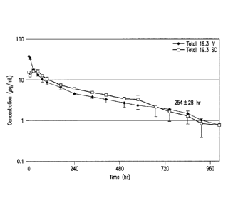
Figure 8 is a graphical representation of the PK of anti-ADDL antibody 19.3
(in
serum) assessed in six rhesus monkeys following administration of a bolus
intravenous (IV) or
subcutaneous (SC) dose of 5 mg/kg. A half-life (tA) of 254 + 28 (274 + 9)
hours was determined
after IV administration and 204 + 49 (219 + 52 ) hours after SC dosing.
Figure 9 is a graphical representation of the PK of anti-ADDL antibody 19.3
assessed in primate (three male rhesus monkeys) cerebrospinal fluid (CSF)
using a cisterna
magna ported rhesus model following administration of a bolus IV dose of 5
mg/kg. At about 48
hours post dose, the anti-ADDL antibody 19.3 was present in the CSF at 0.1% of
the
concentration in serum.
Figures 10A-10D are representations of the ability of anti-ADDL antibody 19.3,
versus two comparator antibodies (Comp 1 and Comp2), to cross the blood-brain-
barrier in a
transgenic mouse model that over-expresses human amyloid precursor protein
(hAPP). Mice
were injected intravenously (IV) with 125I-labeled anti-ADDL antibody 19.3, or
a comparator
antibody, and the blood, CSF and brain samples were collected two hours post-
dose. Upon
assessment of the radioactivity distribution, 0.02% of anti-ADDL antibody 19.3
was present in
the CSF (Figure 10A), while 0.19% was seen in the brain (Figure 10B). Similar
levels were seen
with the two comparator antibodies. Immunocytochemical analysis demonstrated
localization of
anti-ADDL antibody 19.3 (Figure 10C, arrows) and a concentration of anti-ADDL
antibody 19.3
was visible with plaques (Figure 10D). The anti-ADDL antibody19.3 was able to
penetrate into
the brain and bind ADDLs.
Figures 11A-11C are representations of the ability of anti-ADDL antibody 19.3
to
block the deposition of ADDLs into growing plaques in a transgenic mouse model
that over-
expresses hAPP. Biotinylated ADDLs (bADDLs) infused into the hippocampus of 12-
month-old
mice for four weeks (one injection per week) (Figure 11A) labeled existing
plaques (vehicle
alone: Figure 11B; antibody 19.3: Figure 11C, ring). Immunocytochemical
analysis was used to
assess the deposition of new material (ADDLs) (Figures 11B and 11C).
DETAILED DESCRIPTION OF THE INVENTION
The present invention is directed to antibodies, or an antigen binding
fragment,
that bind amyloid f3 (Af3)-derived diffusible ligands (ADDLs), i.e. anti-ADDL
antibodies, and
- 4 -
CA 02805414 2013-01-14
WO 2012/009442 PCT/US2011/043866
attenuate ADDL binding to neurons. Results from a quantitative cell-based
assay revealed that
anti-ADDL antibodies preferentially bound ADDLs, abated the binding of ADDLs
to
hippocampal neurons, crossed the blood-brain barrier, and had an improved
pharmacokinetic
(PK) profile.
In one embodiment the present invention is directed to an isolated antibody,
or an
antigen binding fragment thereof, that binds amyloid 13-derived diffusible
ligands (ADDLs)
comprising:
(a) a light chain variable region comprising,
(i) a CDR1 having the sequence Arg-Ser-Ser-Gln-Ser-Ile-Val-His-Ser-Asn-Gly-
Asn-Thr-Tyr-Leu-Glu (SEQ ID NO: 1),
(ii) a CDR2 having the sequence Lys-Ala-Ser-Asn-Arg-Phe-Ser (SEQ ID NO: 2),
and
(iii) a CDR3 having the sequence Phe-Gln-Gly-Ser-Xaal-Xaa2-Xaa3-Xaa4-Xaa5
(SEQ ID NO: 3), wherein Xaal is Arg, Lys or Tyr, Xaa2 is Val, Ala, or Leu,
Xaa3 is Pro, His, or
Gly, Xaa4 is Ala, Pro, or Val, and Xaa5 is Ser, Gly, or Phe; and
(b) a heavy chain variable region comprising,
(i) a CDR1 having the sequence Gly-Phe-Thr-Phe-Ser-Ser-Phe-Gly-Met-His
(SEQ ID NO: 4),
(ii) a CDR2 having the sequence Tyr-I1e-Ser-Arg-G1y-Ser-Ser-Thr-Ile-Tyr-Tyr-
Ala-Asp-Thr-Val-Lys-Gly (SEQ ID NO: 5), and
(iii) a CDR3 having the sequence Gly-Ile-Thr-Thr-Ala-Leu-Asp-Tyr (SEQ ID NO:
6).
In another embodiment the present invention is directed to an isolated
antibody, or
an antigen binding fragment thereof, that binds amyloid 13-derived diffusible
ligands (ADDLs)
comprising:
(a) a light chain variable region comprising,
(i) a CDR1 having the sequence Arg-Ser-Ser-Gln-Ser-Ile-Val-His-Ser-Xaal -Gly-
Xaa2-Thr-Tyr-Leu-Glu (SEQ ID NO: 53), wherein Xaal is Asn, Ser, Thr, Ala, Asp
or Glu and
Xaa2 is Asn, His, Gin, Ser, Thr, Ala, or Asp;
(ii) a CDR2 having the sequence Lys-Ala-Ser-Xaal-Arg-Phe-Ser (SEQ ID NO:
54), wherein Xaal is Asn, Gin, Ser, Thr, or Ala, and
(iii) a CDR3 having the sequence Phe-Gln-Gly-Ser-Arg-Leu-Gly-Pro-Ser (SEQ
ID NO: 10); and
- 5 -
CA 02805414 2013-01-14
WO 2012/009442 PCT/ES2011/043866
(b) a heavy chain variable region comprising,
(i) a CDR1 having the sequence Gly-Phe-Thr-Phe-Ser-Ser-Phe-Gly-Met-His
(SEQ ID NO: 4),
(ii) a CDR2 having the sequence Tyr-Ile-Ser-Arg-G1y-Ser-Ser-Thr-I1e-Tyr-Tyr-
Ala-Asp-Thr-Val-Lys-Gly (SEQ ID NO: 5), and
(iii) a CDR3 having the sequence Gly-Ile-Thr-Thr-Ala-Leu-Asp-Tyr (SEQ ID NO:
6),
In another embodiment the present invention is an isolated antibody that binds
ADDLs, i.e. an anti-ADDL antibody, or an antigen binding fragment thereof,
having a light chain
variable region CDR3 that is selected from the group consisting of 17.1,
having the sequence
Phe-Gln-Gly-Ser-Arg-Val-Pro-Ala-Ser (SEQ ID NO: 7), 14.2, having the sequence
Phe-Gln-Gly-
Ser-Arg-Val-Pro-Pro-Gly (SEQ ID NO: 8), 13.1, having the sequence Phe-Gin-Gly-
Ser-Lys-Ala-
His-Pro-Ser (SEQ ID NO: 9), 19.3, having the sequence Phe-Gln-Gly-Ser-Arg-Leu-
Gly-Pro-Ser
(SEQ ID NO: 10), 7.2, having the sequence Phe-Gln-Gly-Ser-Tyr-Ala-Pro-Pro-Gly
(SEQ ID NO:
11), 9.2, having the sequence Phe-Gln-Gly-Ser-Arg-Ala-Pro-Pro-Phe (SEQ ID NO:
12), and
11.4, having the sequence Phe-Gln-Gly-Ser-Arg-Val-Pro-Val-Arg (SEQ ID NO: 13).
In a sub-
embodiment the light chain variable region CDR3 is SEQ ID NO: 10.
In still another embodiment of the present invention the isolated anti-ADDL
antibody further comprises a light chain variable region of SEQ ID NO: 15 and
a heavy chain
variable region of SEQ ID NO: 17.
In yet another embodiment of the present invention the isolated anti-ADDL
antibody further comprises a heavy chain constant region of SEQ ID NO: 21.
In another embodiment of the present invention the isolated anti-ADDL antibody
is a monoclonal antibody.
Another embodiment of the present invention is a pharmaceutical composition
comprising an isolated anti-ADDL antibody, or an antigen binding fragment
thereof, in
admixture with a pharmaceutically acceptable carrier.
Another embodiment of the present invention is a method for attenuating
binding
of ADDLs to a neuron comprising contacting the neuron with an isolated anti-
ADDL antibody,
or an antigen binding fragment thereof, so that binding of AP-derived
diffusible ligands to the
neuron is attenuated.
Another embodiment of the present invention is a method for inhibiting the
assembly of ADDLs comprising contacting a sample containing amyloid p 1-42
peptides with an
- 6 -
CA 02805414 2013-01-14
WO 2012/009442 PCT/US2011/043866
isolated anti-ADDL antibody, or antigen binding fragment thereof, thereby
inhibiting the
assembly of ADDLs.
Another embodiment of the present invention is a method for inhibiting the
phosphorylation of tau protein at Ser202/Thr205 comprising contacting a sample
containing a tau
protein with an isolated anti-ADDL antibody, or an antigen binding fragment
thereof, thereby
inhibiting the phosphorylation of tau protein at Ser202/Thr205.
Another embodiment of the present invention is a method for attenuating the
symptoms of a disease associated with ADDLs comprising administering an
effective amount to
a patient in need thereof of the pharmaceutical composition comprising an
isolated anti-ADDL
antibody, or an antigen binding fragment thereof.
Another embodiment of the present invention is a method for identifying a
putative therapeutic agent that attenuates the binding of amyloid 13-derived
diffusible ligands
(ADDLs) to neurons comprising:
(a) contacting a composition comprising a neuron with ADDLs in the presence of
an agent;
(b) contacting the composition with the isolated anti-ADDL antibody, or an
antigen binding fragment thereof; and
(c) detecting the amount of antibody or antigen binding fragment bound in the
presence of the agent,
wherein a decrease in the amount of antibody or antigen binding fragment bound
in the presence of the agent as compared to the amount of antibody bound in
the absence of the
agent indicates that the agent is a putative therapeutic agent for attenuating
binding of ADDLs to
neurons.
Another embodiment of the present invention is a method for detecting ADDLs in
a sample comprising contacting a sample with an isolated anti-ADDL antibody,
or an antigen
binding fragment thereof, and determining the presence of a complex comprising
the ADDLs and
said antibody or antigen binding fragment.
Another embodiment of the present invention is a method for diagnosing a
disease associated with ADDLs comprising contacting a sample with an isolated
anti-ADDL
antibody, or an antigen binding fragment thereof, and detetutining the
presence of a complex
comprising the ADDLs and said isolated antibody or antigen binding fragment,
wherein the
presence of said complex is diagnostic of a disease associated with ADDLs.
- 7 -
Still another embodiment of the present invention is a kit for detecting ADDLs
comprising an isolated anti-ADDL antibody, or an antigen binding fragment
thereof, that binds
ADDLs.
Monoclonal antibodies, which differentially recognize multi-dimensional
conformations of AI3-derived diffusible ligands (ADDLs) are known in the art
(see, U.S. Pat.
No.7,780,963, U.S. Pat. No. 7,731,962, and U.S. Pat. No. 7,811,563)
and have been shown to reduce ADDL binding to neurons
in cell based assays. Anti-ADDL antibodies can distinguish between Alzheimer's
disease (AD)
and control human brain extracts, can identify endogenous oligomers in AD
brain slices and on
hippocampal cells, and can neutralize endogenous and synthetic ADDLs in
solution. Anti-
ADDL antibodies specifically bind one or more multi-dimensional conformations
of ADDLs,
bind particular ADDLs derived from the oligomerization of A1342, while having
reduced affinity
for other Ali peptides, including A131-40.
The present invention is directed to anti-ADDL antibodies, specifically
antibodies
.. 17.1, 14.2, 13.1, 19.3, 19.3133, 19.3S33, 7.2,9.2, and 11.4, that
preferentially bind ADDLs and
that have been characterized as to their specificity and selectivity for
ADDLs. Importantly, the
specificity and selectivity of these anti-ADDL antibodies of the present
invention was not
predictable from the linear epitope of A13 to which they bound, nor was this
activity predictable
from their ability to detect ADDLs by Western blot., or from their ability to
detect immuno-
stained ADDLs bound to neurons. Moreover, the differential ability of the anti-
ADDL
antibodies of the present invention to neutralize ADDLs and block binding to
primary
hippocampal neurons supports the belief that anti-ADDL antibodies act through
binding to a
more relevant, conformational epitope, which prevents ADDL binding to neurons.
One
embodiment of the present invention, anti-ADDL antibody 19.3, not only blocked
the binding of
ADDLs to primary neurons, but also abated ADDL-induced changes to hippocampal
spine
morphology, an indication that the impedance of ADDL-neural binding has
significant
physiological ramifications, for example, neuronal survival, neuronal
connectivity and signal
transduction. Anti-ADDL antibody 19.3 also had an improved pharmacokinetic
(PK) profile, as
compared with a previously known anti-ADDL antibody, 3B3, when assessed in
both in vitro and
in vivo models. In addition, when administered to transgenic mice that over-
express a human
form of amyloid precursor protein (hAPP), anti-ADDL antibody 19.3 was shown to
penetrate the
blood-brain-barrier and concentrate in the brain. Since ADDLs are localized in
the brain and act
there to adversely affect neuronal function, one of skill in the art would
appreciate and recognize
- 8 -
CA 2805414 2017-07-11
that the penetration and concentration of antibody in the brain would be
beneficial for
irnmunotherapy. Taken together, these data demonstrate that selective anti-
ADDL antibodies,
such as antibody 19.3, can block the binding of ADDLs to hippocampal neurons,
which are
critically involved in learning and memory.
The utility of anti-ADDL antibodies for the treatment of AD is based on a
growing body of evidence that suggests that ADDLs, and not amyloid plaques per
se, play a
fundamental role in the cognitive decline associated with this disease (Walsh
and Selkoe, 2004,
Protein Pept. Lett., 11: 213-228). ADDLs are elevated in the AD brain and
induce deficits in
behavioral and electrophysiologica1 endpoints when centrally administered to
rodents (Walsh, et
al., 2002, Nature, 416: 535-539; Cleary, et al., 2004, Nat. Neurosci., 8: 79-
84; Klyubin, et al.,
2005, Nat. Med., 11: 556-561; Balducci, et al., 2010, Proc. Natl. Acad. Sci.
USA, 107: 2295-
2300). Deficits in learning and memory have also been observed in a hAPP
expressing mouse
model, with the onset of impairment associated with elevated ADDL levels
(Westerman, et al.,
2002, J. Neurosci., 22: 1858-1867; Ashe, 2005, Biochem. Soc. Trans., 33: 591-
594; Lee, et al.,
2005, J. Biol. Chem., 281: 4292-4299; Lesne, et al., 2006, Nature, 440: 352-
357). While the
cellular and sub-cellular events that mediate these effects on cognition are
not fully understood, it
is clear that ADDLs bind to the synaptic temtinals localized on the dendritic
processes of
hippocampal neurons (Lacore, et al., 2004, J. Neurosci., 24: 10191-1022) and
alter the
morphology and number of dendritic spines (Lacor et al., 2007, J. Neurosci.,
27: 796-807;
Shankar, et al., 2007, J. Neurosci., 27: 2866-2875; Shughrue, et al., 2010,
Neurobiol. Aging, 31:
189-202). The finding that ADDLs bind to both GABAergic and glutamate neurons
in the
hippocampus (Shughrue, et al., 2010), neurons critically involved in learning
and memory, which
results in the internalization of AMPA receptors (Zhao, et al., 2010, J. Biol.
Chem., 285: 7619-
7632) further supports the belief that ADDLs directly or indirectly modulate
these
neurotransmitter systems (see, for example, Venkitaramani, et al., 2007, J.
Neurosci., 27: 11832-
11837).
In the present invention, a panel of anti-ADDL antibodies derived from anti-
ADDL antibody, 3B3 (U.S. Pat. No. 7,780,963 and U.S. Pat. No. 7,811,563)
were assessed for their ability to block ADDL
binding to primary hippocampal neurons. Selected monoclonal antibodies were
then humanized
and affinity matured for further characterization. Lead antibodies, selected
for their ability to
bind to ADDLs, were further assessed at a single concentration using a three-
pronged ELISA to
determine antibody binding to monomer AO, ADDLs, and fibrillar Af3. As shown
in Figure 1, six
- 9 -
CA 2805414 2017-07-11
CA 02805414 2013-01-14
WO 2012/009442 PCT/US2011/043866
of the seven affinity matured anti-ADDL antibodies, specifically antibodies
14.2, 7.2, 11.4, 13.1,
17.1, and 19.3 were ADDL preferring, when compared with monomer Ap and
fibrillar All
Subsequently an eleven point titration curve and ELISA were used to ascertain
the binding
affinity of anti-ADDL antibodies to ADDLs and monomer Af3 (A131_40) over a
broad range of
concentrations. As shown in Figure 2, the anti-ADDL antibodies 383 and 19.3
were highly
ADDL selective. In addition, antibodies were compared in a cell-based binding
assay to
determine the ability of antibodies to block ADDL binding to neurons. As shown
in Figure 3,
ADDLs, pre-incubated with increasing concentrations of anti-ADDL antibodies
3B3 and 19.3,
were added to primary hippocampal neurons, and a titration curve was used to
show
quantitatively the ability of the antibody to block ADDL binding to neurons.
Taken together,
these results show that anti-ADDL antibodies profoundly attenuate neuronal
binding in a cell-
based format.
An assessment of the amino acid sequence was conducted to identify potential
sites of deamidation. Asparagine and aspartic acid residues present in the
CDRs of therapeutic
antibodies are known to undergo deamidation and isoaspartate formation (Valsak
and Ionescu,
2008, Curr.Pharm.Biotech., 9:468-481; Aswad et al., 2000, J.
Pharm.Biomed.Anal., 21:1129-
1136), the formation of which can alter the binding potency of an antibody
and, in turn, reduce
antibody effectiveness for use as a therapeutic. Thus, those of skill in the
art would recognize
and appreciate that the presence of an asparagine or an aspartic acid within
the CDRs for the 19.3
antibody would not be desirable. Accordingly, Applicants altered the
asparagine residue at
position 33 of the light chain CDR1 to optimize the stability of the anti-ADDL
antibody 19.3
(Table 4B). Derivatives of the 19.3 antibody were produced with the
substitution of serine (SEQ
ID NO: 55), threonine (SEQ ID NO: 56), or glutarnic acid (SEQ ID NO: 67) for
the asparagine at
position 33 (SEQ ID NO: 1) in CDR1. The substitution of aspartic acid (SEQ ID
NO: 68) for the
asparagine as position 33 was also generated as a control. These changes will
remove the
possibility of deamidation of asparagine at position 33 in CDR1. The 19.3
derivatives were
generated as described in Example 3 and characterized as described in Example
4 as to
derivatives with the serine (SEQ ID NO: 55), threonine (SEQ ID NO: 56),
glutamic acid (SEQ
ID NO: 67), and aspartic acid (SEQ ID NO: 68) substitutions, to evaluate the
stability of the new
constructs. As shown in Figures 4B and 4C, respectively, two representative
derivatives,
19.3S33 (SEQ ID NO: 55) and 19.3T33 (SEQ ID NO: 56), had enhanced binding
stability
following a one-month incubation at varying temperatures. Other amino acid
substitutions in the
light chain CDR1 for the asparagine at positions 33 and 35 (SEQ ID NO: 53) and
in the light
- 10 -
CA 02805414 2013-01-14
WO 2012/009442 PCT/US2011/043866
chain CDR2 for the asparagine at position 58 position (SEQ ID NO: 54) are
proposed in Tables
4B and 4C for further evaluation.
To determine the pharmacokinetics of the affinity matured anti-ADDL antibodies
of the present invention, a series of in vitro and in vivo studies were
conducted. The binding of
antibodies to the Fan receptor at pH 6.0 has been shown to be predictive of
antibody half-life in
humans (Zalevsky, et al., 2010, Nat. Biotech., 28(2): 157-159) and at pII 7.3
(USSN 61/307,182)
The binding and dissociation of the anti-ADDL antibodies of the present
invention to
immobilized human FeRn was assessed with a label free interaction analysis,
such as that offered
by BiacoreTM Life Sciences, BiaeoreTM T-100 (GE Healthcare, Piscataway, NT).
An adjusted
sensorgram is used to show the initial binding at pH 6.0 and then the
dissociation of antibodies at
pH 7.3 from 180 seconds. A report point (Stability) was inserted at 5 seconds
after the end of pH
6.0 binding and the "% bound" was calculated as RUStability/RUBinding (%). As
shown in
Figure 5, the off-rate for humanized 3B3 was markedly slower than the seven
anti-ADDL
antibodies of the present invention, which included antibody 19.3, and three
comparator
antibodies. In that a slow off-rate is thought to be an indicator of poor in
vivo PK, an additional
in vivo study was conducted in transgenic FeRn mice (heterozygous 276 human
FeRn mice,
Jackson Laboratories, Bar Harbor, ME). =When the transgenic FeRn mice were
given 10 mgikg
intravenously (IV) of either anti-ADDL antibody 3B3 or 19.3, a significant
difference in
pharmacokinetics was determined. As shown in Figure 7, the half-life (ty2.) of
anti-ADDL
antibody 3B3 was relatively short (29 +9 hours), which was consistent with the
prediction from
the in vitro BiacoreTM data, while the half-life for anti-ADDL antibody 19.3
was significantly
longer (77 6 hours). Generally, poor PK, as seen with antibody 3B3, would
preclude further
development of an antibody for use as a therapeutic due to its short
bioavailability.
To confirm the predicted half-life of anti-ADDL antibody 19.3 in primates, a
primate pharmacokinetics study was conducted for the antibody in a cohort of
eistema magna
ported rhesus monkeys. The animals were dosed with a single intravenous (IV)
bolus or
subcutaneous (SC) injection of anti-ADDL antibody 19.3 (5 mg/kg) and blood
samples collected
after antibody administration. Concurrently, CSF samples were collected from
the cistema
magna port at timed intervals and the concentration of anti-ADDL antibody 19.3
in serum and
.. CSF was determined with an anti-human IgG ELISA assay. When the animals
were
administered anti-ADDL antibody 19.3 by a single IV bolus injection a t112 of
254 + 28 hours
(Figure 8) was observed, while a ti/2 of 204 + 49 hours was observed for the
subcutaneous
-11-
CA 02805414 2013-01-14
WO 2012/009442 PCT/US2011/043866
administration. In addition, Applicants found that anti-ADDL antibody 19.3 was
able to cross
into the primate CSF, where it increased in concentration during the first 48
hours and peaked at
about 0.1% of the antibody dosed (Figure 9).
In an attempt to ascertain the quantity of antibody that penetrates the blood-
brain-
barrier and enters the CSF and brain, anti-ADDL antibody 19.3 and two
comparator antibodies
(Comp 1 and Comp 2) were 125I-labeled and administered to aged (twelve-month
old) mice that
over-express hAPP, a rodent model for AD. Two hours after IV dosing about
0.02% of antibody
19.3 was seen in the CSF (Figure 10A), while about 0.19% of antibody 19.3 was
seen in the
brain (Figure 10B). Similar levels were seen for the two comparator antibodies
(Figure 10A and
10B). When immunocytochemical analysis was carried out on brain sections of
the dosed mice
and the localization of anti-ADDL antibody 19.3 was determined (arrow in
Figure 10C), a
concentration of the antibody associated with the deposition of AO into
plaques was observed
(Figure 10D). This demonstrated that the anti-ADDL antibody 19.3 penetrated
into the CSF and
was concentrated in the brain. Recently it was shown that exogenous ADDLs were
deposited
into plaques when administered to mice that over express hAPP (Gaspar, et al.,
2010, Exp.
Neurol., 223: 394-400). Thus, the findings herein confirmed that the localized
anti-ADDL
antibody19.3 bound to circulating ADDLs associated with plaques.
To further evaluate the in vivo efficacy of anti-ADDL antibodies, the ability
of
antibody 19.3 to block the deposition of ADDLS into growing plaques was
assessed in hAPP
transgenic mice following four weekly infusions of biotinylated ADDLs (bADDLs)
into the
hippoearnpus of 12-month old mice to label existing plaques (Figure 11A). The
animals then
received four weekly intravenous infusions of antibody 19.3 (Figure 11A). The
deposition of
new material (ADDLs) into growing plaques was assessed by irnmunocytoehemical
analysis. As
seen in Figures 11B and I IC, anti-ADDL antibody 19.3 significantly reduced
the deposition of
ADDLs into the periphery of existing plaques (Figure 11C) as compared to mice
treated with
vehicle alone (Figure 11B). Taken together, these results demonstrated that an
anti-ADDL
antibody, specifically the 19.3 antibody, was able to cross the blood-brain-
barrier, bind ADDLs,
and block the deposition of new material into growing plaques.
ADDL binding may also have long-term effects on neurons. Recent studies have
shown that ADDL binding to hippocampal neurons can initiate a signaling
cascade that results in
the phosphotylation of tau (De Felice, et al., 2006, Neurobiol. Aging, 29: 394-
400). One
component of this signaling cascade, GSK-33, has also been shown to be
modulated by ADDL
binding in vivo and in vitro (Ma, et al., 2006, J. Neurosci. Res., 83: 374-
384). Ma, et al., 2006,
- 12 -
CA 02805414 2013-01-14
WO 2012/009442 PCT/US2011/043866
found that passive immunization of hAPP mice with an antibody that reduced
ADDLs, also
reduced GSK-3f3 levels and phosphorylation of tau in the cortex. This finding
supports a link
between AP and phosphorylated tau and suggests that ADDL binding may trigger
events that lead
to the intracellular aggregation of tau. Further, the data suggests that
antibodies that prevent the
binding of ADDLs to neurons and the associated loss of synaptic spines, such
as the antibodies of
the present invention could ameliorate the cognitive and/or pathological
outcomes associated
with Alzheimer's disease and related diseases.
Monoclonal antibodies, which differentially recognize multi-dimensional
conformations of AO-derived diffusible ligands, i.e., ADDLs, have now been
generated. These
antibodies were humanized and, in some embodiments, affinity-matured. The
antibodies
advantageously distinguish between Alzheimer's disease and control human brain
extracts, and
identify endogenous oligomers in Alzheimer's disease brain slices and in
cultured hippoeanapal
cells. Further, the antibodies of the present invention neutralize endogenous
and synthetic
ADDLs in solution. So-called "synthetic" ADDLs are produced in vitro by mixing
purified Aril_
42 under conditions that generate ADDLs. See, U.S. Patent No. 6,218,506. The
antibodies
disclosed herein exhibit a high degree of selectivity for ADDLs, with minimal
detection of
monomer AD species. Moreover, these antibodies differentially block the
ability of ADDL-
containing preparations to bind primary cultures of rat hippocampal neurons
and immortalized
neuroblastoma cell lines, and also block ADDL assembly. This finding
demonstrates that these
antibodies possess a differential ability to recognize a multi-dimensional
conformation of
ADDLs despite similar linear sequence recognition and affinities. Since ADDLs
are known to
associate with a subset of neurons and disrupt normal neuronal function, the
antibodies of this
invention find use in the prevention of ADDL binding to neurons and the
assembly of ADDLs
and, in turn, can be used for the treatment of ADDL-related diseases including
Alzheimer's
disease.
Accordingly, one embodiment of the present invention is an isolated antibody
that
differentially recognizes one or more multi-dimensional conformations of
ADDLs. An tisolatcd!
antibody of the present invention refers to an antibody which is substantially
free of other
antibodies. However, the molecule may include some additional agents or
moieties which do not
deleteriously affect the basic characteristics of the antibody (for example,
binding specificity,
neutralizing activity, etc.).
- 13 -
CA 02805414 2013-01-14
WO 2012/009442 PCT/US2011/043866
An antibody which is capable of specifically binding one or more multi-
dimensional conformations of ADDLs, binds particular ADDLs derived from the
oligomerization
of A131-42, but does not cross-react with other AP peptides, namely Ap1-12,
A131-=28, AI31-40,
and AP 12-28 as determined by western blot analyses as disclosed herein, and
preferentially binds
ADDLs in solution. Specific binding between two entities generally refers to
an affinity of at
least 106, 107, 108, 109, or 101 Affinities greater than 108 M-1 are
desired to achieve
specific binding.
In particular embodiments, an antibody that is capable of specifically binding
a
multi-dimensional conformation of one or more ADDLs is also raised against,
i.e., an animal is
immunized with, multi-dimensional conformations of ADDLs. In other
embodiments, an
antibody that is capable of specifically binding a multi-dimensional
conformation of one or more
ADDLs is raised against a low n-mer-forming peptide such as A131 -42[Nle35-
Dpro371.
The term "epitope" refers to a site on an antigen to which B and/or T cells
respond
or a site on a molecule against which an antibody will be produced and/or to
which an antibody
will bind. For example, an epitope can be recognized by an antibody defining
the epitope.
A linear epitope is an epitope wherein an amino acid primary sequence
comprises
the epitope recognized. A linear epitope typically includes at least 3, and
more usually, at least 5,
for example, about 6 to about 10 amino acids in a unique sequence.
A conformational epitope, in contrast to a linear epitope, is an epitope
wherein the
primary sequence of the amino acids comprising the epitope is not the sole
defining component
of the epitope recognized (for example, an epitope wherein the primary
sequence of amino acids
is not necessarily recognized by the antibody defining the epitope). Typically
a conformational
epitope encompasses an increased number of amino acids relative to a linear
epitope. With
regard to recognition of conformational epitopes, the antibody recognizes a
three-dimensional
.. structure of the peptide or protein. For example, when a protein molecule
folds to form a three-
dimensional structure, certain amino acids and/or the polypeptide backbone
forming the
conformational epitope become juxtaposed enabling the antibody to recognize
the epitope.
Methods of determining conformation of epitopes include but are not limited
to, for example, x-
ray crystallography, two-dimensional nuclear magnetic resonance spectroscopy
and site-directed
spin labeling and electron paramagnetic resonance spectroscopy. See, for
example, Epitope
Mapping Protocols in Methods in Molecular Biology (1996) Vol. 66, Morris
(Ed.).
Arnyloid13-derived diffusible ligands or ADDLs refer to soluble oligomers of
A131-42 which are desirably composed of aggregates of less than eight or nine
A131-42 peptides
-14-
CA 02805414 2013-01-14
WO 2012/009442 PCT/US2011/043866
and are found associated with Alzheimer's disease. This is in contrast to high
molecular weight
aggregation intermediates, which form strings of micelles leading to fibril
formation.
As exemplified herein, the antibodies of the present invention bind or
recognize at
least one multi-dimensional conformation of an ADDL. In particular
embodiments, the
.. antibodies bind at least two, at least three, or at least four multi-
dimensional conformations of an
ADDL. Multi-dimensional conformations of ADDLs are intended to encompass
dimers, timers,
tetramers pentamers, hexamers, heptamers, octamers, nonamers, deeamers, etc.
as defined by
analysis via SDS-PAGE. Because trimer, tetramer, etc. designations can vary
with the assay
method employed (see, e.g., Bitan, et al., 2005, Amyloid, 12:88-95), the
definition of timer,
tetramer, and the like, as used herein, is according to SDS-PAGE analysis. To
illustrate the
differential binding capabilities of the antibodies herein, it has been found
that certain antibodies
will recognize one multi-dimensional conformation, for example, tetramers of
ADDLs (U.S. Pat.
No. 7,780,963, murine antibodies 2D6 and 4E2), 1Nhile other antibodies
recognize several multi-
dimensional conformations, for example, timers and tetramers of ADDLs (U.S.
Pat. No.
7,780,963, murine antibodies 2A10, 2B4, 5F10, and 20C2 and humanized antibody
20C2). As
such, the antibody of the present invention has oligomer-specifie
characteristics. In particular
embodiments, a multi-dimensional conformation of an ADDL is associated with a
specific
polypeptide structure which results in a conformational epitope that is
recognized by an antibody
of the present invention. In other embodiments, an antibody of the invention
specifically binds a
multi-dimensional conformation ADDL having a size range of approximately a
timer or
tetramer, which have molecular weights in excess of >50 kDa.
While antibodies of the present invention may have similar linear epitopes,
such
linear epitopes are not wholly indicative of the binding characteristics of
these antibodies, i.e.,
ability to block ADDL binding to neurons, prevent tau phosphorylation and
inhibit ADDL
assembly, because, as is well-known to the skilled artisan, the linear epitope
may only correspond
to a portion of the antigen's epitope (see, for example, Breitling and Diibel,
1999, Recombinant
Antibodies, John Wiley & Sons, Inc., NY, pg. 115). The antibodies of the
present invention can
be distinguished from those of the art as being capable of differentially
recognizing multi-
dimensional ADDLs and accordingly differentially blocking ADDL binding to
neurons,
differentially preventing tau phosphorylation and differentially inhibiting
ADDL assembly.
An antibody, as used in accordance with the present invention includes, but is
not
be limited to, polyclonal or monoclonal antibodies, and chimeric, human (for
example, isolated
from B cells), humanized, neutralizing, bispecific or single chain antibodies
thereof. In one
- 15-
CA 02805414 2013-01-14
WO 2012/009442 PCT/US2011/043866
embodiment, an antibody of the present invention is monoclonal. For the
production of
antibodies, various hosts including goats, rabbits, chickens, rats, mice,
humans, and others, can
be immunized by injection with synthetic or natural ADDLs. Methods for
producing antibodies
are well-known in the art. See, for example, Kohler and Milstein, 1975,
Nature, 256:495-497:
Harlow and Lane, Antibodies: A Laboratory Manual, Cold Spring Harbor
Laboratory, New York,
1988.
Depending on the host species, various adjuvants can be used to increase the
immunological response. Adjuvants used in accordance with the present
invention desirably
augment the intrinsic response to ADDLs without causing conformational changes
in the
immunogen that affect the qualitative form of the response. Particularly
suitable adjuvants
include 3 De-O-acylated rnonophosphoryl lipid A (MPLTm; RIBI ImmunoChem
Research Inc.,
Hamilton, MT; see GB 2220211) and oil-in-water emulsions, such as squalene or
peanut oil,
optionally in combination with immune stimulants, such as monophosphoryl lipid
A (see, Stoute,
et al., 1997, N. Engl. J. Med., 336:86-91), muramyl peptides (for example, N-
acetylmuramyl-L-
threonyl-D-isoglutamine (thr-MDP), N-acetyl-normuramyl-L-alanyl-D-isoglutamine
(nor-MDP),
N-acetylmuramyl-L-alanyl-D-isoglutaminyl-L-alanine-2-(1'-2tdipalmitoyl-sn-
glycero-3-
hydroxyphosphoryloxy)-ethylatnine (E-PE), N-acetylglucsaminyl-N-acetylmuramyl-
L-Al-D-
isoglu-L-Ala-dipalmitoxy propylamide (DTP-DPP)), or other bacterial cell wall
components.
Specific examples of oil-in-water emulsions include MF59 (WO 90/14837),
containing 5%
Squalene, 0.5% TWEENTm 80, and 0.5% SPAN 85 (optionally containing various
amounts of
MTP-PE) formulated into submicron particles using a microfluidizer such as
Model 110Y
microfluidizer (Microfluidics, Newton, MA); SAP containing 10% Squalene, 0.4%
TWEENTm
80, 5% PLURONIC -blocked polymer L121, and thr-MDP, either microfluidized into
a
submicron emulsion or vortexed to generate a larger particle size emulsion;
and RIBITM adjuvant
system (RAS) (Ribi IrnmunoChem, Hamilton, MT) containing 2% squalene, 0.2%
TWEENTm
80, and one or more bacterial cell wall components such as monophosphoryllipid
A, trehalose
dimycolate (TDM), and cell wall skeleton (CWS).
Another class of adjuvants is saponin adjuvants, such as STIMULONTm (QS-21,
Aquila, Framingham, MA) or particles generated therefrom such as ISCOMs
(immunostimulating complexes) and ISCOMATRIX (CSL Ltd., Parkville,
Australia). Other
suitable adjuvants include Complete Freund's Adjuvant (CFA), Incomplete
Freund's Adjuvant
(IFA), mineral gels such as aluminum hydroxide, and surface-active substances
such as
lysolecithin, PLURON1C polyols, polyanions, peptides, CpG (WO 98/40100),
keyhole limpet
- 16-
CA 02805414 2013-01-14
WO 2012/009442 PCT/US2011/043866
hemocyanin, dinitrophenol, and cytokines such as interleukins (IL-1, 1L-2, and
IL-12),
macrophage colony stimulating factor (M-CSF), and tumor necrosis factor (TNF).
Among
adjuvants used in humans, BCE] (bacilli Calmette-Guerin) and Corynebacterium
parvum are
particularly suitable.
An antibody to a multi-dimensional conformation ADDL is generated by
immunizing an animal with ADDLs. Generally, ADDLs can be generated
synthetically or by
recombinant fragment expression and purification. Synthetic ADDLs can be
prepared as
disclosed herein, or in accordance with the methods disclosed in U.S. Patent
Nos. 6,218,506 and
7,811,563, or in co-pending applications U.S. 2007/0218499, U.S. 2010/0143396,
and U.S.
2010/0240868, all of which are incorporated herein by reference in their
entirety. Further,
ADDLs can be fused with another protein such as keyhole limpet hemocyanin to
generate an
antibody against the chimeric molecule. The ADDLs can be conformationally
constrained to
form an epitope useful as described herein and furthermore can be associated
with a surface for
example, physically attached or chemically bonded to a surface in such a
manner so as to allow
for the production of a conformation which is recognized by the antibodies of
the present
invention.
Monoclonal antibodies to multi-dimensional conformations of ADDLs can be
prepared using any technique which provides for the production of antibody
molecules by
continuous cell lines in culture. These include, but are not limited to, the
hybridoma technique,
the human B-cell hybridoma technique, and the EBV-hybridoma technique (Kohler,
et al.,1975,
Nature 256:495-497; Kozbor, et al. , 1985, J. Im.munol. Methods 81:31-42;
Cote, et al., 1983,
Proe.NatI.Acad.Sci. 80:2026-2030; Cole, et al., 1984, Mol. Cell Biol. 62:109-
120).
In particular embodiments, the antibodies of the present invention are
humanized.
Humanized or chimeric antibodies can be produced by splicing of mouse antibody
genes to
human antibody genes to obtain a molecule with appropriate antigen specificity
and biological
activity (see, MOITiS011, etal., 1984, Proc. Natl. Acad. Sci. 81, 6851-6855;
Neuberger, et at.,
1984, Nature 312:604-608; Takeda, etal., 1985, Nature 314:452-454; Queen. et
al., 1989, Proc.
Natl. Acad. Sci. USA 86:10029-10033; WO 90/07861). For example, a mouse
antibody is
expressed as the Fv or Fab fragment in a phage selection vector. The gene for
the light chain
(and in a parallel experiment, the gene for the heavy chain) is exchanged for
a library of human
antibody genes. Phage antibodies, which still bind the antigen, are then
identified. This method,
commonly known as chain shuffling, provided humanized antibodies that should
bind the same
epitope as the mouse antibody from which it descends (Jespers, et at., 1994,
Biotechnology NY
- 17-
CA 02805414 2013-01-14
WO 2012/009442 PCT/US2011/043866
12:899-903). As an alternative, chain shuffling can be performed at the
protein level (see, Figini,
et al., 1994, J. Mol. Biol. 239:68-78).
Human antibodies can also be obtained using phage-display methods. See, for
example, WO 91/17271 and WO 92/01047. In these methods, libraries of phage are
produced in
which members display different antibodies on their outer surfaces. Antibodies
are usually
displayed as Fv or Fab fragments. Phage displaying antibodies with a desired
specificity are
selected by affinity enrichment to ADDLs. Human antibodies against ADDLs can
also be
produced from non-human transgenie mammals having transgenes encoding at least
a segment of
the human immunoglobulin locus and an inactivated endogenous immunoglobulin
locus. See,
for example, WO 93/12227 and WO 91/10741. Human antibodies can be selected by
competitive binding experiments, or otherwise, to have the same epitope
specificity as a
particular mouse antibody. Such antibodies generally retain the useful
functional properties of
the mouse antibodies. Human polyelonal antibodies can also be provided in the
form of serum
from humans immunized with an immunogenic agent. Optionally, such polyclonal
antibodies
can be concentrated by affinity purification using ADDLs as an affinity
reagent.
As exemplified herein, humanized antibodies can also be produced by veneering
or resurfacing of murine antibodies. Veneering involves replacing only the
surface fixed region
amino acids in the mouse heavy and light variable regions with those of a
homologous human
antibody sequence. Replacing mouse surface amino acids with human residues in
the same
position from a homologous human sequence has been shown to reduce the
immunogenicity of
the mouse antibody while preserving its ligand binding. The replacement of
exterior residues
generally has little, or no, effect on the interior domains, or on the inter-
domain contacts. See,
for example, U.S. Patent No. 6,797,492.
Human or humanized antibodies can be designed to have IgG, IgD, IgA, IgM or
IgE constant regions, and any isotype, including IgGl, IgG2, IgG3 and IgG4. In
particular
embodiments, an antibody of the invention is IgG or IgM, or a combination
thereof. In one
specific embodiment the antibodies of the present invention are IgG2. Those of
skill in the art
would understand that other isoferms can be utilized herein. Exemplary
sequences for these
isoforms are given in SEQ ID NOS: 43-45. Other embodiments of the present
invention embrace
a constant region formed by selective incorporation of human IgG4 sequences
into a standard
human IgG2 constant region. An exemplary mutant IgG2 Fe is IgG2m4, set forth
herein as SEQ
ID NO: 46. Antibodies can be expressed as tetramers containing two light and
two heavy chains,
as separate heavy chains and light chains or as single chain antibodies in
which heavy and light
- 18 -
chain variable domains are linked through a spacer. Techniques for the
production of single chain
antibodies are well-knovvn in the art.
Exemplary humanized antibodies produced by CDR grafting and veneering are
disclosed in U.S. Pat. Nos. 7,780,963, 7,731,962, and 7,811,563.
Diabodies are also contemplated. A diabody refers to an engineered antibody
construct prepared by isolating the binding domains (both heavy and light
chain) of a binding
antibody, and supplying a linking moiety which joins or operably links the
heavy and light chains
on the same polypeptide chain thereby preserving the binding function (see,
Holliger, et al., 1993,
Proc. Natl. Acad. Sci. USA 90:6444; Poljak, 1994, Structure 2:1121-1123). This
forms, in
essence, a radically abbreviated antibody, having only the variable domain
necessary for binding
the antigen. By using a linker that is too short to allow pairing between the
two domains on the
same chain, the domains are forced to pair with the complementary domains of
another chain and
create two antigen-binding sites. These dimeric antibody fragments, or
diabodies, are bivalent
and bispecific. The skilled artisan will appreciate that any method to
generate diabodies can be
used. Suitable methods are described by Holliger, et at, 1993, supra; Poljak,
1994, supra; Zhu,
et al., 1996, Biotechnology 14:192-196, and U.S. Patent No. 6,492,123.
Fragments of an isolated antibody of the invention are also expressly
encompassed by the present invention. Fragments are intended to include Fab
fragments, F(ab)2
fragments, F(ab') fragments, bispecific scFv fragments, Fv fragments and
fragments produced by
a Fab expression library, as well as peptide aptamers. For example, F(ab1)-2
fragments are
produced by pepsin digestion of the antibody molecule of the invention,
whereas Fab fragments
are generated by reducing the disulfide bridges of the F(ab')2 fragments.
Alternatively, Fab
expression libraries can be constructed to allow rapid and easy identification
of monoclonal Fab
fragments with the desired specificity (see, Huse, et al., 1989, Science,
254:1275-1281). In
particular embodiments, antibody fragments of the present invention are
fragments of
neutralizing antibodies which retain the variable region binding site thereof,
i.e. antigen binding
fragment. Exemplary are F(a1302 fragments, F(ab') fragments, and Fab
fragments. See, generally,
Immunology: Basic Processes, 1985, 2nd edition, J. Bellanti (Ed.) pp. 95-97.
Peptide aptamers which differentially recognize multi-dimensional
conformations
of ADDLs can be rationally designed or screened for in a library of aptamers
(for example,
provided by Aptanomics SA, Lyon, France). In general, peptide aptamers are
synthetic
- 19 -
CA 2805414 2017-07-11
recognition molecules whose design is based on the structure of antibodies.
Peptide aptamers
consist of a variable peptide loop attached at both ends to a protein
scaffold. This double
structural constraint greatly increases the binding affinity of the peptide
aptamer to levels
comparable to that of an antibody (nanomolar range).
Exemplary nucleic acid sequences encoding heavy and light chain variable
regions for use in producing antibody and antibody fragments of the present
invention are
disclosed herein in SEQ ID NOS: 14 and 16. As will be appreciated by the
skilled artisan, the
heavy chain variable regions disclosed herein, such as that shown in SEQ ID
NO: 16, can be used
in combination with any one of the light chain variable regions disclosed
herein to generate
antibodies with modified affinities, dissociation, epitopes, and the like.
Antibodies or antibody fragments of the present invention can have additional
moieties attached thereto. For example, a microsphere or micropartiele can be
attached to the
antibody or antibody fragment, as described in U.S. Patent No. 4,493,825.
Moreover, particular embodiment embrace antibody or antibody fragments which
are mutated and selected for increased antigen affinity, neutralizing activity
(i.e,, the ability to
block binding of ADDLs to neuronal cells or the ability to block ADDL
assembly), or a modified
dissociation constant. Mutator strains of E. colt (Low, et al., 1996, J. Mol.
Biol., 260:359-368),
chain shuffling (Figini, et al., 1994, supra), and PCR mutagenesis are
established methods for
mutating nucleic acid molecules encoding antibodies. By way of illustration,
increased affinity
can be selected for by contacting a large number of phage antibodies with a
low amount of
biotinylated antigen so that the antibodies compete for binding. In this case,
the number of
antigen molecules should exceed the number of phage antibodies, but the
concentration of
antigen should be somewhat below the dissociation constant. Thus,
predominantly mutated
phage antibodies with increased affinity bind to the biotinylated antigen,
while the larger part of
the weaker affinity phage antibodies remains unbound. Streptavidin can then
assist in the
enrichment of the higher affinity, mutated phage antibodies from the mixture
(Schier, at al.,
1996, .T. Mol. Biol. 255:28-43). Exemplary affinity-maturated light chain CDR3
amino acid
sequences are disclosed herein (see Table 4), with particular embodiments
embracing a light
chain CDR3 amino acid sequence of SEQ ID NO: 3 and specific embodiments of SEQ
ID NOS:
7-13. The present invention also embraces alternative variations for light
chain CDR1 (SEQ ID
NO: 53) and CDR2 (SEQ ID NO: 54).
- 70 -
CA 2805414 2017-07-11
CA 02805414 2013-01-14
WO 2012/009442 PCT/US2011/043866
For some therapeutic applications it may be desirable to reduce the
dissociation of
the antibody from the antigen. To achieve this, phage antibodies are bound to
biotinylated
antigen and an excess of unbiotinylated antigen is added. After a period of
time, predominantly
the phage antibodies with the lower dissociation constant can be harvested
with streptavidin
(Hawkins, et al.., 1992, J. Mol. Biol. 226:889-96).
Various immunoassays including those disclosed herein can be used for
screening
to identify antibodies, or fragments thereof, having the desired specificity
for multi-dimensional
conformations of ADDLs. Numerous protocols for competitive binding (for
example, ELISA),
latex agglutination assays, immunoradiometric assays, kinetics (for example,
BiacoreTM analysis)
.. using either poly-clonal or monoclonal antibodies, or fragments thereof,
are well-known in the art.
Such immunoassays typically involve the measurement of complex formation
between a specific
antibody and its cognate antigen. A two-site, monoclonal-based immunoassay
utilizing
monoclonal antibodies reactive to two non-interfering epitopes is suitable,
but a competitive
binding assay can also be employed. Such assays cu also be used in the
detection of multi-
.. dimensional conformations of ADDLs in a sample.
An antibody or antibody fragment can also be subjected to other biological
activity assays, e.g., displacement of ADDL binding to neurons or cultured
hippocampal cells or
blockade of ADDL assembly, in order to evaluate neutralizing or
pharmacological activity and
potential efficacy as a prophylactic or therapeutic agent. Such assays are
described herein and are
well-known in the art.
Antibodies and fragments of antibodies can be produced and maintained as
hybridomas or, alternatively, recombinantly produced in any well-established
expression system
including, but not limited to, E. coil, yeast (e.g., Saccharornyces spp. and
Pichia spp.),
baculovirus, mammalian cells (e.g., myeloma, CHO, COS), plants, or transgenic
animals
(Breitling and Dabel, 1999, Recombinant Antibodies, John Wiley & Sons, Inc.,
NY, pp. 119-
132). Antibodies and fragments of antibodies can be isolated using any
appropriate methods
including, but not limited to, affinity chromatography, immunoglobulins-
binding molecules (for
example, proteins A, L, G or H), tags operatively linked to the antibody or
antibody fragment (for
example, His-tag, FLAG -tag, Strep tag, c-myc tag) and the like. See,
Breitling and Dtibel,
1999 supra.
Antibodies and antibody fragments of the present invention have a variety of
uses
including, diagnosis of diseases associated with accumulation of ADDLs,
blocking or inhibiting
binding of ADDLs to neuronal cells, blocking ADDL assembly, prophylactically
or
-21-
CA 02805414 2013-01-14
WO 2012/009442
PCT/US2011/043866
therapeutically treating a disease associated with ADDLs, identifying
therapeutic agents that
prevent binding of ADDLs to neurons, and preventing the phosphorylation of tan
protein at
Ser202/Thr205.
Antibody and antibody fragments of the present invention are useful in a
method
for blocking or inhibiting binding of ADDLs to neuronal cells. This method of
the invention is
carried out by contacting a neuron, in vitro or in vivo, with an antibody or
antibody fragment of
the present invention so that binding of ADDLs to the neuron is blocked. In
particular
embodiments, an antibody or antibody fragment of the present invention
achieves at least a 15%,
20%, 30%, 40%, 50%, 60%, 70%, 80%, 90%, 95%, or 97% decrease in the binding of
ADDLs as
compared to binding of ADDLs in the absence of the antibody or antibody
fragment. The degree
to which an antibody can block the binding of ADDLs to a neuron can be
determined in
accordance with the methods disclosed herein, i.e., immunocytochernistry, or
cell-based alkaline
phosphatase assay, or any other suitable assay. Antibodies particularly useful
for decreasing
binding of ADDLs to neuronal cells include the exemplary anti-ADDL antibodies
shown in U.S.
Pat. Nos.7,731,962, 7,780,963, and 7,811,563, as well as derivatives and
fragments thereof.
Antibody and antibody fragments of the present invention are further useful in
a
method for blocking or inhibiting assembly of ADDLs. This method involves
contacting a
sample containing amyloid 13 1-42 peptides with an antibody or antibody
fragment of the present
invention so that ADDL assembly is inhibited. The degree to which an antibody
can block the
assembly of ADDLs can be determined in accordance with the methods disclosed
herein, i.e.,
FRET or fluorescence polarization or any other suitable assay. Antibodies
particularly useful for
blocking the assembly of ADDLs include anti-ADDL antibodies having a CDR3
amino acid
sequence set forth in SEQ ID NO: 10, as well as derivatives and fragments
thereof.
Antibodies disclosed herein are also useful in methods for preventing the
phosphorylation of tau protein at Ser202/Thr205. This method involves
contacting a sample
containing tau protein with an antibody or antibody fragment of the present
invention so that
binding of ADDLs to neurons is blocked thereby preventing phosphorylation of
tau protein. The
degree to which an antibody can prevent the phosphorylation of tau protein at
Ser202/Thr205 can
be determined in accordance with the methods disclosed herein or any other
suitable assay.
Blocking or decreasing binding of ADDLs to neurons, inhibiting assembly of
ADDLs, and preventing the phosphorylation of tau protein at Ser202/Thr205 all
find application
in methods of prophylactically or therapeutically treating a disease
associated with the
- 22 -
CA 02805414 2013-01-14
WO 2012/009442 PCT/US2011/043866
accumulation of ADDLs. Accordingly, the present invention also embraces the
use of an
antibody or antibody fragment herein to prevent or treat a disease associated
with the
accumulation of ADDLs (for example, Alzheimer's disease or similar memory-
related disorders).
Evidence in the art indicates that elevated levels of AO, but not necessarily
aggregated plaque,
cause Alzheimer's disease-associated dementia and subsequent tau
abnormalities. AO-derived
diffusible ligands are directly implicated in neurotoxicity associated with
Alzheimer's disease.
The art indicates that ADDLs are elevated in transgenic mice and Alzheimer's
disease patients
and modulate functional activity associated with mnemonic processes in animal
models. Thus,
removing this form of AO could provide relief from the neurotoxicity
associated with
Alzheimer's disease. As such, treatment with an antibody of the present
invention that reduces
central nervous system ADDL load could prove efficacious for the treatment of
Alzheimer's
disease. Patients amenable to treatment include individuals at risk of disease
but not exhibiting
symptoms, as well as patients presently exhibiting symptoms. In the case of
Alzheimer's disease,
virtually anyone is at risk of suffering from Alzheimer's disease if he or she
lives long enough.
Therefore, the antibody or antibody fragments of the present invention can be
administered
prophylactically to the general population without the need for any assessment
of the risk of the
subject patient. The present methods are especially useful for individuals who
have a known
genetic risk of Alzheimer's disease. Such individuals include those having
relatives who have
been diagnosed with the disease, and those whose risk is determined by
analysis of genetic or
biochemical markers. Genetic markers of risk for Alzheimer's disease include
mutations in the
APP gene, particularly mutations at position 717 and positions 670 and 671
referred to as the
Hardy and Swedish mutations respectively. Other markers of risk are mutations
in the presenilin
genes, PSI and PS2, and ApoE4, family history of Alzheimer's disease,
hypercholesterolernia or
atherosclerosis. Individuals presently suffering from Alzheimer's disease can
be recognized from
characteristic dementia, as well as the presence of risk factors described
above. In addition, a
number of diagnostic tests are available for identifying individuals who have
Alzheimer's
disease. These include measurement of CSF tau and AO 1-42 levels. Individuals
suffering from
Alzheimer's disease can also be diagnosed by ADRDA criteria or the method
disclosed herein.
In asymptomatic patients, treatment can begin at any age (for example, 10, 20,
30
.. years of age). Usually, however, it is not necessary to begin treatment
until a patient reaches 40,
50, 60 or 70 years of age. Treatment typically entails multiple dosages over a
period of time.
Treatment can be monitored by assaying for the presence of ADDLs over time.
- 23 -
CA 02805414 2013-01-14
WO 2012/009442 PCT/US2011/043866
In therapeutic applications, a pharmaceutical composition or medicament
containing an antibody or antibody fragment of the invention is administered
to a patient
suspected of, or already suffering from such a disease associated with the
accumulation of
ADDLs in an amount sufficient to cure, or at least partially arrest, the
symptoms of the disease
(biochemical, histologic and/or behavioral), including its complications and
intermediate
pathological phenotypes in development of the disease. In prophylactic
applications, a
pharmaceutical composition or medicament containing an antibody or antibody
fragment of the
invention is administered to a patient susceptible to, or otherwise at risk
of, a disease associated
with the accumulation of ADDLs in an amount sufficient to achieve passive
immunity in the
patient thereby eliminating or reducing the risk, lessening the severity, or
delaying the onset of
the disease, including biochemical, histologic and/or behavioral symptoms of
the disease, its
complications and intermediate pathological phenotypes present during
development of the
disease. In some methods, administration of agent reduces or eliminates
myocognitive
impairment in patients that have not yet developed characteristic Alzheimer's
pathology. In
particular embodiments, an effective amount of an antibody or antibody
fragment of the
invention is an amount which achieves at least a 15%, 20%, 30%, 40%, 50%, 60%,
70%, 80%,
90%, 95%, or 97% decrease in the binding of ADDLs to neurons in the patient as
compared to
binding of ADDLs in the absence of treatment. As such, impairment of long-term
potentiation/memory formation is decreased.
Effective doses of the compositions of the present invention, for the
treatment of
the above described conditions vary depending upon many different factors,
including means of
administration, physiological state of the patient, whether the patient is
human or an animal,
other medications administered, and whether treatment is prophylactic or
therapeutic. Usually,
the patient is a human but nonhuman mammals such as dogs or transgenic mammals
can also be
treated.
Treatment dosages are generally titrated to optimize safety and efficacy. For
passive immunization with an antibody or antibody fragment, dosage ranges from
about 0.0001
to 100 mg/kg, and more usually 0.01 to 5 mg/kg, of the host body weight are
suitable. For
example, dosages can be 1 mg/kg body weight or 10 nag/kg body weight or within
the range of 1-
10 mg/kg. hi some methods, two or more antibodies of the invention with
different binding
specificities are administered simultaneously, in which case the dosage of
each antibody
administered falls within the ranges indicated. Antibodies are usually
administered on multiple
-24-
CA 02805414 2013-01-14
WO 2012/009442 PCT/US2011/043866
occasions, wherein intervals between single dosages can be weeldy, monthly or
yearly. An
exemplary treatment regime entails subcutaneous dosing, once biweekly or
monthly. Intervals
can also be irregular as indicated by measuring blood levels of antibody to
ADDLs in the patient.
In some methods, dosage is adjusted to achieve a plasma antibody concentration
of 1-1000
pg/mL and in some methods 25-300 pg/mL. Alternatively, the antibody or
antibody fragment
can be administered as a sustained-release formulation, in which case less
frequent
administration is required. Dosage and frequency vary depending on the half-
life of the antibody
in the patient. In general, human and humanized antibodies have longer half-
lives than chimeric
antibodies and nonhuman antibodies. As indicated above, dosage and frequency
of
administration can vary depending on whether the treatment is prophylactic or
therapeutic. In
prophylactic applications, a relatively low dosage is administered at
relatively infrequent
intervals over a long period of time. Some patients continue to receive
treatment for the rest of
their lives. In therapeutic applications, a relatively high dosage at
relatively short intervals is
sometimes required until progression of the disease is reduced or terminated,
and preferably until
the patient shows partial or complete amelioration of symptoms of disease.
Thereafter, the
patient can be administered a prophylactic regime.
Antibody and antibody fragments of the present invention can be administered
as
a component of a pharmaceutical composition or medicament. Pharmaceutical
compositions or
medicaments generally contain the active therapeutic agent and a variety of
other
pharmaceutically acceptable components. See, Remington: The Science and
Practice of
Pharmacy, Alfonso R. Gennaro, editor, 20th ed. Lippincott Williams & Wilkins:
Philadelphia,
PA, 2000. The preferred form depends on the intended mode of administration
and therapeutic
application. Pharmaceutical compositions can contain, depending on the
formulation desired,
pharmaceutically-acceptable, non-toxic carriers or diluents, which are defined
as vehicles
commonly used to formulate pharmaceutical compositions for animal or human
administration.
Diluents are selected so as not to affect the biological activity of the
combination. Examples of
such diluents are distilled water, physiological phosphate-buffered saline,
Ringer's solutions,
dextrose solution, and Hank's solution.
Pharmaceutical compositions can also contain large, slowly metabolized
macromolecules such as proteins, polysaccharides such as chitosan, polylactic
acids, polyglycolic
acids and copolymers (such as latex-functionalized SEPHAROSETM, agarose,
cellulose, and the
like), polymeric amino acids, amino acid copolymers, and lipid aggregates
(such as oil droplets
or liposomes).
- 25 -
CA 02805414 2013-01-14
WO 2012/009442 PCT/US2011/043866
Administration of a pharmaceutical composition or medicament of the invention
can be carried out in a variety of routes including, but not limited to, oral,
topical, pulmonary,
rectal, subcutaneous, intradertnal, intranasal, intracranial, intramuscular,
intraocular, or
intrathecal or intra-articular injection, and the like. The most typical route
of administration is
intravenous followed by subcutaneous, although other routes can be equally
effective.
Intramuscular injection can also be performed in the arm or leg muscles. In
some methods,
agents are injected directly into a particular tissue where deposits have
accumulated, for example,
intracranial or intrathecal injection. In some embodiments, an antibody or
antibody fragment is
injected directly into the cranium or CSF. In other embodiments, antibody or
antibody fragment
is administered as a sustained-release composition or device, such as a
ME.DIPADTM device.
For parenteral administration, antibody or antibody fragments of the invention
can
be administered as injectable dosages of a solution or suspension of the
substance in a
physiologically acceptable diluent with a pharmaceutical carrier that can be a
sterile liquid such
as water, oils, saline, glycerol, or ethanol. Additionally, auxiliary
substances, such as wetting or
emulsifying agents, surfactants, pH buffering substances and the like can be
present in
compositions. Other components of pharmaceutical compositions are those of
petroleum,
animal, vegetable, or synthetic origin, for example, peanut oil, soybean oil,
and mineral oil. In
general, glycols such as propylene glycol or polyethylene glycol are suitable
liquid carriers,
particularly for injectable solutions. Antibodies can be administered in the
form of a depot
injection or implant preparation which can be formulated in such a manner as
to permit a
sustained-release of the active ingredient.
An exemplary composition contains an isolated antibody, or antibody fragment
thereof, of the present invention formulated as a sterile, clear liquid at a
concentration of at least
10 mg/ml in isotonic buffered saline (10mM histidine, 150 mM sodium chloride,
0.01% (w/v)
POLYSORBATE 80, pH 6.0). An exemplary antibody formulation is filled as a
single dose, 0.6
ml glass vials filled with 3.3 ml of solution per vial. Each vial is stopped
with a TEFLON-coated
stopper and sealed with an aluminum cap.
Typically, compositions are prepared as injectables, either as liquid
solutions or
suspensions; solid forms suitable for solution in, or suspension in, liquid
vehicles prior to
injection can also be prepared. The preparation also can be emulsified or
encapsulated in
liposomes or micro particles such as polylactide, polyglycolide, or copolymer
for enhanced
delivery.
- 26 -
CA 02805414 2013-01-14
WO 2012/009442
PCT/US2011/043866
For suppositories, binders and carriers include, for example, polyalkylcnc
glycols
or triglycerides; such suppositories can be formed from mixtures containing
the active ingredient
in the range of 0.5% to 10%, or more desirably 1%-2%.
Oral formulations include excipients, such as pharmaceutical grades of
mannitol,
lactose, starch, magnesium stearate, sodium saccharine, cellulose, and
magnesium carbonate.
These compositions take the form of solutions, suspensions, tablets, pills,
capsules, sustained-
release foimulations or powders and contain 10%-95% of active ingredient, or
more suitably
25%-70%.
Topical application can result in transdermal or intradermal delivery. Topical
administration can be facilitated by co-administration of the agent with
cholera toxin or
detoxified derivatives or subunits thereof or other similar bacterial toxins
(see Glenn, et al.
(1998) Nature 391:851). Co-administration can be achieved by using the
components as a
mixture or as linked molecules obtained by chemical crosslinking or expression
as a fusion
protein.
Alternatively, transdeimal delivery can be achieved using a skin path or using
transferosomes (Paul, et al., 1995, Fur. J. Immunol. 25:3521-3524; Cevc, et
al., 1998, Biochem,
Biophys. Acta 1368:201-215).
An antibody or antibody fragment of the invention can optionally be
administered
in combination with other agents that are at least partly effective in
treatment of amyloidogenie
disease. For example, the present antibody can be administered with existing
palliative
treatments for Alzheimer's disease, such as acetylcholinesterase inhibitors
such as ARICEPTTm,
EXELONTM, and REMINYLTm and, the NMDA antagonist, NAMENDATm. In addition to
these
approved treatments, the present antibody can be used to provide
synergistic/additive benefit for
any of several approaches currently in development for the treatment of
Alzheimer's disease,
which include without limitation, inhibitors of AP production and aggregation.
Antibody and antibody fragments of the present invention also find application
in
the identification of therapeutic agents that prevent the binding of ADDLs to
neurons (e.g,
hippocampal cell) thereby preventing downstream events attributed to ADDLs.
Such an assay is
carried out by contacting a neuron with ADDLs in the presence of an agent and
using an antibody
of antibody fragment of the invention to deteimine binding of the ADDLs to the
neuron in the
presence of the agent. As will be appreciated by the skilled artisan, an agent
that blocks binding
of ADDLs to a neuron will decrease the amount of ADDLs bound to the neuron as
compared to a
neuron which has not been contacted with the agent; an amount which is
detectable in an
- 27 -
CA 02805414 2013-01-14
WO 2012/009442 PCT/US2011/043866
immunoassay employing an antibody or antibody fragment of the present
invention. Suitable
immunoassays for detecting neuronal-bound ADDLs are disclosed herein.
Agents which can be screened using the method provided herein encompass
numerous chemical classes, although typically they are organic molecules,
preferably small
organic compounds having a molecular weight of more than 100 and less than
about 2,500
daltons. Agents encompass functional groups necessary for structural
interaction with proteins,
particularly hydrogen bonding, and typically include at least an amine,
carbonyl, hydroxyl or
carboxyl group, preferably at least two of the functional chemical groups. The
agents often
contain cyclical carbon or heterocyclic structures and/or aromatic or
polyaromatie structures
substituted with one or more of the above functional groups. Agents can also
be found among
biomolecules including peptides, antibodies, saccharides, fatty acids,
steroids, purines,
pyrimidines, derivatives, structural analogs or combinations thereof. Agents
are obtained from a
wide variety of sources including libraries of natural or synthetic compounds.
A variety of other reagents such as salts and neutral proteins can be included
in the screening assays. Also, reagents that otherwise improve the efficiency
of the assay, such
as protease inhibitors, nuclease inhibitors, anti-microbial agents, and the
like can be used. The
mixture of components can be added in any order that provides for the
requisite binding.
Agents identified by the screening assay of the present invention will be
beneficial for the treatment of amyloidogenic diseases and/or tauopathies. In
addition, it is
contemplated that the experimental systems used to exemplify these concepts
represent research
tools for the evaluation, identification and screening of novel drug targets
associated with
amyloid beta induction of tau phosphorylation.
The present invention also provides methods for detecting ADDLs and
diagnosing a disease associated with accumulation of ADDLs using an antibody
or antibody
fragment herein. A disease associated with accumulation of ADDLs is intended
to include any
disease wherein the accumulation of ADDLs results in physiological impairment
of long-term
potentiation/memory formation. Diseases of this type include, but are not
limited to, Alzheimer's
disease and similar memory-related disorders.
In accordance with these methods, a sample from a patient is contacted with an
antibody or antibody fragment of the invention and binding of the antibody or
antibody fragment
to the sample is indicative of the presence of ADDLs in the sample. As used in
the context of the
present invention, a sample is intended to mean any bodily fluid or tissue
which is amenable to
-28-
analysis using immunoassays. Suitable samples which can be analyzed in
accordance with the
methods of the invention include, but are not limited to, biopsy samples and
thud samples of the
brain from a patient (for example, a mammal such as a human). For in vitro
purposes (for
example, in assays monitoring oligomer formation), a sample can be a neuronal
cell line or tissue
sample. For diagnostic purposes, it is contemplated that the sample can be
from an individual
suspected of having a disease associated with accumulation of ADDLs or from an
individual at
risk of having a disease associated with accumulation of ADDLs, for example,
an individual with
a family history which predisposes the individual to a disease associated with
accumulation of
ADDLs.
Detection of binding of the antibody or antibody fragment to ADDLs in the
sample can be carried out using any standard immunoassay (for example, as
disclosed herein), or
alternatively when the antibody fragment is, for example, a peptide aptamer,
binding can be
directly detected by, for example, a detectable marker protein (for example,
13-galactosidase, GFP
or luciferase) fused to the aptamer. Subsequently, the presence or absence of
the ADDL-
antibody complex is correlated with the presence or absence, respectively, of
ADDLs in the
sample and therefore the presence or absence, respectively, of a disease
associated with
accumulation of ADDLs. It is contemplated that one or more antibodies or
antibody fragments
of the present invention can be used in conjunction with current non-invasive
immuno-based
imaging techniques to greatly enhance detection and early diagnosis of a
disease associated with
accumulation of ADDLs.
To facilitate diagnosis, the present invention also pertains to a kit
containing an
antibody or antibody fragment herein. The kit includes a container holding one
or more
antibodies or antibody fragments which recognize multi-dimensional
conformation of ADDLs
and instructions for using the antibody for the purpose of binding to ADDLs to
form an antibody-
antigen complex and detecting the formation of the antibody-antigen complex
such that the
presence or absence of the antibody-antigen complex correlates with presence
or absence of
ADDLs in the sample. Examples of containers include m-ultiwell plates which
allow
simultaneous detection of ADDLs in multiple samples.
The invention is described in greater detail by the following non-limiting
examples.
- 29 -
CA 2805414 2017-07-11
CA 02805414 2013-01-14
WO 2012/009442 PCT/US2011/043866
EXAMPLES
The following abbreviations are used herein: Ab: antibody; AP: amyloid beta
protein; AD: Alzheimer's disease; ADDL: amyloid-P (AP)-derived diffusible
ligand; Ag: antigen;
APP: amyloid precursor protein; bADDLs: biotinylated ADDLs; CSF: cerebrospinal
fluid;
DMSO: dimethyl sulfoxide; hAPP: human amyloid precursor protein; HAT medium:
hypoxanthine-aminopterin-thymidine medium; IIFIP: hexafluoro-2-propanol; IV:
intravenous;
LB agar: lysogeny broth agar; SC: subcutaneous; PBS: phosphate buffered
saline; TEA:
Triethylamine.
Example 1
General Materials and Methods
A. Generation of ADDL Monoclonal Antibodies
Soluble AP oligomers, a species of which is referred to herein as "synthetic"
ADDLs, were mixed 1:1 with complete Freund's adjuvant (first and second
vaccination) or
incomplete Freund's adjuvant (all subsequent vaccinations) and were given by
subcutaneous
(first two vaccinations) or intraperitoneal injection into three mice in a
total volume of 1
mL/mouse. Each injection consisted of purified ADDLs equivalent to 194 25 ug
total protein.
Mice were injected approximately every three weeks. After six injections, one
mouse died and
its spleen was frozen. The spleen from the mouse with the highest titer serum
was then fused
with 5P2/0 rnyeloma cells in the presence of polyethylene glycol and plated
out into six 96-well
plates. The cells were cultured at 37 C with 5% CO2 for 10 days in 200 pL of
hypoxanthine-
aminopterin-thymidine (HAT) selection medium, which is composed of an enriched
synthetic
medium, such as Iscove's Modified Dulbecco's Medium (IMDM), (Sigma-Aldrich,
St. Louis,
MO), supplemented with 10% fetal bovine serum (FBS), 1 pg/mL HYBRI-MAX
(azaserine-
hypoxanthine; Sigma-Aldrich, MO), and 30% conditioned media collected from
SP2/0 cell
culture. The cultures were fed once with IMDM (Sigma-Aldrich, St. Louis, MO)
supplemented
with 10% FBS on day 10, and the culture supernatants were removed on day 14 to
screen for
positive wells in ELISA. The positive cultures were further cloned by limiting
dilutions with
probability of 0.3 cells per well. The positive clones were confirmed in ELISA
and further
expanded. Monoclonal antibodies were then produced and purified for use (QED
Bioscience,
San Diego, CA).
- 30 -
CA 02805414 2013-01-14
WO 2012/009442 PCT/US2011/043866
B. Preparation of ADDLs and bADDLs
ADDLs were prepared using previously described methods (Hepler, et al., 2006,
Biochemistry, 45: 15157-15167; Shughrue, etal., 2010, Neurobiol. Aging, 31:
189-202).
Briefly, synthetic Ar31-42 peptide (American Peptide, Sunnyvale, CA) was
dissolved in
hexafluoro-2-propanol (HFIP) at a concentration of 10 mg/ml, and incubated at
room
temperature (RI) for one hour. The peptide solution was dispensed into 50 !al
aliquots in
polypropylene 1.5 ml microcentrifttge tubes. The HFIP was removed using a
Speed Vac
(Thermo-Fisher Scientific, Waltham, MA), and the resulting peptide films were
stored desiccated
at -70 C until needed. A 0.5 mg dried HFIP film was dissolved in 22 ul of
anhydrous dimethyl
sulfoxide (DMSO) with agitation for 10 minutes on a vortex mixer.
Subsequently, 1 ml of cold
Ham's F12 media without phenol red (United Biosource, San Francisco, CA) was
added rapidly
to the DMSO/peptide mixture. The tube was capped, inverted to insure complete
mixing and
incubated overnight at 4 C. The next morning the samples were centrifuged for
ten minutes at
12,000 x g in a Beckman microccntrifuge (Beckman Coulter, Brea, CA) operated
at 2-8 C. The
supernatant was collected and filtered through ym 50 (50,000 kDa molecular
cutoff) Centricont
centrifugal filter (Millipore, Billerica, MA) to enrich the oligomeric
species. Biotinylated
ADDLs (bADDLs) were prepared using the same methods, but starting with N-
tcrminal
biotinylated Ap1-42 peptide (American Peptide, Sunnyvale, CA).
C. Monomer and Fibril Preparations
To generate monomer preparations, RT AP1-40 or Ain -42 peptide film was
dissolved in 2 rriL of 25 mM borate buffer (pH 8.5) per mg of peptide, divided
into aliquots, and
frozen at -70 C until used. The fibril preparations were made by adding 2 rnI,
of 10 mM
hydrochloric acid per mg of AJ31-42 peptide film. The solution was mixed on a
vortex mixer at
the lowest possible speed for five to ten minutes and the resulting
preparation was stored at 37 C
for 18 to 24 hours before use.
D. Primary Neurons
Primary neuronal cultures were prepared from rat hippocampal and/or cortical
tissues purchased from BrainBits (Springfield, IL). After dissociation, cells
were plated at a
35,000 cells/well in 96-well plates pre-coated with larninin and poly-D-lysine
(Corning Life
Sciences, Lowell, MA). Cells were maintained at 37 C with 5% CO2 in media
(Neurobasal
-31-
CA 02805414 2013-01-14
WO 2012/009442 PCT/US2011/043866
supplemented with 2% B27, 1% L-glutamine, and 1% penistrep; Invitrogen,
Carlsbad, CA) for
two-three weeks and then used for binding studies.
E. Cell-based ADDL Binding Assay
To measure the effect of anti-ADDL antibodies on blocking ADDL binding, anti-
ADDL antibodies were mixed with 500nM bADDLs, with the final antibody
concentrations
ranging from 1.8nM to 450nM. As a control, the same concentration of heat-
denatured antibody
(98 C for 30 minutes) was mixed with bADDLs. The antibody-bADDL mixtures were
incubated
in siliconized microcentrifuge tubes (Fischer Scientific, Pittsburgh, PA) at
37 C for one hour
with constant end-to-end rotation at a low speed. The mixtures were then
applied to primary
hippocampal and/or cortical cultures and incubated at 37 C for one hour. The
incubation was
terminated by removing the culture medium. Cells were subjected to fixation
and post-fixation
treatments as described above. Cells were then incubated with streptavidin
conjugated with
alkaline phosphate (AP) at 4 C overnight, washed five times with PBS and
reacted with the
Tropix CDP -Star chemiluminescent substrate (Life TechnologiesTm, Carlsbad,
CA) at room
temperature for 30 minutes. The bADDL binding intensity was measured and
recorded with an
EnVisione microplate reader (PerkinElmer, Waltham, MA).
F. ELISA
Biotinylated ADDLs (bADDLs) or monomer Ap1-40 or A01-42 was added to a
high-capacity streptavidin-coated plate (Sigma-Aldrich, St. Louis, MO) with
100 f.tt per well of
coating reagent in PBS at 1 uM and incubated for two hours at room
temperature. The plates
were washed in PBS with 0.05% Tween (six times) and then PBS alone (three
times) prior to
blocking wells with 5% non-fat dry milk in PBS for one hour at room
temperature. The wells
were then washed and a serial dilution of antibody samples added to the plates
and allowed to
bind for two hours at room temperature. After incubation and washing, the
antibody binding was
detected with a goat anti-human IgG-Fc secondary antibody conjugated to horse
radish
peroxidase (1-IRP) (1:1,000; one hour at room temperature). The FIRP label was
visualized with
tetramethyl benzidine (Virolabs, Chantilly, VA) as a substrate and read at 450
urn on a
microplate reader.
-32-
CA 02805414 2013-01-14
WO 2012/009442 PCT/US2011/043866
Example 2
Selection of Anti-ADDL Antibodies
A. Panning humanized antibody library
An affinity mature library of a humanized anti-ADDL antibody, h3B3, (See, U.S.
2006/0228349 and U.S. 2008/0175835) was constructed in which part of the light
chain CDR3
amino acid sequences were subject to random mutagenesis. To cover the entire
CDR3 region,
two sub-libraries were built. One library was composed of the parental heavy
chain variable
region and mutated amino acids in the left half of the light chain CDR3 and
the other in the right
half of the light chain CDR3. A similar strategy was used for heavy chain CDRs
random
mutagenesis with three sub-libraries.
Humanized 3B3 (h3B3) was subject to affinity maturation using methods known
in the art. The h3B3 variable regions were cloned in a Fab display vector
(pFab3D). In this
vector, the variable regions for heavy and light chains were in-frame inserted
to match the CHI
domain of the constant region and the kappa constant region, respectively. In
Fab3D, myc
epitope and six consecutive histidine amino acids follow the CHI sequence,
which is then linked
to the phage plII protein for display. All positions in the heavy and light
chain CDR3s were
randomly mutagenized using degenerate oligonucleotide sequences built in the
PCR primers. To
accommodate the physical size, the sub-libraries were constructed with each
focusing on 5-6
amino acids. The vector DNA of human 383 (H3B3) was used as template DNA to
amplify both
heavy and light chains with the urinated PCR primers (Table 1). After PCR
amplification, the
synthesized DNA fragments were run on a 1.3% agarose gel, the primers removed
and the
variable fragments digested with restriction enzymes: 13siWI and Xbai cloning
sites for light
chain variable cloning, XlioI and ApaI for heavy chain variable cloning.
Table 1
383 Affinity Forward PCR Reverse PCR Primers
Maturation Primer
Library
Light Chain SEQ ID NO: 22 SEQ ID NO: 23
Libraries SEQ ID NO: 24
Heavy Chain SEQ ID NO: 25 SEQ ID NO: 26
Libraries SEQ ID NO: 27
To construct an affinity maturation library in pFab3D phage display vector,
pFab3D-3133 DNA was digested with the same pair of the restriction enzymes,
purified and the
PCR fragments for heavy or light chain variables ligated with T4 ligase
(Invitrogen) overnight at
- 33 -
CA 02805414 2013-01-14
WO 2012/009442 PCT/US2011/043866
16 C. The ligation products were then transfected into E. coil TG1
electroporation-competent
cells (Stratagene, Agilent Technologies, Santa Clara, CA) and aliquots of the
bacterial culture
plated on LB agar-earbenicillin (50 pg/mL) plates to titer library size. The
remaining cultures
were either plated on a large plate with carbenicillin and incubated at 30 C
overnight for E. coil
library stock or infected with helper phage M131(07 (Invitrogen, Carlsbad, CA,
1011pfii/mL) by
incubating at room temperature and 37 C for ten minutes. Then 2YT medium with
carbenicillin
(50 p,g/mL) was added and incubated at 37 C for one hour with shaking.
Kanamycin (70 pg/m1)
was then added and the cultures grown overnight at 30 C with shaking.. The
phage culture
supernatant was tittered and concentrated by precipitation with 20% (v/v) PEG
(polyetbleneglycol)/NaC1, resuspended in PBS, sterilized with a 0.22 p.m
filter, and aliquots
made for phage library pm-ling.
Phage library panning was then conducted as summarized in Table 2,
Table 2
Panning Rounds - Round 1 Round 2 Round 3 Round 4
Antigen 180nM 60nM 20nM lOnM
concentration
Input phages from the Fab display phage libraries (100 iii, about 1011-12 pfu)
were blocked with
900 pL of blocking solution (3% non-fat dry milk in PBS) to reduce nonspecific
binding to the
phage surface. Streptavidin-coated beads were prepared by collecting 200 pL of
the bead
suspension in a magnetic separator and removing supernatants. The beads were
then suspended
in 1 mL of blocking solution and put on a rotary mixer for 30 minutes. To
remove non-specific
streptavidin binding phage, the blocked phage library was mixed with the
blocked streptavidin-
coated beads and placed on a rotary mixer for thirty minutes. Phage
suspensions from the de-
selection process were transferred to a new tube and 200 L of antigen, 10%
bADDL was added
and incubated for two hours for antibody and antigen binding. After the
incubation, the mixture
was added into the blocked Streptavidin-coated beads and incubated on a rotary
mixer for one
.. hour to capture the Ab/Ag complex on streptavidin beads. The beads with
captured 10%
bADDL/ phage complexes were washed five times with PBS/0.05% Tween 20 and then
twice
with PBS alone.. The bound phages were eluted from the bADDL with 200 pL of
100mM TEA
(Sigma Aldrich, St. Louis, MO) and incubated for twenty minutes. The eluted
phage were then
transferred to a 50 mL tube, neutralized with 1004 of 1M Tris-HC1, p1-17.5,
and added to 10
mL of E. coil TG1 cells with an OD 600 urn between 0.6-0.8. After incubation
at 37 C with
- 34 -
CA 02805414 2013-01-14
WO 2012/009442 PCT/US2011/043866
shaking for one hour, culture aliquots were plated on LB agar-carbenicillin
(50 ug/mL) plates to
titer the output phage number, and the remaining bacteria centrifuged and
suspended with 500 ul
2xYT medium (Teknova, Hollister, CA, plated on bioassay YT agar plates
(Teknova, Hollister,
CA) containing 100 ug/m1 ampicillin and 1% glucose. The bioassay plates were
grown
.. overnight at 30 C.
After each round of panning, single colonies were randomly picked to produce
phage in 96-well plates. The procedures for phage preparation in 96-well plate
were similar to
that described above except no phage precipitation step was used. Culture
plates containing
colonies growing in 120 uL of 2xTY medium with 100 lag/miampicillin and 0.1%
glucose were
incubated overnight in a HiGro shaker (Genornic Solutions, Ann Arbor, MI) at
30 C with
shaking at 450 rpm. The phage supernatants (ab0ut100 iaL) were directly used
for analysis in the
ADDT, binding ELISA described above. One difference is that the binding of
phage to ADDLs
was detected with an anti-M13-antibody conjugated to HRP (Amersham Bioscience,
GE
Healthcare, Waukesha, WI).
Example 3
Identification of anti-ADDL antibodies
From the light chain affinity maturation effort, a panel of seven clones
showed
strong binding activities to ADDLs when compared with h3B3 in a phage/Fab
ELISA (data not
shown). The seven clones were selected for conversion to IgGs and the
monoclonal antibodies
produced and purified for further characterization.
A. Anti-ADDL antibody selection
Following the library panning and screening described in Example 2, seven
leading Fab clones (Tables 3-5) were selected for IgG conversion. Table 3
shows the amino acid
.. similarity for the clones selected from the light chain affinity maturation
library relative to
parental antibody, h3B3. Table 4A summarizes the number of amino acid
differences in the
CDR3 of the light chain of the selected clones from the CDR3 of the light
chain for the parental
antibody, h3B3. Table 4B summarizes the number of amino acid differences in
the CDR1 of the
light chain of the selected clones from the CDR3 of the light chain for the
parental antibody,
19.3. Table 4C summarizes the number of amino acid differences in the CDR2 of
the light chain
of the selected clones from the CDR3 of the light chain for the parental
antibody, 19.3. Table 5
is an alignment of a portion (positions 21-117) of the light chain variable
regions for the selected
clones and the parental antibody, h3B3. CDR3 of each clone is shown in bold.
-35-
CA 02805414 2013-01-14
WO 2012/009442 PCT/US2011/043866
Table 3
Antibody 11.4 17.1 14.2 13.1 19.3 7.2 9.2 h3B3-
humanized
LC
11.4 98 98 96 96 96 97 97
17.1 98 96 97 96 97 97
14.2 96 97 98 98 98
13.1 97 97 97 96
19.3 96 97 96
7.2 98 97
9.2 97
Table 4A
Antibody LC-CDR3 sequences Number of Amino
Acid Differences
from h3B3
113133 (parental) FQGSHVPPT (SEQ ID NO: 28) 0
19.3 FQGSRLGPS (SEQ ID NO: 10) 4
17.1 FQGSRVPAS (SEQ ID NO: 7) 3
14.2 FQGSRVPPG (SEQ ID NO: 8) 2
13.1 FQGSKAHPS (SEQ ID NO: 9) 4
7.2 FQGSYAPPG (SEQ ID NO: 11) 3
9.2 FQGSRAPPF (SEQ ID NO: 12) 3
11.4 FQGSRVPVR (SEQ ID NO: 13) 3
Table 413
I Antibody LC-CDR1 sequences Number of Amino
Acid Differences
from 19.3 (parental)
19.3 (parental) RSSQSIVHSNGNTYLE (SEQ ID NO: 1) 0
19.3 N33S RSSQSIVHSSGNTYLE (SEQ ID NO: 55) _ 1
19.3 N33T RSSQSIVHSTGNTYLE (SEQ ID NO: 56) 1
19.3 N33A RSSQSIVHSAGNTYLE (SEQ ID NO: 57) 1
19.3 N33E RSSQSIVHSEGNTYLE (SEQ ID NO: 67) _ 1
19.3 N33D RSSQSIVHSDGNTYLE (SEQ ID NO: 68) 1
19.3 N335-N35Q RSSQSIVHSSGQTYLE (SEQ ID NO: 59) 2
19.3 N33S-N355 RSSQSIVHSSGSTYLE (SEQ ID NO: 60) 2
19.3 N335-N35T RSSQSIVHSSGTTYLE (SEQ ID NO: 61) 2
19.3 N33S-N35A RSSQSIVHSSGATYLE (SEQ ID NO: 62) 2
-36-
CA 02805414 2013-01-14
WO 2012/009442 PCT/US2011/043866
Table 4C
r Antibody LC-CDR2 sequences Number of Amino
Acid Differences
from 19.3 (parental)
19.3 (parental) KASNR.FS (SEQ ID NO: 2) 0
19.3 N58Q KASQRFS (SEQ ID NO: 63) 1
19.3 N58S KASSRFS (SEQ ID NO: 64) 1
19.3 N58T KASTRFS (SEQ ID NO: 65) 1
19.3 N58A KASARFS (SEQ ID NO: 66) 1
Table 5
17.1
PASISCRSSQSIVHSNGNTYLEWYLQKPGQSPQLLIYKASNRFSGVPDRFSGSGSGTDFTLKISRVE
AEDVGVYYCFQGSRVPASFGQGTKLEIK (SEQ ID NO: 33)
14.2
PASISCRSSQSIVHSNONTYLEWYLQKPGQSPQLLIYKASNRFSGVPDRESGSGSGTDFTLKISRVE
AEDVGVY YCFQGSRVPPGFGQGTKLEEK (SEQ ID NO: 34)
13.1
PASISCRSSQSIVHSNGNTYLEWYLQKPOQSPQLLIYKASNRFSGVPDRFSGSGSGTDFTLKISRVE
AEDVGVYYCFQGSKAHPSFGQGTKLEIK (SEQ ID NO: 35)
19.3
PASISCRSSQSIVHSNGNTYLEWYLQKPGQSPQLLIYKASNRFSGVPDRFSGSGSGTDFTLIUSRVE
AEDVGVYYCFQGSRLGPSFGQGTKLEIK (SEQ ID NO: 36)
7.2
PASISCRSSQSIVHSNGNTYLEWYLQKPGQSPQLLIYKASNRFSGVPDRFSGSGSGTDFTLKISRVE
AEDVGVYYCFQGSYAPPGFGQGTKLEIK (SEQ ID NO: 37)
9.2
PASISCRSSQSWHSNGNTYLEWYLQKPGQSPQLLIYKASNRFSGVPDRFSGSGSGTDFTLKISRVE
AEDVGVYYCFQGSRAPPFFGQGTKLEIK (SEQ ID NO: 38)
11.4
PASISCRSSQSIVHSNGNTYLEWYLQKPGQSPQLLIYKASNRFSGVPDRFSGSGSGMFTLKISRVE
AEDVGVYYCFQGSR'VPVREGQGTKLEIK (SEQ ID NO: 39)
h3 B3
PASISCRSSQSIVHSNGNTYLEWYLQKPGQSPQLLIYKASNRFSGVPDRFSGSGSGTDFTLKISRVE
AEDVGVYYCFQGSTIVPPTEGQGTKLEIK (SEQ TD NO: 40)
B. IgG conversion
The converted IgGs can be expressed using plasmid based vectors. The
expression vectors were built such that they contain all the necessary
components except the
variable regions. In the basic vectors, the expression of both light and heavy
chains was driven
by human CMV promoter and bovine growth hormone polyadenylation signal. For
the seven
clones selected for IgG conversion, the heavy chain variable region was in-
frame fused with a
human IgG2 heavy chain constant region (SEQ ID NOS: 20 and 21), while the
light chain
-37-
CA 02805414 2013-01-14
WO 2012/009442 PCT/US2011/043866
variable region was in-frame fused with the kappa light chain constant region
(SEQ ID NOS: 18
and 19). The heavy (SEQ ID NOS: 29 and 30) and light (SEQ ID NOS: 31 and 32)
chain leader
sequences, which mediate the secretion of the antibodies into the culture
media, were also in-
frame fused with the variable regions accordingly. For the heavy chain
expression vectors, the
constant region can be selected from a different subclass isotype, e.g., IgG1
or IgG2. Between
the leader sequence and the constant region, the intergenic sequences contains
cloning sequences
for seamless in-frame fusion of the incoming variable region with the leader
sequence at its
end and the constant region at its 3'-end using In-Fusion cloning strategy
(Clontech, Mountain
View, CA). The In-Fusion rm Dry-Down PCR Cloning Kits (Clontech, Mountain
View, CA) was
used for PCR amplification of the variable regions. The dry-down cloning kit
contains all the
necessary components for PCR reaction. PCR primers and template DNAs were
added. The
expression vectors carry oriP from the EBV viral genome. The oriP/EI3NA1 pair
is often used to
prolong the presence of the expression vector inside the transfected cells and
widely used for the
extension of the expression duration (Lindner, et al., 2007, Plasmid 58:1-12)
for prolonged
expression in 293EBNA cells, bacterial sequences for a kanamycin selection
marker, and a
replication origin in E. coli. When the variable regions were inserted, the
IgGs were directly
expressed in mammalian cells. All heavy chain variable regions herein were
cloned into an IgGI
expression vector (pV1INSA-BF-HCG1) and the light chain variable regions were
cloned into a
matching kappa or lambda expression vector (pV1INSA-GS-FB-LCK).
C. Antibody Cloning
The cloning procedure for the resulting antibody expression vectors was as
follows. The variable regions were PCR amplified in which the PCR reactions
were carried out
in a volume of 25 tL containing high fidelity PCR master mix, template volume
1 fit and
forward and reverse primers: 1 pi, each, PCR conditions: I cycle of 94 C, 2
minutes; 25 cycles
of 94 C, 1,5 minutes; 60 C, 1.5 minutes; 72 C, 1.5 minutes and 72 C, 7
minutes; 4 C until
removed. The PCR products were then digested with Dpnl and purified with
QIAquick plate kit
(Qiagen, Venlo, The Netherlands). 100 ng of the corresponding previously
linearized heavy
chain or light chain vectors annealed to 10 ng of the PCR fragment with an In-
Fusion reaction
(IN-Fusion Dry-Down Cloning Kit, Clontech, Mountain View, CA). The reaction
mixture was
transformed to XL2 Blue MRP competent cells and plated overnight on Agar
plates containing
50 lig/mL kanamycin. Light chain constructs were digested with HindIII NotI
and heavy chain
-38-
CA 02805414 2013-01-14
WO 2012/009442 PCT/US2011/043866
constructs were digested with AspI HindlII to check structure by restriction
analysis. The
DNA sequences for all the clones were confirmed by sequencing.
D. Antibody Expression in Mammalian Cells and Purification
Sequencing confirmed constructs of light chain and heavy chain DNA were
transfected in 293 Freestyle cells (Invitrogen, Carlsbad, CA). The 293
Freestyle cells were
transfected using 293 Transfectin (Invitrogen, Carlsbad, CA). EBNA monolayer
cells were
transfected using PEI based transfection reagents. Transfected cells were
incubated at 37 C/5%
CO2 for seven days in Opti-MEM serum free medium (Invitrogen, Carlsbad, CA).
The medium
was collected, spun down, filtered through 0.22 urn filtration system
(Millipore, Billerica, MA),
and then concentrated by a Centricong centrifuge filter (Millipore, Billerica,
MA). Concentrated
medium were mixed 1:1 with binding buffer (Pierce, Thermo Fisher Scientific,
Rockford, IL),
and then was loaded onto pre-equilibrated protein A/G column (Pierce, Thema
Fisher Scientific,
Rockford, IL) or HI trap rProtein A FF from GE Healthcare, Waukesha, WI. The
loaded column
was washed with binding buffer and eluted with elution buffer (Pierce, Thermo
Fisher Scientific,
Rockford, IL). Eluted antibody was neutralized immediately and dialyzed
against buffer PBS for
overnight. Dialyzed antibody was concentrated with an Amicon centrifuge filter
(Pierce, Thermo
Fisher Scientific, Rockford, IL) and protein concentration was determined by
OD2gc1im with the
extinct coefficient of 1.34 rag/mL. Purified antibody was analyzed using SDS-
PAGE
.. (Invitrogen, Carlsbad, CA), or protein labehip (Caliper LifeSciences,
Hopkinton, MA). SDS-
PAGE was run under non-reduced conditions.
The mutagenesis of the asparagine at position 33 (N33) of the light chain CDR1
for the antibody 19.3 into N33S (SEQ ID NO: 55), N33T (SEQ ID NO: 56), N33E
(SEQ ID NO:
67), or N33D (SEQ ID NO: 68) was carried out by site directed mutagenesis from
the WT
expression vector of pV1JASN-GS-19.3-LCK using QuikChange II XL Site-Directed
Mutagenesis Kit (Agilent Technologies, La Jolla, CA). The codon AAT for N was
mutated to
AGT for S in 19.3S33 (SEQ ID NO: 55), ACT for T in 19.3T33 (SEQ ID NO: 56),
GAA for E in
19.3E33 (SEQ ID NO: 67), or GAT for D in 19.3D33 (SEQ ID NO: 68), and the new
condons in
that position were confirmed by DNA sequencing. To generate full-length IgG
antibodies for
these mutants, the respective light chain plasmids were paired with the
cognate heavy chain
plasmid, pV1JNSA-19.3-HCG2, for transient transfection in 293 FreeStyle cells
(Invitrogen,
Carlsbad, CA). The expression and purification methods were described above in
this example.
- 39 -
CA 02805414 2013-01-14
WO 2012/009442 PCT/US2011/043866
Aliquots of purified mutant antibodies along with the 19.3 parental antibody
(SEQ ID NO: 1)
were incubated under various conditions at 4 C, 25 C or 40 C for a month
before being subjected
to the ELISA analysis shown in Figures 4A-4C.
Example 4
Characterization of Anti-ADDL Antibodies
The selected anti-ADDL antibodies, i.e. those derived from the parental
antibody,
h3B3, where first assessed in a thee-pronged Ap ELISA to evaluate binding of
the antibody to
monomer All, ADDLs, and fibrillar Ap. As shown in Figure 1, with the exception
of antibody
9.2, all of the anti-ADDL antibodies showed preferential binding to ADDLs
relative to h3B3,
selective (Comp 1 and 3: bind only ADDLs), non-selective (Comp 2: bind all
forms of Af3
evaluated) comparators, and a control (no antibody). Antibody 9.2 showed low
binding to all
forms of Ap, which suggested that its binding affinity was adversely affected
during IgG
conversion and/or antibody production. A full titration curve (Figure 2) was
generated for each
antibody and h3B3 to determine their binding affinity for ADDLs, as compared
with monomer
All. Notwithstanding that six of the seven affinity matured antibodies showed
preferential
binding to ADDLs, Applicants have previously shown that some anti-ADDL
antibodies having
preferential binding to ADDLs are not able to prevent ADDL binding to primary
hippocatnpal
neurons (Shughrue, et al., 2010, Neurobiol. Aging, 31: 189-202, Figure 1).
In that preferential binding to ADDLs alone may not be an accurate predictor
of
effectiveness, it would be desirable to identify anti-ADDL antibodies that
also block ADDL
binding to neurons, which can be evaluated in a cell-based binding assay as
follows. Antibodies
were pre-incubated with ADDLs and then added to primary hippocampal cultures
to assess their
blockade of ADDL binding. The results of this study showed that the anti-ADDL
antibodies
herein, specifically antibody 19.3, dramatically reduced ADDL binding to
neurons (Figure 3).
However, a marked reduction in antibody activity in this assay was observed
when the antibodies
were heat-denatured (Figure 3).
Determination of EC50. High protein binding plates (Costar, Coming, Lowell,
MA), were coated with target ligand in PBS overnight at 4 C. The concentration
of coating
protein was 100 pmol/well for AJ340 (American Peptide, Sunnyvale, CA) and 50
pmol/well for
ADDLs. ADDLs were generated as described in Example 1B. Next day, plates were
washed
five times with PBS + 0.05% Tween-20 (Sigma Aldrich, St. Louis, MO) and
blocked overnight
with Casein blocking buffer (ThermoScientific, Waltham, MA) and 0.05% Tween-
20, Three
- 40 -
CA 02805414 2013-01-14
WO 2012/009442 PCT/US2011/043866
representative antibodies, 19.3 (Fig. A), 19.3S33 (Fig. 4B), and 19.3T33 (Fig.
4C), generated as
described in Example 3, were tested at 15 pg/m1 to 0 Jagimi in a 12-point
three-fold dilution
series. After 2 hours at room temperature incubation, the plates were washed
and alkaline
phosphatase conjugated anti-human IgG (ThermoScientific, Waltham, MA) was
added at 0.08
.. pg/ml. After 45 minutes at room temperature incubation, the plates were
washed and Tropix
CDP -Star chemi luminescent substrate (Life TechnologiesTm, Carlsbad, CA) was
added.
Luminescence was detected after 30 minutes on an EnVision microplate reader
(PerkinElmer,
Waltham, MA). Curve fits were completed using GraphPad Prism (GraphPad
Software, Inc.,
San Diego, CA) software.
Example 5
In vitro FeRn binding of anti-ADDL antibodies
To characterize the ability of anti-ADDL antibodies to bind and to dissociate
immobilized human FeRn, the seven anti-ADDL antibodies herein were evaluated
in a Biaeore
1 5 FcRn binding assay, a surrogate system used to evaluate antibody PK and
predict the terminal
half life (t112) of antibodies in non-human primates.
Briefly, purified human FeRn protein was immobilized onto a Biacore CM5
biosensor chip and PBSP (50 mM NaPO4, 150 rnM NaC1 and 0.05% (viv) Surfactant
20) pH 7.3
was used as running buffer. The mAbs were diluted with PBSP pH 6.0 to 100 nM,
allowed to
bind FeRn for 3 min to reach equilibrium and followed by dissociation in pH
7.3 running buffer.
A report point (Stability) was inserted at 5 seconds after the end of mAb
binding and the "%
bound" was calculated as RUStability/RUBinding (%)- Applicants found that
monoclonal
antibodies (mAbs) with identical Fe sequences but different Fab domains can
bind and dissociate
from FeRn with considerable differences (data not shown). Moreover, an
apparent correlation
between dissociation at neutral pH and in vivo pharmacokinetics was observed,
in which mAbs
with slow-dissociation fractions (i.e. higher "% bound") tended to exhibit
shorter tv2 in vivo.
This feature was used as an in vitro screening tool for antibody
pharmacokinetics.
A comparison was made of the seven anti-ADDL antibodies herein, along with
h3B3, two A.DDL preferring antibodies (Comp 1 and 3) and a non-selective (Comp
2: binds all
.. AP forms evaluated) comparator in the FeRn binding assay. A sensorgram was
generated (Figure
5) showing the initial binding of the antibody at pH 6.0 and then the
dissociation of the antibody
at pH 7.3 from 180 seconds. As shown in Figure 5, there was a noticeable
difference between
- 41 -
CA 02805414 2013-01-14
WO 2012/009442 PCT/US2011/043866
h3B3 and the other antibodies assessed. While h3B3 had a high percent bound to
FeRn, the
seven anti-ADDL antibodies of the present invention, as well as the two
comparator antibodies
exhibited considerably lower binding.
Example 6
Characterization of anti-ADDL antibody 19.3
Affinity matured antibody 19.3 was selected for further characterization. The
complete DNA sequence and the deduced amino acid sequence for the variable
region of the
light chain was determined, SEQ ID NOS: 14 and 15, respectively. Alignment of
the heavy
(SEQ ID NO: 17) and light (SEQ ID NO: 15) chain variable regions is shown in
Figure 6A,
together with the closest germ line sequence (SEQ ID NO: 47). A 3D model of
heavy and light
chain variable regions and the location of the six complementary determining
regions (CDRs) are
shown in Figure 6B.
BiacorcTM (GE Healthcare, Waukesha, WI) and KinExA (Sapidyne, Boise, it))
analyses were carried out to ascertain the binding affinity of anti-ADDL
antibody 19.3 for
ADDLs and determine the selectivity of 19.3 for ADDLs versus monomer AP.
BiacoreTM and
KinExA based technologies are widely used for the measurement of biding
affinity between
macromolecules such as antibody and protein target. In the Surface Plasmon
Resonance (SPR)
technology on which BiacoreTM is based, quantitative measurements of the
binding interaction
between one or more molecules are dependent on the immobilization of a target
molecule to the
sensor chip surface. Binding partners to the target can be captured as they
pass over the chip.
Surface Plasmon Resonance (SPR) detects changes in mass in the aqueous layer
close to the
sensor chip surface by measuring changes in refractive index. When molecules
in the test
solution bind to a target molecule the mass increases (ka), when they
dissociate the mass falls
(kd). This simple principle forms the basis of the sensorgram ¨ a continuous,
real-time
monitoring of the association and dissociation of the interacting molecules.
The sensorgram
provides quantitative information in real-time on specificity of binding,
active concentration of
molecule in a sample, kinetics and affinity.
The KinExA technology from Sapidyne Instruments, Boise, Idaho, measures
binding constants to characterize biomolecular binding events in the solution
phase, not binding
events between a solution phase and a solid phase. In solution, the binding
partners reach
equilibrium after sufficient incubation. The unbound molecules are quantified
with a titration,
-42-
CA 02805414 2013-01-14
WO 2012/009442 PCT/US2011/043866
which will reflect the portion of molecules bound to the partners. The KinExA
method does not
require modification of molecules under study. With KinExA, the reaction being
measured
occurs between unmodified molecules in solution. Therefore, concerns of how
modification
alters "native" binding reactions are eliminated. The KinExA method allows a
wider range of
binding constants as tight as 1043 M. The KinExA software performs data
analyses which are
based on exact solutions to classic binding equations (kd mathematics), not
pseudo first-order
approximations. KinExA does not require arbitrary data manipulations or range
selections.
As shown in Table 6, antibody 19.3 had a 4.8 nM affinity for ADDLs as compared
to a 150 nM affinity for monomer AP in the BiacoreTM assay. The thirty fold
selectivity of
antibody 19.3 for ADDLs over AP monomer was markedly better than that seen for
the parental
antibody, h3B3, which exhibited only a 10 fold preference for ADDLs versus AP
monomer.
Table 6
Antibody ADDLs A31-40 Ratio
(nM) (nM)
monomer/ADDL)
3133 10.0 104.6 10
19.3 4.8 150.0 31
Similarly, antibody 19.3 was evaluated in a KinExA based equilibrium constant
measurement. As shown in Table 7, antibody 19.3 had an equilibrium constant of
2.7 nM, which
represents more than a six fold preference for ADDL oligomers versus A1340
monomer binding
in the same assay.
Table 7
Antibody ADDLs A13140 Ratio
(nM) OM)
(A13 monomer/ADDL)
3133 3.3 45.0 13.6
19.3 2.7 16.7 6.2
Example 7
Biophysical characterization of anti-ADDL antibody 19.3
Biophysical characterization to assess the potential for antibody aggregate
formation was carrier out to show that the anti-ADDL antibodies herein are
stable under stressed
conditions and suitable for use as a therapeutic. Anti-ADDL antibody 19.3 was
concentrated to
>50 mg/mL and placed in a number of formulations with a pH ranging from 5.0 to
8Ø Two sets
of samples were incubated at 37 C and 45 C for one week. A third set of
samples was placed at
-70 C to initiate a series of five freeze/thaw cycles. Size exclusion
chromatography analysis
- 43 -
CA 02805414 2013-01-14
WO 2012/009442 PCT/US2011/043866
indicated that the antibody preparations were predominantly (>95%) in the
monomer state, with
small amount of dimers, which were typical for monoclonal antibody
preparations. The amount
of dirners and higher molecular weight oligomers did not increase after the
temperature stress
across all buffers and no fragmentation was observed. As summarized in Table
8, the near
ultraviolet turbidity analysis also indicated lack of aggregation. The
freeze/thaw stressed samples
showed buffer-dependent increase in turbidity, which was comparable to other
monoclonal
antibodies. Viscosity at 50 mg/mL was below 2 centipoise, indicating an
acceptable injection
viscosity, as the 20 centipoise level is generally considered to be a
practical limit for
subcutaneous injections. Differential scanning calorimetry also revealed
acceptable thermal
stability, with Fab unfolding at about 72 C and the least stable CH2 domain
unfolding above
65 C. Taken together, antibody 19.3 demonstrated very good structural
stability with biophysical
properties compatible with subcutaneous delivery.
Table 8
Antibody Initial Aggregates (%) Initial Fragments (%)
19.3 2.2 0.0
Control 1 1.6 0.4
Control 2 2.6 0.0
Example 8
Pharmacokinetic Analysis of 19.3 and Efficacy in a Model of AD
A. Phannacokinetics study in human FcRn mice
Human FeRn mice (heterozygous Tg276) (Jackson Laboratories, Bar Harbor, ME)
have recently been suggested as a valuable surrogate system for evaluating
monoclonal antibody
pharmacokinetics. To characterize the pharmacokinetics of the anti-ADDL
antibody 19.3 in
human FeRn mice, three animals received a single intravenous injection of
antibody 19.3 at 10
mg/kg via tail vein. A series of 10 pt of blood samples were then collected at
time points 0, 25,
50, 75, 100, 150, 250 and 350 hours after IV administration of antibody 19.3
or h3B3 and a
validated anti-human IgG immunoassay was used to determine blood levels of
antibody. As
shown in Figure 7, blood levels for antibody 19.3 declined in a biphasic
manner with an apparent
ti/2 77 6 hours, which was considerably longer than the half life for the
parental antibody, h3B3,
of about 29 9 hours. These half lives were in agreement with the difference
predicted by the in
vitro FeRn binding assay (Figure 5). The elimination phase terminal half life
was determined
- 44 -
CA 02805414 2013-01-14
WO 2012/009442
PCT/US2011/043866
using non-compartmental model (WinNonlinO, Pharsight, Sunnyvale, CA) and data
points
between day 3 and day 15 post dose.
B. Pharmacokineties study in non-human primates
To confirm the predicted t112 of 19,3 in primates, a primate pharmacokinetics
study was conducted for anti-ADDL antibody 19.3 in a cohort of cistema magna
ported rhesus
monkeys. Six animals (three male/three female) were dosed with a single
intravenous bolus or
subcutaneous injection of antibody 19.3 (5 mg/kg) and blood samples collected
after antibody
administration. Concurrently, CSF samples were collected from the cistema
magna port at 0, 2,
4, 8, 12, 24, 30, 48, 54 and 72 hours and the concentration of antibody 19.3
in the serum and CSF
Ivas determined with an anti-human IgG EL1SA assay. When the animals were
administered a
single IV bolus injection of antibody 19.3, a tii2 of 254 + 28 hours was
observed, while a tu2 of
204 + 49 hours was seen after subcutaneous administration (Figure 8). In
addition, Applicants
found that antibody 19.3 was able to cross into the primate CSF, where it
increased in
concentration during the first 48 hours and peaked at about 0.1% of the
antibody dosed (Figure
9).
C. Distribution of 1251-labeled anti-ADDL antibody 19.3 in mouse brain
In an attempt to determine the concentration of antibody that reached the
brain,
twelve-month-old male Tg2576 mice (line B6; SJL-TgN APPSWE) were injected
(tail vein) with
200 jag of 125I-labeled 19.3 antibody (-8 mg/kg), or one of two comparator
antibodies, and the
blood and CSF collected two hours later. The residual radioactivity was
cleared from the vessels
of the brain via cardiac perfusion with PBS prior to the removal of the brain.
A sample of blood,
CSF and the whole brain was then placed in a gamma counter to determine the
amount of radio-
labeled antibody present in each sample. After counting, the brains were fixed
in 4%
paraformaldehyde for 48 hours and then processed for free-floating
immunocytochemistry. The
localization of antibody 19,3 in the mouse brain was detected with an anti-
human secondary
antibody and a standard ABC detection method. This in-ununoreactivity was then
combined with
thioflavin S staining (a stain that detects plaques) to determine the
colocalization of antibody
with plaques in the mouse brain.
As shown in Figures 10A and 10B radiolabeled antibody 19.3 was able to
penetrate the blood-brain-barrier into the mouse CSF and brain. Moreover, the
data indicated
that antibody 19.3 was enriched in the brain (0.19%) when compared with levels
seen in the CSF
-45-
CA 02805414 2013-01-14
WO 2012/009442 PCT/ES2011/043866
(0.02%). To determine if this concentration in the brain was due to the
association of antibody
193 with AP, the brains were fixed and processed for immunocytochernistry.
Analysis of
antibody distribution in the aged Tg2576 mouse brain revealed that antibody
19.3 was associated
with thioflavin S positive amyloid plaques in the brain (Figure 10C and 10D).
These data
provided the first evidence that antibody 19.3 was able to penetrate into the
transgenic mouse
brain and bind to AP species of interest.
Example 9
Plaque deposition model
To further assess the ability of anti-ADDL antibody 19.3 to abate ADDL
deposition into amyloid plaques in the brain, twelve-month-old male Tg2576
mice (Taconic,
NY) were unilaterally cannulated weekly and bADDLs (50 pmol/gL) infused weekly
for four
weeks into the hippocampus (Figure 11A). One week after the last bADDL
treatment, half of the
mice (n=5/treatment) were dosed (tail vein) weekly, for four weeks with PBS,
while the
remaining animals were dosed weekly with 200 lig of anti-ADDL antibody (about
8 mg/kg). All
animals were euthanized one week after the last treatment and their brains
processed for
immuno-cytoehemistry. For the detection of bADDL and plaques, brain sections
were incubated
with Streptavidin Alexa Fluor 594 (Invitrogen, Carlsbad, CA), mounted onto
slides and the
plaques stained with thioflavin S. Fluorescent images of the plaques were then
captured with a
PerkinElmer Rapid Confocal Imager with UltraVIEW ERS software and the
difference in plaque
growth quantified. The details of this model were recently published (Gaspar
et. al., 2010, Exp.
Neurol., 223: 394-400). After one month of treatment, a significant reduction
in the deposition
of new ADDLs into existing plaques was seen in animals treated with antibody
19.3 (Figure
11C), when compared to animals treated with vehicle alone (Figure 11B).
- 46 -