Note: Descriptions are shown in the official language in which they were submitted.
CA 02806029 2013-01-18
WO 2012/018476 PCT/US2011/043316
ANTIBODIES RELATING TO PIVKA-II AND USES THEREOF
FIELD OF THE INVENTION
The present disclosure relates to antibodies and immunoassay methods that may
be used,
for example, in the diagnosis, treatment and prevention of hepatocellular
carcinoma (HCC), liver
cancer and related conditions.
BACKGROUND OF THE INVENTION
The protein Prothrombin II, also known as Factor II, undergoes a post-
synthetic
modification in the presence of Vitamin K wherein ten glutamate (GLA) residues
in the GLA-
domain are carboxylated to g-carboxy glutamic acid. The carboxylation process
is aberrant and
incomplete in the diseased state and the process by which prothrombin is
converted to PIVKA-II
(Protein Induced by Vitamin K Absence). PIVKA-II is a large glycoprotein
having a 72 KDa
molecular mass and known to be elevated in the case of HCC patients (Liebman
et al., The New
England Journal of Medicine (1984), 310 (22), pages 1427-1431; Fujiyama et
al.,
Hepatogastroenterology (1986), 33, pages 201-205; Marreo et al., Hepatology
(2003), 37, pages
1114-1121). At present, available methods for detecting HCC or liver cancer by
use of
biomarkers are ineffective (Koteish et al., J. Vasc. Interv. Radiol. (2002),
13, pages 185-190;
Yuen et al., Best Practice & Research Clinical Gastroenterology (2005), 19,
pages 91-99; see
also Herai et al., Japanese Journal of Clinical Laboratory Automation (2007),
32(2), pages 205-
210; Durazo et al., Journal of Gastroenterology and Hepatology (2008), 23,
pages 1541-1548;
Yamaguchi et al., Clin. Chem. Lab. Med. (2008), 46(3), pages 411-416).
Further, only a few
monoclonal antibodies are known which have the binding specificity required to
be useful in
immunoassays that effectively detect such conditions or to treat such
conditions (Naraki et al.,
Biochemica et Biophysica Acta (2002) 1586, pages 287-298). Thus, a great need
exists in
oncology for the development of antibodies that can be used effectively for
detecting HCC or
liver cancer.
SUMMARY OF THE INVENTION
In one aspect, the present disclosure provides a hybridoma cell line
designated by
American Type Culture Collection (ATCC) deposit designation PTA-10541. The
present
disclosure also provides a monoclonal antibody produced by the hybridoma cell
line designated
by American Type Culture Collection (ATCC) deposit designation PTA-10541.
CA 02806029 2013-01-18
WO 2012/018476 PCT/US2011/043316
In another aspect, the present disclosure provides an isolated binding protein
comprising
an antigen binding portion that binds to amino acids 1-13 of Prothrombin
Induced Vitamin K
Antagonist II (PIVKA-II). In an exemplary embodiment, the isolated binding
protein has a
binding dissociation constant of about 4.0 x 10 -9 M or lower.
In another aspect, the present disclosure provides an isolated nucleic acid
molecule
encoding a binding protein that binds to PIVKA-II, wherein the binding protein
has a variable
heavy chain region, the amino acid sequence of the variable heavy chain region
having at least
70% sequence identity with the amino acid sequence of the monoclonal antibody
produced by
the hybridoma cell line designated by American Type Culture Collection (ATCC)
deposit
designation PTA-10541. With reference to the binding protein, the isolated
nucleic acid
molecule may encode a binding protein having an antigen binding portion binds
to amino acids
1-13 of PIVKA-II. The isolated nucleic acid molecule may be provided in a
vector. An isolated
host cell may comprise such a vector.
In another aspect, the present disclosure provides a purified amino acid
sequence having
at least 70% sequence identity with the amino acid sequence of the monoclonal
antibody
produced by the hybridoma cell line designated by American Type Culture
Collection (ATCC)
deposit designation PTA-10541.
In another aspect, the present disclosure provides a method of producing a
binding
protein capable of binding to PIVKA-II, the method comprising the steps of: a)
constructing a
vector comprising the nucleic acid molecule of described above operably linked
to a regulatory
element; b) transforming the resulting vector into a host cell; c) culturing
the host cell for a time
and under conditions sufficient to produce the binding protein. The disclosure
further provides
an isolated binding protein produced according to such a method.
In another aspect, the present disclosure provides a method for detecting
PIVKA-II
antigen in a test sample, the method comprising the steps of: a) contacting
the test sample with
an antibody having an antigen binding portion that binds to amino acids 1-13
of PIVKA-II and
for a time and under conditions sufficient for the formation of antibody-
antigen complexes; and
b) detecting the presence of the antibody-antigen complexes, wherein the
presence of the
antibody-antigen complexes indicates the presence of PIVKA-II in the test
sample. The
antibody can be a monoclonal antibody produced by a hybridoma cell line having
ATCC deposit
designation PTA-10541.
2
CA 02806029 2013-01-18
WO 2012/018476 PCT/US2011/043316
In the above method for detecting as PIVKA-II in a test sample, and in any of
the
methods described herein, the test sample can be whole blood, serum or plasma.
In the methods,
an antibody can be labeled with a detectable label and the method can include
measuring the
signal generated by or emitted from the detectable label and detecting the
PIVKA-II antigen in
the test sample. The detectable label can be a radioactive label, an enzymatic
label, a
chemiluminescent label, a fluorescence label, a thermometric label, and an
immuno-polymerase
chain reaction label. The detectable label can be, for example, an acridinium
compound. When
the detectable label is an acridinium compound, the method may further
comprise: a) generating
or providing a source of hydrogen peroxide to the antibody-antigen complexes;
b) adding a basic
solution to the mixture of step (a); and c) measuring the light signal
generated or emitted in step
(b) and detecting PIVKA-II in the sample.
In another aspect, the present disclosure provides a method of detecting PIVKA-
II
antigen in a test sample comprising the steps of: a) contacting the test
sample with a first
antibody having an antigen binding portion that binds to amino acids 13-27 of
PIVKA-II, for a
time and under conditions sufficient for the formation of first antibody-
antigen complexes; b)
adding a second antibody to the first antibody/antigen complexes, wherein the
second antibody
has an antigen binding portion that binds to amino acids 1-13 of PIVKA-II and
is conjugated to a
detectable label, for a time and under conditions sufficient to form first
antibody/antigen/second
antibody complexes; and c) measuring the signal generated by or emitted from
the detectable
label and detecting the PIVKA-II antigen in the test sample. The first
antibody can be a
monoclonal antibody produced by a hybridoma cell line having ATCC deposit
designation PTA-
9638. The second antibody can be a monoclonal antibody produced by a hybridoma
cell line
having ATCC deposit designation PTA-10541.
In another aspect, the present disclosure provides a method of detecting PIVKA-
II
antigen in a test sample comprising the steps of: a) contacting the test
sample with 1) a PIVKA-II
reference antigen, wherein the reference antigen is attached to a detectable
label capable of
generating a detectable signal and 2) an antibody to PIKVA-II antigen, for a
time and under
conditions sufficient to form PIVKA-II reference antigen/antibody complexes;
b) detecting a
signal generated by the detectable label, wherein the amount of PIVKA-II
antigen detected in the
test sample is inversely proportional to the amount of PIVKA-II reference
antigen bound to the
antibody. In the method, the antibody can comprise an antigen-binding domain
that binds to
3
CA 02806029 2013-01-18
WO 2012/018476 PCT/US2011/043316
amino acids 1-13 of PIVKA-II, and can be for example a monoclonal antibody
produced by a
hybridoma cell line having ATCC deposit designation PTA-10541.
In another aspect, the present disclosure provides a method of producing a
hybridoma cell
line that expresses a binding protein comprising an antigen-binding domain
that binds to amino
acids 1-13 of PIVKA-II, comprising the steps of: a) immunizing a GANP mouse
with an antigen
comprising amino acids 1-17 of PIVKA-II for a time and under conditions
sufficient for the
mouse to produce antibodies against the antigen; b) harvesting and purifying
eight cells from the
spleen of the mouse; c) fusing the spleen cells with myeloma cells in order to
produce
hybridomas; and d) selecting a hybridoma cell line which expresses the binding
protein
comprising an antigen-binding domain which binds to amino acids 1-13 of PIVKA-
II. In the
method, the hybridoma cell line can be the cell line having ATCC deposit
designation PTA-
10541.
In another aspect, the present disclosure provides a pharmaceutical
composition
comprising a binding protein comprising an antigen-binding domain that binds
to amino acids 1-
13 of PIVKA-II, and a pharmaceutically acceptable carrier. In the
pharmaceutical composition,
the binding protein may comprise a monoclonal antibody produced by a hybridoma
cell line
having ATCC deposit designation PTA-10541.
In another aspect, the present disclosure provides a method of diagnosing HCC
or liver
cancer in a patient suspected of having HCC or liver cancer comprising the
steps of: a) isolating
a biological sample from the patient; b) contacting the biological sample with
an antibody
comprising an antigen binding portion that binds to amino acids 1-13 of PIVKA-
II antigen for a
time and under conditions sufficient for formation of PIVKA-II
antigen/antibody complexes; and
c) detecting presence of the PIVKA-II antigen/antibody complexes; d)
dissociating the PIVKA-II
antigen present in the complexes from the antibody present in the complexes;
and e) measuring
the amount of dissociated PIVKA-II antigen, wherein an amount of dissociated
PIVKA-II
antigen greater than a predetermined level indicates a diagnosis of HCC or
liver cancer in the
patient. In the method, the predetermined level can be for example about 40
mAU/mL. In the
method, the antibody can be a monoclonal antibody produced by the hybridoma
cell line having
ATCC deposit designation PTA-10541.
In another aspect, the present disclosure provides a method of diagnosing HCC
or liver
cancer in a patient suspected of having HCC or liver cancer, comprising the
steps of: a) isolating
4
CA 02806029 2013-01-18
WO 2012/018476 PCT/US2011/043316
a biological sample from the patient; b) contacting the biological sample with
a first antibody
having an antigen binding domain that binds to amino acids 13-27 of PIVKA-II
antigen for a
time and under conditions sufficient for the formation of PIVKA-II
antigen/antibody complexes;
c) adding a conjugate to the resulting PIVKA-II antigen/antibody complexes for
a time and under
conditions sufficient to allow the conjugate to bind to the bound PIVKA-II
antigen, wherein the
conjugate comprises a second antibody having an antigen binding domain that
binds to amino
acids 1-13 of PIVKA-II and is attached to a detectable label capable of
generating a detectable
signal; d) detecting the presence of PIVKA-II antigen which may be present in
the biological
sample by detecting a signal generated by the detectable label; and e)
measuring the amount of
PIVKA-II antigen present in the test sample by measuring the intensity of the
signal, wherein an
amount of PIVKA-II antigen greater than a predetermined level is indicative of
the presence of
HCC or liver cancer in the patient. In the method, the predetermined level can
be about 40
mAU/mL. In the method, the first antibody can be a monoclonal antibody
produced by the
hybridoma cell line having ATCC deposit designation PTA-9638 (mAb 3C10). The
second
antibody can be a monoclonal antibody produced by the hybridoma cell line
having ATCC
deposit designation PTA-10541. The first antibody can be immobilized on a
solid phase either
before or after the formation of the first antibody-antigen complexes.
In another aspect, the present disclosure provides a kit for detecting and/or
quantifying an
amount of PIVKA-II in a test sample, the kit comprising a container containing
a monoclonal
antibody produced by the hybridoma cell line having ATCC deposit designation
PTA-10541, or
a binding protein having an antigen binding domain that binds to amino acids 1-
13 of PIVKA-
II. The kit may further comprise a container containing a binding protein
having an antigen
binding domain that binds to amino acids 13-27 of PIVKA-II.
In another aspect, the present disclosure provides a kit for detecting and/or
quantifying an
amount of PIVKA-II in a test sample, the kit comprising: a detection reagent
comprising an
antibody having an antigen binding portion that binds to amino acids 1-13 of
PIVKA-II; and
instructions for detecting and/or quantifying the amount of PIVKA-II in the
test sample. In the
kit, the antibody may be a monoclonal antibody produced by the hybridoma cell
line designated
by American Type Culture Collection (ATCC) deposit designation PTA-10541. In
the kit, a
detectable label can be attached to the antibody, wherein the detectable label
is capable of
generating a detectable signal. In the above kit, and in any of the above
methods, when a
5
CA 02806029 2013-01-18
WO 2012/018476
PCT/US2011/043316
detectable label is used, the detectable label can be selected from the group
consisting of a
radioactive label, an enzymatic label, a chemiluminescent label, a
fluorescence label, a
thermometric label, and an immuno-polymerase chain reaction label, and in
particular may be an
acridinium compound. The acridinium compound may be an acridinium-9-
carboxamide having
a structure according to formula I:
R1 xe
- J ' 1o R8
- 0
R10
0 i\IR2
R15 02 R11
R1 J 1101 R12
R13
I
wherein R1 and R2 are each independently selected from the group consisting
of: alkyl,
alkenyl, alkynyl, aryl or aralkyl, sulfoalkyl, carboxyalkyl and oxoalkyl, and
wherein R3 through
R15 are each independently selected from the group consisting of: hydrogen,
alkyl, alkenyl,
alkynyl, aryl or aralkyl, amino, amido, acyl, alkoxyl, hydroxyl, carboxyl,
halogen, halide, nitro,
cyano, sulfo, sulfoalkyl, carboxyalkyl and oxoalkyl; and optionally, if
present, X is an anion.
Alternatively, the acridinium compound may be an acridinium-9-carboxylate aryl
ester
having a structure according to formula II:
6
CA 02806029 2013-01-18
WO 2012/018476
PCT/US2011/043316
R1 X e
R4 R3 1 R7N R8
R5 0 / 0 R9
R6 R10
0 0
R15 Ri 1
Ri4 1111 Ri 2
R13
II
wherein R1 is an alkyl, alkenyl, alkynyl, aryl or aralkyl, sulfoalkyl,
carboxyalkyl and
oxoalkyl; and wherein R3 through R15 are each independently selected from the
group
consisting of: hydrogen, alkyl, alkenyl, alkynyl, aryl or aralkyl, amino,
amido, acyl, alkoxyl,
hydroxyl, carboxyl, halogen, halide, nitro, cyano, sulfo, sulfoalkyl,
carboxyalkyl and oxoalkyl;
and optionally, if present, X is an anion. In the above kit, when an
acridinium compound is
included as the detectable label, the kit may further comprise a basic
solution. The basic solution
can be a solution having a pH of at least about 10. The kit may also include a
hydrogen peroxide
source, such as a buffer or a solution containing hydrogen peroxide. The
hydrogen peroxide
source may comprise a hydrogen peroxide generating enzyme, such as a hydrogen
peroxide
generating enzyme selected from the group consisting of: (R)-6-hydroxynicotine
oxidase, (S)-2-
hydroxy acid oxidase, (S)-6-hydroxynicotine oxidase, 3-aci-nitropropanoate
oxidase, 3-
hydroxyanthranilate oxidase, 4-hydroxymandelate oxidase, 6-hydroxynicotinate
dehydrogenase,
abscisic-aldehyde oxidase, acyl-CoA oxidase, alcohol oxidase, aldehyde
oxidase, amine oxidase,
amine oxidase (copper-containing), amine oxidase (flavin-containing), aryl-
alcohol oxidase,
aryl-aldehyde oxidase, catechol oxidase, cholesterol oxidase, choline oxidase,
columbamine
oxidase, cyclohexylamine oxidase , cytochrome c oxidase, D-amino-acid oxidase,
D-arabinono-
1,4-lactone oxidase, D-arabinono-1,4-lactone oxidase, D-aspartate oxidase, D-
glutamate oxidase,
D-glutamate(D-aspartate) oxidase, dihydrobenzophenanthridine oxidase,
dihydroorotate oxidase,
7
CA 02806029 2013-01-18
WO 2012/018476
PCT/US2011/043316
dihydrouracil oxidase, dimethylglycine oxidase, D-mannitol oxidase, ecdysone
oxidase,
R1 X e
R4 R3 1 R7N 9 R8
R5 R9
R6 R10
0 0
R15 R11
Ri4 110 Ri2
R13
II
ethanolamine oxidase, galactose oxidase , glucose oxidase, glutathione
oxidase, glycerol-3-
phosphate oxidase, glycine oxidase, glyoxylate oxidase, hexose oxidase,
hydroxyphytanate
oxidase, indole-3-acetaldehyde oxidase, lactic acid oxidase, L-amino-acid
oxidase, L-aspartate
oxidase, L-galactonolactone oxidase, L-glutamate oxidase, L-gulonolactone
oxidase, L-lysine 6-
oxidase, L-lysine oxidase, long-chain-alcohol oxidase, L-pipecolate oxidase, L-
sorbose oxidase,
malate oxidase, methanethiol oxidase, monoamino acid oxidase , N6-methyl-
lysine oxidase, N-
acylhexosamine oxidase, NAD(P)H oxidase, nitroalkane oxidase, N-methyl-L-amino-
acid
oxidase, nucleoside oxidase, oxalate oxidase, polyamine oxidase, polyphenol
oxidase, polyvinyl-
alcohol oxidase, prenylcysteine oxidase, protein-lysine 6-oxidase, putrescine
oxidase, pyranose
oxidase, pyridoxal 5'-phosphate synthase, pyridoxine 4-oxidase,
pyrroloquinoline-quinone
synthase, pyruvate oxidase, pyruvate oxidase (CoA-acetylating), reticuline
oxidase, retinal
oxidase, rifamycin-B oxidase, sarcosine oxidase, secondary-alcohol oxidase,
sulfite oxidase,
superoxide dismutase, superoxide reductase, tetrahydroberberine oxidase,
thiamine oxidase,
tryptophan a,I3-oxidase, urate oxidase (uricase, uric acid oxidase), vanillyl-
alcohol oxidase,
xanthine oxidase, xylitol oxidase and combinations thereof When the detectable
label is an
acridinium compound, the kit may further comprise a basic solution. The basic
solution can be
for example a solution having a pH of at least about 10. The kit may further
comprise a hydrogen
peroxide source. The hydrogen peroxide source can comprise a buffer or a
solution containing
hydrogen peroxide. The hydrogen peroxide source can comprise a hydrogen
peroxide generating
enzyme. The hydrogen peroxide generating enzyme can be selected from the group
consisting
8
CA 02806029 2013-01-18
WO 2012/018476 PCT/US2011/043316
of: (R)-6-hydroxynicotine oxidase, (S)-2-hydroxy acid oxidase, (S)-6-
hydroxynicotine oxidase,
3-aci-nitropropanoate oxidase, 3-hydroxyanthranilate oxidase, 4-
hydroxymandelate oxidase, 6-
hydroxynicotinate dehydrogenase, abscisic-aldehyde oxidase, acyl-CoA oxidase,
alcohol
oxidase, aldehyde oxidase, amine oxidase, amine oxidase (copper-containing),
amine oxidase
(flavin-containing), aryl-alcohol oxidase, aryl-aldehyde oxidase, catechol
oxidase, cholesterol
oxidase, choline oxidase, columbamine oxidase, cyclohexylamine oxidase ,
cytochrome c
oxidase, D-amino-acid oxidase, D-arabinono-1,4-lactone oxidase, D-arabinono-
1,4-lactone
oxidase, D-aspartate oxidase, D-glutamate oxidase, D-glutamate(D-aspartate)
oxidase,
dihydrobenzophenanthridine oxidase, dihydroorotate oxidase, dihydrouracil
oxidase,
dimethylglycine oxidase, D-mannitol oxidase, ecdysone oxidase, ethanolamine
oxidase,
galactose oxidase , glucose oxidase, glutathione oxidase, glycerol-3-phosphate
oxidase, glycine
oxidase, glyoxylate oxidase, hexose oxidase, hydroxyphytanate oxidase, indole-
3-acetaldehyde
oxidase, lactic acid oxidase, L-amino-acid oxidase, L-aspartate oxidase, L-
galactonolactone
oxidase, L-glutamate oxidase, L-gulonolactone oxidase, L-lysine 6-oxidase, L-
lysine oxidase,
long-chain-alcohol oxidase, L-pipecolate oxidase, L-sorbose oxidase, malate
oxidase,
methanethiol oxidase, monoamino acid oxidase , N6-methyl-lysine oxidase, N-
acylhexosamine
oxidase, NAD(P)H oxidase, nitroalkane oxidase, N-methyl-L-amino-acid oxidase,
nucleoside
oxidase, oxalate oxidase, polyamine oxidase, polyphenol oxidase, polyvinyl-
alcohol oxidase,
prenylcysteine oxidase, protein-lysine 6-oxidase, putrescine oxidase, pyranose
oxidase, pyridoxal
5'-phosphate synthase, pyridoxine 4-oxidase, pyrroloquinoline-quinone
synthase, pyruvate
oxidase, pyruvate oxidase (CoA-acetylating), reticuline oxidase, retinal
oxidase, rifamycin-B
oxidase, sarcosine oxidase, secondary-alcohol oxidase, sulfite oxidase,
superoxide dismutase,
superoxide reductase, tetrahydroberberine oxidase, thiamine oxidase,
tryptophan a,I3-oxidase,
urate oxidase (uricase, uric acid oxidase), vanillyl-alcohol oxidase, xanthine
oxidase, xylitol
oxidase and combinations thereof
In another aspect, the present disclosure provides a kit for detecting and/or
quantifying an
amount of PIVKA-II in a test sample, comprising a first isolated binding
protein comprising an
antigen binding portion that binds to amino acids 13-27 of PIVKA-II, and a
second isolated
binding protein comprising an antigen binding portion that binds to amino
acids 1-13 of PIVKA-
II. The first and second isolated binding proteins can be monoclonal
antibodies. The first
monoclonal antibody can be an antibody produced by the hybridoma cell line
designated by
9
WO 2012/018476
CA 02806029 2013-01-18
PCT/US2011/043316
American Type Culture Collection (ATCC) deposit designation PTA-9638, and the
second
monoclonal antibody can be an antibody produced by the hybridoma cell line
designated by
American Type Culture Collection (ATCC) deposit PTA-10541.
In another aspect, the present disclosure provides a method for determining an
amount of
PIVKA-II in a sample comprising use of at least two different binding proteins
wherein each
binding protein comprises an antigen binding portion that specifically binds
to a subset of amino
acids 1-33 of PIVKA-II, wherein the antigen binding portion of each binding
protein binds to a
different subset of amino acids 1-33 of PIVKA-II. The binding proteins can be
monoclonal
antibodies. A first monoclonal antibody can have an antigen binding portion
that binds to
PIVKA-II, and a second monoclonal antibody can have an antigen binding portion
that binds to
PIVKA-II and an antigen binding portion that binds to at least a subset of
amino acids 1-33 of
prothrombin. The first monoclonal antibody can have for example an antigen
binding portion
that binds to amino acids 13-27 of PIVKA-II, and the second monoclonal
antibody can have for
example an antigen binding portion that binds to amino acids 1-13 of PIVKA-II.
The first
monoclonal antibody can be for example an antibody produced by the hybridoma
cell line
designated by American Type Culture Collection (ATCC) deposit designation PTA-
9638, and
the second monoclonal antibody can be an antibody produced by the hybridoma
cell line
designated by American Type Culture Collection (ATCC) deposit PTA-10541.
BRIEF DESCRIPTION OF THE FIGURES
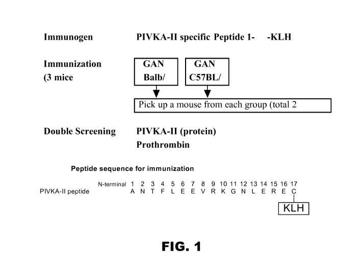
FIG.1 is a schematic diagram showing use of Peptide KLH to immunize three
germinal
center-associated DNA primase (GANP) transgenic Balb/c mice and three GANP
transgenic
C57BL/6 mice. FIG. 2 is a bar graph of the results of hybridoma
screening using sandwich reactivity
using mAb 3C10 (anti-PIVKA-II 17-24).
FIG. 3 shows the binding curve of mAb 6H6 (anti-PIVKA-II 1-13) and an A1exa488
labeled PIVKA-II peptide (1-13), showing a Kd of mAb 6H6 for the PIVKA-II 1-13
peptide of
37 4nM.
FIG. 4 shows the binding curve of mAb 6H6 and an A1exa488 labeled prothrombin
peptide (1-13), showing a Kd of the 6H6 mAb for the prothrombin peptide of 4.6
0.5 04.
antibody reactivity.FIG. 5A is a schematic diagram of the human PIVKA II
molecule showing sites of
10
WO 2012/018476 CA 02806029 2013-01-18
PCT/US2011/043316
FIG. 5B is a schematic diagram of an automated immunoassay format using mAb
6H6.
FIG. 6 is a graph of RLU (relative light units) values for each of six PIVKA-
II calibrators
used to measure performance of the control conjugate (MAC1-18) and the 6H6
conjugate,
according to the assay format show in FIG. 5.
FIG. 7 is a graph of the correlation of assay results using MAC1-18 versus
Picolumi
across a Picolumi value range of 0-20000 mAU/mL.
FIG. 8 is a graph of the correlation of assay results using 6H6 versus
Picolumi across a
Picolumi value range of 0-20000 mAU/mL.
FIG. 9 illustrates the reactivity to PIVKA-II and Prothrombin in connection
with the top
five selected hybridomas in each group, showing reactivity of hybridomas from
GANP
transgenic mice or wild type mice to 35 PIVKA-II and Prothrombin.
FIG. 10 shows the signals of several antibodies, showing strong reactivity of
Mab 3C10
to the PIVKA-II antigen.
FIG. 11 shows the subtracted PIVKA-II signal and background in connection with
the
procedure noted in Example 6.
FIG. 12 illustrates the equilibrium dissociation constants (Li) of antigens
measured in
direct binding experiments, in which Alexa-488 labeled PIVKA-II Gla domain
peptide (13-27)
was kept at 0.05nM, while the concentration of BHQ-mAb varied from 50nM to
0.0002nM.
FIG. 13 illustrates the equilibrium dissociation constants (Li) of antigens
measured in
direct binding experiments, in which Alexa-488 labeled PIVKA-II Gla domain
peptide (13-27)
was kept at 0.2nM, while the concentration of mAbs varied from 1pM to sub nano-
molar.
FIG. 14 illustrates FCS measurements of individual samples in which A1exa488-
PIVKA-
II peptide (13-27 cyc) was premixed with mAb 3C10 and various amounts of Glu-
substituted
peptide (G1a14, Gla 16, Gla 19, Gla 20, Gla 25, G1a26) were then added to the
antigen-antibody
complex.FIG. 15 illustrates additional FCS measurements of each sample in
which A1exa488-
PIVKA-II peptide (13-27 cyc) was premixed with mAb 3C10, and various amounts
of PIVKA-II
from different preparations were added to the antigen- antibody complex.
FIG. 16 illustrates the results obtained when competitive binding measurements
of
various PIVKA-II Gla domain (13-27) analogs with A1exa488-PIVKA-II (13-27) and
mAb 3C10
were used to test cross-reactivity with mAb 3C10.
11
CA 02806029 2013-01-18
WO 2012/018476
PCT/US2011/043316
DETAILED DESCRIPTION OF THE INVENTION
A. Introduction and Definitions
The GLA domain of the PIVKA-II protein consists of amino acids 1-46 (or 44-88
of
prothrombin sequence), including ten GLA amino acids. The PIVKA protein exists
in multiple
forms that vary as to the position and number of decarboxylated GLA's.
Currently available
immunoassays for PIVKA-II detect only a portion of the protein, primarily the
sequence of
amino acids 17-23 of the cyclic disulfide bond, and surrounding sequences,
i.e., amino acids 13-
27. As a result, GLA's outside of amino acids 17-23 including decarboxylated
GLA's are not
detected. The new antibodies and methods disclosed herein provide a way to
detect amino acids
1-17 of PIVKA, and the decarboxylated residues in the region of amino acids 17-
23. This can be
achieved, for example, by using a first anti-PIVKA antibody having an antigen
binding portion
that binds to amino acids 13-27 of PIVKA-II, and a second anti-PIVKA antibody
having an
antigen binding portion that binds to amino acids 1-13 of PIVKA-II. The second
antibody can
strongly react with decarboxylated amino acid residues of PIVKA, and
moderately react with the
carboxylated (normal) amino acid residues. Use of both antibodies in an assay
can detect both
PIVKA 13-27 and PIKVA 1-27 with a high level of specificity, and thus
generates a stronger
signal than that generated by detection of PIKVA 13-27 alone.
The present disclosure thus provides a binding protein and, in particular, a
monoclonal
antibody, hereinafter "6H6" that binds to one or more epitopes of PIVKA-II,
with a Kd for the
PIVKA-II peptide of 37 4 nM or less, and preferably in a range of lx1 0 -9 M
or greater,
preferably about lx10 10M- or greater. In particular, the binding protein or
antibody of the
present disclosure has a dissociation constant (KO to the 1-13 amino acid
region of PIVKA-II of
about 1 xl 0 -9 M or greater, preferably about 1x10 -10 M or greater. The
antibody is thus capable
of specifically recognizing and binding to PIVKA-II. Once it is bound to PIVKA-
II, it is not
replaced by prothrombin. In a situation in which the antibody is exposed to
PIVKA-II and
prothrombin at the same time, it is noteworthy that the 6H6 antibody of the
present disclosure
has about 10 to about 1000 times lower affinity to prothrombin than to PIVKA-
II. The subject
invention also includes isolated nucleotide sequences (and fragments thereof)
encoding the
variable light and heavy chains of the antibodies of the present disclosure as
well as those
nucleotide sequences (or fragments thereof) having sequences comprising,
corresponding to,
identical to, hybridizable to, or complementary to, at least about 70% (e.g.,
70% 71%, 72%,
12
CA 02806029 2013-01-18
WO 2012/018476 PCT/US2011/043316
73%, 74%, 75%, 76%, 77%, 78% or 79%), preferably at least about 80% (e.g.,
80%, 81%, 82%,
83%, 84%, 85%, 86%, 87%, 88% or 89%), and more preferably at least about 90%
(e.g, 91%,
92%, 93%, 94%, 95%, 96%, 97%, 98%, 99% or 100%) identity to these encoding
nucleotide
sequences. (All integers (and portions thereof) between and including 70% and
100% are
considered to be within the scope of the present disclosure with respect to
percent identity.)
Such sequences may be derived from any source (e.g., either isolated from a
natural source,
produced via a semi-synthetic route, or synthesized de novo). In particular,
such sequences may
be isolated or derived from sources other than described in the examples
(e.g., bacteria, fungus,
algae, mouse or human). In addition to the nucleotide sequences described
above, the present
disclosure also includes amino acid sequences of the variable light and heavy
chains of the
antibodies described herein (or fragments of these amino acid sequences).
Further, the present disclosure also includes amino acid sequences (or
fragments thereof)
comprising, corresponding to, identical to, or complementary to at least about
70% (e.g., 70%,
71%, 72%, 73%, 74%, 75%, 76%, 77%, 78% or 79%), preferably at least about 80%
(e.g., 80%
81%, 82%, 83%, 84%, 85%, 86%, 87%, 88% or 89%), and more preferably at least
about 90%
identity (e.g., 90%, 91%, 92%, 93%, 94%, 95%, 96%, 97%, 98%, 99% or 100%), to
the amino
acid sequences of the proteins of the present disclosure. (Again, all integers
(and portions
thereof) between and including 70% and 100% (as recited in connection with the
nucleotide
sequence identities noted above) are also considered to be within the scope of
the present
disclosure with respect to percent identity.) For purposes of the present
disclosure, a "fragment"
of a nucleotide sequence is defined as a contiguous sequence of approximately
at least 6,
preferably at least about 8, more preferably at least about 10 nucleotides,
and even more
preferably at least about 15 nucleotides corresponding to a region of the
specified nucleotide
sequence. The term "identity" refers to the relatedness of two sequences on a
nucleotide-by-
nucleotide basis over a particular comparison window or segment. Thus,
identity is defined as
the degree of sameness, correspondence or equivalence between the same strands
(either sense or
antisense) of two DNA segments (or two amino acid sequences). "Percentage of
sequence
identity" is calculated by comparing two optimally aligned sequences over a
particular region,
determining the number of positions at which the identical base or amino acid
occurs in both
sequences in order to yield 15 the number of matched positions, dividing the
number of such
positions by the total number of positions in the segment being compared and
multiplying the
13
CA 02806029 2013-01-18
WO 2012/018476
PCT/US2011/043316
result by 100. Optimal alignment of sequences may be conducted by the
algorithm of Smith &
Waterman, App. Math. 2:482 (1981), by the algorithm of Needleman & Wunsch, J.
Mol. Biol.
48:443 (1970), by the method of Pearson & Lipman, Proc. Natl. Acad. Sci. (USA)
85:2444
(1988) and by computer programs which implement the relevant algorithms (e.g.,
Clustal Macaw
Pileup (http://cmgm.stanford.edu/biochem218/11Multiple.pdf; Higgins et al.,
CABIOS. 5L151-
153 (1989)), FASTDB (Intelligenetics), BLAST (National Center for Biomedical
Information;
Altschul et al., Nucleic Acids Research 25:3389-3402 (1997)), PILEUP (Genetics
Computer
Group, Madison, WI) or GAP, BESTFIT, FASTA and TFASTA (Wisconsin Genetics
Software
Package Release 7.0, Genetics Computer Group, Madison, WI). (See U.S. Patent
No.
5,912,120.)For purposes of the present disclosure, "complementarity" is
defined as the degree of
relatedness between two DNA segments. It is determined by measuring the
ability of the sense
strand of one DNA segment to hybridize with the antisense strand of the other
DNA segment,
under appropriate conditions, to form a double helix. A "complement" is
defined as a sequence
which pairs to a given sequence based upon the canonic base-pairing rules. For
example, a
sequence A-G-T in one nucleotide strand is "complementary" to T-C-A in the
other strand. In the
double helix, adenine appears in one strand, thymine appears in the other
strand. Similarly,
wherever guanine is found in one strand, cytosine is found in the other. The
greater the
relatedness between the nucleotide sequences of two DNA segments, the greater
the ability to
form hybrid duplexes between the strands of the two DNA segments. "Similarity"
between two
amino acid sequences is defined as the presence of a series of identical as
well as conserved
amino acid residues in both sequences. The higher the degree of similarity
between two amino
acid sequences, the higher the correspondence, sameness or equivalence of the
two
sequences. ("Identity between two amino acid sequences is defined as the
presence of a series of
exactly alike or invariant amino acid residues in both sequences.) The
definitions of
"complementarity", "identity" and "similarity" are well known to those of
ordinary skill in the
art. "Encoded by" refers to a nucleic acid sequence which codes for a
polypeptide sequence,
wherein the polypeptide sequence or a portion thereof contains an amino acid
sequence of at
least 3 amino acids, more preferably at least 8 amino acids, and even more
preferably at least 15
amino acids from a polypeptide encoded by the nucleic acid sequence.
14
WO 2012/018476 CA 02806029 2013-01-18 PCT/US2011/043316
The term "biological activity" as used herein refers to all inherent
biological properties of
PIVKA-II. Such properties include, for example, the ability to bind to the
antibodies described
herein. "Functional equivalent" as used herein, refers to a protein (e.g., an
antibody) having the
same characteristics (e.g., binding affinity) of the antibodies of the present
disclosure.
The term "polypeptide" as used herein, refers to any polymeric chain of amino
acids. The
terms "peptide" and "protein" are used interchangeably with the term
polypeptide and also refer
to a polymeric chain of amino acids. The term "polypeptide" encompasses native
or artificial
proteins, protein fragments and polypeptide analogs of a protein sequence. A
polypeptide may be
monomeric or polymeric. The term "isolated protein" or "isolated polypeptide"
is a protein or
polypeptide that by virtue of its origin or source of derivation is not
associated with naturally
associated components that accompany it in its native state; is substantially
free of other proteins
from the same species; is expressed by a cell from a different species; or
does not occur in
nature. Thus, a polypeptide that is chemically synthesized or synthesized in a
cellular system
different from the cell from which it naturally originates will be "isolated"
from its naturally
associated components. A protein may also be rendered substantially free of
naturally associated
components by isolation, using protein purification techniques well known in
the art.
The term "recovering" as used herein, refers to the process of rendering a
chemical
species such as a polypeptide substantially free of naturally associated
components by isolation,
e.g., using protein purification techniques well known in the art.
The terms "binding", "specific binding" or "specifically binding", as used
herein, in
reference to the interaction of an antibody, a protein, or a peptide with a
second chemical
species, mean that the interaction is dependent upon the presence of a
particular structure (e.g.,
an antigenic determinant or epitope) on the chemical species; for example, an
antibody
recognizes and binds to a specific protein structure rather than to proteins
generally. If an
antibody is specific for epitope "A", the presence of a molecule containing
epitope A (or free,
unlabeled A), in a reaction containing labeled "A" and the antibody, will
reduce the amount of
labeled A bound to the antibody.
The term "antibody", as used herein, broadly refers to any immunoglobulin (Ig)
molecule
comprised of four polypeptide chains, two heavy (H) chains and two light (L)
chains, or any
functional fragment, mutant, variant, or derivation thereof, which retains the
essential epitope
binding features of an Ig molecule. Such mutant, variant, or derivative
antibody formats are
15
CA 02806029 2013-01-18
WO 2012/018476 PCT/US2011/043316
known in the art. Nonlimiting embodiments of which are discussed below. In a
full-length
antibody, each heavy chain is comprised of a heavy chain variable region
(abbreviated herein as
HCVR or VH) and a heavy chain constant region. The heavy chain constant region
is comprised
of three domains, CH1, CH2 and CH3. Each light chain is comprised of a light
chain variable
region (abbreviated herein as LCVR or VL) and a light chain constant region.
The light chain
constant region is comprised of one domain, CL. The VH and VL regions can be
further
subdivided into regions of hypervariability, termed complementarity
determining regions (CDR),
interspersed with regions that are more conserved, termed framework regions
(FR). Each VH
and VL is composed of three CDRs and four FRs, arranged from amino-terminus to
carboxy-
terminus in the following order: FR1, CDR1, FR2, CDR2, FR3, CDR3, FR4.
Immunoglobulin
molecules can be of any type (e.g., IgG, IgE, IgM, IgD, IgA and IgY), class
(e.g., IgG 1, IgG2,
IgG3, IgG4, IgAl and IgA2) or subclass.
The term "antigen-binding portion" of an antibody (or simply "antibody
portion"), as
used herein, refers to one or more fragments of an antibody that retain the
ability to specifically
bind to an antigen (e.g., one or more epitopes of PIVKA-II). It has been shown
that the antigen-
binding function of an antibody can be performed by one or more fragments of a
full-length
antibody. Such antibody embodiments may also be bispecific, dual specific, or
multispecific,
specifically binding to two or more different antigens. Examples of binding
fragments
encompassed within the term "antigen-binding portion" of an antibody include
(i) a Fab
fragment, a monovalent fragment consisting of the VL, VH, CL and CH1 domains;
(ii) a F(ab') 2
fragment, a bivalent fragment comprising two Fab fragments linked by a
disulfide bridge at the
hinge region; (iii) a Fd fragment consisting of the VH and CH1 domains; (iv) a
Fv fragment
consisting of the VL and VH domains of a single arm of an antibody, (v) a dAb
fragment (Ward
et al., (1989) Nature 341:544-546, Winter et al., International App.
Publication No. WO
90/05144 Al herein incorporated by reference), which comprises a single
variable domain; and
(vi) an isolated complementarity determining region (CDR). Furthermore,
although the two
domains of the Fv fragment, VL and VH, are coded for by separate genes, they
can be joined,
using recombinant methods, by a synthetic linker that enables them to be made
as a single
protein chain in which the VL and VH regions pair to form monovalent molecules
(known as
single chain Fv (scFv); see e.g., Bird et al. (1988) Science 242:423-426; and
Huston et al. (1988)
Proc. Natl. Acad. Sci. USA 85:5879-5883). Such single chain antibodies are
also encompassed
16
CA 02806029 2013-01-18
WO 2012/018476 PCT/US2011/043316
within the term "antigen-binding portion" of an antibody. Other forms of
single chain antibodies,
such as diabodies, are also encompassed. Diabodies are bivalent, bispecific
antibodies in which
VH and VL domains are expressed on a single polypeptide chain, but using a
linker that is too
short to allow for pairing between the two domains on the same chain, thereby
forcing the
domains to pair with complementary domains of another chain and creating two
antigen binding
sites (see e.g., Holliger, P., et al. (1993) Proc. Natl. Acad. Sci. USA
90:6444-6448; Poljak, R.J.,
et al. (1994) Structure 2:1121- 1123). Such antibody binding portions are
known in the art
(Kontermann and Dubel eds., Antibody Engineering (2001) Springer-Verlag. New
York. 790 pp.
(ISBN 3-540-41354-5).
The term "antibody construct" as used herein refers to a polypeptide
comprising one or
more the antigen binding portions of the present disclosure linked to a linker
polypeptide or an
immunoglobulin constant domain. Linker polypeptides comprise two or more amino
acid
residues joined by peptide bonds and are used to link one or more antigen
binding portions. Such
linker polypeptides are well known in the art (see e.g., Holliger, P., et al.
(1993) Proc. Natl.
Acad. Sci. USA 90:6444-6448; Poljak, R.J., et al. (1994) Structure 2:1121-
1123). An
immunoglobulin constant domain refers to a heavy or light chain constant
domain. Human IgG
heavy chain and light chain constant domain amino acid sequences are known in
the art. Still
further, an antibody or antigen-binding portion thereof may be part of a
larger immunoadhesion
molecule, formed by covalent or noncovalent association of the antibody or
antibody portion
with one or more other proteins or peptides. Examples of such immunoadhesion
molecules
include use of the streptavidin core region to make a tetrameric scFv molecule
(Kipriyanov,
S.M., et al. (1995) Human Antibodies and Hybridomas 6:93-101) and use of a
cysteine residue, a
marker peptide and a C-terminal polyhistidine tag to make bivalent and
biotinylated scFv
molecules (Kipriyanov, S.M., et al. (1994) Mol. Immunol. 31:1047-1058).
Antibody portions,
such as Fab and F(ab') 2 fragments, can be prepared from whole antibodies
using conventional
techniques, such as papain or pepsin digestion, respectively, of whole
antibodies. Moreover,
antibodies, antibody portions and immunoadhesion molecules can be obtained
using standard
recombinant DNA techniques, as described herein.
An "isolated antibody", as used herein, is intended to refer to an antibody
that is
substantially free of other antibodies having different antigenic
specificities (e.g., an isolated
antibody that specifically binds at least one epitope of PIVKA-II with which
the antibodies of the
17
CA 02806029 2013-01-18
WO 2012/018476 PCT/US2011/043316
present disclosure are reactive and is substantially free of antibodies that
specifically bind
antigens or epitopes other than those present within PIVKA-II.
The terms "Kabat numbering", "Kabat definitions" and "Kabat labeling" are used
interchangeably herein. These terms, which are recognized in the art, refer to
a system
of numbering amino acid residues which are more variable (i.e. hypervariable)
than other amino
acid residues in the heavy and light chain variable regions of an antibody, or
an antigen binding
portion thereof (Kabat et al. (1971) Ann. NY Acad, Sci. 190:382-391 and Kabat,
E.A., et al.
(1991) Sequences of Proteins of Immunological Interest, Fifth Edition, U.S.
Department of
Health and Human Services, NIH Publication No. 91-3242).
As used herein, the term "CDR" refers to the complementarity determining
region within
antibody variable sequences. There are three CDRs in each of the variable
regions of the heavy
chain and the light chain, which are designated CDR1, CDR2 and CDR3, for each
of the
variable regions.
The term "CDR set" as used herein refers to a group of three CDRs that occur
in a single
variable region capable of binding the antigen. The exact boundaries of these
CDRs have been
defined differently according to different systems. The system described by
Kabat (Kabat et al.,
Sequences of Proteins of Immunological Interest (National Institutes of
Health, Bethesda, MD
(1987) and (1991)) not only provides an unambiguous residue numbering system
applicable to
any variable region of an antibody, but also provides precise residue
boundaries defining the
three CDRs. These CDRs may be referred to as Kabat CDRs. Chothia and coworkers
(Chothia
& Lesk, J. Mol. Biol. 196:901-917 (1987) and Chothia et al., Nature 342:877-
883 (1989)) found
that certain sub- portions within Kabat CDRs adopt nearly identical peptide
backbone conformations, despite having great diversity at the level of amino
acid sequence.
These sub-portions were designated as Ll, L2 and L3 or H1, H2 and H3 where the
"L" and the
"H" designates the light chain and the heavy chains regions, respectively.
These regions may be
referred to as Chothia CDRs, which have boundaries that overlap with Kabat
CDRs. Other
boundaries defining CDRs overlapping with the Kabat CDRs have been described
by Padlan
(FASEB J. 9:133-139 (1995)) and MacCallum (LT Mol Riot 262(5):732-45 (1996)).
Still other
CDR boundary definitions may not strictly follow one of the above systems, but
will nonetheless
overlap with the Kabat CDRs, although they may be shortened or lengthened in
light
of prediction or experimental findings that particular residues or groups of
residues or even entire
18
CA 02806029 2013-01-18
WO 2012/018476 PCT/US2011/043316
CDRs do not significantly impact antigen binding. The methods used herein may
utilize CDRs
defined according to any of these systems, although preferred embodiments use
Kabat or Chothia
defined CDRs.
As used herein, the term "canonical" residue refers to a residue in a CDR or
framework
that defines a particular canonical CDR structure as defined by Chothia et al.
(J. Mol. Biol.
196:901-907 (1987); Chothia et al., J. Mot. Biol. 227:799 (1992), both are
incorporated herein by
reference). According to Chothia et al., critical portions of the CDRs of many
antibodies have
nearly identical peptide backbone confirmations despite great diversity at the
level of amino acid
sequence. Each canonical structure specifies primarily a set of peptide
backbone torsion angles
for a contiguous segment of amino acid residues forming a loop.
As used herein, the term "key" residues refer to certain residues within the
variable region
that have more impact on the binding specificity and/or affinity of an
antibody, in particular a
humanized antibody. A key residue includes, but is not limited to, one or more
of the following:
a residue that is adjacent to a CDR, a potential glycosylation site (can be
either N- or 0-
glycosylation site), a rare residue, a residue capable of interacting with the
antigen, a
residue capable of interacting with a CDR, a canonical residue, a contact
residue between heavy
chain variable region and light chain variable region, a residue within the
Vernier zone, and a
residue in the region that overlaps between the Chothia definition of a
variable heavy chain
CDR1 and the Kabat definition of the first heavy chain framework. As used
herein, "Vernier"
zone refers to a subset of framework residues that may adjust CDR structure
and fine-tune the fit
to antigen as described by Foote and Winter (1992, J. Mot. Biol. 224:487-499,
which is
incorporated herein by reference). Vernier zone residues form a layer
underlying the CDRs and
may impact on the structure of CDRs and the affinity of the antibody.
The term "activity" includes activities such as the binding
specificity/affinity of an
antibody for an antigen, for example, the antigen or antigens which the
antibodies of the present
disclosure are reactive.
The term "epitope" includes any polypeptide determinant capable of specific
binding to
an immunoglobulin or T-cell receptor. In certain embodiments, epitope
determinants include
chemically active surface groupings of molecules such as amino acids, sugar
side chains,
phosphoryl, or sulfonyl and, in certain embodiments, may have specific three-
dimensional
structural characteristics, and/or specific charge characteristics. An epitope
is a region of an
19
CA 02806029 2013-01-18
WO 2012/018476
PCT/US2011/043316
antigen that is bound by an antibody. In certain embodiments, an antibody is
said to specifically
bind an antigen when it preferentially recognizes its target antigen in a
complex mixture of
proteins and/or macromolecules.
The term "surface plasmon resonance", as used herein, refers to an optical
phenomenon
that allows for the analysis of real-time biospecific interactions by
detection of alterations in
protein concentrations within a biosensor matrix, for example using the
BlAcore system
(Pharmacia Biosensor AB, Uppsala, Sweden and Piscataway, NJ). For further
descriptions, see
Jonsson, U., et al. (1993) Ann. Biol. Clin. 51:19-26; Jonsson, U., et al.
(1991)
Biotechniques 11:620-627; Johnsson, B., et al. (1995) J. Mol. Recognit. 8:125-
131; and
Johnnson, B., et al. (1991) Anal. Biochem. 198:268-277.
The term "Kon", as used herein, is intended to refer to the on rate constant
for association
of an antibody to the antigen to form the antibody/antigen complex as is known
in the art.
The term "Koff", as used herein, is intended to refer to the off rate constant
for
dissociation of an antibody from the antibody/antigen complex as is known in
the art.
The term" Kd", as used herein, is intended to refer to the dissociation
constant of a
particular antibody-antigen interaction as is known in the art.
The term "labeled binding protein" as used herein, refers to a protein with a
label
incorporated that provides for the identification of the binding protein.
Preferably, the label is a
detectable marker, e.g., incorporation of a radiolabeled amino acid or
attachment to a
polypeptide of biotinyl moieties that can be detected by marked avidin (e.g.,
streptavidin containing a fluorescent marker or enzymatic activity that can be
detected by optical
or colorimetric methods). Examples of labels for polypeptides include, but are
not limited to,
the following: radioisotopes or radionuclides (e.g., 3H, 14C5 355 90y5 99Te5
"In, 1251 1311 177Lu 5 5
166Ho or 1535m); fluorescent labels (e.g., FITC, rhodamine, lanthanide
phosphors), enzymatic
labels (e.g., horseradish peroxidase, luciferase, alkaline phosphatase);
chemiluminescent
markers; biotinyl groups; predetermined polypeptide epitopes recognized by a
secondary reporter
(e.g., leucine zipper pair sequences, binding sites for secondary antibodies,
metal binding
domains, epitope tags); and magnetic agents, such as gadolinium chelates.
The term "antibody conjugate" refers to a binding protein, such as an
antibody,
chemically linked to a second chemical moiety, such as a therapeutic or
cytotoxic agent.
20
CA 02806029 2013-01-18
WO 2012/018476 PCT/US2011/043316
The term "agent" is used herein to denote a chemical compound, a mixture of
chemical
compounds, a biological macromolecule, or an extract made from biological
materials.
Preferably the therapeutic or cytotoxic agents include, but are not limited
to, pertussis toxin,
taxol, cytochalasin B, gramicidin D, ethidium bromide, emetine, mitomycin,
etoposide,
tenoposide, vincristine, vinblastine, colchicin, doxorubicin, daunorubicin,
dihydroxy anthracin
dione, mitoxantrone, mithramycin, actinomycin D, 1-dehydrotestosterone,
glucocorticoids,
procaine, tetracaine, lidocaine, propranolol, and puromycin and analogs or
homologs thereof
The terms "crystal", and "crystallized" as used herein, refer to an antibody,
or antigen-
binding portion thereof, that exists in the form of a crystal. Crystals are
one form of the solid
state of matter, which is distinct from other forms such as the amorphous
solid state or the liquid
crystalline state. Crystals are composed of regular, repeating, three-
dimensional arrays of atoms,
ions, molecules (e.g., proteins such as antibodies), or molecular assemblies
(e.g.,
antigen/antibody complexes). These three-dimensional arrays are arranged
according to specific
mathematical relationships that are well-understood in the field. The
fundamental unit,
or building block, that is repeated in a crystal is called the asymmetric
unit. Repetition of the
asymmetric unit in an arrangement that conforms to a given, well-defined
crystallographic
symmetry provides the "unit cell" of the crystal. Repetition of the unit cell
by regular
translations in all three dimensions provides the crystal. See Giege, R. and
Ducruix, A. Barrett,
Crystallization of Nucleic Acids and Proteins, a Practical Approach, 2nd ed.,
pp. 20 1-16,
Oxford University Press, New York, New York, (1999).
The term "polynucleotide" as referred to herein means a polymeric form of two
or more
nucleotides, either ribonucleotides or deoxvnucleotides or a modified form of
either type of
nucleotide. The term includes single and double stranded forms of DNA but
preferably is
double-stranded DNA.
The term "isolated polynucleotide" as used herein shall mean a polynucleotide
(e.g., of
genomic, cDNA, or synthetic origin, or some combination thereof) that, by
virtue of its origin, is
not associated with all or a portion of a polynucleotide with which the
"isolated polynucleotide"
is found in nature; is operably linked to a polynucleotide that it is not
linked to in nature; or does
not occur in nature as part of a larger sequence.
The term "vector", as used herein, is intended to refer to a nucleic acid
molecule capable
of transporting another nucleic acid to which it has been linked. One type of
vector is a
21
CA 02806029 2013-01-18
WO 2012/018476 PCT/US2011/043316
"plasmid", which refers to a circular double stranded DNA loop into which
additional DNA
segments may be ligated. Another type of vector is a viral vector, wherein
additional DNA
segments may be ligated into the viral genome. Certain vectors are capable of
autonomous
replication in a host cell into which they are introduced (e.g., bacterial
vectors having a bacterial
origin of replication and episomal mammalian vectors). Other vectors (e.g.,
non-episomal
mammalian vectors) can be integrated into the genome of a host cell upon
introduction into the
host cell, and thereby are replicated along with the host genome. Moreover,
certain vectors
are capable of directing the expression of genes to which they are operatively
linked. Such
vectors are referred to herein as "recombinant expression vectors" (or simply,
"expression vectors"). In general, expression vectors of utility in
recombinant DNA techniques
are often in the form of plasmids. In the present specification, "plasmid" and
"vector" may
be used interchangeably as the plasmid is the most commonly used form of
vector. However, the
invention is intended to include such other forms of expression vectors, such
as viral
vectors (e.g., replication defective retroviruses, adenoviruses and adeno-
associated viruses),
which serve equivalent functions.
The term "operably linked" refers to a juxtaposition wherein the components
described
are in a relationship permitting them to function in their intended manner. A
control sequence
"operably linked" to a coding sequence is ligated in such a way that
expression of the coding
sequence is achieved under conditions compatible with the control sequences.
"Operably linked"
sequences include both expression control sequences that are contiguous with
the gene of interest
and expression control sequences that act in trans or at a distance to control
the gene of interest.
The term "expression control sequence" as used herein refers to polynucleotide
sequences
that are necessary to effect the expression and processing of coding sequences
to which they are
ligated. Expression control sequences include appropriate transcription
initiation, termination,
promoter and enhancer sequences; efficient RNA processing signals such as
splicing and
polyadenylation signals; sequences that stabilize cytoplasmic mRNA; sequences
that enhance
translation efficiency (i.e., Kozak consensus sequence); sequences that
enhance protein stability;
and when desired, sequences that enhance protein secretion. The nature of such
control sequences differs depending upon the host organism; in prokaryotes,
such control
sequences generally include promoter, ribosomal binding site, and
transcription termination
22
CA 02806029 2013-01-18
WO 2012/018476 PCT/US2011/043316
sequence; in eukaryotes, generally, such control sequences include promoters
and transcription
termination sequence.
The term "control sequences" is intended to include components whose presence
is
essential for expression and processing, and can also include additional
components
whose presence is advantageous, for example, leader sequences and fusion
partner
sequences. "Transformation", as defined herein, refers to any process by which
exogenous DNA
enters a host cell. Transformation may occur under natural or artificial
conditions using various
methods well known in the art. Transformation may rely on any known method for
the
insertion of foreign nucleic acid sequences into a prokaryotic or eukaryotic
host cell. The method
is selected based on the host cell being transformed and may include, but is
not limited to, viral
infection, electroporation, lipofection, and particle bombardment. Such
"transformed" cells
include stably transformed cells in which the inserted DNA is capable of
replication either as an
autonomously replicating plasmid or as part of the host chromosome. They also
include cells
that transiently express the inserted DNA or RNA for limited periods of time.
The term "recombinant host cell" (or simply "host cell"), as used herein, is
intended to
refer to a cell into which exogenous DNA has been introduced. It should be
understood that such
terms are intended to refer not only to the particular subject cell but also
to the progeny of such
a cell. Because certain modifications may occur in succeeding generations due
to either mutation
or environmental influences, such progeny may not, in fact, be identical to
the parent cell, but are
still included within the scope of the term "host cell" as used herein.
Preferably, host
cells include prokaryotic and eukaryotic cells selected from any of the
Kingdoms of life.
Preferred eukaryotic cells include protist, fungal, plant and animal cells.
Most preferably, host
cells include but are not limited to the prokaryotic cell line E. coli;
mammalian cell lines CHO,
HEK 293 and COS; the insect cell line Sf9; and the fungal cell
Saccharomyces cerevisiae. Standard techniques may be used for recombinant
DNA, oligonucleotide synthesis, and tissue culture and transformation (e.g.,
electroporation,
lipofection). Enzymatic reactions and purification techniques may be performed
according to
manufacturer's specifications or as commonly accomplished in the art or as
described herein. The
foregoing techniques and procedures may be generally performed according to
conventional
methods well known in the art and as described in various general and more
specific references
that are cited and discussed throughout the present specification. See e.g.,
Sambrook et al.
23
CA 02806029 2013-01-18
WO 2012/018476 PCT/US2011/043316
Molecular Cloning: A Laboratory Manual (2d ed., Cold Spring Harbor Laboratory
Press, Cold
Spring Harbor, N.Y. (1989)), which is incorporated herein by reference for any
purpose. "Transgenic organism", as known in the art and as used herein, refers
to an organism
having cells that contain a transgene, wherein the transgene introduced into
the organism (or an
ancestor of the organism) expresses a polypeptide not naturally expressed in
the organism. A
"transgene" is a DNA construct, which is stably and operably integrated into
the genome of a cell
from which a transgenic organism develops, directing the expression of an
encoded gene product
in one or more cell types or tissues of the transgenic organism.
The terms "regulate" and "modulate" are used interchangeably, and, as used
herein, refers
to a change or an alteration in the activity of a molecule of interest
Modulation may be an
increase or a decrease in the magnitude of a certain activity or function of
the molecule of
interest. Exemplary activities and functions of a molecule include, but are
not limited to, binding
characteristics, enzymatic activity, cell receptor activation, and signal
transduction. Correspondingly, the term "modulator," as used herein, is a
compound capable of
changing or altering an activity or function of a molecule of interest. For
example, a
modulator may cause an increase or decrease in the magnitude of a certain
activity or function of
a molecule compared to the magnitude of the activity or function observed in
the absence of the
modulator. In certain embodiments, a modulator is an inhibitor, which
decreases the magnitude
of at least one activity or function of a molecule. Exemplary inhibitors
include, but are not
limited to, proteins, peptides, antibodies, peptibodies, carbohydrates or
small organic molecules.
Peptibodies are described, e.g., in International Application Publication No.
WO 01/83525.
The term "agonist", as used herein, refers to a modulator that, when contacted
with a
molecule of interest, causes an increase in the magnitude of a certain
activity or function of the
molecule compared to the magnitude of the activity or function observed in the
absence of the
agonist.
The term "antagonist" or "inhibitor", as used herein, refer to a modulator
that, when
contacted with a molecule of interest causes a decrease in the magnitude of a
certain activity or
function of the molecule compared to the magnitude of the activity or function
observed in the
absence of the antagonist.
As used herein, the term "effective amount" refers to the amount of a therapy
which is
sufficient to reduce or ameliorate the severity and/or duration of a disorder
or one or more
24
CA 02806029 2013-01-18
WO 2012/018476 PCT/US2011/043316
symptoms thereof, prevent the advancement of a disorder, cause regression of a
disorder, prevent
the recurrence, development, onset or progression of one or more symptoms
associated with a
disorder, detect a disorder, or enhance or improve the prophylactic or
therapeutic effect(s) of
another therapy (e.g., prophylactic or therapeutic agent).
The term "sample", as used herein, is used in its broadest sense. A
"biological sample", as
used herein, includes, but is not limited to, any quantity of a substance from
a living thing or
formerly living thing. Such living things include, but are not limited to,
humans, mice,
rats, monkeys, dogs, rabbits and other mammalian or non-mammalian animals.
Such substances
include, but are not limited to, blood, serum, urine, synovial fluid, cells,
organs, tissues (e.g.,
brain), bone marrow, lymph nodes, cerebrospinal fluid, and spleen.
Unless otherwise defined herein, scientific and technical terms used in
connection with
the present disclosure shall have the meanings that are commonly understood by
those of
ordinary skill in the art. The meaning and scope of the terms should be clear;
however, in the
event of any latent ambiguity, definitions provided herein take precedent over
any dictionary or
extrinsic definition. Further, unless otherwise required by context, singular
terms shall include
pluralities and plural terms shall include the singular.
In this application, the use of "or" means "and/or" unless stated otherwise.
Furthermore,
the use of the term "including", as well as other forms, such as "includes"
and "included", is not
limiting. Also, terms such as "element" or "component" encompass both elements
and
components comprising one unit and elements and components that comprise more
than one
subunit unless specifically stated otherwise. Generally, nomenclatures used in
connection with,
and techniques of, cell and tissue culture, molecular biology, immunology,
microbiology,
genetics and protein and nucleic acid chemistry and hybridization described
herein are those well
known and commonly used in the art.
As used herein, the term "hydrogen peroxide generating enzyme" refers to an
enzyme
that is capable of producing as a reaction product the chemical compound
having the molecular
formula H202, i.e. hydrogen peroxide. Non-limiting examples of hydrogen
peroxide generating
enzymes are listed below in Table A.
25
CA 02806029 2013-01-18
WO 2012/018476 PCT/US2011/043316
Table A
ACCEPTED COMMON NAME IUBMB ENZYME PREFERRED
NOMENCLATURE SUBSTRATE
(R)-6-hydroxynicotine oxidase EC 1.5.3.6 (R)-6-hydroxynicotine
(S)-2-hydroxy acid oxidase EC 1.1.3.15 S)-2-hydroxy acid
(S)-6-hydroxynicotine oxidase EC 1.5.3.5 (S)-6-hydroxynicotine
3-aci-nitropropanoate oxidase EC 1.7.3.5 3 -ac i-nitropropanoate
3-hydroxyanthranilate oxidase EC 1.10.3.5 3-hydroxyanthranilate
4-hydroxymandelate oxidase EC 1.1.3.19 (S)-2-hydroxy-2-(4-
hydroxyphenyl)acetate
6-hydroxynicotinate dehydrogenase EC 1.17.3.3 6-hydroxynicotinate
Abscisic-aldehyde oxidase EC 1.2.3.14 abscisic aldehyde
acyl-CoA oxidase EC 1.3.3.6 acyl-CoA
Alcohol oxidase EC 1.1.3.13 a primary alcohol
Aldehyde oxidase EC 1.2.3.1 an aldehyde
amine oxidase
amine oxidase (copper-containing) EC 1.4.3.6 primary monoamines,
diamines and histamine
amine oxidase (flavin-containing) EC 1.4.3.4 a primary amine
aryl-alcohol oxidase EC 1.1.3.7 an aromatic primary
alcohol
(2-naphthyl)methanol
3-methoxybenzyl
alcohol
aryl-aldehyde oxidase EC 1.2.3.9 an aromatic aldehyde
Catechol oxidase EC 1.1.3.14 Catechol
cholesterol oxidase EC 1.1.3.6 Cholesterol
Choline oxidase EC 1.1.3.17 Choline
columbamine oxidase EC 1.21.3.2 Columbamine
cyclohexylamine oxidase EC 1.4.3.12 Cyclohexylamine
cytochrome c oxidase EC 1.9.3.1
D-amino-acid oxidase EC 1.4.3.3 a D-amino acid
D-arabinono-1,4-lactone oxidase EC 1.1.3.37 D-arabinono-1,4-lactone
D-arabinono-1,4-lactone oxidase EC 1.1.3.37 D-arabinono-1,4-lactone
D-aspartate oxidase EC 1.4.3.1 D-aspartate
D-glutamate oxidase EC 1.4.3.7 D-glutamate
D-glutamate(D-aspartate) oxidase EC 1.4.3.15 D-glutamate
dihydrobenzophenanthridine EC 1.5.3.12 dihydrosanguinarine
oxidase
dihydroorotate oxidase EC 1.3.3.1 (S)-dihydroorotate
dihydrouracil oxidase EC 1.3.3.7 5,6-dihydrouracil
dimethylglycine oxidase EC 1.5.3.10 N,N-dimethylglycine
D-mannitol oxidase EC 1.1.3.40 Mannitol
26
CA 02806029 2013-01-18
WO 2012/018476 PCT/US2011/043316
Ecdysone oxidase EC 1.1.3.16 Ecdysone
ethanolamine oxidase EC 1.4.3.8 Ethanolamine
Galactose oxidase EC 1.1.3.9 D-galactose
Glucose oxidase EC 1.1.3.4 13-D-glucose
glutathione oxidase EC 1.8.3.3 Glutathione
Glycerol-3-phosphate oxidase EC 1.1.3.21 sn-glycerol 3-phosphate
Glycine oxidase EC 1.4.3.19 Glycine
glyoxylate oxidase EC 1.2.3.5 Glyoxylate
hexose oxidase EC 1.1.3.5 D-glucose,
D-galactose
D-mannose
maltose
lactose
cellobiose
hydroxyphytanate oxidase EC 1.1.3.27 L-2-hydroxyphytanate
indole-3-acetaldehyde oxidase EC 1.2.3.7 (indo1-3-yl)acetaldehyde
lactic acid oxidase Lactic acid
L-amino-acid oxidase EC 1.4.3.2 an L-amino acid
L-aspartate oxidase EC 1.4.3.16 L-aspartate
L-galactonolactone oxidase EC 1.3.3.12 L-galactono-1,4-lactone
L-glutamate oxidase EC 1.4.3.11 L-glutamate
L-gulonolactone oxidase EC 1.1.3.8 L-gulono-1,4-lactone
L-lysine 6-oxidase EC 1.4.3.20 L-lysine
L-lysine oxidase EC 1.4.3.14 L-lysine
long-chain-alcohol oxidase EC 1.1.3.20 A long-chain-alcohol
L-pipecolate oxidase EC 1.5.3.7 L-pipecolate
L-sorbose oxidase EC 1.1.3.11 L-sorbose
malate oxidase EC 1.1.3.3 (S)-malate
methanethiol oxidase EC 1.8.3.4 Methanethiol
monoamino acid oxidase
1V6-methyl-lysine oxidase EC 1.5.3.4 6-N-methyl-L-lysine
N-acylhexosamine oxidase EC 1.1.3.29 N-acetyl-D-glucosamine
N-glycolylglucosamine
N-acetylgalactosamine
N-acetylmannosamine.
NAD(P)H oxidase EC 1.6.3.1 NAD(P)H
nitroalkane oxidase EC 1.7.3.1 a nitroalkane
N-methyl-L-amino-acid oxidase EC 1.5.3.2 an N-methyl-L-amino
acid
nucleoside oxidase EC 1.1.3.39 Adenosine
Oxalate oxidase EC 1.2.3.4 Oxalate
polyamine oxidase EC 1.5.3.11 1-N-acetylspermine
polyphenol oxidase EC 1.14.18.1
Polyvinyl-alcohol oxidase EC 1.1.3.30 polyvinyl alcohol
prenylcysteine oxidase EC 1.8.3.5 an S-prenyl-L-cysteine
27
CA 02806029 2013-01-18
WO 2012/018476 PCT/US2011/043316
Protein-lysine 6-oxidase EC 1.4.3.13 peptidyl-L-lysyl-
peptide
putrescine oxidase EC 1.4.3.10 butane-1,4-diamine
Pyranose oxidase EC 1.1.3.10 D-glucose
D-xylose
L-sorbose
D-glucono-1,5-lactone
Pyridoxal 5'-phosphate synthase EC 1.4.3.5 pyridoxamine 5'-
phosphate
pyridoxine 4-oxidase EC 1.1.3.12 Pyridoxine
pyrroloquinoline-quinone synthase EC 1.3.3.11 6-(2-amino-2-
carboxyethyl)-7,8-
dioxo-1,2,3,4,5,6,7,8-
octahydroquinoline-2,4-
dicarboxylate
Pyruvate oxidase EC 1.2.3.3 Pyruvate
Pyruvate oxidase (CoA-acetylating) EC 1.2.3.6 Pyruvate
Reticuline oxidase EC 1.21.3.3 Reticuline
retinal oxidase EC 1.2.3.11 Retinal
Rifamycin-B oxidase EC 1.10.3.6 rifamycin-B
Sarcosine oxidase EC 1.5.3.1 Sarcosine
secondary-alcohol oxidase EC 1.1.3.18 a secondary alcohol
sulfite oxidase EC 1.8.3.1 Sulfite
superoxide dismutase EC 1.15.1.1 Superoxide
superoxide reductase EC 1.15.1.2 Superoxide
tetrahydroberberine oxidase EC 1.3.3.8 (S)-tetrahydroberberine
Thiamine oxidase EC 1.1.3.23 Thiamine
tryptophan a,13-oxidase EC 1.3.3.10 L-tryptophan
urate oxidase (uricase, uric acid EC 1.7.3.3 uric acid
oxidase)
Vanillyl-alcohol oxidase EC 1.1.3.38 vanillyl alcohol
Xanthine oxidase EC 1.17.3.2 Xanthine
xylitol oxidase EC 1.1.3.41 Xylitol
The methods and techniques of the present disclosure are generally performed
according
to conventional methods well known in the art and as described in various
general and more
specific references that are cited and discussed throughout the present
specification unless
otherwise indicated. Enzymatic reactions and purification techniques are
performed according
to manufacturer's specifications, as commonly accomplished in the art or as
described herein.
The nomenclatures used in connection with, and the laboratory procedures and
techniques of,
analytical chemistry, synthetic organic chemistry, and medicinal and
pharmaceutical chemistry
described herein are those well known and commonly used in the art. Standard
techniques are
28
CA 02806029 2013-01-18
WO 2012/018476 PCT/US2011/043316
used for chemical syntheses, chemical analyses, pharmaceutical preparation,
formulation, and
delivery, and treatment of patients.
According to the invention and, in particular, for the purpose of assessing
the binding
affinities of the antibodies of the present disclosure, a process may be used
as described in
International Application Publication No. WO 2004/067561, which is
incorporated herein by
reference in its entirety. The process comprises unfolding a natural,
recombinant or
synthetic peptide or a derivative thereof; exposing the at least partially
unfolded peptide or
derivative thereof to a detergent, reducing the detergent action and
continuing incubation. For the
purpose of unfolding the peptide, hydrogen bond breaking agents such as, for
example,
hexafluoroisopropanol (HFIP) may be allowed to act on the protein. Times of
action of a few
minutes, for example about 10 to 60 minutes, are sufficient when the
temperature of action is
from about 20 to 50 C and in particular about 35 to 40 C. Subsequent
dissolution of the residue
evaporated to dryness, preferably in concentrated form, in suitable organic
solvents miscible with
aqueous buffers, such as, for example, dimethyl sulfoxide (DMSO), results in a
suspension of the
at least partially unfolded peptide or derivative thereof, which can be used
subsequently. If
required, the stock suspension may be stored at low temperature, for example
at about -20 C, for
an interim period. Alternatively, the peptide or the derivative thereof may be
taken up in slightly
acidic, preferably aqueous, solution, for example, an about 10 mM aqueous HC1
solution. After
an incubation time of usually a few minutes, insoluble components are removed
by
centrifugation. A few minutes at 10000 g is expedient. These method steps are
preferably carried
out at room temperature, i.e. a temperature in the range from 20 C to 30 C.
The supernatant
obtained after centrifugation contains the peptide or the derivative thereof
and may be stored at
low temperature, for example at about -20 C, for an interim period.
B. Preparation of Monoclonal Antibodies
Monoclonal antibodies can be prepared using a wide variety of techniques known
in the
art including the use of hybridoma, recombinant, and phage display
technologies, or
a combination thereof For example, it is preferred that monoclonal antibodies
of the present
disclosure be produced using hybridoma techniques including those known in the
art and taught,
for example, in Harlow et al., Antibodies: A Laboratory Manual (Cold Spring
Harbor Laboratory
Press, 2nd ed. 1988); Hammerling, et al., in: Monoclonal Antibodies and T-Cell
Hybridomas
563-681 (Elsevier, N.Y., 1981) (said references incorporated by reference in
their entireties).
29
CA 02806029 2013-01-18
WO 2012/018476
PCT/US2011/043316
The term "monoclonal antibody" as used herein is not limited to antibodies
produced
through hybridoma technology. The term "monoclonal antibody" refers to an
antibody that is
derived from a single clone, including any eukaryotic, prokaryotic, or phage
clone, and not the
method by which it is produced.
Methods for producing and screening for specific antibodies using hybridoma
technology
are routine and well known in the art. In one embodiment, the present
disclosure provides
methods of generating monoclonal antibodies as well as antibodies produced by
the method
comprising culturing a hybridoma cell secreting an antibody of the present
disclosure wherein,
preferably, the hybridoma is generated by fusing splenocytes isolated from a
mouse immunized
with an antigen of the present disclosure with myeloma cells and then
screening the hybridomas
resulting from the fusion for hybridoma clones that secrete an antibody able
to bind a
polypeptide of the present disclosure. Briefly, mice can be immunized with the
antigen of
interest. In a preferred embodiment, the antigen is administered with an
adjuvant to stimulate the
immune response. Such adjuvants include complete or incomplete Freund's
adjuvant, RIBI
(muramyl dipeptides) or ISCOM (immunostimulating complexes). Such adjuvants
may
protect the polypeptide from rapid dispersal by sequestering it in a local
deposit, or they may
contain substances that stimulate the host to secrete factors that are
chemotactic for macrophages
and other components of the immune system. Preferably, if a polypeptide is
being administered,
the immunization schedule will involve two or more administrations of the
polypeptide, spread
out over several weeks.After immunization of an animal with the antigen,
antibodies and/or antibody-producing
cells may be obtained from the animal. An antibody-containing serum is
obtained from the
animal by bleeding or sacrificing the animal. The serum may be used as it is
obtained from the
animal, an immunoglobulin fraction may be obtained from the serum, or the
antibodies may be
purified from the serum. Serum or immunoglobulins obtained in this manner are
polyclonal,
thus having a heterogeneous array of properties.
Once an immune response is detected, e.g., antibodies specific for the antigen
are
detected in the mouse serum, the mouse spleen is harvested and splenocytes
isolated.
The splenocytes are then fused by well-known techniques to any suitable
myeloma cells, for
example cells from cell line 5P20 available from the American Type Culture
Collection
(Manassas, VA). Hybridomas are selected and cloned by limited dilution. The
hybridoma clones
30
CA 02806029 2013-01-18
WO 2012/018476 PCT/US2011/043316
are then assayed by methods known in the art for cells that secrete antibodies
capable of binding
to the peptide or antigen of interest. Ascites fluid, which generally contains
high levels of
antibodies, can be generated by immunizing mice with positive hybridoma
clones.
In another embodiment, antibody-producing immortalized hybridomas may be
prepared
from the immunized animal. After immunization, the animal is sacrificed and
the splenic B
cells are fused to immortalized myeloma cells as is well known in the art.
See, e.g., Harlow and
Lane, supra. In a preferred embodiment, the myeloma cells do not secrete
immunoglobulin polypeptides (a non-secretory cell line). After fusion and
antibiotic selection,
the hybridomas are screened using the antigen, or a portion thereof, or a cell
expressing
the antigen. In a preferred embodiment, the initial screening is performed
using an enzyme-
linked immunoassay (ELISA) or a radioimmunoassay (RIA), preferably an ELISA.
An example
of ELISA screening is provided in International Application Publication No. WO
00/37504,
herein incorporated by reference.
Antibody-producing hybridomas are selected, cloned and further screened for
desirable
characteristics, including robust hybridoma growth, high antibody production
and desirable
antibody characteristics, as discussed further below. Hybridomas may be
cultured and expanded
in vivo in syngeneic animals, in animals that lack an immune system, e.g.,
nude mice, or in cell
culture in vitro. Methods of selecting, cloning and expanding hybridomas are
well known
to those of ordinary skill in the art.
In a preferred embodiment, the hybridomas are mouse hybridomas, as described
above.
In another preferred embodiment, the hybridomas are produced in a non-human,
non-
mouse species such as rats, sheep, pigs, goats, cattle or horses. In another
embodiment, the
hybridomas are human hybridomas, in which a human non-secretory myeloma is
fused with a
human cell expressing the antibody.
C. Other Methods Of Producing Antibodies
As noted above, antibodies of the present disclosure may be produced by any of
a number
of techniques known in the art. For example, the antibody may be produced
based upon
expression from host cells, wherein expression vector(s) encoding the heavy
and light chains is
(are) transfected into a host cell by standard techniques. The various forms
of the term
"transfection" are intended to encompass a wide variety of techniques commonly
used for the
introduction of exogenous DNA into a prokaryotic or eukaryotic host cell,
e.g., electroporation,
31
CA 02806029 2013-01-18
WO 2012/018476 PCT/US2011/043316
calcium-phosphate precipitation, DEAE-dextran transfection and the like.
Although, it is
possible to express the antibodies of the present disclosure in either
prokaryotic or eukaryotic
host cells, expression of antibodies in eukaryotic cells is preferable, and
most preferable
in mammalian host cells, because such eukaryotic cells (and in particular
mammalian cells) are
more likely than prokaryotic cells to assemble and secrete a properly folded
and immunologically active antibody.
Preferred mammalian host cells for expressing the recombinant antibodies of
the present
disclosure include Chinese Hamster Ovary (CHO cells) (including dhfr- CHO
cells, described in
Urlaub and Chasin, (1980) Proc. Natl. Acad. Sci. USA 77:4216 -4220, used with
a DHFR
selectable marker, e.g., as described in R.J. Kaufman and P.A. Sharp (1982)
Mol. Biol. 159:601 -
621), NSO myeloma cells, COS cells and 5P2 cells. When recombinant expression
vectors
encoding antibody genes are introduced into mammalian host cells, the
antibodies are produced
by culturing the host cells for a period of time sufficient to allow for
expression of the antibody
in the host cells or, more preferably, secretion of the antibody into the
culture medium in which
the host cells are grown. Antibodies can be recovered from the culture medium
using
standard protein purification methods.
Host cells can also be used to produce functional antibody fragments, such as
Fab
fragments or scFv molecules. It will be understood that variations on the
above procedure are
within the scope of the present disclosure. For example, it may be desirable
to transfect a host
cell with DNA encoding functional fragments of either the light chain and/or
the heavy chain of
an antibody of this invention. Recombinant DNA technology may also be used to
remove some,
or all, of the DNA encoding either or both of the light and heavy chains that
is not necessary for
binding to the antigens of interest. The molecules expressed from such
truncated DNA molecules
are also encompassed by the antibodies of the present disclosure. In addition,
bifunctional
antibodies may be produced in which one heavy and one light chain are an
antibody of the
present disclosure and the other heavy and light chain are specific for an
antigen other than the
antigens of interest by crosslinking an antibody of the present disclosure to
a second antibody by
standard chemical crosslinking methods.
In a preferred system for recombinant expression of an antibody, or antigen-
binding
portion thereof, of the present disclosure, a recombinant expression vector
encoding both
the antibody heavy chain and the antibody light chain is introduced into dhfr-
CHO cells by
32
CA 02806029 2013-01-18
WO 2012/018476 PCT/US2011/043316
calcium phosphate-mediated transfection. Within the recombinant expression
vector,
the antibody heavy and light chain genes are each operatively linked to CMV
enhancer/AdMLP
promoter regulatory elements to drive high levels of transcription of the
genes. The recombinant
expression vector also carries a DHFR gene, which allows for selection of CHO
cells that have
been transfected with the vector using methotrexate selection/amplification.
The selected
transformant host cells are cultured to allow for expression of the antibody
heavy and light
chains and intact antibody is recovered from the culture medium. Standard
molecular biology
techniques are used to prepare the recombinant expression vector, transfect
the host cells, select
for transformants, culture the host cells and recover the antibody from the
culture medium. Still
further the invention provides a method of synthesizing a recombinant antibody
of the present
disclosure by culturing a host cell of the present disclosure in a suitable
culture medium until a
recombinant antibody of the present disclosure is synthesized. The method can
further comprise
isolating the recombinant antibody from the culture medium.
D. Preparation of Antibodies For Diagnostic and Other Applications
As noted above, preferably, antibodies of the present disclosure exhibit a
high binding
affinity to one or more epitopes of PIVKA-II, e.g., as assessed by any one of
several in vitro and
in vivo assays known in the art (e.g., see examples below).
In certain embodiments, the antibody comprises a heavy chain constant region,
such as an
IgGl, IgG2, IgG3, IgG4, IgA, IgE, IgM or IgD constant region. Preferably, the
heavy
chain constant region is an IgG1 heavy chain constant region or an IgG4 heavy
chain constant
region. Furthermore, the antibody can comprise a light chain constant region,
either a kappa light
chain constant region or a lambda light chain constant region. Preferably, the
antibody comprises
a kappa light chain constant region. Alternatively, the antibody portion can
be, for example, a
Fab fragment or a single chain Fv fragment.
Replacements of amino acid residues in the Fc portion to alter antibody
effector function
are known in the art (Winter, et al. U.S. Patent Nos. 5,648,260 and
5,624,821). The Fc portion of
an antibody mediates several important effector functions e.g. cytokine
induction, ADCC,
phagocytosis, complement dependent cytotoxicity (CDC) and half-life/clearance
rate of antibody
and antigen-antibody complexes. In some cases, these effector functions are
desirable
for therapeutic antibody but in other cases might be unnecessary or even
deleterious, depending
33
CA 02806029 2013-01-18
WO 2012/018476 PCT/US2011/043316
on the therapeutic objectives. Certain human IgG isotypes, particularly IgG1
and IgG3, mediate
ADCC and CDC via binding to FcyRs and complement Clq, respectively. Neonatal
Fc receptors
(FcRn) are the critical components determining the circulating half-life of
antibodies. In still
another embodiment, at least one amino acid residue is replaced in the
constant region of
the antibody, for example the Fc region of the antibody, such that effector
functions of the
antibody are altered.
One embodiment provides a labeled binding protein wherein an antibody or
antibody
portion of the present disclosure is derivatized or linked to another
functional molecule
(e.g., another peptide or protein). For example, a labeled binding protein of
the present disclosure
can be derived by functionally linking an antibody or antibody portion of the
present disclosure
(by chemical coupling, genetic fusion, noncovalent association or otherwise)
to one or more
other molecular entities, such as another antibody (e.g., a bispecific
antibody or a diabody),
a detectable agent, a cytotoxic agent, a pharmaceutical agent, and/or a
protein or peptide that can
mediate associate of the antibody or antibody portion with another molecule
(such as
a streptavidin core region or a polyhistidine tag).
Immunoassays according to the present disclosure employ one or more anti-PIVKA-
II
antibodies, each of which binds specifically to PIVKA-II. In certain
embodiments, an antibody
as disclosed herein has a detectable label. Detectable labels suitable for use
in the detection
antibodies of the present disclosure include any compound or composition
having a moiety that
is detectable by spectroscopic, photochemical, biochemical, immunochemical,
electrical, optical,
or chemical means. Such labels include, for example, an enzyme,
oligonucleotide, nanoparticle
chemiluminophore, fluorophore, fluorescence quencher, chemiluminescence
quencher, or biotin.
Thus for example, in an immunoassay employing an optical signal, the optical
signal is measured
as an analyte concentration dependent change in chemiluminescence,
fluorescence,
phosphorescence, electrochemiluminescence, ultraviolet absorption, visible
absorption, infrared
absorption, refraction, surface plasmon resonance. In an immunoassay employing
an electrical
signal, the electrical signal is measured as an analyte concentration
dependent change in current,
resistance, potential, mass to charge ratio, or ion count. In an immunoassay
employing a change-
of-state signal, the change of state signal is measured as an analyte
concentration dependent
change in size, solubility, mass, or resonance.
34
CA 02806029 2013-01-18
WO 2012/018476 PCT/US2011/043316
Useful labels according to the present disclosure include magnetic beads
(e.g.,
DynabeadsTm), fluorescent dyes (e.g., fluorescein, Texas Red, rhodamine, green
fluorescent
protein) and the like (see, e.g., Molecular Probes, Eugene, Oregon., USA),
chemiluminescent
compounds such as acridinium (e.g., acridinium-9-carboxamide),
phenanthridinium, dioxetanes,
luminol and the like, radiolabels (e.g., 3H, 1251, 35S, 14C, or 32P),
catalysts such as enzymes
(e.g., horse radish peroxidase, alkaline phosphatase, beta-galactosidase and
others commonly
used in an ELISA), and colorimetric labels such as colloidal gold (e.g., gold
particles in the 40-
80 nm diameter size range scatter green light with high efficiency) or colored
glass or plastic
(e.g., polystyrene, polypropylene, latex, etc.) beads. Patents teaching the
use of such labels
include U.S. Pat. Nos. 3,817,837; 3,850,752; 3,939,350; 3,996,345; 4,277,437;
4,275,149; and
4,366,241.
The label can be attached to each antibody prior to, or during, or after
contact with the
biological sample. So-called "direct labels" are detectable labels that are
directly attached to or
incorporated into the detection antibody prior to use in the assay. Direct
labels can be attached to
or incorporated into the antibody by any of a number of means well known to
those of skill in the
art. In contrast, so-called "indirect labels" typically bind to each antibody
at some point during
the assay. Often, the indirect label binds to a moiety that is attached to or
incorporated into the
detection agent prior to use. Thus, for example, each antibody can be
biotinylated before use in
an assay. During the assay, an avidin-conjugated fluorophore can bind the
biotin-bearing
detection agent, to provide a label that is easily detected.
In another example of indirect labeling, polypeptides capable of specifically
binding
immunoglobulin constant regions, such as polypeptide A or polypeptide G, can
also be used as
labels for detection antibodies. These polypeptides are normal constituents of
the cell walls of
streptococcal bacteria. They exhibit a strong non-immunogenic reactivity with
immunoglobulin
constant regions from a variety of species (see, generally Kronval, et al.
(1973)J. Immunol., 111:
1401-1406, and Akerstrom (1985) J. Immunol., 135: 2589-2542). Such
polypeptides can thus be
labeled and added to the assay mixture, where they will bind to each antibody,
as well as to the
autoantibodies, labeling all and providing a composite signal attributable to
analyte and
autoantibody present in the sample.
Some labels useful in the present disclosure may require the use of an
additional
reagent(s) to produce a detectable signal. In an ELISA, for example, an enzyme
label (e.g., beta-
35
CA 02806029 2013-01-18
WO 2012/018476 PCT/US2011/043316
galactosidase) will require the addition of a substrate (e.g., X-gal) to
produce a detectable signal.
In immunoassays using an acridinium compound as the direct label, a basic
solution and a source
of hydrogen peroxide are added.
In an exemplary embodiment, a chemiluminescent compound is used as a direct
label
conjugated to an antibody. The chemiluminescent compound can be for example an
acridinium
compound. When an acridinium compound is used as the detectable label, then
the above-
described method may further include generating or providing a source of
hydrogen peroxide to
the mixture resulting from contacting the test sample with the labeled
antibody, and adding at
least one basic solution to the mixture to generate a light signal. The light
signal generated or
emitted by the mixture is then measured to detect the PIVKA-II in the test
sample.
The source of hydrogen peroxide may be a buffer solution or a solution
containing
hydrogen peroxide or an enzyme that generates hydrogen peroxide when added to
the test
sample. The basic solution serves as a trigger solution, and the order in
which the at least one
basic solution and detectable label are added is not critical. The basic
solution used in the
method is a solution that contains at least one base and that has a pH greater
than or equal to 10,
preferably, greater than or equal to 12. Examples of basic solutions include,
but are not limited
to, sodium hydroxide, potassium hydroxide, calcium hydroxide, ammonium
hydroxide,
magnesium hydroxide, sodium carbonate, sodium bicarbonate, calcium hydroxide,
calcium
carbonate and calcium bicarbonate. The amount of basic solution added to the
test sample
depends on the concentration of the basic solution used in the assay. Based on
the concentration
of the basic solution used, one skilled in the art could easily determine the
amount of basic
solution to be used in the method described herein.
36
CA 02806029 2013-01-18
WO 2012/018476 PCT/US2011/043316
In a chemiluminescence immunoassay according to the present disclosure and
using an
acridinium compound as the detectable label, preferably the acridinium
compound is an
acridinium-9-carboxamide. Specifically, the acridinium-9-carboxamide has a
structure according
to formula I:
R1 Xe
I -7 Rs
R6 o R9
= i\TR2
R1 5 =
R12
R13
wherein Rl and R2 are each independently selected from the group consisting
of: alkyl, alkenyl,
alkynyl, aryl or aralkyl, sulfoalkyl, carboxyalkyl and oxoalkyl, and
wherein R3 through R15 are each independently selected from the group
consisting of:
hydrogen, alkyl, alkenyl, alkynyl, aryl or aralkyl, amino, amido, acyl,
alkoxyl, hydroxyl,
carboxyl, halogen, halide, nitro, cyano, sulfo, sulfoalkyl, carboxyalkyl and
oxoalkyl;
and further wherein any of the alkyl, alkenyl, alkynyl, aryl or aralkyl may
contain one or more
heteroatoms; and
optionally, if present, X is an anion.
Methods for preparing acridinium 9-carboxamides are described in Mattingly, P.
G. J.
Biolumin. Chemilumin., 6, 107-14; (1991); Adamczyk, M.; Chen, Y.-Y.,
Mattingly, P. G.; Pan,
Y. J. Org. Chem., 63, 5636-5639 (1998); Adamczyk, M.; Chen, Y.-Y.; Mattingly,
P. G.; Moore,
J. A.; Shreder, K. Tetrahedron, 55, 10899-10914 (1999); Adamczyk, M.;
Mattingly, P. G.;
Moore, J. A.; Pan, Y. Org. Lett., 1, 779-781 (1999); Adamczyk, M.; Chen, Y.-
Y.; Fishpaugh, J.
R.; Mattingly, P. G.; Pan, Y.; Shreder, K.; Yu, Z. Bioconjugate Chem., 11, 714-
724 (2000);
Mattingly, P. G.; Adamczyk, M. In Luminescence Biotechnology: Instruments and
Applications;
Dyke, K. V. Ed.; CRC Press: Boca Raton, pp. 77-105 (2002); Adamczyk, M.;
Mattingly, P. G.;
Moore, J. A.; Pan, Y. Org. Lett., 5, 3779-3782 (2003); and U.S. Patent Nos.
5,468,646,
37
WO 2012/018476 CA
02806029 2013-01-18
PCT/US2011/043316
5,543,524 and 5,783,699 (each incorporated herein by reference in their
entireties for their
teachings regarding same).
Alternatively, the acridinium compound can be an acridinium-9-carboxylate aryl
ester;
the acridinium-9-carboxylate aryl ester can have a structure according to
formula II:
R1 x0
R4 0 40/R3 11C) R7 R8
R5 R6 R1 R9
R15 00 RI 1
Ri 4 a R12
R13
I I
Examples of acridinium-9-carboxylate aryl esters having the above formula II
that can be
used in the present disclosure include, but are not limited to, 10-methy1-9-
(phenoxycarbonyl)acridinium fluorosulfonate (available from Cayman Chemical,
Ann Arbor,
MI). Methods for preparing acridinium 9-carboxylate aryl esters are described
in McCapra, F.,
et al., Photochem. Photobiol., 4, 1111-21(1965); Razavi, Z et al.,
Luminescence, 15:245-249
(2000); Razavi, Z et al., Luminescence, 15:239-244 (2000); and U.S. Patent No.
5,241,070 (each
incorporated herein by reference in their entireties for their teachings
regarding same).
Another embodiment of the present disclosure provides a crystallized binding
protein.
Preferably, the invention relates to crystals of whole antibodies and
fragments thereof as
disclosed herein, and formulations and compositions comprising such crystals.
In one
embodiment the crystallized binding protein has a greater half-life in vivo
than the soluble
counterpart of the binding protein. In another embodiment, the binding protein
retains biological
activity after crystallization. Crystallized binding protein of the present
disclosure may
be produced according methods known in the art and as disclosed in
International App.
Publication No. WO 02/072636, incorporated herein by reference. Another
embodiment of the
present disclosure provides a glycosylated binding protein wherein the
antibody or antigen
38
WO 2012/018476 CA 02806029 2013-01-18 PCT/US2011/043316
binding portion thereof comprises one or more carbohydrate residues. Nascent
in vivo protein
production may undergo further processing, known as post-translational
modification. In
particular, sugar (glycosyl) residues may be added enzymatically, a process
known as
glycosylation. The resulting proteins bearing covalently linked
oligosaccharide side chains are
known as glycosylated proteins or glycoproteins. Antibodies are glycoproteins
with one or
more carbohydrate residues in the Fc domain, as well as the variable domain.
Carbohydrate
residues in the Fc domain have important effect on the effector function of
the Fc domain, with
minimal effect on antigen binding or half-life of the antibody (R. Jefferis,
Biotechnol. Prog. 21
(2005), pp. 11- 16). In contrast, glycosylation of the variable domain may
have an effect on the
antigen binding activity of the antibody. Glycosylation in the variable domain
may have
a negative effect on antibody binding affinity, likely due to steric hindrance
(Co, M.S., et al.,
Mo/. Immunol. (1993) 30:1361-1367), or result in increased affinity for the
antigen (Wallick,
S.C., et al., Exp. Med. (1988) 168:1099-1109; Wright, A., et al., EMBO Jr.
(1991) 10:2717
2723).
One aspect of the present disclosure is directed to generating glycosylation
site mutants
in which the 0- or N- linked glycosylation site of the binding protein has
been mutated. One
skilled in the art can generate such mutants using standard well-known
technologies. The
creation of glycosylation site mutants that retain the biological activity but
have increased or
decreased binding activity are another object of the present disclosure.
In still another embodiment, the glycosylation of the antibody or antigen-
binding portion
of the present disclosure is modified. For example, an aglycoslated antibody
can be made (i.e.,
the antibody lacks glycosylation). Glycosylation can be altered to, for
example, increase the
affinity of the antibody for antigen. Such carbohydrate modifications can be
accomplished by,
for example, altering one or more sites of glycosylation within the antibody
sequence. For
example, one or more amino acid substitutions can be made that result in
elimination of one or
more variable region glycosylation sites to thereby eliminate glycosylation at
that site.
Such aglycosylation may increase the affinity of the antibody for antigen.
Such an approach is
described in further detail in International App. Publication No. WO
03/016466A2, and U.S. Pat.
Nos. 5,714,350 and 6,350,861, each of which is incorporated herein by
reference in its entirety.
Additionally or alternatively, a modified antibody of the present disclosure
can be made
that has an altered type of glycosylation, such as a hypofucosylated antibody
having reduced
39
CA 02806029 2013-01-18
WO 2012/018476 PCT/US2011/043316
amounts of fucosyl residues or an antibody having increased bisecting GlcNAc
structures. Such
altered glycosylation patterns have been demonstrated to increase the ADCC
ability of
antibodies. Such carbohydrate modifications can be accomplished by, for
example, expressing
the antibody in a host cell with altered glycosylation machinery. Cells with
altered glycosylation
machinery have been described in the art and can be used as host cells in
which to
express recombinant antibodies of the present disclosure to thereby produce an
antibody with
altered glycosylation. See, for example, Shields, R. L. et al. (2002) J. Biol.
Chem. 277:26733-
26740; Umana et al. (1999) Nat. Biotech. 17:176-1, as well as, European Patent
NO.: EP
1,176,195; International App. Publication Nos. WO 03/035835 and WO 99/54342
80, each
of which is incorporated herein by reference in its entirety.
Protein glycosylation depends on the amino acid sequence of the protein of
interest, as
well as the host cell in which the protein is expressed. Different organisms
may produce different
glycosylation enzymes (e.g., glycosyltransferases and glycosidases), and have
different
substrates (nucleotide sugars) available. Due to such factors, protein
glycosylation pattern, and
composition of glycosyl residues, may differ depending on the host system in
which the
particular protein is expressed. Glycosyl residues useful in the invention may
include, but are not
limited to, glucose, galactose, mannose, fucose, n-acetylglucosamine and
sialic acid. Preferably
the glycosylated binding protein comprises glycosyl residues such that the
glycosylation pattern
is human.
It is known to those skilled in the art that differing protein glycosylation
may result in
differing protein characteristics. For instance, the efficacy of a therapeutic
protein produced in a
microorganism host, such as yeast, and glycosylated utilizing the yeast
endogenous pathway may
be reduced compared to that of the same protein expressed in a mammalian cell,
such as a CHO
cell line. Such glycoproteins may also be immunogenic in humans and show
reduced half-life in
vivo after administration. Specific receptors in humans and other animals may
recognize specific
glycosyl residues and promote the rapid clearance of the protein from the
bloodstream. Other
adverse effects may include changes in protein folding, solubility,
susceptibility to
proteases, trafficking, transport, compartmentalization, secretion,
recognition by other proteins or
factors, antigenicity, or allergenicity. Accordingly, a practitioner may
prefer a therapeutic protein
with a specific composition and pattern of glycosylation, for example
glycosylation composition
40
CA 02806029 2013-01-18
WO 2012/018476 PCT/US2011/043316
and pattern identical, or at least similar, to that produced in human cells or
in the species-specific
cells of the intended subject animal.
Expressing glycosylated proteins different from that of a host cell may be
achieved by
genetically modifying the host cell to express heterologous glycosylation
enzymes.
Using techniques known in the art a practitioner may generate antibodies or
antigen-binding
portions thereof exhibiting human protein glycosylation. For example, yeast
strains have been
genetically modified to express non-naturally occurring glycosylation enzymes
such that
glycosylated proteins (glycoproteins) produced in these yeast strains exhibit
protein
glycosylation identical to that of animal cells, especially human cells (U.S
Patent Application
Publication Nos. 20040018590 and 20020137134 and International App.
Publication No. WO
05/100584A2).
The term "multivalent binding protein" is used in this specification to denote
a binding
protein comprising two or more antigen binding sites. The multivalent binding
protein is
preferably engineered to have the three or more antigen binding sites, and is
generally not a
naturally occurring antibody. The term "multispecific binding protein" refers
to a binding protein
capable of binding two or more related or unrelated targets.. Dual variable
domain (DVD)
binding proteins as used herein, are binding proteins that comprise two or
more antigen binding
sites and are tetravalent or multivalent binding proteins. Such DVDs may be
monospecific, i.e.,
capable of binding one antigen or multispecific, i.e. capable of binding two
or more antigens.
DVD binding proteins comprising two heavy chain DVD polypeptides and two light
chain DVD
polypeptides are referred to a DVD Ig. Each half of a DVD Ig comprises a heavy
chain DVD
polypeptide, and a light chain DVD polypeptide, and two antigen binding sites.
Each binding site
comprises a heavy chain variable domain and a light chain variable domain with
a total of 6
CDRs involved in antigen binding per antigen binding site. DVD binding
proteins and methods
of making DVD binding proteins are disclosed in U.S. Patent Application No.
11/507,050
and incorporated herein by reference.
One aspect of the present disclosure pertains to a DVD binding protein
comprising
binding proteins capable of binding to one or more epitopes of PIVKA-II.
Preferably, the DVD
binding protein is capable of binding the epitope and a second target.
In addition to the binding proteins, the present invention is also directed to
an anti-
idiotypic (anti-Id) antibody specific for such binding proteins of the present
disclosure. An anti-
41
CA 02806029 2013-01-18
WO 2012/018476 PCT/US2011/043316
Id antibody is an antibody, which recognizes unique determinants generally
associated with the
antigen-binding region of another antibody. The anti-Id can be prepared by
immunizing an
animal with the binding protein or a CDR containing region thereof The
immunized animal
will recognize, and respond to the idiotypic determinants of the immunizing
antibody and
produce an anti-Id antibody. The anti-Id antibody may also be used as an
"immunogen" to
induce an immune response in yet another animal, producing a so-called anti-
anti-Id antibody.
Further, it will be appreciated by one skilled in the art that a protein of
interest may be
expressed using a library of host cells genetically engineered to express
various glycosylation
enzymes, such that member host cells of the library produce the protein of
interest with
variant glycosylation patterns. A practitioner may then select and isolate the
protein of interest
with particular novel glycosylation patterns. Preferably, the protein having a
particularly selected
novel glycosylation pattern exhibits improved or altered biological
properties.
E. Methods Using The Antibodies
Given their ability to bind to PIVKA-II, or epitopes or portions thereof, the
antibodies of
as disclosed herein can be used to detect and/or quantify an amount of PIVKA-
II in a biological
sample (such as, for example, serum, blood, tissue or plasma), using a
conventional competitive
or non-competitive immunoassay (e.g., an enzyme linked immunosorbent assay
(ELISA), a
radioimmunoassay (RIA), immunometric, sandwich assay or tissue
immunohistochemistry). Such detection may then result in a diagnosis of HCC or
liver cancer for
the patient from which the biological sample was obtained.
A method for detecting PIVKA-II in a biological sample comprises, for example,
contacting a biological sample with an antibody of the present disclosure (or
an antibody portion
thereof), and detecting PIVKA-II or a portion (e.g., epitope thereof) by
detecting formation of
an antigen/antibody complex, for a time and under conditions sufficient for
the formation of first
antibody-antigen complexes. The antibody may be directly or indirectly labeled
with a detectable
substance to facilitate detection and/or quantification of the bound or
unbound antigen (i.e.,
PIVKA-II).
A method of detecting PIVKA-II antigen in a test sample may alternatively
comprise the
steps of: a) contacting the test sample with a first antibody having an
antigen binding portion that
binds to amino acids 13-27 of PIVKA-II, for a time and under conditions
sufficient for the
formation of first antibody-antigen complexes; b) adding a second antibody to
the first
42
CA 02806029 2013-01-18
WO 2012/018476 PCT/US2011/043316
antibody/antigen complexes, wherein the second antibody has an antigen binding
portion that
binds to amino acids 1-13 of PIVKA-II and is conjugated to a detectable label,
for a time and
under conditions sufficient to form first antibody/antigen/second antibody
complexes; and c)
measuring the signal generated by or emitted from the detectable label and
detecting the PIVKA-
II antigen in the test sample. The first antibody is for example mAb 3C10,
i.e., a monoclonal
antibody produced by a hybridoma cell line having ATCC deposit designation PTA-
9638. The
second antibody is for example 6H6, i.e., a monoclonal antibody produced by a
hybridoma cell
line having ATCC deposit designation PTA-10541.
Quantification methods based on immunoassays are well-known and may include
for
example comparing the amount of PIVKA-II as determined from the immunoassay
output to a
predetermined level such as a threshold or cut-off value, above which a level
of PIVKA-II is
indicative of HCC or liver cancer. An exemplary threshold or cut-off level is
about 40 mAU/mL
but can range up to about 100 mAU/mL (0.1 AU/mL) for a human subject. For
example, the
cut-off level can be about 50, about 60, about 70, about 80, or about 90
mAU/mL. It will be
appreciated that the predetermined cut-off value used may vary based on many
factors including
the age, gender, ethnicity, and clinical history of the subject.
A method of diagnosing HCC or liver cancer in a patient suspected of having
HCC or
liver cancer can thus comprise, for example, the steps of: a) isolating a
biological sample from
the patient; b) contacting the biological sample with an antibody comprising
an antigen binding
portion that binds to amino acids 1-13 of PIVKA-II antigen for a time and
under conditions
sufficient for formation of PIVKA-II antigen/antibody complexes; c) detecting
presence of the
PIVKA-II antigen/antibody complexes; d) dissociating the PIVKA-II antigen
present in the
complexes from the antibody present in the complexes; and e) measuring the
amount of
dissociated PIVKA-II antigen, wherein an amount of PIVKA-II antigen greater
than a
predetermined level indicates a diagnosis of HCC or liver cancer in the
patient. The
predetermined level can be for example about 40 mAU/mL, but can a higher level
up to about as
100 mAU/mL as explained above. Alternatively, a method of diagnosing HCC or
liver cancer
in a patient can comprise the steps of: a) isolating a biological sample from
the patient; b)
contacting the biological sample with an antibody comprising an antigen
binding portion that
binds to amino acids 1-13 of PIVKA-II antigen for a time and under conditions
sufficient for
formation of PIVKA-II antigen/antibody complexes; and c) detecting presence of
the PIVKA-II
43
CA 02806029 2013-01-18
WO 2012/018476 PCT/US2011/043316
antigen/antibody complexes; d) dissociating the PIVKA-II antigen present in
the complexes from
the antibody present in the complexes; and e) measuring the amount of
dissociated PIVKA-II
antigen, wherein an amount of dissociated PIVKA-II antigen greater than a
predetermined level
indicates a diagnosis of HCC or liver cancer in the patient.
Alternatively, a method of diagnosing HCC or liver cancer in a patient can
comprise the
use of two or more anti-PIVKA antibodies. For example, such a method can
comprise the steps
of: a) isolating a biological sample from the patient; b) contacting the
biological sample with a
first antibody having an antigen binding domain that binds to amino acids 13-
27 of PIVKA-II
antigen for a time and under conditions sufficient for the formation of PIVKA-
II
antigen/antibody complexes; c) adding a conjugate to the resulting PIVKA-II
antigen/antibody
complexes for a time and under conditions sufficient to allow the conjugate to
bind to the bound
PIVKA-II antigen, wherein the conjugate comprises a second antibody having an
antigen
binding domain that binds to amino acids 1-13 of PIVKA-II and is attached to a
detectable label
capable of generating a detectable signal; d) detecting the presence of PIVKA-
II antigen which
may be present in the biological sample by detecting a signal generated by the
detectable label;
and e) measuring the amount of PIVKA-II antigen present in the test sample by
measuring the
intensity of the signal, wherein an amount of PIVKA-II antigen greater than a
predetermined
level is indicative of the presence of HCC or liver cancer in the patient. In
the method, the
predetermined level can be about 40 mAU/mL, but can a higher level up to about
as 100
mAU/mL as explained above. The first antibody can be a monoclonal antibody
produced by the
hybridoma cell line having ATCC deposit designation PTA-9638 (mAb 3C10), and
the second
antibody can be a monoclonal antibody produced by the hybridoma cell line
having ATCC
deposit designation PTA-10541. The first antibody can be immobilized on a
solid phase either
before or after the formation of the first antibody-antigen complexes.
The methods encompass thus use of at least two different binding proteins that
bind to
PIVKA-II, wherein each binding protein comprises an antigen binding portion
that specifically
binds to a subset of amino acids 1-33 of PIVKA-II, and wherein the antigen
binding portion of
each binding protein binds to a different subset of amino acids 1-33 of PIVKA-
II. For example,
a method using two binding proteins can use a first monoclonal antibodies such
as mAb 3C10
which has an antigen binding portion that binds to PIVKA-II (i.e., an antibody
produced by the
hybridoma cell line designated by American Type Culture Collection (ATCC)
deposit
44
CA 02806029 2013-01-18
WO 2012/018476
PCT/US2011/043316
designation PTA-9638), and a second monoclonal antibody such as 6H6 that has
an antigen
binding portion that binds to PIVKA-II and also an antigen binding portion
that binds to at least
a subset of amino acids 1-33 of prothrombin (i.e., an antibody produced by the
hybridoma cell
line designated by American Type Culture Collection (ATCC) deposit PTA-10541).
The use of
at least two different antibodies that are both capable of specific binding to
PIVKA-II but have
different antigen binding domains and bind to different subsets of the amino
acids 1-33 of the
PIVKA-II protein will produce a much higher signal than use of a single
monoclonal antibody.
Suitable detectable substances for labeling the antibody include various
enzymes, prosthetic groups, fluorescent materials, luminescent materials and
radioactive
materials. Examples of suitable enzymes include horseradish peroxidase,
alkaline
phosphatase, p-galactosidase, or acetylcholinesterase; examples of suitable
prosthetic group
complexes include streptavidin/biotin and avidin/biotin; examples of suitable
fluorescent
materials include umbelliferone, fluorescein, fluorescein isothiocyanate,
rhodamine,
dichlorotriazinylamine fluorescein, dansyl chloride or phycoerythrin; an
example of a
luminescent material includes luminol; and examples of suitable radioactive
material include
radionuclides (e.g., 3H,14C5 35s5 90y5 99Te5 '"In, 12515 13115 177Lu 5 166H0
or 1535m).
As an alternative to labeling the antibody, the antigen can be assayed in
biological fluids
by a competition immunoassay utilizing recombinant standards labeled with a
detectable
substance and an unlabeled antibody. In this assay, the biological sample, the
labeled
recombinant antigen standard and the antibody are combined, and the amount of
labeled peptide
standard bound to the unlabeled antibody is determined. The amount of antigen
in the biological
sample is inversely proportional to the amount of labeled antigen standard
bound to the
antibody. In this method, the antibody can comprise an antigen-binding domain
that binds to
amino acids 1-13 of PIVKA-II, i.e., 6H6, a monoclonal antibody produced by a
hybridoma cell
line having ATCC deposit designation PTA-10541.
To illustrate the above assays in connection with the present disclosure, an
antibody to
PIVKA-II (or to epitopes or portions of full length PIVKA-II), such as 6H6 (or
mAb 3C10), is
for example coated on a solid phase (or is present in a liquid phase). The
test or biological
sample (e.g., serum, plasma, urine, etc.) is then contacted with the solid
phase. If PIVKA-II
antigen is present in the sample, the antibody bound to the solid phase will
bind to the PIVKA-
II antigen which may then be detected by either a direct or indirect method.
The direct method
45
CA 02806029 2013-01-18
WO 2012/018476 PCT/US2011/043316
comprises simply detecting presence of the complex itself and thus presence of
the PIVKA-
II antigen. In the indirect method, a conjugate is added to the bound PIVKA-II
antigen. The
conjugate comprises a second antibody (usually different from the first
antibody coated onto the
solid phase), which binds to the bound PIVKA-II antigen, attached to a signal
generating
compound or label. Should the second antibody bind to the bound antigen, the
signal-generating
compound generates a measurable signal. Such signal then indicates presence of
the antigen in
the test sample. It should be noted that the initial capture antibody (for
detecting PIVKA-II
antigens) used in the immunoassay may be covalently or non-covalently (e.g.,
ionic,
hydrophobic, etc.) attached to the solid phase. Linking agents for covalent
attachment are known
in the art and may be part of the solid phase or derivatized to it prior to
coating.
Examples of solid phases used in diagnostic immunoassays are porous and non-
porous
materials, latex particles, magnetic particles, microparticles (see U.S.
Patent No.
5,705,330), beads, membranes, microtiter wells and plastic tubes. The choice
of solid phase
material and method of labeling the antigen or antibody present in the
conjugate, if desired,
are determined based upon desired assay format performance characteristics.
As noted above, the conjugate (or indicator reagent) will comprise an antibody
(or
perhaps anti-antibody, depending upon the assay), attached to a signal-
generating compound or
label. This signal-generating compound or "label" is itself detectable or may
be reacted with one
or more additional compounds to generate a detectable product. Examples of
signal-generating
compounds include chromogens, radioisotopes (e.g., 1251, 1311, 32P, 3H, 35S
and 14C),
chemiluminescent compounds (e.g., acridinium), particles (visible or
fluorescent), nucleic acids,
complexing agents, or catalysts such as enzymes (e.g., alkaline phosphatase,
acid
phosphatase, horseradish peroxidase, beta-galactosidase and ribonuclease). In
the case of enzyme
use (e.g., alkaline phosphatase or horseradish peroxidase), addition of a
chromo-, fluoro-,
or lumo-genic substrate results in generation of a detectable signal. Other
detection systems such
as time-resolved fluorescence, internal-reflection fluorescence, amplification
(e.g., polymerase
chain reaction) and Raman spectroscopy are also useful.
Examples of biological fluids which may be tested by the above immunoassays
include
plasma, urine, whole blood, dried whole blood, serum, cerebrospinal fluid,
saliva, tears,
nasal washes or aqueous extracts of tissues and cells.
46
CA 02806029 2013-01-18
WO 2012/018476 PCT/US2011/043316
Alternatively, in order to detect the presence of PIVKA-II in a biological
sample, one
may coat the solid phase with PIVKA-II antigen and then contact the solid
phase with
labeled antibody to PIVKA-II antigen, such as monoclonal antibody 6H6 (or mAb
3C10), for a
time and under conditions sufficient to allow the immobilized antigen to bind
to the labeled
antibody. Subsequent thereto, the test sample may be added to the antigen-
antibody complex. If
PIVKA-II is present in the test sample, it will then bind to the bound labeled
antibody.
A detectable signal is then generated by the label indicating presence of the
PIVKA-II antigen in
the test sample.
Additionally, in an alternative assay format, one may use a PIVKA-II
recombinant
standard labeled with a detectable substance and an unlabeled antibody such as
6H6 (or mAb
3C10). In this assay, the biological test sample, the labeled recombinant
PIVKA-II antigen
standard and the 6H6 (or mAb 3C10) monoclonal antibody are combined, and the
amount of
labeled PIVKA-II standard bound to the unlabeled antibody is determined. The
amount of
PIVKA-II antigen in the biological sample is inversely proportional to the
amount of labeled
PIVKA-II antigen standard bound to the antibody.
Other assay formats which may be used for purposes of the present disclosure,
in order to
simultaneously detect antigens and antibodies include, for example, Dual assay
strip blots,
a rapid test, a Western blot, as well as the use of paramagnetic particles in,
for example, an
Architect 0 assay (Frank Quinn, The Immunoassay Handbook, Second edition,
edited by
David Wild, pages 363-367, 2001). Such formats are known to those of ordinary
skill in the art.
It should also be noted that the elements of the assays described above are
particularly
suitable for use in the form of a kit. The kit may also comprise one container
such as vial, bottles
or strip, with each container with a pre-set solid phase, and other containers
containing the
respective conjugates. These kits may also contain vials or containers of
other reagents needed
for performing the assay, such as washing, processing and indicator reagents.
Any of the exemplary formats herein and any assay or kit according to the
invention can
be adapted or optimized for use in automated and semi-automated systems
(including those in
which there is a solid phase comprising a microparticle), as described, e.g.,
in U.S. Patent
Nos. 5,089,424 and 5,006,309, and as, e.g., commercially marketed by Abbott
Laboratories
(Abbott Park, IL) including but not limited to Abbott's ARCHITECT , AxSYM,
IMX, PRISM,
and Quantum II platforms, as well as other platforms.
47
CA 02806029 2013-01-18
WO 2012/018476 PCT/US2011/043316
Additionally, the assays and kits of the present invention optionally can be
adapted or
optimized for point of care assay systems, including Abbott's Point of Care (i-
STATIm) electrochemical immunoassay system. Immunosensors and methods of
manufacturing
and operating them in single-use test devices are described, for example in
U.S. Patent No.
5,063,081 and published U.S. Patent Application Nos. 20030170881, 20040018577,
20050054078, and 20060160164 (incorporated by reference herein for their
teachings regarding
same).
Further, it has been noted that PIVKA-II may induce malignancy of a tumor
(Shiraha, J.
Biol. Chem. 2005 Feb 25;280(8):6409-15). Thus, the present disclosure also
provides methods
for reducing PIVKA-II activity, in a human suffering from a disease or
disorder with which
PIVKA-II activity is associated (e.g., liver cancer or HCC). This method
comprises administering to the subject an antibody (i.e., 6H6) or portion
thereof (e.g., Fab'
fragment) of the present disclosure such that PIVKA-II activity in the subject
is reduced
(i.e., passive immunization). Moreover, an antibody of the present disclosure
(or fragment
thereof) can be administered to a non-human mammal for therapeutic purposes,
other veterinary
purposes or for study of the effect of the antibody in an animal having a
condition mimicking
that found in humans. In particular, such animal models may be useful for
evaluating the
therapeutic efficacy of antibodies of the present disclosure (e.g., testing of
dosages and time
courses of administration).
Non-limiting examples of disorders that can be treated with the antibodies of
the present
disclosure include those disorders discussed in the section below pertaining
to
pharmaceutical compositions of the antibodies of the present disclosure.
F. Pharmaceutical Compositions
As noted above, the invention also provides pharmaceutical compositions
comprising an
antibody, or antigen-binding portion thereof, of the present disclosure and a
pharmaceutically
acceptable carrier. A pharmaceutical composition may comprise for example a
binding protein
having an antigen binding portion that binds to amino acids 1-13 of PIVKA-II.
The binding
protein may be a monoclonal antibody as described herein, for example a
monoclonal antibody
produced by a hybridoma cell line designated by American Type Culture
Collection (ATCC)
deposit designation PTA-10541.The pharmaceutical compositions comprising
antibodies of the
present disclosure are for use in, but not limited to, diagnosing, detecting,
or monitoring a
48
CA 02806029 2013-01-18
WO 2012/018476 PCT/US2011/043316
disorder, in preventing, treating, managing, or ameliorating of a disorder or
one or more
symptoms thereof, and/or in research. The pharmaceutical compositions can be
used for
example for use in treating or diagnosing cancer such as HCC or liver cancer.
A pharmaceutical
composition may comprise one or more antibodies of the present disclosure. In
another embodiment, the pharmaceutical composition comprises one or more
antibodies of the
present disclosure and one or more prophylactic or therapeutic agents other
than antibodies of the
present disclosure for treating a disorder in which PIVKA-II activity is
detrimental. Preferably,
the prophylactic or therapeutic agents known to be useful for or having been
or currently being
used in the prevention, treatment, management, or amelioration of a disorder
or one or more
symptoms thereof. In accordance with these embodiments, the composition may
further comprise of a carrier, diluent or excipient.
The antibodies and antibody-portions of the present disclosure can be
incorporated into
pharmaceutical compositions suitable for administration to a subject.
Typically, the
pharmaceutical composition comprises an antibody (e.g., 6H6) or antibody
portion of the present
disclosure and a pharmaceutically acceptable carrier. As used herein,
"pharmaceutically
acceptable carrier" includes any and all solvents, dispersion media, coatings,
antibacterial
and antifungal agents, isotonic and absorption delaying agents, and the like
that are
physiologically compatible. Examples of pharmaceutically acceptable carriers
include one or
more of water, saline, phosphate buffered saline, dextrose, glycerol, ethanol
and the like, as well
as combinations thereof In many cases, it will be preferable to include
isotonic agents, for
example, sugars, polyalcohols such as mannitol, sorbitol, or sodium chloride
in the composition.
Pharmaceutically acceptable carriers may further comprise minor amounts of
auxiliary
substances such as wetting or emulsifying agents, preservatives or buffers,
which enhance the
shelf life or effectiveness of the antibody or antibody portion.
Various delivery systems are known and can be used to administer one or more
antibodies of the present disclosure or the combination of one or more
antibodies of the present
disclosure and a prophylactic agent or therapeutic agent useful for
preventing, managing,
treating, or ameliorating a disorder or one or more symptoms thereof, e.g.,
encapsulation in
liposomes, microparticles, microcapsules, recombinant cells capable of
expressing the antibody
or antibody fragment, receptor mediated endocytosis (see, e.g., Wu and Wu, J.
Biol.
Chem. 262:4429-4432 (1987)), construction of a nucleic acid as part of a
retroviral or other
49
CA 02806029 2013-01-18
WO 2012/018476 PCT/US2011/043316
vector, etc. Methods of administering a prophylactic or therapeutic agent of
the present
disclosure include, but are not limited to, parenteral administration (e.g.,
intradermal,
intramuscular, intraperitoneal, intravenous and subcutaneous), epidural
administration,
intratumoral administration, and mucosal adminstration (e.g., intranasal and
oral routes).
In addition, pulmonary administration can be employed, e.g., by use of an
inhaler or nebulizer,
and formulation with an aerosolizing agent. See, e.g., U.S. Pat. Nos.
6,019,968, 5,985,320,
5,985,309, 5,934, 272, 5,874,064, 5,855,913, 5,290,540, and 4,880,078; and
International App.
Publication Nos. WO 92/19244, WO 97/32572, WO 97/44013, WO 98/31346, and WO
99/66903, each of which is incorporated herein by reference their entireties.
In one embodiment,
an antibody as disclosed herein, combination therapy, or a composition as
presently disclosed is
administered using Alkermes AIR pulmonary drug delivery technology (Alkermes,
Inc.,
Cambridge, MA). In a specific embodiment, prophylactic or therapeutic agents
of the present
disclosure are administered intramuscularly, intravenously, intratumorally,
orally, intranasally,
pulmonary, or subcutaneously. The prophylactic or therapeutic agents may be
administered by
any convenient route, for example by infusion or bolus injection, by
absorption through
epithelial or mucocutaneous linings (e.g., oral mucosa, rectal and intestinal
mucosa, etc.) and
may be administered together with other biologically active agents.
Administration can
be systemic or local.
In a specific embodiment, it may be desirable to administer the prophylactic
or
therapeutic agents of the present disclosure locally to the area in need of
treatment; this may be
achieved by, for example, and not by way of limitation, local infusion, by
injection, or by means
of an implant, said implant being of a porous or non-porous material,
including membranes and
matrices, such as sialastic membranes, polymers, fibrous matrices (e.g.,
Tissue18), or collagen
matrices. In one embodiment, an effective amount of one or more antibodies of
the present
disclosure is administered locally to the affected area to a subject to
prevent, treat, manage,
and/or ameliorate a disorder or a symptom thereof In another embodiment, an
effective amount
of one or more antibodies of the present disclosure is administered locally to
the affected area of
a subject, in combination with an effective amount of one or more therapies
(e.g., one or more
prophylactic or therapeutic agents) other than an antibody of the present
disclosure, to
prevent, treat, manage, and/or ameliorate a disorder or one or more symptoms
thereof.
50
CA 02806029 2013-01-18
WO 2012/018476 PCT/US2011/043316
In another embodiment, the prophylactic or therapeutic agent can be delivered
in a
controlled release or sustained release system. In one embodiment, a pump may
be used
to achieve controlled or sustained release (see Langer, supra; Sefton, 1987,
CRC Crit. Ref
Biomed. Eng. 14:20; Buchwald et al., 1980, Surgery 88:507; Saudek et al.,
1989, N. Engl.
J. Med. 321:574). In another embodiment, polymeric materials can be used to
achieve controlled
or sustained release of the therapies of the present disclosure (see e.g.,
Medical Applications
of Controlled Release, Langer and Wise (eds.), CRC Pres., Boca Raton, FL
(1974); Controlled
Drug Bioavailability, Drug Product Design and Performance, Smolen and Ball
(eds.),
Wiley, New York (1984); Ranger and Peppas, 1983, J. Macromol. Sci. Rev.
Macromol. Chem.
23:61; see also Levy et al., 1985, Science 228:190; During et al., 1989, Ann.
Neurol.
25:351; Howard et al., 1989, J. Neurosurg. 7 1:105); U.S. Pat. No. 5,679,377;
U.S. Pat. No.
5,916,597; U.S. Pat. No. 5,912,015; U.S. Pat. No. 5,989,463; U.S. Pat. No.
5,128,326;
International App. Publication No. WO 99/15154; and International App.
Publication No.
WO 99/20253. Examples of polymers used in sustained release formulations
include, but are not
limited to, poly(2-hydroxy ethyl methacrylate), poly(methyl methacrylate),
poly(acrylic acid),
poly(ethylene-co-vinyl acetate), poly(methacrylic acid), polyglycolides (PLG),
polyanhydrides,
poly(N-vinyl pyrrolidone), poly(vinyl alcohol), polyacrylamide, poly(ethylene
glycol),
polylactides (PLA), poly(lactide-co- glycolides) (PLGA), and polyorthoesters.
In a
preferred embodiment, the polymer used in a sustained release formulation is
inert, free of
leachable impurities, stable on storage, sterile, and biodegradable. In yet
another embodiment, a
controlled or sustained release system can be placed in proximity of the
prophylactic or
therapeutic target, thus requiring only a fraction of the systemic dose (see,
e.g., Goodson, in
Medical Applications of Controlled Release, supra, vol. 2, pp. 115-138
(1984)).
Controlled release systems are discussed in the review by Langer (1990,
Science
249:1527-1533). Any technique known to one of skill in the art can be used to
produce
sustained release formulations comprising one or more therapeutic agents of
the present
disclosure. See, e.g., U.S. Pat. No. 4,526,938, International App. Publication
No. WO
91/05548, International App. Publication No. WO 96/20698, Ning et al., 1996,
"Intratumoral
Radioimmunotherapy of a Human Colon Cancer Xeno graft Using a Sustained-
Release Gel,"
Radiotherapy & Oncology 39:179-189, Song et al., 1995, "Antibody Mediated Lung
Targeting
of Long-Circulating Emulsions," PDA Journal of Pharmaceutical Science &
Technology 50:372-
51
CA 02806029 2013-01-18
WO 2012/018476 PCT/US2011/043316
397, Cleek et al., 1997, "Biodegradable Polymeric Carriers for a bFGF Antibody
for
Cardiovascular Application," Pro. Intl. Symp. Control. Rel. Bioact. Matter.
24:853-854, and
Lam et al., 1997, "Microencapsulation of Recombinant Humanized Monoclonal
Antibody for
Local Delivery," Proc. Ina. Symp. Control Rel. Bioact. Matter. 24:759- 760,
each of which is
incorporated herein by reference in their entireties.
In a specific embodiment, where the composition of the present disclosure is a
nucleic
acid encoding a prophylactic or therapeutic agent, the nucleic acid (encoded
an antibody of the
present disclosure) can be administered in vivo to promote expression of its
encoded prophylactic
or therapeutic agent, by constructing it as part of an appropriate nucleic
acid expression vector
and administering it so that it becomes intracellular, e.g., by use of a
retroviral vector (see
U.S. Pat. No. 4,980,286), or by direct injection, or by use of microparticle
bombardment (e.g., a
gene gun; Biolistic, Dupont), or coating with lipids or cell-surface receptors
or transfecting
agents, or by administering it in linkage to a homeobox-like peptide which is
known to enter the
nucleus (see, e.g., Joliot et al., 1991, Proc. Natl. Acad. Sci. USA 88:1864-
1868). Alternatively, a
nucleic acid can be introduced intracellularly and incorporated within host
cell DNA
for expression by homologous recombination.
A pharmaceutical composition of the present disclosure is formulated to be
compatible
with its intended route of administration. Examples of routes of
administration include, but are
not limited to, parenteral, e.g., intravenous, intradermal, subcutaneous,
oral, intranasal
(e.g., inhalation), transdermal (e.g., topical), transmucosal, and rectal
administration. In a
specific embodiment, the composition is formulated in accordance with routine
procedures as a
pharmaceutical composition adapted for intravenous, subcutaneous,
intramuscular, oral,
intranasal, or topical administration to human beings. Typically, compositions
for intravenous
administration are solutions in sterile isotonic aqueous buffer. Where
necessary, the composition
may also include a solubilizing agent and a local anesthetic such as lidocaine
to ease pain at the
site of the injection.
If the compositions of the present disclosure are to be administered
topically, the
compositions can be formulated in the form of an ointment, cream, transdermal
patch, lotion, gel,
shampoo, spray, aerosol, solution, emulsion, or other form well known to one
of skill in the art.
See, e.g., Remington's Pharmaceutical Sciences and Introduction to
Pharmaceutical Dosage
Forms, 19th ed., Mack Pub. Co., Easton, Pa. (1995). For non- sprayable topical
dosage forms,
52
CA 02806029 2013-01-18
WO 2012/018476 PCT/US2011/043316
viscous to semi-solid or solid forms comprising a carrier or one or more
excipients compatible
with topical application and having a dynamic viscosity preferably greater
than water
are typically employed. Suitable formulations include, without limitation,
solutions, suspensions,
emulsions, creams, ointments, powders, liniments, salves, and the like, which
are, if desired,
sterilized or mixed with auxiliary agents (e.g., preservatives, stabilizers,
wetting agents, buffers,
or salts) for influencing various properties, such as, for example, osmotic
pressure. Other suitable
topical dosage forms include sprayable aerosol preparations wherein the active
ingredient,
preferably in combination with a solid or liquid inert carrier, is packaged in
a mixture with a
pressurized volatile (e.g., a gaseous propellant, such as freon) or in a
squeeze bottle. Moisturizers
or humectants can also be added to pharmaceutical compositions and dosage
forms if
desired. Examples of such additional ingredients are well known in the art.
If the method according to the present disclosure comprises intranasal
administration of a
composition, the composition can be formulated in an aerosol form, spray, mist
or in the form
of drops. In particular, prophylactic or therapeutic agents for use according
to the present
disclosure can be conveniently delivered in the form of an aerosol spray
presentation
from pressurized packs or a nebuliser, with the use of a suitable propellant
(e.g.,
dichlorodifluoromethane, trichlorofluoromethane, dichlorotetrafluoroethane,
carbon dioxide or
other suitable gas). In the case of a pressurized aerosol, the dosage unit may
be determined by
providing a valve to deliver a metered amount. Capsules and cartridges
(composed of, e.g.,
gelatin) for use in an inhaler or insufflator may be formulated containing a
powder mix of
the compound and a suitable powder base such as lactose or starch.
If the method of the present disclosure comprises oral administration,
compositions can
be formulated orally in the form of tablets, capsules, cachets, gel caps,
solutions, suspensions,
and the like. Tablets or capsules can be prepared by conventional means with
pharmaceutically acceptable excipients such as binding agents (e.g.,
pregelatinised maize starch,
polyvinylpyrrolidone, or hydroxypropyl methylcellulose); fillers (e.g.,
lactose, microcrystalline
cellulose, or calcium hydrogen phosphate); lubricants (e.g., magnesium
stearate, talc, or
silica); disintegrants (e.g., potato starch or sodium starch glycolate); or
wetting agents (e.g.,
sodium lauryl sulphate). The tablets may be coated by methods well-known in
the art. Liquid
preparations for oral administration may take the form of, but not limited to,
solutions, syrups or
suspensions, or they may be presented as a dry product for constitution with
water or other
53
WO 2012/018476 CA 02806029 2013-01-18 PCT/US2011/043316
suitable vehicle before use. Such liquid preparations may be prepared by
conventional means
with pharmaceutically acceptable additives such as suspending agents (e.g.,
sorbitol syrup,
cellulose derivatives, or hydrogenated edible fats); emulsifying agents (e.g.,
lecithin or acacia);
non-aqueous vehicles (e.g., almond oil, oily esters, ethyl alcohol, or
fractionated vegetable oils);
and preservatives (e.g., methyl or propyl-p-hydroxybenzoates or sorbic acid).
The preparations
may also contain buffer salts, flavoring, coloring, and sweetening agents as
appropriate. Preparations for oral administration may be suitably formulated
for slow release,
controlled release, or sustained release of a prophylactic or therapeutic
agent(s).
The method of the present disclosure may comprise pulmonary administration,
e.g., by
use of an inhaler or nebulizer, of a composition formulated with an
aerosolizing agent. See,
e.g., U.S. Pat. Nos. 6,019,968, 5,985,320, 5,985,309, 5,934,272, 5,874,064,
5,855,913,
5,290,540, and 4,880,078; and International App. Publication Nos. WO 92/19244,
WO 97/32572, WO 97/44013, WO 98/31346, and WO 99/66903, each of which is
incorporated
herein by reference their entireties. In a specific embodiment, an antibody of
the present
disclosure, combination therapy, and/or composition of the present disclosure
is administered
using Alkermes AIR pulmonary drug delivery technology (Alkermes, Inc.,
Cambridge, MA).
The method of the present disclosure may comprise administration of a
composition
formulated for parenteral administration by injection (e.g., by bolus
injection or continuous
infusion). Formulations for injection may be presented in unit dosage form
(e.g., in ampoules or
in multi-dose containers) with an added preservative. The compositions may
take such forms
as suspensions, solutions or emulsions in oily or aqueous vehicles, and may
contain formulatory
agents such as suspending, stabilizing and/or dispersing agents.
Alternatively, the active
ingredient may be in powder form for constitution with a suitable vehicle
(e.g., sterile pyrogen-
free water) before use.
The methods of the present disclosure may additionally comprise of
administration of
compositions formulated as depot preparations. Such long acting formulations
may be
administered by implantation (e.g., subcutaneously or intramuscularly) or by
intramuscular injection. Thus, for example, the compositions may be formulated
with suitable
polymeric or hydrophobic materials (e.g., as an emulsion in an acceptable oil)
or ion
exchange resins, or as sparingly soluble derivatives (e.g., as a sparingly
soluble salt).
54
CA 02806029 2013-01-18
WO 2012/018476 PCT/US2011/043316
The methods of the present disclosure encompass administration of W
compositions
formulated as neutral or salt forms. Pharmaceutically acceptable salts include
those formed
with anions such as those derived from hydrochloric, phosphoric, acetic,
oxalic, tartaric acids,
etc., and those formed with cations such as those derived from sodium,
potassium, ammonium,
calcium, ferric hydroxides, isopropylamine, triethylamine, 2-ethylamino
ethanol, histidine,
procaine, etc. Generally, the ingredients of compositions are supplied either
separately or mixed
together in unit dosage form, for example, as a dry lyophilized powder or
water free
concentrate in a hermetically sealed container such as an ampoule or sachette
indicating the
quantity of active agent. Where the mode of administration is infusion,
composition can
be dispensed with an infusion bottle containing sterile pharmaceutical grade
water or saline.
Where the mode of administration is by injection, an ampoule of sterile water
for injection or
saline can be provided so that the ingredients may be mixed prior to
administration.
In particular, the invention also provides that one or more of the
prophylactic or
therapeutic agents, or pharmaceutical compositions of the present disclosure
is packaged in
a hermetically sealed container such as an ampoule or sachette indicating the
quantity of the
agent. In one embodiment, one or more of the prophylactic or therapeutic
agents,
or pharmaceutical compositions of the present disclosure is supplied as a dry
sterilized
lyophilized powder or water free concentrate in a hermetically sealed
container and can be
reconstituted (e.g., with water or saline) to the appropriate concentration
for administration to a
subject. Preferably, one or more of the prophylactic or therapeutic agents or
pharmaceutical compositions of the present disclosure is supplied as a dry
sterile lyophilized
powder in a hermetically sealed container at a unit dosage of at least 5 mg,
more preferably at
least 10 mg, at least 15 mg, at least 25 mg, at least 35 mg, at least 45 mg,
at least 50 mg, at least
75 mg, or at least 100 mg. The lyophilized prophylactic or therapeutic agents
or pharmaceutical
compositions of the present disclosure should be stored at between 2 C and 8
C in its original
container and the prophylactic or therapeutic agents, or pharmaceutical
compositions of the
present disclosure should be administered within 1 week, preferably within 5
days, within 72
hours, within 48 hours, within 24 hours, within 12 hours, within 6 hours,
within 5 hours, within 3
hours, or within 1 hour after being reconstituted. In an alternative
embodiment, one or more
of the prophylactic or therapeutic agents or pharmaceutical compositions of
the present
disclosure is supplied in liquid form in a hermetically sealed container
indicating the quantity
55
CA 02806029 2013-01-18
WO 2012/018476 PCT/US2011/043316
and concentration of the agent. Preferably, the liquid form of the
administered composition is
supplied in a hermetically sealed container at least 0.25 mg/ml, more
preferably at least 0.5
mg/ml, at least 1 mg/ml, at least 2.5 mg/ml, at least 5 mg/ml, at least 8
mg/ml, at least 10 mg/ml,
at least 15 mg/kg, at least 25 mg/ml, at least 50 mg/ml, at least 75 mg/ml or
at least 100 mg/mi.
The liquid form should be stored at between 2 C and 8 C in its original
container.
The antibodies and antibody portions of the present disclosure can be
incorporated into a
pharmaceutical composition suitable for parenteral administration. Preferably,
the antibody
or antibody portions will be prepared as an injectable solution containing 0.1-
250 mg/ml
antibody. The injectable solution can be composed of either a liquid or
lyophilized dosage
form in a flint or amber vial, ampule or pre-filled syringe. The buffer can be
L-histidine (1-50
mM), optimally 5-10mM, at pH 5.0 to 7.0 (optimally pH 6.0). Other suitable
buffers include but
are not limited to, sodium succinate, sodium citrate, sodium phosphate or
potassium phosphate.
Sodium chloride can be used to modify the toxicity of the solution at a
concentration of 0-300
mM (optimally 150 mM for a liquid dosage form). Cryoprotectants can be
included for
a lyophilized dosage form, principally 0-10% sucrose (optimally 0.5-1.0%).
Other suitable
cryoprotectants include trehalose and lactose. Bulking agents can be included
for a
lyophilized dosage form, principally 1-10% mannitol (optimally 2-4%).
Stabilizers can be used
in both liquid and lyophilized dosage forms, principally 1-50 mM L-Methionine
(optimally 5-10
mM). Other suitable bulking agents include glycine, arginine, can be included
as 0-0.05%
polysorbate-80 (optimally 0.005-0.01%). Additional surfactants include but are
not limited
to polysorbate 20 and BRIJ surfactants. The pharmaceutical composition
comprising the
antibodies and antibody-portions of the present disclosure prepared as an
injectable solution
for parenteral administration, can further comprise an agent useful as an
adjuvant, such as those
used to increase the absorption, or dispersion of a therapeutic protein (e.g.,
antibody). A
particularly useful adjuvant is hyaluronidase, such as Hylenex0 (recombinant
human
hyaluronidase). Addition of hyaluronidase in the injectable solution improves
human bioavailability following parenteral administration, particularly
subcutaneous
administration. It also allows for greater injection site volumes (i.e.
greater than 1 ml) with less
pain and discomfort, and minimum incidence of injection site reactions. (See
International App.
Publication No. WO 04/078140 and U.S. Patent App. Publication No.
U52006104968,
incorporated herein by reference.)
56
CA 02806029 2013-01-18
WO 2012/018476 PCT/US2011/043316
The compositions of this invention may be in a variety of forms. These
include, for
example, liquid, semi-solid and solid dosage forms, such as liquid solutions
(e.g., injectable and
infusible solutions), dispersions or suspensions, tablets, pills, powders,
liposomes and
suppositories. The preferred form depends on the intended mode of
administration
and therapeutic application. Typical preferred compositions are in the form of
injectable or
infusible solutions, such as compositions similar to those used for passive
immunization
of humans with other antibodies. The preferred mode of administration is
parenteral (e.g.,
intravenous, subcutaneous, intraperitoneal, intramuscular). In a preferred
embodiment, the
antibody is administered by intravenous infusion or injection. In another
preferred embodiment,
the antibody is W administered by intramuscular or subcutaneous injection.
Therapeutic compositions typically must be sterile and stable under the
conditions of
manufacture and storage. The composition can be formulated as a solution,
microemulsion, dispersion, liposome, or other ordered structure suitable to
high drug
concentration. Sterile injectable solutions can be prepared by incorporating
the active compound
(i.e., antibody or antibody portion) in the required amount in an appropriate
solvent with one or a
combination of ingredients enumerated above, as required, followed by filtered
sterilization. Generally, dispersions are prepared by incorporating the active
compound into a
sterile vehicle that contains a basic dispersion medium and the required other
ingredients from
those enumerated above. In the case of sterile, lyophilized powders for the
preparation of sterile
injectable solutions, the preferred methods of preparation are vacuum drying
and spray
drying that yields a powder of the active ingredient plus any additional
desired ingredient from a
previously sterile filtered solution thereof The proper fluidity of a solution
can be maintained,
for example, by the use of a coating such as lecithin, by the maintenance of
the required particle
size in the case of dispersion and by the use of surfactants. Prolonged
absorption of injectable
compositions can be brought about by including, in the composition, an agent
that delays
absorption, for example, monostearate salts and gelatin.
The antibodies and antibody portions of the present invention can be
administered by a
variety of methods known in the art, although for many therapeutic
applications, the preferred
route/mode of administration is subcutaneous injection, intravenous injection
or infusion. As will
be appreciated by the skilled artisan, the route and/or mode of administration
will vary
depending upon the desired results. In certain embodiments, the active
compound may be
57
CA 02806029 2013-01-18
WO 2012/018476 PCT/US2011/043316
prepared with a carrier that will protect the compound against rapid release,
such as a controlled
release formulation, including implants, transdermal patches, and
microencapsulated
delivery systems. Biodegradable, biocompatible polymers can be used, such as
ethylene vinyl
acetate, polyanhydrides, polyglycolic acid, collagen, polyorthoesters, and
polylactic acid.
Many methods for the preparation of such formulations are patented or
generally known
to those skilled in the art. See, e.g., Sustained and Controlled Release Drug
Delivery Systems,
J.R. Robinson, ed., Marcel Dekker, Inc., New York, 1978. In certain
embodiments, an antibody
or antibody portion of the present disclosure may be orally administered, for
example, with an
inert diluent or an assimilable edible carrier. The compound (and other
ingredients, if desired)
may also be enclosed in a hard or soft shell gelatin capsule, compressed into
tablets, or
incorporated directly into the subject's diet. For oral therapeutic
administration, the compounds
may be incorporated with excipients and used in the form of ingestible
tablets, buccal tablets,
troches, capsules, elixirs, suspensions, syrups, wafers, and the like. To
administer a compound of
the present disclosure by other than parenteral administration, it may be
necessary to coat
the compound with, or co-administer the compound with, a material to prevent
its inactivation.
Supplementary active compounds can also be incorporated into the compositions.
In
certain embodiments, an antibody or antibody portion of the present disclosure
is coformulated
with and/or coadministered with one or more additional therapeutic agents that
are useful for
treating disorders in which PIVKA-II activity is detrimental. For example, an
anti-PIVKA-II
antibody or antibody portion of the present disclosure may be coformulated
and/or
coadministered with one or more additional antibodies that bind other targets
(e.g., antibodies
that bind other cytokines or that bind cell surface molecules).
Furthermore, one or more antibodies of the present disclosure may be used in
combination with two or more of the foregoing therapeutic agents. Such
combination therapies
may advantageously utilize lower dosages of the administered therapeutic
agents, thus avoiding
possible toxicities or complications associated with the various
monotherapies. In certain
embodiments, an antibody to PIVKA-II or fragment thereof is linked to a half-
life extending
vehicle known in the art. Such vehicles include, but are not limited to, the
Fc domain,
polyethylene glycol, and dextran. Such vehicles are described, e.g., in U.S.
Patent
Application Serial No. 09/428,082 and published International Patent
Application No. WO
99/25044, which are hereby incorporated by reference for any purpose.
58
CA 02806029 2013-01-18
WO 2012/018476 PCT/US2011/043316
In a specific embodiment, nucleic acid sequences comprising nucleotide
sequences
encoding an antibody of the present disclosure or another prophylactic or
therapeutic agent of the
present disclosure are administered to treat, prevent, manage, or ameliorate a
disorder or one or
more symptoms thereof by way of gene therapy. Gene therapy refers to therapy
performed by the
administration to a subject of an expressed or expressible nucleic acid. In
this embodiment of the
present disclosure, the nucleic acids produce their encoded antibody or
prophylactic or
therapeutic agent of the present disclosure that mediates a prophylactic or
therapeutic effect. Any
of the methods for gene therapy available in the art can be used according to
the present
disclosure. For general reviews of the methods of gene therapy, see Goldspiel
et al., 1993,
Clinical Pharmacy 12:488-505; Wu and Wu, 1991, Biotherapy 3:87-95; Tolstoshev,
1993, Ann.
Rev. Pharmacol. Toxicol. 32:573-596; Mulligan, Science 260:926- 932 (1993);
and Morgan and
Anderson, 1993, Ann. Rev. Biochem. 62:191-217; May, 1993, TIBTECH 11(5):155-
215.
Methods commonly known in the art of recombinant DNA technology which can be
used
are described in Ausubel et al. (eds.), Current Protocols in Molecular
Biology, John Wiley
&Sons, NY (1993); and Kriegler, Gene Transfer and Expression, A Laboratory
Manual,
Stockton Press, NY (1990). An detailed description of various methods of gene
therapy is
provided for example in U.S. Patent Application Publication No. U52005 0042664
Al, which is
incorporated herein by reference in its entirety.
Antibodies of the present disclosure or antigen binding portions thereof can
be used alone
or in combination to treat diseases associated with the liver. For example,
the antibody may
be used as a targeted therapy to prevent autocline cancer growth, and may be
attached to a toxic,
chemotherapeutic agent (i.e., small molecule or large molecule having
cytotoxic
properties). Further, the antibody may be labeled for imaging purposes. It
should be understood
that the antibodies of the present disclosure or antigen binding portion
thereof can be used
alone or in combination with one or more additional agents, e.g., a
therapeutic agent (for
example, a small molecule or biologic), said additional agent being selected
by the skilled
artisan for its intended purpose. The additional agent also can be an agent
that imparts a
beneficial attribute to the therapeutic composition e.g., an agent that
affects the viscosity of
the composition. It should further be understood that the combinations which
are to be included
within this invention are those combinations useful for their intended
purpose. The agents set
forth below are illustrative for purposes and not intended to be limited. The
combinations, which
59
CA 02806029 2013-01-18
WO 2012/018476 PCT/US2011/043316
are part of this invention, can be the antibodies of the present disclosure
and at least one
additional agent selected from the lists below. The combination can also
include more than one
additional agent, e.g., two or three additional agents if the combination is
such that the formed
composition can perform its intended function.
The pharmaceutical compositions of the present disclosure may include a
"therapeutically
effective amount" or a "prophylactically effective amount" of an antibody or
antibody portion of
the present disclosure. A "therapeutically effective amount" refers to an
amount effective, at
dosages and for periods of time necessary, to achieve the desired therapeutic
result. A
therapeutically effective amount of the antibody or antibody portion may be
determined by a
person skilled in the art and may vary according to factors such as the
disease state, age, sex, and
weight of the individual, and the ability of the antibody or antibody portion
to elicit a
desired response in the individual. A therapeutically effective amount is also
one in which any
toxic or detrimental effects of the antibody, or antibody portion, are
outweighed by
the therapeutically beneficial effects. A "prophylactically effective amount"
refers to an amount
effective, at dosages and for periods of time necessary, to achieve the
desired prophylactic result.
Typically, since a prophylactic dose is used in subjects prior to or at an
earlier stage of
disease, the prophylactically effective amount will be less than the
therapeutically effective
amount.
Dosage regimens may be adjusted to provide the optimum desired response (e.g.,
a
therapeutic or prophylactic response). For example, a single bolus may be
administered, several
divided doses may be administered over time or the dose may be proportionally
reduced or
increased as indicated by the exigencies of the therapeutic situation. It is
especially advantageous
to formulate parenteral compositions in dosage unit form for ease of
administration and
uniformity of dosage. Dosage unit form as used herein refers to physically
discrete units suited as
unitary dosages for the mammalian subjects to be treated; each unit containing
a predetermined
quantity of active compound calculated to produce the desired therapeutic
effect in association
with the required pharmaceutical carrier. The specification for the dosage
unit forms of the
present disclosure are dictated by and directly dependent on (a) the unique
characteristics of
the active compound and the particular therapeutic or prophylactic effect to
be achieved, and (b)
the limitations inherent in the art of compounding such an active compound for
the treatment of
sensitivity in individuals.
60
WO 2012/018476 CA 02806029 2013-01-18 PCT/US2011/043316
An exemplary, non-limiting range for a therapeutically or prophylactically
effective
amount of an antibody or antibody portion of the present disclosure is 0.1-20
mg/kg, more
preferably 1-10 mg/kg. It is to be noted that dosage values may vary with the
type and severity of
the condition to be alleviated. It is to be further understood that for any
particular
subject, specific dosage regimens should be adjusted over time according to
the individual need
and the professional judgment of the person administering or supervising the
administration of
the compositions, and that dosage ranges set forth herein are exemplary only
and are not
intended to limit the scope or practice of the claimed composition.
G. Adaptations of the Compositions and Methods of the Present Disclosure
All patents and publications mentioned in the specification are indicative of
the levels of
those skilled in the art to which the present disclosure pertains. All patents
and publications are
herein incorporated by reference to the same extent as if each individual
publication was
specifically and individually indicated to be incorporated by reference.
The present disclosure illustratively described herein suitably may be
practiced in the
absence of any element or elements, limitation or limitations that are not
specifically disclosed
herein. Thus, for example, in each instance herein any of the terms
"comprising," "consisting
essentially of' and "consisting of' may be replaced with either of the other
two terms. The terms
and expressions which have been employed are used as terms of description and
not of
limitation, and there is no intention that the use of such terms and
expressions exclude any
equivalents of the features shown and described or portions thereof, but it is
recognized that
various modifications are possible within the scope of the present disclosure
claimed. Thus, it
should be understood that although the present disclosure has been
specifically disclosed by
preferred embodiments and optional features, modification and variation of the
concepts herein
disclosed may be resorted to by those skilled in the art, and that such
modifications and
variations are considered to be within the scope of this invention as defined
by the appended
claims.
EXAMPLES
By way of example, and not of limitation, examples of the present disclosures
shall now
be given.
61
CA 02806029 2013-01-18
WO 2012/018476 PCT/US2011/043316
EXAMPLE 1: Development of the 6H6 monoclonal antibody
Design of immunogen: A seventeen mer peptide in the PIVKA-II (i.e., Protein
induced
by Vitamin K in absence of blood coagulation Factor II) specific region of
PIVKA-II 1-17 was
selected as immunogen. There were 4 decarboxylated amino acids of Glutamic
acid in the 17
mer peptide in PIVKA-II, while prothrombin (factor-II) had 4 carboxylated
glutamic acid (GLA)
in the 17 mer peptide. The PIVKA-II specific 17 mer peptide, with the C-
terminus cysteine was
selectively conjugated to maleimide activated keyhole limpet hemocyanin
(KLH,). The use of
PIVKA-II (1-17) C-terminal conjugated-KLH presents the N-terminal portion of
the PIVKA-II
as the antigen. Synthesis of the peptide and conjugation to the KLH was
conducted with a
standard method. The N-terminal region of the peptide was bound to the KLH.
The 6H6
monoclonal antibody was produced using the following synthetic peptide (SEQ ID
NO: 18)
linked to keyhole limpet hemocyanin (KLH) as a carrier.
Peptide sequence for immunization
N-terminal 1 2 3 4 5 6 7 8 9 10 11 12 13 14 15 16 17
PIVKA-II peptide ANT F L EEVRKGNL EREC
I
KLH
Immunization: Peptide KLH was used to immunize three germinal center-
associated
DNA primase (GANP) transgenic Balb/c mice and three GANP transgenic C57BL/6
mice as
shown in Figure 1. The method of GANP transgenic mice production and method of
immunization were followed in accordance with the method described in
Sakaguchi et al., The
Journal of Immunology 174 (2005), pages 4485-4494. Reactivity determination to
PIVKA-II and
Prothrombin: PIVKA-II antigen was prepared by heating dried prothrombin powder
(Sigma
F5132) at 110 C for 8 hours. (See Bajaj et al., J. Biol. Chem. (1982 Apr 10),
257(7), pages 3726-
31.) After more than 8 weeks from immunization, mouse serum was bled and
reactivity to
PIVKA-II and reactivity to prothrombin were determined using the following
procedures: One
ug/mL of PIVKA-II or 5 ug/mL of Prothrombin were added into the 96 wells of an
Enzyme
Immunoassay (EIA) plate, and PIVKA-II or prothrombin was coated onto the well
surface.
After blocking by a solution including Block Ace, mouse serum was diluted and
then added to
the wells. After a washing step, anti-mouse antibody labeled by horseradish
peroxidase (HRP)
62
CA 02806029 2013-01-18
WO 2012/018476 PCT/US2011/043316
was added. After another washing step, substrate solution was added, and then
absorbance was
measured by spectrophotometer. Mice that showed the highest reactivity to
PIVKA-II and the
lowest reactivity to Prothrombin in each group were selected for the next
step.
Fusion: Spleen cells from the 1 mouse selected from each group GANP transgenic
Balb/c, and GANP transgenic C57BL/6 were fused to myeloma cells with a
standard method as
described in Sakaguchi et al., The Journal of Immunology 174 (2005), pages
4485-4494. The
hybridoma cells were diluted by a limiting dilution method, and then the
culture supernatant was
used for the screening of the hybridomas.
Screening of Hybridoma: Screening of the hybridomas was performed by use of
the
following procedures: (1) Reactivity to PIVKA-II: One ug/mL of PIVKA-II was
added into the
96 well EIA plate, and PIVKA-II was coated onto the well surface. After
blocking by a solution
including Block Ace, supernatants of the hybridomas were then added to the
wells. After a
washing step, anti-mouse antibody labeled by horseradish peroxidase was added.
After another
washing step, substrate solution was added and then absorbance was measured by
spectrophotometer. The wells showed high reactivity were selected for the next
step. (2)
Sandwich reactivity using mAb 3C10 (anti PIVKA-II 17-24 antibody): Ten ug/mL
of antibody
to mouse Fc was added into the 96 well EIA plate, and anti mouse Fc antibody
was coated onto
the well surface. After blocking by a solution including Block Ace, 1:100 fold
diluted
supernatants of the hybridomas were then added to the wells. After washing
step, heterophilic
blocker reagent (HBR) was added to the wells to cap the remaining reaction
site of the anti
mouse Fc antibody previously coated. After washing step, biotinylated anti
PIVKA 17-24
monoclonal antibody (Clone #3C10) was added to the wells. After washing step,
Avidine
labeled by horseradish peroxidase was added. After another washing step,
substrate solution was
added and then absorbance was measured by spectrophotometer. The results were
shown in the
Figure 2. Hybridomas showed absorbance more than 1 OD were picked up for the
next step.
Establishment of Clones: Cloning of the hybridomas 6H6 hybridoma was conducted
using a standard procedure as described in Sakaguchi et al., The Journal of
Immunology 174
(2005), pages 4485-4494. Clones of 6H6 were then established.
EXAMPLE 2: Characterization of the 6H6 Monoclonal Antibody Affinity
Fluorescence correlation spectroscopy (FCS) was used to determine the Kys of
mAb 6H6
and PIVKA-II peptide (1-13), mAb 6H6 and Prothrombin peptide (1-13). FCS is a
solution
63
CA 02806029 2013-01-18
WO 2012/018476 PCT/US2011/043316
phase, single molecule level fluorescence technique that can measure the
diffusion coefficient of
fluorescent molecules. Large difference in the molecular mass of the free and
antibody bound
Alexa488-peptide results in a substantial change in diffusion coefficient,
which in turn can be
used to monitor the peptide and antibody interactions.
The sequence of the PIVKA-II peptide (1-13) was A1exa488-CANTFLEEVRKGNL
(SEQ ID NO: 19) and the sequence of prothrombin peptide (1-13) was A1exa488-
CANTFLE*E*VRKGNL (SEQ ID NO: 20). The concentration of labeled peptide was
determined by absorption in a 1 cm cuvette using /495 = 71000 M-lcm-1. The
concentration of
mAb 6H6 was determined using /280 = 218000 M-lcm-1. The FCS experiments were
performed
using a dual-channel fluorescence correlation spectrometer ALBA (ISS,
Champaign, IL)
integrated with an inverted Nikon Eclipse TE300 fluorescence microscope (Nikon
InsTech Co.,
Ltd., Kanagawa, Japan). Detailed information is described in S.Y. Tetin et al.
(2006),
"Interactions of two monoclonal antibodies with BNP: high resolution epitope
mapping using
fluorescence correlation spectroscopy." Biochemistry 45(47): 14155-65). The
equilibrium
dissociation constants (K,i) of the peptides and their antibody were measured
in direct binding
experiments by monitoring changes in autocorrelation curves of fluorescently
labeled peptide in
the presence of mAb 6H6. The Alexa-488 labeled peptide was kept at 2nM, while
the antibodies
concentration incrementally increased from the sub-nanomolar to micromolar in
the series of 15
samples. The fraction of antibody bound peptide was calculated from each
autocorrelation curve
using a two component-fitting model. The fitting routine and calculation of Kd
are described in
S.Y Tetin et al. (2006).
All binding measurements were performed in 10mM HEPES buffer, pH 7.4,
containing
0.15M NaC1, 3mM EDTA, and 0.005% surfactant P20. Figure 3 shows the binding
curve of
mAb 6H6 and A1exa488-PIVKA-II peptide. The Kd of mAb 6H6 for the PIVKA-II
peptide is
37 4nM. Figure 4 shows the binding curve of mAb 6H6 and A1exa488-prothrombin
peptide (1-
13). Each data point on the curve was extracted from the fit of each
autocorrelation curve (data
not shown). The Kd of the 6H6 mAb and the prothrombin peptide is 4.6 0.5uM.
Labeling of PIVKA-II Peptide Cysl-13: To prepare the Alexa 488 PIVKA-II
peptide (1-
13), 6 mg of PIVKA-II (cys 1-13) was weighed into a 4 mL glass vial and
dissolved in 2 mL of
50 mM MES pH 6.2, to this solution was added 1 mg of Alexa Fluor 488 malimide
in 0.2 mL of
DMF (i.e., dimethylformide). The mixture was incubated for 2 hrs at room
temperature. The
64
CA 02806029 2013-01-18
WO 2012/018476 PCT/US2011/043316
A1exa488 PIVKA-II peptide (1-13) was purified on a Phenomenex Luna 10 u,
C18(2) 250 x 50
mm column (Phenomenex, Torrance, CA) using a gradient of acetonitrile water
(10-40%) for 60
minutes. The pure fraction of the peak was pooled and lyophilized to obtain
0.6 mg of the dry
powder. The concentration of labeled peptide was determined by absorption in 1
cm cuvette
using E495 = 71000 M-lcm-1.
EXAMPLE 3: Use of 6H6 in Immunoassay Automated Immunoassay:
The hybridoma was cultured in serum free media. Antibodies in the culture
supernatant
were purified with a Protein A column. The purified 3C10 PIVKA-II specific
monoclonal
antibodies were coated to the magnetic microparticles. (A carboxyl group was
attached to the
surface of the microparticles (Abbott Laboratories, IL) with a covalent bond
using 1-Ethyl-343-
dimethylaminopropyl]carbodiimide hydrochloride (EDC).) The coated
microparticles were
dispersed into the buffer solution, which included bovine serum albumin (BSA
to make reagent
A.
Acridinium conjugates were made from the 6H6 monoclonal antibody described
above.
The antibodies were labeled using N-hydroxysuccinimide (NHS) activated
acridinium ester
(Abbott Laboratories, IL). The labeled antibody was diluted into the buffer
containing BSA to
make Reagent B was prepared. Buffer solution including Triton X-100 was
prepared as Reagent
C. The immunoassay was automatically conducted with the following procedures
utilized with
the automated immunoassay system of ARCHITECT i2000 (Abbott Laboratories, IL).
In
particular, 50 uL of Reagent A and 50 uL of reagent C were mixed with 50 uL of
sample. The
mixture was incubated at 37 C for 18 minutes to allow binding of antibody
coated on the
magnetic microparticles and reactive substance (PIVKA-II) in the sample.
Magnetic
microparticles were attracted by a magnet and then the residual solutions were
removed. The
magnetic microparticles were washed by phosphate buffered saline (PBS) so that
impurities
nonspecifically bound on the magnetic microparticle surface were removed.
Fifty uL of Reagent
B was then added to the microparticle and then the complex of (antibody coated
magnetic
microparticle) - (PIVKA-II in sample) ¨ (acridinium labeled antibody) was
formed. After a
washing step by PBS, peroxide was added in the alkaline condition, and then
acridinium ester
produced a luminescent signal that was detected by a photo multiplier tube
(PMT).
PIVKA-II solution was tested with the Architect immunoassay using the 4
antibodies
coated on the magnetic microparticles (Figure 2). Clone 3C10 showed the
strongest reactivity to
65
CA 02806029 2013-01-18
WO 2012/018476 PCT/US2011/043316
the PIVKA-II antigen. These results indicated that 3C10 antibody showed high
specificity for
PIVKA-II and was highly reactive with PIVKA-II. The assay is calibrated using
6 calibrators
with concentrations of 0 to 30000 mAU/mL PIVKA in a phosphate buffer
containing BSA and
anti-microbial agents at pH 7.4. A diagram of the human PIVKA-II molecule is
shown in
Figure 5A with sites of antibody reactivity. A diagram of the assay format is
shown is Figure
5B.
Evaluation of Assay Performance: The six calibrators were assayed in
replicates of 5 and
the mean RLU (relative light units) values for each calibrator using the
control conjugate react to
human prothrombin (Abbott Japan, Tokyo, Japan) and the 6H6 conjugate were
compared in
Table B and as shown in Figure 6. Both conjugates gave good response in the
assay.
Table B:
Anti-
prothrombin Anti PIVKA-II
1-13 (6H6)
(control)
Calibrator PIVKA Value RLU RLU
A 0 mAU/mL 190 547
B 20 mAU/mL 683 1142
C 100 mAU/mL 2596 2970
D 750 mAU/mL 19034 19034
E 7500 mAU/mL 203782 198142
F 30000 mAU/mL 870813 854921
The results from the two ARCHITECT PIVKA-II assays were compared to the
results
from a commercial PIVKA-II assay (Picolumi, EISAI, Japan), which uses a
polyclonal anti-
human prothrombin conjugate. Samples from apparently healthy persons (ProMedDx
LLC,
Norton, Massachusetts) and samples from patients with hepatocellular carcinoma
(Clinical
Research Center of Cape Cod, West Yarmouth, Massachusetts) were used. The
correlation for
both assay formats versus Picolumi is shown in Figures 7-8. These results
demonstrate the
ability of these assays to detect PIVKA-II in human serum.
EXAMPLE 4: Development of 3C10 Cell Line
Design of immunogen: Seventeen mer peptides in the PIVKA-II specific region of
PIVKA-II 13-27 were selected as immunogens. The 15 mer peptide in PIVKA-II had
six
decarboxylated amino acids of Glutamic acid, and prothrombin (factor-II) had
six carboxylated
glutamic acid (GLA) in the 15 mer peptide. The PIVKA-II specific 15 mer
peptide, with a linker
66
CA 02806029 2013-01-18
WO 2012/018476 PCT/US2011/043316
at the N-terminus wherein the linker was x-LERECVEETCCSYEEA (SEQ ID NO: 21;
disulfide
bond between two cysteine)(x= epsilon-aminocapronic acid), conjugated with
keyhole limpet
hemocyanin (KLH,) was designed as the immunogen. Synthesis of the peptide and
conjugation
to the KLH was conducted with a standard method. The N-terminal region of the
peptide was
bound to the KLH. Immunization: Peptide KLH was used to immunize wild type
Balb/c, wild
type C57BL/6 mice, germinal center-associated DNA primase (GANP) transgenic
Balb/c mice,
and GANP transgenic C57BL/6 mice. The method of GANP transgenic mice
production and
method of immunization were followed in accordance with the method described
in Sakaguchi et
al., The Journal of Immunology 174 (2005), pages 4485-4494. Reactivity
determination to
PIVKA-II and Prothrombin: PIVKA-II antigen was prepared by heating dried
prothrombin powder (Sigma F5132) at 110 C for 8 hours. (See Bajaj et al., J.
Biol. Chem. (1982
Apr 10), 257(7), pages 3726-31.)
After more than 8 weeks from immunization, mouse serum was bled and reactivity
to
PIVKA-II and reactivity to prothrombin were determined using the following
procedures: Five
ug/mL of PIVKA-II or 5 ug/mL of Prothrombin were added into the 96 wells of an
Enzyme
Immunoassay (EIA) plate, and PIVKA-II or prothrombin was coated onto the well
surface. Mouse serum was diluted and then added to the wells. After a washing
step, anti-mouse
antibody labeled by horseradish peroxidase (HRP) was added. After another
washing step,
substrate solution was added, and then absorbance was measured by
spectrophotometer. Mice
that showed the highest reactivity to PIVKA-II and the lowest reactivity to
Prothrombin in each
group were selected for the next step.
Fusion: Spleen cells from the 4 mice selected from each group of wild type
Balb/c, wild
type C57BL/6, GANP transgenic Balb/c, and GANP transgenic C57BL/6 were fused
to
myeloma cells with a standard method as described in Sakaguchi et al., The
Journal of
Immunology 174 (2005), pages 4485-4494. The hybridoma cells were diluted by a
limiting
dilution method, and then the culture supernatant was used for the screening
of the hybridomas.
Screening of Hybridoma: Screening of the hybridomas was performed by use of
the
following procedures: One ug/mL of PIVKA-II or 5 ug/mL of Prothrombin was
added into the
96 well EIA plate, and PIVKA-II or Prothrombin was coated onto the well
surface. After
blocking by a solution including Block Ace, supernatants of the hybridomas
were then added to
the wells. After a washing step, anti-mouse antibody labeled by horseradish
peroxidase was
67
CA 02806029 2013-01-18
WO 2012/018476 PCT/US2011/043316
added. After another washing step, substrate solution was added and then
absorbance was
measured by spectrophotometer. The top 5 hybridomas in each group were
selected by the
following criteria: (1) no reactivity to prothrombin and then (2) top 5
reactivity to PIVKA-II No
hybridomas that were obtained from wild type mice reacted with PIVKA-II
strongly. Hybridoma
#3C10 from GANP transgenic C57BL/6 showed strong reactivity to PIVKA-II and no
reactivity
to prothrombin. It was thought that the method using GANP transgenic mouse
with PIVKA-II
peptide as immunogen could produce clones that produced antibody which had
higher reactivity
to PIVKA-II than wild mouse as well as no reactivity to the prothrombin.
Establishment of Clones: Cloning of hybridomas #3C10 and #2H4 were conducted
using
a standard procedure as described in Sakaguchi et.al., The Journal of
Immunology 174 (2005),
pages 4485-4494. Clones of 3C10 and 2H4 were then established. Using the same
procedures of
fusion, screening of hybridomas and establishment of clones as described above
for one of each
group of GANP transgenic Balb/c and GANP transgenic C57BL/6 mice, clone #12D6
from
GANP transgenic C57BL/6 mouse and clone #7B10 from GANP transgenic Balb/c
mouse were
established. These clones had strong reactivity to PIVKA-II and no reactivity
to Prothrombin.
EXAMPLE 5: Hybridoma screening with automated immunoassay using the Architect
System
Automated Immunoassay: Each hybridoma was cultured in to serum free media.
Antibodies in the culture supernatant were purified with a Protein A column.
The antibodies
were coated to the magnetic microparticles. (A carboxyl group was attached to
the surface of the
microparticles (Abbott Laboratories, IL) with a covalent bond using 1- Ethyl-3-
(3-
dimethylaminopropyllcarbodiimide hydrochloride (EDC).) The coated
microparticles were
dispersed into the buffer solution, which included bovine serum albumin (BSA)
and then
Reagent A was prepared. Anti-Prothrombin antibody (code # PA150) from Hyphen
Biomed
(France) was labeled by N-hydroxysuccinimide (NHS) activated acridinium ester
(Abbott
Laboratories, IL). The labeled antibody was diluted into the buffer containing
BSA, and then
Reagent B was prepared. Buffer solution including Triton X-100 was prepared as
Reagent C.
The immunoassay was automatically conducted with the following procedures
utilized with the
automated immunoassay system of ARCHITECT i2000 (Abbott Laboratories, Abbott
Park, IL).
In particular, 50 uL of Reagent A and 50 uL of reagent C were mixed with 50 uL
of sample. The
mixture was incubated at 37 C for 18 minutes to allow binding of antibody
coated on the
magnetic microparticles and reactive substance (PIVKA-II) in the sample.
Magnetic
68
CA 02806029 2013-01-18
WO 2012/018476 PCT/US2011/043316
microparticles were attracted by a magnet and then the residual solutions were
removed. The
magnetic microparticles were washed by phosphate buffered saline (PBS) so that
impurities
nonspecifically bound on the magnetic microparticle surface were removed.
Fifty uL of Reagent
B was then added to the microparticle and then the complex of (antibody coated
magnetic
microparticle) - (PIVKA-II in sample) - (acridinium labeled antibody) was
formed. After
a washing step by PBS, peroxide was added in the alkaline condition, and then
acridinium ester
produced a luminescent signal that was detected by a photo multiplier tube
(PMT). PIVKA-II
solution was tested with the Architect immunoassay using the four antibodies
coated on the
magnetic microparticles (Figure 10). Clone 3C10 showed the strongest
reactivity to the PIVKA-
II antigen. These results indicated that 3C10 antibody showed high specificity
for PIVKA-II
and was highly reactive with PIVKA-II.
EXAMPLE 6: Reactivity of Clones 3C10, 2H4, 7B10 and 12D6 to Plasma Substances
Using an
Automated Immunoassay
Two normal plasma specimens known to have the PIVKA-II value of 23 mAU/mL and
23.5 mAU/mL were tested with the Architect immunoassay using the 4 antibodies
from clone
#3C10, 2H4, 7B10, and 12D6 coated on the magnetic microparticles. Clone 3C10
and 7B10
showed no or little signal from the plasma (Figure 11). This result indicated
that 3C10 and 7B10
had no cross reactivity to the plasma substances including Factor II
(Prothrombin), Factor IX,
Factor X, Factor VII, Protein C, Protein S, and Protein Z. In particular,
since Factor II is the
precursor of PIVKA-II Factor HH and has a GLA domain that contains
carboxylated glutamic
acid, and these amino acids are absent in PIVKA-II, the antibody 3C10 is
specific to
these changes and does not recognize Factor II/prothrombin. Other coagulation
factors such as
Factor IX, Factor X and Factor VII also contain the GLA domain with a few
amino acids
being preferentially different (i.e., homologous proteins). Hence, the
antibody 3C10 does not
recognize any of these proteins although they are very similar in amino acid
sequence to PIVKA-
II.
EXAMPLE 7: Characterization of the Antibodies
a) Material and methods:
Sequences of the peptides synthesized (SEQ ID NO: 22 and SEQ ID NO: 23):
PIVKA-II 13 14 15 16 17 18 19 20 21 22 23 24 25 26 27
69
CA 02806029 2013-01-18
WO 2012/018476 PCT/US2011/043316
LERECVEET CS YEE A
Human Prothrombin L E* R E* C V E* E* T CS Y E* E* A
E*: 4-carboxyglutamic acid = Gla
(Naraki et al., Biochimica et Biophysica Acta, (2002), 1586, pages 287-298).
PIKVA-II 17-27 peptides synthesized to evaluate the epitope specificity of the
length of the
peptide were (SEQ ID NOS: 1-10):
041)0
CialithwisolDESO NH2
10.m.
Cyclized PIVKA11 peptide
nete
µ,..41160s
41471"151100000115) NH2
AC
PIVKAIT 17-27
Sequence number 9
N142
Ac =
PR/KM-17-24
Sequence 10
70
WO 2012/018476 CA 02806029 2013-01-18 PCT/US2011/043316
000011660419111130.0 Prothrombin 13-27 sequence 8
00060=0410(4104100 PIVKA1113-27 sequence 1
41,11000e V 60410900090 PIVKAll 13-27 GLA14 sequence 2
40.0090060606080 PIVKA11 13-27 GLA16 sequence 3
0001)(14.301,081104100 PIVKAII 13-27 GLA 19 sequence 4
000 E (140000C400660 P1VKA 13-27 GLA 20 sequence 5
4100096000111,6080 PIVKAII 13-27 GLA 25 sequence 6
0411006000000111Mite PIVKAII 13-27 GLA 26 sequence 7
Homologous series of peptides (all cyclic peptides implying the di-sulphide
formation;
variable residues are shown in bold):
LERECMEEKCSFEEA (13-27 Gla domain PIVKA IX; SEQ ID NO: 11)
LERECMEETCSYEEA (13-27 Gla domain PIVKA Factor X; SEQ ID NO: 12)
LERECKEEQCSFEEA (13-27 Gla domain PIVKA Factor VII; SEQ ID NO: 13)
LERECIEEICDFEEA (13-27 Gla domain PIVKA Protein C; SEQ ID NO: 14)
LERECIEELCNKEEA (13-27 Gla domain PIVKA Protein S; SEQ ID NO: 15)
LEKECYEEICVYEEA (13-27 Gla domain PIVKA Protein Z; SEQ ID NO: 16)
LERECVEETCSYEEA (13-27 PIVKA-II SEQUENCE; SEQ ID NO: 17)
71
CA 02806029 2013-01-18
WO 2012/018476 PCT/US2011/043316
Sequence homology analysis using Biology workbench: The GLA domain of
prothrombin has sequence homology with other co-aggulation proteins. The
protein sequence of
Prothrombin, Protein Z, Protein S, Protein C, Factor X and Factor IX were
retrieved from the
Swiss-pro database and the GLA domain of these proteins was copied and fed
into Biology
workbench software (San Diego Supercomputer Center (SDSC), La Jolla, CA) for
sequence
alignment. The sequence alignment showed homology in the region of interest
(i.e., 13 to 27)
embedded in the GLA region of Prothrombin.
Peptide Synthesis: Peptides were synthesized using commercially available Fmoc
protected amino acids on a Pioneer synthesizer from ABI (Foster City, CA) or
using a CS Bio
synthesizer (Menlo Park, CA). The amino acids were activated with coupling
reagents such as
PyBOP (i.e., benzotriazol-l-ylosytripyrrolidinophsphonium hexafluorophosphate)
or
PyAOP (i.e., 7-azabenzotriazol-1-yloxy-tris-(pyrrolidono)phosphonium
hexafluorophosphate)
The Fmoc protection was removed on the instrument, and the N-terminal amine
was not capped.
The peptides were cleaved using 2.5 % water, 2.5 % tri-isopropyl silane, and
95% TFA (i.e.,
trifluroacetic acid) reagent mixture for 1-2 hrs at room temperature. The
cleaved peptide was
precipitated with ether, dissolved in 50% aq. acetonitrile, and lyophilized to
obtain the required
peptide. This is the general procedure that was utilized for peptide synthesis
for sequences # 1 to
20. (See below.)
Cyclization of PIVKA-II: 50 mg diAcm PIVKA-II peptide (13- 27) was mixed in 20
mL
of acetic acid ("AcOH"):H20 mixture, (1:1 v/v). Two mL of 1N HC1 was added
followed by
addition of 30 milligrams of iodine as a solution in 1 mL of methanol
("Me0H"):AcOH (1:1
v/v)(Greg Fields ed., Methods in Enzymology, Vol. 289, pp. 198-221, 1997). The
reaction mixture was stirred for 45 minutes under dark conditions. The
reaction mixture was a
clear brown solution without any suspended particles.
After 45 minutes, the reaction was quenched by adding a 10% solution of
ascorbic acid.
In particular, approximately 100 mg of an ascorbic acid solution (i.e.,
approximately 10 mL) was
added (which is commercially available from Aldrich, Milwaukee, WI) drop-wise
until
the solution was clear. The solution was diluted 4 times with water and
purified by preparative
HPLC. A Phenomenex Luna 10 u, C18 (2) 250 x 50 mm column (Phenomenex,
Torrance, CA)
was used for purification, using a gradient of acetonitrile water (10-40%) for
60 minutes. The
peptide was collected in fractions as the peak rose, and the fractions were
checked by HPLC. The
72
CA 02806029 2013-01-18
WO 2012/018476 PCT/US2011/043316
fractions with the highest purity (i.e., >98%) were pooled and lyophilized.
One hundred and ten
mgs of cyclized cyclized PIVKA-II peptide (13-27) were obtained.
Labeling of PIVKA-II Peptide: To prepare the Alexa 488 PIVKA-II peptide (13-
27), 4
mg of cyclized PIVKA-II (13-27) were weighed into a 4 mL glass vial and
treated with 2 mg
of Alexa Fluor 488 TFP active ester in 1 mL of DMF (i.e., dimethylformide). To
this mixture
was added 0.2 mL of DIEA (i.e., diisopropylethylamine) and the mixture was
incubated for 2
hrs. The A1exa488 PIVKA-II peptide (13-27) was purified on a Phenomenex Luna
10 u, C18(2)
250 x 50 mm column (Phenomenex, Torrance, CA) using a gradient of acetonitrile
water (10-
40%) for 60 minutes. The pure fraction of the peak was pooled and lyophilized
to obtain 0.6 mg
of the dry powder. The concentration of labeled peptide was determined by
absorption in 1 cm
cuvette using E495 = 71000 M-lcm-1.
Labeling of the Antibody: Anti-PIVKA-II mAb 3C10 was selectively labeled with
Black
Hole Quencher (BHQ, Biosearch Technologies, Inc. Novato, CA). Purification and
labeling procedures were provided by the vendor. The unlabeled BHQ-10s were
removed on a
G-25 column equilibrated with PBS. The concentrations of the labeled mAbs were
determined
using /280 = 218000 M1 cm-1, with corrections for contributions from BHQ
(218000 M1 cm-1).
The molar incorporation ratio (I.R. dye/protein) was calculated based on the
concentration of
the protein and chromophore. The I.R. for mAb 3C10 is 2.3.
Fluorescence-based methods: Fluorescence anisotropy and fiirster resonance
energy
transfer (FRET) were used to determine the dissociation constants of Alexa-488
labeled PIVKA-
II Gla domain peptide (13-27) and monoclonal antibodies developed against this
peptide. In
particular, fluorescence correlation spectroscopy (FCS) was used to compare
the binding strength
of the Gla-substituted PIVKA-II peptide (13-27) mutants and identify the
epitopic Gla residues
of the PIVKA-II peptide (13-27). FCS is a solution phase, single molecule
level fluorescence
technique that can measure the diffusion coefficient of fluorescent molecule.
Large differences in
the molecular masses of the free and antibody bound A1exa488- PIVKA-II (13-27)
results in a
substantial change in diffusion coefficient, which in turn can be used to
monitor the analyte and
antibody interactions.
Instrumentation: All equilibrium fluorescence measurements were performed on
an SLM
8100 photon counting spectrofluorimeter (SLM; no longer in existence). For
anisotropy
measurement, samples were excited at 480 nm, and emission fluorescence signals
were collected
73
CA 02806029 2013-01-18
WO 2012/018476
PCT/US2011/043316
through a polarizer and a 530/30 nm interference filter. Anisotropy values for
each sample were
measured 5 times, and the average value was recorded. For fluorescence
intensity
measurements, 25 samples were excited at 480 nm. Total emission fluorescence
signals were
collected through a 530/30 nm interference filter (polarizer removed to
improve sensitivity).
Total fluorescence signals for each sample were measured 5 times, and the
average value was
recorded. FSC experiments were performed using a dual-channel fluorescence
correlation
spectrometer ALBA (ISS, Champaign, IL) integrated with an inverted Nikon
Eclipse
TE300 fluorescence microscope (Nikon InsTech Co., Ltd., Kanagawa, Japan).
Detailed
information is described in Tetin et al., Biochemistry, 2006, 45:14155-65.
Determination of the Dissociation Constants: The equilibrium dissociation
constants (K,i)
of antigens (with the antibody of interest) were measured in direct binding
experiments
by monitoring changes in fluorescence anisotropy or fluorescence intensity.
The Alexa-488
labeled antigen was kept at concentrations well below the Li, while the
antibodies' concentration
incrementally increased from the pico-molar range to sub-micromolar in the
series of fifteen
samples. Since there is no fluorescence intensity quenching of Alexa488-
antigen when it binds to
the antibody, the change in anisotropy is directly proportional to the
fraction of antigen bound to
antibody (Fb) as follows:
Fb(0= A(1-14 trta -A miE =( 1 )
where A() is the anisotropy of Alexa488-antigen at each antibody
concentration, Anima is
the anisotropy of A1exa488- antigen alone, and Amax is the anisotropy of
antibody
bound Alexa488-antigen. The concentration of the unbound antibody binding
sites [ABS free]
can be calculated from the following formula:
[ABS j={.ABS1¨Erlx Fb ( 2 )
74
WO 2012/018476 CA
02806029 2013-01-18
PCT/US2011/043316
The binding data were then fitted with the simple binding model to calculate
the
equilibrium dissociation constant:
Fb K d +[ABS[ABSj five (3)
For high affinity monoclonal antibody 3C10 (mAb 3C10), a lower concentration
of
A1exa488-antigen (50pM) is required for the binding measurement, which is
below the
sensitivity of anisotropy measurement. A different approach is therefore used.
In particular, by
introducing a Black-hole quencher (none fluorescent chromophore) onto the mAb
3C10,
the fluorescence intensity of A1exa488-antigen is quenched upon its binding to
mAb 3C10. The
quenching (Q) of fluorescence 5 intensity of the antigen (I') at each antibody
concentration is
calculated from equation 4.
MANmax (4)
where Imax is the fluorescence intensity of the antigen in the absence of
antibody. Imin is
the fluorescence intensity of the antigen at highest antibody concentration.
Assuming that the
value of Q/Qmax can be directly translated into the fraction of Alexa488-
antigen bound to its
monoclonal antibody, the concentration of the unbound antibody binding sites,
[ABSfree] can be
calculated from the following formula:
[ABS fTeg [A BS,,,ta ¨ [Tõlix
Q 5)
75
WO 2012/018476 CA 02806029 2013-01-18 PCT/US2011/043316
where [ABSthtau 1 and [Tthtau 1 are the antibody binding sites and total
concentrations of the
A1exa488-peptide, respectively. The binding data were then fitted with the
simple binding model
to calculate the equilibrium dissociation constant.
Q K d +[ABS ftw]( )
All binding measurements were performed in 10mM HEPES buffer, pH 7.4,
containing
0.15M NaC1, 3mM EDTA, and 0.005% surfactant P20. The bind titration curves of
Alexa-488
labeled PIVKA-II Gla domain peptide (13-27) and the mAbs are shown in Figures
12 and 13.
The dissociation constants and changes in anisotropy of A1exa488- antigen upon
its binding to
mAbs are listed in Table C below:
Table C:
Kd (nM) Anisotropy Changes
mAb 1E9 92(+/-)12 0.05- 0.17
mAb 7E410 65 (+/-) 8 0 . 05 - >0 . 16
mAb 12136 14 (+/- )2 0 . 05- >0 .3.4
trab 2144 f2 ( +/ ) 0 . 4 0.05->0.17
mAb 3C10 0.15(+/-)0.1 0.05->0.095
Epitope Mapping By Fluorescence Correlation Spectroscopy: The competitive
binding
measurements of Glu-substituted peptide with Alexa488-PIVKA-II (13-27) and mAb
3C10
identified 10 specific Gla residues in the 13-27 region that play a critical
role in epitope
recognition for mAb 3010. The results showed that residues Gla 19, 20 and 25
are involved in
epitope recognition for mAb 3C10, as replacement with Glu at each of those
positions partially
or completely eliminates the recognition by the mAb 3C10 (see Figure 14).
Potency of Various Preparations of PIVKA-II: Competitive binding measurements
of
various preparations of PIVKA-II with 20 A1exa488-PIVKA-II (13-27) and mAb
3C10 were
76
CA 02806029 2013-01-18
WO 2012/018476 PCT/US2011/043316
used to compare the potency of various lots of PIVKA-II. The results showed
that, after 4 hours
of heating, the potency of the sample reached its highest value and would not
improve if
heated longer than four hours (see Figure 15). 25 Cross-reactivity of PIVKA-II
Gla Domain (13-
27) Analog: Competitive binding measurements of various PIVKA-II Gla domain
(13-27)
analogs with A1exa488-PIVKA-II (13-27) and mAb 3C10 were used to test their
cross-activity
with mAb 3C10. 2nM 30 A1exa488-PIVKA-II (13-27) and 10 nM mAb 3C10 were
premixed
to ensure all A1exa488-PIVKA-II (13-27) were bound to the antibody. Then,
various PIVKA-II
Gla domain (13-27) analogs were added to the sample. PIVKA-II (13-27) was
added as
a positive control, and the original sample was used as a negative control.
FCS measurements
were performed on each sample after overnight incubation. Figure 16
illustrates the auto-
correlation curves from each sample and the calculated diffusion coefficient
(D). The results
showed that PIVKA-II (13-27) can displace A1exa488-PIVKA-II (13-27) from mAb
3C10, 10
yielding a high D value; while all other PIVKA-II peptide analog can not
displace A1exa488-
PIVKA-II (13-27) from mAb 3C10, indicating they have no cross-reactivity with
mAb3C10.
77