Note: Descriptions are shown in the official language in which they were submitted.
METHOD OF DETERMINING ACUTE MYELOID LEUKEMIA
RESPONSE TO TREATMENT WITH FARNESYLTRANSFERASE
INHIBITORS
Background
Acute Myelogenous Leukemia ("AML") has a low prevalence in the US at about
50,000 patients, which is believed to be well below the 200,000 patients
required for
being labeled as an orphan disease. The prevalence of AML is greater in older
patients,
in whom the disease also tends to be far more difficult to treat. Elderly
patients, typically
defined as being at least 60 years old (although some classifications require
patients to be
above at least 65 or even 70 years old), succumb to the disease at a far
higher rate.
Response (to treatment) rates and survival in elderly AML patients average 30%
to 50%
for a complete recovery with a median relapse-free survival (RFS) of only
about 9 to 12
months. Very few elderly patients survive beyond 2 years.
Managing the treatment of elderly AML patients presents many challenges.
Although about seventy percent (70%) of patients achieve remission of AML with
conventional induction therapy, because of the toxic effects of the therapy
and extremely
poor outcome in elderly patients, conventional induction therapy is often not
offered to
the elderly. The treatment options for elderly patients, then, are often
little more than
investigational treatments or palliative care. Nevertheless, it is possible to
identify sub-
groups of elderly patients that are likely to respond to conventional
induction therapy.
For instance, elderly patients with favorable cytogenetics and without
elevated multiple
drug resistance protein (MDR1) expression respond well to induction therapy.
However,
delay in identifying such patients resulting in a delay in the initiation of
induction
therapy, for instance, while waiting for results of the cytogenetic
evaluation¨which may
take a week or so to be completed¨has a markedly deleterious effect on the
prognosis.
Other markers of poor response include the presence of the FLT3/ITD mutation,
or the
expression of the CD34 antigen. Thus, effective management of elderly AML
patients'
treatment requires quick decisions, which, in turn, requires rapid assays to
select the
appropriate treatment. Some patients respond well to treatment while others
decline and
suffer from the side-effects.
AML patients may also be divided into those having relapsed/refractory disease
and those with newly diagnosed disease. A relapsed or refractory disease state
patient
1
CA 2806112 2018-10-23
CA 02806112 2013-01-18
WO 2012/016021
PCT/US2011/045693
has either become non-responsive to treatment or the disease has returned.
Either
relapsed or refractory state is associated with poor prognosis.
Unfortunately the initial success of many AML treatments is often followed by
relapses. Further, most treatments are not known to be effective in all
patients and partial
remission is largely ineffective in prolonging survival.
Farnesyl transferase inhibitors (FTIs) offer an alternative treatment even in
elderly
patients. Farnesyl transferase inhibitors (FTIs) inhibit the covalent
attachment of the
carbon farnesyl moieties to the C-terminal CAAX motif of various proteins.
FTIs,
significantly, appear to be better tolerated by elderly patients than
conventional induction
therapy. But, only about 15% to 25% of the patients respond to treatment with
a farnesyl
transferase inhibitor. Farnesyltransferase inhibitors, such as Tipifarnib,
function by
competitively inhibiting the addition of a farnesyl moiety to signaling
molecules such as
RAS that are implicated in cancers. Such inhibition is expected to hamper
their function.
Many FTIs of interest for this disclosure are described in US Patent
Publication
20030050323.
Tipifarnib, also referred to as R115777 or its trade name ZARNESTRATm, the
first farnesyltransferase inhibitor (FTI) to be tested in the clinic, has
shown promise for
treating many diseases. It has demonstrated significant activity in
hematological
disorders including AML, multiple myeloma (MM), Myelodysplastic Syndrome
(MDS),
.. juvenile myelomonocytic leukemia (JMML), myelofibrosis with myeloid
metaplasia
(MMM) and chronic myclogcnous leukemia (CML), with complete response rates in
AML and MDS of up to approximately 15%. Moreover, Tipifarnib often acts
synergistically with other treatments. This synergy often provides an elderly
patient with
few other options, the ability to undergo treatment with a farnesyl inhibitor
in
combination with another agent while having tolerable side-effects and
superior
outcomes than with treatment just one agent in isolation. Notably, prior
clinical trials of
tipifarnib by itself did not lead to noticeable increase in survival, and some
combinations
with other agents, such as Cytarabine (ara-C') may even have increased
mortality. The
combination of tipifarnib with etoposide appears to overcome such drawbacks.
The preferred FTI, tipifarnib, inhibits the growth of many tumors/cell lines.
In
particular, cell lines expressing N-ras or H-ras mutations exhibit significant
inhibition of
2
CA 02806112 2013-01-18
WO 2012/016021 PCT/US2011/045693
cell proliferation. However, only about half of cell lines with K-ras
mutations, when
tested, were inhibited by FTI R115777 and then too at much higher doses. FTI
R115777
also exhibited synergy with many agents in inhibiting the growth of
tumors/cell lines.
Notably, some cancers and other proliferative disorders are characterized by
mutations in
or sensitivity to different types of ras mutations. Accordingly, FTIs,
including R115777,
are not expected to be equally efficacious in treating all types of cancers
and proliferative
disorders. Indeed, where K-ras plays an important role, FTIs are unlikely to
be as
effective as when only N-ras or H-ras have an important role.
Another set of alternative treatments to conventional induction therapy are
based
on Podophyllotoxin and are described in US Patent Publication US20030050323.
Podophyllotoxin is extracted from the mandrake plant. It is the parent
compound from
which two glycosides have been developed which compounds show significant
therapeutic activity in several human neoplasms, including pediatric leukemia,
small cell
carcinomas of the lung, testicular tumors, Hodgkin's disease, and large cell
lymphomas.
These derivatives are etoposide (VP-16) which has the chemical name 41-
demethylepipodophyllotoxin-944,6-0-(R)-ethylidene-beta-D-glucopyranoside] and
teniposide (VM-26) which has the chemical name 41-dem ethylepipodophyllotoxin-
9-
[4,6-0-(R)-thenylidene-beta-Dglycopyranoside]. These compounds' mechanisms of
action involves the induction of DNA strand breaks by an interaction with DNA
topoisomerase II or the formation of free radicals. Both etoposide and
teniposide,
however, cause toxic side-effects especially myclosuppression.
To increase the inhibitory efficacy of anti-tumor podophyllotoxin derivatives
against tumor growth and also to provide a means for the use of lower dosages
of anti-
tumor podophyllotoxin derivatives, synergistic combinations with other
treatments have
been explored. FTIs, and in particular, tipifarnib, exhibit synergies with
etoposide, which
allows less toxic effective doses ¨a significant consideration in elderly AML
patients.
The side-effects from a combination of tipifarnib and a derivative of
Podophyllotoxin, such as an etoposide, are more tolerable. However, unlike the
seventy
percent (70%) response rate in younger AML patients, the response rate to a
combination
of etoposide and tipifarnib typically ranges from about 15% to 25%. Since not
all AML
3
Although the preferred FTI, R115777, has been effective in combination
therapy,
it has not been possible to reliably predict such synergy between R115777 and
other
agents in a particular patient, in part because the extent of inhibition of
farnesyl
transferase activity does not correlate well with clinical changes. For
instance, the
mutation status of the RAS gene was considered to be a candidate biomarker for
patient
response to FTIs. This rationale was based on pre-clinical evidence that
specific point
mutations within the RAS genes cause constitutive activation of the RAS
Pathway in
many cancers. It is generally accepted that with tumors heavily reliant on the
activation
of one or two pathways, patients with such tumors should respond to drugs that
inhibit
those pathways. However, sometimes many pathways can be activated by multiple
events and it has been found that RAS can be up-regulated in the absence of
activating
RAS mutations. Furthermore, no correlation between RAS mutations and response
to
FTIs has been demonstrated in clinical studies as has been pointed out in US
Patent
Publication 20070048782. Indeed, while several early clinical studies of FTIs
focused on
cancers that exhibited high frequencies of RAS mutations, the response rate
was
disappointingly low in those trials. Thus, the problem of predicting the
response of a
particular patient to farnesyl transferase inhibitors in combination with
other treatments
awaits a suitable diagnostic assay that is rapid, accurate and affordable to
make the
prediction ability clinically useful.
4
CA 2806112 2018-01-08
CA 02806112 2013-01-18
WO 2012/016021 PCT/US2011/045693
Summary
This disclosure identifies markers that predict response to treatment with a
combination of a farnesyl transferase inhibitor and an etoposide. These
markers enable
identification of an oncology therapy with a low response profile that is not
withheld
from potential responders while avoiding subjecting likely non-responders to
undesirable
side-effects.
The preferred embodiments allow reliably and rapidly predicting if a
particular
patient is likely to respond to an FTI combination treatment, which treatment
includes an
FTI with one or more of etoposide, teniposide, tamoxifen, sorafenib,
paclitaxel,
temozolomide, topotecan, trastuzumab and cisplatinum. A preferred FTI
combination
treatment comprises tipifamib with etoposide. Further, this disclosure meets
the need to
select the most effective treatment among many possible treatments, and to
switch to a
more effective treatment. One of the goals of treating AML, with its multiple
causes,
complications and treatments, is to timely and accurately predict the
effectiveness of a
particular treatment in a patient, especially if the patient is elderly. The
disclosed
personalized predictions of likely response to FTI combination treatments
should allow
the potentially non-responsive patients to be offered alternative treatments
while treating
likely responders with FTI combination treatments.
Preferred treatments include, tipifarnib, which is an orally available,
nonpeptidomimetic farnesyltransferase inhibitor with demonstrated complete
recovery
rates in AML and MDS of up to 15% in myeloid malignancies including in elderly
adults
with AML who are not candidates for traditional cytotoxic therapy. Tipifamib
is also
effective in high-risk myelodysplasia, and myeloproliferative disorders
including
agnogenic myeloid metaplasia and imatinib resistant chronic myelogenous
leukemia.
Significant improvements in this response rate are desirable to avoid dosing
patients with
tipifarnib who are highly likely to be non-responsive to it.
The method, for identifying whether a subject diagnosed with a myeloid
disorder
is a candidate for FTI combination treatment, comprises administering a first
assay
having a first outcome. If this outcome, or the reciprocal of this outcome, is
less than a
predetermined threshold, then the subject is flagged as unlikely to be aided
by a first
group of treatments, each of which requires administration of a farnesyl
transferase
5
CA 02806112 2013-01-18
WO 2012/016021
PCT/US2011/045693
inhibitor in combination with an agent selected from the group consisting of
etoposide,
teniposide, tamoxifen, sorafenib, paclitaxel, temozolomide, topotecan,
trastuzumab and
eisplatinum. If the subject is not flagged, then a treatment from the group of
treatments is
selected for administration to the subject.
The choice of the predetermined threshold is preferably such that the subject
is
flagged if there is a high negative predictive value for benefit from a
treatment selected
from the group of treatments to avoid denying effective treatment to as large
a group as
may be reasonable. Thus, effectively the high negative predictive value
requires flagging
subjects least likely to be aided by treatment with the farnesyl transferase
inhibitor in
combination with another agent. It should be noted that flagging a subject may
be either
a positive act¨determining that the subject will likely benefit from a
treatment¨or a
negative act¨determining that the subject will not benefit from a treatment.
Thus,
flagging should also be understood as identifying a group or even defining a
group.
Alternatively, the choice of the predetermined threshold can be such that the
subject is flagged if there is a high positive predictive value for benefit
from a treatment
selected from the group of treatments to improve the likelihood of benefit
from the
treatment. This typically will be favored if there are many competing
treatments
available that can be distinguished from each other.
Further, even for subjects identified as likely to benefit from a treatment
selected
.. from the group of treatments, the treatment is selected based on a relative
positive
predictive value of the treatment¨preferably relative to other treatments in
the group. In
a preferred embodiment, the myeloid disorder is acute myeloid leukemia.
In another aspect, the disclosed method may also be used to identify whether,
in
response to detecting a reduction in a subject's response to a past treatment,
the subject
should be switched over to a different future treatment. The different
treatments may
include a palliative treatment. Alternatively, the different treatment may
comprise a
different combination of an FTI with a medication like etoposide, tamoxifen,
sorafenib,
paclitaxel, Temozolomide, Topotecan, Trastuzumab and cisplatinum.
A positive predictive value of a treatment with a Farnesyl transferase
inhibitor in
combination with another agent is determined based on a fraction of subjects
expected to
exhibit a positive response to the treatment, wherein the positive response
comprises
6
CA 02806112 2013-01-18
WO 2012/016021 PCT/US2011/045693
complete remission, wherein, further, complete remission is defined by the
presence of
less than 5% myeloblasts with normal maturation of all cell lines, an ANC of
at least
1O00/AL and a platelet count of at least 100,004L, absence of blasts in
peripheral blood,
absence of identifiable leukemic cells in the bone marrow, clearance of
disease-
associated cytogenetic abnormalities, and clearance of any previously existing
extramedullary disease.
In addition, in a preferred embodiment, the positive response further includes
partial remission, wherein partial remission is defined by presence of
trilineage
hematopoiesis in the bone marrow with recovery of ANC and platelets to the
above stated
levels, but with 5 to 25% bone marrow blasts, and at least 50% decrease in
bone marrow
blast percentage from baseline. Further, in another preferred embodiment, the
positive
response further includes hematologic improvement. Hematologic improvement is
defined by at least a 50% decrease in marrow blasts or decrease in any
measurable
extramedullary disease, recovery of ANC to 500 to 1000/lit, platelet count to
20,000 to
100,000/ L, or improvement in transfusion requirements.
In a preferred embodiment of the disclosed method a level of expression of
genes
RASGPR1 and APTX is estimated using a polymerase chain reaction (PCR). The PCR
reactions may be performed in a single tube together with a reference PCR
reaction. The
sample for such amplification may be one or more of (i) a bone marrow sample;
and/or
(ii) a blood sample. The ratio of the expression levels of two markers,
RASGRP1 and
APTX, may be estimated using an external normalization control. In a preferred
embodiment, amplification of amplicons comprising
CTGGACGATCTCATTGACAGCTGCATTCAATCTTTTGATGCAGATGGAAACCT
GTGTCGAAGTAACCAACTGTTGCAAG SEQ No. 1 for RASGRP1 and
CGCTTCCGATTGGGCTACCACGCCATTCCGAGTATGAGCCATGTACATCTTCA
TGTGATCAGCCAGGATTTTGATTCT SEQ No. 2 for APTX is undertaken using the
primer pairs selected from the group consisting of
(i) 5'-CGCTTCCGATTGGGCTAC-3' SEQ No. 3 APTXupper primer
(ii) 5'- AGAATCAAAATCCTGGCTGATC-3' SEQ No. 4 APTX lower primer,
(iii) 5'- CTGGACGATCTCATTGACAGC-3' SEQ No. 5 RASGPR1, upper
primer, and
7
CA 02806112 2013-01-18
WO 2012/016021 PCT/US2011/045693
(iv) 5'- CTTGCAACAGTTGGTTACTTCG -3' SEQ No. 6 R4SGPR1, lower
primer.
The performance and utility of two-gene expression ratio (RASGRP1:APTX) in
predicting a clinically meaningful response to FTIs like R115777, RASGRP1 and
APTX
was identified by studying bone marrow from older adults with previously
untreated,
poor-risk acute myeloid leukemia (AML) for N-RAS mutations using global gene
expression, and/or quantitative PCR (qPCR) of specific genes. Microarray
profiling
identified a two-gene expression ratio (RASGRP1:APTX) as providing the
greatest
accuracy for predicting response to tipifarnib. This classifier predicted
response to
tipifarnib in patients with relapsed or refractory AML, with a negative
predictive value
and positive predictive value of 92% and 28% respectively (odds ratio of 4.4).
Therefore,
in both newly diagnosed and relapsed or refractory AML, this classifier
improves the
overall response rate by approximately 50% while maintaining a high NPV, and
significantly improves patient overall survival. The two-gene classifier may
be
implemented with the aid of qPCR, using which in a study a negative predictive
value
(NPV) and positive predictive value (PPV) of 81% and 50% respectively (odds
ratio of
4.3) were observed. Such data indicate that a simple two-gene expression assay
can be
used to identify AML patients who are likely to respond to tipifarnib
(R115777). Further,
the two-gene assay may be used not only in newly diagnosed patients, but also
in those
exhibiting refractory or relapsed AML, for instance following induction
therapy, and for
providing maintenance therapy.
In an exemplary embodiment, a rapid two-gene ratio RASGRP1 :APTX is
determined by the steps of collecting a peripheral whole blood sample,
isolating the RNA
from the sample, amplifying the amplicons described above using the primers
described
above, amplifying in the same set of reactions the amplicons described above
in
Universal RNA or another external control¨a reference that includes R4SGRP1
and
APTX RNA species, measuring the Ct values for each reaction, rejecting samples
or
reactions in which the Ct is above 40 cycles, more preferably rejecting
samples or
reactions in which the Ct is above 37 cycles, even more preferably rejecting
samples or
reactions in which the Ct is above 35 cycles and most preferably rejecting
samples or
8
CA 02806112 2013-01-18
WO 2012/016021 PCT/US2011/045693
reactions in which the Ct is above 30 cycles. Then, the RASGRP hAPTX ratio is
calculated as described next.
RASGRP1 :APTX ratio =2^-4A-B)-(C-D))
Where A: Sample RasGRP1 Ct value
B: JY (or Universal) RNA (+) RasGRP1 Ct value
C: Sample APTX Ct Value
D: JY (or Universal) RNA (+) APTX Ct Value
Outcome rendered by the assay is compared against the response. To estimate
assay performance, Area under the Curve (AUC) value is preferably calculated
based on
Receiver Operator Characteristic (ROC) curve analysis, for instance, using a
MedCale
software package.
In the preferred method, if the ratio exceeds a predetermined threshold, then
the
patient is classified as being a likely responder. Else, the patient is a non-
responder. The
predetermined threshold is defined by, preferably, the AUC corresponding to
the desired
assay performance or another performance criterion such as sensitivity or
specificity or a
maximized sum of sensitivity and specificity. Thus, the particular threshold
value may
differ, for instance, due to the reference RNA (Y or Universal or another RNA
set) used,
but the specification desired performance of the threshold in stratifying
patients allows
use of different reference RNA and other experimental conditions in an RTPCR
assay
while generating comparable patient stratification.
This disclosure allows selection of a threshold, wherein the ratio of
expression
levels RASGRP1 and APTX is compared to the threshold, for identifying a
responder to a
treatment with a combination of a farnesyl inhibitor and another agent, which
is selected
from or is a derivative of a member selected from the group consisting of
etoposide,
teniposide, tamoxifen, sorafenib, paclitaxel, Temozolomide, Topotecan,
Trastuzumab and
eisplatinum. The selection of the threshold, in a preferred exemplary
embodiment,
comprises processing a blood sample to generate a ratio of expression levels
RASGRP1
and APTX. The threshold is selected to increase one or more of a measure from
the set
consisting of a positive predictive value of the treatment, a negative
predictive value of
identifying a responder, an AUC in a ROC analysis, a sensitivity and a
specificity. In a
preferred embodiment, the expression levels of RASGRP1 or APTX are measured
using
9
CA 02806112 2013-01-18
WO 2012/016021 PCT/US2011/045693
RT-PCR although other methods of measuring expression of a gene of interest
may be
substituted.
It should be noted that instead of Universal RNA (from STRATAGENETm
another external control RNA may be used with no loss of generality. However,
the
predetermined threshold for the RASGRP1:APTX ratio may need to be adjusted.
The
predetermined threshold may be evaluated using a ROC analysis so as to keep
the AUC
constant. For instance, using JY RNA (obtained from the JY cell line) as a
reference a
threshold of 5.2 was determined. Switching to the more widely available
standardized
Universal RNA resulted in an adjustment of the threshold to 7.3 to ensure that
AUC was
consistent. The difference in the threshold reflects the different relative
presence of
RASGRP1 and APTX in the reference RNA. Other reagents may further make a
difference in the threshold calculation.
In addition, the threshold may also be adjusted based on the sensitivity or
specificity requirements¨if any. Thus, when putative non-responders are
candidates for
an alternative therapy then it is advisable to select a threshold to maximize
the number of
patients eligible for either therapy to improve the overall likelihood of
combating AML
in the most patients. In this regard, in view of the ability of younger
patients to undergo
induction therapy with relatively high remission rates, a different threshold
may be used
when evaluating such younger patients for treatment with a FTI, alone or in
combination
with another agent, than the threshold used to evaluate elderly patients who
are not
offered the induction therapy. Such a threshold may be chosen to reflect a
high
specificity to identify patients highly likely to respond to treatment with an
FTI like
tipifarnib. Alternatively, induction therapy in combination with an FTI, even
though not
known to be synergistic, will provide the patients with timely effective
treatment-
timeliness being a critical factor in combating AML. This ensures that
patients are not
denied possible therapy.
Optionally, in an exemplary embodiment, RNA from HMBS is also amplified and
detected to check on sample integrity so that an abnormally low value of HMBS
RNA
flags the sample as being questionable. For HMBS RNA a preferred amplicon is
CCTGCCCACTGTGCTTCCTCCTGGCTTCACCATCGGAGCCATCTGCAAGCGGG
AAAACCCTCATGAT Seq. No. 7, which is amplified using the primers
CA 02806112 2013-01-18
WO 2012/016021 PCT/US2011/045693
CCTGCCCACTGTGCTTCCT SEQ No.8 HMBS upper primer, and
ATCATGAGGGTTTTCCCGCT, SEQ No. 9 HMBS, lower primer.
The detection of the amplicons is preferably made using the following probes:
FAM-CATTCAATCTTTTGATGCAGATGGAAACCTG-BHQ1, RASGPR1, Taqman
probe, SEQ No. 10;
Gold 540-CACGCCATTCCGAGTATGAGCCATGTAC-BHQ2, APTX, TaqMan probe,
SEQ No. 11; and
Cy5-GCTTCACCATCGGAGCCATCTGCA-BHQ1, HMBS, TaqMan probe, SEQ No.
12. As will be readily noted, the probes can be varied not only in the choice
of the
sequences but also as to the specific tags used on them with little loss of
generality.
This disclosure also demonstrates that the two-gene ratio RASGRP1:APTX can be
rapidly assayed by qPCR performed in a single tube using standardized
reagents. This
assay has predictive utility in identifying likely responders among newly
diagnosed AML
as well as relapsed or refractory AML patients¨including elderly patients.
Further the
.. assay can use a peripheral blood sample instead of the customary bone
marrow sample,
obtaining which requires a far more invasive a procedure than that required to
obtain the
peripheral blood sample.
The two-gene ratio is useful in a method for prescribing tipinifarb to a
subject
diagnosed with a myeloid disorder. In one such method evaluation of the
expression of
RASGRP1 and APTX is made in a sample, such as bone marrow or blood, by
amplification of signals from ribonucleic acid targets using at least one
primer from the
group consisting of
(i) 5'-CGCTTCCGATTGGGCTAC-3'
(ii) 5'- AGAATCAAAATCCTGGCTGATC-3'
(iii) 5'- CTGGACGATCTCATTGACAGC-3' and
(iv) 5'- CTTGCAACAGTTGGTTACTTCG -3'.
Next, the level of expression of genes RASGPR1 is estimated relative to one or
more of the group consisting of expression levels of APTX, beta-actin and
HMBS,
preferably in a single tube in a multiplex format. The ratio of expression
levels of
RASGRP1 relative to APTX is determined. If the ratio in a subject is greater
than a
threshold, which preferably is about 5.1 or about 5.2, the subject is
prescribed tipifarnib.
11
In a preferred embodiment, tipifarnib is prescribed with another agent
synergistic with
tipifarnib. Such an agent may be one of or a derivative of a member selected
from the
group consisting of etoposide, teniposide, tamoxifen, sorafenib, paclitaxel,
Temozolomide,
Topotecan, Trastuzumab and cisplatinum. The most preferred administration is
of tipifarnib
and etoposide.
The invention also facilitates a method for administering tipinifarb and
etoposide to
a patient diagnosed with a myeloid disorder. As before, it is first determined
if the ratio of
RASGRP1 and APTX expression exceeds a threshold of about 5.1 or about 5.2.
And, if the
ratio exceeds this threshold, tipifarnib is administered. These and other
details are
described next with the aid of the following figures, many of which together
with parts of
the specification are based on, and shared with, the US Patent No. 7,932.036.
In one embodiment, there is provided tipifarnib and etoposide combination for
use
in the treatment of a patient diagnosed with a hematological disorder, wherein
the patient is
identified as a subject for treatment with a therapeutically effective amount
of tipifarnib
and a therapeutically effective amount of etoposide by determining, in a
sample, wherein
the sample comprises at least one of a bone marrow sample and a whole blood
sample of
the patient, if a ratio of RASGRP1 and APTX expression, each level computed
using the
AACt method, exceeds a AACt threshold corresponding to a specified sensitivity
or
specificity or a maximized sum of sensitivity and specificity in a ROC
analysis in a test
population.
In another embodiment, there is provided the use of tipifarnib and etoposide
in the
manufacture of a medicament for use in the treatment of a patient diagnosed
with a
hematological disorder, wherein the patient is identified by determining, in a
sample of the
patient, if a ratio of RASGRP1 and APTX expression, each level computed using
the AACt
method, exceeds a AACt threshold corresponding to a specified sensitivity or
specificity or
a maximized sum of sensitivity and specificity in a ROC analysis in a test
population, and
wherein the sample comprises at least one of a bone marrow sample and a whole
blood
sample of the patient.
In yet another embodiment, there is provided the use of a therapeutically
effective
amount of tipifarnib in combination with a therapeutically effective amount of
etoposide in
the treatment of a patient diagnosed with a hematological disorder, the
patient being
identified as a responder to the treatment by determining that a ratio of
RASGRP1 and
12
CA 2806112 2018-01-08
APTX expression in a sample of the patient, each level computed using the AACt
method,
exceeds a AACt threshold corresponding to a specified sensitivity or
specificity or a
maximized sum of sensitivity and specificity in a ROC analysis in a test
population, and
wherein the sample comprises at least one of a bone marrow sample and a whole
blood
sample of the patient.
Brief Description of the Drawings
Figure 1 depicts the performance of the RASGRP I gene as a predictor of
response
to tipifarnib in AML. The accuracy rates (A) and Kaplan-Meier survival curves
(B) using
the RASGRP1 gene classifier in newly diagnosed AML.
Figure 2 depicts the performance of the RASGRP1:APTX gene pair as a predictor
of response to tipifarnib in AML. The overall survival of newly diagnosed AML
patients
(A) and relapsed/refractory AML patients (C) stratified with the 2-gene
classifier are
plotted using Kaplan-Meier analysis. The accuracy rates of the two-gene
classifier in
newly diagnosed AML (B) and relapsed/refractory AML (D) are shown.
Figure 3 depicts the performance of RASGRPI :APTX gene classifier using qPCR.
(A) The accuracy rates of the RASGRP1 gene classifier in newly diagnosed AML
for all 30
patients are shown using a cutoff of 0 was used to stratify patients. (B) The
associated
overall survival of the stratified patients are plotted using Kaplan-Meier
analysis.
Figure 4 depicts the performance of the RASGRP1 gene as a predictor of
response
to tipifarnib in relapsed and refractory AML. The accuracy rates (A) and
Kaplan Meier
survival curves (B) using the RASGRP1 gene classifier in relapsed/refractory
AML.
Figure 5 depicts the overall survival of non-FTI treated AML patients
stratified with
the RASGRP I :APTX gene expression ratio. Three cDNA probes for both RASGRP1
and
APTX were present in the available data set. We first calculated the mean
value for each
gene and then calculated the RASGRP1 :APTX ratio of these values. Patients
whose ratio
was above 1 were classified as progressors and those with a ratio below 1 were
classified as
responders. Kaplan-Meier analysis was then performed.
Figure 6 depicts the correlation of Affymetrix and qPCR data. Nine RNA samples
that were analyzed on both the Affymetrix GeneChip and by qPCR were compared
by
linear regression analysis. The Y-axis is used to plot the qPCR values in the
form of a
normalized ACt corresponding to a ratio of RASGRP1:APTX. It should be noted
that this
value, strictly speaking, is not a ratio but a normalized A-Ct corresponding
to the
13
CA 2806112 2018-01-08
CA 02806112 2013-01-18
WO 2012/016021 PCT/US2011/045693
ratio even though the terms are used interchangeably. As a result, as the
level of
RASGRP1 increases, its corresponding Ct value decreases and all else being the
same, the
A Ct value decreases. The X-axis represents the corresponding RASGRP1:APTX
ratio
values generated from the array data for the same samples, which values
increase as the
ratio increases. As a result the slope of the line showing the correlation
between the
normalized A-Ct and the array generated RASGRP 1:APTX ratios is negative.
Figure 7 depicts the amplification of RasGRP1, APTX and HMBS RNA in a
triplex format in a single tube showing the required close correspondence, low
variability
and high reproducibility.
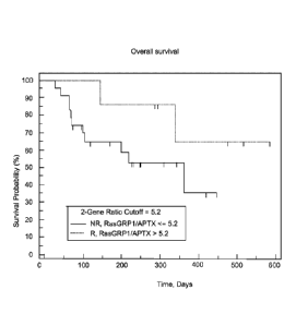
Figures 8 and 13 depict accuracy of the improved qPCR assay in a Phase 2 study
of tipifarnib + etoposide study in elderly AML using Kaplan Meier analysis of
patients
stratified using an optimal ratio cutoff 5.2..
Figures 9 and 14 depict the ROC analysis indicating a discriminative value of
the
2-gene ratio as 80% (AUC= 0.80) for predicting overall response with a
complete
.. remission (CR) patient group used as the response criteria.
Figures 10 and 15 show there is no association between the 2-gene ratio and
clinical response or overall survival in patients not treated with an FTI.
Overall survival
of 41 AML patients treated with intensive induction chemotherapy with ara-C,
anthracycline, and a third agent (flavopiridol or etoposide): stratification
by high vs. low
.. 2-gene ratio.
Figure 11 depicts the work flow for comparing and determining the preferred
sample collection problem.
Figures 12A and 12B show the effect of the sample collection protocols on the
result of the two-gene assay. Figure 12B in particular shows the scatter for
each patient
illustrating the effect of the sample collection protocol. The Y-axis shows
the ratio of
RASGRPLAPTX in the sample to the RASGRP 1:APTX in a calibration/reference RNA,
which in this case is JY RNA. Thus, the value on the Y-axis is a ratio of
ratios arrived at
by the AA Ct method. The threshold used in the preferred assay is a threshold
based on a
desirable stratification of patients using the AA Ct method for quantitating
the levels of
RASGRP1 and APTX.
14
CA 02806112 2013-01-18
WO 2012/016021 PCT/US2011/045693
Detailed Description
The therapeutic agents referred to in this specification include FTIs. They
take on
a multitude of forms but share the essential inhibitory function of
interfering with or
lessening the farnesylation of proteins implicated in cancer and proliferative
diseases.
Preferably, the FTIs are those indicated for the treatment of leukemias such
as AML.
Numerous FTIs arc within the scope of the disclosure and include those
described
in US Patents 5,976,851; 5,972,984; 5,972,966; 5,968,965; 5,968,952;
6,187,786;
6,169,096; 6,037,350; 6,177,432; 5,965,578; 5,965,539; 5,958,939; 5,939,557;
5,936,097;
5,891,889; 5,889,053; 5,880,140; 5,872,135; 5,869,682; 5,861,529; 5,859,015;
5,856,439;
5,856,326; 5,852,010; 5,843,941; 5,807,852; 5,780,492; 5,773,455; 5,767,274;
5,756,528;
5,750,567; 5,721,236; 5,700,806; 5,661,161; 5,602,098; 5,585,359; 5,578,629;
5,534,537;
5,532,359; 5,523,430; 5,504,212; 5,491,164; 5,420,245; 5,238,922 and US
Publication
20030050323. Non-peptidal, so-called "small molecule" therapeutics are
preferred.
More preferred FTIs are quinolines or quinoline derivatives such as:
7-(3-chloropheny1)-9-[(4-chloropheny1)-1H-imidazol-1-ylmethyl]-2,3-dihydr- o-
1H,5H-benzo[ij]quinolizin-5-one,
7-(3-chloropheny1)-9-[(4-chloropheny1)-1H-imidazol-1-ylmethyl]-1,2-dihydr-o-
41-1-pyrrolo[3,2,1-ij]quinoline-4-one,
8-[amino(4-chl orophenyl)(1-methy1-1H-imi dazol-5-y1),methyl]-6-(3 -chloroph-
eny1)-1,2-dihydro-4H-pyrrolo[3,2,1-ij]quinolin-4-one, and
8-[amino(4-chlorophenyl)(1-methy1-1H-imidazol-5-y1)methyl]-6-(3-chlorophe-
nyl)-2,3-dihydro-1H,5H-benzo[ij]quinolizin-5-one. The most preferred FTI is
(B)-6-
[amino(4-chlorophenyl)(1-methy1-1H-imidazol-5-yHmethyl]-4-(3-ch- loropheny1)-1-
methy1-2(1H)-quinolinone).
It is desirable to classify response to treatment to facilitate both and
understanding
of the effect of the treatment and to compare different treatments. There are
many
criteria for evaluating treatments. For instance, a count of a thousand
neutrophils may
suffice to identify a response in some embodiments, while other embodiments
may
require 1,400 or 1,500 neutrophils. Similarly, the platelet count may vary
anywhere from
100,000 to 140,000. And, improvement in one cell line of a certain percentage
may be
required or in other evaluations improvement in two or even improvement in all
three cell
CA 02806112 2013-01-18
WO 2012/016021 PCT/US2011/045693
lines. The time duration over which such changes are determined may range from
one
month or two months or even more. In a preferred embodiment, a patient who
responds
to an FTI is one in whom at least a reduction of more than 50% of blast cells
is seen in
bone marrow following treatment with the FTI. Typically, the degree of
improvement
.. required for partial response tends to be variable, and improvement
represented by
hematologic improvement is extremely variable between evaluations by different
investigators and/or physicians. Alternative similar standards to evaluate a
response to
the administration of an FTI are intended to be within scope of claims
directed to
predicting response to treatment¨unless a contrary intent is expressly
indicated.
In a preferred embodiment, positive responses to treatment comprise rates for
Complete Remission (CR), Partial Remission (PR), and Hematologic Improvement
(HI).
The remaining disease descriptors are Progressive disease (PD) with the
remainder of the
non-responders adjudged to be exhibiting Stable disease (SD). Each of the
positive
responder classifications are described next.
Complete remission (CR) may be marked by bone marrow showing less than 5%
myeloblasts with normal maturation of all cell lines, an ANC of at least 1000/
L and a
platelet count of 100,000 pL, absence of blasts in peripheral blood, absence
of
identifiable leukemic cells in the bone marrow, clearance of disease-
associated
cytogenetic abnormalities, and clearance of any previously existing
extramedullary
disease. A CR must be confirmed 4 to 6 weeks after the initial documentation.
If
possible, at least one bone marrow biopsy should be performed to confirm the
CR. With
CR it is expected bone marrow will appear to be normal with fewer than five
percent
blasts, normal maturation, and no dysplasia. In the peripheral blood, a
haemoglobin of
greater than 11 grams, neutrophils of over 1,500 per millimeter squared and
platelets over
100,000, no blasts, and no dysplasia will be encountered. Further, to consider
AML
cured, ideally the risk of relapse in a patient with CR must be the same as
the risk of
AML in the general population.
Partial remission (PR) is preferably identified by the presence of trilineage
hematopoiesis in the bone marrow with recovery of ANC and platelets to the
above stated
levels, but with 5 to 25% bone marrow blasts, and at least 50% decrease in
bone marrow
16
CA 02806112 2013-01-18
WO 2012/016021 PCT/US2011/045693
blast percentage from baseline. A PR must be confirmed 4 to 6 weeks after the
initial
documentation.
Hematologic Improvement (HI) is preferably marked by at least 50% decrease in
marrow blasts or decrease in any measurable extramedullary disease, recovery
of ANC to
500 to 1000 t1L, platelet count to 20,000 to 100,000 L, or improvement in
transfusion
requirements.
Stable disease (SD) is identified by any response to treatment not meeting CR,
PR, HI, or PD criteria.
Progressive disease (PD) is marked by any one of the following:
= >50% increase in bone marrow blast percentage from best assessment
= >50% increase in circulating blasts
= New appearance of circulating blasts (on at least 2 consecutive
occasions)
= Development of extramedullary disease
= In patients who present with an initial marrow blast percentage
sufficiently high
to preclude the ability to base disease progression on a >50% increase in
marrow blast
percentage, disease progression should be based upon peripheral blood
criteria, new
appearance of circulating blasts (on at least 2 consecutive occasions), and/or
development
of extramedullary disease.
The duration of response is preferably measured from the time measurement
.. criteria are met for CR or PR (whichever is first recorded) until the first
date that
recurrent or progressive disease is objectively documented. The duration of CR
is
measured from the time measurement criteria are first met for CR until the
first date that
recurrent disease is objectively documented.
The duration of stable disease is measured in patients with stable disease
from the
start of the treatment until the criteria for progression are met.
Progression-Free Survival ("PFS") represents the time between study entry and
the first date of objective documentation of recurrent or progressive disease,
or the
occurrence of death from any cause. Overall Survival is measured from time of
enrollment onto this study to time of death.
The mere presence of nucleic acid sequences having the potential to express
proteins or peptides ("genes") within the genome is not determinative of
whether a
17
CA 02806112 2013-01-18
WO 2012/016021 PCT/US2011/045693
protein or peptide is expressed in a given cell. Whether or not a given gene
capable of
expressing proteins or peptides does so and to what extent such expression
occurs, if at
all, is determined by a variety of complex factors. Irrespective of
difficulties in
understanding and assessing these factors, assaying gene expression can
provide useful
information about the cellular response to a given stimulus such as the
introduction of a
drug or other therapeutic agent. Relative indications of the degree to which
genes are
active or inactive can be found in gene expression profiles. The gene
expression profiles
are used to identify and treat patients who will likely benefit from a given
therapy or
exclude patients from a given therapy where the patient likely would
experience little or
no beneficial response to the drug or therapy.
Preferred methods for establishing gene expression profiles (including those
used
to arrive at the relevant biological pathways) include determining the amount
of RNA
that is produced that can code for a protein or peptide. This is accomplished
by reverse
transcription PCR (RT-PCR), competitive RT-PCR, real time RT-PCR, differential
display RT-PCR, Northern Blot analysis and other related tests. While it is
possible to
conduct these techniques using individual PCR reactions, it is best to amplify
copy DNA
(cDNA) or copy RNA (cRNA) produced from mRNA. Some methods for determining
gene expression can be found in US Patents 6,271,002; 6,218,122; 6,218,114;
and
6,004,755.
One preferred method involves computing the two-gene ratio RASGRPLAPTX to
determine whether a person is likely to respond to the use of an FTI
therapeutic agent.
The term 'ratio' or the 'two-gene ratio RASGRPPAPTX' as applied to gene
expression
values has a range of technical interpretations in this disclosure that are
readily discerned
from the context. At a basic level the meaning is the same although the form
may differ.
For instance, when using qPCR techniques, a AC t value corresponding to a
ratio of two
genes of interest is readily generated, as is well known to one having
ordinary skill in the
art. This value may be generated by using normalized Ct values for the
expression levels
of each of the genes by for instance, subtracting the mean Ct value for that
gene and
dividing by the standard deviation in the Ct values for that gene. The
difference between
such normalized Ct values for the two genes, the AC t value, corresponds to
the ratio of
expression of the genes in that as the ratio increases, the AC t value
decreases and vice-
18
versa. Examples of such normalized AC t values are seen on the Y-axis of
Figure 6 for
RASGRP1 and APTX. Such AC t values, or even normalized AC t values may be
referred to
as the two-gene ratio RASGRP1:APTX in this disclosure. For example a threshold
of 0 in
terms of ACt value may be shown on the Y-axis such that responders are below
the
threshold. This threshold of 0 corresponds to a threshold of 1 when two-gene
ratio
RASGRP1:APTX is expressed in terms of array data, such as those plotted along
the X-
axis of Figure 6. Figure 6 merely illustrates that it is readily possible to
go from one way
of determining the RASGRP1:APTX ratio to another. Alternatively, the ratio may
be
expressed as a positive number based on the AACt value, which compares various
samples to a standard calibrator/reference RNA. Use of such a common
calibrator makes
an assay more portable and reliable since the threshold can and does change
based on the
experimental conditions since the threshold is primarily defined by its
performance in
stratifying patients in a test environment. In a preferred embodiment using
RTPCR, the
two-gene ratio RASGRP1:APTX , expressed as a positive number based on the AACt
value as described elsewhere in this disclosure, leads to a value of 5.2 for
stratifying
responders to tipifarnib from non-responders to tipifarnib. Example two-gene
ratio values
of RASGRP1:APTX, expressed as a positive number based on the AACt values, are
plotted on the Y-axis of Figure 12B and are also referred to as two-gene ratio
RASGRP1:APTX with the context making clear which interpretation should be
used.
Strictly speaking, a person having ordinary skill in the art will realize that
a threshold or
two-gene ratio R4SGRP1:APTX, expressed as a value based on one or more of the
AACt
value, in terms of array data, as a AC t value and just the AACt value while
comparable
may not lend themselves to ready interconvertability in the absence of
additional
information to aid in such a mapping. The claims and description herein should
be read in
light of this consideration. The two-gene ratio RASGRP1:APTX is indicated, for
clarity,
as the two-gene AACtratio RASGRP1:APTX or the two-gene ACt ratio RASGRP1:APTX
or the AACt threshold or the ACt threshold, but when such clarification is not
provided the
context readily provides the correct interpretation.
Having established a threshold to distinguish a responder from a non-
responder,
the two-gene ratio is fixed in a medium such as a computer readable medium as
described
below. A patient sample is obtained that contains diseased cells (such as
hematopoietic
19
CA 2806112 2018-01-08
CA 02806112 2013-01-18
WO 2012/016021 PCT/US2011/045693
blast cells in the case of AML). In a preferred embodiment, sample RNA is then
obtained and amplified from the diseased patient cell and amplified using PCR
and the
two-gene ratio calculated with the aid of an external normalization control.
Then, in a
preferred embodiment, if the two-gene ratio is greater than a predetermined
threshold, the
patient is identified as a likely responder, else as a non-responder.
In similar fashion, the two-gene ratio can be used to monitor response to a
treatment comprising an FTI at various periods throughout the course of
treatment. If the
two-gene ratio is consistent with a responder then the patient's therapy is
continued. If it
is not, then the patient's therapy is altered. Such analysis permits
intervention and
therapy adjustment prior to detectable clinical indicia or in the face of
otherwise
ambiguous clinical indicia.
Preferred embodiments may cover representations of the gene expression
profiles
useful for treating, diagnosing, prognosticating, staging, and otherwise
assessing diseases
that are reduced to a medium that can be automatically read such as computer
readable
media (magnetic, optical, and the like). Preferred embodiments can also
include
instructions for assessing the gene expression profiles in such media. For
example,
preferred embodiments may comprise a CD ROM having computer instructions for
comparing gene expression profiles of the portfolios of genes described above.
The
preferred embodiments may also have gene expression profiles digitally
recorded therein
so that they may be compared with gene expression data from patient samples.
Alternatively, the profiles can be recorded in different representational
format. A
graphical recordation is one such format. Clustering algorithms such as those
incorporated in "OlVINTVIZ" and "TREE VIEW" computer programs mentioned above
can best assist in the visualization of such data.
The biological effect of a drug may be a consequence of drug-mediated changes
in the rate of transcription or degradation of one or more species of RNA, the
rate or
extent of translation or post-translational processing of one or more
polypeptides, the rate
or extent of the degradation of one or more proteins, the inhibition or
stimulation of the
action or activity of one or more proteins, and so forth. In addition to the
preferred FTI's,
the preferred drugs include those that modulate the MAPK/ERK signaling
pathways,
TGF-13, WNT or apoptotic pathways. These include, without limitation, tyrosine
kinase
CA 02806112 2013-01-18
WO 2012/016021 PCT/US2011/045693
inhibitors, MEK kinase inhibitors, P13K kinase inhibitors, MAP kinase
inhibitors,
apoptosis modulators and combinations thereof. Exemplary drugs that are most
preferred
among these are the "GLEEVEC" tyrosine kinase inhibitor of Nov artis, U-0126
MAP
kinase inhibitor, PD-098059 MAP kinase inhibitor, SB-203580 MAP kinase
inhibitor,
and antisense, ribozyme, and DNAzyme Bel-XL anti-apoptotics. Examples of other
useful drugs include, without limitation, the calanolides of US Patent
6,306,897; the
substituted bicyclics of US Patent 6,284,764; the indolines of US Patent
6,133,305; and
the antisense oligonucleotides of US Patent 6,271,210.
Pharmaceutically useful compositions comprising the drugs described herein may
be formulated according to known methods such as by the admixture of a
pharmaceutically acceptable carrier. Examples of such carriers and methods of
formulation may be found in Remington's Pharmaceutical Sciences. To form a
pharmaceutically acceptable composition suitable for effective administration,
such
compositions will contain an effective amount of the drug. The effective
amount of the
drug may vary according to a variety of factors such as the individual's
condition, weight,
sex and age. Other factors include the mode of administration. The
pharmaceutical
compositions may be provided to the individual by a variety of routes such as
subcutaneous, topical, oral and intramuscular.
The drugs described herein include chemical derivatives of the base molecules
of
the drug. That is, they may contain additional chemical moieties that are not
normally a
part of the base molecule. Such moieties may improve the solubility, half-
life,
absorption, etc. of the base molecule. Alternatively the moieties may
attenuate
undesirable side effects of the base molecule or decrease the toxicity of the
base
molecule. Examples of such moieties are described in a variety of texts, such
as
Remington's Pharmaceutical Sciences.
Compounds identified according to the methods disclosed herein may be used
alone at appropriate dosages defined by routine testing in order to obtain
optimal
inhibition or activity while minimizing any potential toxicity. In addition,
co-
administration or sequential administration of other agents may be desirable.
The drugs described herein can be administered in a wide variety of
therapeutic
dosage forms in conventional vehicles for administration. For example, the
drugs can be
21
CA 02806112 2013-01-18
WO 2012/016021 PCT/US2011/045693
administered in such oral dosage forms as tablets, capsules (each including
timed release
and sustained release fornmlations), pills, powders, granules, elixirs,
tinctures, solutions,
suspensions, syrups and emulsions, or by injection. Likewise, they may also be
administered in intravenous (both bolus and infusion), intraperitoneal,
subcutaneous,
topical with or without occlusion, or intramuscular form, all using forms well
known to
those of ordinary skill in the pharmaceutical arts. An effective but non-toxic
amount of
the compound desired can be employed as a modulating agent.
For combination treatment with more than one active agent, where the active
agents are in separate dosage formulations, the active agents can be
administered
.. concurrently, or they each can be administered at separately staggered
times.
The dosage regimen utilizing the compounds or modulators described herein is
selected in accordance with a variety of factors including type, species, age,
weight, sex
and medical condition of the patient; the severity of the condition to be
treated; the route
of administration; the renal and hepatic function of the patient; and the
particular drug
employed. A physician or veterinarian of ordinary skill can readily determine
and
prescribe the effective amount of the drug required to prevent, counter or
arrest the
progress of the condition. Optimal precision in achieving concentrations of
drug within
the range that yields efficacy without toxicity requires a regimen based on
the kinetics of
the drug's availability to target sites. This involves a consideration of the
distribution,
equilibrium, and elimination of a drug.
The drugs described herein form the active ingredient, and are typically
administered in admixture with suitable pharmaceutical diluents, excipients or
carriers
(collectively referred to herein as "carrier" materials) suitably selected
with respect to the
intended form of administration, that is, oral tablets, capsules, elixirs,
syrups and the like,
and consistent with conventional pharmaceutical practices.
For instance, for oral administration in the form of a tablet or capsule, the
active
drug component can be combined with an oral, non-toxic pharmaceutically
acceptable
inert carrier such as ethanol, glycerol, water and the like. Moreover, when
desired or
necessary, suitable binders, lubricants, disintegrating agents and coloring
agents can also
.. be incorporated into the mixture. Suitable binders include, without
limitation, starch,
gelatin, natural sugars such as glucose or beta-lactose, corn sweeteners,
natural and
22
synthetic gums such as acacia, tragacanth or sodium alginate,
carboxymethylcellulose,
polyethylene glycol, waxes and the like. Lubricants used in these dosage forms
include,
without limitation, sodium oleate, sodium stearate, magnesium stearate, sodium
benzoate,
sodium acetate, sodium chloride and the like. Disintegrators include, without
limitation.
starch, methyl cellulose, agar, bentonite, xanthan gum and the like.
For liquid forms the active drug component can be combined in suitably
flavored
suspending or dispersing agents such as the synthetic and natural gums, for
example,
tragacanth, acacia, methyl-cellulose and the like. Other dispersing agents
that may be
employed include glycerin and the like. For parenteral administration, sterile
suspensions
and solutions are desired. Isotonic preparations, which generally contain
suitable
preservatives, are employed when intravenous administration is desired.
The compounds or modulators may alternatively be administered parenterally via
injection of a formulation consisting of the active ingredient dissolved in an
inert liquid
carrier. Injection may be either intramuscular, intraluminal, intratracheal,
or
subcutaneous. The injectable formulation consists of the active ingredient
mixed with an
appropriate inert liquid carrier. Acceptable liquid carriers include the
vegetable oils such
as peanut oil, cotton seed oil, sesame oil and the like as well as organic
solvents such as
solketal, glycerol formal and the like. As an alternative, aqueous parenteral
formulations
may also be used. The vegetable oils are the preferred liquid carriers. The
formulations
are prepared by dissolving or suspending the active ingredient in the liquid
carrier such
that the final formulation contains from 0.005 to 10% by weight of the active
ingredient.
The disclosure is further illustrated by the following non-limiting examples.
23
CA 2806112 2018-01-08
EXAMPLE 1
Materials and Methods
Clinical Evaluation
In an exemplary example, bone marrow samples are collected from an open label,
multicenter, non-comparative phase 2 study investigating the efficacy and
safety of
farnesyltransferase inhibition with tipifarnib (R115777, ZARNESTRA ) in older
adults
with previously untreated, poor-risk AML.
Sample Collection and Processing
Bone marrow samples were collected from consenting patients before treatment
with tipifarnib followed by mononuclear cells preferably being processed on
site. Bone
marrow aspirates were diluted with PBS and centrifuged with ficollTm-
diatrizoate
(1.077g/m1). Enriched leukemic blood cells were washed twice with PBS,
resuspended
in FBS with 10% DMSO and immediately frozen at -70 C to -80 C. Total RNA was
extracted from cell samples using the Trizol Kit (Qiagen, Santa Clarita, CA).
RNA
quality may be determined by assessing the presence of ribosomal bands on an
Agilent
Bioanalyzer. Good quality samples were further processed for microarray
analysis. DNA
was isolated from the same sample of Trizol-processed bone marrow as per the
manufacturer's instructions (Qiagen, Santa Clarita, CA). Samples were assayed
for
global gene expression, N-RAS mutations, and/or qPCR of specific genes (Fig
1).
N-RAS mutational status
Analysis of activating mutations in N-RAS was determined by PCR and RFLP
analysis as previously described. End etal. (2001). Exons 1 and 2 of the N-RAS
gene
were simultaneously amplified in a single multiplex reaction and an aliquot
was used for
a second round of PCR. Resistance to cleavage at natural or primer induced
restriction
enzyme sites in second-round amplicons indicated the presence of a mutation
that had
abolished the site at the loci being analyzed. Restriction enzymes for the
analysis of
specific loci were Bsl I (N-ras codons 12 and 13), Msc 1 (N-ras codon 61,
positions 1 and
2), and Bfa I (N-ras codon 61, position 3). Reactions were digested overnight
and PCR
products were analyzed on an Agilent Bioanalyzer.
24
CA 2806112 2018-01-08
Microarray analysis
Synthesis of cDNA and cRNA were performed according to Affymetrix (Santa
Clara, CA) protocols. Since the yield of many samples was low, two rounds of
linear
amplification were performed as previously described in US Patent Publication
No.
20070048782. For hybridization, 111,1g of cRNA were fragmented randomly by
incubation at 94 C for 35 min in 40 mM Tris-acetate, pH 8.1, 100 mM potassium
acetate,
and 30 mM magnesium acetate. Fragmented cRNA was hybridized to U1 33A arrays
at
45 C for 16 h in a rotisserie oven set at 60 rpm. Following hybridization,
arrays were
washed (with 6x SSPE and 0.5x SSPE containing TritonTm X-100 (0.005%)), and
stained
with streptavidin-phycoerythrin (SAPE; Molecular Probes, Eugene, OR).
Quantification
of bound labeled probe was conducted using the Agilent G2500A GeneArray
scanner
(Agilent Technologies, Palo Alto, CA).
The total fluorescence intensity for each array was scaled to the uniform
value of
600. Chip performance was quantified by calculating a signal to noise ratio
(raw average
signal/noise). Chips were removed from further analysis if their signal-to-
noise ratio was
less than 20 or if the present calls on the chip was less than 30%. Genes were
only
included in further analysis if they were called "present" in at least 10% of
the chips.
Approximately 12,000 Affymetrix probe sets remained following this cut-off.
The
quality of the gene expression data were further controlled by identifying
outliers based
on principal components analysis and by analyzing the normal distributions of
the gene
intensities (Partek Pro V5.1). The microarray data have been deposited in
NCBIs Gene
Expression Omnibus (GEO, http://www.ncbi.nlm.nih.gov/geo/).
Response definitions
Response to tipifarnib was defined as patients who had a complete response
(CR),
a partial response (PR), or hematological improvement (HI). Briefly, HI was
defined as
any bone marrow blast count less than 5% or a reduction in bone marrow blasts
by at
least half. Progressive disease (PD) was defined as either >50% increase in
bone marrow
or circulating blast % from baseline, or new appearance of circulating blasts
(on at least 2
consecutive occasions). Stable disease (SD) was defined as any response not
meeting CR,
PR, HI, or PD criteria.
CA 2806112 2018-01-08
CA 02806112 2013-01-18
WO 2012/016021 PCT/US2011/045693
Statistical analysis
Receiver Operator Characteristic (ROC) analysis was utilized to test the
overall
predictive value of individual genes and/or multigene classifiers. The
following gene
filtering criteria were used to identify genes differentially expressed
between responders
and patients with progressive disease: Specificity for identifying "responder"
with 100%
sensitivity >= 40%, T-test p value (10g2 transformed data with unequal
variance) <0.05,
fold change > 2. The genes that passed these criteria were ranked by AUC (Area
under
the ROC curve).
To build a classifier the response score was used to calculate each patient's
likelihood of responding to tipifarnib therapy. The score was defined as the
linear
combination of weighted expression signals with the t-statistic as the weight.
The
threshold was determined from the ROC curve of the training set to ensure 100%
sensitivity and the highest specificity. To determine how many genes needed to
be
included in the predictor, leave-one-out cross validation (LOOCV) was carried
out. The
response scores for the 'left-out' samples based on different numbers of genes
were
recorded. The performances of the predictors with different numbers of genes
were
assessed based on misclassification error rate, sensitivity, specificity, p
values measuring
the separation of Kaplan-Meier curves of the two predicted groups. And the
best
predictor was selected accordingly.
The Top Scoring Pair (TSP) algorithm was first introduced by Geman et al.
(2004). In essence, the algorithm ranks all the gene pairs (genes i and j)
based on the
absolute difference (Dij) in the frequency of event where gene i has higher
expression
value than gene j in samples among class Cl to C2. In the cases of there are
multiple top
scoring pairs (all sharing the same Dij), the top pair by a secondary rank
score that
.. measures the magnitude to which inversions of gene expression levels occur
from one
class to the other within a pair of genes is selected. The top pair with
highest frequency
of absolute Dij > 2 fold in all samples will be selected as candidate pair.
The candidate
pair was then assessed in an independent testing data set.
Leave-one-out cross validation (LOOCV) was carried out in the training data
set
to evaluate how the algorithm perform. The performances of the predictors were
26
CA 02806112 2013-01-18
WO 2012/016021 PCT/US2011/045693
assessed based on maximum misclassification error rate. All the statistical
analyses were
done using R (R Development Core Team, 2006).
Real-Time Quantitative RT-PCR
For each sample, 1 lag of total RNA (as assessed by 0D260) was reversed
transcribed using the High Capacity cDNA Reverse Transcription kit (Applied
Biosystems, Foster City, CA) according to the manufacturer's instructions.
Samples
were then incubated at 25 C for 10 minutes and then 37 C for 30 minutes for
optimum
RNA conversion. QPCR was performed using the ABI Prism 7900HT sequence
detection system (Applied Biosystems, Foster City, CA) with all samples run in
triplicate.
Each reaction contained 5 I TaqMang Universal PCR Master Mix containing UNG
(Applied Biosystems, Foster City, CA), 4.5 I of cDNA template and 0.5 I of
20 x
Assay on Demand Gene Expression Assay Mix or 9 pmol of both forward and
reverse
primer and 2.5 pmol of probe (Applied Biosystems, Foster City, CA), in a total
reaction
volume of 10 1. All primer, probe sets were chosen due to the small amplicon
size (less
than 100 nucleotides) and FAM fluorogenic probes were used. Primers and probes
used
were APTX (product number 4331182 Applied Biosystems) and RASGRP1 (product
number 4351372 Applied Biosystems). The RASGRPLAPTX expression ratio was
calculated by normalizing the raw Ct values by subtracting the mean Ct from
the sample
set, dividing by the standard deviation, and then calculating the difference
of the
normalized Ct values of each gene (APTX ¨ RASGRP1).
Results
This study examined gene expression profiles of leukemic bone marrow samples
from patients enrolled in a Phase 2 clinical trial of the farnesyltransferase
inhibitor
tipifarnib in elderly patients with previously untreated poor-risk acute
myelogenous
leukemia. Lancet et al. (2006). Bone marrow from 67 patients was collected
before
treatment with tipifarnib and leukemic myeloid cells were enriched by Ficoll-
density
centrifugation (Table 1). Good quality total RNA from 13 responders (9 CR, 4
HI), 8
stable disease and 13 progressive disease patients was amplified, labeled, and
hybridized
to the Affymetrix U133A GeneChip. A total of 30 samples were evaluated by qPCR
for
validation of specific genes and 32 samples were evaluated for AT-RAS
mutational status.
27
CA 02806112 2013-01-18
WO 2012/016021
PCT/US2011/045693
Table 1. Comparison of profiled patients.
Parameter All treated patients PGx profiled patients
Total patients, n 158 67
microarray assay, n 34
qPCR assay, n 30
N-Ras assay, n 32
N-Ras mutation, n (/o) 11(34)
median age, y (range) 74 (34-85) 73 (63-85)
sex, n male (`)/0) 95 (60) 41(61)
Prior MDS, yes (/o) 119 (75) 48 (72)
CR, no. (A) 22 (14) 14 (21)
PR, no. (`)/0) 3 (2) 1 (2)
HI, no. (%) 12 (8) 7 (10)
SD, no. (`)/0) 50 (32) 15 (22)
PD, no. (`)/0) 58 (37) 30 (44)
NE, no. (`)/0) 13 (8) 0 (0)
CR = complete response; PR = partial response; HI = hematological improvement,
SD = stable
disease, PD = progressive disease, NE = not evaluable; PGx = pharmacogenomics
Ras mutational status and patient outcome.
DNA from the bone marrow of 32 AML patients was screened for N-Ras
activating mutations (codons 12, 13, 61). Thirty-four percent (11/32) of
patients
exhibited N-Ras mutations with one patient having mutations at multiple codons
(Table
2). There was no statistically significant correlation between N-RAS
mutational status
and response to tipifarnib or overall survival.
Table 2.
SUBJID RESPONSE N-Ras Mutation OS Alive Microarray qPCR SEX AGE Prior MDS
100101 HI ND 378 NO ND YES MALE 68 NO
100104 PD ND 728 NO YES YES FEMALE 63 NO
100109 PD ND 68 NO YES YES FEMALE 81 NO
100110 CR ND 983 YES YES YES FEMALE 74 NO
100112 PD ND 169 NO ND YES FEMALE 69 YES
100113 CR ND 211 NO ND YES MALE 82 YES
100116 PD ND 14 NO ND YES FEMALE 72 YES
100121 SD ND 252 NO YES ND MALE 72 YES
100204 SD N-12 493 NO ND ND FEMALE 69 YES
100205 PD WT 754 NO YES ND MALE 74 YES
100208 PD WT 29 NO YES ND MALE 76 YES
100209 PD N61(1,2) 209 NO YES ND MALE 73 YES
100210 PD N-12, N-13 654 NO YES ND MALE 68 YES
100212 SD N-12 1200 YES ND ND MALE 70 YES
100213 CR WT 257 NO YES ND FEMALE 81 YES
100214 CR N-13 395 NO ND ND FEMALE 73 YES
100215 SD WT 54 NO ND ND MALE 82 NO
100216 SD N-13 116 NO ND ND MALE 77 YES
28
CA 02806112 2013-01-18
WO 2012/016021
PCT/US2011/045693
100302 PD N-12 48 NO YES ND FEMALE 73 NO
100307 HI WT 179 NO YES ND MALE 68 YES
100310 SD WT 242 NO ND ND FEMALE 76 YES
100316 SD WT 273 NO ND ND FEMALE 66 NO
100317 PD WT 39 NO ND ND MALE 76 NO
100319 SD WT 233 NO YES ND MALE 71 NO
100320 HI WT 374 NO ND ND FEMALE 78 NO
100322 CR WT 237 YES YES ND MALE 73 YES
100324 HI WT 248 NO YES ND MALE 85 YES
100330 HI N-12 153 NO YES ND FEMALE 67 NO
100333 SD N-12 364 NO YES ND MALE 65 YES
100336 CR N-12 67 NO YES ND MALE 80 YES
100337 PD WT 38 NO ND ND MALE 72 YES
100338 PD N-12 8 NO YES ND MALE 78 NO
100339 PD WT 25 NO YES ND MALE 75 NO
100340 SD WT 32 NO ND ND FEMALE 83 NO
100341 CR WT 433 NO YES ND MALE 67 YES
100604 SD WT 64 NO YES ND MALE 63 YES
100605 PD WT 74 NO ND ND MALE 67 YES
101008 CR WT 548 NO YES ND MALE 82 NO
101021 CR ND 991 YES YES YES FEMALE 69 YES
101025 CR ND 735 YES ND YES MALE 70 YES
101029 PD ND 64 NO ND YES MALE 70 YES
101038 SD ND 151 NO YES ND FEMALE 75 YES
101039 PD ND 50 NO ND YES FEMALE 85 YES
101043 SD ND 200 NO YES ND FEMALE 79 YES
101046 PD ND 53 NO YES YES FEMALE 66 YES
101049 CR WT 564 NO YES ND MALE 65 YES
101057 CR WT 386 NO YES ND MALE 85 YES
101067 PD ND 88 NO ND YES FEMALE 76 YES
101069 PD ND 94 NO ND YES MALE 81 YES
101075 HI ND 659 YES YES YES MALE 71 YES
101077 SD ND 574 YES YES ND FEMALE 75 YES
101078 PD ND 190 NO ND YES FEMALE 77 NO
101079 PD ND 429 NO ND YES FEMALE 70 YES
101083 PD ND 71 NO ND YES MALE 73 YES
101091 CR ND 671 YES ND YES MALE 71 YES
101092 PD ND 136 NO ND YES FEMALE 69 YES
101094 HI ND 579 YES ND YES MALE 65 YES
101095 PD ND 108 NO YES YES MALE 82 YES
101096 CR ND 390 YES ND YES MALE 69 YES
101101 PD ND 91 NO ND YES MALE 69 YES
101102 PD ND 76 NO YES YES MALE 69 YES
101103 PD ND 29 NO ND YES FEMALE 80 NO
101108 PR ND 123 NO NO YES MALE 70 YES
101109 SD ND 656 YES YES ND MALE 68 YES
101114 PD ND 69 NO YES YES MALE 72 YES
101121 PD ND 43 NO ND YES MALE 78 NO
101122 PD ND 44 NO ND YES FEMALE 80 NO
29
CA 02806112 2013-01-18
WO 2012/016021 PCT/US2011/045693
ND = not determined; WT = wildtype; CR = complete response; PR = partial
response; HI = hematological
improvement, SD = stable disease, PD = progressive disease, OS = Overall
survival.
Identification of predictive genes from the newly diagnosed AML cohort
The next aim was to identify genes predictive of response to tipifarnib in the
newly diagnosed AML population. To this end discovery experiments were
performed in
the 13 responders (9 CR and 4 HI) and 13 patients with progressive disease.
Patients
with stable disease were not utilized in this analysis since these patients
cannot be clearly
defined as either responders or non-responders. Using the same approach as was
utilized
for identifying markers for relapsed and refractory AML (20070048782) we
identified 45
probesets (corresponding to 38 unique genes) that were predictive of response
(Table 3).
The selection criteria aimed at identifying genes that would predict
responders with a
high sensitivity (approaching 100%) with a specificity cut-off of 40% and a
mean gene
expression difference of at least two-fold. The genes were ranked based on the
area
under the curve (AUC) defined from a receiver operator characteristic (ROC)
analysis of
the training set. This value represents the overall predictive value of the
gene with an
AUC of 1.0 indicating perfect classification. Each gene was first tested on
the training
set using a LOOCV method. The top gene, the RAS guanyl-releasing protein 1
(RASGRP1), showed an AUC of 0.95.
30
CA 02806112 2013-01-18
WO 2012/016021 PCT/US2011/045693
Table 3. 45 probesets predictive of response to tipifarnib in newly
diagnosed AML
Probe Set ID Gene Symbol Gene Title pvalue
spec tstat FC AUC
205590_at RASGRP1
RAS guanyl releasing protein 1 2.64E-06 0.54 6.40 4.01 0.95
217028 at CXCR4 chemokine (C-X-C motif) receptor 4
4.41E-05 0.69 5.08 2.35 0.92
206687_s_at PTPN6 protein tyrosine phosphatase, non-receptor type 6
8.23E-05 0.77 -4.75 -2.15 0.91
210439_at ICOS inducible T-cell co-stimulator
1.27E-04 0.77 4.56 3.81 0.91
206641_at TNFRSF17
tumor necrosis factor receptor superfamily, member 17 3.79E-02 0.62 2.24
2.55 0.91
213539_at CD3D CD3d molecule, delta (CD3-TCR complex)
1.75E-04 0.69 4.63 2.82 0.91
208018_s_at HCK hemopoietic cell kinase
2.62E-04 0.62 -4.28 -3.14 0.90
203063_at PPM1F protein phosphatase 1F (PP2C domain containing)
3.66E-04 0.85 -4.17 -2.31 0.90
208130_s_at TBXAS1 thromboxane A synthase 1
2.70E-04 0.46 -4.26 -2.51 0.89
216834 at RGS1 regulator of G-protein signalling 1
3.90E-04 0.62 4.16 3.48 0.87
213388_at PDE4DIP
phosphodiesterase 4D interacting protein (myomegalin) 1.47E-03 0.54 -3.64 -
2.01 0.86
38487_at STAB1 stabilin 1
7.95E-04 0.54 -3.87 -2.45 0.86
210982_s_at HLA-DRA
major histocompatibility complex, class II, DR alpha 4.23E-03 0.69 -3.25 -
3.07 0.85
210321_at GZMH granzyme H (cathepsin G-like 2, protein h-CCPX)
1.64E-03 0.54 3.55 2.83 0.85
217147_s_at TRAT1 T cell receptor associated transmembrane adaptor 1
1.19E-03 0.54 3.72 2.82 0.85
206298_at ARHGAP22 Rho GTPase activating protein 22
7.89E-04 0.62 -3.88 -2.19 0.85
202990_at PYGL phosphorylase, glycogen; liver
1.95E-03 0.46 -3.50 -2.01 0.85
221671 x at IGKC immunoglobulin kappa constant
1.62E-03 0.46 3.56 3.10 0.85
221651_x_at IGKC immunoglobulin kappa constant
1.65E-03 0.46 3.57 2.92 0.85
207651_at GPR171
G protein-coupled receptor 171 1.13E-03 0.62 3.70 3.01 0.85
202988_s_at RGS1 regulator of G-protein signalling 1
1.48E-03 0.54 3.59 2.95 0.84
213418_at HSPA6 heat shock 70kDa protein 6
1.63E-02 0.62 -2.61 -2.34 0.83
209901_x_at AlF1 allograft inflammatory factor 1
3.52E-03 0.54 -3.24 -2.48 0.83
205488_at GZMA granzyme A
4.43E-03 0.46 3.18 2.75 0.83
217022 sat IGHA1 immunoglobulin heavy constant alpha 1
3.43E-03 0.69 3.36 2.56 0.83
207339_s_at LTB lymphotoxin beta (TNF superfamily, member 3)
1.34E-03 0.46 3.65 2.40 0.83
206337_at CCR7 chemokine (C-C motif) receptor 7
1.14E-03 0.54 3.71 2.08 0.83
208894_at HLA-DRA
major histocompatibility complex, class II, DR alpha 6.14E-03 0.46 -3.05 -
2.58 0.82
39729_at PRDX2 peroxiredoxin 2
5.81E-03 0.54 3.05 2.13 0.82
209500_x_at TNFSF13
tumor necrosis factor (ligand) superfamily, member 13 1.23E-03 0.46 -3.68 -
2.02 0.82
214677_x_at IGL@ immunoglobulin lambda locus
4.69E-03 0.46 3.17 2.86 0.82
210314 x at TNFSF13
tumor necrosis factor (ligand) superfamily, member 13 3.48E-03 0.46 -3.24 -
2.05 0.81
209138_x_at IGL@ lmmunoglobulin lambda locus
4.17E-03 0.54 3.17 3.41 0.80
207831_x_at DHPS deoxyhypusine synthase
1.09E-02 0.62 -2.77 -2.05 0.80
215121_x_at IGL@ immunoglobulin lambda locus
1.20E-02 0.46 2.72 4.42 0.79
215946_x_at CTA-246H3.1 similar to omega protein
1.10E-02 0.46 2.76 2.46 0.79
204069_at MEIS1 Meis1, myeloid ecotropic viral integration site 1
homolog 1.01E-02 0.62 -2.89 -2.14 0.78
204698_at ISG20 interferon stimulated exonuclease gene 20kDa
6.93E-03 0.46 2.95 2.39 0.78
209906_at C3AR1 complement component 3a receptor 1
1.49E-02 0.54 -2.65 -2.05 0.77
205608 sat ANGPT1 angiopoietin 1
6.40E-03 0.46 -3.11 -2.18 0.76
205927_s_at CTSE cathepsin E
2.02E-02 0.46 2.55 2.05 0.76
215051_x_at AlF1 allograft inflammatory factor 1
1.54E-02 0.62 -2.62 -2.03 0.76
205609_at ANGPT1
angiopoietin 1 4.12E-02 0.54 -2.20 -3.11 0.73
202890_at MAP7 microtubule-associated protein 7
3.30E-02 0.62 -2.30 -2.31 0.73
203485_at RTN 1 reticulon 1
2.60E-02 0.54 -2.40 -2.29 0.72
31
CA 02806112 2013-01-18
WO 2012/016021 PCT/US2011/045693
Spec = specificity, FC = fold change, AUG = area under the curve of Receiver
Operator
Characteristic Analysis, negative t-statistic indicates gene is down in
responders.
Whether increasing the number of genes in the classifier improved its
predictive
value was also examined. Using the LOOCV approach and then plotting
sensitivity,
specificity, and overall error rate of each classifier, the top gene alone was
found to
provide the best predictive value (data not shown). Adding genes to the
classifier in a
linear fashion did not improve its predictive value. Using a cutoff that
biases for high
sensitivity, the LOOCV demonstrated that the expression of the RASGRP1 gene
allowed
for a NPV 88.9%, and a PPV of 70.6%, with an overall predictive accuracy of
76.9% (Fig
1A). In addition, Kaplan Meier analysis showed a significant difference in
median
overall survival of the responders (386 days) and those with progressive
disease (68 days)
(Fig 1B). Over expression of this single gene therefore predicted response to
tipifarnib in
newly diagnosed AML with a high negative predictive value.
Identification of a Top Scoring Pair classifier
The predictive value of RASGRP1 was not improved if additional genes were
added to the classifier using a linear approach. An alternative gene selection
algorithm
was utilized to select genes that would improve the predictive value of
RAAS'GRPI alone.
To this end the Top Scoring Pair (TSP) algorithm was utilized to identify the
best pair of
genes that would provide the greatest predictive accuracy. Geman et al.
(2004). This
approach was utilized to exploit the greatest difference in expression between
two genes
and may be useful when aiming to develop a qPCR based diagnostic assay. The
TSP
from the training set was RASGRP1 and aprataxin (APTX). RASGRP1 and APTX were
over- and under-expressed in responders, respectively. A robust LOOCV showed
that
this top scoring pair (TSP) provided 85.7% NPV and 91.7% PPV in the training
set of
samples with an overall error rate of only 8% (Fig 2A). The difference in
overall survival
between predicted responders and non-responders was 357 days (Fig 2B). These
data
demonstrate that the model-building algorithm has a low associated prediction
error rate.
32
CA 02806112 2013-01-18
WO 2012/016021 PCT/US2011/045693
Validation of the RASGRP1:APTX classifier in an independent set of relapsed or
refractory AML
External validation of the TSP classifier was performed in an independent
microarray dataset comprising of 54 relapsed/refractory AML patient samples. A
diagnostic assay that predicts likely response to a cancer therapy should have
a high
sensitivity (and negative predictive value) since it is important to capture
as many
potential responders as possible. Therefore, to define an appropriate cutoff
for testing the
TSP classifier the need to obtain a high sensitivity of predicting responders
while
maintaining an acceptable level of specificity was considered. In the training
set, the
level of specificity that could be achieved ranged from approximately 30% to
100% when
the sensitivity was set at 100% to 80%, respectively. To ensure the classifier
would
predict as many responders as possible a conservative cutoff that provided a
specificity of
approximately 60% in the training set was tested. When this cutoff was applied
to the
independent testing set of relapsed/refractory AML, the RASGRP1:APTX gene
classifier
stratified responders with 92% NPV and 27.6% PPV (compared to 18.5%
prevalence)
(Fig 3C). The associated odds ratio for being a responder was 4.38. While this
was
similar to the predictive accuracy of RASGRP1 alone, the application of the
TSP
classifier demonstrated a better NPV and an improved difference in overall
survival of 98
days between predicted responders and progressors (Fig 3A), compared to only
56 days
.. for RASGRP1 (Fig 4).
QPCR validation of the RASGRP1:APTX expression ratio
A two-gene expression ratio allows the use of a more clinically relevant qPCR
detection system. Thirty samples (20 PD, 6 CR, 3 HI and 1 PR) provided enough
total
RNA for qPCR. Therefore, the RASGRP1:APTX gene expression ratio was evaluated
as
a predictor of response to tipifamib using TaqMane qPCR in these 30 samples
(10
responders, 20 progressive disease) from the newly diagnosed AML clinical
study. Nine
of these samples had been assayed on the microarray platform, however 21 had
not been
utilized in the discovery set due to poor quality RNA. Therefore, two thirds
of this test
set was comprised of completely independent samples.
33
CA 02806112 2013-01-18
WO 2012/016021 PCT/US2011/045693
Evaluation of the 9 samples indicated there was good correlation (r = 0.74) of
the
R4SGRP1:APTX expression ratio between the two platforms (Fig 6). Using a cut-
point
of 0, the two-gene classifier correctly predicted the treatment outcome in 20
of the 30
patients with PPV and NPV of 50% and 81%, respectively (Fig 3B). The median
overall
survival of the predicted resistant patients was 82 days while those
classified as
responders had a median value of 295 days (Fig 3C).
The RASGRPLAPTX classifier does not have prognostic utility independent of FTI
treatment
The two-gene expression ratio was tested in an independent microarray dataset
of
116 AML patients treated with chemotherapeutic regimes. When the RASGRP1:APTX
classifier was applied to this set of patients, utilizing a similar cut-off as
for the tipifarnib-
treated population, no significant separation in overall survival was seen
(Fig 5). Nor
were significant survival differences observed when a range of other cut-offs
was utilized
(Table 4). This indicated that the RASGRPLAPTX classifier specifically
stratifies
patients who have been treated with tipifarnib and is not relevant to non-
FTIs. On the
other hand when the prognostic signature was applied to our set of relapsed
and
refractory AML patients there was a clear stratification in terms of overall
survival.
Table 4.
cutoff p value Responders Progressors median OS No. Responders No.
Progressors
median OS
0.5 0.956 336 414 13 103
0.6 0.342 672 374 24 92
0.7 0.266 511 335 34 82
0.8 0.269 511 326 47 69
0.9 0.101 540 316 57 59
1 0.215 483 326 64 52
2 0.795 374 570 94 22
3 0.209 346 909 104 12
OS = overall survival
EXAMPLE 2
This example describes an improved RT-PCR assay suitable for applying the two-
gene assay to FTI combination therapy. During the analytical assay development
Tallman assays for 3 markers: RASGRP1 (guanine nucleotide exchange factor that
activates RAS), APTX (aprataxin involved in DNA excision repair) and HMBS
(used as an
34
CA 02806112 2013-01-18
WO 2012/016021 PCT/US2011/045693
internal control) were designed. HMBS could be used as a control in an
embodiment or
be dispensed with in other embodiments. Sequences of Taqman primer probe sets
and
their amplicons are listed above in the Summary section and in the Sequences
Section of
this disclosure. Namely, for APTX - SEQ # 3-4 and 2, RASGRP1 primer probe set
SEQ
#56 and 1; and IIMBS - SEQ # 7-9.
Quantitative RT-PCR assays were developed and optimized using ABI-7500
platform to assess the 2-gene ratio performance with FAM-labeled RASGRP1, Gold
540-
APTX and Cy5-HMBS in a single-tube triplex format. JY cellular RNA and
Universal
RNA (Stratagene) were used as external normalization.
RASGRP1 :APTX ratio =2^-((A-B)-(C-D))
Where A: Sample RASGRP1 Ct value
B: JY (or Universal) RNA (+) RasGRP1 Ct value
C: Sample APTX Ct Value
D: JY (or Universal) RNA (+) APTX Ct Value
Other sources of cellular RNA could be substituted provided the assay
parameters, in particular, the threshold, are recalculated for the particular
cellular RNA
employed for external normalization. The reference RNA used for external
normalization is preferably from a cell line grown under defined conditions
and that is
relatively non-responsive to FTIs. Without being bound by theory, a suitable
reference
RNA may be derived from a cell line that is neutral in its response to FTI.
Such a
reference cell line may be developed from patients testing in the threshold
region based
on the two-gene assay, but who actually are non-responsive to FTI treatment.
Thus,
without being bound by theory, such patients are a likely source for
developing an
improved reference RNA for the two-gene ratio test. One possible method for
developing and evaluating a cell line may be to select a cell line that, when
used as an
external control in the two-gene assay, tends to increase the threshold. This
criteria is
reasonable because such a cell line(s) will damp (due to a possible
correlation in its two-
gene response and that being measured) the measured two-gene ratio the least.
Alternatively, the external control may be selected using a defined
combination of APTX
and RASGRP1 RNA to allow for absolute quantitation or using AUC to maximize
discrimination between responders and non-responders. The choice of the
external
CA 02806112 2013-01-18
WO 2012/016021 PCT/US2011/045693
control is not the only source of variability in the threshold employed in the
two-gene
assay.
Two RT-PCR formats were developed and their performance assessed using a
standard curve analysis. Both formats are demonstrably equivalent in
performance with a
high Pearson correlation using both raw Cts of 3 markers and derived from them
2-gene
ratio values (R2=1 - 0.93, P<0.000; Table 5).
Table 5. Pearson Correlation between RUO and GMP RT-PCR assays
RUO vs GMP RT-PCR format
RasGRP1, Mean C, 0.97
APTX, Mean C, 0.99
HMBS, Mean Ct 1.00
Ratio normalized to JY control RNA 0.93
RUO format uses a commercially available RNA-to-Ct One Step RT-PCR Kit
.. distributed by INVITROGEN (Cat. No.4392938). This format has a 20 1il
reaction
volume with 50 ng total RNA input according to the manufacturer's
instructions.
GMP format is based on the VERIDEV) BLN RT-PCR Kit components
(GeneSearch Breast Lymph Node (BLN) Test Kit, IVD, Cat #2900004) with
GeneSearch BLN Enzyme Mix, IVD, P/N 7700040 and BLN Base Master Mix, P/N
7700031 used for preparation of RT-PCR master mix. The GMP format has a 25 p,1
reaction with 50 ng RNA input using an optimized RT step described next.
RT-PCR multiplexed protocol in a GMP format
50ng of total RNA was used as a target input in a triplex qRT-PCR which was
carried out on an Applied Biosystems Prism 7500 Sequence Detection System in a
25. uL
reaction. RNA samples (including normalization control RNAs) were thawed on
ice and
diluted to 10 ng/pi. The qRT-PCR was carried out using reagents from the
VERIDEX
BLN RT-PCR Kit (GeneSearch Breast Lymph Node (BLN) Test Kit, IVD, Cat
#2900004): GeneSearch BLN Enzyme Mix, IVD, P/N 7700040 and BLN Base Master
Mix, P/N 7700031.
36
CA 02806112 2013-01-18
WO 2012/016021 PCT/US2011/045693
25X probe-primer master mix was prepared comprising: 400 nM SEQ No. 1 and
2, respectively, and 200 nM FAM-labeled RasGRP1 (SEQ No.10); 400 nM SEQ No. 3
and 4, and 200 nM Gold 540-APTX (SEQ No. 11), and 400 nM of SEQ No. 8 and 9
and
200 nM Cy5-HMBS (SEQ No. 12) as summarized in Table 6.
Table 6. 25X Probe/Primer Mix
Stock to Final
3p1ex-1 (25X) Sequence Add/rxn 500 Conc.
in 25
Marker
iaL
RASGRP1 RASGRP1 SEQ No. 1 0.10 50.00
0.4 M
RASGRP1 SEQ No. 2 0.10 50.00
0.4 M
RASGRP1 (Fam) SEQ No. 10 0.05 25.00
0.2 M
APTX APTX SEQ No. 3 0.10 50.00
0.4 M
APTX SEQ No. 4 0.10 50.00
0.4 M
APTX SEQ No. 11 (Gold) 0.05 25.00
0.204
HMBS HMBS SEQ No. 8 0.10 50.00
0.4 M
HMBS SEQ No. 9 0.10 50.00
0.4 M
HMBS SEQ No. 12 0.05 25.00
0.2 M
idTe 0.25 125.00
Total 1.00 500.00
Each reaction consisted of 9 !IL of BLN Base Master Mix, 10 uL of 2.5X BLN
Enzyme mix and 1 ul of 25X primer/probe mix. The primer/probe mix had a final
concentration of 400nM of forward and reverse primers and 200 nM of
fluorescent
probes for each marker. 5 ul of total RNA from patient samples or
normalization
controls (Universal or JY RNA) was added to 20 pi of RT-PC master mix.
A threshold/cutoff of 5.2 was determined based on JY cellular RNA used as an
external normalization control.
The qRT-PCR assays were carried out using the following cycling parameters: 1
min at 95 C for denaturation step; 30 minutes at 58 C (RT reaction); 5% ramp
to 70 C
37
CA 02806112 2013-01-18
WO 2012/016021 PCT/US2011/045693
and incubation for 2 min followed by 40 cycles at 95 C for 15 seconds
(denaturation) and
60 C for 1 minute (annealing/extension). This optimization of probe-primer
concentrations of the GMP RT-PCR format in a single-tube multiplexed set-up
attained a
robust and sensitive detection of RNA targets from both bone marrow and
peripheral
blood samples at the level of 0.6 ng with Ct values around 31, i.e. in a low
variability and
highly reproducibility range of Cts (Figure 3). This reaction format is
suitable for
commercialization of the test, being based on the GMP grade reagents
formulated and
manufactured for the FDA-approved BLN kit, such as Tth Polymerase (enzyme mix)
and
Base master mix. Some details for the Master Mix based protocol are provided
in Table
7.
Table 7. Master Mix based protocol
Reagents
BLN Enzyme mix, BLN Master Mix
Prepare bulk RT-PCR Master Mix
Master Mix
RT-PCR 1 rxn. ( L)
BLN Base Master Mix 9
2.5X BLN Enzyme Mix 10
25X Primer/Probe mix 1
Total 20
Prepare bulk RT-PCR Mix by multiplying reagent volumes on the table by the
number of
Samples plus exra 10%
Add 20 itiL Master Mix in each well of a 96-well plate
Add 5 1tL diluted RNA sample to appropriate well
Add 5 ti,L, of Universal RNA (external control) and (Nf water¨No Target
Control)
Seal the plate. Vortex briefly and centrifuge at 1500 rpm for 1 min.
Place in AB1 7500(1)
38
CA 02806112 2013-01-18
WO 2012/016021 PCT/US2011/045693
Data Analysis
1. Inclusion Criteria
The cycle threshold (Ct) values obtained from ABI7500 data output files were
used for data analysis. Ct Cutoffs for "No Test" for each Marker were selected
as
follows:
HMBS (internal control maker) Ct should not be greater than 30;
RASGRP1 Ct - not greater than 35 and
APTX Ct - not greater than 35.
If a sample had any one of the two markers, RASGRP1 or APTX, above the Ct
cutoff level, it was considered as "No Test" (excluded from the data
analysis). Results
presented in Table 8 demonstrate equivalency between singlex and triplex
(single-tube)
formats of RT-PCR set-up when comparing slope values and PCR efficiency. Table
9
presents further improvements by extending RT step to 30 minutes.
Table 8. Performance of Singlex vs. Triplex RT-PCR formats
PCR QC RASGRP1-Fam APTX-Gold HMBS-Cy5
Slope -3.6 -3.6 -3.6
Triplex Y-Intercept 29.5 29.6 29.5
RT-PCR Efficiency 91% 90% 91%
Singlex RT- Slope -3.5 -3.6 -3.6
PCR in 3 Y-Intercept 30.0 29.8 29.6
channels Efficiency 92% 89% 89%
Table 9. Triplex RT-PCR Performance Optimization
30 min RT, Primers-400nM, Probes 200nM
Target ng input Marker Average Ct PCR
Efficiency
100 24.0 Slope -3.4
50 RasGRP1-Fam 25.1 Intercept 30.8
25.9 Efficiency 95%
39
CA 02806112 2013-01-18
WO 2012/016021 PCT/US2011/045693
12.5 26.9
6.25 28.2
gDNA(8ng) Undetermined
NTC Undetermined
100 23.7 Slope -3.3
50 24.7 Intercept 30.2
25 25.6 Efficiency 102%
12.5 APTX-Gold 26.5
6.25 27.7
gDNA(8ng) Undetemined
NTC Undetermined
100 22.6 Slope -3.3
50 23.5 Intercept 29.1
25 24.4 Efficiency 100%
12.5 HMBS-Cy5 25.4
6.25 26.7
gDNA(8ng) 35.8
NTC Undetermined
Using GMP RT-PCR format, Universal RNA (from AGILENTTm) and JY RNA
controls (from in-house cultured cell line) were run in RT-PCR with 37 patient
samples
from Phase 2 T+E study. This analysis was later repeated with 40 patient
samples. Both
analysis, based on the outcome of ROC curve analysis, are presented in Table
10, to show
that the assay performance is equivalent when using JY or Universal RNA
controls for
raw Ct normalization with an adjustment to the threshold.
The use of Universal RNA as the external control resulted in a threshold of
7.3
compared to a threshold of 5.2 when JY RNA was used as the external control.
In these
threshold determinations, the calculations treated CR as the Response
criteria¨with all
other response types treated as non-responsive.
CA 02806112 2013-01-18
WO 2012/016021 PCT/US2011/045693
Table 10. ROC Curve Analysis: CR vs. NR for TthPol GMP RT-PCR format
Normalization AUC SE 95% Cutoff Sensitivity 95% Specificity 95% Sample
Control CI by CI CI number
ROC
analysis
JY RNA (37 0.8 0.97 0.629 >5.2 66.67 30.1 92.86 76..5 CR=9
patient to to to
NR=28
analysis) 0.908 92.1 98.9
Universal 0.8 0.97 0.629 >5.2 66.67 30.1
92.86 76..5 CR=9
RNA (37 to to to NR=28
patient 0.908 92.1 98.9
analysis)
JY RNA (40 0.8 0.0881 0.636 >5.2 63.64 30.9
93.1 77.2 CR=11
patient to to to
NR=29
analysis) 0.904 88.8 99.0
Universal 0.8 0.0881 0.636 >7.3 63.64 30.9
93.1 77.2 CR=11
RNA (40 to to to NR=29
patient 0.904 88.8 99.0
analysis)
Although the threshold in the 2-gene assay is dimensionless (being the ratio
of
RASGRP1 and APTX), without being bound by theory, it is believed that the
threshold
value may depend on the standards conditions and the reagents used. Therefore,
it is
preferable to specify the threshold relative to a performance based criterion,
such as a
ROC curve based criterion. Thus, the threshold/cutoff may be adjusted to meet
other
requirements such as keeping the AUC value constant with a change in the
external
control. In this case, using Universal RNA instead of JY RNA as the external
control
resulted in a higher threshold/cutoff of 7.3 with AUC remaining unchanged.
41
CA 02806112 2013-01-18
WO 2012/016021 PCT/US2011/045693
EXAMPLE 3
This example shows the two-gene assay is effective in newly diagnosed AML
patients. The 2-gene response predictive assay was tested first on leukemic
blasts from a
subset of 84 newly diagnosed AML patients enrolled in Phase 1 dosing study of
tipifarnib
and etoposide using the technical RT-PCR protocol described in Blood 2008,
111:2589.
The clinical plan of Phase 1 trial is described in Blood 2009, 113:4841.
Briefly, fifty-one (51) unpublished evaluable patient samples from this study
were
analyzed for a preliminary assessment of the performance of the 2-gene assay.
Table 11
summarizes the results using a threshold of 5 with RR standing for the
response rate with
responders exhibiting CR or PR, PPV for positive predictive value and NPV for
the
negative predictive value. 13 out of 51 patients responded for an RR of about
0.25.
Among those who exceeded the threshold of 5 of the two-gene assay, half (9 out
of 18)
responded resulting in a PPV of about 0.5. Of the patients who did not exceed
the
threshold, 29 out of 33 did not respond leading to a NPV of 0.88.
Table 11. Results of the two-gene assay on 51 patients
CR/PR Remainder Total Measure Measure
Value
>5 9 9 18 RR 0.25
5< 4 29 33 PPV 0.5
Total 13 38 51 NPV 0.88
Compare RUO and GMP RT-PCR Formats
Using both RUO and optimized GMP RT-PCR formats gene expression profiles
.. from 33 bone marrow samples from newly diagnosed AML patients evaluable for
clinical
response were analyzed. One patient sample (HI) from the RUO RT-PCR data set
didn't
meet analysis inclusion criteria, thus was excluded from both data sets.
Patient
annotations by best response are presented in Table 12.
42
CA 02806112 2013-01-18
WO 2012/016021 PCT/US2011/045693
Table 12 Breakdown of Responders and Non-responders
Best Response Number of Patients
PD 17
SD 3
CR 7
PR 4
HI 1
Total Evaluable Patients 32
At a later date corresponding, the 33 patients were analyzed again and were
found
to have 7 complete remission (CR), 4 HI, 3 PR, 7 PD and 11 SD cases (Table
13). When
using CR as response criteria, 7 patients were classified as Responders and 25
as Non-
responders while when using CR/PR/H1 as response criteria, 12 patients were
classified
as Responders and 20 as Non-responders.
Table 13 Breakdown of Responders and Non-responders
Best Response Number of Patients
PD 7
SD 11
CR 7
PR 3
HI 4
Total Evaluable Patients 32
A preliminary statistical analysis to evaluate a possible cutoff value
separating
.. 'normal (Responders, R) from 'abnormal' (Non-Responders, NR) test results
was carried
out with responders being patients exhibiting CR, PR or HI A ROC curve
analysis for
both RT-PCR formats indicated almost equivalent assay performance with AUC
value of
the 2-gene ratio equal to 71% (Table 14). When using JY RNA as a normalization
43
CA 02806112 2013-01-18
WO 2012/016021 PCT/US2011/045693
control the cutoff of 4.7 was determined for the RUO protocol and 5.2 - for
the GMP
format based on the ROC curve analysis algorithm. Without being bound by
theory, the
difference in the thresholds and other parameters based on the format employed
is
believed to be, in part, due to the specificity or efficiency of the RT-PCR
procedure and
statistical fluctuations.
Table 14. Comparison of RUO and GMP formats
Table 14. Comparison of RUO and GMP formats
2-Gene
RT-PCR 95% 95%
Ratio Sensitivity Specificity AUC
NPV PPV
FORMAT CI CI
Cutoff
68.3
ABI, 21.2
4.7 50% 90% to 0.71 74% 67%
RUO -78.8
98.5
75.1
VRX, 21.2
5.2 50% 95% to 0.71 76% 86%
GMP -78.8
99.2
As in the case of the data presented in Tables 12 and 13, at the later date
analysis,
which treated responders as patients exhibiting complete remission (CR)
only¨PR and
HI patients were included in the group of non-responders, led to the results
in Table 15,
which lead to thresholds of 5.1 corresponding to the RUO protocol and 5.2 ¨
corresponding to the GMP format based on the ROC curve analysis algorithm.
20
44
CA 02806112 2013-01-18
WO 2012/016021 PCT/US2011/045693
Table 15. Comparison of RUO and GMP formats
2-Gene
RT-PCR Sensitivity, 95%
Specificity, 95% NPV, PPV,
Ratio AUC
Format Cl Cl
Cutoff
ABI, 42.2- 59.3-
5.1 85.7 80 0.83 95 55
RUO 97.6 93.1
VRX, 29.3- 73.9-
5.2 71.4 92 0.83 92 71
GMP 95.5 98.8
When using a cutoff value of 5.2 for the GMP RT-PCR data set the overall
response rate (for patients exhibiting CR or PR or HI) increased from 38%
(ORR) to 86%
(PPV) with a negative predictive value of 76% (Table 16) using JY RNA. In
other
words, 86% of the patients classified as responders by the two-gene assay
actually
responded, which is an improvement. This outcome is comparable to the 70%
response
expected from induction therapy in younger patients. In other words, 86% (6
out of
seven patients) of those exhibiting a 2-gene ratio value of greater than 5.2
in the GMP
RT-PCR format assay responded to treatment with a combination of tipifarnib
and
etoposide (which corresponds to a PPV of 86%). In contrast, 24% (six out of
twenty-five
patients) with a 2-gene ratio value of less than or equal to 5.2 responded to
treatment with
a combination of tipifarnib and etoposide, but were not detected with the 2-
gene test. It
should be noted that the threshold/cutoff may be adjusted to meet other
requirements
such as keeping the AUC value constant with a change in the external control.
In this
case, using Universal RNA instead ofJY RNA as the external control resulted in
a higher
threshold/cutoff of about 8 (from about 5).
CA 02806112 2013-01-18
WO 2012/016021 PCT/US2011/045693
A later analysis treating responders as only exhibiting complete recovery led
to
Table 17. Results using the GMP format with Responders defined by CR/PR/HI
2-Gene Non- PPV NPV ORR Sens Spec
Responder
Ratio Responder Total
Cutoff NR
>5.2 5 2 7 71%
<5.2 2 23 25 92%
7 25 32 22%
71.4% 92%
the results in Table 17 showing superior NPV and higher sensitivity with
comparable
specificity.
The Kaplan-Meier curve analysis with Hazard Ratio (HR) of 2.3 indicated a
positive trend in favor of utility of a 2-gene ratio assay in predicting
response between R
and NR to the combination of tipifarnib and etoposide in elderly newly
diagnosed AML
patients (Figures 8 and 13¨the two differ in the definitions used for
identifying
esponders). Based on the equivalency of the assay performance characteristics
the GMP
format was pursued for further development as a companion diagnostic assay.
Table 16. Results using the GMP format with Responders defined by CR/PR/HI
2-Gene Non- PPV NPV ORR Spec Sens
Responder
Ratio Responder Total
Cutoff NR
>5.2 6 1 7 86%
<5.2 6 19 25 76%
12 20 32 38% 95%
50%
Further GMP RT-PCR Format Testing
Following further improvements the single tube multiplexed RT-PCR assay in the
GMP format was tested in 37 patients by adding 4 evaluable whole blood samples
to the
46
CA 02806112 2013-01-18
WO 2012/016021 PCT/US2011/045693
existing set of 33 bone marrow samples. Because bone marrow and whole blood
samples
generated equivalent raw Ct values for the three markers, they were merged in
a single
data set, which is summarized in Table 18. Receiver-operator characteristic
(ROC)
analysis indicated a discriminative value of the 2-gene ratio as 80% (AUC=
0.80) for
predicting overall response in a complete remission (CR) patient group used as
response
criteria corresponding to an overall response ORR (24%) computed as
CR(9)/total patient
n (37). This is illustrated in Figures 9 and 14. At the 2-gene ratio cutoff of
5.2 the assay
sensitivity was 67% and specificity was 93%.
Table 18 merged data with results from whole blood and bone marrow samples
Patient Classification
Best Response
(J07901, baseline)
NR 19 NR
SD 3 NR
CR 9
PR 4 NR
HI 2 NR
Total evaluable patients 37 28 NR vs. 9 R
With the definition of responders in Table 18, for the expanded patient set
used in
Table 18 the overall performance of the assay is described by Table 19.
47
CA 02806112 2013-01-18
WO 2012/016021 PCT/US2011/045693
EXAMPLE 4
This example shows, consistent with prior examples that the two-gene assay is
specific for treatments that comprise an FTI. The synergistic presence of
another agent
does not appreciably reduce the requirement for an FTI for the two-gene assay
to be
effective.
While it is plain that in an identifiable subset of patients suffering from
AML,
which is identified by the 2-gene assay, it is possible to effectively treat
such patients
with combination therapies comprising an FTI, like Zarnestra, it has been
expected that in
the absence of an FTI in the combination therapy, the 2-gene assay will not be
helpful.
To empirically assess the specificity of the 2-gene assay, it was tested on 41
samples from AML patients not treated with the FTI, Zarnestra to see if the
response
correlated with the 2-gene assay results. These AML patients were treated with
Table 19 GMP format performance with CR the only response criterion
2-Gene Non- PPV NPV ORR Spec Sens
Responder
Ratio Responder Total
Cutoff NR
>5.2 6 2 8 75%
<5.2 3 26 29 90%
9 28 37 24% 93% 67%
traditional chemotherapeutic regimens consisting of ara-C, anthracyclines and
VP16
(AcDVP16, n=23) and FLAM (n=18). Figure 10 shows that there was no significant
association between the 2-gene ratio and clinical response or overall
survival. Thus,
these results further confirmed that the 2-gene ratio assay does not predict
response to
therapy in non-FTI-treated AML patients. The ROC AUC was determined to be 0.5,
which demonstrates no significant value in predicting clinical response to
chemotherapeutic regimens that do not include an FTI.
Furthermore, when subjects were classified as either high or low ratios based
on
the median of 2-gene ratio (0.959), 25th pantile (0.561), 75th (pantile
(1.248), there was
48
no demonstrated benefit in overall survival. Following survival analyses were
performed:
1) all patients, 2) VP16 only and 3) ELAM only. The results (Figures 10 and
15, all
patients) showed there were no significant differences in overall survival
(Days) between
the two groups stratified by median ratio as well as in 2 sub-groups
stratified by treatment
regimen. This was also found to be the case when using cutpoints at the 25th
and 75th
quantile (data not shown).
EXAMPLE 5
This example illustrates that not all sample collection techniques are
equivalent.
Excessive RNA degradation and other sources of variability dependent on the
sample
collection protocol can influence the two-gene assay.
The sample collection protocol typically requires the use of bone marrow
samples
in prior implementations of the two-gene assay. This is not only quite
intrusive, but also
painful for patients suffering from AML. The new protocol allows use of whole
blood as
is described in this example.
To identify a preferred sample collection protocol (sample type) for the assay
equivalency testing study was performed between the two sample types (Bone
Marrow
vs. Whole Blood) using three (3) collection protocols (Paxgene system vs. a
standard
collection process in Heparin and EDTA tubes followed by a Ficoll-PaqueTM
centrifugation method), respectively. 11 AML patients provided by the Sidney
Kimmel
Comprehensive Cancer Center at Johns Hopkins University were tested.
Approximately 11 mL blood was drawn and 8-10 mL bone marrow was aspirated
from each patient using 3 collection methods for each of 2 sample types
according to the
work flow depicted in Figure 11. The following six protocols were tested
according to
this study design as depicted in Figure 11:
Protocol 1: PAXgeneTM Bone Marrow System Catalog No. 764114 (PreAnalytix,
A QIAGEN / BD COMPANY). 2 mL volume of BMA was collected in PAXgeneIM
Bone Marrow tubes and shipped to VRX within 24 hrs at ambient temperature on
refrigerated cold packs. RNA was isolated upon receipt of the samples using
PAXgene
Bone Marow RNA kit (Cat. No.764133).
49
CA 2806112 2018-01-08
CA 02806112 2013-01-18
WO 2012/016021 PCT/US2011/045693
Protocol 2: BMA collection was done in Sodium Heparin tubes (2 mL sample
volume) and shipped to VRX within 24 hrs at ambient temp on refrigerated cold
packs.
Ficoll-Hypaque density gradient centrifugation step with direct freezing of
cell pellets at -
80C and RNA extraction using Qiagen Blood Midi kit was performed at Veridex.
Protocol 3: BMA collection was done in EDTA tubes (2 mL sample volume) and
shipped to VRX within 24 hrs at ambient temp on refrigerated cold packs.
Ficoll-
Hypaque density gradient centrifugation step with direct freezing of cell
pellets at -80C
and RNA extraction using Qiagen Blood Midi kit was performed at VERIDEX .
Protocol 4: PAXgeneTM Blood System. 2.5 mL blood was collected in
PAXgcncTM Blood tubes (Cat. no. 762165, PreAnalytix, A QIAGEN / BD COMPANY)
and shipped to VRX within 24 hrs at ambient temp on refrigerated cold packs.
RNA was
isolated at Veridex upon receipt of the samples using PAXgene Blood RNA kit
(Cat. no.
762164)
Protocol 5: Whole blood collected in Sodium Heparin tubes (4mL Blood) was
shipped to VRX within 24 hrs at ambient temp on refrigerated cold packs.
Transfer of 3
mL blood to 4 mL CPT tube with subsequent Ficoll separation of mononuclear
cells and
direct freezing of cell pellets at -80C was performed at Veridex. RNA
extraction was
performed using Qiagen Blood Midi kit.
Protocol 6: Whole blood collected in EDTA tubes (4mL Blood) was shipped to
VRX within 24 hrs at ambient temp on refrigerated cold packs. Transfer of 3 mL
blood to
4 mL CPT tube with subsequent Ficoll separation of mononuclear cells and
direct
freezing of cell pellets at -80C was performed at Veridex. RNA extraction was
performed
using Qiagen Blood Midi kit.
Based on the results from Agilent BioAnalyzer QC, PAXGene system use
resulted in good quality RNA targets for both bone marrow and blood samples
for all
eleven patients compared to Heparin and EDTA protocols, most commonly used in
clinical laboratory settings.
A direct method of RNA integrity assessment (RNA QC) is to run RNA on
Agilent BioAnalyzer and calculate RIN values (RNA Integrity Number). As
recommended by Qiagen using PAXgene system, RNA with RIN 7 and above (to 10)
is
considered a good-quality target. Please see an additional data in Table 17
with RIN
CA 02806112 2013-01-18
WO 2012/016021 PCT/US2011/045693
values for the sample stability study with 11 patient samples. RIN values
highlighted in
yellow correspond to poor quality RNA samples (from Heparin and EDTA tubes).
PAXgene system in comparison with the other 2 types of collection tubes
generated only
good quality RNA based on the evaluable by yield samples. In Table 20, NE
stands for
not evaluated; NA stands for not available; a RIN value of about 2 to 5
indicates
degraded RNA; and a RIN value of about 5 to 7 indicates marginal RNA.
Table 20 Correlation by RIN values between sample collection/processing
protocols
Agilent Profile (RIN)
Blood Marrow
Sample ID Heparin EDTA Paxgene Heparin EDTA Paxgene
1 10.0 10.0 8.7 9.7 9.6 9.0
2 9.0 9.2 8.9 6.6 7.2 8.9
3 NE NE 9.4 NE 2.1 8.9
4 9.6 9.6 NE 7.9 9.3 NE
5 9.3 8.3 8.0 7.3 7.3 8.4
6 5.7 6.7 9.1 3.2 3.6 9.1
7 NE NE 9.8 NE NE NE
8 3.4 2.9 9.1 2.6 2.4 9.6
9 4.9 3.9 9.4 8.6 8.4 8.1
9.3 9.2 9.9 9.6 8.9 9.8
11 7.7 NA 9.2 6.6 5.8 9.5
Samples marked NE were not analyzed for the insufficient sample yields after
10 the sample prep step.
Figure 12 (panels A and B) shows that bone marrow samples number 3 and 6
demonstrated substantial RNA degradation in Heparin tubes, which degradation
elevated
the values of 2-gene ratios. Raw Cts are inversely correlated with RIN values:
for the
lower RIN values the higher Cts are generated in all 3 channels in case of
Heparin and
EDTA protocols.
51
CA 02806112 2013-01-18
WO 2012/016021 PCT/US2011/045693
As is seen in Figures 12A and 12B, none of the samples exceed the threshold of
5
for the two-gene assay. The scatter in the measured two-gene ratio due to the
collection
protocol varies significantly as is Figure 12B. Patients 3 and 6 appear to
have the most
scatter with the Heparin and Paxgene bone marrow collection protocols defining
the two
extreme boundaries for these patients. An examination of the data in TABLE 20
shows
that the whole blood collection protocols correlate well with each other while
the same
cannot be said for the bone marrow collection protocols. Indeed, based on
Table 20 and
Figure 12A and 12B, it can be argued that the two-gene assay should preferably
be
performed using whole blood. To ensure consistency, the PAXgene PB protocol is
preferred. Without being bound by theory, it is believed that RNA degradation
might be
more rapidly occurring in bone marrow samples than in whole blood, thus lower
level of
correlation is demonstrated between EDTA (Heparin) tubes and PAXgene system.
Further, collection of bone marrow samples in heparin tubes appears to less
reliable than other methods, a detail that should improve sample collection
procedures in
.. general for assays other than for just the two-gene assay disclosed here.
Pearson correlation coefficients (R2) and p-values for bone marrow (BM) and
blood samples (PB) with the six (6) collection/processing protocols of Figure
11 are
presented in Table 21. There is a good correlation observed between two-gene
ratios for
RNA samples obtained from Heparin, EDTA and PAXGene tubes in case of blood
samples (values highlighted in red). However, there is a poor correlation
observed
between PAXGene bone marrow samples and other collection procedures. This
observation is consistent with the lack of stabilization in Heparin tubes
resulting in
instability of RNA templates when bone marrow specimens are stored (shipped)
under
ambient conditions within 24 hrs before processing. These observations also
support that
PAXGene Whole Blood system as one of the preferred collection/processing
protocols of
this disclosure in view of the observed RNA integrity and reliability of 2-
gene ratio
values in such samples.
52
CA 02806112 2013-01-18
WO 2012/016021 PCT/US2011/045693
Table 21 Pearson correlation between sample collection/processing protocols
Heparin Heparin
EDTA PB EDTA BM Paxgene PB
PB BM
Heparin BM 0.936
p-value 0
EDTA BM 0.933 0.913
p-value 0 0
EDTA PB 0.714 0.683 0.784
p-value 0.014 0.02 0.004
Paxgene PB 0.816 0.839 0.917 0.717
p-value 0.002 0.001 0 0.013
Paxgene BM 0.323 0.329 0.383 0.635 0.453
p-value 0.333 0.323 0.246 0.036 0.162
EXAMPLE 6
This example, based in part on the patients described previously as part of
Example 3, illustrates the adjustment of the tipifarnib dose to balance the
toxicity with
effective treatment of the patients with the aid of the two-gene assay to
direct FTI
combination therapy. This later analysis, performed in the context of
determining better
dosing strategies for tipifarnib and etoposide, also modified the analysis
relating to RUO
and GMP RT-PCR formats by treating CR as Responders, the rest being non-
responders.
The results of this later analysis have been presented in earlier described
Examples.
53
CA 02806112 2013-01-18
WO 2012/016021 PCT/US2011/045693
DISCUSSION
Stratification of patient populations to predict therapeutic response is
becoming
increasingly valuable in the clinical management of cancer patients. For
example,
companion diagnostics are required for the stratification of patients who are
candidates
for targeted therapies such as trastuzumab (Herceptin, Genentech) in
metastatic breast
cancer, and cetuximab (Erbitux, Merck) in colorectal cancer to provide quality
care.
Predictive biomarkers are also being utilized for imatinib (Gleevec, Novartis)
in
gastrointestinal stromal tumors, and for erlotinib (Tarceva, OSI
Pharmaceuticals) and
gefitinib (Iressa, Astra-Zeneca) in lung cancer. Currently there is no method
available to
predict response to a combination therapy that includes an FTI.
RASGRP1 was identified as the most robust single predictive gene expression
marker with an overall predictive accuracy of 77% in the cross-validated
training set for
treatment with just an FTI. RASGRP1 is a guanine nucleotide exchange factor
(GEF)
that specifically activates RAS. Expression of RASGRP1 has been found in
brain, T-
cells, cells of monocytic lineage, and primitive hematopoietic precursors.
This disclosure provides the ratio of expression of RASGRP1 and APTX as a
robust predictor of response to FTI combination therapy. This two-gene
classifier
showed predictive utility in the discovery set of newly diagnosed AML.
The disclosed simple qPCR-based diagnostic assay has wider utility in the
clinic
than gene expression microarrays due to its ability to assay poor quality
clinical samples
that may not be profiled by current microarray technologies. Further, the
assay can detect
a signal in whole blood samples instead of requiring bone marrow cells. The
methodology for selecting an external control for the assay, a simpler
calculation of the
ratio based on the sample, the external control and just RASGRP1 and APTX
makes it
possible to offer patients, particularly elderly patients, the two-gene assay
to determine
whether a FTI combination therapy will assist them in fighting off AML.
The disclosed retrospective analysis of bone marrow aspirates collected in the
context of the current tipifarnib + etoposide trial validate the 2-gene
signature as a
reproducible predictor of response to tipifarnib. The analysis also suggest
that it is
possible to prospectively discriminate those patients with AML who are likely
to respond
to tipifarnib vs. those who are not. The data substantiate the notion that the
2-gene
54
signature signature is relatively specific for tipifarnib (or a farnesyl
transferase inhibitor),
since there appcared to be no predictive relationship between the RASGRP1:APTX
mRNA ratio and two intensive investigational chemotherapy regimens, each of
which
included ara-C and an anthracycline with a third agent (flavopiridol or
etoposide).
Although the positive predictive value of the test in the context of the
tipifarnib +
etoposide Phase II trial was 78%, the negative predictive value (NPV) of the
test was
slightly lower (87%) than the NPV reported for tipifarnib monotherapy studies,
which
have been in the range of-95 A. This apparent discrepancy in NPV between
single and
combination therapies might relate, in part, to response to etoposide.
In summary, one skilled in the art will appreciate this disclosure is
susceptible to
many variations and alternative implementations without departing from its
teachings or
spirit. For instance, using an algebraic or mathematical variant of the two-
gene ratio may
be used to implement the method. The scope of the claims appended below
includes
many such modifications.
55
CA 2806112 2018-01-08
CA 02806112 2013-01-18
WO 2012/016021
PCT/US2011/045693
SEQUENCES
R4SGRP1 Amplicon SEQ No. 1
ctggacgatctcattgacagctgcattcaatcttttgatgcagatggaaacctgtgtcgaagtaaccaactgttgcaag
APTXAmplicon SEQ No. 2
cgettccgattgggctaccacgccattccgagtatgagccatgtacatcttcattgtgatcagccaggattttgattct
APTXUPPER PRIMER SEQ No. 3
cgcttccgattgggctac
APTX LOWER PRIMER SEQ No.4
agaatcaaaatcctggctgatc
RASGRP1 UPPER PRIMER SEQ No. 5
ctggacgatacattgacagagcattcaatatttgatgcagatggaaacctgtgtcgaagtaaccaactgttgcaag
RASGRP1 LOWER PRIMER SEQ No.6
cttgcaacagttggttacttcg,
HMBS Amplicon SEQ No. 7
ccttgcccactgtgcttcctccctggcttcaccatcggagccatctgcaagcgggaaaaccctcatgat
HMBS UPPER PRIMER SEQ No. 8
cctgcccactgtgcttcct
HMBS LOWER PRIMER SEQ No.9
atcatgagggttttcccgct
RASGRP1 TAQMAN PROBE SEQ No. 10
FAM-cattcaatctfttgatgcagatggaaacctg-BHQ1
A PTX TAQMAN PROBE SEQ No. 11
Gold 540-cacgccattccgagtatgagccatgtac-BHQ2
HMBS TaqMan probe, SEQ No. 12
Cy5-gcttcaccatcggagccatctgca-BHQ1,
56
CA 02806112 2013-01-18
WO 2012/016021 PCT/US2011/045693
REFERENCES
Ahel et al. (2006) The neurodegenerative disease protein aprataxin resolves
abortive DNA ligation intermediates Nature 443:713-716
Baylin et al. in Nature Clinical Practice (2005) 2:S1-S3
Bivona et al. (2003) Phospholipase Cgamma activates Ras on the Golgi apparatus
by means of RasGRP1 Nature 424:694-698
Bos (1989) ras oncogenes in human cancer: a review Cancer Res 49:4682-4689
Bullinger et at. (2004) Use of gene-expression profiling to identify
prognostic
subclasses in adult acute myeloid leukemia N Engl J Med 350:1605-1616
Burger et at. (2005) Activating mutations in c-KIT and PDGFRalpha are
exclusively found in gastrointestinal stromal tumors and not in other tumors
overexpressing these imatinib mesylate target genes Cancer Biol Ther 4:1270-
1274
Chang et al. (2003) Gene expression profiling for the prediction of
therapeutic
response to docetaxel in patients with breast cancer Lancet 362:362-369
Chen et at. (2005) FLT3/ITD mutation signaling includes suppression of SHP-1 J
Biol Chem 280:5361-5369
Cox et al (2002) Farnesyltransferase inhibitors: promises and realities Curr
Opin
Pharmacol 2:388-393
Ebinu et al. (1998) RasGRP, a Ras guanyl nucleotide- releasing protein with
calcium- and diacylglycerol-binding motifs Science 280:1082-1086
Ehmann et al. (2006) Detection of N-RAS and K-RAS in their active GTP-bound
form in acute myeloid leukemia without activating RAS mutations Leuk Lymphoma
47:1387-1391
End et al. (2001) Characterization of the antitumor effects of the selective
famesyl
protein transferase inhibitor R115777 in vivo and in vitro Cancer Res 61:131-
137
Feldkamp et al. (2001) Isotype-specific RasGTP-levels predict the efficacy of
farnesyl transferase inhibitors against human astrocytomas regardless of Ras
mutational
status Cancer Res 61:4425-4431
Geman et al. (2004) Classifying gene expression profiles from pairwise mRNA
.. comparisons Stat Appl Genet Mol Biol 3:30
57
CA 02806112 2013-01-18
WO 2012/016021 PCT/US2011/045693
Holleman et al. (2004) Gene-expression patterns in drug-resistant acute
lymphoblastic leukemia cells and response to treatment N Engl J Med 351:533-
542
Elmer et at. (2005) Activation of the RAS pathway is predictive for a
chemosensitive phenotype of acute myelogenous leukemia blasts Clin Cancer Res
11:3217-322
Jansen et al. (2005) Molecular classification of tamoxifen-resistant breast
carcinomas by gene expression profiling J Clin Oncol 23:732-740
Karp et al. (2001) Clinical and biologic activity of the farnesyltransferase
inhibitor R115777 in adults with refractory and relapsed acute leukemias: a
phase 1
clinical-laboratory correlative trial Blood 97:3361-3369
Karp, Karen Flatten, Eric J. Feldman, Jacqueline M. Greer et al. Active oral
regimen for elderly adults with newly diagnosed acute myelogenousleukemia: a
preclinical and phase 1 trial of the farnesyltransferase inhibitor tipifarnib
(R115777,
Zarnestra) combined with etoposide. Blood 2009, 113:4841-4852.
Kawasaki et al. (1998) A Rap guanine nucleotide exchange factor enriched
highly
in the basal ganglia Proc Natl Acad Sci 95:13278-13283
Lancet et al. (2006) A phase II study of the farnesyltransferase inhibitor
tipifarnib
in poor-risk and elderly patients with previously untreated acute myelogenous
leukemia
Blood 2:2
Leith et at. Acute Myeloid Leukemia in the Elderly: Assessment of Multidrug
Resistance (MDR1) and Cytogenetics Distinguishes Biologic Subgroups With
Remarkably Distinct Responses to Standard Chemotherapy. A Southwest Oncology
Group Study. Blood, Vol. 89, 1997: pp. 3323-3329.
Lossos et at. (2004) Prediction of survival in diffuse large-B-cell lymphoma
based
on the expression of six genes N Engl J Med 350:1828-1837
Lubet et al. (2006) Effects of the famesyl transferase inhibitor R115777
(Zarnestra) on mammary carcinogenesis: prevention, therapy, and role of HaRas
mutations Mol Cancer Ther 5:1073-1078
Lynch et al. (2004) Activating mutations in the epidermal growth factor
receptor
underlying responsiveness of non-small-cell lung cancer to gefitinib N Engl J
Med
350:2129-2139
58
CA 02806112 2013-01-18
WO 2012/016021 PCT/US2011/045693
Ma et al. (2004) A two-gene expression ratio predicts clinical outcome in
breast
cancer patients treated with tamoxifen Cancer Cell 5:607-616
Mesa et al (2006) Tipifamib: famesyl transferase inhibition at a crossroads
Expert
Rev Anticancer Ther 6:313-319
Moroni et al. (2005) Gene copy number for epidermal growth factor receptor
(EGFR) and clinical response to antiEGFR treatment in colorectal cancer: a
cohort study
Lancet Oncol 6:279-286
Perez de Castro et al. (2004) A Ras activation in Jurkat T cells following low-
grade stimulation of the T-cell receptor is specific to N-Ras and occurs only
on the Golgi
apparatus Mol Cell Biol 24:3485-3496
Potti et al. (2006) Genomic signatures to guide the use of chemotherapeutics
Nat
Med 12:1294-1300
Raponi M, Wang Y, and Hongtao F. W02008112749 Al. Methods of
determining acute myeloid leukemia response to treatment with
famesyltransferase.
Raponi M, Harousseau JL, Lancet JE, et al. Identification of molecular
predictors
of response in a study of tipifarnib treatment in relapsed and refractory
acute
myelogenous leukemia. Clin Cancer Res. 2007;13: 2254-2260.
Raponi, Jeffrey E. Lancet, Hongtao Fan, Lesley Dossey, Grace Lee, Ivana Gojo,
Eric J. Feldman, Jason Gotlib, Lawrence E. Morris, Peter L. Greenberg, John J.
Wright,
Jean-Luc Harousseau, Bob Lowenberg, Richard M. Stone, Peter De Porre, Yixin
Wang,
and Judith E. Karp. A 2-gene classifier for predicting response to the
farnesyltransferase
inhibitor tipifamib in acute myeloid leukemia. Blood 2008, 111: 2589-2596.
Rao et al. (2004) Phase III Double-Blind Placebo-Controlled Study of Farnesyl
Transferase Inhibitor R115777 in Patients With Refractory Advanced Colorectal
Cancer J
Clin Oncol 22:3950-3957
Reuter et al. (2000) Targeting the Ras signaling pathway: a rational,
mechanism-
based treatment for hematologic malignancies? Blood 96:1655-1669
Reuther et al. (2001) Leukemia-associated Rho guanine nucleotide exchange
factor, a Dbl family protein found mutated in leukemia, causes transformation
by
.. activation of RhoA J Biol Chem 276:27145-27151
59
CA 02806112 2013-01-18
WO 2012/016021 PCT/US2011/045693
Reuther et al. (2002) RasGRP4 is a novel Ras activator isolated from acute
myeloid leukemia J Biol Chem 277:30508-30514
Rosenwald et at. (2002) The use of molecular profiling to predict survival
after
chemotherapy for diffuse large-B-cell lymphoma N Engl J Med 346:1937-1947
Rowinsky et al (1999) Ras protein farnesyltransferase: A strategic target for
anticancer therapeutic development J Clin Oncol 17:3631-3652
Sahai et al. (2002) RHO-GTPases and cancer Nat Rev Cancer 2:133-142
Seidman et al. (2001) Weekly trastuzumab and paclitaxel therapy for metastatic
breast cancer with analysis of efficacy by HER2 immunophenotype and gene
amplification J Clin Oncol 19:2587-2595
Ship et al. (2002) Diffuse large B-cell lymphoma outcome prediction by gene-
expression profiling and supervised machine learning Nat Med 8:68-74
Solit et al. (2006) BRAF mutation predicts sensitivity to MEK inhibition
Nature
439:358-362
Sterpetti et al. (1999) Activation of the Lbc Rho exchange factor proto-
oncogene
by truncation of an extended C terminus that regulates transformation and
targeting Mol
Cell Biol 19:1334-1345
Stone (2006) Regulation of Ras in lymphocytes: get a GRP Biochem Soc Trans
34:858-861
Tognon et at. (1998) Regulation of RasGRP via a phorbol ester-responsive Cl
domain Mol Cell Biol 18:6995-7008
Tsao et al. (2005) Erlotinib in lung cancer - molecular and clinical
predictors of
outcome N Engl J Med 353:133-144
Van Cutsem et al. (2004) Phase III trial of gemcitabine plus tipifarnib
compared
with gemcitabine plus placebo in advanced pancreatic cancer J Clin Oncol
22:1430-1438
Waters et al. (2006) Developing gene expression signatures of pathway
deregulation in tumors Mol Cancer Ther 5:2444-2449
Weinstein et al. (2006) Mechanisms of disease: Oncogene addiction--a rationale
for molecular targeting in cancer therapy Nat Clin Pract Oncol 3:448-457
White et at. (1997) K- and N-Ras are geranylgeranylated in cells treated with
farnesyl protein transferase inhibitors J Biol Chem 272:14459-14464
CA 02806112 2013-01-18
WO 2012/016021 PCT/US2011/045693
Yeoh et al. (2002) Classification, subtype discovery, and prediction of
outcome in
pediatric acute lymphoblastic leukemia by gene expression profiling Cancer
Cell 1:133-
143
61