Note: Descriptions are shown in the official language in which they were submitted.
81722065
METHODS OF MAKING COLLAGEN FIBER MEDICAL CONSTRUCTS
AND RELATED MEDICAL CONSTRUCTS, INCLUDING TUBES
RELATED APPLICATIONS
[0001] This application claims priority to U.S. Application
61/352,213, filed
June 7, 2010 and U.S. Application 61/422,363, filed December 13, 2010.
FIELD OF THE INVENTION
100021 The invention relates to biomedical materials and products.
BACKGROUND OF 'THE INVENTION
[00031 Koob et al. have described methods of producing
nordihydroguaiaretic
acid (NDGA) polymerized collagen fibers for various biomedical applications,
some
with tensile strengths similar to that of neural tendon (e.g., about 91 MPa).
See, for
example, Koob and Hernandez, Material properties of polymerized NDGA-collagen
composite fibers: development of biologically based tendon constructs,
Biornaterials
2002 Jan; 23 (1): 203-12; and U.S. Patent Number 6,565,960.
SUMMARY OF EMBODIMENTS OF THE INVENTION
[00041 Embodiments of the present invention are directed to methods of
making collagen constructs for medical use and related constructs.
[0005] Particular embodiments are directed to methods of manufacturing
a
medical construct. The method can comprise providing at least one collagen
fiber at a
length of between about 1 m to about 100 m and applying at least one layer of
a
gelatin slurry onto the at least one collagen fiber to form a construct.
CA 2806396 2018-04-12
CA 02806396 2013-01-23
WO 2011/156319
PCT/US2011/039375
2
[0006] The method can further include winding the at least one collagen
fiber
a number of revolutions about a length of a support member (e.g. mandrel)
having a
long axis, the winding having at least one defined pitch and/or fiber angle
relative to a
long axis of the support member (e.g. mandrel). The support member can be
ribbed,
smooth, textured, patterned, embossed, and/or rough. The support member can be
elongate, planar, flat, tubular, or frustoconical.
[0007] The gelatin slurry can comprise one or more minerals and/or
particulates. The amount or volume of gelatin slurry applied to the at least
one
collagen fiber can vary.
[0008] The method can include placing a gel of soluble collagen about an
outer surface of the support member before the winding step. Multiple layers
of
collagen gel can be placed on the support member. One or more of the collagen
gel
layers can comprise one or more minerals. The method can include placing a
collagen gel on a ribbed support member and/or the collagen gel can optionally
comprise one or more minerals to form a rough inner surface of the construct.
[0009] The method can further include placing a gel of soluble collagen
over
the at least one collagen fiber during and/or after the winding step. Multiple
layers of
collagen gel can be placed over the at least one collagen fiber. One or more
of the
collagen gel layers can comprise one or more minerals. The collagen gel can
comprise one or more minerals to form a rough outer surface of the construct.
[0010] Further embodiments of the present invention are directed to methods
of manufacturing a medical construct that include: placing collagen gel about
an outer
surface of a support member; allowing the collagen gel to dry to form a film
on the
support member; then winding at least one collagen fiber about the support
member
over the film; applying a gelatin slurry comprising at least one mineral to a
surface of
the at least one fiber on the support member; allowing the wound collagen
fiber with
the gelatin slurry to dry; then applying a collagen gel over the dried
collagen fiber
with the gelatin slurry; and allowing the applied collagen gel to dry to form
an outer
layer of film.
[0011] Other embodiments of the present invention are directed to medical
devices. The medical devices can comprise a tube with a wall surrounding an
axially
extending cavity. The wall can have at least one wound collagen fiber derived
from
extruded soluble dermal collagen and a gelatin film attached to the at least
one
81722065
3
collagen fiber. The gelatin film can include one or more minerals and a
gelatin concentration
of between about 0.1% to about 40% weight per volume.
[0012] Particular embodiments are directed to devices (typically tubes,
sleeves or
patches) having a wall with at least three layers, including an intermediate
layer of at least one
collagen fiber arranged in a (repeating) pattern along a length thereof and
attached to a gelatin
film. The fiber and gelatin film is sandwiched by a collagen film outer
surface and a collagen
film inner surface. The wall can have one or more integrated minerals. The
devices are
entirely scalable in all dimensions, length, diameter, wall thickness,
relative amount of
mineral per collagen, etc. Typically, the devices are tubes that have a length
that is between
about 5 cm to about 15 cm, a diameter that is between about 3 mm to about 20
mm, and a wall
thickness between about 0.1 mm to about 2 mm. The devices can be particularly
suitable for
allo-grafts or auto-grafts such as tendon or ligament implants.
[0013] The devices can be configured as tubes with rough inner and/or
outer surfaces.
The devices can taper in size about its length or have a substantially
constant width (e.g.,
diameter). The devices can be rough, ribbed, smooth, textured, or patterned.
The percent of
the at least one mineral in a solution and the constituents of the solution
may vary from that
described.
[0014] Certain embodiments of the invention are directed to a plurality of
elongated
collagen fibers, wherein the collagen fibers have a length of between about 1
m to about
100 m and are coated at least partially with a gelatin slurry comprising one
or more minerals.
[0014a] Thus, in an aspect of the present invention, there is provided a
medical device,
comprising: a tube with a wall surrounding an axially extending cavity, the
wall having at
least one wound collagen fiber derived from extruded soluble dermal collagen
and a gelatin
film attached to the at least one wound collagen fiber, the gelatin film
comprising one or more
minerals and having a gelatin concentration of from about 0.1% to about 40%
weight per
volume, wherein the at least one wound collagen fiber has a pattern with a
number of
revolutions about the tube cavity with overlying intersecting segments over at
least a major
portion of a length of the tube.
CA 2806396 2018-04-12
81722065
3a
[0014b] In another aspect, there is provided at least one elongated
collagen fiber,
wherein the at least one collagen fiber has a length of from about 1 m to
about 100 m and is at
least partially coated with a gelatin film comprising one or more minerals and
having a gelatin
concentration of from about 0.1% to about 40% weight per volume.
10014e1 In another aspect, there is provided an implantable medical device
comprising:
a tube with a wall surrounding an axially extending cavity, the wall having at
least one
collagen fiber with a continuous length arranged in a pattern having a number
of revolutions
about the tube cavity with overlying intersecting segments over at least a
major portion of a
length of the tube; a gelatin film attached to the at least one collagen
fiber, the gelatin film
comprising one or more minerals and having a gelatin concentration of from
about 0.1% to
about 40% weight per volume; at least one layer of collagen film attached to
the at least one
collagen fiber residing over the gelatin film; and at least one further layer
of collagen film
attached to the at least one collagen fiber residing under the gelatin film.
[0014d] In another aspect, there is provided a method of manufacturing a
medical
construct, comprising: providing at least one collagen fiber at a length of
from about 1 m to
about 100 m; winding the at least one collagen fiber a number of revolutions
about a length of
a support member having a long axis, the winding having at least one defined
pitch and/or
fiber angle relative to the long axis of the support member; and applying at
least one layer of a
gelatin slurry onto the at least one collagen fiber during the winding to form
the medical
construct, wherein the gelatin slurry comprises one or more minerals selected
from the group
consisting of calcium phosphate, calcium hydrogen phosphate, calcium
dihydrogen
phosphate, monotite, brushite, calcium pyrophosphate, tricalcium phosphate,
tetracalcium
phosphate, octacalcium phosphate, amorphous calcium phosphate, hydroxyapatite,
carbonateapatite, calcite, and calcium sulfate.
[0014e] In another aspect, there is provided a method of manufacturing a
medical
construct, comprising: placing a soluble collagen gel having a substantially
cylindrical shape
on an outer surface of a support member; drying the soluble collagen gel to a
low moisture
content state to form an inner layer of collagen film on the support member;
then winding at
least one collagen fiber about the support member over the collagen film;
applying a gelatin
CA 2806396 2018-04-12
81722065
3b
slurry comprising at least one mineral to a surface of the at least one
collagen fiber on the
support member; at least partially drying the wound collagen fiber with the
gelatin slurry to a
low moisture content state to form a gelatin film; then placing a further
soluble collagen gel
over the at least partially dried collagen fiber with the gelatin film; and
drying the applied
collagen gel to form an outer layer of collagen film.
[0014f1 In another aspect, there is provided a method as described herein,
wherein the
multiple layers comprise different components and/or different concentrations
of components,
and wherein the components are selected from the group consisting of collagen
and minerals.
100151 It is noted that aspects of the invention described with respect to
one
embodiment, may be incorporated in a different embodiment although not
specifically
described relative thereto. That is, all embodiments and/or features of any
embodiment can be
combined in any way and/or combination. Applicant reserves the right to change
any
originally filed claim or file any new claim accordingly, including the right
to be able to
amend any originally filed claim to depend from and/or incorporate any feature
of any other
claim although not originally claimed in that manner. These and other objects
and/or aspects
of the present invention are explained in detail in the specification set
forth below.
BRIEF DESCRIPTION OF THE DRAWINGS
CA 2806396 2018-04-12
CA 02806396 2013-01-23
WO 2011/156319
PCT/US2011/039375
4
[0016] Figure lA is a schematic cross-section (in an axial direction) of an
exemplary collagen fiber construct on an exemplary support member according to
embodiments of the present invention.
[0017] Figure 1B is an end view of the device shown in Figure 1A (shown
without the support member) according to embodiments of the present invention.
[0018] Figures 2A-2D are digital photographs of a prototype of a collagen
fiber construct that may be particularly suitable for a medical construct,
such as a
nerve guide, according to embodiments of the present invention.
[0019] Figure 3A is a top perspective view of a lathe that can be used to
wind
collagen fiber(s) onto a tubular support member according to embodiments of
the
present invention.
[0020] Figure 3B is a side perspective view of the device shown in Figure
3A.
[0021] Figure 3C is a side perspective view of the lathe with a
substantially
planar elongate support member according to embodiments of the present
invention.
[0022] Figure 3D is a side perspective view of a planar support member with
a wound collagen fiber(s) according to other embodiments of the present
invention.
[0023] Figure 3E is a side perspective view of a tubular support member
with
an insert according to embodiments of the present invention.
[0024] Figure 4 is a schematic illustration of different collagen fiber
configurations that may be used for winding a construct according to
embodiments of
the present invention.
100251 Figure SA is a schematic illustration of a tubular construct with
segments having increased fiber density according to embodiments of the
present
invention.
[0026] Figure 5B is a schematic illustration showing that the tubular
structure
of Figure SA can be separated or cut into multiple different components (shown
as
two) according to embodiments of the present invention.
[0027] Figure 6A is a schematic illustration of a substantially planar
construct
with segments having increased fiber density according to embodiments of the
present
invention.
[0028] Figure 6B is a schematic illustration of the construct shown in
Figure
6A illustrating that the construct can be separated into multiple components
(shown as
four) according to embodiments of the present invention.
CA 02806396 2013-01-23
WO 2011/156319
PCT/US2011/039375
[0029] Figure 7 is a front view of a winding apparatus that can be used to
wind (braid) collagen fiber according to embodiments of the present invention.
[0030] Figure 8A is a schematic illustration of a collagen fiber medical
construct according to embodiments of the present invention.
[0031] Figure 8B is a schematic illustration of a collagen fiber medical
construct according to embodiments of the present invention.
[0032] Figure 9 is a schematic illustration of a medical kit according to
embodiments of the present invention.
[0033] Figure 10 is a flow chart of operations that can be used to
fabricate a
construct according to embodiments of the present invention.
[0034] Figure 11 is a flow chart of an exemplary winding protocol
according
to particular embodiments of the present invention.
[0035] Figure 12 is a flow chart of operations that can be used to
fabricate a
construct comprising a gelatin slurry according to embodiments of the present
invention.
[0036] Figure 13A is a digital photograph of a support member being
wrapped with a collagen fiber according to embodiments of the present
invention.
[0037] Figure 13B is a digital photograph of a dry collagen tube
comprising
an inner collagen gel wrapped with at least one collagen fiber and collagen
containing
at least one mineral (e.g., hydroxyapatite) on a support member according to
embodiments of the present invention.
[0038] Figure 1313 is a digital photograph of a dry collagen tube
comprising
an inner collagen gel wrapped with at least one collagen fiber and collagen
containing
at least one mineral (e.g., hydroxyapatite) on a support member according to
embodiments of the present invention.
[0039] Figure 13C is a digital photograph of a cross-linked collagen fiber
construct (e.g., tube) on a support member according to embodiments of the
present
invention.
[00401 Figure 13D is a digital photograph of a top view of a prototype of
a
cross-linked collagen fiber construct prepared with a collagen/hydroxyapatite
solution
according to embodiments of the present invention.
[0041] Figure 13E is a digital photograph of a side perspective view of a
prototype of a cross-linked collagen fiber construct prepared with a
collagen/hydroxyapatite solution according to embodiments of the present
invention.
CA 02806396 2013-01-23
WO 2011/156319
PCT/US2011/039375
6
[0042] Figure 13F is a scanning electron microscope image of a cut edge of
a
collagen fiber construct prepared with collagewhydroxyapatite painting while
winding at least one collagen fiber according to embodiments of the present
invention.
[0043] Figure 14A is a digital photograph of a wet or hydrated/partially
hydrated collagen fiber construct comprising an inner collagen gel layer
wrapped with
an intermediate layer comprising at least one collagen fiber and
gelatin/hydroxyapatite, and an outer collagen gel layer comprising a mineral
on a
support member according to embodiments of the present invention.
[0044] Figure 14B is a digital photograph of the construct of Figure 14A,
but
cross-linked on the support member according to embodiments of the present
invention.
[0045] Figure 14C is a digital photograph of a top view of a prototype of
a
cross-linked collagen fiber construct prepared with a gelatinthydroxyapatite
solution
according to embodiments of the present invention.
[0046] Figure 141) is a digital photograph of a side perspective view of a
prototype of a cross-linked collagen fiber construct prepared with a
gelatin/hydroxyapatite solution according to embodiments of the present
invention.
[0047] Figures 14E is a digital photograph of an end view of a prototype
of a
cross-linked collagen fiber construct prepared with a gelatin/hydroxyapatite
solution
according to embodiments of the present invention.
[0048] Figure 14 F is a scanning electron microscope image of a cut edge
of a
collagen fiber construct demonstrating the collagen gel layers and the
gelatin/hydroxyapatite layer according to embodiments of the present
invention.
[0049] Figure 14 G is an enlarged scanning electron microscope image of a
cut edge of a collagen fiber construct demonstrating the collagen gel layers
and the
gelatin/hydroxyapatite layer according to embodiments of the present
invention.
[0050] Figure 15A is a scanning electron microscope image of a cut edge of
a
collagen fiber construct prepared with collagen painting while winding the at
least one
collagen fiber according to embodiments of the present invention.
[00511 Figure 15B is a scanning electron microscope image of a cut edge of
a
collagen fiber construct prepared with gelatin painting while winding the at
least one
collagen fiber according to embodiments of the present invention.
CA 02806396 2013-01-23
WO 2011/156319
PCT/US2011/039375
7
[0052] Figure 15C is a scanning electron microscope image of a cut edge of
a
collagen fiber construct prepared with collagen/hydroxyapatite painting while
winding the at least one collagen fiber according to embodiments of the
present
invention.
[0053] Figure 1511 is a scanning electron microscope image of a cut edge of
a
collagen fiber construct prepared with gelatin/hydroxyapatite painting while
winding
the at least one collagen fiber according to embodiments of the present
invention.
[0054] Figure 16A is a digital photograph of a collagen/hydroxyapatite gel
according to embodiments of the present invention.
[0055] Figure 16B is a digital photograph of a wet or hydrated/partially
hydrated collagen/hydroxyapatite gel wrapped collagen fiber construct
comprising a
rough outer surface on a support member according to embodiments of the
present
invention.
100561 Figures 16C-D are digital photographs of a top view of a prototype
of
a cross-linked collagen fiber construct comprising a rough outer surface
according to
embodiments of the present invention.
[0057] Figure 16E is a digital photograph of an end view of a prototype of
a
cross-linked collagen fiber construct comprising a rough outer surface
according to
embodiments of the present invention.
[0058] Figure 17A is a digital photograph of a collagenthydroxyapatite gel
according to embodiments of the present invention.
100591 Figure 17B is a digital photograph of a support member wrapped with
an inner collagen gel layer and an intermediate layer comprising at least one
collagen
fiber and gelatin/hydroxyapatite according to embodiments of the present
invention.
100601 Figure 17C is a digital photograph of a collagen fiber construct
comprising a rough outer surface on a support member according to embodiments
of
the present invention.
[0061] Figure 17D is a digital photograph of a prototype of a cross-linked
collagen fiber construct on a support member comprising a rough outer surface
according to embodiments of the present invention.
[00621 Figure 17E is a digital photograph of a top view of a prototype of a
cross-linked collagen fiber construct comprising a rough outer surface
according to
embodiments of the present invention.
CA 02806396 2013-01-23
WO 2011/156319
PCT/US2011/039375
8
[0063] Figure 17F is a digital photograph of a side perspective view of a
prototype of a cross-linked collagen fiber construct comprising a rough outer
surface
according to embodiments of the present invention.
[0064] Figure 18A is a digital photograph of a prototype of a cross-linked
collagen fiber construct prepared with about 1 mL/tube of
gelatin/hydroxyapatite
solution or about 3 mL/tube of gelatin/hydroxyapatite solution according to
embodiments of the present invention.
[0065] Figure 18B is a digital photograph of a prototype of a cross-linked
collagen fiber construct prepared with about 1 mL/tube, about 2 mL/tube, or
about 3
mL/tube of gelatin/hydroxyapatite solution according to embodiments of the
present
invention.
[0066] Figure 18C is a chart displaying the wall thickness (mm) of
exemplary
collagen fiber constructs prepared with about 1 mL/tube, about 2 mL/tube, or
about 3
mL/tube of gelatin/hydroxyapatite solution and the ratio of wall thickness to
the outer
radius of the tube according to embodiments of the present invention.
[0067] Figure 180 is a set of digital photographs of a prototype of a
collagen
fiber construct during a 3-point bending test according to embodiments of the
present
invention.
[0068] Figure 18E is a graph of deflection (mm) v. force (N) from a 3-point
bending test performed on collagen fiber constructs prepared with about 1
mL/tube,
about 2 mL/tube, or about 3 mL/tube of gelatinlhydroxyapatite solution
according to
embodiments of the present invention.
[0069] Figure 18F is a chart of the peak force (N) experienced by the tubes
during a 3-point bending test according to embodiments of the present
invention. The
asterisk indicates a value not included in the mean and standard deviation.
[0070] Figure 18G is a bar graph displaying the average wall thickness (mm)
and peak force (N) experienced by collagen fiber constructs prepared with
about 1
tilt/tube, about 2 mL/tube, or about 3 mL/tube of gelatin/hydroxyapatite
solution
during a 3-point bending test according to embodiments of the present
invention.
[0071] Figure 19A is a digital photograph of a collagen fiber construct on
a
ribbed support member according to embodiments of the present invention.
[0072] Figure 19B is a digital photograph of a collagen fiber construct on
a
support member before cross-linking according to embodiments of the present
invention.
CA 02806396 2013-01-23
WO 2011/156319
PCT/US2011/039375
9
100731 Figure 19C is a digital photograph of a top view of a prototype of a
cross-linked collagen fiber construct with a rough outer and inner surface on
a ribbed
support member according to embodiments of the present invention.
[0074] Figure 19D is a digital photograph of a side perspective view of a
prototype of a cross-linked collagen fiber construct with a rough outer and
inner
surface according to embodiments of the present invention.
[0075] Figure 19E is a digital photograph of a top perspective view of a
prototype of a cross-linked collagen fiber construct with a rough outer and
inner
surface according to embodiments of the present invention.
[0076] Figure 20A is a digital photograph of a collagen construct
comprising
an inner collagen gel layer and an intermediate layer comprising at least one
collagen
fiber and gelatin/hydroxyapatite wrapped on a frustoconical support member
according to embodiments of the present invention.
[0077] Figure 20B is a digital photograph of a top view of a prototype of a
tapered cross-linked collagen fiber construct with a rough outer surface
according to
embodiments of the present invention.
10078] Figure 20C is a digital photograph of a collagen fiber construct on
a
frustoconical support member according to embodiments of the present
invention.
[0079] Figure 20D is a digital photograph of a top view of a prototype of a
tapered collagen fiber construct with a rough outer surface according to
embodiments
of the present invention.
[0080] Figure 20E is a digital photograph of a frustoconical support member
wrapped with an inner collagen gel layer and an intetinediate layer comprising
at least
one collagen fiber and gelatin/hydroxyapatite according to embodiments of the
present invention.
[0081] Figure 20F is a digital photograph of the construct of Figure 20E,
further including a collagen gel/hydroxyapatite layer wrapped on the collagen
fiber
construct on the frustoconical support member according to embodiments of the
present invention.
[0082] Figure 20C is a digital photograph of a cross-linked collagen fiber
construct with a rough outer surface on a frustoconical support member
according to
embodiments of the present invention.
CA 02806396 2013-01-23
WO 2011/156319
PCT/US2011/039375
[0083] Figure 20H is a digital photograph of a prototype of a tapered
collagen
fiber construct with a rough outer surface according to embodiments of the
present
invention.
DETAILED DESCRIPTION
[0084] The present invention now is described more fully hereinafter with
reference to the accompanying drawings, in which embodiments of the invention
are
shown. This invention may, however, be embodied in many different forms and
should not be construed as limited to the embodiments set forth herein;
rather, these
embodiments are provided so that this disclosure will be thorough and
complete, and
will fully convey the scope of the invention to those skilled in the art.
[0085] Like numbers refer to like elements throughout. In the figures, the
thickness of certain lines, layers, components, elements or features may be
exaggerated for clarity. Broken lines illustrate optional features or
operations unless
specified otherwise.
[0086] The terminology used herein is for the purpose of describing
particular embodiments only and is not intended to be limiting of the
invention. As
used herein, the singular forms "a", "an" and "the" are intended to include
the plural
forms as well, unless the context clearly indicates otherwise. It will be
further
understood that the terms "comprises" and/or "comprising," when used in this
specification, specify the presence of stated features, integers, steps,
operations,
elements, and/or components, but do not preclude the presence or addition of
one or
more other features, integers, steps, operations, elements, components, and/or
groups
thereof. As used herein, the term "and/or" includes any and all combinations
of one
or more of the associated listed items. As used herein, phrases such as
"between X
and Y" and "between about X and Y" should be interpreted to include X and Y.
As
used herein, phrases such as "between about X and Y" mean "between about X and
about Y." As used herein, phrases such as "from about X to Y" mean "from about
X
to about Y."
[0087] Unless otherwise defined, all terms (including technical and
scientific
terms) used herein have the same meaning as commonly understood by one of
ordinary skill in the art to which this invention belongs. It will be further
understood
that terms, such as those defined in commonly used dictionaries, should be
interpreted as having a meaning that is consistent with their meaning in the
context
CA 02806396 2013-01-23
WO 2011/156319
PCT/US2011/039375
11
of the specification and relevant art and should not be interpreted in an
idealized or
overly formal sense unless expressly so defined herein. Well-known functions
or
constructions may not be described in detail for brevity and/or clarity.
[9088] It will be understood that when an clement is referred to as being
"on", "attached" to, "connected" to, "coupled" with, "contacting", etc.,
another
element, it can be directly on, attached to, connected to, coupled with or
contacting
the other element or intervening elements may also be present. In contrast,
when an
element is referred to as being, for example, "directly on", "directly
attached" to,
"directly connected" to, "directly coupled" with or "directly contacting"
another
element, there are no intervening elements present. It will also be
appreciated by
those of skill in the art that references to a structure or feature that is
disposed
"adjacent" another feature may have portions that overlap or underlie the
adjacent
feature.
[0089] It will be understood that, although the terms first, second, etc.
may
be used herein to describe various elements, components, regions, layers
and/or
sections, these elements, components, regions, layers and/or sections should
not be
limited by these terms. These terms are only used to distinguish one element,
component, region, layer or section from another region, layer or section.
Thus, a
first element, component, region, layer or section discussed below could be
termed a
second element, component, region, layer or section without departing from the
teachings of the present invention. The sequence of operations (or steps) is
not
limited to the order presented in the claims or figures unless specifically
indicated
otherwise.
1.0090] Spatially relative terms, such as "under", "below", "lower",
"over",
"upper" and the like, may be used herein for ease of description to describe
one
element or feature's relationship to another element(s) or feature(s) as
illustrated in the
figures. It will be understood that the spatially relative terms are intended
to
encompass different orientations of the device in use or operation in addition
to the
orientation depicted in the figures. For example, if a device in the figures
is inverted,
elements described as "under" or "beneath" other elements or features would
then be
oriented "over" the other elements or features. Thus, the exemplary term
"under" can
encompass both an orientation of over and under. The device may be otherwise
oriented (rotated 90 degrees or at other orientations) and the spatially
relative
descriptors used herein interpreted accordingly. Similarly, the terms
"upwardly",
CA 02806396 2013-01-23
WO 2011/156319
PCT/US2011/039375
12
"downwardly", "vertical", "horizontal" and the like are used herein for the
purpose of
explanation only unless specifically indicated otherwise.
100911 The term "patch" refers to a piece or segment of biomaterial that
can be
placed on and/or affixed to target anatomical structure, typically soft
tissue, to treat,
protect, repair and/or reinforce a target site. The patch can be any geometric
shape
but is typically substantially planar and may, in position, conform to the
shape of
underlying or overlying tissue.
[0092] 't he term "implantable" and derivatives thereof means the device
can
be inserted, embedded, grafted or otherwise acutely or chronically attached or
placed
in or on a patient. The term "construct" refers to a device and/or material in
a final
form for use or in a pre-final form. The term "pitch" means winding or wound
at an
angle relative to a first plane normal to the longitudinal axis of a core or
cavity.
100931 The terms "winding" and "wound" and derivatives thereof means to
wrap about an object or center at least once, typically repeatedly, e.g., to
turn in a
series of circular motions. In some embodiments, at least one collagen fiber
(multiple fibers, one or more fiber bundles) turns or rotates its
circumferential
position about a centerline or long axis. The winding may define a coil (e.g,,
a series
of connected typically substantially concentric rings or spirals), woven
and/or
braided fiber arrangement with a number of revolutions or turns about a core
and/or
tube, typically in a regular pattern (but an irregular pattern may also be
used) about a
length of at least one layer of a tube or cylindrical shape.
[0094] Embodiments of the present invention comprise collagen, typically
dermal collagen. However, the collagen can be of any form and from any origin.
The
collagen can be any of the identified collagen genotypes, for example, the
interstitial
fiber forming collagen types I, II and III, as well as any other substantially
fiber
forming types of collagen, for example collagen VI. The collagen can be acid
soluble
collagen or pepsin solubilized or soluble collagen. The collagen can be from
mammalian cells synthesized in vitro. The collagen can he from molecularly
engineered constructs and synthesized by bacterial, yeast or any other
molecularly
manipulated cell type. For example, the collagen can be sea cucumber dermis
collagen, bovine, caprine, porcine, ovine or other suitable donor mammal,
marine
animal collagen such as chinoderms, molecularly engineered collagen, or
gelatin (e.g.,
in any suitable form including solid, gel, hydrogels, liquids, or foams). In
addition,
the collagen can be digested with a protease before, where used, oxidizing and
81722065
13
polymerizing steps. The collagen can be in the form of microfibrils, fibrils,
natural
fibers, or synthetic fibers.
[0095] In some embodiments, the collagen can be solubilized, dissolved
or
otherwise transferred into an acid solution, for example, acetic acid (e.g.,
about 0.01
M to about 1.0 M, typically about 0.5 M), hydrochloric acid (between about pH
1 to
about pH 3, typically about pH 2.0), or any other suitable acid at appropriate
concentration (e.g., about pH 1.0 to about pH 3.0, typically about pH 2.0).
Dialysis
may optionally be used to neutralize a soluble collagen solution. The collagen
can
also or alternatively be dissolved in a neutral buffered solution either with
or without
salts, e.g., phosphate buffer at about pH 7,0, or phosphate buffered saline at
about pH
7Ø The phosphate buffer can be at any concentration of sodium phosphate
between
about 0.01 M and about 0.5 M, but more typically between about 0.02 M and
about
0.1M. The butler can also be any buffer, including, but not limited to, for
example,
sodium acetate, 4-(2-hydroxyethyl)-1-piperazineethanesulfonic acid (HEPES), or
3-
(N-morpholino) propanesulfonic acid (MOPS). The collagen can be present in a
quantity that is at least about 0.1% to about 10%, typically between about
0.1% to
about 5% (e.g., about 0.1, 0.2, 0.3, 0.4, 1.0, 2.0, 4.0%) weight per volume,
or weight
per volume in the neutral buffer solution before tibrillogenesis and fiber
formation. In
a dried fiber collagen, collagen can be present in an amount of weight by
volume of
between about 50-100% (e.g., at least about 75%, 90%, 95% or 100%) before
crosslinking (where crosslinking is used).
[0096] Collagen "microfibrils," "fibrils," "fibers," and "natural
fibers" refer to
naturally-occurring structures found in a tendon. Microfibrils are about 3.5
am to
about 50 mn in diameter. Fibrils are about 50 nm to about 50 p.m in diameter.
Natural fibers are above about 50 um in diameter. A "synthetic fiber" refers
to any
fiber-like material that has been formed and/or chemically or physically
created or
altered from its naturally-occurring state. For example, an extruded fiber of
fibrils
formed from a digested tendon is a synthetic fiber but a tendon fiber newly
harvested
from a mammal is a natural fiber.
[0097] Of course, synthetic collagen fibers can include non-collagenous
components or biocompatible materials, such as particulates, hydroxyapatite
and other
mineral phases, or drugs that facilitate tissue growth or other desired
effects. See, US
Patent 6,821,530. For example, the fibers and/or constructs formed from same,
can
include compositions that can contain
CA 2806396 2018-04-12
CA 02806396 2013-01-23
WO 2011/156319
PCT/US2011/039375
14
carbon nano-tubes, zinc nano-wires, nano-crystalline diamond, or other nano-
scale
particulates; and larger crystalline and non-crystalline particulates such as
calcium
phosphate, calcium sulfate, apatite minerals. For example, the compositions
can also
or alternatively contain therapeutic agents such as bisphosphonates, anti-
inflammatory
steroids, growth factors such as basic fibroblast growth factor, tumor growth
factor
beta, bone morphogenie proteins, platelet-derived growth factor, and insulin-
like
growth factors; cheinotactic factors such fibronectin and hyaluronan; and
extracellular
matrix molecules such as aggrecan, biglyean, decorin, fibromodulin, COMP,
elastin,
and fibrillin. In some embodiments, the fibers and/or fiber-derived constructs
can
contain cells, engineered cells, stem cells, and the like. Combinations of the
above or
other materials can be embedded, coated and/or otherwise directly or
indirectly
attached to the collagen fibers and/or construct formed of same.
[00981 The term "collagen gel" means a semi-solid (e.g., gelatinous
density)
material that includes collagen fiber, fibrils and/or microfibrils, typically
dermal
collagen, that has been acid or pepsin solubilized (e.g., soluble collagen)
and
processed to maintain the collagen in its molecular form. The collagen
concentration
of the soluble collagen and/or resulting soluble collagen gel can be between
about
0.1% to about 4% weight per volume. The collagen can be solublized, dissolved,
and/or suspended in a solution (e.g., water or buffer solution). The solution
can be a
neutralized solution with a pH of about pH 7.0 to about 7.4. The pH can be
about
7.0, 7.1, 7.2, 7.3, or 7.4. In some embodiments the pH is about 7.2. The
buffer can
be any buffer, including, but not limited to, for example, sodium acetate, 4-
(2-
hydroxyethyl)-1-piperazineethanesulfonic acid (HEPES), or 3-(N-morpholino)
propanesulfonic acid (MOPS) at a pH of about pH 7.0 to about 7.4. The soluble
collagen gel may be formed to be in a cylindrical shape of a defined length
and
diameter, typically with a diameter of between about 0.1 cm to about 1 cm, and
a
length of between about 5 cm to about 100 m, more typically between about 1 m
to
about 50 m.
[00991 The collagen gel can comprise non-collagenous components or
biocompatible materials, such as one or more particulates and/or minerals.
Exemplary minerals include, but are not limited to, calcium phosphate, calcium
hydrogen phosphate, calcium dihydrogen phosphate, monotite, brushite, calcium
pyrophosphate, tricalcium phosphate, tetracalcium phosphate, octacalcium
phosphate, amorphous calcium phosphate, hydroxyapatite, carbonateapatite,
calcite,
81722065
and calcium sulfate. One or more minerals can be present in a quantity from
about
0.1% to about 5%, typically between about 0.1% to about 1% (e.g., 0.1, 0.2,
0.4, 0.6,
0.8, or 1%) weight per volume. When one or more minerals and/or particulates
are
present in the collagen gel, the collagen gel can be used to create a rough or
textured
surface. "Rough" as used herein refers to an unequal or varied surface that
can
contain surface texture, ridges, and/or bumps. In some embodiments at least
one
mineral is present in the collagen gel to create a rough inner and/or outer
surface.
The higher the mineral concentration in the collagen gel, typically, the
rougher the
surface and/or resulting tube. A high mineral concentration can provide a
surface
and/or a tube that is lighter in color than a surface and/or tube containing
no
minerals.
1001001 The collagen fibers and collagen gel can be produced in batch or
continuous-type systems, including wet gel collagen extrusion systems, which
produce cylindrical lengths of gel that can be allowed to substantially dry
(actively or
passively) to obtain a suitable length of fiber. Examples of some collagen
fiber
production processes that can generate soluble collagen in suitable lengths
are
dest,Tibed in U.S. Patent No. 6,565,960, and pending U.S. Patent Application
Publication No. US-2008-0188933-Al.
[001011 The collagen fibers can be spooled for supplying to an automated
or
semi-automated winder to form the biomedical constiuct. The collagen fibers
may be
formed with a relatively thin diameter, such as, for example between about
0.05 mm
to about 0.2 mm (average), such as about .08 mm dry diameter (average) and
about a
0.13 mm wet diameter (average).
1001021 The term "gelatin" refers to denatured collagen. Gelatin can be
derived
from collagen in a well known manner or can be obtained from commercial
suppliers,
such as Sigma-Aldrich, located in St. Louis, MO. An exemplary method of
obtaining gelatin is by heating collagen at a suitable temperature to cause it
to become
denatured. Denaturation results in the irreversible transformation of collagen
into a
random coiled structure, which is gelatin. Gelatin can be derived from one or
more
sources of collagen and derived from one or more types of collagen, such as
but not
limited to, types I, II, III, and/or VI. Exemplary sources from which gelatin
is derived
include, but are not limited to, sea cucumber dermis collagen, bovine,
caprine,
porcine, ovine or other suitable donor mammal collagen, and marine animal
collagen
CA 2806396 2018-04-12
CA 02806396 2013-01-23
WO 2011/156319
PCT/US2011/039375
16
such as chinoderms. The gelatin can be derived from collagen obtained from
mammalian cells synthesized in vitro. The gelatin can be derived from collagen
obtained from molecularly engineered constructs and synthesized by bacterial,
yeast
or any other molecularly manipulated cell type.
[00103] The term "gelatin slurry" as used herein refers to a mixture of
gelatin in
a solvent (e.g., water or buffer solution). The gelatin slurry can be a
homogeneous or
heterogeneous mixture. Gelatin in the gelatin slurry can he suspended,
solubilized,
and/or dissolved (e.g., completely or partially) in a solvent to form a
gelatin slurry.
The gelatin slurry can comprise other components, such as, but not limited to,
one or
more minerals and/or particulates, that can be suspended, solubilized, and/or
dissolved in the solvent. The buffer can be any buffer, including, but not
limited to,
for example, sodium acetate, 4-(2-hydroxyethyl)-1-piperazineethanesulfonic
acid
(HEPES), or 3-(N-motpholino) propanesulfonic acid (MOPS) at a pH of about pH
6.5
to about 7.8. The pH of the buffer can be about 6.5, 6.6, 6.7, 6.8, 6.9, 7.0,
7.1, 7.2,
7.3, 7.5, 7.4, 7.6 or 7.8. In some embodiments the pH is about 7.2. The
gelatin can
also or alternatively be dissolved in a neutral buffered solution either with
or without
salts, e.g., phosphate buffer at about pH 6.5 to about 7.8, or phosphate
buffered saline
at about pH 6.5 to about 7.8. The phosphate buffer can be at any concentration
of
sodium phosphate between about 0.01 M and about 0.5 M, but more typically
between about 0.02 M and about 0.1 M. The gelatin can be present in a quantity
from
about 0.1% to about 60%, typically between about 2% to about 40% (e.g., about
2, 3,
4, 5, 6, 7, 8, 10, 15, 20, 25, 30, 35, or 40%) weight per volume.
[00104] The gelatin slurry can be heated to create a viscous slurry at a
temperature that keeps the gelatin from gelling or solidifying during
application,
and/or to dissolve or solubilize the gelatin in the solvent. When a gelatin
slurry is
cooled to a sufficient temperature a "gelatin hydrogel" is fonned. The term
"gelatin
hydrogel" as used herein refers to a semi-solid (e.g., gelatinous density)
material
formed by the gelatin slurry that includes gelatin and can comprise other
components,
such as, but not limited to, one or more minerals and/or particulates. The
gelatin in
the gelatin slurry and in the resulting gelatin hydrogel are composed of
denatured
collagen and cannot be used to produce collagen fibers, fibrils, and/or
microfibrils.
To be clear, in contrast, the term "collagen gel" as used herein refers to a
gel that
includes collagen fiber, fibrils and/or microfibrils that has been acid or
pepsin
solubilized (e.g., soluble collagen) and processed to maintain the collagen in
its
CA 02806396 2013-01-23
WO 2011/156319
PCT/US2011/039375
17
molecular form, whereas the terms "gelatin hydrogel" and "gelatin slurry" as
used
herein refer to compositions of gelatin, which is denatured collagen that
cannot be
used to produce collagen fibers, fibrils, and/or microfibrils. Stated
differently, gelatin
is denatured collagen which does not maintain collagen in its molecular form
since it
is irreversibly transformed into a random coiled structure.
[001051 The gelatin slurry and/or the gelatin hydrogel, which may or may
not
be attached to at least one collagen fiber, can be cross-linked with a
suitable
polymerizing (i.e., cross-linking) material, such as, but not limited to,
NDGA,
carbodiimide, glutaraldehyde, formaldehyde, tannic acid, isocyanates, and
epoxy
resins, or may be used in a non-cross-linked state. Alternatively or in
addition, the
gelatin slurry and/or gelatin hydrogel can be stabilized with treatments, such
as, but
not limited to, one or more of dehydrothermal treatment, glycation, and
ultraviolet
light. The gelatin slurry and/or the gelatin hydrogel treated with a
polymerizing
material and/or a stabilization treatment can be resistant to liquification at
37 C and/or
thermally stable at temperatures over about 37 C. The gelatin slurry and/or
the
gelatin hydrogel treated with a polymerizing material and/or a stabilization
treatment
can be thermally stable at temperatures up to about 120 C, typically at
temperatures
between about 37 C to about 104 C. The polymerized and/or stabilized gelatin
hydrogel can be stronger and/or stiffer than an untreated gelatin slurry
and/or gelatin
hydrogel (e.g., an untreated gelatin hydrogel has a compressive stiffness of
about 0.70
MPa, compared to about 4.71 MPa for NDGA-treated gelatin hydrogel). The
polymerized and/or stabilized gelatin hydrogel can be nearly elastic under
dynamic
compression loads (e.g., rebounds substantially completely after compression
to over
80%, while untreated gelatin hydrogels fracture when compressed to 80%). The
polymerized and/or stabilized gelatin hydrogel can undergo large deformations
without comprising its mechanical properties. According to some embodiments,
the
gelatin slurry and/or the gelatin hydrogel, if polymerized (i.e., cross-
linked) and/or
stabilized, can be polymerized and/or stabilized at any time either before,
during,
and/or after application and/or drying to at least one collagen fiber, where
applied.
[00106] One or more minerals can be added to the gelatin slurry. The
mineral
can support bone ingrowth and/or osteointegration. In some embodiments the
mineral
can integrate into bone structures and support bone growth without breaking
down or
dissolving. Exemplary minerals include, but are not limited to, calcium
phosphate,
calcium hydrogen phosphate, calcium dihydrogen phosphate, monotite, brushite,
CA 02806396 2013-01-23
WO 2011/156319
PCT/US2011/039375
18
calcium pyrophosphate, tricalcium phosphate, tetracalcium phosphate,
octacalcium
phosphate, amorphous calcium phosphate, hydroxyapatite, carbonateapatite,
calcite,
and calcium sulfate. The one or more minerals can be present in a quantity
from
about 0.1% to about 50% weight per volume, typically between about 0.1% to
about
30% (e.g., 0.1, 0.2, 0.5, 0.7, 1, 2, 3, 4, 5, 6, 8, 10, 12, 15, 20, 25, or
30%) weight per
volume. When one or more minerals and/or particulates are present in the
gelatin
slurry, the resulting gelatin hydrogel can have suspended particulates that
are visually
detectable with and/or without the use of a scanning electron microscope (SEM)
and/or the resulting surface of a tube can be rough. For example, a higher
mineral
concentration in the gelatin slurry can provide a rougher resulting tube
and/or a tube
that is lighter in color. The mineral, in some particular embodiments, is
hydroxyapatite. Exemplary gelatin slurries are provided in Tables 1 and 2
below.
Table 1: Exemplary gelatin slurries.
Component Concentration (wt/vol)
Mineral (e.g., Hydroxyapatite) Gelatin
0.5% 3%
1% 20%
2% 2%
2,5% 15%
3% 35%
5% 10%
5% 1.5%
8% 40%
10% 2%
10% 10%
12% 0.5%
15% 2.5%
15% 25%
20% 1%
20% 3%
20% 25%
23% 3.5%
25% 25%
25% 35%
30% 2%
30% 5%
30% 40%
Table 2: Exemplary gelatin slurries comprising a combination of minerals.
Component Concentration (wt/vol) 1
CA 02806396 2013-01-23
WO 2011/156319
PCT/US2011/039375
19
Gelatin Ilydroxyapatite Calcium Sulfate Tricalcium
Phosphate
0.5% 7.5% 7.5%
1% 2% 2% 2%
1% 12% 12%
_
1.5% 5% 5%
2% 7.5% 2.5%
2% 10% 10% 10%
3% 2% 10% 5%
5% 1.5% 2%
5% 7.5% 12.5%
10% 2.5% 2.5% 2.5%
10% 15% 5%
15% 7.5% 7.5%
20% 2.5% 10.5%
20% 10% 15%
25% 3,5% 7.5%
30% 3.5% 1.5% 0.5%
30% 15% 15%
35% 15% 10% 5%
35% 6% 9%
40% 5% 2.5%
[00107] The gelatin slurry can be heated prior to application typically
above
room temperature, such as up to about 120 C or even more. In some embodiments,
the gelatin slurry can be heated and/or kept at between about room temperature
and
about 100 C, typically between about room temperature and about 70 C to keep
the
gelatin from gelling or solidifying during application, and/or to dissolve or
solubilize
the gelatin and/or one or more minerals in the solvent.
1001081 During application of the gelatin slurry onto a construct (e.g.,
collagen
fiber), the gelatin slurry can be heated above room temperature, such as
between
about 20 C and about 70 C, between about 20 C and about 60 C, typically
between
about 45 C to about 55 C to keep the gelatin from gelling or solidifying
during
CA 02806396 2013-01-23
WO 2011/156319
PCT/US2011/039375
application, and/or to dissolve or solubilize the gelatin and/or one or more
minerals in
the solvent.
[00109] The gelatin slurry can be heated by known methods and devices, such
as, but not limited to, heating using a water bath, heating block, heating
pad, solar or
light source, microwave, or bunsen burner. The temperature to which the
gelatin
slurry is heated can depend on the concentration of gelatin and/or other
components
present in the slurry. Typically, if a high concentration of gelatin and/or
other
components is present in the gelatin slurry, then the gelatin slurry may need
to be
heated to a higher temperature to create a viscous slurry at a temperature
that keeps
the gelatin from gelling or solidifying during application, and/or to dissolve
or
solubilize the gelatin and/or other components in the solvent. Generally, the
higher
the concentration of gelatin in the slurry, the higher the temperature needed
to create a
viscous slurry at a temperature that keeps the gelatin from gelling or
solidifying
during application, and/or to dissolve or solubilize the gelatin in the
solvent.
However, other components present in the gelatin slurry, e.g., minerals, may
affect the
viscosity of the gelatin slurry, the temperature at which the gelatin slurry
gels or
solidifies, and/or the solubility of the gelatin and/or minerals in the
solvent. Thus, the
temperature to which the gelatin slurry is exposed or heated to can vary.
[001101 The term "film" refers to a thin layer of collagen gel, gelatin
slurry
(typically comprising one or more minerals), and/or gelatin hydrogel
(typically
comprising one or more minerals) that has dried. The collagen gel, gelatin
slurry,
and/or gelatin hydrogel can be actively and/or passively dried. Exemplary
methods
of drying the collagen gel, gelatin slurry, and/or gelatin hydrogel include,
but are not
limited to, air drying, drying under heat, or drying in an oven or dryer using
conduction, convection, infrared/radiant, or radio frequency. The moisture
content
of the resulting collagen film and/or gelatin film can be less than about 25%
by
weight of the film, less than about 15% by weight of the film, but is
typically less
than about 5% by weight of the film to provide a state of the collagen film
and/or
gelatin film at a low moisture content.
[00111] Several layers of the collagen gel, gelatin slurry, and/or gelatin
hydrogel can be applied or used to generate the desired film thickness or
coverage.
For example, between about 1-20 layers of collagen gel, gelatin slurry, or
gelatin
hydrogel can be applied to form a collagen film or gelatin film, typically
between
about 1-10 layers of collagen gel, gelatin slurry, or gelatin hydrogel can be
applied to
CA 02806396 2013-01-23
WO 2011/156319
PCT/US2011/039375
21
form a collagen film or gelatin film. As will be discussed further below, in
particular
embodiments, between about 1-20 layers of collagen gel are placed about an
outer
surface of a support member (e.g., 20 Figure 1A, 20 Figure 3C, 20r Figure 19A,
and 20f Figure 20A) and allowed to dry, then, at least one collagen fiber is
wound a
number of revolutions about a length of the support member and while winding
the
at least one collagen fiber between about 1-20 layers of a gelatin slurry are
applied to
the at least one collagen fiber and optionally allowed to dry, then, between
about 1-
20 layers of collagen gel are placed onto the at least one collagen fiber with
the
gelatin hydrogel or gelatin film and allowed to dry.
[00112] The one or more layers of collagen gel, gelatin slurry, and/or
gelatin
hydrogel can comprise different components and/or comprise the same components
present in different concentrations. In certain embodiments, each of the
layers
comprise the same components, e.g., minerals and particulates, and in other
embodiments the layers comprise different components. In particular
embodiments,
each of the layers comprise the same components, but in each layer the
concentration
of the components is different. For example, in certain embodiments, the
mineral
concentration in a first (inner) layer can be less than the mineral
concentration in the
outer layer. The film can be present in a thickness that is between about 5
microns
and about 1 mm, typically between about 5 microns and about 700 microns, and
more typically between about 5 microns and about 500 microns. The film of
gelatin
hydrogel is typically thicker than the film of collagen gel.
[00113] The collagen and/or gelatin film can be permeable and flexible and
optically transmissive, e.g., translucent or transparent, or may be opaque.
The color
of the collagen gel, gelatin slurry, and/or gelatin hydrogel can vary
depending on the
components present in the gel and their concentration. In certain embodiments
the
greater the mineral concentration present in the collagen gel, gelatin slurry,
and/or
gelatin hydrogel, the lighter the gel and/or resulting construct (e.g., tube).
The color
or transmissive characteristics of the collagen film and/or gelatin film can
vary
within the film. The color or transmissve characteristics of the collagen film
and/or
gelatin film may change when hydrated. The film can infuse into, migrate
and/or
bond to a collagen fiber to form a collagen fiber laminate. A "laminate" as
used
herein refers to the joining of two materials by any manner, such as, but not
limited
to, by adhesion to one another. The materials can be the same or different.
The
collagen fiber can be coiled or wound (dry). The film is not required. In some
CA 02806396 2013-01-23
WO 2011/156319
PCT/US2011/039375
22
embodiments, a collagen gel, where used, can provide a smooth (and typically a
substantially constant diameter) surface over and/or under the at least one
collagen
fiber. In other embodiments, a collagen gel comprising one or more minerals,
e.g.,
hydroxyapatite, where used, can provide a rough layer (e.g., inner and/or
outer
surface) over and/or under the at least one collagen fiber.
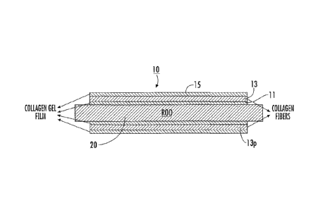
[00114] Referring now to the figures, Figure 1A, an exemplary elongate
construct 10 is shown on a support member 20. As shown, the construct 10
includes
an inner layer of collagen film 11, an intermediate layer of at least one
wound
collagen fiber 13, and an outer layer of collagen film 15. Referring to
Figures 131?,
15A, and 15C, the intermediate layer 13s can comprise at least one wound
collagen
fiber and one or more layers of collagen gel and/or collagen film optionally
including
one or more minerals and/or particulates. Referring to Figure 15B, for
example, the
intermediate layer 14 can comprise at least one wound collagen fiber and one
or more
layers of gelatin slurry, gelatin hydrogel, and/or gelatin film optionally
including one
or more minerals and/or particulates. An intermediate layer comprising at
least one
wound collagen fiber and one or more layers of gelatin slurry, gelatin
hydrogel,
and/or gelatin film comprising one or more minerals 14m is demonstrated in
Figure
15D.
[00115] In other embodiments, the construct 10 can be formed without the
inner and/or outer layer of film 11 and/or may optionally include other
materials or
constituents and/or layers. For example, hydroxyapatite can be placed into the
collagen fiber and/or collagen gel material. This configuration can be
particularly
suitable to augment fixation of auto graft tendons (typically with one or more
interference screws).
[00116] As shown in Figure 111, the construct 10 can have a wall 10w with a
suitable thickness defined by the at least one collagen fiber 13 and the film
layers
(where used) and/or other coatings and/or materials placed thereon. The
construct can
have a uniform or non-uniform stiffness. The construct 10 can have an open
through
cavity or may be filled or partially filled with a nerve-growth media or other
therapeutic material (e.g., an anti-inflammatory, antibiotic and/or the like).
[00117] As also shown, the at least one collagen fiber 13 has an angular
fiber
pattern 13p of repeating intersecting collagen fiber segments along its
length. The
angular pattern 13p can be defined by a number of revolutions of the at least
one fiber
13 about the support member 20 at a given pitch or pitches for at least one
layer
CA 02806396 2013-01-23
WO 2011/156319
PCT/US2011/039375
23
(typically more than one layer). The support member 20 is used to wrap the at
least
one collagen fiber around its exterior surface to form a desired shape.
[00118] The support member 20 can include a lubricious and/or smooth
surface, an embossed surface with lower contact surface area, a rough surface,
a
patterned surface and/or a textured surface, typically of a polymer material.
In
particular embodiments, the support member 20 can include a ribbed surface 20r
(Figure 19C), which may have a spiral configuration. The ribbed surface of the
support member (where used) can provide consistent regions of higher (e.g.,
ridges)
and lower (e.g., valleys) contact surfaces along the length of the support
member 20.
In other embodiments, the ribs 20r can be configured asymmetrically to vary
about
the circumference and/or length of the support member 20. The ribs can have
the
same height and/or thickness or can vary. For example, the ribs 20r can be
evenly
spaced apart along the length of the support member 20 or the distance between
the
ribs can vary along the length of the support member 20. In some embodiments,
the
support member 20 can include an anti-slip surface with ridges or a sleeve can
be
placed over the support member (not shown) to contact the next layer (e.g.,
inner film
11 or fiber 13). In some embodiments, the support member 20 comprises Teflon
or
other suitable low friction and/or anti-stick material. The support member 20
can be
tubular, e.g., cylindrical, as shown in Figures 1A, 3A, 3B and 3E or may be
substantially flat and rectangular 20' as shown in Figures 3C and 31). Other
geometries may also be used, such as, for example, a frustoconical or funnel
shape.
Typically, the support member 20 is elongate and has a substantially circular,
oval,
polygonal or other cross-sectional shape. The support member 20 can have a
consistent diameter along its length (Figure 1711) or the support member can
be
frustoconical or tapered 201 along its length (Figures 20A, 20C, 20E, 20F, and
20G).
1001191 The design and dimensions of the support member can affect the
design and dimensions of the resulting construct 10. For example, a support
member
with a ribbed surface 20r can provide a construct 10 with a rough inner
surface 11r or
a support member that is frustoconical or tapered 20f along its length can
provide a
construct that is similarly tapered (Figure 19C). In some embodiments, the
support
member is frustoconical 20f and is about 5 cm in length with a diameter that
is
between about 3 mm to about 6,5 mm. In certain embodiments, the resulting
construct 10 formed on a tapered support member can be particularly suitable
for
tendons.
CA 02806396 2013-01-23
WO 2011/156319
PCT/US2011/039375
24
1001201 The at least one collagen fiber 13 can be organized into various
arrays
including braids, weaves, knits, parallel arrays, twisted configurations, and
the like.
The orientation of one or more of the fibers 13 within the resulting material
10 (see,
e.g., Figures 2A-20) can be targeted to meet the specific mechanical
requirements of
the medical application. Fiber density can vary from dense to loose geometries
and
the numbers and size of the one or more collagen fibers used can vary as well
as the
thickness of the film to provide specific mechanical properties. The fiber(s)
13 can be
continuous length fibers or may be formed by attaching a series of collagen
fibers in
an end-to-end orientation 13j (Figure 4).
[001211 Figures 2A-21) are digital photographs of a prototype of a
construct
10. This construct 10 may be particularly suitable as a medical construct,
such as an
auto and/or allo-graft, a nerve tube or guide 10n (Figure 8A), or other
medical
construct. The construct 10 is tubular 10t with an open cavity and has a
flexible
elastic configuration. The construct 10 can be formed using a single fiber 13
formed
in wound multiple layers, the fiber 13 can have a length between about 1- 6 m,
typically about 5 m. The construct 10 can be formed using a single fiber 13 of
a
continuous length that is wrapped in several layers about the support member
20. Use
of a single fiber 13 can reduce the likelihood of any fraying associated with
multiple
fibers (such as those wound in one lengthwise direction). The construct 10 can
have a
length between about 1 cm to about 20 cm (or more), typically between about 5
cm to
about 15 cm, and the inner diameter can be between about 1 mm to about 10 mm,
typically between about 3 mm to about 20 mm, with the wall thickness being
about
0.1 mm to about 3 mm, typically between about 0.1 mm to about 2 mm.
[00122] The construct 10 can have reversible elasticity with sufficient
rigidity
or strength to prevent undue tendon or nerve compression or the like, while
allowing
flexibility sufficient to allow the construct 10 to spring back into its
original shape
after being exposed to a strain or tension caused by normal body movement that
deforms the shape. The nerve guide lOn can be used for any nerve location,
e.g.,
peripheral nerves (such as in a hand or finger), spinal cord nerves, and the
like. The
construct 10 can be used for other repairs or treatments as will be discussed
further
below. The construct 10 is biocompatible (or at least non-cytotoxic) and can
provide
a desired half-life suitable for its intended function.
[00123] The construct 10 and/or the fiber 13 can be cross-linked with a
suitable
polymerizing material, such as, but not limited to, NDGA, or may be used in a
non-
CA 02806396 2013-01-23
WO 2011/156319
PCT/US2011/039375
cross-linked state. The NDGA cross-linking can increase the strength of the
device
10 but may decrease the resiliency, elasticity or flexibility. In some
embodiments, the
collagen fiber 13 is not cross-linked during the winding process, but may
optionally
be cross-linked after the winding process (typically after the collagen film
has been
applied to the outer surface and dried).
1001241 The support member 20 can be configured to facilitate removal of
the
construct 10. For example, the construct 10 may be wound tightly against the
outer
surface of the support member 20 and allowed to dry. The support member 20 can
be
configured to reduce in cross-sectional size or disassemble with the construct
10 held
thereon to allow easy removal of the elongate construct. In some embodiments,
the
support member 20 can be a multi-piece device that provides this size change.
In
other embodiments, the support member 20 may be cooled while the construct is
heated to provide a size difference. In particular embodiments, the support
member
20 can cooperate with an insert 201 (Figure 3D) that provides the desired size
adjustability. In other embodiments, the construct 10 can be removed from the
support member without such a size adjustment (e.g., its inner surface may be
sufficiently lubricous or a suitable liquid or other material can be used to
slide the
construct off the support member. In other embodiments, the construct 10 can
be cut
in a lengthwise (e.g., "X") direction and taken off the support member 20. In
some
embodiments, the construct 10 may be cut or otherwise separated in a long axis
direction with a longitudinal slit lOs and used for a cuff 10c (Figure 8B)
that can be
positioned about a nerve or other tissue to protect that tissue (and the cuff
may be
sutured together along at least a portion of the long axis and/or may be
sutured or
otherwise anchored into position). The cuff 10c may be configured to provide a
snug
or alternatively, a non-constricting, encasement for injured tissue such as
injured
peripheral nerves for protection of the neural environment. The wall of the
cuff with
the longitudinal slit lOs can be spread open for easy placement over the
injured target
tissue. The resilience of the collagen conduit allows the cuff to recover and
maintain
closure once the device is placed around the tissue.
[00125] As shown in Figures 3A-3B, the construct 10 can be made by winding
at least one collagen fiber 13 around a support member 20 using a computer-
guided
and/or controlled lathe system 100. The lathe system can be configured to
rotate the
support member 20 and to move the support member back and forth in a length
direction to alter the location of the fiber on the support member 20 relative
to the
CA 02806396 2013-01-23
WO 2011/156319
PCT/US2011/039375
26
introduction point of the fiber (e.g., the fiber introduction point may be
stationary). In
other embodiments, the fiber(s) 13 can be supplied through a head that moves
relative
to the support member 20 (e.g., the support member can be stationary) or both
the
fiber introduction head and the support member may move relative to teach
other.
[00126] Different size (e.g., diameter) support members 20 can be used
depending on the target product. For example, transverse small cross-section
support
members (e.g., diameter rods) can be used for manufacturing devices for use in
vein
and artery replacements or repairs, while larger transverse cross-section
support
members (e.g. diameter rods) can be used to manufacture devices for aortic or
large
artery replacements or repairs and/or various shunts.
[00127] An example of a small lathe 100, typically a micro or miniature
lathe,
suitable for fabricating embodiments of the constructs is the Model 4410 lathe
available from Sherline Products, Inc., having a place of business in Vista,
CA. Two
user-selectable inputs can operate the lathe system: one controls the speed
that the
support member that spins and the other controls the pattern (fiber angle) in
which the
at least one fiber 13 is laid onto the support member. The operation can be
configured
so that the fiber is self-pulling from a spool in communication with a channel
in the
feeder head based on the speed of the spinning support member 20. The lathe
100 can
co-wind a plurality of fibers or fiber bundles substantially concurrently
about the
support member 20.
[00128] The at least one collagen fiber 13 can be coated with one or more
layers of collagen gel 11, 15 and/or other suitable bio-compatible material
during
and/or after winding the at least one collagen fiber 13 to seal the fiber(s)
13 within the
bioeomposite material and/or to form a smooth inner and/or outer surface of
the
construct 10. Figure 3B illustrates that collagen gel can be applied to the
fiber 13 on
the support member during the winding. Figure 3B illustrates that a brush 111
can be
used to apply the gel. Other application techniques may be used, such as
spray, pour,
drop, and the like. The application of the soluble collage gel may be manual
or
automated and applied by electro-mechanical devices.
[00129] The winding can be performed so that at least one layer of the at
least
one collagen fiber has a substantially constant pitch for at least a major
portion of a
length thereof or so that at least one layer of the at least one collagen
fiber has a
variable pitch for at least a major portion of a length thereof.
81722065
27
[00130] Figure 4 illustrates that different configurations of fibers 13
may be
used. Examples of fiber configurations include a single fiber 131, a plurality
of fibers
131_ 13n (typically n=2 to 100) that can be concurrently co-wound about the
support
member 20, a fiber bundle 13b, and a twisted, woven or braided fiber bundle
13t. In
some embodiments, a plurality of collagen fibers 13 are twisted, woven, and/or
braided together to form a twisted, woven, and/or braided fiber bundle 131.
The
twisted, woven, and/or braided fiber bundle 13t can be wound about a support
member 20. In certain embodiments, the plurality of collagen fibers 13
comprises
between about 3 to about 30 fibers, typically between about 6 to about 15
fibers. For
the fiber bundles 13b, 13t, two or more fibers 13 can be grouped together to
form the
fiber bundle 13b, 13t and that bundle 13b, 131 applied or wrapped about the
support
member 20, similar to a single fiber. One or more fiber bundles 13b, 131 may
be used
to form the construct 10. In certain embodiments, a plurality of fibers
comprises a
fiber bundle 13b, 13t. In some embodiments, the fiber bundle 13b, 13t is
combined
with between about 6 to about 27 fiber bundles 13b, 13t. Combinations of the
different fiber types may also be used for some constructs 10. That is, for
example, a
twisted fiber 13t can be co-wound with a single fiber 131 and/or a single
fiber 131 may
be used to form one layer and a twisted 131 to form a different layer, and the
like.
Exemplary configurations of fibers 13 are described in U.S. Patent Application
Publication Nos, 2008/0188933, 2008/0215150, 2008/0200992, 2009/0216233, and
2009/0287308.
[00131] The collagen fiber 13 can be wound using various fiber angles
(e.g.,
pitch angles), such as, angles between about 2-70 degrees, typically between
about 5-
60 degrees, such as, for example, 5, 10, 15, 20, 25, 30, 35, 40, 45, 50, 54
and 55
degrees, or other odd or even numbers between 5-70. Where constructs of
multiple
layers are used, one layer may have a first pitch and another layer may have a
different pitch.
[00132] Figure 5A illustrates that a construct 10 can be wound with
increased
fiber density 52 along certain segments, typically forming end rings 52r. This
increased fiber density 52 can provide sufficient rigidity to allow a suture
to attach
thereto. As shown in Figure 5A, the construct 10 is tubular let and may
optionally
include an increased density segment 52 at an intermediate location. Figure 5B
illustrates that the construct 10 can be used as formed, or may be cut or
separated
CA 2806396 2018-04-12
81722065
28
along a Y-axis into two components lOta, 10th. For the latter, the
intermediate
increased density ring 52 can form end rings for the separated construct lOta,
10tb.
[001331 Figure 6A illustrates a construct 10 that is relatively flat 10f
and/or
rectangular. Again, the construct 10f can optionally include increased fiber
density
segments 52 that may be suitable for end rings 52r. Figure 6B illustrates that
the
construct 10f can be cut along the X-axis and separated into at least two
components
that form biocompatible patches. The intermediate increased density ring(s)
52,
where used, can optionally form end rings 52 for the separated construct 10fa,
10th,
etc.
1001341 Figure 7 illustrates an example of another automated winding
system
100' that can be used to form the construct 10. This embodiment uses several
fibers
13, each independently wound and/or wrapped to weave or braid the fibers about
the
support member 20 to form the construct 10. The system 100' includes a plate
122
supporting spindles 124, a forming plate 126, a support member (shown as a
cylindrical mandrel) 20 that extends through an aperture in the forming plate
126, and
braid puller 128. An exemplary microbraider is believed to be available from
Kokubun Ltd of Japan. See also, Figure 2 and col. 2 of U.S. Patent No.
7,135,040.
1001351 The fibers 13 can be wound before or after cross-linking (or not
cross-
linked at all). If wound before, the fibers can, where desired, be polymerized
with
any suitable cross-linking materials, to promote collagen organization, such
as, for
example, NDGA, but other cross-linking materials may be used, including, for
example, glutaraldehyde. The (dried) collagen fiber can also be treated with
other
methods to improve the tensile properties of the fiber. The (dried) collagen
fibers 13
can be cross-linked with agents such as glutaraldehyde, formaldehyde, epoxy
resins,
tannic acid, or any other chemical agent that produces covalent cross-links
between
collagen molecules within fibrils or between fibrils. Alternatively, the fiber
13 can be
treated to induce cross-linking between collagen molecules such as, but not
limited to,
one or more of a carbodiimide treatment, ultraviolet irradiation either with
or without
carbohydrates to initiate glycation adducts, and dehydrothermal treatment
coupled
with any of the aforementioned methods.
[00136] Figure 9 illustrates a medical kit 250 that includes a medical
device or
implant 10 or 10'. The kit 250 may optionally include other components, such
as, for
example, a container of surgical adhesive, sutures 210, suture anchors, and
the like.
CA 2806396 2018-04-12
CA 02806396 2013-01-23
WO 2011/156319
PCT/US2011/039375
29
The device or implant 10, 10' may be held hydrated in a flexible sealed
package of
sterile liquid 230. The kit 250 may include a temperature warning so that the
construct 10, 10' is not exposed to unduly hot temperatures that may degrade
the
implant. A temperature sensor 252 may optionally be included on the package of
the
kit to alert the clinician as to any excessive or undue temperature exposure
prior to
implantation. For example, it may be desirable to hold or store the kit 250
(and
implant or device 10, 10') at a temperature that is less than about 370 C
and/or 100 F
prior to implantation. The kit 250 may be packaged in a housing with a
temperature
controlled or insulated chamber 250c to facilitate an appropriate temperature
range.
[00137] Figure 10 is a flow chart of operations that can be used to carry
out
embodiments of the present invention. In some embodiments, the at least one
collagen fiber is wound a number of revolutions about a length of a support
member
having a long axis. The winding can have a defined pitch and/or fiber angle
relative
to the long axis of the support member to form an elongate construct with at
least one
wound collagen fiber (block 150). The winding step can form multiple overlying
layers of the at least one collagen fiber in one or more fiber angles so that
the at least
one fiber intersects itself at different locations along a length of the
construct.
[00138] Optionally, a collagen gel can be placed onto the support member
and
the gel can dry to form a film on the outer surface of the support member
before the
winding step (block 155). The collagen film can be dried or allowed to dry on
the
support member (e.g., rod). As the fiber(s) is wound about the support member,
a
soluble collagen can be applied (e.g., wrapped, painted, sprayed, dripped and
the like)
onto the fiber(s) and/or support member so that the fiber(s) becomes wet while
one or
more layers are wound on the lathe.
[00139] The at least one collagen fiber can be supplied to the
winder/support
member in a substantially dry state and may be provided as a spooled (dry)
quantity
of the at least one collagen fiber (block 152). The fiber(s) can be supplied
and wound
in a non-cross-linked state,
[00140] In some embodiments, the winding step can be carried out to create
multiple adjacent overlying layers of the at least one fiber, the adjacent
layers being
coextensive for at least a major portion of a length of the construct (block
153). A
liquid or gel comprising soluble collagen can be placed onto the at least one
wound
collagen fiber to cover at least the outer surface in a collagen film (block
165).
CA 02806396 2013-01-23
WO 2011/156319
PCT/US2011/039375
[00141] Optionally, the placing of the collagen gel or liquid is carried
out by
placing collagen gel having a cylindrical shape around the at least one wound
collagen fiber and the support member (block 158).
[00142] Optionally, the collagen can be polymerized -while the elongate
construct is held on the support member using a suitable cross-linker, such
as, for
example, NDGA, then removing the construct from the support member (block
166).
[00143] The winding can be carried out so that the at least one fiber turns
about
the support member in one of a clockwise or counterclockwise direction along a
first
lengthwise direction for a first layer, then reverses to travel in an opposing
lengthwise
direction and continues to turn about the support member in the same clockwise
or
counterclockwise direction for a second adjacent layer (block 180, Figure 11).
[00144] Alternatively, in particular embodiments, the winding may be
carried
out so that the at least one collagen fiber turns (is wrapped) about the
support member
in one of a clockwise or counterclockwise direction along a first lengthwise
direction
for a first layer, then reverses to travel in an opposing lengthwise direction
and turns
about the support member in the other clockwise or counterclockwise direction
a
second adjacent layer.
[00145j In some embodiments, the winding step has a first pitch for the
winding of the at least one collagen fiber on the first layer and a second
smaller or
greater pitch for the winding of the at least one collagen fiber on the second
layer. In
some embodiments, the at least one fiber on the second layer resides between
gaps
defined by the at least one fiber wound with the defined pitch on the first
layer.
[00146] Figure 12 is a flow chart of operations that can be used to carry
out
embodiments of the present invention where a gelatin slurry is applied to the
at least
one collagen fiber. In certain embodiments, the at least one collagen fiber is
wound a
number of revolutions about a length of a support member having a long axis.
The
winding can have a defined pitch and/or fiber angle relative to the long axis
of the
support member to form an elongate construct with at least one wound collagen
fiber
(block 350). As the fiber(s) is wound about the support member, a gelatin
slurry is
applied (e.g, wrapped, painted, sprayed, dripped, dipped, and the like) onto
the
fiber(s) 13 and/or support member 20 so that the fiber(s) 13 is wetted while
one or
more layers of the fibers are wound on the lathe (block 355).
[00147] The gelatin slurry is typically painted on a fiber during and/or
after the
fiber is wound on the support member. However, it is contemplated that a
collagen
CA 02806396 2013-01-23
WO 2011/156319
PCT/US2011/039375
31
fiber can be pre-sprayed, coated, painted, dipped or the like with a gelatin
slurry
before winding (block 340). The at least one collagen fiber with the gelatin
slurry can
be polymerized after the winding step with a suitable cross-linker, such as,
for
example, NDGA (block 342). in some embodiments, a plurality of elongated
collagen fibers having a length of between about 1 m to about 100 m are coated
with a
gelatin slurry comprising one or more minerals.
1001481 Also, it is contemplated that a gelatin slurry can be applied to a
non-
wound collagen fiber construct as well, e.g., braided, twisted, or parallel
fibers and the
like to form a gelatin film on the collagen fiber construct. The non-wound
collagen
fiber construct can be cross-linked with a suitable polymerizing material,
such as, but
not limited to, NDGA, carbodiimide, glutaraldehyde, formaldehyde, tannic acid,
isocyanates, and epoxy resins, or may be used in a non-cross-linked state.
[00149] As described above, the gelatin slurry can comprise one or more
minerals, such as, but not limited to, hydroxyapatite (block 357). During the
application of the gelatin slurry, the concentration of the components, e.g.
gelatin
and/or one or more minerals, can vary. For example, the concentration of the
one or
more minerals in the gelatin slurry can vary along a length of a support
member
and/or at different layers. For example, the mineral concentration can be
greater at
the end of the application step than at the beginning of the application step
or vice
versa to result in a variation in the mineral concentration along the fiber
and/or
support member. In certain embodiments a concentration gradient of one or more
minerals is established along the fiber and/or support member. Alternatively,
the
concentration of the gelatin and/or one or more minerals can be higher or
lower in
certain regions of the fiber and/or support member. The gelatin slurry can be
heated
before and/or during application to the fiber(s), as described above (block
358).
1001501 The thickness of the gelatin intermediate layer 14 can vary
depending
on the amount or volume of gelatin slurry applied to the at least one collagen
fiber,
which can vary the wall thickness of a resulting construct. The gelatin
intermediate
layer 14 comprises at least one collagen fiber, which may be infused,
embedded,
incorporated, and/or attached to the gelatin slurry, gelatin hydrogel, and/or
gelatin
film. The gelatin intermediate layer 14 optionally comprising one or more
minerals
can vary in thickness depending on the amount or volume of gelatin slurry
applied to
the at least one collagen fiber. As illustrated in Figures 18B and 18C, the
gelatin
intermediate layer comprising one or more minerals 14m can vary in thickness
CA 02806396 2013-01-23
WO 2011/156319
PCT/US2011/039375
32
depending on the volume of gelatin slurry applied to the at least one collage
fiber. A
tube with a thicker wall can provide increased mechanical strength compared to
a tube
with a thinner wall (Figure 18G). The application of force to a tube with a
thicker
wall can, in some embodiments, result in more deformation compared to a tube
with a
thinner wall (Figure 18E). When a gelatin slurry is applied to the at least
one wound
collagen fiber it can be actively and/or passively dried to form a gelatin
film (block
359).
[00151] The incorporation of one or more minerals into the gelatin slurry
can
result in the minerals being partially or substantially retained in the
construct or tube.
The one or more minerals can be partially or substantially retained in the
construct.
The minerals can be integrated into the construct so that they are
substantially
retained even when exposed via a cut edge. The construct can retain 50%, 60%,
70%,
75%, 80%, 85%, 90%, 95%, 98%, 99% or 100% of the mineral concentration present
in the construct after addition of the gelatin slurry. The mineral
concentration can be
determined by well known methods. Exemplary methods include, but are not
limited
to, quantitative chemical analytical techniques such as titrations (e.g., EDTA
titrations) and colorimetrie methods such as ascorbic acid colorimetric
methods,
atomic absorption spectroscopy, mass spectrometry such as inductively coupled
plasma mass spectrometry (ICP-MS), X-ray fluorescence, X-ray diffraction, and
electron probe mieroanalyzer.
[00152] The application of a gelatin slurry optionally comprising one or
more
minerals onto a collagen fiber(s) can inhibit the tube layers and/or minerals
from
delaminating, as exemplified in Figures 15A-D. The mineral particles can
incorporate or integrate into the collagen fiber. The mineral particles can be
embedded and/or dissolved in the gelatin hydrogel or gelatin film after
cooling and/or
drying. The collagen fiber can be embedded or incorporated into the gelatin
hydrogel
or gelatin film.
[00153] Optionally, prior to the winding of the at least one collagen fiber
(block 350) a collagen gel optionally comprising one or more minerals can be
placed
onto the support member and the collagen gel can dry to form a collagen film
on the
outer surface of the support member before the winding step (block 310). The
collagen gel can be dried or allowed to dry on the support member (e.g., rod)
either
actively or passively. In certain embodiments, a collagen gel comprises at
least one
mineral. In some embodiments, a collagen gel is placed onto a ribbed support
CA 02806396 2013-01-23
WO 2011/156319
PCT/US2011/039375
33
member to form a construct with a rough inner surface. Multiple layers of
collagen
gel can be placed onto the support member. For example, between about 1 to
about
20 layers of collagen gel can be applied, typically between about 1 to about
10 layers
of collagen gel can be applied. When multiple layers of collagen gel are
applied, the
layers can comprise different components and/or the same components present in
different concentrations. For example, in some embodiments, between about 1 to
about 5 layers of collagen gel with no minerals can be placed onto the support
member, followed by between about 1 to about 5 layers of collagen gel with at
least
one mineral or vice versa.
[001541 Optionally, after application of the gelatin slurry, a liquid or
gel
comprising soluble collagen and optionally one or more minerals can be placed
onto
the at least one wound collagen fiber with the gelatin slurry to cover at
least the outer
surface in a collagen film (block 315). In particular embodiments, the soluble
collagen comprises at least one mineral and is placed onto the at least one
wound
collagen fiber with the gelatin slurry to form a construct with a rough outer
surface.
Multiple layers of soluble collagen can be placed onto the at least one wound
collagen
fiber with the gelatin slurry. For example, between about 1 to about 20 layers
of
soluble collagen can be applied, typically between about 1 to about 10 layers
of
soluble collagen can be applied. When multiple layers of soluble collagen are
applied, the layers can comprise different components and/or the same
components
present in different concentrations. For example, in some embodiments between
about 1 to about 5 layers of soluble collagen comprising at least one mineral
can be
placed onto the at least one wound collagen fiber with the gelatin slurry,
followed by
between about 1 to about 5 layers of soluble collagen with no minerals or vice
versa.
[00155] The method can include cutting the construct in an axial direction
to
form a flat collagen fiber patch. The method can include winding the collagen
fibers
in a plurality of axially spaced apart segments with increased collagen fiber
density, at
least some of which are provided as reinforced segments for suturing. The
reinforced
segments can be formed at end portions of the tube and optionally at one or
more
intermediate locations therebetween. The methods can produce a nerve guide
having
sufficient strength and elasticity to withstand buckling and to be able to
bend and to
elastically return to its original shape after bending to inhibit occlusive
pressures or
restrictions on nerves.
CA 02806396 2013-01-23
WO 2011/156319
PCT/US2011/039375
34
[00156] Embodiments of the invention can be used for a number of different
medical applications, including, but not limited to, auto-grafis, allo-grafts,
nerve
guides, wound bed patches, muscle or organ patches, cardiac patches, valve
replacements or repairs, hernia patches, skin patches, burn treatment patches,
skin/tissue repair patches or cuffs, blood vessel (artery, vein, and the like)
repairs,
sleeves that can reside about repairing tendon to prevent or inhibit
adhesions,
indwelling tubes for delivery of therapeutic agents, duets such as lymphatic,
hepatic,
pancreatic and cystic ducts, tubes such as ureter and urethra tubes and the
like.
[00157] The devices are entirely scalable in all dimensions, length,
diameter,
wall thickness, relative amount of mineral per collagen, etc. Typically, the
devices
are tubes that have a length that is between about 5 cm to about 15 cm, a
diameter that
is between about 3 mm to about 20 mm, and a wall thickness about 0.1 mm to
about 2
mm. In particular embodiments, the devices can be used with/for allo-grafts or
auto-
grafts such as tendon or ligament implants.
[001581 The devices can be configured as tubes with rough inner 11r and/or
rough outer surfaces 15r, as exemplified in Figures 16C, 17E, and 19E. The
devices
can taper in size about its length (Figures 2013, 20D, and 2014) or have a
substantially
constant width (e.g., diameter) (Figures 16C-E and 19C-E).
[00159] The present invention is explained in greater detail in the
following
non-limiting Examples.
EXAMPLES
[00160] In the following examples the exemplary collagen fiber constructs
(e.g., sleeves or tubes) are cross-linked with nor-dihydroguaiaretie acid
(NDGA).
However, this cross-linking agent is for exemplary purposes only. The present
invention is not intended to be limited to cross-linked constructs (i.e., the
construct
may be used in a non-cross-linked state) or to cross-linked constructs where
NDGA is
the cross-linking agent. For example, other cross-linking agents, such as, but
not
limited to carbodiimide, glutaraldehyde, formaldehyde, tannic acid,
isoeyanates, and
epoxy resins, and/or stabilization treatments, such as, but not limited to,
one or more
of dehydrothermal treatment, glycation, and ultraviolet light may be used in
the
present invention.
Example 1
CA 02806396 2013-01-23
WO 2011/156319
PCT/US2011/039375
[00161] Figures 2A-2D illustrate exemplary sleeves or tubes of wound NDGA-
collagen fibers that may be particularly suitable for medical constructs, such
as nerve
guides. The inner diameter of the tube can vary between about 1 and lOmm. The
thickness of the wall can vary between about 0.1 and 3 mm. The length of the
tube
can vary from between about 1 to 6 cm or more.
[00162] The tube can be made of dermal collagen that is acid or pepsin
soluble.
The soluble collagen can be made by neutralizing acid soluble collagen and
keeping
the soluble collagen at a desired low temperature to maintain the collagen in
molecular form, (e.g., about 4 C). Collagen gels can be produced from acid
soluble
collagen by neutralization, injection molding in a "feflon tube of diameter
between
0.1 cm to 1.0 cm and incubation for at least about 4 hours at 37 C. The
resulting gel
can be extruded into deionized water to form a gel cylinder with a diameter
between
about 0.1 cm to 1.0 cm (and can have a length between about 1 -100 m. Collagen
concentration of the soluble collagen and collagen gel can be from about 0.1-
4%
weight per volume. The gel cylinder can be used in the gel form or allowed to
dry,
actively or passively (suspended in air), to form a collagen fiber having a
diameter
between about 0.05 mm (average) to about 0.2 mm (average).
[00163] The first step to make this prototype tube is to wrap the collagen
gel of
specified collagen concentration and diameter onto a Teflon rod of selected
diameter. The collagen gel layer was allowed to dry on the rod at room
temperature
to form a thin layer of collagen film. The thickness of this collagen film can
be varied
by applying more or less layers of collagen gel, either is a single
application of in
several applications.
[00164] The second step is to wind dry collagen fibers on to the collagen
film
coated Teflon rod. The pitch of the fiber relative to the long axis of the
tube can be
specified. The thickness of the collagen winding can be adjusted, for example,
corresponding to the number of layers of fibers that are laid on (and/or the
number of
fibers bundled together for the winding). During the fiber winding process,
soluble
collagen is applied (e.g., painted) onto the surface of the laid-on fibers.
The thickness
of the final soluble collagen layer can be varied to achieve specific
thickness. The
soluble collagen coated fiber wound cylinder is allowed to dry.
[00165] The third step in making the tube is the same as the first step,
e.g., to
wrap a collagen gel on to the collagen fiber would Teflon rod and the gel
layer is
allowed to dry to form a collagen film enwrapping the collagen fiber tube. The
81722065
36
thickness of the penultimate collagen film can be varied by the number of
layers of
wrapped gel.
[001661 The dried tube can be used "as- is" (used in a non-cross-linked
state
and hydrated when in the body or prior to placement in the body), or it can be
cross-
linked with any agent or action that cross-links the collagen. The (nerve)
tube is then
taken off the Teflon* rod. In the present example, the tube is cross-linked
with nor-
dihydroguaiaretic acid (NDGA), see, e.g., U.S. Patent No. 6,565,960, and U.S.
Patent
Application Publication No. US-2008-0161917-Al.
Example 2
[00167] Figures 13A-F depict various stages in the formation of an
exemplary
collagen fiber construct of the present invention that comprises an
intermediate layer
comprising at least one wound collagen fiber and one or more layers of
collagen/HA
applied to the collagen fiber. This exemplary embodiment may be particularly
suitable for medical constructs. Hydroxyapatite (HA) can be mixed in a
neutralized
collagen solution at pH 7.2 at a very high concentration to form a collagen/HA
solution. A 7 mm Teflon rod can be wrapped with between about 5 to about 6
layers
of collagen gel and dried. Uncrosslinked collagen fibers can be wound on the
collagen coated Teflon" rod. Figure 13A is a digital photograph of the rod
being
wrapped with the uncrosslinked collagen fibers. While the collagen fiber is
being
wound, the collagen/HA solution can be painted along the fiber winding. Figure
13B
is a digital photograph of the dry collagen tube comprising the inner collagen
gel
wrapped with the collagen fiber and painted with the collagen/HA solution on
the rod.
Eight layers of collagen gel can be wrapped on the fibers wound on the Teflon
rod.
The tube can then be crosslinked in 100 mL standard NDGA solution for
approximately 24 hours and then washed by phosphate buffer pH 9 and 70%
ethanol
for 1 hour each. Figure 13C is a digital photograph of the completed tube
comprising
the inner collagen gel wrapping, the wound collagen fiber with the
collagen/HA, and
the outer collagen gel wrapping that has been cross-linked with NDGA on the
rod.
The tube can be air dried and taken off the Teflon* rod. Figures 13D-F
illustrate the
completed exemplary NDGA crosslinked collagen fiber constructs (e.g, tubes or
sleeves).
CA 2806396 2018-04-12
CA 02806396 2013-01-23
WO 2011/156319
PCT/US2011/039375
37
Example 3
[00168] Figures 14A-G depict various stages in the formation of an
exemplary
collagen fiber construct of the present invention that comprises an
intermediate layer
comprising at least one wound collagen fiber and one or more layers of
gelatin/IIA
applied to the collagen fiber. This exemplary embodiment may be particularly
suitable for medical constructs. 1 gram of hydroxyapatite can be mixed in 5 mL
of
3% Sigma gelatin A solution, to yield a gelatin/HA solution with a
concentration of
HA particles of 20%. A 7 mm Teflon rod can be wrapped with 5-6 layers of
collagen gel and dried. Uncrosslinked collagen fibers can be wound on the
collagen
coated Teflon rod, meanwhile the gelatin/HA solution can be painted along the
fiber
winding. Eight layers of collagen gel can be wrapped on the fibers wound on
the
Teflon rod. Figure 14A is a digital photograph of the completed device still
wet on
the rod with the three layers: the inner collagen gel layer, the intermediate
layer
comprising the wound collagen fiber and gelatin/hydroxyapatite, and the outer
collagen gel layer. The tube can be crosslinked in 100 mL standard NDGA
solution
for approximately 24 hours, and then washed by phosphate buffer pH 9 and 70%
ethanol for 1 hour each. Figure 14B is a digital photograph of the completed
device
that has been cross-linked on the rod with NDGA. The tube can be air dried and
taken off the Teflon rod. Figures 14C-G illustrate the completed exemplary
NDGA
crosslinked collagen fiber constructs (e.g., tubes or sleeves).
Example 4
[001691 Figures 16A-E depict various stages in the formation of an
exemplary
collagen fiber construct of the present invention that comprises a rough outer
surface.
This exemplary embodiment may be particularly suitable for medical constructs.
Hydroxyapatite can be mixed in neutralized collagen solution (pH 7.2) at a
concentration of 1% (300 mg/30 mL = 1 g/100 mL) for wrapping gel
(collagen/HA),
such collagen/HA gel is depicted in Figure 16A. Hydroxyapatite can be mixed in
2%
gelatin at a concentration of 10% for painting purposes (gelatin/HA, total 5
mL for 3
tubes, 500 mg HA/5 mL 2% Gelatin = 10 g/100 mL).
1001701 A 6.35mm (1/4") Teflon rod can be wrapped with 6 layers of Devro
collagen gel and dried. Uncrosslinked collagen fibers can be wound on the
collagen
coated Teflon rod, meanwhile the warm gelatin/HA solution (50-55 C) can be
painted along the fiber winding under heat. The painting with the gelatin/HA
solution
CA 02806396 2013-01-23
WO 2011/156319
PCT/US2011/039375
38
can be continued until finish a 5 mL solution for all 3 tubes. Two layers of
collagen/HA gel can be wrapped on the fibers wound on the Teflon rod. Figure
16B
is a digital photograph of the wet collagen/HA gel wrapped collagen fiber
construct
comprising a rough outer surface on the rod. Then, four layers of "pure"
collagen gel
(i.e., collagen gel with collagen, but no additional components or minerals)
can be
wrapped on the tubes. The tubes can be crosslinked in standard NDGA solution
for
approximately 24 hours, and then washed by phosphate buffer pH 10.3 (or pll 9)
and
70% ethanol for 1 hour each. The construct can be air dried and taken off the
Teflon
rod (or hydrated if the construct cannot be taken off). Figures 16C-E
illustrate the
completed exemplary NDGA crosslinked collagen fiber constructs (e.g., tubes or
sleeves).
Example 5
[00171] Figures 17A-F depict various stages in the formation of an
exemplary
collagen fiber construct of the present invention that comprises a rough outer
surface.
This exemplary embodiment may be particularly suitable for medical constructs.
Hydroxyapatite can be mixed in neutralized collagen solution (pH 7.2) at a
concentration of 1% for wrapping gel (collagen/HA, Devro 0.5% collagen, dilute
with
DI salt solution at 1:1, adjust pH 7.22, 900 mg HA/90 mL neutralized collagen
solution = 1 g/100 mL = 1%). The collagen/HA wrapping gel is depicted in
Figure
17A. Hydroxyapatite can be mixed in 2% gelatin at 10% for painting purpose
(gelatin/HA, total 40 mL for 24 tubes, 4 g HA/40 mL 2%, Gelatin = 10 g/100 mL
=-
10%). Collagen/HA gel can be formed in the incubator at 37 C for 4 hours
before
extruded into DI water, which is the same as the procedure for forming general
collagen gel. Gelatin/HA painting solution can be kept in a water bath at 45-
50 C to
warm up the gelatin during fiber winding and painting process.
[001721 A 6.35mm (1/4") Teflon rod can be wrapped with 4 layers of Devro
collagen (0.25%) gel and dried. Uncrosslinked collagen fibers can be wound on
the
collagen coated Teflon rod, meanwhile the warm gelatin/HA solution (50 C) can
be
painted along the fiber winding. The average volume of gelatin/HA painting
solution
used can be approximately 1.5 mL per tube. Figure 17B is a digital photograph
of the
rod wrapped with the inner collagen gel layer and the intermediate layer
comprising
the collagen fiber painted with gelatin/hydroxyapatite. The tube can then be
dried.
One layer of collagen/HA gel can be wrapped on the fibers wound on the Teflon
rod.
CA 02806396 2013-01-23
WO 2011/156319
PCT/US2011/039375
39
Figure 17C is a digital photograph of the collagen fiber construct comprising
the
rough outer surface on the rod. Then, three layers of pure collagen gel can be
wrapped on the tube and air dried. The tube can be crosslinked in standard
NDGA
solution for approximately 24 hours, and then washed by phosphate buffer pH
10.3
(or pH 9) and DI water. The tube can be air dried and taken off the Teflon
rod (or
hydrated if the tubes cannot be taken off). Figures 171)-F illustrate the
completed
exemplary NDGA crosslinked collagen fiber constructs (e.g., tubes or sleeves).
Example 6
[001731 Figures 18A-B illustrate exemplary sleeves or tubes of wound NDGA-
collagen fibers that may be particularly suitable for medical constructs. A
6.35 mm
(1/4") Teflon rod can be wrapped with 6 layers of Devro collagen gel and
dried to
provide a tube with an inner diameter of 6.35 mm. Uncrosslinked collagen
fibers can
be wound on the collagen coated Teflon" rod, meanwhile a warm gelatin/HA
solution
(50-55 C) can be painted along the fiber winding under heat and until dry.
The
volume of gelatin/HA solution used for painting can be 1 mL/tube, 2 mL/tube,
or
3mL/tube. Six layers of pure collagen gel can be wrapped on the tubes.
[001741 The wall thickness (mm) of exemplary 1 mL/tubes, 2 mL/tubes, and 3
mL/tubes is displayed in Figure 18C along with the ratio of wall thickness to
the
outer radius of the tube for these tubes. The amount of deformation or
deflection
(mm) in exemplary 1 mL/tubes, 2 mL/tubes, and 3 mL/tubes was measured using a
3-
point bending test, as exemplified in Figures 181)-G. Observations from this
test
were that in general, the thicker the wall, the more permanent deformation was
caused
if the bending test kept pressing at the same point. .
Example 7
100175] Figures 19A-E depict various stages in the formation of an
exemplary
collagen fiber construct of the present invention that comprises a rough inner
and
outer surface. This exemplary embodiment may be particularly suitable for
medical
constructs, Hydroxyapatite can be mixed in neutralized collagen solution (pH
7.2) at
concentration of 1% for wrapping gel (collagen/HA, Devro 0.5% collagen, dilute
with
Di salt solution at 1:1, adjust pH 7.22, 500 mg HA/50 naL neutralized collagen
solution = 1 g/100 mL = 1%). Hydroxyapatite can be mixed in 2% gelatin at 10%
for
painting purpose (gelatin/HA, total 4 inL for 4 tubes, 400 mg HA/4 mL 2%
Gelatin =
CA 02806396 2013-01-23
WO 2011/156319
PCT/US2011/039375
10 g/100 mL = 10%). Collagen/HA gel can be formed in the incubator at 37 C for
4
hours before extruded into DI water, which is the same as the procedure for
forming
general collagen gel. Gelatin/HA painting solution can be kept in a water bath
at 45-
C to warm up the gelatin during fiber winding and painting process.
1001761 A 6.35 mm (1/4") spiral Teflon rod can be wrapped with 4 layers of
general Devro collagen (0.25%) gel, dried, and wrapped with 2 layers of
collagen/HA
gel. Uncrosslinked collagen fibers can be wound on the collagen coated Teflon
rod,
meanwhile a warm gelatin/HA solution (50 C) can be painted along the fiber
winding. The average volume of gelatin/HA painting solution used can be
approximately 1-1.5 mL per tube. The tube can then be air dried. Two layers of
collagen/HA gel can be wrapped on the fibers wound on the Teflon rod and then
4
layers of general Devro collagen gel can be wrapped on the fibers wound on the
Teflon rod. Figures 19A and 19B are digital photographs of the collagen fiber
construct with a rough inner and outer surface on a spiral rod before cross-
linking.
The tube can be crosslinked in standard NDGA solution for approximately 24
hours,
and then washed with phosphate buffer pH 10.3 (or pH 9) and DI water for 1
hour
each. Figure 19C is a digital photograph of the cross-linked collagen fiber
construct
with a rough inner and outer surface on the spiral rod. The tube can be air
dried and
"screwed" off the Teflon spiral rod (or hydrated if the tube cannot be taken
off) or
split or cut along a portion or all of its length. Figures 19D-E illustrate
the completed
exemplary NDGA crosslinked collagen fiber constructs (e.g., tubes or sleeves).
Example 8
[00177] Figures 20A-B depict various stages in the formation of an
exemplary
tapered collagen fiber construct of the present invention. This exemplary
embodiment
may be particularly suitable for medical constructs. 500 mg of hydroxyapatite
(677418-10G, HA nanopowder) can be mixed in 1 mL 2% Sigma gelatin A solution,
to yield a gelatin/EA solution with a concentration of HA particles of 10% in
gelatin.
[00178] Four layers of "pure" collagen gel (i.e., collagen gel with
collagen, but
no additional components or minerals) can be wrapped on a tapered Teflon rod.
Unerosslinked collagen fibers can be wrapped on the collagen coated Teflon
rod,
meanwhile a gelatin/HA solution can be painted along the fiber winding. Figure
20A
is a digital photograph of the collagen tube comprising the inner collagen gel
layer
and the intermediate layer comprising the collagen fiber painted with the
CA 02806396 2013-01-23
WO 2011/156319
PCT/US2011/039375
41
gelatin/hydroxyapatite on a tapered rod. Two layers of collagen/HA gel (1% HA
in
neutralized collagen) can be wrapped on the fiber wound tube to make a rough
surface. Then, four layers of 0.25% regular collagen gel can be wrapped on the
HA
coated tube. The tube can be crosslinked in 100 mL standard NDGA solution for
approximately 24 hours, and then washed by phosphate buffer pH 10.3 and DI
water.
The tube can be air dried and taken off the rod. Figures 20B depicts the
completed
exemplary tapered NDGA crosslinked collagen fiber construct (e.g., tubes or
sleeves).
[00179] Figures 20C-D depict various stages in the formation of an
exemplary
tapered collagen fiber construct of the present invention, which may be
particularly
suitable for medical constructs. This exemplary tapered collagen fiber
construct (e g.,
tube) can have an increased concentration of hydroxyapatite in the gelatin and
collagen to provide a tube that is more rough and lighter in color. The
collagen/HA
can be prepared as follows 20 mL neutralized collagen + 300 mg HA, final HA in
collagen gel is 1.5%. Two layers of collagen/HA can be wrapped on the tube
using
approximately 10 mL of the collagen/HA gel. Figure 20C is a digital photograph
of
the collagen fiber construct on the tapered rod with the collagen
gel/hydroxyapatite
layer applied. The gelatin/HA can be prepared as follows 5 mL 2.5% gelatin A +
750
mg HA, final HA in gelatin solution is 15%. Approximately 2.5-3 mL of
gelatin/HA
can be painted on while winding the fibers. Figures 20D depicts the completed
exemplary tapered NDGA crosslinked collagen fiber construct (e.g., tubes or
sleeves).
1001801 Figures 20E-H depict various stages in the formation of an
exemplary
tapered collagen fiber construct of the present invention, which may be
particularly
suitable for medical constructs. This exemplary tapered collagen fiber
construct (e.g.,
tube) can have more uniform stiffness from the end bigger in diameter to the
end
smaller in diameter along the longitudinal direction. Four layers of regular
collagen
gel can be wrapped on a tapered Teflon rod. Unerosslinked collagen fibers can
be
wound on the collagen coated Teflon rod, meanwhile a gelatin/HA solution can
be
painted along the fiber winding. Figure 20E is a digital photograph of the
tapered rod
wrapped with the collagen gel inner layer and the intermediate layer
comprising the
collagen fiber painted with gelatin/hydroxyapatite and is awaiting application
of the
rough surface. The concentration of HA particle can be 15% in 2.5% gelatin
solution.
One layer of collagen/HA gel (1.5% HA in neutralized collagen) can be wrapped
on
the fiber wound tube to make a rough surface. Figure 20F is a digital
photograph of
the construct of Figure 20E that has been wound with the collagen
gel/hydroxyapatite
CA 02806396 2013-01-23
WO 2011/156319
PCT/US2011/039375
42
layer. Three layers of 0.25% "pure" collagen gel can be wrapped on the HA
coated
tube. The tube can be crosslinked in 100 mL standard NDGA solution for
approximately 24 hours, and then washed with phosphate buffer pH 10.3 and DI
water. Figure 20G is a digital photograph of the tapered NDGA cross-linked
collagen fiber construct with a rough outer surface on the rod. "lhe tube can
be air
dried. The tube can be sectioned and rehydrated, then taken off the rod.
Figures 2011
depicts the completed exemplary tapered NDGA crosslinked collagen fiber
construct
(e.g., tubes or sleeves) with a rough outer surface.
[001811 The foregoing is illustrative of the present invention and is not
to be
construed as limiting thereof. Although a few exemplary embodiments of this
invention have been described, those skilled in the art will readily
appreciate that
many modifications are possible in the exemplary embodiments without
materially
departing from the novel teachings and advantages of this invention.
Accordingly, all
such modifications are intended to be included within the scope of this
invention as
defined in the claims. The invention is defined by the following claims, with
equivalents of the claims to be included therein.