Note: Descriptions are shown in the official language in which they were submitted.
CA 02806726 2013-01-25
WO 2012/018857 PCT/US2011/046325
PREDICTION OF AND MONITORING CANCER THERAPY RESPONSE BASED
ON GENE EXPRESSION PROFILING
RELATED APPLICATIONS
This application claims priority to USSN 61/369,928, filed on August 2, 2010,
which
is herein incorporated by reference in its entirety.
FIELD OF THE INVENTION
This invention concerns gene sets relevant to the treatment of epithelial
cancers, and
methods for assigning treatment options to epithelial cancer patients based
upon knowledge
derived from gene expression studies of cancer tissue.
BACKGROUND OF THE INVENTION
Previous work has shown that epithelial-to-mesenchymal transition ("EMT") is
associated with metastasis and cancer stem cells (Creighton et al., 2009; Mani
et al., 2008;
Morel et al., 2008; Yang et al., 2006; Yang et al., 2004; Yauch et al., 2005).
Importantly,
induction of EMT across epithelial cancer types (e.g., lung, breast) also
results in resistance
to cancer therapies, including chemotherapies and kinase-targeted anti-cancer
agents (e.g.,
erlotinib). Those skilled in the art will recognize that the EMT produces
cancer cells that are
invasive, migratory, and have stem-cell characteristics, which are all
hallmarks of cells that
have the potential to generate metastases.
EMT is a process in which adherent epithelial cells shed their epithelial
characteristics
and acquire, in their stead, mesenchymal properties, including fibroblastoid
morphology,
characteristic gene expression changes, increased potential for motility, and
in the case of
cancer cells, increased invasion, metastasis and resistance to chemotherapy.
(See Kalluri et
al., J Clin Invest 119(6):1420-28 (2009); Gupta et al., Cell 138(4):645-59
(2009)). Recent
studies have linked EMTs with both metastatic progression of cancer (see Yang
et al., Cell
117(7):927-39 (2004); Frixen et al., J Cell Biol 113(1):173-85 (1991); Sabbah
et al., Drug
Resist Updat 11(4-5):123-51 (2008)) and acquisition of stem-cell
characteristics (see Mani et
al., Cell 133(4):704-15 (2008); Morel et al., PLoS One 3(8):e288 (2008)),
leading to the
hypothesis that cancer cells that undergo an EMT are capable of metastasizing
through their
acquired invasiveness and, following dissemination, through their acquired
self-renewal
1
WO 2012/018857 CA 02806726 2013-01-25 PCT/US2011/046325
potential; the latter trait enables them to spawn the large cell populations
that constitute
macroscopic metastases.
Given these observations, one might predict that cancers harboring significant
populations (or subpopulations) of cells having undergone EMT would be likely
to exhibit
reduced responsiveness to chemotherapies and anti-kinase targeted therapies.
SUMMARY OF THE INVENTION
The present invention is a method for deriving a molecular signature of
epithelial
cancers that would not be responsive to chemotherapies and anti-kinase
targeted therapies.
The present invention also covers any patient stratification scheme that takes
advantage of the
biomarkers described herein, whether for the purpose of treatment selection
and/or prognosis
determination. Treatment selection could be either positive or negative and
with respect to
any class of anti-cancer agents. The method utilizes assays for the expression
of biomarker
genes that are upregulated in cancer cells post-EMT (Table 1) and assays for
other biomarker
genes upregulated in cells that have not undergone EMT (Table 2). Using these
biomarker
assays, it is possible to identify cancers that would not be responsive to
conventional cancer
therapies.
The invention provides methods of predicting the likelihood that a patient's
epithelial
cancer will respond to a standard-of-care therapy, following surgical removal
of the primary
tumor, by determining the expression level in cancer (i. e. , in an epithelial
cancer cell from the
removed primary tumor) of genes in Tables 1 and/or 2, wherein the
overexpression of genes
in Table 1 indicates an increased likelihood that the tumor will be resistant
to the standard-of-
care therapy and overexpression of genes in Table 2 indicates an increased
likelihood that the
tumor will be sensitive to the standard-of-care therapy.
Overexpression of genes in Table 1 (or any suitable subset thereof) indicates
an
increased likelihood that the epithelial cancer will be resistant to standard-
of-care therapies
such as paclitaxel but sensitive to a cancer stem-cell selective agent ("CSS
agent") such as,
for example, but not limited to, salinomycin. Moreover, underexpression of
genes in Table 2
(or any suitable subset thereof) indicates an increased likelihood that the
epithelial cancer will
be resistant to standard-of-care therapy such as paclitaxel but sensitive to a
CSS agent such as
salinomycin.
2
WO 2012/018857 CA 02806726 2013-01-25 PCT/US2011/046325
Additionally, those skilled in the art will recognize that the underexpression
of genes
in Table 1 indicates an increased likelihood that the tumor will be sensitive
to standard-of-
care. Similarly, the overexpression of genes in Table 2 indicates an increased
likelihood that
the tumor will be resistant to standard-of-care therapy.
Those skilled in the art will recognize that determining the expression level
of genes
in Tables 1 and/or 2 occurs in vitro in the removed primary tumor.
Specifically, those skilled in the art will recognize that the overexpression
of genes in
Table 1 indicates an increased likelihood that the tumor will be resistant to
standard-of-care
therapy. For example, the overexpression of genes in Table 1 indicates an
increased
likelihood that the tumor will be resistant to paclitaxel.
Examples of standard-of-care therapy can include, but are not limited to,
kinase-
targeted therapy, such as EGFR-inhibition, radiation, a hormonal therapy,
paclitaxel and/or
any combination(s) thereof.
In various embodiments, those skilled in the art will recognize that the
expression
level of the genes assayed may constitute any subset of the genes in Table 1
and/or Table 2.
Specifically, the gene subset is any subset of genes is one for which an
appropriate statistical
test (i.e., Gene Set Enrichment Analysis ("GSEA")) demonstrates that the genes
in the subset
are differentially expressed in populations treated with a cancer therapy at a
level of
significance (e.g. p-value) less than 0.1, relative to an appropriate control
population (e.g.,
DMSO treatment). Any appropriate statistical test(s) known to those skilled in
the art and/or
any appropriate control population(s) known to those skilled in the art can be
used in
identifying the gene subsets. For example, the appropriate control
population(s) can be any
population of cells (i.e., cancer cells) that have not been treated with a
given cancer therapy.
Examples of cancer therapy may include, but are not limited to, salinomycin
treatment
and paclitaxel treatment. Moreover, in various embodiments, the subset of
genes may
include 2, 3, 4, 5, 6, 7, 8, 9, 10, 11, 12, 13, 14, 15, 16, 17, 18, 19, 20,
21, 22, 23, 24, 25, 26,
27, 28, 29, or 30 of the genes in Table 1 and/or Table 2.
The overexpression of genes in Table 1 may also indicate an increased
likelihood that
the tumor will be sensitive to therapeutic agents that are toxic to cancer
cells resistant to
standard-of-care therapies. Moreover, the overexpression of genes in Table 1
may also
indicate an increased likelihood that the tumor will be sensitive to
therapeutic agents that are
toxic to cancer stem cells or to therapeutic agents that target invasive
and/or metastatic cancer
3
WO 2012/018857 CA 02806726 2013-01-25 PCT/US2011/046325
cells. In still other embodiments, the overexpression of genes in Table 1 may
indicate an
increased likelihood that the tumor will be sensitive to therapeutic agents
that are toxic to
cancer cells that have undergone an epithelial-to-mesenchymal transition.
Moreover, the
overexpression of genes in Table 1 also indicates an increased likelihood that
the tumor will
be sensitive to a CSS agent (e.g., salinomycin).
Also provided are methods of predicting the likelihood that a patient's
epithelial
cancer will respond to standard-of-care therapy, following surgical removal of
the primary
tumor, comprising determining the expression level in cancer (i.e., in an
epithelial cancer cell
from the removed tumor) of genes in Table 2. Those skilled in the art will
recognize that the
reduced expression of genes in Table 2 indicates an increased likelihood that
the tumor will
be resistant to standard-of-care therapy. Standard-of-care therapy can
include, but is not
limited to, a kinase-targeted therapy, such as EGER-inhibition; a radiation
therapy; a
hormonal therapy; paclitaxel; and/or any combination(s) thereof.
Those skilled in the art will recognize that determining the expression level
of genes
in Table 2 occurs in vitro in the removed primary tumor. Again, those skilled
in the art will
recognize that the expression level of the genes assayed may constitute any
subset of the
genes in Table 2. Specifically, the gene subset is any subset of genes is one
for which an
appropriate statistical test (i.e., Gene Set Enrichment Analysis ("GSEA"))
demonstrates that
the genes in the subset are differentially expressed in populations treated
with a cancer
therapy at a level of significance (e.g. p-value) less than 0.1, relative to
an appropriate control
population (e.g., DMSO treatment). Any appropriate statistical test(s) known
to those
skilled in the art and/or any appropriate control population(s) known to those
skilled in the art
can be used in identifying the gene subsets. For example, the appropriate
control
population(s) can be any population of cells (i.e., cancer cells) that have
not been treated with
a given cancer therapy.
Examples of cancer therapy may include, but are not limited to, salinomycin
treatment
and paclitaxel treatment. Moreover, in various embodiments, the subset of
genes may
include 2, 3, 4, 5, 6, 7, 8, 9, 10, 11, 12, 13, 14, 15, 16, 17, 18, 19, 20,
21, 22, 23, 24, 25, 26,
27, 28, 29, or 30 of the genes in Table 2.
In these methods, the reduced expression of genes in Table 2 may indicate an
increased likelihood that the tumor will be sensitive to therapeutic agents
that are toxic to
cancer cells resistant to standard-of-care therapies. Similarly, the reduced
expression of
4
CA 02806726 2013-01-25
WO 2012/018857 PCT/US2011/046325
genes in Table 2 may indicate an increased likelihood that the tumor will be
sensitive to
therapeutic agents that are toxic to cancer stem cells. Likewise, the reduced
expression of
genes in Table 2 may indicate an increased likelihood that the tumor will be
sensitive to
therapeutic agents that are toxic to cancer cells that have undergone an
epithelial-to-
mesenchymal transition.
The invention further provides methods of identifying therapeutic agents that
target
cancer stem cells or epithelial cancers that have undergone an epithelial to
mesenchymal
transition by screening candidate agents to identify those that increase the
levels of
expression of the genes in Table 2, wherein an increase in the expression of
genes in Table 2
indicates that the candidate agent targets cancer stem cells or epithelial
cancers that have
undergone an epithelial to mesenchymal transition. Moreover, the reduced
expression of
genes in Table 2 also indicates an increased likelihood that the tumor will be
sensitive to a
CSS agent (e.g., salinomycin).
Such methods are preferably performed in vitro on cancer (i.e., on epithelial
cancer
cells obtained following surgical removal of a primary tumor).
The methods of identifying therapeutic agents that target cancer stem cells or
epithelial cancers that have undergone an EMT according to the invention can
be performed
independently, simultaneously, or sequentially.
Those skilled in the art will recognize that in these screening methods, any
subset of
genes in Table 2 is evaluated for its expression levels. Preferably, the
subset of genes is one
for which a statistical test demonstrates that the genes in the subset are
differentially
expressed in populations treated with a cancer therapy (e.g., salinomycin
treatment or
paclitaxel treatment) at a level of significance (e.g., p-value) less than
0.1, relative to an
appropriate control population (e.g., DMSO treatment). For example, the subset
of genes
may include 2, 3, 4, 5, 6, 7, 8, 9, 10, 11, 12, 13, 14, 15, 16, 17, 18, 19,
20, 21, 22, 23, 24, 25,
26, 27, 28, 29, or 30 of the genes in Table 2.
Any appropriate statistical test(s) known to those skilled in the art and/or
any
appropriate control population(s) known to those skilled in the art can be
used in identifying
the gene subsets. For example, the appropriate control population(s) can be
any population of
cells (i.e., cancer cells) that have not been treated with a given cancer
therapy.
In still further embodiments, the invention provides methods of identifying
therapeutic agents that target cancer stem cells or epithelial cancers that
have undergone an
5
WO 2012/018857 CA 02806726 2013-01-25 PCT/US2011/046325
epithelial to mesenchymal transition comprising screening candidate agents to
identify those
that decrease the levels of expression of the genes in Table 1, wherein a
decrease in the
expression of genes in Table 1 indicates that the candidate agent targets
cancer stem cells or
epithelial cancers that have undergone an epithelial to mesenchymal
transition. Such
methods are preferably performed in vitro on cancer (i.e., epithelial cancer
cells obtained
following surgical removal of a primary tumor).
In these methods, any subset of genes in Table 1 is evaluated for its
expression levels.
Preferably, the subset of genes is one for which a statistical test
demonstrates that the genes
in the subset are differentially expressed in populations treated with a
cancer therapy (e.g.,
salinomycin treatment or paclitaxel treatment) at a level of significance
(e.g., p-value) less
than 0.1, relative to an appropriate control population (e.g., DMSO
treatment). For example,
the subset of genes may include 2, 3, 4, 5, 6, 7, 8, 9, 10, 11, 12, 13, 14,
15, 16, 17, 18, 19,20,
21, 22, 23, 24, 25, 26, 27, 28, 29, or 30 of the genes in Table 1.
Any appropriate statistical test(s) known to those skilled in the art and/or
any
appropriate control population(s) known to those skilled in the art can be
used in identifying
the gene subsets. For example, the appropriate control population(s) can be
any population of
cells (i.e., cancer cells) that have not been treated with a given cancer
therapy.
In other embodiments, the invention provides methods of predicting the
likelihood
that a patient's epithelial cancer will respond to therapy, following surgical
removal of the
primary tumor, comprising determining the expression level in cancer of genes
in Table 1.
Those skilled in the art will recognize that the overexpression of genes in
Table 1 indicates an
increased likelihood that the tumor will be sensitive to therapy with
salinomycin or other CSS
agents. Moreover, the overexpression of genes in Table 1 indicates an
increased likelihood
that the tumor will be resistant to standard-of-care therapy such as, for
example, paclitaxel.
Those skilled in the art will recognize that in such methods, determining the
expression level of genes in Table 1 occurs in vitro in the removed primary
tumor. In any of
these methods of predicting the likelihood that a patient's epithelial cancer
will respond to
therapy, any subset of genes in Table 1 is evaluated for its expression
levels. Preferably, the
subset of the genes whose expression is evaluated is one for which a
statistical test
demonstrates that the genes in the subset are differentially expressed in
populations treated
with a cancer therapy (e.g., salinomycin treatment or paclitaxel treatment) at
a level of
significance (e.g., p-value) less than 0.1, relative to an appropriate control
population (e.g.,
6
WO 2012/018857 CA 02806726 2013-01-25 PCT/US2011/046325
DMSO treatment). Those skilled in the art will recognize that the subset of
genes can include
2, 3, 4, 5, 6, 7, 8, 9, 10, 11, 12, 13, 14, 15, 16, 17, 18, 19, 20, 21, 22,
23, 24, 25, 26, 27, 28,
29, or 30 of the genes in Table 1.
Those skilled in the art will readily recognize that any appropriate
statistical test(s)
known to those skilled in the art and/or any appropriate control population(s)
known to those
skilled in the art can be used in identifying the gene subsets. For example,
the appropriate
control population(s) can be any population of cells (i. e. , cancer cells)
that have not been
treated with a given cancer therapy.
In some embodiments, the methods of the invention provide intermediate
information
that may be useful to a skilled practitioner in selecting a future course of
action, therapy,
and/or treatment in a patient. For example, any of the methods described
herein can further
involve the step(s) of summarizing the data obtained by the determination of
the gene
expression levels. By way of non-limiting example, the summarizing may include
prediction
of the likelihood of long term survival of said patient without recurrence of
the cancer
following surgical removal of the primary tumor. Additionally (or
alternatively), the
summarizing may include recommendation for a treatment modality of said
patient.
Also provided by the instant invention are kits containing, in one or more
containers,
at least one detectably labeled reagent that specifically recognizes one or
more of the genes in
Table 1 and/or Table 2. For example, the kits can be used to determine the
level of
expression of the one or more genes in Table 1 and/or Table 2 in cancer (i. e.
, in an epithelial
cancer cell). In some embodiments, the kit is used to generate a biomarker
profile of an
epithelial cancer. Kits according to the invention can also contain at least
one pharmaceutical
excipient, diluent, adjuvant, or any combination(s) thereof.
Moreover, in any of the methods of the invention, the RNA expression levels
are
indirectly evaluated by determining protein expression levels of the
corresponding gene
products. For example, in one embodiment, the RNA expression levels are
indirectly
evaluated by determining chromatin states of the corresponding genes.
Those skilled in the art will readily recognize that the RNA is isolated from
a fixed,
wax-embedded breast cancer tissue specimen of said patient; the RNA is
fragmented RNA;
and/or the RNA is isolated from a fine needle biopsy sample.
7
WO 2012/018857 CA 02806726 2013-01-25 PCT/US2011/046325
In any of the methods described herein, the cancer may be an epithelial
cancer, a lung
cancer, breast cancer, prostate cancer, gastric cancer, colon cancer,
pancreatic cancer, brain
cancer, and/or melanoma cancer.
The invention additionally provides in vitro for determining whether or
predicting the
likelihood that a patient's epithelial cancer will respond to a standard-of-
care therapy. Such
methods involve the steps of determining the expression level in cancer (i.e.,
in an epithelial
cancer cell obtained following surgical removal of a primary tumor from a
patient having
epithelial cancer) of genes in Tables 1 and/or 2, wherein the overexpression
of genes in Table
1 indicates an increased likelihood that the patient's epithelial cancer will
be resistant to the
standard-of-care therapy and overexpression of genes in Table 2 indicates an
increased
likelihood that the patient's epithelial cancer will be sensitive to the
standard-of-care therapy.
More specifically, the overexpression of genes in Table 1 indicates an
increased likelihood
that the tumor will be resistant to standard-of-care therapy and/or an
increased likelihood that
the tumor will be resistant to paclitaxel. Moreover, the overexpression of
genes in Table 1
indicates an increased likelihood that the tumor will be sensitive to
therapeutic agents that are
toxic to cancer cells resistant to standard-of-care therapies; an increased
likelihood that the
tumor will be sensitive to therapeutic agents that are toxic to cancer stem
cells or to
therapeutic agents that target invasive, metastatic, or invasive and
metastatic cancer cells;
and/or an increased likelihood that the tumor will be sensitive to therapeutic
agents that are
toxic to cancer cells that have undergone an epithelial-to-mesenchymal
transition.
Similarly, the reduced expression of genes in Table 2 indicates an increased
likelihood that the tumor will be resistant to standard-of-care therapy; an
increased likelihood
that the tumor will be sensitive to therapeutic agents that are toxic to
cancer cells resistant to
standard-of-care therapies; an increased likelihood that the tumor will be
sensitive to
therapeutic agents that are toxic to cancer stem cells; and/or an increased
likelihood that the
tumor will be sensitive to therapeutic agents that are toxic to cancer cells
that have undergone
an epithelial-to-mesenchymal transition.
Those skilled in the art will readily recognize that the standard-of-care
therapy can be
a kinase-targeted therapy, such as EGFR-inhibition; a radiation; a hormonal
therapy;
paclitaxel; and/or any combination thereof.
In any of these in vitro methods, the expression level of the genes assayed
constitutes
any subset of the genes in Table 1 and/or Table 2. Specifically, the subset of
genes is one for
8
CA 02806726 2013-01-25
WO 2012/018857 PCT/US2011/046325
which a statistical test (e.g., Gene Set Enrichment Analysis) demonstrates
that the genes in
the subset are differentially expressed in populations treated with a cancer
therapy at a level
of significance (e.g., p-value) less than 0.1, relative to an appropriate
control population (e.g.,
DMSO treatment). Examples of cancer therapy include, but are not limited to
salinomycin
treatment and paclitaxel treatment. Those skilled in the art will recognize
that the subset of
genes assayed can include 2, 3, 4, 5, 6, 7, 8, 9, 10, 11, 12, 13, 14, 15, 16,
17, 18, 19, 20, 21,
22, 23, 24, 25, 26, 27, 28, 29, or 30 of the genes in Table 1 and/or Table 2.
The details of one or more embodiments of the invention have been set forth in
the
accompanying description below. Although any methods and materials similar or
equivalent
to those described herein can be used in the practice or testing of the
present invention, the
preferred methods and materials are now described. Other features, objects,
and advantages
of the invention will be apparent from the description and from the claims. In
the
specification and the appended claims, the singular forms include plural
references unless the
context clearly dictates otherwise. Unless defined otherwise, all technical
and scientific
terms used herein have the same meaning as commonly understood by one of
ordinary skill
in the art to which this invention belongs. All patents and publications cited
in this
specification are incorporated by reference in their entirety.
BRIEF DESCRIPTION OF THE FIGURES
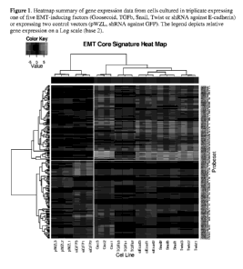
Figure 1: Heatmap summary of gene expression data from cells cultured in
triplicate
expressing one of five EMT-inducing factors (Goosecoid, TGFb, Snail, Twist or
shRNA
against E-cadherin) or expressing two control vectors (pWZL, shRNA against
GFP). The
legend depicts relative gene expression on a Log scale (base 2).
Figure 2: Gene-set enrichment analysis using subsets of genes in Table 1.
Shown is
the enrichment level of subsets of EMT-associated genes in HMLER cancer cells
treated with
paclitaxel. The gene sets are named EMT_UP_NUM, where NUM is the number of
genes in
the subset. The plots show the enrichment score as a function of rank and
indicate that each
of the EMT_UP gene sets is enriched in its expression in cells following
paclitaxel treatment.
Figure 3: Gene-set enrichment analysis with subsets of genes in Table 2. Shown
is
the enrichment level of subsets of non-EMT-associated genes in HMLER cancer
cells treated
with paclitaxel. The gene sets are named EMT_DN_NUM, where NUM is the number
of
genes in the subset. The plots show the enrichment score as a function of rank
and indicate
9
WO 2012/018857 CA 02806726 2013-01-25PCT/US2011/046325
that each of the EMT_DN gene sets is enriched in its expression in cells that
are treated with
DMSO control relative to cells treated with paclitaxel.
Figure 4: Gene-set enrichment analysis with subsets of genes in Table 2. Shown
is
the enrichment level of subsets of non-EMT-associated genes in HMLER cancer
cells treated
with salinomycin. The gene sets are named EMT_DN_NUM, where NUM is the number
of
genes in the subset. The plots show the enrichment score as a function of rank
and indicate
that each of the EMT_DN gene sets is enriched in its expression in cells
following
salinomycin treatment relative to control treatment.
Figure 5: Gene-set enrichment analysis with subsets of genes in Table 1. Shown
is
the enrichment level of subsets of EMT-associated genes in HMLER cancer cells
treated with
salinomycin. The gene sets are named EMT_UP_NUM, where NUM is the number of
genes
in the subset. The plots show the enrichment score as a function of rank and
indicate that each
of the EMT_UP gene sets is enriched in its expression in cells that are
treated with DMSO
control relative to cells treated with salinomycin.
DETAILED DESCRIPTION OF THE INVENTION
Prior to setting forth the invention, it may be helpful to an understanding
thereof to set
forth definitions of certain terms that will be used hereinafter.
A "biomarker" in the context of the present invention is a molecular indicator
of a
specific biological property; a biochemical feature or facet that can be used
to detect and/or
categorize an epithelial cancer. "Biomarker" encompasses, without limitation,
proteins,
nucleic acids, and metabolites, together with their polymorphisms, mutations,
variants,
modifications, subunits, fragments, protein-ligand complexes, and degradation
products,
protein-ligand complexes, elements, related metabolites, and other analytes or
sample-derived
measures. Biomarkers can also include mutated proteins or mutated nucleic
acids. In the
instant invention, measurement of mRNA is preferred.
A "biological sample" or "sample" in the context of the present invention is a
biological sample isolated from a subject and can include, by way of example
and not
limitation, whole blood, blood fraction, serum, plasma, blood cells, tissue
biopsies, a cellular
extract, a muscle or tissue sample, a muscle or tissue biopsy, or any other
secretion,
excretion, or other bodily fluids.
10
WO 2012/018857 CA 02806726 2013-01-25PCT/US2011/046325
The phrase "differentially expressed" refers to differences in the quantity
and/or the
frequency of a biomarker present in a sample taken from patients having for
example,
epithelial cancer as compared to a control subject. For example without
limitation, a
biomarker can be an mRNA or a polypeptide which is present at an elevated
level (i.e.,
overexpressed) or at a decreased level (i.e., underexpressed) in samples of
patients with
cancer as compared to samples of control subjects. Alternatively, a biomarker
can be a
polypeptide which is detected at a higher frequency (i.e., overexpressed) or
at a lower
frequency (i.e., underexpressed) in samples of patients compared to samples of
control
subjects. A biomarker can be differentially present in terms of quantity,
frequency or both.
Previous work has shown that agents that selectively target cells induced into
EMT
also selectively kill cancer stem cells. Since cancer cells induced into EMT
are also highly
invasive, the hypothesis is that anti-cancer therapies that target invasive
and/or metastatic
cancer cells are likely to also target cancer cells induced into EMT.
According to one embodiment, this invention provides a method for determining
which patient subpopulations harbor tumors responsive to three classes of
essentially
overlapping anti-cancer therapies or treatments -- i.e., (a) therapies that
target
invasive/metastatic cells, (b) therapies that target cancer stem cells and (c)
therapies that
target cells post-EMT. Specifically, the invention provides methods for
determining which
therapies or treatments would be effective in cancers that express genetic
biomarkers that are
upregulated in cancer cells post-EMT (Table 1) and would not be effective in
cancers that
express genetic markers upregulated in cancer cells that have not undergone an
EMT (Table
2).
The cancers that the methods of this invention are contemplated to be useful
for
include any epithelial cancers, and specifically include breast cancer,
melanoma, brain,
gastric, pancreatic cancer and carcinomas of the lung, prostate, and colon.
The anti-cancer therapies and treatments in which the methods of this
invention are
contemplated to be useful for include standard-of-care therapies such as
paclitaxel, DNA
damaging agents, kinase inhibitors (e.g., erlotinib), and radiation therapies,
as well as
therapies that target cancer stem cells and/or therapies that target cells
post-EMT, including,
for example, CSS agents such as salinomycin.
A set of genes differentially expressed in cancer cells that have undergone an
EMT
(Table 1) and genes expressed in cancer cells that have not undergone an EMT
(Table 2) was
11
CA 02806726 2013-01-25
WO 2012/018857 PCT/US2011/046325
determined. These genes were obtained by collecting RNA and performing
microarray gene-
expression analyses on breast cancer cells that were cultured either
expressing one of 5 EMT-
inducing genetic factors or 2 control genetic factors that did not induce EMT
(control
vectors). Cells were cultured in triplicate for each treatment condition. A
global analysis of
the gene expression data is shown as a heatmap in Figure 1, where the top sets
of genes in
Tables 1 and 2 were used to construct the heatmap.
To demonstrate that the responsiveness of cancer cell populations to therapy
can be
both measured by and predicted by the various subsets of the genes identified
in Tables 1 and
2, HMLER breast cancer populations were treated with a commonly used anti-
cancer
chemotherapy paclitaxel (Taxol) or with control DMSO treatment. mRNA was then
isolated,
and global gene expression data was collected. The collective expression
levels of the genes
in Tables 1 and 2 after paclitaxel treatment were then determined. For these
analyses, which
are shown in Figures 2 and 3, collections of gene subsets of various sizes
were chosen.
Those skilled in the art will recognize that determining the expression level
of genes
in Tables 1 and/or 2 occurs in vitro in the removed primary tumor.
The analyses show that the genes expressed in Table 1 and/or many subsets
thereof
are over-expressed upon treatment with paclitaxel, indicating that these genes
identify cancer
cellular subpopulations that are resistant to treatment with paclitaxel. As a
consequence,
measurement of the expression of the genes in Table 1 would serve to identify
tumors that
would fail to be responsive to paclitaxel treatment when applied as a single
agent.
Also covered in this invention is any subset of the genes in Table 1 for which
a
statistical test (such as, for example, Gene Set Enrichment Analysis (see
Subramanian,
Tamayo, et al., PNAS 102:15545-50 (2005) and Mootha, Lindgren et al., Nat.
Genet 34:267-
73 (2003), each of which is herein incorporated by reference in its entirety)
demonstrates that
the genes in the subset are over-expressed in paclitaxel-treated populations
at a level of
significance (e.g. p-value) less than 0.1, more preferably less than 0.05,
relative to an
appropriate control population (e.g., DMSO treatment). In one embodiment it
was
contemplated that the subset of genes from Table 1 comprises at least 2 genes,
10 genes, 15
genes, 20 genes or 30 genes (or any range intervening therebetween). For
example, the
subset might include 2, 3, 4, 5, 6, 7, 8, 9. 10, 11, 12, 13, 14, 15, 16, 17,
18, 19, 20, 21, 22, 23,
24, 25, 26, 27, 28, 29, or 30 genes.
12
WO 2012/018857 CA 02806726 2013-01-25PCT/US2011/046325
Those skilled in the art will recognize that any other appropriate statistical
test(s) for
gene enrichment or differential expression can also be used to identify the
desired subset of
genes from Table 1. For example, the summation of the log-transformed gene
expression
scores for the genes in a set could identify a metric that could be used to
compare differential
gene expression between two profiles using a t-test, modified t-test, or non-
parametric test
such as Mann-Whitney.
Moreover, those skilled in the art will also recognize that any appropriate
control
population(s) can also be used to identify the desired subset of genes from
Table 1. For
example, the appropriate control population(s) can be any population of cells
(i.e., cancer
cells) that have not been treated with a given cancer therapy.
Alternatively, the subsets of the genes in Table 1 may be identified as any
subset for
which a statistical test (such as, for example, Gene Set Enrichment Analysis)
demonstrates
that the genes in the subset are under-expressed in salinomycin-treated
populations at a level
of significance (e.g. p-value) less than 0.1, more preferably less that 0.05,
relative to an
appropriate control population (e.g., DMSO treatment). In one embodiment it
was
contemplated that the subset of genes from Table 1 comprises at least 2 genes,
10 genes, 15
genes, 20 genes or 30 genes (or any range intervening therebetween). For
example, the
subset might include 2, 3, 4, 5, 6, 7, 8, 9. 10, 11, 12, 13, 14, 15, 16, 17,
18, 19, 20, 21, 22, 23,
24, 25, 26, 27, 28, 29, or 30 genes. For those skilled in the art, any other
appropriate
statistical test(s) for gene expression or differential expression can also be
used to identify the
desired subset of genes from Table 1. For example, the summation of the log-
transformed
gene expression scores for the genes in a set could identify a metric that
could be used to
compare differential gene expression between two profiles using a t-test,
modified t-test, or
non-parametric test such as Mann-Whitney.
Likewise, any appropriate control population(s) can also be used to identify
the
desired subset of genes from Table 1. For example, the appropriate control
population(s) can
be any population of cells (i.e., cancer cells) that have not been treated
with a given cancer
therapy.
Those skilled in the art will recognize that the statistical test used to
determine
suitable subsets of the genes in Table 1 could be Gene Set Enrichment Analysis
(GSEA) (see
Subramanian, Tamayo, et al., PNAS 102:15545-50 (2005) and Mootha, Lindgren et
al., Nat.
Genet 34:267-73 (2003), each of which is herein incorporated by reference in
its entirety) as
13
CA 02806726 2013-01-25
WO 2012/018857
PCT/US2011/046325
used for the purposes of elucidation in this application, or it could be any
other statistical test
of enrichment or expression known in the art. For example, the summation of
the log-
transformed gene expression scores for the genes in a set could identify a
metric that could be
used to compare differential gene expression between two profiles using a t-
test, modified t-
test, or non-parametric test such as Mann-Whitney.
The populations of cells being treated for the purposes of this evaluation
could be
cancer cells of any type or normal cellular populations.
Table 1. Genes identified that are over-expressed in cancer populations having
undergone an
EMT, relative to cancer populations that have not undergone an EMT.
Mean Fold
OverExpression
Symbol Description GenBank
Upon EMT
DON Decorin AF138300
137.6156
collagen, type III, alpha 1 (Ehlers-Danlos
COL3A1 syndrome type IV, autosomal dominant) AU144167
132.1195
COL1A2 collagen, type 1, alpha 2 AA788711
88.05054
FBN1 fibrillin 1 (Marfan syndrome) NM 000138
76.51337
gremlin 1, cysteine knot superfamily, homolog
GREM1 (Xenopus laevis) NM 013372
75.35859
POSTN periostin, osteoblast specific factor D13665
73.18114
NID1 nidogen 1 BF940043
51.91502
FBLN5 fibulin 5 NM 006329
34.4268
syndecan 2 (heparan sulfate proteoglycan 1,
5D02 cell surface-associated, fibroglycan) AL577322
32.48001
00L5A2 collagen, type V, alpha 2 NM 000393
26.66545
PRG1 proteoglycan 1, secretory granule J03223
23.46014
transcription factor 8 (represses interleukin 2
TCF8 expression) A1806174
22.83413
ectonucleotide
pyrophosphatase/phosphodiesterase 2
ENPP2 (autotaxin) L35594
22.72739
nuclear receptor subfamily 2, group F, member
NR2F1 1 A1951185
20.64471
COL6A1 collagen, type VI, alpha 1 AA292373
17.36271
RGS4 regulator of G-protein signalling 4 AL514445
16.63788
CDH11 cadherin 11, type 2, OB-cadherin (osteoblast) D21254
16.61483
PRRX1 paired related homeobox 1 NM 006902
14.73362
OLFML3 olfactomedin-like 3 NM_020190
14.0984
sparc/osteonectin, cwcv and kazal-like domains
SPOOK proteoglycan (testican) AF231124
13.99112
wingless-type MMTV integration site family,
WNT5A member 5A NM 003392
13.33384
MAP1B microtubule-associated protein 1B AL523076
13.0877
BG109855 12.44401
pentraxin-related gene, rapidly induced by IL-1
PTX3 beta NM 002852
12.01196
C5orf13 chromosome 5 open reading frame 13 U36189
11.95863
IGFBP4 insulin-like growth factor binding protein 4 NM 001552
11.09963
14
CA 02806726 2013-01-25
WO 2012/018857
PCT/US2011/046325
PCOLCE procollagen C-endopeptidase enhancer NM 002593
11.04575
TNFAIP6 tumor necrosis factor, alpha-induced protein 6 NM_007115
11.02984
L0051334 NM_016644
10.91454
cytochrome P450, family 1, subfamily B,
CYP1B1 polypeptide 1 NM 000104
10.47429
tissue factor pathway inhibitor (lipoprotein-
TFPI associated coagulation inhibitor) BF511231
10.42648
PVRL3 poliovirus receptor-related 3 AA129716
10.30262
ROR1 receptor tyrosine kinase-like orphan receptor 1 NM_005012
10.10474
FBLN1 fibulin 1 NM 006486
10.09844
BIN1 bridging integrator 1 AF043899
9.928529
LUM Lumican NM 002345
9.727574
ral guanine nucleotide dissociation stimulator-
RGL1 like 1 AF186779
9.643922
PTGFR prostaglandin F receptor (FP) NM 000959
8.939536
transforming growth factor, beta receptor III
TGFBR3 (betaglycan, 300kDa) NM 003243
8.838
COL1A1 collagen, type 1, alpha 1 Y15916
8.667645
DLC1 deleted in liver cancer 1 AF026219
8.610518
PM P22 peripheral myelin protein 22 L03203
8.560648
PRKCA protein kinase C, alpha A1471375
8.338108
matrix metallopeptidase 2 (gelatinase A, 72kDa
MMP2 gelatinase, 72kDa type IV collagenase) NM 004530
8.268926
CTGF connective tissue growth factor M92934
8.168776
CDH2 cadherin 2, type 1, N-cadherin (neuronal) M34064
7.987921
guanine nucleotide binding protein (G protein),
GNG11 gamma 11 NM 004126
7.953115
PPAP2B phosphatidic acid phosphatase type 2B AA628586
7.907272
NEBL Nebulette AL157398
7.817894
MYL9 myosin, light polypeptide 9, regulatory NM 006097
7.780485
potassium large conductance calcium-activated
KCNMA1 channel, subfamily M, alpha member 1 A1129381
7.747227
IGFBP3 insulin-like growth factor binding protein 3 BF340228
7.57812
CSPG2 chondroitin sulfate proteoglycan 2 (versican) NM 004385
7.318764
sema domain, seven thrombospondin repeats
(type 1 and type 1-like), transmembrane domain
(TM) and short cytoplasmic domain,
SEMA5A (semaphorin) 5A NM 003966
7.298702
Cbp/p300-interacting transactivator, with
CITED2 Glu/Asp-rich carboxy-terminal domain, 2 AF109161
7.220907
membrane metallo-endopeptidase (neutral
MME endopeptidase, enkephalinase, CALLA, CD10) A1433463
7.05859
DOCK10 dedicator of cytokinesis 10 NM 017718
6.972809
DNAJB4 DnaJ (Hsp40) homolog, subfamily B, member 4 BG252490
6.782043
PCDH9 protocadherin 9 A1524125
6.711987
NID2 nidogen 2 (osteonidogen) NM 007361
6.54739
HAS2 hyaluronan synthase 2 NM 005328
6.520398
PTGER4 prostaglandin E receptor 4 (subtype EP4) AA897516
6.396133
TRAM2 translocation associated membrane protein 2 A1986461
6.275542
SYT11 synaptotagmin XI BC004291
6.149546
BGN Biglycan AA845258
5.838023
CYBRD1 cytochrome b reductase 1 NM 024843
5.710828
CHN1 chimerin (chimaerin) 1 BF339445
5.687127
DPT Dermatopontin A1146848
5.573023
15
CA 02806726 2013-01-25
WO 2012/018857
PCT/US2011/046325
integrin, beta-like 1 (with EGF-like repeat
ITGBL1 domains) AL359052
5.511939
FLJ22471 NM_025140
5.364784
LOC22136
2 AL577024
5.35364
MLPH Melanophilin NM 024101
5.296062
ANXA6 annexin A6 NM 001155
5.18628
echinoderm microtubule associated protein like
EML1 1 NM_004434
5.138332
cAMP responsive element binding protein 3-like
CREB3L1 1 AF055009
5.073214
FLJ10094 NM_017993
4.998863
leucine-rich repeats and immunoglobulin-like
LRIG1 domains 1 AB050468
4.9963
SNED1 sushi, nidogen and EGF-like domains 1 N73970
4.993945
serpin peptidase inhibitor, clade F (alpha-2
antiplasmin, pigment epithelium derived factor),
SERPINF1 member 1 NM 002615
4.969153
disabled homolog 2, mitogen-responsive
DAB2 phosphoprotein (Drosophila) NM 001343
4.913939
Wiskott-Aldrich syndrome protein interacting
WASPIP protein AW058622
4.882974
FN1 fibronectin 1 AJ276395
4.869319
C10orf56 chromosome 10 open reading frame 56 AA131324
4.795629
DAPK1 death-associated protein kinase 1 NM 004938
4.726984
LOXL1 lysyl oxidase-like 1 NM 005576
4.720305
inhibitor of DNA binding 2, dominant negative
1D2 helix-loop-helix protein NM 002166
4.672064
prostaglandin E receptor 2 (subtype EP2),
PTGER2 53kDa NM 000956
4.427892
COL8A1 collagen, type VIII, alpha 1 BE877796
4.38653
DDR2 discoidin domain receptor family, member 2 NM 0061 82
4.338932
SEPT6 septin 6 D50918
4.30699
HRASLS3 HRAS-like suppressor 3 B0001387
4.281926
pleckstrin homology domain containing, family C
PLEKHC1 (with FERM domain) member 1 AW469573
4.272913
THY1 Thy-1 cell surface antigen AA218868
4.253587
ribosomal protein S6 kinase, 90kDa,
RPS6KA2 polypeptide 2 A1992251
4.225143
GALC galactosylceramidase (Krabbe disease) NM 000153
4.222742
fibrillin 2 (congenital contractural
FBN2 arachnodactyly) NM 001999
4.205916
FSTL1 follistatin-like 1 B0000055
4.175243
NRP1 neuropilin 1 BE620457
4.162874
TNS1 tensin 1 AL046979
4.131713
TAGLN Transgelin NM 003186
4.131083
cyclin-dependent kinase inhibitor 20 (p18,
CDKN2C inhibits CDK4) NM 001262
4.124788
MAGEH1 melanoma antigen family H, 1 NM 014061
4.094423
latent transforming growth factor beta binding
LTBP2 protein 2 NM 000428
4.000998
PBX1 pre-B-cell leukemia transcription factor 1 AL049381
3.997339
TBX3 T-box 3 (ulnar mammary syndrome) NM 016569
3.992244
16
WO 2012/018857 CA 02806726 2013-01-25PCT/US2011/046325
The analyses also show that the genes in Table 2 and many subsets thereof are
under-
expressed upon treatment with paclitaxel, indicating that these genes identify
cellular
subpopulations that are sensitive to treatment with paclitaxel. As a
consequence,
measurement of the expression of the genes in Table 2 would serve to identify
tumors that
would be responsive to paclitaxel treatment when applied as a single agent.
Those skilled in the art will recognize that determining the expression level
of genes
in Table 2 occurs in vitro in the removed primary tumor.
Also covered in this invention is any subset of the genes in Table 2 for which
a
statistical test (such as, for example, Gene Set Enrichment Analysis)
demonstrates that the
genes in the subset are under-expressed in paclitaxel-treated populations at a
level of
significance (e.g. p-value) less than 0.1, more preferably less than 0.05,
relative to an
appropriate control population (e.g., DMSO treatment). In one embodiment it
was
contemplated that the subset of the genes from Table 2 comprises at least 2
genes, 6 genes, 10
genes, 15 genes, 20 genes or 30 genes (or any range intervening therebetween).
For example,
the subset might include 2, 3, 4, 5, 6, 7, 8, 9. 10, 11, 12, 13, 14, 15, 16,
17, 18, 19, 20, 21, 22,
23, 24, 25, 26, 27, 28, 29, or 30 genes. Those skilled in the art will
recognize that any other
appropriate statistical test(s) for gene enrichment or differential expression
can also be used
to identify the desired subset of genes from Table 2. For example, the
summation of the log-
transformed gene expression scores for the genes in a set could identify a
metric that could be
used to compare differential gene expression between two profiles using a t-
test, modified t-
test, or non-parametric test such as Mann-Whitney.
Moreover, those skilled in the art will also recognize that any appropriate
control
population(s) can also be used to identify the desired subset of genes from
Table 2. For
example, the appropriate control population(s) can be any population of cells
(i.e., cancer
cells) that have not been treated with a given cancer therapy.
Alternatively, the subsets of the genes in Table 2 may be identified as any
subset for
which a statistical test (such as Gene Set Enrichment Analysis) demonstrates
that the genes in
the subset are over-expressed in salinomycin-treated populations at a level of
significance
(e.g. p-value) less than 0.1, more preferably less than 0.05, relative to an
appropriate control
population (e.g., DMSO treatment). In one embodiment it was contemplated that
the subset
of the genes from Table 2 comprises at least 2 genes, 6 genes, 10 genes, 15
genes, 20 genes
or 30 genes (or any range intervening therebetween). For example, the subset
might include
17
WO 2012/018857 CA 02806726 2013-01-25PCT/US2011/046325
2, 3, 4, 5, 6, 7, 8, 9. 10, 11, 12, 13, 14, 15, 16, 17, 18, 19, 20, 21, 22,
23, 24, 25, 26, 27, 28,
29, or 30 genes. Those skilled in the art will recognize that any other
appropriate statistical
test(s) for gene enrichment or differential expression can also be used to
identify can also be
used to identify the desired subset of genes from Table 2. For example, the
summation of the
log-transformed gene expression scores for the genes in a set could identify a
metric that
could be used to compare differential gene expression between two profiles
using a t-test,
modified t-test, or non-parametric test such as Mann-Whitney.
Likewise, those skilled in the art will also recognize that any appropriate
control
population(s) can also be used to identify the desired subset of genes from
Table 2. For
example, the appropriate control population(s) can be any population of cells
(i.e., cancer
cells) that have not been treated with a given cancer therapy.
The statistical test used could be Gene Set Enrichment Analysis (GSEA) (see
Subramanian, Tamayo, et al., PNAS 102:15545-50 (2005) and Mootha, Lindgren et
al., Nat.
Genet 34:267-73 (2003), each of which is herein incorporated by reference in
its entirety) as
used for the purposes of elucidation in this application, or it could be any
other statistical test
of enrichment or expression known in the art. By way of non-limiting example,
the
summation of the log-transformed gene expression scores for the genes in a set
could identify
a metric that could be used to compare differential gene expression between
two profiles
using a t-test, modified t-test, or non-parametric test such as Mann-Whitney.
The populations of cells being treated for the purposes of this evaluation
could be
cancer cells of any type or normal cellular populations.
18
CA 02806726 2013-01-25
WO 2012/018857
PCT/US2011/046325
Table 2. Genes identified that are over-expressed in cancer populations that
have not
undergone an EMT, relative to cancer populations that have undergone an EMT.
Mean Fold
OverExpression In
Symbol Description Gen Bank
Non-EMT
serpin peptidase inhibitor, clade B
SERPINB2 (ovalbumin), member 2 NM 002575
36.74103
tumor-associated calcium signal
TACSTD1 transducer 1 NM 002354
35.91264
SPRR1A small proline-rich protein 1A A1923984
34.99944
SPRR1B small proline-rich protein 1B (cornifin) NM 003125
29.33599
ILIA interleukin 1, alpha M15329
28.86922
KLK10 kallikrein 10 B0002710
25.16523
fibroblast growth factor receptor 3
FGFR3 (achondroplasia, thanatophoric dwarfism) NM_000142
24.74251
CDH1 cadherin 1, type 1, E-cadherin (epithelial) NM_004360
23.74645
SLPI secretory leukocyte peptidase inhibitor NM 003064
21.4404
KRT6B keratin 6B A1831452
20.84833
FXYD domain containing ion transport
FXYD3 regulator 3 B0005238
19.01308
peptidase inhibitor 3, skin-derived
P13 (SKALP) L10343
18.10103
RAB25 RAB25, member RAS oncogene family NM 020387
17.64907
SAA2 serum amyloid A2 M23699
17.20791
RBM35A RNA binding motif protein 35A NM 017697
15.20696
TMEM3OB transmembrane protein 30B AV691491
14.98036
EVA1 epithelial V-like antigen 1 AF275945
14.69364
kallikrein 7 (chymotryptic, stratum
KLK7 corneum) NM 005046
14.42981
RBM35B RNA binding motif protein 35A NM 024939
13.49619
5100A14 S100 calcium binding protein Al 4 NM 020672
13.44819
serpin peptidase inhibitor, clade B
SERPINB13 (ovalbumin), member 13 AJ001698
13.29747
ubiquitin carboxyl-terminal esterase L1
UCHL1 (ubiquitin thiolesterase) NM 004181
13.27334
aldehyde dehydrogenase 1 family,
ALDH1A3 member A3 NM 000693
13.10531
CKMT1B creatine kinase, mitochondrial 1B NM 020990
12.4713
ANXA3 annexin A3 M63310
12.4013
NMU neuromedin U NM 006681
12.15367
KRT15 keratin 15 NM 002275
12.09266
FST Follistatin NM 013409
11.85793
FGFBP1 fibroblast growth factor binding protein 1 NM_005130
11.49472
S100 calcium binding protein A7
5100A7 (psoriasin 1) NM 002963
11.07673
TP73L tumor protein p73-like AF091627
10.93454
FLJ12684 NM_024534
10.70372
SCNN1A sodium channel, nonvoltage-gated 1 alpha NM_001038
10.3172
KLK5 kallikrein 5 AF243527
10.20992
S100 calcium binding protein A8
5100A8 (calgranulin A) NM_002964
10.10418
CCND2 cyclin D2 AW026491
9.950438
MAP7 microtubule-associated protein 7 AW242297
9.942027
19
CA 02806726 2013-01-25
WO 2012/018857
PCT/US2011/046325
CXADR coxsackie virus and adenovirus receptor NM_001338
9.872805
KRT17 keratin 17 NM 000422
9.74958
CDH3 cadherin 3, type 1, P-cadherin (placental) NM_001793
9.735938
TRIM29 tripartite motif-containing 29 NM 012101
9.373189
SPINT1 serine peptidase inhibitor, Kunitz type 1 NM 003710
9.353589
TGFA transforming growth factor, alpha NM 003236
9.30496
interleukin 18 (interferon-gamma-inducing
IL18 factor) NM 001562
9.218934
CA9 carbonic anhydrase IX NM 001216
9.196596
keratin 16 (focal non-epidermolytic
KRT16 palmoplantar keratoderma) AF061812
9.177365
gap junction protein, beta 3, 31kDa
GJB3 (connexin 31) AF099730
9.030588
VSNL1 visinin-like 1 NM 003385
8.637896
ID B interleukin 1, beta NM 000576
8.629518
CA2 carbonic anhydrase II M36532
8.606222
CNTNAP2 contactin associated protein-like 2 A0005378
8.592036
ARHGAP8 Rho GTPase activating protein 8 Z83838
8.434017
keratin 5 (epidermolysis bullosa simplex,
Dowling-Meara/Kobner/VVeber-Cockayne
KRT5 types) NM 000424
8.14695
ARTN Artemin NM 003976
8.125857
calcium/calmodulin-dependent protein
CAMK2B kinase (CaM kinase) II beta AF078803
8.125181
ZBED2 zinc finger, BED-type containing 2 NM 024508
8.046492
TPD52L1 tumor protein D52-like 1 NM 003287
7.949147
erythrocyte membrane protein band 4.1
EPB41L4B like 4B NM 019114
7.911
KLK8 kallikrein 8 (neuropsin/ovasin) NM 007196
7.895551
C1orf116 chromosome 1 open reading frame 116 NM_024115
7.889643
LEPREL1 leprecan-like 1 NM 018192
7.85189
JAG2 jagged 2 Y14330
7.562273
DSC2 desmocollin 2 NM 004949
7.425664
cytochrome P450, family 27, subfamily B,
CYP27B1 polypeptide 1 NM 000785
7.293746
HOOK1 hook homolog 1 (Drosophila) NM 015888
7.275468
lectin, galactoside-binding, soluble, 7
LGALS7 (galectin 7) NM 002307
7.241758
HBEGF heparin-binding EGF-like growth factor NM 001945
7.202511
CDP-diacylglycerol synthase
CDS1 (phosphatidate cytidylyltransferase) 1 NM 001263
7.130583
RNF128 ring finger protein 128 NM 024539
7.12999
PRR5 NM_015366
7.124753
KRT6A keratin 6A J00269
7.042267
LAMA3 laminin, alpha 3 NM 000227
6.95736
adaptor-related protein complex 1, mu 2
AP1M2 subunit NM 005498
6.911026
SLAC2-B AB014524
6.847038
GRHL2 grainyhead-like 2 (Drosophila) NM 024915
6.781949
suppression of tumorigenicity 14 (colon
ST14 carcinoma, matriptase, epithin) NM 021978
6.733796
DSC3 desmocollin 3 NM_001941
6.68478
CD24 antigen (small cell lung carcinoma
CD24 cluster 4 antigen) M58664
6.653991
LAMB3 laminin, beta 3 L25541
6.6375
20
CA 02806726 2013-01-25
WO 2012/018857
PCT/US2011/046325
TSPAN1 tetraspanin 1 AF133425
6.619673
SYK spleen tyrosine kinase NM 003177
6.585623
SNX10 sorting nexin 10 NM 013322
6.540949
NM_024064 6.518229
CTSL2 cathepsin L2 AF070448
6.516422
solute carrier family 2 (facilitated glucose
SLC2A9 transporter), member 9 NM 020041
6.458325
TMEM40 transmembrane protein 40 NM 018306
6.408648
COL17A1 collagen, type XVII, alpha 1 NM 000494
6.405184
C10orf10 chromosome 10 open reading frame 10 AL136653
6.37754
ST6 (alpha-N-acetyl-neuraminy1-2,3-beta-
galactosy1-1,3)-N-acetylgalactosaminide
ST6GALNAC2 alpha-2,6-sialyltransferase 2 NM 006456
6.224336
ANXA8 annexin A8 NM_001630
6.199621
ABLIM1 actin binding LIM protein 1 NM 006720
6.19859
RLN2 relaxin 2 NM 005059
6.139665
VGLL1 vestigial like 1 (Drosophila) BE542323
6.116473
NRG1 neuregulin 1 NM 013959
5.854395
matrix metallopeptidase 9 (gelatinase B,
92kDa gelatinase, 92kDa type IV
MM P9 collagenase) NM 004994
5.737173
desmoglein 3 (pemphigus vulgaris
DSG3 antigen) NM 001944
5.731926
gap junction protein, beta 5 (connexin
GJB5 31.1) NM 005268
5.684999
NDRG1 N-myc downstream regulated gene 1 NM 006096
5.681532
MAPK13 mitogen-activated protein kinase 13 B0000433
5.587721
DST Dystonin NM 001723
5.560135
CORO1A coronin, actin binding protein, 1A U34690
5.510182
IRF6 interferon regulatory factor 6 AU144284
5.499117
KIBRA AK001727
5.491803
SPINT2 serine peptidase inhibitor, Kunitz type, 2 AF027205
5.466358
arachidonate 15-lipoxygenase, second
ALOX15B type NM 001141
5.461662
serpin peptidase inhibitor, clade B
SERPINB1 (ovalbumin), member 1 NM 030666
5.348966
chloride channel, calcium activated, family
CLCA2 member 2 AF043977
5.30091
MY05C myosin VC NM 018728
5.269624
CSTA cystatin A (stefin A) NM 005213
5.215624
ITGB4 integrin, beta 4 NM 000213
5.180603
MBP myelin basic protein AW070431
5.108643
AQP3 aquaporin 3 N74607
5.084832
solute carrier family 7 (cationic amino acid
SLC7A5 transporter, y+ system), member 5 AB018009
5.084409
GPR87 G protein-coupled receptor 87 NM 023915
5.073566
MALL mal, T-cell differentiation protein-like B00031 79
4.957731
macrophage stimulating 1 receptor (c-met-
MST1R related tyrosine kinase) NM 002447
4.955876
SOX15 SRY (sex determining region Y)-box 15 NM_006942
4.948873
LAMC2 laminin, gamma 2 NM 005562
4.941675
CST6 cystatin ELM NM 001323
4.931341
MFAP5 microfibrillar associated protein 5 AW665892
4.871412
KRT18 keratin 18 NM 000224
4.799686
21
CA 02806726 2013-01-25
WO 2012/018857
PCT/US2011/046325
JUP junction plakoglobin NM 021991 4.719454
DSP Desmoplakin NM 004415 4.716772
MTSS1 metastasis suppressor 1 NM 014751 4.715399
fibroblast growth factor receptor 2
(bacteria-expressed kinase, keratinocyte
growth factor receptor, craniofacial
dysostosis 1, Crouzon syndrome, Pfeiffer
FGFR2 syndrome, Jackson-Weiss syndrome) NM 022969 4.67323
PKP3 plakophilin 3 AF053719 4.646421
STAC 5H3 and cysteine rich domain NM 003149 4.643331
RAB38 RAB38, member RAS oncogene family NM 022337 4.544243
SFRP1 secreted frizzled-related protein 1 NM 003012 4.465928
RHOD ras homolog gene family, member D B0001338 4.45418
TPD52 tumor protein D52 BG389015 4.453563
F11R F11 receptor AF154005 4.39018
tumor necrosis factor receptor
TNFRSF6B superfamily, member 6b, decoy NM 003823 4.342302
BCL2-interacting killer (apoptosis-
BIK inducing) NM 001197 4.323681
XDH xanthine dehydrogenase U06117 4.309678
phospholipase A2, group IVA (cytosolic,
PLA2G4A calcium-dependent) M68874 4.308364
PTHLH parathyroid hormone-like hormone J03580 4.294946
NEF3 neurofilament 3 (150kDa medium) NM 005382 4.274928
sortilin-related receptor, L(DLR class) A
SORL1 repeats-containing AV728268 4.257894
solute carrier family 6 (neurotransmitter
SLC6A8 transporter, creatine), member 8 NM 005629 4.205508
proline rich Gla (G-carboxyglutamic acid)
PRRG4 4 (transmembrane) NM 024081 4.187822
CLDN1 claudin 1 NM 021101 4.185384
K1AA0888 AB020695 4.162009
GPR56 G protein-coupled receptor 56 AL554008 4.153478
synuclein, alpha (non A4 component of
SNCA amyloid precursor) BG260394 4.149795
fibronectin leucine rich transmembrane
FLRT3 protein 3 NM 013281 4.130167
ILI RN interleukin 1 receptor antagonist U65590 4.12988
discoidin domain receptor family, member
DDR1 1 L11315 4.125646
v-yes-1 Yamaguchi sarcoma viral related
LYN oncogene homolog M79321 4.107271
FLJ20130 NM_017681 4.09499
STAP2 B0000795 4.089544
potassium channel, subfamily K, member
KCNK1 1 NM_002245 4.084162
TSPAN13 tetraspanin 13 NM 014399 4.079691
LISCH7 NM_015925 4.025813
PERP PERP, TP53 apoptosis effector NM 022121 4.024473
Next, identical analyses as those described above were performed in the
context of
treatment with a different anti-cancer agent-salinomycin-that was previously
identified as
22
WO 2012/018857 CA 02806726 2013-01-25PCT/US2011/046325
specifically killing invasive cancer stem cells. The opposite expression
change (relative to
paclitaxel) was observed upon treatment with salinomycin. The analyses, shown
in Figures 4
and 5, indicate that the genes expressed in Table 1 and any subsets thereof
are under-
expressed upon treatment with salinomycin, indicating that these genes
identify cellular
subpopulations that are sensitive to treatment with a CSS agent such as
salinomycin. As a
consequence, measurement of the expression of the genes in Table 1 (or any
appropriate
subsets thereof identified according to the methods disclosed herein) would
serve to identify
tumors that would be responsive to a CSS agent (e.g., salinomycin treatment)
when applied
as a single agent.
The analyses also show that the genes expressed in Table 2 and any subset
thereof are
over-expressed upon treatment with salinomycin (relative to control),
indicating that these
genes identify cellular subpopulations that are resistant to treatment with a
CSS agent such as
salinomycin. As a consequence, measurement of the expression of the genes in
Table 2 (or
any appropriate subsets thereof identified according to the methods disclosed
herein) would
serve to identify tumors that would fail to be responsive to a CSS agent (e.g,
salinomycin
treatment) when applied as a single agent.
It follows that measurement of the expression of the genes in Tables 1 and/or
2 as
well as various subsets thereof for which a statistical test demonstrates that
the genes in the
subset are differentially expressed in response to treatment with a cancer
treatment (e.g.,
salinomycin treatment or paclitaxel treatment) at a level of significance
(e.g., p value) less
than 0.1, relative to an appropriate control population (e.g., DMSO treatment)
can be used to
identify cancer cell populations that are or are not responsive to any given
therapy or
treatment. Distinct subpopulations of cells are identified using the
expression levels of the
genes in Tables 1 and/or 2 (or any appropriate subsets thereof) and these
distinct
subpopulations could respond distinctively to any particular therapeutic or
treatment regimen,
thereby allowing these genes to serve as biomarkers dictating therapy choice
following
primary tumor removal.
All documents and patents or patent applications referred to herein are fully
incorporated by reference.
23
CA 02806726 2013-01-25
WO 2012/018857 PCT/US2011/046325
References:
1. Piyush Gupta, Tamer T. Onder, Sendurai Mani, Mai-jing Liao, Eric S. Lander,
Robert A.
Weinberg. A Method for the Discovery of Agents Targeting and Exhibiting
Specific Toxicity
for Cancer Stem Cells. Patent pending. (WHI07-20; MIT 12947WB;
WO/2009/126310).
2. Piyush B. Gupta, Tamer T. Onder, Guozhi Jiang, Tai Kao, Charlotte
Kuperwasser,
Robert A. Weinberg, Eric S. Lander. "Identification of selective inhibitors of
cancer stem
cells by high-throughput screening." Cell. (2009) Aug; 138(4):645-659.
3. Thomson S, Petti F, Sujka-Kwok I, Epstein D, Haley JD. Kinase switching in
mesenchymal-like non-small cell lung cancer lines contributes to EGFR
inhibitor resistance
through pathway redundancy. Clin Exp Metastasis. 2008;25(8):843-54. Epub 2008
Aug 12.
PubMed PMID: 18696232.
4. Ban- S, Thomson S, Buck E, Russo S, Petti F, Sujka-Kwok I, Eyzaguirre A,
Rosenfeld-
Franklin M, Gibson NW, Miglarese M, Epstein D, Iwata KK, Haley JD. Bypassing
cellular
EGF receptor dependence through epithelial-to-mesenchymal-like transitions.
Clin Exp
Metastasis. 2008;25(6):685-93. Epub 2008 Jan 31. Review. PubMed PMID:
18236164;
PubMed Central PMCID: PMC2471394.
5. Buck E, Eyzaguirre A, Ban- S, Thompson S, Sennello R, Young D, Iwata KK,
Gibson
NW, Cagnoni P, Haley JD. Loss of homotypic cell adhesion by
epithelial-mesenchymal transition or mutation limits sensitivity to epidermal
growth factor
receptor inhibition. Mol Cancer Ther. 2007 Feb;6(2):532-41. PubMed PMID:
17308052.
6. Woodward WA, Debeb BG, Xu W, Buchholz TA. Overcoming radiation resistance
in
inflammatory breast cancer. Cancer. 2010 Jun 1;116(11 Suppl):2840-5. PubMed
PMID:20503417.
7. Bao, S., Wu, Q., McLendon, R.E., Hao, Y., Shi, Q., Hjelmeland, A.B.,
Dewhirst, M.W.,
Bigner, D.D., and Rich, J.N. (2006). Glioma stem cells promote radioresistance
by
preferential activation of the DNA damage response. Nature 444, 756-760.
8. Ban-, S., Thomson, S., Buck, E., Russo, S., Petti, F., Sujka-Kwok, I.,
Eyzaguirre, A.,
Rosenfeld-Franklin, M., Gibson, N.W., Miglarese, M., et al. (2008). Bypassing
cellular EGF
receptor dependence through epithelial-to-mesenchymal-like transitions.
Clinical &
experimental metastasis 25, 685-693.
9. Buck, E., Eyzaguirre, A., Rosenfeld-Franklin, M., Thomson, S., Mulvihill,
M., Ban-, S.,
Brown, E., O'Connor, M., Yao, Y., Pachter, J., et al. (2008). Feedback
mechanisms promote
cooperativity for small molecule inhibitors of epidermal and insulin-like
growth factor
receptors. Cancer research 68, 8322-8332.
10. Creighton, C.J., Li, X., Landis, M., Dixon, J.M., Neumeister, V.M.,
Sjolund, A., Rimm,
D.L., Wong, H., Rodriguez, A., Herschkowitz, J.I., et al. (2009). Residual
breast cancers after
conventional therapy display mesenchymal as well as tumor-initiating features.
Proceedings
of the National Academy of Sciences of the United States of America 106, 13820-
13825.
24
CA 02806726 2013-01-25
WO 2012/018857 PCT/US2011/046325
11. Horwitz, K.B., and Sartorius, C.A. (2008). Progestins in hormone
replacement therapies
reactivate cancer stem cells in women with preexisting breast cancers: a
hypothesis. The
Journal of clinical endocrinology and metabolism 93, 3295-3298.
Mani, S.A., Guo, W., Liao, M.J., Eaton, E.N., Ayyanan, A., Zhou, A.Y., Brooks,
M.,
Reinhard, F., Zhang, C.C., Shipitsin, M., et al. (2008). The epithelial-
mesenchymal transition
generates cells with properties of stem cells. Cell 133, 704-715.
12. Morel, A.P., Lievre, M., Thomas, C., Hinkal, G., Ansieau, S., and
Puisieux, A. (2008).
Generation of breast cancer stem cells through epithelial-mesenchymal
transition. PLoS ONE
3, e2888.
13. Thomson, S., Buck, E., Petti, F., Griffin, G., Brown, E., Ramnarine, N.,
Iwata, K.K.,
Gibson, N., and Haley, J.D. (2005). Epithelial to mesenchymal transition is a
determinant of
sensitivity of non-small-cell lung carcinoma cell lines and xenografts to
epidermal growth
factor receptor inhibition. Cancer research 65, 9455-9462.
14. Yang, A.D., Fan, F., Camp, E.R., van Buren, G., Liu, W., Somcio, R., Gray,
M.J.,
Cheng, H., Hoff, P.M., and Ellis, L.M. (2006). Chronic oxaliplatin resistance
induces
epithelial-to-mesenchymal transition in colorectal cancer cell lines. Clin
Cancer Res 12,
4147-4153.
15. Yang, J., Mani, S.A., Donaher, J.L., Ramaswamy, S., Itzykson, R.A., Come,
C.,
Savagner, P., Gitelman, I., Richardson, A., and Weinberg, R.A. (2004). Twist,
a master
regulator of morphogenesis, plays an essential role in tumor metastasis. Cell
117, 927-939.
16. Yauch, R.L., Januario, T., Eberhard, D.A., Cavet, G., Zhu, W., Fu, L.,
Pham, T.Q.,
Soriano, R., Stinson, J., Seshagiri, S., et al. (2005). Epithelial versus
mesenchymal phenotype
determines in vitro sensitivity and predicts clinical activity of erlotinib in
lung cancer
patients. Clin Cancer Res 11, 8686-8698.
17. Taube, J.H, Herschkowitz, J.I., Komurov, K., Zhou, A.Y., Gupta, S., Yang,
J., Hartwell,
K., Onder, T.T., Gupta, P.B., Evans, K.W., Hollier, B.G., Ram, P.T., Lander,
E.S., Rosen,
J.M., Weinberg, R.A., Mani, S.A. (2010). A Core EMT Interactome Gene
Expression
Signature is Associated with Claudin-Low and Metaplastic Breast Cancer
Subtypes. Proc.
Natl Acad. Sci 107, 15449-15454.
OTHER EMBODIMENTS
While the invention has been described in conjunction with the detailed
description
thereof, the foregoing description is intended to illustrate and not limit the
scope of the
invention, which is defined by the scope of the appended claims. Other
aspects, advantages,
and modifications are within the scope of the following claims.
25