Note: Descriptions are shown in the official language in which they were submitted.
WO 2012/015976 CA 02806755 2013-01-25PCT/US2011/045615
SYSTEM FOR SACRO-ILIAC STABILIZATION
FIELD OF THE INVENTION
The invention relates generally to stabilization and/or
fixation of adjacent bony structures of the skeletal system,
and more particularly to a minimally invasive system for
stabilizing and/or fixating the sacro-iliac joint of the
human.
BACKGROUND
Back pain may be decreased or eliminated through
stabilization or fusion of certain skeletal joints of the
body, such as the sacro-iliac ("SI") joint of the spine.
Referring to Figure 1A, the SI joint (6) is located at the
juncture of the ilium (4), the upper bone of the pelvis, and
the sacrum (2) at the base of the spine. While the sacroiliac
joint (6) has a limited range of motion, dysfunction of the
joint has been identified and associated with fairly
significant negative impacts upon normal activity in some
cases. Important soft tissue structures, such as ligaments,
vessels, and nerves surround the SI joint, making intervention
challenging. It would be valuable to have a means for
minimally invasively stabilizing and/or fixating the SI joint
in patients requiring such intervention, from an approach that
does not compromise the important surrounding soft tissue
structures.
SUMMARY
One embodiment is directed to a system for stabilizing an
SI joint, comprising a defect-creating tool assembly
configured to be advanced from a posterior approach into an SI
junction defined between sacrum and ilium structures of a
patient, the tool assembly being configured to create a defect
1
WO 2012/015976 CA 02806755 2013-01-25 PCT/US2011/045615
defined at least in part by portions of both the sacrum and
the ilium, the defect having a three dimensional shape defined
in part by at least one noncircular cross sectional shape in a
plane substantially perpendicular to the longitudinal axis of
the tool assembly; and a prosthesis configured to fit into the
defect created by the tool assembly. The tool assembly may
comprise one or more coring devices configured to dislodge and
remove one or more portions of the sacrum, ilium, or both.
The system may further comprise a tool assembly advancing
device selected from the group consisting of a hammer, a
drill, a solenoid, and a piston. The system may further
comprise an image capture device configured to
intraoperatively capture images of the tool assembly relative
to portions of the sacrum and ilium. The image capture device
may be selected from the group consisting of a fluoroscope, a
CT system, an ultrasound system, a radiography system, and a
magnetic resonance imaging system. The system may further
comprise a fixation catalyst configured to fit into the defect
along with the prosthesis, the catalyst selected from the
group consisting of: demineralized bone matrix, autograft
bone material, allograft bone material,
polymethylmethacrylate, calcium-based bone void filler
material, and bone morphogenic protein, such as one selected
from the group consisting of BMP-1, BMP-7, and OP-1. The at
least one noncircular cross sectional shape may be selected
from the group consisting of: an oval shape, an elliptical
shape, a multilobed shape, an "H" shape, an "arcuate-H" shape,
a rectangular shape, and a square shape. The at least one
noncircular cross sectional shape may further comprise one or
more leg portions extending away from the noncircular cross
sectional shape. One or more of the leg portions may comprise
a shape selected from the group consisting of a straight leg,
an arcuate leg, and a multisegmented leg. The tool assembly
2
WO 2012/015976 CA 02806755 2013-01-25PCT/US2011/045615
may be configured to create a defect shape which varies in
cross section relative to the longitudinal axis of the tool
assembly. The tool assembly may be configured to create a
defect having a proximal cross sectional shape which is
greater in area that a corresponding distal cross sectional
shape.
Another embodiment is directed to a method of stabilizing
an SI joint, for the purpose of better understanding the
invention. The method comprises advancing a tool assembly
from a posterior approach into an SI junction defined between
sacrum and ilium structures of a patient, the tool assembly
being configured to create a defect defined at least in part
by portions of both the sacrum and the ilium, the defect
having a three dimensional shape defined in part by at least
one noncircular cross sectional shape in a plane substantially
perpendicular to the longitudinal axis of the tool assembly;
creating a defect with the tool assembly; retracting the tool
assembly; and deploying a prosthesis into the defect. The
method may further comprise advancing an elongate guiding
member into the SI junction, confirming a position of the
guiding member in the SI junction, and using the guiding
member as a mechanical guide while advancing the tool assembly
into the SI junction. Confirming may comprise
intraoperatively capturing images of the guiding member
relative to portions of the sacrum and ilium. The images may
be captured with a modality selected from the group consisting
of fluoroscopy, CT, ultrasound, radiography, and magnetic
resonance imaging. Creating a defect may comprise
mechanically actuating at least a portion the tool assembly,
such as by inducing insertion/retraction or rotational motion
to a portion of the tool assembly. Advancing a tool assembly
from a posterior approach may comprise manually inserting.
Advancing a tool assembly from a posterior approach may
3
WO 2012/015976 CA 02806755 2013-01-25 PCT/US2011/045615
comprise urging the tool assembly forward using a tool
selected from the group consisting of a hammer, a drill, a
solenoid, and a piston. Advancing a tool assembly from a
posterior approach may comprise dislodging one or more
portions of the sacrum, ilium, or both. The tool assembly may
comprise one or more coring devices configured to dislodge and
remove one or more portions of the sacrum, ilium, or both. At
least one noncircular cross sectional shape may be selected
from the group consisting of: an oval shape, an elliptical
shape, a multilobed shape, an "H" shape, an "arcuate-H" shape,
a rectangular shape, and a square shape. The at least one
noncircular cross sectional shape may further comprise one or
more leg portions extending away from the noncircular cross
sectional shape. One or more of the leg portions may comprise
a shape selected from the group consisting of a straight leg,
an arcuate leg, and a multisegmented leg. The method may
further comprise inserting into at least a portion of the
prosthesis a material selected from the group consisting of:
demineralized bone matrix, autograft bone material, allograft
bone material, polymethylmethacrylate, calcium-based bone void
filler material, and bone morphogenic protein, such as one
selected from the group consisting of BMP-1, BMP-7, and OP-1.
The tool assembly may be configured to create a defect shape
which varies in cross section relative to the longitudinal
axis of the tool assembly. The tool assembly may be
configured to create a defect having a proximal cross
sectional shape which is greater in area that a corresponding
distal cross sectional shape.
BRIEF DESCRIPTION OF THE DRAWINGS
Figures 1A-1C illustrate aspects of sacro-iliac anatomy.
4
WO 2012/015976 CA 02806755 2013-01-25PCT/US2011/045615
Figure 2 illustrates two approaches to the sacro-iliac
joint.
Figures 3A-3G illustrate aspects of a stabilization
prosthesis deployment from a posterior approach.
Figures 4A-4G illustrate aspects of a stabilization
prosthesis deployment from a lateral approach.
Figures 5A-5J illustrate aspects of stabilization
prosthesis deployments from both posterior and lateral
approaches.
Figures 6A-6E illustrate aspects of stabilization
prosthesis deployments from both posterior and lateral
approaches.
Figures 7A-7B illustrate aspects of a defect-creating
tool assembly.
Figures 8A-8B illustrate aspects of a defect-creating
tool assembly.
Figures 9A-9B illustrate aspects of a defect-creating
tool assembly.
Figures 10A-10B illustrate aspects of a defect-creating
tool assembly.
Figures 11A-11E illustrate various embodiments of defect-
creating paradigms to stabilize the sacro-iliac joint from a
posterior approach.
5
WO 2012/015976 CA 02806755 2013-01-25 PCT/US2011/045615
Figures 12A-12C illustrate one embodiment of a prosthesis
suitable for stabilizing the sacro-iliac joint in accordance
with the invention.
Figures 13A-13B illustrate one embodiment of a prosthesis
suitable for stabilizing the sacro-iliac joint in accordance
with the invention.
Figures 14A-14C illustrate one embodiment of a prosthesis
suitable for stabilizing the sacro-iliac joint in accordance
with the invention.
Figures 15A-15D illustrate one embodiment of a prosthesis
suitable for stabilizing the sacro-iliac joint in accordance
with the invention.
Figures 16A-16E illustrate embodiments of a prosthesis
deployment system and method in accordance with the invention.
DETAILED DESCRIPTION
Referring again to Figure 1A, the SI joint (6) is defined
by the interface between articulating surfaces of the sacrum
(2) and the ilium (4). Each of these bony structures comprises
a combination of trabecular bone (10) and cortical bone (8),
and generally the surfaces of the bones most adjacent the SI
joint (6) comprise cortical bone (8), which is more compact,
dense, and hard relative to trabecular bone (10), which
generally is located at interior regions of bony structures.
Figure 1B depicts a close up illustration of a portion of the
leftmost SI joint (6) illustrated in Figure 1A. For
illustrative simplicity, a uniform layer of cortical bone (8)
is shown adjacent a deeper layer of trabecular bone (10) on
both of the depicted sacrum (2) and ilium (4) portions; in
6
WO 2012/015976 CA 02806755 2013-01-25 PCT/US2011/045615
live anatomy, such layers are far less uniform and
homogeneous. Figure 1C illustrates a view of the same
structure from a different orthogonal perspective. From the
perspective of Figure 1C, a posterior approach to the SI joint
(6) would be substantially perpendicular to the page upon
which Figure 1C is printed. Indeed, referring to Figure 2, a
variation similar to that depicted in Figure 1B is
illustrated, showing an approximate approach vector for a
lateral approach to the SI joint (6) versus a posterior
approach, using the orientation paradigms introduced in
Figures 1A-1C. Such paradigm is used to illustrate various
embodiments of the subject invention in various figures that
follow Figures 1A-2.
Referring to Figures 3A-3G, an SI joint (6) stabilization
or fixation embodiment is depicted. As shown in Figure 3A, a
tool assembly (12) comprising an elongate delivery probe (16)
and a bone defect-creating distal portion (14) is advanced, or
inserted (18) from a posterior approach toward an SI joint
(6). In one embodiment, the defect-creating distal portion
(14) comprises a drill bit which may be operated manually,
pneumatically, or electromechanically. In another embodiment,
the defect-creating distal portion comprises a coring tool
configured to create one or more osteotomies, thereby removing
bony material to create a defect. A guide probe (not shown in
Figure 3A; shown as element 206 in Figure 16A), such as a
guidewire or needle, may be utilized to probe minimally
invasively into the SI joint (6) to confirm, with the
assistance of image capture technologies such as radiography,
fluoroscopy, ultrasound, computed tomography ("CT"), magnetic
resonance ("MRI"), and the like, that the guide probe has
indeed reached the SI joint (6); thereafter other tools
and/or assemblies may be advanced using the guide probe as a
mechanical guide, such as in socalled "over the wire"
7
WO 2012/015976 CA 02806755 2013-01-25PCT/US2011/045615
techniques. Alternatively, one or more of the aforementioned
imaging modalities may be utilized to observe the position and
orientation of the tool assembly (12) itself as it is advanced
(18) toward and into the SI joint (6). Referring to Figure
3B, the tool assembly has been advanced into a desired
position, and when removed or retracted (20), as shown in
Figure 3C, leaves behind a defect (22) configured to
facilitate placement of a fixation / stabilization prosthesis.
In the depicted embodiment, a thin layer of cortical bone (8)
preferably remains at least in some aspects of the defect
(22), to define the defect volume. In another embodiment, the
cortical bone (8) is substantially removed, leaving trabecular
bone material (10) to substantially define the defect volume.
Referring to Figure 3D, a prosthesis delivery assembly (26) is
advanced (18) into the defect (22). The prosthesis delivery
assembly (26) may comprise a distal expandable member (28)
coupled by an interconnect member (32) to a proximal
expandable member (30), which may be removably coupled to an
elongate delivery probe (34) using a coupling which is
threaded, latched, controllably fracturable or breakable, or
controllably erodible (such as by the techniques describe in
US Patent No. 5,122,136). The proximal (30) and distal (28)
expandable members may comprise porous structures such as
small expandable cages, rolls of material, or expandable
meshes, such as stentlike structures, which may be
controllably expanded once in position using means such as
hydraulic pressure and expandable balloon lumens. Such
expandable members (28, 30) may also be self expanding,
subsequent to release of a binding member, such as a small
circumferential tensile member configured to be controllably
erodable, breakable, untie-able, or fracturable from a
proximal control location by the operator. Referring to
Figure 3E, the expandable members (28, 30) are in a deployment
8
WO 2012/015976 CA 02806755 2013-01-25 PCT/US2011/045615
position within the defect (22). Referring to Figure 3F, the
expandable members (28, 30) have been expanded. In the
depicted embodiment, such expansion has intentionally expanded
the outer dimensions of the expandable members beyond the
previous outer dimensions of the defect (22), thus creating a
substantially interlocked interface between the bones (2, 4)
and prostheses members (28, 30, 32). Referring to Figure 3G,
the elongate delivery probe is retracted (20), leaving the
deployed prosthesis in place. Other materials may also be
deployed into the fixation / stabilization environment to
catalyze or facilitate mechanical and/or biological fixation,
including but not limited to demineralized bone matrix,
autograft bone material, allograft bone material,
polymethylmethacrylate, calcium-based bone void filler
material, and bone morphogenic protein, such as the varieties
known by the names "BMP-1", "BMP-7", and "OP-1". In one
embodiment, one or more of such materials are contained within
the expandable members (28, 30) when they are deployed.
Referring to Figures 4A-4G, an analogous configuration
may be utilized from a lateral approach to fix or stabilize an
SI joint (6). As shown in Figure 4A, a tool assembly (12) is
advanced (18) from a lateral approach, and may be utilized to
create and leave behind a defect (22) after retraction (20),
as shown in Figures 4B-4C. As shown in Figures 4D-4G, the
expandable members (28, 30) may be utilized along with the
interconnecting member (32) to place the SI joint (6) at least
partially in stabilizing lateral compression. Indeed, in one
embodiment suitable for use from a posterior or lateral
approach, the interconnect member (32) may be remotely
adjustable in length, such as by turning the delivery probe
(34) relative to the deployed prosthesis assembly (28, 30, 32)
to rotate a threaded interface, to controllably create tension
9
WO 2012/015976 CA 02806755 2013-01-25 PCT/US2011/045615
in the interconnect member (32), and thereby compression in at
least portions of the couple bony structures (2, 4).
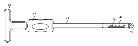
Referring to Figure 5A, a prosthesis deployment and
defect creation assembly is depicted having a distal portion
(40) configured to drill or core through bony material,
providing a defect volume through which an expandable
prosthesis (44) may be advanced. Once in position, which may
be confirmed as described above using various intraoperative
imaging modalities, relative rotation and/or linear deflection
between two handles (37, 38) may be utilized to pull the
distal portion (40), through a tension member (42), toward the
elongate shaft (41), thereby putting the expandable prosthesis
(44) in compression, and ultimately pulling the distal portion
through the center of the prosthesis (44), leaving behind a
deployed expanded prosthesis, as shown, for example, in
Figures 5C and Figures 5G-J. Figures 5B and 5C illustrate
close up orthogonal views of an embodiment of an expandable
prosthesis having end portions (46, 47), and a slotted
midportion (48), with slots (50) that complementarily define
defectable elongate members (49) configured to yield and bend
outward, as shown in Figure 5C, when the end portions (46, 47)
are compressed toward each other. Referring to Figures 5D-5F,
another embodiment is depicted wherein centrifugal forces
associated with angular velocity may be utilized to expand a
drilling member prosthesis, shown in compact form in Figure
5D, to expand to an expanded form, as shown incrementally in
Figures 5E and 5F. The proximal portion (58) is configured
without defects and has a releasable coupling, such as a
threaded coupling (60), for mechanical interfacing with a
rotation / insertion drive shaft (not shown). Below a
designed angular velocity threshold, the drilling member
prosthesis (52) functions as a drill bit and may be rotated
and advanced to create a defect and be positioned to a
10
WO 2012/015976 CA 02806755 2013-01-25PCT/US2011/045615
preferred location within or adjacent to bony tissue. When in
position, as may be confirmed, for example, using the
aforementioned imaging modalities, the operator may elect to
expand/deploy the prosthesis by exceeding the angular velocity
threshold, thus causing the relatively large massed central
portions (62) of connecting members (56) formed by the defects
(54) to migrate outward, thereby expanding the overall radius
of the prosthesis (52), as shown in Figure 5F. Such a
configuration may also be used in installations such as those
depicted in Figures 5G-5J.
Referring to Figure 5G, a single radially expandable
prosthesis (52) is deployed from a posterior approach to
stabilize or fixate an SI joint (6). Referring to Figure 5H,
two radially expandable prostheses (52) are deployed from a
posterior approach to stabilize or fixate an SI joint (6). In
one embodiment they may be coupled with a tensile or
stabilizing interconnect member (not shown). In another
embodiment they may simply reside adjacent one another.
Referring to Figures 51 and 5J, analogous deployment
embodiments are illustrated from a lateral approach.
Referring to Figure 6A, another expandable prosthesis
assembly (64) is depicted, comprising a main body (70) coupled
to four leg members (72) which are configured to bend and
rotate away from the main body (70) when two wedge members
(68) are advanced toward each other with the use of a screw
comprising a threaded shaft (67) and screw head (65);
preferably one or both of the wedge members have a threaded
interface with the screw shaft (67) to provide advancement of
the wedge members (68) relative to each other when the screw
shaft (67) is rotated relative to the wedge members (68) and
body (70) using an actuator or manual rotational driver
interfaced with the screw head (65). Referring to Figure 6B,
subsequent to creation of a defect (22) utilizing
11
WO 2012/015976 CA 02806755 2013-01-25PCT/US2011/045615
configurations such as those described in reference to Figure
3B, and expandable prosthesis assembly (64) may be advanced
into place and controllably expanded to stabilize the SI joint
(6). Referring to Figure 6C, one or more such assemblies (64)
may be utilized, and they may be coupled together with an
intercoupling member (76), which may be controllably elongated
or decreased in dimension, as described above, to create
tension or compression in the surrounding bony structures.
Referring to Figures 6D and 6E, analogous configurations are
depicted utilizing a lateral approach.
Referring to Figures 7A-10B, several coring or osteotome
tool embodiments are depicted; they may be utilized from
posterior or lateral approaches to create defects which may be
subsequently occupied by one or more prosthesis components to
stabilize or fix an SI joint.
Referring to Figure 7A, a three leading point osteotome
(78) embodiment is depicted having one advanced lead point
cutting apex (81) and two following lead point cutting apices
(82) located distally. A lumen or recess (82) is defined
through the middle of the osteotome (78) to contain captured
bone tissue. Referring to Figure 7B, a straight end view
shows that the osteotome (78) embodiment of Figure 7A has a
generally oval cross sectional shape. The lead point cutting
apices (81, 82) are configured to assist with positioning,
orienting, and generally advancing the osteotome (78) as it
accesses and traverses the bony structures comprising an SI
joint.
Referring to Figure 8A, a two jawed osteotome (84)
embodiment is depicted having a first jaw portion (85), a
second jaw portion (86), an included jaw angle (88), and a
defined lumen or cavity (82), all of which are configured to
create controlled defects at the SI joint. As shown in the
12
WO 2012/015976 CA 02806755 2013-01-25PCT/US2011/045615
straight end view of Figure 8B, this embodiment also has a
generally oval cross sectional shape.
Referring to Figure 9A, an embodiment similar to that of
Figure 7A is depicted, with the exception that the distal
taper angle and cutting apices (91, 92) are more mild
geometrically. The straight end view of Figure 9B shows that
this embodiment also has a generally oval cross sectional
shape.
Figure 10A depicts an embodiment similar to that of
Figure 9A, with the exception that more material has been
recessed away in between each of the apices (95, 96), to form
more pronounced distal contact points as this osteotome (94)
is placed in contact with, and advanced through, bony
structures comprising the SI joint. The straight end view of
Figure 10B shows that this embodiment also has a generally
oval cross sectional shape.
Referring to Figures 11A-11E, various osteotome
configurations may be utilized to create fixation /
stabilization defects of various geometries - and these
defects may be subsequently occupied by prostheses configured
to stabilize and/or fix the SI joint. Each of the
illustrative embodiments of Figures 11A-11E is shown in
reference to a posterior approach, but lateral SI joint
stabilizing approaches with similar configurations may also be
utilized. Referring to Figure 11A, in one embodiment, the
size, position, shape, and orientation of a defect may be
planned utilizing simiulated defect images (98) overlaid upon
preoperative or intraoperative images of the subject SI joint
(6) anatomy. Referring to Figure 11B, a generally oval defect
may be created from a posterior approach using tools such as
one or more drills, osteotomes (such as those described in
reference to Figures 7A-10B), or other orthopaedic bone
intervention tools. Referring to Figure 11C, a bi-lobed
13
WO 2012/015976 CA 02806755 2013-01-25PCT/US2011/045615
defect comprising two lobes (103, 104) connected by a
midportion (105). A prosthesis occupying such geometry from a
posterior approach provides several inherent stability
qualities. For example, the lobes (103, 104) may be utilized
relative to the cortical bone (8) positioning to interlock the
sacral bone portion (2) relative to the ilium bone portion (4)
and prevent relative motion of the prosthesis or bones.
Further, the relatively large surface area may be an advantage
for biological fixation (i.e., of bone tissue to porous
material which preferably comprises the outer surface of a
suitable prosthesis). Figure 11D depicts a "H" shaped defect
configuration which may be occupied by an "H" shaped
prosthesis comprising a central portion (112) and two end
portions (108, 110). Such a configuration also provides
desirable stability and fixation qualities. Figure 11E
depicts an "arcuate H" shaped defect (114) which may be
occupied by an "arcuate H" shaped prosthesis. The defect and
prosthesis may have a main body portion (116) and four or more
arm portions (118), each of which may be generally arcuate
shaped by virtue of a single joint-like bend (120), as in the
embodiment of Figure 11E, or a more gradual bend to define a
smoothly-arced arm (not shown). Other geometries may be
utilized.
Referring to Figures 12A-15D, various generally hollow
prosthesis configurations are depicted for press fit insertion
into a defect which may be created using the aforementioned
techniques. Their internal cavities may contain fixation
catalysts at deployment time, as described above, and their
walls generally comprise slots, holes, and other geometric
features which are configured to enhance initial "scratch fit"
fixation (i.e., when such prostheses are press fit into a
defect with a hammer or other tool; in certain embodiments
the prostheses may be configured to be under loads immediately
14
WO 2012/015976 CA 02806755 2013-01-25PCT/US2011/045615
when deployed, for interference / load bearing fit qualities;
in other embodiments, the prosthesis geometry may be matched
to the defect geometry without inherent stresses at
deployment), as well as subsequent biological fixation.
Generally they may be machined or otherwise formed from
materials such as nickel titanium surgical superalloy to
mimick, at least to a relative degree, the mechanical
properties of adjacent bony structural tissues.
Referring to Figures 12A-12C, a single taper prosthesis
embodiment (122) is depicted in various orthogonal views
having a plurality of slots (124) and distal holes (128)
configured to optimize structural performance and promote
biological fixation. A tapered distal portion (126) is
geometrically configured to assist with insertion upon
deployment. An inverse taper (132) of the midportion (between
the two outer lobes cross-sectionally) is defined
longitudinally. A substantial interior volume (134) may be
occupied by deployment tools and/or fixation catalysts, as
described above.
Referring to Figures 13A-13B, another single taper
prosthesis embodiment (136) is depicted in two orthogonal
views. The embodiment of Figures 13A-B is somewhat similar to
that of Figures 12A-12C; one significant departure is a
positive taper of the midportion (in between the end portions
(138)) - to define two ridges with interrupted ridge (150)
features and a generally longitudinally tapering (142) ridge /
midportion geometry that extends out farther proximally (146)
than distally (144). A substantial interior volume (151) may
be occupied by deployment tools and/or fixation catalysts, as
described above.
Referring to Figures 14A-14C, a dual taper prosthesis
embodiment (152) is depicted in three orthogonal views. This
embodiment also has a plurality of slots (162) and distal end
15
WO 2012/015976 CA 02806755 2013-01-25PCT/US2011/045615
(168) holes. The midportion (156) in between the two outer
lobed portions (154) has an inverse taper providing a smaller
cross sectional geometry proximally. The outer portions (154)
each define an interrupted ridge (164) that defines two
winglets (160, 161) and tapers in the inverse relative to the
tapering of the midportion. An interior volume is easily
accessed proximally (166).
Figures 15A-15D depict another dual taper prosthesis
embodiment (170) having three winglet ridges (188, 190, 192 /
194, 196, 198) on each of two outer portions (172), and
inverted tapering longitudinally of the modportion (174)
relative to the end portions (172).
Referring to Figures 16A-16E, aspects of a deployment
system and method utilizing configurations such as those
described above in reference to Figures 1A-15D are
illustrated. Referring to Figure 16A, an
osteotome/cannulation device (208) is temporarily coupled to
an obturator (210). A guiding member (206), such as a
guidewire or needle, has been partially advanced into an SI
joint (6) and such advanced position confirmed using one or
more imaging modalities as described above. A guiding member
lumen or channel (208) may be utilized to guide the
obturator/osteotome assembly (210 / 204) as they are advanced
through portions of the bony structures (2, 4) defining the SI
joint (6). The proximal end (216) of the osteotome (204) is
fitted against an enlarged obturator portion which may be
driven forward with a hammer or the like to advance the
osteotome distal end (214). As shown in greater detail in
Figure 16B, the distal end of the osteotome (214) may comprise
one or more teeth or apices (212) configured to assist with
creation of a defect of one or more bony structure portions.
After a defect of desired geometry and position has been
created, such as by advancing the osteotome forward into the
16
WO 2012/015976 CA 02806755 2013-01-25PCT/US2011/045615
bony structures defining the SI joint (6), a prosthesis
deployment assembly, such as that depicted in Figures 16C and
16D, may be utilized to advance a prosthesis, such as those
depicted in Figures 12A-15D, into place within a defect (200).
Two handles (222, 224) coupled to two shaft members (224, 226)
may be utilized to advance and controllably deploy a
prosthesis (122), such as the one shown coupled to the
assembly in Figure 16D, from a prosthesis interface (228),
such as that depicted in Figures 16C and 16D. Referring to
Figure 16E, a deployed prosthesis (122) is depicted in situ
providing stabilization and/or fixation to the SI joint (6)
and bony structures (2, 4) defining it.
17