Note: Descriptions are shown in the official language in which they were submitted.
CA 02807004 2013-01-29
WO 2012/016135 PCT/US2011/045879
BALLOON WITH SURFACE ELECTRODES AND INTEGRAL COOLING
FOR RENAL NERVE ABLATION
SUMMARY
Embodiments of the disclosure are directed to ablating target tissue of the
body,
such as innervated renal tissue, using an intravascular ablation device with
integral
cooling. Embodiments of the disclosure are directed to systems, apparatuses,
and methods
for ablating target tissue of the body, such as innervated renal tissue, using
balloon
supported ablation electrodes and an integral cooling arrangement for cooling
the ablation
electrodes.
According to various embodiments, an ablation apparatus includes a catheter
arrangement having a flexible shaft and a balloon disposed at a distal end of
the shaft. The
balloon is configured for deployment within a target vessel of the body.
Ablation
electrodes are supported by a wall of the balloon and arranged in a predefined
pattern.
The ablation electrodes are configured to deliver electrical energy sufficient
to ablate
target tissue proximate a wall of the target vessel when the balloon is in a
deployed
configuration. A cooling arrangement is encompassed at least in part by the
balloon and
configured to provide cooling to at least the electrodes during ablation such
that a location
at which steady-state ablative heating begins is translated from an electrode-
tissue
interface at the target vessel wall to a location a predetermined distance
away from the
electrode-tissue interface.
In some embodiments, an ablation apparatus includes a catheter arrangement
comprising a flexible shaft having a proximal end, a distal end, a length, and
a lumen
arrangement extending between the proximal and distal ends. The length of the
shaft is
sufficient to access a patient's renal artery relative to a percutaneous
access location. A
therapy unit is provided at the distal end of the shaft and coupled to the
lumen
arrangement. The therapy unit is dimensioned for deployment within a patient's
renal
artery, and includes a balloon fluidly coupled to the lumen arrangement and
transformable
between a low-profile introduction configuration and a larger-profile deployed
configuration. The balloon comprises a wall configured to contact an inner
wall of the
renal artery when in the deployed configuration. Ablation electrodes are
supported by the
balloon wall and arranged in a predefined pattern. The ablation electrodes are
configured
to deliver electrical energy sufficient to ablate perivascular renal nerves
adjacent the renal
1
WO 2012/016135 CA 02807004 2013-01-29 PCT/US2011/045879
artery when the balloon is in the deployed configuration. A cooling
arrangement is
encompassed at least in part by the balloon and configured to provide cooling
to at least
the electrodes during ablation such that a location at which steady-state
ablative heating
begins is translated from an electrode-tissue interface at the inner renal
artery wall to a
location a predetermined distance away from the electrode-tissue interface.
In accordance with other embodiments, a method of ablating tissue involves
expanding an ablation device within a target vessel, wherein a vessel-
contacting surface of
the ablation device supports ablation electrodes arranged in a predefined
pattern. The
method also involves delivering electrical energy through a wall of the target
vessel
sufficient to ablate target tissue proximate the target vessel wall, and
cooling at least the
ablation electrodes during ablation such that the target vessel is cooled and
steady-state
ablative heating begins at a predefined distance away from the electrodes.
Methods may
further involve compressing portions of the target vessel wall at a tissue-
electrode
interface associated with each of the electrodes, and delivering electrical
energy through
the compressed target vessel wall portions. The target vessel may include a
renal artery
and the target tissue may include perivascular renal nerve tissue, for
example.
These and other features can be understood in view of the following detailed
discussion and the accompanying drawings.
BRIEF DESCRIPTION OF THE DRAWINGS
Figure 1 is an illustration of a right kidney and renal vasculature including
a renal
artery branching laterally from the abdominal aorta;
Figures 2A and 2B illustrate sympathetic innervation of the renal artery;
Figure 3A illustrates various tissue layers of the wall of the renal artery;
Figures 3B and 3C illustrate a portion of a renal nerve;
Figure 4 shows a therapy device of an ablation catheter which includes a
cooling
arrangement and balloon supported ablation electrodes in accordance with
various
embodiments;
Figure 5 shows a therapy device of an ablation catheter which includes a
cooling
arrangement and balloon supported ablation electrodes in accordance with
various
embodiments;
2
CA 02807004 2013-01-29
WO 2012/016135
PCT/US2011/045879
Figure 6 is a cross-sectional view of a portion of a therapy device showing an
electrode-tissue interface defined between a balloon supported ablation
electrode and a
wall of a renal artery in accordance with various embodiments;
Figure 7 is a cross-sectional view of a portion of a therapy device showing an
electrode-tissue interface defined between a balloon supported ablation
electrode and a
wall of a renal artery in accordance with various embodiments;
Figures 8-10 show features of a cooling arrangement for a portion of a therapy
device which includes a balloon supported ablation electrode in accordance
with various
embodiments; and
Figure 11 shows a therapy system configured to perform renal denervation using
a
therapy device of an ablation catheter which includes a cooling arrangement
and balloon
supported ablation electrodes in accordance with various embodiments.
Embodiments of the disclosure are directed to apparatuses and methods for
DESCRIPTION
ablating target tissue using electrical energy delivered by a multiplicity of
cooled ablation
electrodes supported by an expandable therapy device. Embodiments of the
disclosure are
directed to apparatuses and methods for ablating target tissue located
adjacent to a body
vessel using a multiplicity of cooled ablation electrodes supported by an
expandable
therapy device deployed in the body vessel. Embodiments are directed to
ablating target
tissue of the body using cooled ablation electrodes situated at a wall of the
target vessel
proximate the target tissue, such that the cooled ablation electrodes
translate a location at
which steady-state ablative heating begins from an electrode-tissue interface
at the target
vessel wall to a desired location a predetermined distance away from the
electrode-tissue
interface. Particular embodiments of the disclosure are directed to
apparatuses and
methods for ablating perivascular renal nerves for the treatment of
hypertension.
Radiofrequency (RF) ablation of renal nerves, which lie proximate to the
adventitia
of the renal artery, may be an effective treatment for chronic hypertension.
It has been
difficult to effectively ablate perivascular renal sympathetic nerves by
access from the
renal artery, without injury to the renal artery wall. To reduce concern for
potential
stenotic narrowing of the artery after the ablation procedure, minimizing
arterial injury
during such an ablation procedure is important.
3
WO 2012/016135 CA 02807004 2013-01-29 PCT/US2011/045879
Embodiments of the disclosure incorporate a housing mounted at a distal end of
a
therapy catheter for supporting ablation electrodes and cooling components of
the therapy
device. The housing encompasses at least a portion of a cooling arrangement
and supports
a number of ablation electrodes on an outer surface of the housing. The
housing is
preferably transformable between a low-profile introduction configuration and
a larger-
profile deployment configuration. The low-profile introduction configuration
allows the
therapy catheter to be readily advanced through the venous or arterial system
to a desired
body location, for example. The larger-profile deployed configuration allows
the therapy
catheter to be stabilized at the desired body location, such as within the
renal artery. In
various embodiments, the expandable structure comprises a balloon, such as a
cooling
balloon or a cryoballoon.
Various embodiments of the disclosure include a balloon catheter with
electrodes
on the balloon to perform ablation of target tissue while cooling the luminal
surface of a
renal artery prevents undesirable heating of non-targeted tissue of the renal
artery,
particularly the endothelium of the artery. Apparatuses of the disclosure can
provide a
number of benefits, including one or more of reduced injury to the artery,
ablation with a
single treatment rather than multiple treatments which reduces treatment time,
and
ablation in a manner that is more controllable and repeatable.
An RF electrode can be cooled to limit temperature increase at the electrode
surface while allowing increased temperature at a distance from the electrode.
When the
electrode is in contact with tissue, the distance where steady-state heating
starts is
preferably on the order of about 0.5 mm to about 1 mm into the tissue. For
example, it is
desirable that steady-state ablative heating begins at a distance of at least
about 0.5 mm
away from the electrode surface. Heat is conducted out from that point. In a
blood vessel,
limiting heat at the electrode-vessel surface (also referred to herein as an
electrode-tissue
interface) can limit injury at the vessel surface, which can reduce thermal
injury to, and
yield improved healing of, the vessel surface. One or more temperature
sensors, such as
thermocouples, can be provided at the site of the electrodes to measure the
temperature at
or proximate the electrodes. In some embodiments, a temperature sensor is
positioned
near or at the site of each electrode on the balloon, allowing for precision
temperature
measurements at individual electrode locations of the ablation electrode
arrangement.
Cooling of the electrodes, and other portions of the balloon wall if desired,
can be
effected using several different cooling mechanisms. In some embodiments, a
therapy
4
CA 02807004 2013-01-29
WO 2012/016135 PCT/US2011/045879
catheter incorporates a phase-change cryothermal capability such as by
spraying a cryogen
to cool at least the electrode supporting portions of an inflated balloon.
Temperature
and/or pressure sensors or other sensor elements (e.g., impedance sensors) can
be
incorporated near or at the electrode locations or other locations to
facilitate monitoring
and control of the ablation procedure. In other embodiments, the therapy
catheter
incorporates a heat exchange apparatus configured to receive a liquid coolant
capable of
causing freezing of tissue proximate of the target tissue, such as the wall of
the renal
artery. In some embodiments, the therapy catheter incorporates one or more
solid-state
thermoelectric cooling devices, such as Peltier devices. The cooling and
ablation
electrode components of the therapy catheter can interface with external
control units to
control device functioning and monitor or display temperatures, power used,
impedance,
blood pressure, or other parameters.
Various embodiments of the disclosure are directed to apparatuses and methods
for
renal denervation for treating hypertension. Hypertension is a chronic medical
condition
in which the blood pressure is elevated. Persistent hypertension is a
significant risk factor
associated with a variety of adverse medical conditions, including heart
attacks, heart
failure, arterial aneurysms, and strokes. Persistent hypertension is a leading
cause of
chronic renal failure. Hyperactivity of the sympathetic nervous system serving
the
kidneys is associated with hypertension and its progression. Deactivation of
nerves in the
kidneys via renal denervation can reduce blood pressure, and may be a viable
treatment
option for many patients with hypertension who do not respond to conventional
drugs.
The kidneys are instrumental in a number of body processes, including blood
filtration, regulation of fluid balance, blood pressure control, electrolyte
balance, and
hormone production. One primary function of the kidneys is to remove toxins,
mineral
salts, and water from the blood to form urine. The kidneys receive about 20-
25% of
cardiac output through the renal arteries that branch left and right from the
abdominal
aorta, entering each kidney at the concave surface of the kidneys, the renal
hilum.
Blood flows into the kidneys through the renal artery and the afferent
arteriole,
entering the filtration portion of the kidney, the renal corpuscle. The renal
corpuscle is
composed of the glomerulus, a thicket of capillaries, surrounded by a fluid-
filled, cup-like
sac called Bowman's capsule. Solutes in the blood are filtered through the
very thin
capillary walls of the glomerulus due to the pressure gradient that exists
between the blood
in the capillaries and the fluid in the Bowman's capsule. The pressure
gradient is
5
CA 02807004 2013-01-29
WO 2012/016135 PCT/US2011/045879
controlled by the contraction or dilation of the arterioles. After filtration
occurs, the
filtered blood moves through the efferent arteriole and the peritubular
capillaries,
converging in the interlobular veins, and finally exiting the kidney through
the renal vein.
Particles and fluid filtered from the blood move from the Bowman's capsule
through a number of tubules to a collecting duct. Urine is formed in the
collecting duct
and then exits through the ureter and bladder. The tubules are surrounded by
the
peritubular capillaries (containing the filtered blood). As the filtrate moves
through the
tubules and toward the collecting duct, nutrients, water, and electrolytes,
such as sodium
and chloride, are reabsorbed into the blood.
The kidneys are innervated by the renal plexus which emanates primarily from
the
aorticorenal ganglion. Renal ganglia are formed by the nerves of the renal
plexus as the
nerves follow along the course of the renal artery and into the kidney. The
renal nerves
are part of the autonomic nervous system which includes sympathetic and
parasympathetic
components. The sympathetic nervous system is known to be the system that
provides the
bodies "fight or flight" response, whereas the parasympathetic nervous system
provides
the "rest and digest" response. Stimulation of sympathetic nerve activity
triggers the
sympathetic response which causes the kidneys to increase production of
hormones that
increase vasoconstriction and fluid retention. This process is referred to as
the renin-
angiotensin-aldosterone-system (RAAS) response to increased renal sympathetic
nerve
activity.
In response to a reduction in blood volume, the kidneys secrete renin, which
stimulates the production of angiotensin. Angiotensin causes blood vessels to
constrict,
resulting in increased blood pressure, and also stimulates the secretion of
the hormone
aldosterone from the adrenal cortex. Aldosterone causes the tubules of the
kidneys to
increase the reabsorption of sodium and water, which increases the volume of
fluid in the
body and blood pressure.
Congestive heart failure (CHF) is a condition that has been linked to kidney
function. CHF occurs when the heart is unable to pump blood effectively
throughout the
body. When blood flow drops, renal function degrades because of insufficient
perfusion
of the blood within the renal corpuscles. The decreased blood flow to the
kidneys triggers
an increase in sympathetic nervous system activity (i.e., the RAAS becomes too
active)
that causes the kidneys to secrete hormones that increase fluid retention and
vasorestriction. Fluid retention and vasorestriction in turn increases the
peripheral
6
CA 02807004 2013-01-29
WO 2012/016135 PCT/US2011/045879
resistance of the circulatory system, placing an even greater load on the
heart, which
diminishes blood flow further. If the deterioration in cardiac and renal
functioning
continues, eventually the body becomes overwhelmed, and an episode of heart
failure
decompensation occurs, often leading to hospitalization of the patient.
Figure 1 is an illustration of a right kidney 10 and renal vasculature
including a
renal artery 12 branching laterally from the abdominal aorta 20. In Figure 1,
only the right
kidney 10 is shown for purposes of simplicity of explanation, but reference
will be made
herein to both right and left kidneys and associated renal vasculature and
nervous system
structures, all of which are contemplated within the context of embodiments of
the
disclosure. The renal artery 12 is purposefully shown to be disproportionately
larger than
the right kidney 10 and abdominal aorta 20 in order to facilitate discussion
of various
features and embodiments of the present disclosure.
The right and left kidneys are supplied with blood from the right and left
renal
arteries that branch from respective right and left lateral surfaces of the
abdominal aorta
20. Each of the right and left renal arteries is directed across the crus of
the diaphragm, so
as to form nearly a right angle with the abdominal aorta 20. The right and
left renal
arteries extend generally from the abdominal aorta 20 to respective renal
sinuses
proximate the hilum 17 of the kidneys, and branch into segmental arteries and
then
interlobular arteries within the kidney 10. The interlobular arteries radiate
outward,
penetrating the renal capsule and extending through the renal columns between
the renal
pyramids. Typically, the kidneys receive about 20% of total cardiac output
which, for
normal persons, represents about 1200 mL of blood flow through the kidneys per
minute.
The primary function of the kidneys is to maintain water and electrolyte
balance
for the body by controlling the production and concentration of urine. In
producing urine,
the kidneys excrete wastes such as urea and ammonium. The kidneys also control
reabsorption of glucose and amino acids, and are important in the production
of hormones
including vitamin D, renin and erythropoietin.
An important secondary function of the kidneys is to control metabolic
homeostasis of the body. Controlling hemostatic functions include regulating
electrolytes,
acid-base balance, and blood pressure. For example, the kidneys are
responsible for
regulating blood volume and pressure by adjusting volume of water lost in the
urine and
releasing erythropoietin and renin, for example. The kidneys also regulate
plasma ion
concentrations (e.g., sodium, potassium, chloride ions, and calcium ion
levels) by
7
CA 02807004 2013-01-29
WO 2012/016135 PCT/US2011/045879
controlling the quantities lost in the urine and the synthesis of calcitrol.
Other hemostatic
functions controlled by the kidneys include stabilizing blood pH by
controlling loss of
hydrogen and bicarbonate ions in the urine, conserving valuable nutrients by
preventing
their excretion, and assisting the liver with detoxification.
Also shown in Figure 1 is the right suprarenal gland 11, commonly referred to
as
the right adrenal gland. The suprarenal gland 11 is a star-shaped endocrine
gland that rests
on top of the kidney 10. The primary function of the suprarenal glands (left
and right) is
to regulate the stress response of the body through the synthesis of
corticosteroids and
catecholamines, including cortisol and adrenaline (epinephrine), respectively.
Encompassing the kidneys 10, suprarenal glands 11, renal vessels 12, and
adjacent
perirenal fat is the renal fascia, e.g., Gerota's fascia, (not shown), which
is a fascial pouch
derived from extraperitoneal connective tissue.
The autonomic nervous system of the body controls involuntary actions of the
smooth muscles in blood vessels, the digestive system, heart, and glands. The
autonomic
nervous system is divided into the sympathetic nervous system and the
parasympathetic
nervous system. In general terms, the parasympathetic nervous system prepares
the body
for rest by lowering heart rate, lowering blood pressure, and stimulating
digestion. The
sympathetic nervous system effectuates the body's fight-or-flight response by
increasing
heart rate, increasing blood pressure, and increasing metabolism.
In the autonomic nervous system, fibers originating from the central nervous
system and extending to the various ganglia are referred to as preganglionic
fibers, while
those extending from the ganglia to the effector organ are referred to as
postganglionic
fibers. Activation of the sympathetic nervous system is effected through the
release of
adrenaline (epinephrine) and to a lesser extent norepinephrine from the
suprarenal glands
11. This release of adrenaline is triggered by the neurotransmitter
acetylcholine released
from preganglionic sympathetic nerves.
The kidneys and ureters (not shown) are innervated by the renal nerves 14.
Figures
1 and 2A-2B illustrate sympathetic innervation of the renal vasculature,
primarily
innervation of the renal artery 12. The primary functions of sympathetic
innervation of the
renal vasculature include regulation of renal blood flow and pressure,
stimulation of renin
release, and direct stimulation of water and sodium ion reabsorption.
Most of the nerves innervating the renal vasculature are sympathetic
postganglionic fibers arising from the superior mesenteric ganglion 26. The
renal nerves
8
CA 02807004 2013-01-29
WO 2012/016135 PCT/US2011/045879
14 extend generally axially along the renal arteries 12, enter the kidneys 10
at the hilum
17, follow the branches of the renal arteries 12 within the kidney 10, and
extend to
individual nephrons. Other renal ganglia, such as the renal ganglia 24,
superior mesenteric
ganglion 26, the left and right aorticorenal ganglia 22, and celiac ganglia 28
also innervate
the renal vasculature. The celiac ganglion 28 is joined by the greater
thoracic splanchnic
nerve (greater TSN). The aorticorenal ganglia 26 is joined by the lesser
thoracic
splanchnic nerve (lesser TSN) and innervates the greater part of the renal
plexus.
Sympathetic signals to the kidney 10 are communicated via innervated renal
vasculature that originates primarily at spinal segments T10-T12 and Li.
Parasympathetic
signals originate primarily at spinal segments S2-S4 and from the medulla
oblongata of the
lower brain. Sympathetic nerve traffic travels through the sympathetic trunk
ganglia,
where some may synapse, while others synapse at the aorticorenal ganglion 22
(via the
lesser thoracic splanchnic nerve, i.e., lesser TSN) and the renal ganglion 24
(via the least
thoracic splanchnic nerve, i.e., least TSN). The postsynaptic sympathetic
signals then
travel along nerves 14 of the renal artery 12 to the kidney 10. Presynaptic
parasympathetic signals travel to sites near the kidney 10 before they synapse
on or near
the kidney 10.
With particular reference to Figure 2A, the renal artery 12, as with most
arteries
and arterioles, is lined with smooth muscle 34 that controls the diameter of
the renal artery
lumen 13. Smooth muscle, in general, is an involuntary non-striated muscle
found within
the media layer of large and small arteries and veins, as well as various
organs. The
glomeruli of the kidneys, for example, contain a smooth muscle-like cell
called the
mesangial cell. Smooth muscle is fundamentally different from skeletal muscle
and
cardiac muscle in terms of structure, function, excitation-contraction
coupling, and
mechanism of contraction.
Smooth muscle cells can be stimulated to contract or relax by the autonomic
nervous system, but can also react on stimuli from neighboring cells and in
response to
hormones and blood borne electrolytes and agents (e.g., vasodilators or
vasoconstrictors).
Specialized smooth muscle cells within the afferent arteriole of the
juxtaglomerular
apparatus of kidney 10, for example, produces renin which activates the
angiotension II
system.
The renal nerves 14 innervate the smooth muscle 34 of the renal artery wall 15
and
extend lengthwise in a generally axial or longitudinal manner along the renal
artery wall
9
CA 02807004 2013-01-29
WO 2012/016135 PCT/US2011/045879
15. The smooth muscle 34 surrounds the renal artery circumferentially, and
extends
lengthwise in a direction generally transverse to the longitudinal orientation
of the renal
nerves 14, as is depicted in Figure 2B.
The smooth muscle 34 of the renal artery 12 is under involuntary control of
the
autonomic nervous system. An increase in sympathetic activity, for example,
tends to
contract the smooth muscle 34, which reduces the diameter of the renal artery
lumen 13
and decreases blood perfusion. A decrease in sympathetic activity tends to
cause the
smooth muscle 34 to relax, resulting in vessel dilation and an increase in the
renal artery
lumen diameter and blood perfusion. Conversely, increased parasympathetic
activity
tends to relax the smooth muscle 34, while decreased parasympathetic activity
tends to
cause smooth muscle contraction.
Figure 3A shows a segment of a longitudinal cross-section through a renal
artery,
and illustrates various tissue layers of the wall 15 of the renal artery 12.
The innermost
layer of the renal artery 12 is the endothelium 30, which is the innermost
layer of the
intima 32 and is supported by an internal elastic membrane. The endothelium 30
is a
single layer of cells that contacts the blood flowing though the vessel lumen
13.
Endothelium cells are typically polygonal, oval, or fusiform, and have very
distinct round
or oval nuclei. Cells of the endothelium 30 are involved in several vascular
functions,
including control of blood pressure by way of vasoconstriction and
vasodilation, blood
clotting, and acting as a barrier layer between contents within the lumen 13
and
surrounding tissue, such as the membrane of the intima 32 separating the
intima 32 from
the media 34, and the adventitia 36. The membrane or maceration of the intima
32 is a
fine, transparent, colorless structure which is highly elastic, and commonly
has a
longitudinal corrugated pattern.
Adjacent the intima 32 is the media 33, which is the middle layer of the renal
artery 12. The media is made up of smooth muscle 34 and elastic tissue. The
media 33
can be readily identified by its color and by the transverse arrangement of
its fibers. More
particularly, the media 33 consists principally of bundles of smooth muscle
fibers 34
arranged in a thin plate-like manner or lamellae and disposed circularly
around the arterial
wall 15. The outermost layer of the renal artery wall 15 is the adventitia 36,
which is
made up of connective tissue. The adventitia 36 includes fibroblast cells 38
that play an
important role in wound healing.
10
CA 02807004 2013-01-29
WO 2012/016135 PCT/US2011/045879
A perivascular region 37 is shown adjacent and peripheral to the adventitia 36
of
the renal artery wall 15. A renal nerve 14 is shown proximate the adventitia
36 and
passing through a portion of the perivascular region 37. The renal nerve 14 is
shown
extending substantially longitudinally along the outer wall 15 of the renal
artery 12. The
main trunk of the renal nerves 14 generally lies in or on the adventitia 36 of
the renal
artery 12, often passing through the perivascular region 37, with certain
branches coursing
into the media 33 to enervate the renal artery smooth muscle 34.
Embodiments of the disclosure may be implemented to provide varying degrees of
denervation therapy to innervated renal vasculature. For example, embodiments
of the
disclosure may provide for control of the extent and relative permanency of
renal nerve
impulse transmission interruption achieved by denervation therapy delivered
using a
treatment apparatus of the disclosure. The extent and relative permanency of
renal nerve
injury may be tailored to achieve a desired reduction in sympathetic nerve
activity
(including a partial or complete block) and to achieve a desired degree of
permanency
(including temporary or irreversible injury).
Returning to Figures 3B and 3C, the portion of the renal nerve 14 shown in
Figures
3B and 3C includes bundles 14a of nerve fibers 14b each comprising axons or
dendrites
that originate or terminate on cell bodies or neurons located in ganglia or on
the spinal
cord, or in the brain. Supporting tissue structures 14c of the nerve 14
include the
endoneurium (surrounding nerve axon fibers), perineurium (surrounds fiber
groups to
form a fascicle), and epineurium (binds fascicles into nerves), which serve to
separate and
support nerve fibers 14b and bundles 14a. In particular, the endoneurium, also
referred to
as the endoneurium tube or tubule, is a layer of delicate connective tissue
that encloses the
myelin sheath of a nerve fiber 14b within a fasciculus.
Major components of a neuron include the soma, which is the central part of
the
neuron that includes the nucleus, cellular extensions called dendrites, and
axons, which are
cable-like projections that carry nerve signals. The axon terminal contains
synapses,
which are specialized structures where neurotransmitter chemicals are released
in order to
communicate with target tissues. The axons of many neurons of the peripheral
nervous
system are sheathed in myelin, which is formed by a type of glial cell known
as Schwann
cells. The myelinating Schwann cells are wrapped around the axon, leaving the
axolemma
relatively uncovered at regularly spaced nodes, called nodes of Ranvier.
Myelination of
axons enables an especially rapid mode of electrical impulse propagation
called saltation.
11
CA 02807004 2013-01-29
WO 2012/016135 PCT/US2011/045879
In some embodiments, a treatment apparatus of the disclosure may be
implemented
to deliver denervation therapy that causes transient and reversible injury to
renal nerve
fibers 14b. In other embodiments, a treatment apparatus of the disclosure may
be
implemented to deliver denervation therapy that causes more severe injury to
renal nerve
fibers 14b, which may be reversible if the therapy is terminated in a timely
manner. In
preferred embodiments, a treatment apparatus of the disclosure may be
implemented to
deliver denervation therapy that causes severe and irreversible injury to
renal nerve fibers
14b, resulting in permanent cessation of renal sympathetic nerve activity. For
example, a
treatment apparatus may be implemented to deliver a denervation therapy that
disrupts
nerve fiber morphology to a degree sufficient to physically separate the
endoneurium tube
of the nerve fiber 14b, which can prevent regeneration and re-innervation
processes.
By way of example, and in accordance with Seddon's classification as is known
in
the art, a treatment apparatus of the disclosure may be implemented to deliver
a
denervation therapy that interrupts conduction of nerve impulses along the
renal nerve
fibers 14b by imparting damage to the renal nerve fibers 14b consistent with
neruapraxia.
Neurapraxia describes nerve damage in which there is no disruption of the
nerve fiber 14b
or its sheath. In this case, there is an interruption in conduction of the
nerve impulse down
the nerve fiber, with recovery taking place within hours to months without
true
regeneration, as Wallerian degeneration does not occur. Wallerian degeneration
refers to a
process in which the part of the axon separated from the neuron's cell nucleus
degenerates.
This process is also known as anterograde degeneration. Neurapraxia is the
mildest form
of nerve injury that may be imparted to renal nerve fibers 14b by use of a
treatment
apparatus according to embodiments of the disclosure.
A treatment apparatus may be implemented to interrupt conduction of nerve
impulses along the renal nerve fibers 14b by imparting damage to the renal
nerve fibers
consistent with axonotmesis. Axonotmesis involves loss of the relative
continuity of the
axon of a nerve fiber and its covering of myelin, but preservation of the
connective tissue
framework of the nerve fiber. In this case, the encapsulating support tissue
14c of the
nerve fiber 14b are preserved. Because axonal continuity is lost, Wallerian
degeneration
occurs. Recovery from axonotmesis occurs only through regeneration of the
axons, a
process requiring time on the order of several weeks or months. Electrically,
the nerve
fiber 14b shows rapid and complete degeneration. Regeneration and re-
innervation may
occur as long as the endoneural tubes are intact.
12
CA 02807004 2013-01-29
WO 2012/016135 PCT/US2011/045879
A treatment apparatus may be implemented to interrupt conduction of nerve
impulses along the renal nerve fibers 14b by imparting damage to the renal
nerve fibers
14b consistent with neurotmesis. Neurotmesis, according to Seddon's
classification, is the
most serious nerve injury in the scheme. In this type of injury, both the
nerve fiber 14b
and the nerve sheath are disrupted. While partial recovery may occur, complete
recovery
is not possible. Neurotmesis involves loss of continuity of the axon and the
encapsulating
connective tissue 14c, resulting in a complete loss of autonomic function, in
the case of
renal nerve fibers 14b. If the nerve fiber 14b has been completely divided,
axonal
regeneration causes a neuroma to form in the proximal stump.
A more stratified classification of neurotmesis nerve damage may be found by
reference to the Sunderland System as is known in the art. The Sunderland
System
defines five degrees of nerve damage, the first two of which correspond
closely with
neurapraxia and axonotmesis of Seddon's classification. The latter three
Sunderland
System classifications describe different levels of neurotmesis nerve damage.
The first and second degrees of nerve injury in the Sunderland system are
analogous to Seddon's neurapraxia and axonotmesis, respectively. Third degree
nerve
injury, according to the Sunderland System, involves disruption of the
endoneurium, with
the epineurium and perineurium remaining intact. Recovery may range from poor
to
complete depending on the degree of intrafascicular fibrosis. A fourth degree
nerve injury
involves interruption of all neural and supporting elements, with the
epineurium remaining
intact. The nerve is usually enlarged. Fifth degree nerve injury involves
complete
transection of the nerve fiber 14b with loss of continuity.
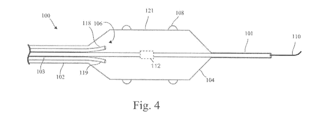
Figure 4 shows an embodiment of the disclosure which includes a therapy
catheter
100 configured for placement within a lumen of a target vessel of the body,
such as
patient's renal artery. The therapy catheter 100 shown in Figure 4 includes a
therapy
device 104 provided at a distal end of a shaft 102 of the therapy catheter
100. The therapy
device 104 includes a multiplicity of electrodes 108 supported by an
expandable housing
121 and configured to deliver ablative electrical energy (e.g., RF energy or
other form of
high frequency AC energy) to target tissue located adjacent the target vessel.
The therapy
device 104 further includes a cooling arrangement 106 configured to cool each
of the
electrodes 108 and, if desired, other portions of a wall of the housing 121.
During ablation, the electrodes 108 are cooled by the cooling arrangement 106
such that a location at which steady-state ablative heating begins is
translated from an
13
CA 02807004 2013-01-29
WO 2012/016135 PCT/US2011/045879
electrode-tissue interface to a location a predetermined distance away from
the electrode-
tissue interface. Translating the location at which study-state ablative
heating begins away
from the electrode-tissue interface provides for effective ablating of target
tissue while
intervening target vessel wall tissue is thermally protected.
As is further shown in Figure 4, the therapy device 104 is fluidly and
electrically
coupled to a lumen arrangement 103 which runs along the length of the shaft
102. The
lumen arrangement 103 includes an electrical conductor arrangement, a
pressurizable
lumen arrangement, and a guidewire lumen 101 dimension to receive a guidewire
110.
The guidewire 110 can be used by the clinician to access a patient's venous or
arterial
system, locate a target vessel, such as the patient's renal artery, and
advanced the therapy
device 104 into the lumen of the target vessel. The proximal end of the shaft
102 is fluidly
and electrically coupled to an external control system via the lumen
arrangement 103, an
embodiment of which is described hereinbelow with reference to Figure 11.
In the embodiment shown in Figure 4, the lumen arrangement 103 includes a
supply lumen 118 through which a thermal transfer fluid is supplied to the
therapy device
104 from an external source coupled to a proximal end of the shaft 102. The
lumen
arrangement 103 also includes a return lumen 119, through which spent thermal
transfer
fluid is returned to the proximal end of the shaft 102. According to some
embodiments,
the cooling arrangement 106 can include a phase-change cryothermal mechanism,
a
simpler heat exchanger system with liquid coolant, or a solid-state
thermoelectric cooling
device, for example. Depending on the particular cooling arrangement employed,
one or
both of the supply and return lumen's 118, 119 may or may not be required.
Various
cooling elements and support, connection, and control arrangements and
methodologies
that can be adapted for use in embodiments of the present disclosure are
disclosed in
commonly owned U.S. Patent No. 7,238,184 and U.S. Patent Application No.
13/157,844
filed June 10, 2011, which are incorporated herein by reference.
According to various embodiments, the electrodes 108 are cooled using a
thermal
transfer fluid supplied by an external coolant source and transported through
the lumen
arrangement 103 of the shaft 102. A variety of thermal transfer fluids may be
employed,
including cold saline or cold saline and ethanol mixture, Freon or other
fluorocarbon
refrigerants, nitrous oxide, liquid nitrogen, and liquid carbon dioxide, for
example. The
cooling arrangement 106 of the therapy unit 104 may include a tube (e.g., a
cryoprobe),
lumen, manifold, and/or a balloon arrangement through which the thermal
transfer fluid
14
CA 02807004 2013-01-29
WO 2012/016135 PCT/US2011/045879
passes. The cooling arrangement 106 may be integral or separate from the
expandable
housing 121. In some configurations, the cooling arrangement 106 may be
configured to
cool a substantial portion of the housing wall, including locations where the
electrodes 108
are mounted. In other configurations, the cooling arrangement 106 may be
configured to
cool only those portions of the housing wall where the electrodes 108 are
mounted.
In accordance with various embodiments, the electrodes 108 are energized by a
conductive thermal transfer fluid within the housing 121. An electrical
conductor extends
along the lumen arrangement of the shaft 102 and is in electrical
communication with the
conductive fluid. In some configurations, the electrical conductor is
electrically coupled
to an electrode 112 positioned on the shaft 102 within the housing 121. High
frequency
AC power is communicated to the electrodes 108 supported by the housing 121
via the
electrical conductor, electrode 112, and electrically conductive fluid within
the housing
121. Various embodiments may incorporate selected structural, electrical,
thermal, and
control features of the devices disclosed in the commonly owned U.S. Serial
No.
13/188,677 on July 22, 2011, which claims priority to U.S. Provisional
Application Nos.
61/411,795, filed on November 9, 2010, and 61/369,442, filed on July 30, 2010,
each of
which is incorporated herein by reference. In other embodiments, the
electrodes 108 are
energized by electrical conductors that couple each electrode 108 to a
conductor
arrangement of the shaft 102. The electrodes 108 can be connected to an
external control
system individually or in series.
In some embodiments, the thermal transfer fluid, when released inside the
cooling
arrangement 106 (e.g., a cryoballoon) via the supply lumen 118, undergoes a
phase change
that cools some or all of the housing 121 and each of the electrodes 108 by
absorbing the
latent heat of vaporization from the tissue surrounding the therapy unit 104,
and by
cooling of the vaporized gas as it enters a region of lower pressure inside
the cooling
arrangement 106 (the Joule-Thomson effect). As a result of the phase change
and the
Joule-Thompson effect, heat is extracted from the surroundings of the housing
121,
thereby cooling at least the electrodes 108 (and other portions of the housing
wall if
desired) which are in contact with vessel wall tissue. In configurations where
cooling is
limited to the electrodes 108, a manifold can be implemented within the
housing 121 or
housing wall to transport thermal transfer fluid to and from the electrodes
108. The gas
released inside the cooling arrangement 106 may be exhausted through the
return lumen
119 of the shaft 102. The pressure inside the cooling arrangement 106 may be
controlled
15
CA 02807004 2013-01-29
WO 2012/016135 PCT/US2011/045879
by regulating one or both of a rate at which thermal transfer fluid is
delivered and a rate at
which the exhaust gas is extracted. The lumen 118, 119 of the lumen
arrangement 103
which transport thermal transfer fluid are preferably lined with or otherwise
incorporate
insulation material(s) having appropriate thermal and mechanical
characteristics suitable
for a selected thermal transfer fluid.
Embodiments of the present invention may incorporate selected balloon,
catheter,
lumen, control, and other features of the devices disclosed in the following
commonly
owned U.S. patents and published patent applications: U.S. Patent Publication
Nos.
2009/0299356, 2009/0299355, 2009/0287202, 2009/0281533, 2009/0209951,
2009/0209949, 2009/0171333, 2008/0312644, 2008/0208182, 2008/0058791 and
2005/0197668, and U.S. Patent Nos. 5868735, 6290696, 6648878, 6666858,
6709431,
6929639, 6989009, 7022120, 7101368, 7172589, 7189227, and 7220257, which are
incorporated herein by reference. Embodiments of the present invention may
incorporate
selected balloon, catheter, and other features of the devices disclosed in
U.S. Patent Nos.
6355029, 6428534, 6432102, 6468297, 6514245, 6602246, 6648879, 6786900,
6786901,
6811550, 6908462, 6972015, and 7081112, which are incorporated herein by
reference.
In various embodiments, the cooling arrangement 106 can include one or more
thermoelectric elements configured to thermally couple to the wall of the
housing 121 at
or near the electrodes 108 and operate in a hypothermic mode. The
thermoelectric
elements preferably comprise solid-state thermoelectric elements, such as
Peltier elements.
Various Peltier-effect elements and support, connection, and control
arrangements and
methodologies that can be adapted for use in embodiments of the present
invention are
disclosed in commonly owned U.S. Patent No. 7,238,184, which is incorporated
herein by
reference.
In some embodiments, for example, the expandable housing 121 includes or is
constructed as a balloon which is fluidly coupled to the lumen arrangement 103
and
transformable between a low-profile introduction configuration and a larger-
profile
deployed configuration. The housing 121 is typically constructed from
polymeric
material, and preferably has a diameter dimensioned to fit within a target
vessel, such as a
renal artery of an average patient. It is understood that different models of
ablation
catheters 100 can be constructed each having specific housing configurations
and
dimensions appropriate for a given population of patients. In some
embodiments, the
housing 121 may comprise an expandable element, such as a pressurizable
balloon or a
16
CA 02807004 2013-01-29
WO 2012/016135 PCT/US2011/045879
mechanically expandable arrangement (e.g., an expandable-collapsible mesh
structure).
Use of such an expandable element in the construction of the housing 121
allows for use
of a common housing design for a population of patients having varying
anatomy. In
accordance with various embodiments in which a pressurizable balloon is used
in the
construction of the housing 121, a thermal transfer fluid may be used for
pressurizing the
balloon and cooling of vessel tissue and the electrodes 108.
The balloon 121 includes a wall configured to contact an inner wall of a
target
vessel when in its deployed configuration. A multiplicity of ablation
electrodes 108 are
supported by the balloon wall and are preferably arranged in a predefined
pattern. The
electrodes 108 may, for example, be arranged to form one or more
circumferential
patterns. By way of further example, the electrodes 108 may be arranged to
form a helical
or spiral pattern. The ablation electrodes 108 are configured to deliver
electrical energy
sufficient to ablate target tissue located adjacent to the target vessel when
the balloon 121
is in its deployed configuration. All or at least part of the cooling
arrangement 106 is
encompassed by the balloon 121.
As discussed previously, the cooling arrangement 106 is configured to cool at
least
the electrodes 108 during ablation, such that a location at which steady-state
ablative
heating begins is translated from an electrode-tissue interface at the inner
vessel wall to a
location a predetermined distance away from electrode-tissue interface. In
some
embodiments, the cooling arrangement is configured to cool the electrodes 108
such that
the steady-state ablative heating begins at a distance of about 0.5 mm to
about 1 mm from
the electrodes 108 (away from the electrode-tissue interface and towards
target tissue). In
other embodiments, the location at which steady-state ablative heating begins
is translated
from the electrode-tissue interface to a distance of about 1 mm.
As is shown in Figure 5, one or more temperature sensors 115 can be situated
on
the therapy device 104 to provide for temperature sensing at or near the
electrodes 108
and/or the target vessel wall. In the embodiment shown in Figure 5, each of
the electrodes
108 is mounted to the wall of the housing 121 along with a corresponding
temperature
sensor 115. In some configurations, the electrodes 108 can be mounted so as to
directly
contact the corresponding temperature sensor 115. In such a configuration, the
temperature of each electrode 108 can be individually monitored and energy
delivered
from each electrode 108 can be individually controlled. Although in some
embodiments it
may be desirable to connect the electrodes 108 in series to a common
conductor, it may be
17
WO 2012/016135 CA 02807004 2013-01-29PCT/US2011/045879
more desirable to provide individual connectivity with at least some of the
electrodes 108,
allowing for selective energizing of the electrodes 108.
With further reference to the embodiment shown in Figure 5, the therapy unit
104
incorporates a cooling arrangement 106 in which cooling of the housing wall
and
electrodes 108 is provided by blood passing through the target vessel within
which the
therapy unit 104 is deployed. The embodiment shown in Figure 5 includes a
cooling
channel 150 that extends through a longitudinal portion of the housing 121.
The cooling
channel 150 includes an inlet 152 which is configured to divert blood flowing
through the
target vessel into the cooling channel 150. The cooling channel 150 further
includes an
outlet 154 through which heated blood returns to the target vessel. Although
the cross-
sectional illustration of the embodiment shown in Figure 5 shows a single
cooling channel
150, it is understood that two or more cooling channels 150 may be
incorporated into the
housing 121 (e.g., between 2 and 6).
Figure 6 illustrates a portion of a therapy unit 104 of an ablation catheter
100
positioned within a lumen of a renal artery 12 in its deployed configuration.
More
particularly, Figure 6 shows an ablation electrode 108 supported by the wall
121a of a
balloon 121. According to some embodiments, an electrical conductor 117 is
connected to
the electrode 108 and extends within or along the balloon wall 121a. The
conductor 117
extends along the length of the shaft 102 and terminates at a coupling at the
proximal end
of the ablation catheter 100. The electrical conductor 117 may alternatively
be disposed in
an interior or exterior lumen provided along the interior or exterior of the
balloon 121. In
other embodiments, the electrical conductor 117 terminates at a location
within the balloon
other than at the electrode(s) 108. For example, and as previously discussed
with
reference to the embodiment of Figure 4, an electrode can be situated on the
shaft of the
balloon structure and coupled to the electrical conductor 117 which extends
along the
length of the catheter's shaft. High frequency alternating current is
conducted from the
shaft electrode to the electrode(s) 108 via an electrically conductive thermal
transfer fluid
within the balloon 121.
The electrode 108 is shown mounted to the outer surface of the balloon wall
121a.
In the embodiment shown in Figure 6, a thermal conductor 160 is affixed to the
balloon
wall 121a and can serve as a base structure to facilitate mounting of the
electrode 108 to
the balloon wall 121a. The thermal conductor 160 preferably enhances the
transfer of
thermal energy between the cooling media 107 and the electrode 108. Although
the
18
CA 02807004 2013-01-29
WO 2012/016135 PCT/US2011/045879
thermal conductor 160 is shown extending through the thickness of the balloon
wall 121a,
the thermal conductor 160 can extend into the balloon interior 123 or only
partially within
the balloon wall 121a. The thermal conductor 160 may be fabricated using a
matrix of
polymeric and conductive material, which provides for pliancy of the thermal
conductor
160.
As is further shown in the embodiment of Figure 6, the electrode 108 includes
a
protuberance 109 defining a tissue contacting surface which serves to compress
a portion
of the renal artery wall 15 when the balloon 121 is in its pressurized
deployed
configuration. The protuberance 109 of the electrode 108 is shown to have a
continuous
curved shape. The pressurized balloon 121 forces the protuberance 109 of the
electrode
108 against the renal artery wall 15, thereby compressing a portion of the
renal artery wall
shown as compression region, Rc, surrounding the electrode protuberance 109.
Compressing the renal artery wall 15 using the electrode protuberance 109
reduces
the width of a renal artery wall portion 15a in the area of the electrode 108
and shortens
15 the distance between the electrode 108 and target tissue (e.g.,
perivascular renal nerves
37). The effective reduction in the distance between the electrode 108 and the
perivascular renal nerves 37 adjacent the renal artery 12 can facilitate a
reduction in the
amount of electrical energy needed to ablate the perivascular renal nerve
tissue, due to a
reduced amount of tissue through which the electrical energy must pass. A
reduction in
the amount of electrical energy needed to ablate target tissue can result in a
reduction in
the total amount of heat generated during ablation, resulting in reduced risk
of thermal
injury to non-targeted tissue.
A significant reduction in the total heat generated within the renal artery
wall 15 is
realized by cooling the electrode 108 during ablation. As previously
discussed, it has been
found that cooling the electrode 108 using a cooling arrangement of the type
discussed
herein advantageously translates outwardly the location at which steady-state
ablative
heating begins a predetermined distance away (i.e., a predetermined distance
away from
the tissue-electrode interface defined between the electrode protuberance 109
and adjacent
renal artery wall tissue and in a direction of the perivascular renal nerve
tissue).
The magnitude of this translation may be influenced by a number of factors
including the amount of power delivered to the electrode 108, shape, size, and
material of
the electrode protuberance 109, temperature of the electrode 108 during
cooling, renal
artery wall thickness, the amount of renal artery wall compression, and other
properties of
19
CA 02807004 2013-01-29
WO 2012/016135 PCT/US2011/045879
the renal artery and neighboring tissue, among others. In general, the
magnitude of this
translation can range between about 0.5 mm to about 1 mm. An appreciable
reduction in
thermal injury to the artery wall is realizable when the start of steady-state
heating is
translated about 0.5 mm from the electrode-tissue interface, with further
reductions in
artery wall injury being realized until a translation of about 1 mm is
achieved. Because
artery anatomy differs between individual patients, it is understood that the
range of about
0.5 mm to about 1 mm is an estimated range in which a beneficial reduction in
thermal
injury to the artery wall can be achieved for most patients. This range may be
greater or
smaller by about +/- 0.1 mm, +/- 0.2 mm, or +/- 0.3 mm (for one or both
extremes of the
range), for example, for some patients. In qualitative terms, the magnitude of
this
translation is preferably such that target tissue is effectively ablated while
non-targeted
tissue is subject to an acceptable level of thermal injury (e.g., little or no
permanent
thermal injury).
Figure 7 shows a portion of the therapy unit 104 of an ablation catheter 100
positioned within a lumen of the renal artery 12 in its deployed
configuration. The therapy
unit 104 shown in Figure 7 is similar in most aspects to that shown in Figure
6, but differs
in terms of the shape of the protuberance 109 of ablation electrode 108.
Whereas the
protuberance 109 of the electrode 108 in the embodiment of Figure 6 has a
continuous
curved shape, the protuberance 109 of the electrode 108 in the embodiment of
Figure 7
has a complex curved shape. The profile of the protuberance 109 of the
electrode in
Figure 7 includes a discontinuity such that a lower portion of the electrode
108 has a more
gradual slope relative to that of an upper portion of the electrode 108. The
smaller radius
of curvature of the upper portion of the electrode 108 serves to concentrate
greater
compressive force at the tip of the electrode 108 when compared to an
electrode 108
having a continuous curved shape. The protuberance 109 of Figure 7 provides
for
increased compression of the renal artery wall portion 15a in contact with the
electrode
108, resulting in a further reduction in separation distance between the
electrode 108 and
the target tissue (perivascular renal nerves 37) located adjacent to the renal
artery 12.
It is understood that, in some embodiments, the electrodes 108 can be flush or
nearly flush with the outer surface of the housing 121 of the therapy unit
104. Many of the
attributes described herein with regard to cooled electrodes 108 having
protuberances 109
can be realized when using flush or near-flush mounted ablation electrodes
108, but with
some degree of reduced benefits.
20
CA 02807004 2013-01-29
WO 2012/016135 PCT/US2011/045879
Figures 8-10 show a portion of a therapy unit 104 of an ablation catheter 100
including different cooling arrangements incorporated into a balloon 121 in
accordance
with various embodiments of the disclosure. Figure 8 shows an embodiment in
which
blood passing through the vessel is used for cooling within the therapy unit
104 (see, e.g.,
embodiment of Figure 5). The sectional view of Figure 8 shows an ablation
electrode 108
supported by the wall 121a of a balloon 121 of the therapy unit 104. The
electrode 108 is
mounted on or otherwise coupled to a temperature sensor 115. In some
embodiments, an
inner surface of the balloon wall 121a is lined with a thermally conductive
layer of
material 180, such as a metallic foil layer. The thermally conductive layer
180 serves to
enhance the transfer of thermal energy from the blood 170 flowing through the
vessel,
thereby enhancing cooling of the electrode 108. It is noted that the
configuration and
material of the temperature sensor 115 may be selected to also enhance thermal
energy
transfer between the electrode 108 and the blood 170. For example, the
temperature
sensor 115 may be constructed as a heat sink. An electrical insulator 162 may
be used to
electrically insulate the electrode 108 from the thermally conductive layer
180.
Figure 9 shows an embodiment in which a cooling media 107 is supplied to the
balloon 121 via a manifold 111. The embodiment shown in Figure 9 is
essentially the
same as that shown in Figure 8, but differs in terms of the cooling
arrangement
configuration. In Figure 9, the manifold 111 disperses the cooling media 107
within the
balloon 121 as either a gas or a liquid depending on the configuration of the
cooling
arrangement (e.g., a phase-change or heat exchange cooling arrangement). As
previously
discussed, the manifold 111 can be configured to disperse the cooling media
107 to all or
most of the balloon wall 121a or only to those portions where electrodes 108
are mounted,
in which case the conductive metallic layer 180 can either be excluded or
limited to
balloon wall regions adjacent the electrodes 180.
Figure 10 shows an embodiment in which thermoelectric cooling devices 190 are
incorporated in the cooling arrangement. As shown in Figure 10, one or more
thermoelectric cooling devices 190 are coupled to an inner surface of the
balloon wall
121a. The thermoelectric cooling devices 190, for example, can be mounted to
the
thermally conductive layer 180, which provides for lateral conduction of
thermal energy
along the balloon wall 121a. In some configurations, a patch 180 of conductive
metallic
material can be affixed to the inner surface of the balloon wall 121a under
individual
electrodes 180 or under a subset of the electrodes 180. A thermoelectric
cooling device
21
CA 02807004 2013-01-29
WO 2012/016135 PCT/US2011/045879
190 can be affixed to each of the conductive metallic material patches 180.
The
thermoelectric cooling devices 190 are preferably individually controlled
during ablation,
allowing for enhanced control of the temperature at each electrode 108. It is
understood
that a therapy unit 104 can incorporate more than one cooling arrangement of a
type
described herein, and that the cooling arrangements may be modified based on
the
application of a given therapy unit 104.
Referring now to Figure 11, there is shown a system 300 for ablating tissue
that
influences sympathetic renal nerve activity in accordance with various
embodiments. The
system 300 shown in Figure 11 includes a therapy device 104 provided at the
distal end of
a therapy catheter 100 deployed within a patient's renal artery 12. The
therapy catheter
100 includes a flexible shaft 102 within which a lumen arrangement 103 is
provided. The
shaft 102 is preferably sufficient in length to reach a patient's renal artery
12 from a
percutaneous access location 129. It may be desirable to use an external
sheath 105 to
facilitate delivery of the therapy device 104 into the renal artery 12. The
catheter shaft
102 may include a distal hinge 356 that facilitates navigation of a near 90
turn into the
renal artery 12 from the aorta 20.
The therapy device 104 includes an electrode arrangement and a cooling
arrangement of a type previously described. The electrode arrangement is
electrically
coupled to an external radiofrequency (RF) generator 320. A power control 322
and
timing control 324 provide for automatic or semi-automatic control of
electrical energy
delivery from the therapy unit 104. The cooling arrangement of the therapy
device 104 is
shown fluidly coupled to a coolant source 340. A temperature control 324 is
preferably
coupled to one or more temperature sensors provided at the therapy device 104.
The
temperature control 324 generates temperature signals which are used by the RF
generator
320 and coolant source 340 to adjust (automatically via a processor of the
system 300 or
semi-automatically) power delivered to the ablation electrodes 108 and thermal
transfer
fluid delivered and/or removed to/from the cooling arrangement of the therapy
device 104.
A pump system 341 is shown coupled to the coolant source 340. The pump system
341 is coupled to a fluid reservoir system which may be configured to store a
variety of
cryogens, such as cold saline or cold saline and ethanol mixture, Freon or
other
fluorocarbon refrigerants, nitrous oxide, liquid nitrogen, and liquid carbon
dioxide, for
example.
22
CA 02807004 2013-01-29
WO 2012/016135 PCT/US2011/045879
Various embodiments disclosed herein are generally described in the context of
ablation of perivascular renal nerves for control of hypertension. It is
understood,
however, that embodiments of the disclosure have applicability in other
contexts, such as
performing ablation from within other vessels of the body, including other
arteries, veins,
and vasculature (e.g., cardiac and urinary vasculature and vessels), and other
tissues of the
body, including various organs (e.g., the prostate for BPH ablation).
It is to be understood that even though numerous characteristics of various
embodiments have been set forth in the foregoing description, together with
details of the
structure and function of various embodiments, this detailed description is
illustrative
only, and changes may be made in detail, especially in matters of structure
and
arrangements of parts illustrated by the various embodiments to the full
extent indicated
by the broad general meaning of the terms in which the appended claims are
expressed.
23