Note: Descriptions are shown in the official language in which they were submitted.
WO 2012/022725 CA 02807226 2013-01-
31-1- PCT/EP2011/064051
Case 26845 WO
CONVERSION OF SOMATIC CELLS TO INDUCED REPROGRAMMED NEURAL STEM CELLS (IRNSCS)
This application relates to a method for converting somatic cells to Neural
Stem Cells
(NSCs). Moreover this application relates to a method for converting human
fibroblasts,
kerotinocytes or adipocytes to neural stem cells based on linked steps of
genes transduction and
chemically defined medium induction.
The dogma that fully differentiated somatic cells have absolutely irreversible
properties
was generally accepted for a long time. This began to change when a series of
pioneering
experiments showed that silent gene expression profiles can be completely
reactivated by the
fusion of different pairs of cell types (Blau, H. M. How fixed is the
differentiated state? Lessons
from heterokaryons. Trends Genet. 5, 268-272 (1989)). More recently it was
shown that transfer
of nuclei from a somatic cell type into an enucleated egg cell could lead to
the complete
reversion of the somatic cells' gene expression profile, and to the formation
of a pluripotent cell
state able to generate new entire animals (see e.g. Gurdon, J. B. & Melton, D.
A. Nuclear
reprogramming in cells. Science 322, 1811-1815 (2008)). Yamanaka and
colleagues (Takahashi,
K. & Yamanaka, S. Induction of pluripotent stem cells from mouse embryonic and
adult
fibroblast cultures by defined factors. Cell 126, 663-676 (2006)) demonstrated
that somatic cells
can be reprogrammed to induced pluripotent stem cells (iPSCs) by transduction
of four defined
factors (Sox2, Oct4, K1f4, c-Myc). Different types of somatic cells including
fibroblasts,
keratynocytes and adipocytes have been reprogrammed to an iPSC pluripotent
state. During the
past years the question arose whether specific somatic cell types could be
transdifferentiated to a
completely different somatic cell type such as a neuron. Wernig and colleagues
addressed this
question showing the direct conversion of mouse fibroblasts to functional
neurons by
transduction of three crucial genes: Mashl, Brn2 and Mytll (Wernig at al.
Direct conversion of
fibroblasts to functional neurons by defined factors. Nature 25;463(7284):1035-
41 (2010).
However the neurons obtained are postmitotic cells which are by definition not
able to
proliferate and which do not tolerate freezing and thawing procedures.
U52010/0021437
discloses a method for generating induced pluripotent stem cells from
fibroblasts and inducing
those cells to differentiate into neural phenotypes.
However, direct conversion of differentiated somatic cells to neural stem
cells has not been
described so far. Neural stem cells are multipotent stem cells and are
reported to be propagated
under specific conditions. They require a chemically defined medium, for
example N2B27
medium (N2B27 is a 1:1 mixture of DMEM/F12 (Gibco, Paisley, UK) supplemented
with N2
CG / 06.07.2011
CA 02807226 2013-01-31
WO 2012/022725 PCT/EP2011/064051
-2-
and B27 (both from Gibco)) supplemented with FGF (fibroblast growth factor 2)
and EGF
(epidermal growth factor). They can grow as a monolayer adherent culture, e.g.
on Poly-
ornithine/Lamin coated plate or as floating neurospheres in non-adherent cell
culture plates. The
two types of neural stem cell cultures (neurospheres, adherent cultures) have
been reported to be
completely inter-convertible. Neural stem cells can be grown indefinitely and
still remain truly
multipotent. Upon special conditions they differentiate into the cell types
that compose the adult
brain, including neurons, astrocytes and oligodendrocytes. Neural stem cells
are considered
possible therapeutic agents for treating patients with neurodegenerative
diseases such as
Alzheimer's disease, Parkinson's disease, stroke, and spinal cord injury.
It is known that neural stem cells can be generated either in vitro from
Embryonic Stem
Cells (ESCs) (Chambers et al. Nature 27;3 (2009)) or they can be isolated
directly from brain
samples (Reynolds BA, Rietze RL (2005) Nat Methods 2:333-336). However these
methods
known so far have many major drawbacks as they either raise a number of highly
sensitive
ethical considerations and / or they necessitate complicated and laborious
technologies which
suffer from serious troubles with reproducibility. So far no method has been
described wherein
neural stem cells can be directly derived from differentiated somatic cells.
In principle, neural
stem cells could be obtained from iPSCs that have been derived from
differentiated cells.
However, this would imply culturing of iPSCs. iPSCs have been reported to
expand indefinitely
but the culture conditions are complicated and require huge efforts. In
addition the derivation of
neural stem cells from pluripotent stem cells has been reported to fluctuate
due to stochastic
mechanisms. A common obstacle of iPSCs and ESCs is that even a small number of
undifferentiated cells can result in the formation of teratomas (germ cell
tumors comprising
several cell types), which pose serious contaminations that may not be
ignored. Somatic stem
cells, such as neural stem cells do not form teratomas. Hence there remains a
need for an easy
accessible and reproducible technology for the generation of neural stem
cells. The present
invention provides a method for converting somatic cells directly to neural
stem cells. The new
method alleviates the necessity of obtaining iPS cells and hence removes the
risk of teratoma
formation. Such cells without the ability to form teratomas are useful and
safe for regenerative
medicine applications. Preferably said somatic cells are mammalian somatic
cells, most
preferably human somatic cells. Said human somatic cells can be obtained from
a healthy
individual or from a patient. Preferably said somatic cells are fibroblast
cells, adipocytes or
keratinocytes, most preferably fibroblast cells. Said fibroblast cells,
adipocytes or keratinocytes
can be easily and safely derived from a patient or healthy individual, for
example by non-
invasive methods such as skin biopsy or from plucked hair. The method of this
invention allows
to convert somatic cells such as fibroblasts cells, adipocytes or
keratinocytes from healthy or
diseased individuals directly to neural stem cells. These healthy individuals
or patients specific
neural stem cells can be expanded indefinitely. Culturing is easy and well
characterized. It is
CA 02807226 2013-01-31
WO 2012/022725 PCT/EP2011/064051
-3-
possible to freeze and thaw healthy individuals and patients specific neural
stem cells aliquots
reproducibly. In particular, patient derived neural stem cells represent a
disease relevant in vitro
model to study the pathophysiology of CNS diseases. Conversion of patients
specific somatic
cells directly to neural stem cells represents an easy accessible and
reproducible technology to
generate BioBanks of patient specific neural stem cells. Such BioBanks have
great relevance for
CNS related diseases, as a clear pathology has been described in at least one
of the three cell
types generated from the neural stem cells: neurons, oligodentrocytes and
astrocytes. Hence the
neural stem cells obtained with the method described herein are valuable
disease models to
screen effective and safe drugs.
A variety of neurodegenerative diseases are characterized by neuronal cell
loss. The
regenerative capacity of the adult brain is rather limited in response to
brain injury and
neurodegenerative disease. Further, pharmacological interventions often become
increasingly
less effective as the susceptible neuronal populations are progressively lost.
The neural stem
cells obtained with the method described herein can also be used in
regenerative medicine to
treat neurodegenerative diseases like Parkinson's disease, Alzheimer's
disease, Huntington's
disease, Amyotrophic lateral sclerosis (ALS/Lou Gehrig's Disease) or spinal
cord injury. With
the innovative method described herein it is now possible to provide
sufficient amounts of
neuronal precursor cells for use in cell transplantation therapies. The neural
stem cells can either
be obtained from somatic cells isolated from a healthy individual or from a
patient. Patient
specific neural stem cells obtained by the method described herein are an
attractive new donor
source for autologous cell transplantation therapies, thereby abrogating any
immune rejection
due to immunological incompatibility between patient and donor. This strategy
would eliminate
the requirement of immune suppressants in cell transplantation therapy.
Moreover, the creation
of Biobanks of neural stem cells derived from healthy individuals with various
HLA
homozygous alleles can be used as donor banks for treatment of individuals in
need.
Heterologous transplantation of neural stem cells with a compatible HLA type
reduces the risk of
undesirable immune responses which could lead to rejection of the transplanted
cells.
To achieve the inventive breakthrough described here, it was necessary to
bypass some of
the existing limitations of reprogramming, as well as to combine genes
transduction with the
employment of a step of induction with a specific medium.
Provided herein is a method for converting somatic cells to Neural Stem Cells
(NSC), said
method comprising the steps of:
a) providing somatic cells
b) reprogramming said somatic cells to neural stem cells by introducing at
least two genes
and
CA 02807226 2013-01-31
WO 2012/022725 PCT/EP2011/064051
-4-
c) inducing for the reprogramming with growth factors and a small molecule;
In a further embodiment, said method additionally comprises
d) incubating the product of step b) and c) under conditions suitable for
proliferation of the
neural stem cells. Typically the product of step b) and c) can be easily
identified in a cell culture
as neurospheres. Preferably said conditions suitable for proliferation of the
neural stem cells
comprise harvesting of said neurospheres and expanding them in a chemically
defined medium.
Preferably, said medium is an expansion medium and the neurospheres are
cultured in non-
adherent culturing conditions. Non-limiting examples of expansion media are
described further
below.
The term "somatic cell" as used herein refers to any cell forming the body of
an organism
that are not germ line cells (e. g. sperm and ova, the cells from which they
are made
(gametocytes)) and undifferentiated stem cells. Internal organs, skin, bones,
blood and
connective tissue are all made up of somatic cells. Preferred somatic cells
used in the method
described herein are fibroblast cells, adipocytes or keratinocytes and are
preferably obtained
from skin biopsy.
Preferably, the somatic cells used for conversion into neural stem cells are
of mammalian
origin, most preferably of human origin. Said human somatic cells can be
obtained from a
healthy individual or from a patient. Preferably said somatic cells are chosen
from the group of
fibroblast cells, adipocytes or keratinocytes. These donor cells can be easily
obtained from any
suitable source. Preferred herein are sources that allow isolation of donor
cells without invasive
procedures on the human body. Methods for isolating fibroblast cells are well
known in the art.
Fibroblast cells may be obtained from any suitable source, for example from
various organ
tissues or skin tissue. Preferred fibroblasts are lung fibroblasts, foreskin
fibroblasts, and adult
dermal fibroblasts. In a special embodiment of this invention, said human
fibroblasts are
obtained from a patient, for example by skin biopsy (e.g. Reprogramming of
human somatic
cells to pluripotency with defined factors. George Q. Daley et al. Nature
2008; A method for the
isolation and serial propagation of keratinocytes, endothelial cells, and
fibroblasts from a single
punch biopsy of human skin, Normand et al. In Vitro Cellular & Developmental
Biology -
Animal, 1995). Adipocytes and keratinocytes can also be easily derived by skin
biopsy or
plucked hair (Isolation and cultivation of human keratinocytes from skin or
plucked hair for the
generation of induced pluripotent stem cells, Belmonte et al. Nature Protocols
2010) and are also
preferred donor cells for the method of this invention.
One preferred aspect of the present invention is a method for generating
patient specific
neural stem cells. Another aspect of the present invention is a method for
generating neural stem
cells from somatic cells obtained from a healthy individual.
CA 02807226 2013-01-31
WO 2012/022725 PCT/EP2011/064051
-5-
As used herein, "neural stem cells" refers to a subset of pluripotent cells
which express
some neural markers including, for example, nestin. The neural stem cells
obtained by the
method described herein are also referred to as "irNSCs": induced reprogrammed
neural stem
cells. Neural stem cells can be expanded indefinitely and may differentiate
into neurons or glial
cells (e.g. astrocytes and oligodendrocytes). The term "patient specific
neural stem cell" refers to
neural stem cells obtained from somatic cells of a patient and are also
referred to as autologous
neural stem cells. "Neural stem cells obtained from a healthy individual" as
used herein refers to
neural stem cells obtained from somatic cells of an individual that is not
suspected to suffer from
any disorder or disease.
As used herein, the term "reprogramming" refers to one or more steps needed to
convert a
somatic cell to a less-differentiated cell, for example for converting
fibroblast cells, adipocytes or
keratinocytes into neural stem cells. Reprogramming of a somatic cell to a
neural stem cell is
achieved by introducing at least two genes involved in the maintenance of
neural stem cell
properties. Genes suitable for reprogramming of somatic cells to neural stem
cells include, but
are not limited to Sox2 (Seq ID No. 1), Brn2 (Seq ID No. 2), Bmil (Seq ID No.
3), Mashl (Seq
ID No. 4), Sox 11 (Seq ID No. 5), NCam (Seq ID No. 6), Kpnal (Seq ID No.7),
Foxg 1 (Seq ID
No. 8), Emx2 (Seq ID No.9) and Pax6 (Seq ID No. 10). In a preferred embodiment
at least two
genes are introduced, in another preferred embodiment three genes are
introduced. A preferred
combination of genes to be introduced into the somatic cells comprises Bmil
and Sox2. In a
further preferred embodiment this combination of at least two genes
additionally comprises
Mashl. In another embodiment this combination of at least two genes
additionally comprises one
gene selected from the group of Mashl, Emx2, Foxgl, Pax6 and Soxll. In a
further embodiment
the combination of at least two genes comprises Bmil and Sox2 and Mashl.
The term "introducing of genes", as used herein, refers to any method that
leads to the
stable expression of said gene in a somatic cell. Said genes are introduced
into somatic cells by
methods known in the art, either by delivery into the cell via reprogramming
vectors or by
activation of said genes via small molecules. Examples of reprogramming
vectors are
retroviruses, lentiviruses, adenoviruses, plasmids and transposons. Preferred
herein is the use of
a lentivirus for the delivery of said genes. Examples of small molecules
suitable for robust
activation of said genes are DNA methylation inhibitors, histone deacytelase
inhibitors, ergolines
(e.g. lysergic acid ethylamide), flavones (e.g. 7' hydroxyflavone), paullones
(e.g. Kenpaullone)
(Reprogramming of murine fibroblasts to induced pluripotent stem cells with
chemical
complementation of K1f4 PNAS 2009 106 (22) 8912-8917), L-type channel agonists
(e.g.
BIX01294), BayK8644 and 5' azacytidine (Induction of Pluripotent Stem Cells
from Mouse
Embryonic Fibroblasts by Oct4 and K1f4 with Small-Molecule Compounds
CA 02807226 2013-01-31
WO 2012/022725
PCT/EP2011/064051
-6-
Yan Shi et al. Cell Stem Cell - 6 November 2008 (Vol. 3, Issue 5, pp. 568-
574)). For successful
induction of the reprogramming the somatic cells are grown in a suitable
medium supplemented
with growth factors and a small molecule. As used herein, the term "growth
factor" means a
biologically active polypeptide which causes cell proliferation, and includes
both growth factors
and their analogues. These include, without limitation, epidermal growth
factor, transforming
growth factors, nerve growth factor, acidic and basic fibroblast growth factor
and angiogenesis
factor, platelet-derived growth factor, insulin and insulin-like growth
factors including
somatomedins, myxoma and vaccinia virus-derived growth factors. Preferred
growth factors
used herein are BDNF (brain-derived neutrotrophic factor), FGF2 (fibroblast
growth factor 2)
and EGF (epidermal growth factor). The growth factors may be used alone or in
pairwise
combination, or most preferably all three factors are used together. In
addition the fibroblasts are
cultured in the presence of at least one small molecule. The term "small
molecule", or "small
compound" as used herein, refers to organic or inorganic molecules either
synthesized or found
in nature, generally having a molecular weight less than 10,000 grams per
mole, optionally less
than 5,000 grams per mole, and optionally less than 2,000 grams per mole. In
one preferred
embodiment said small molecule comprises an inhibitor of the Rho-associated
coiled-coil
forming protein serine/threonine kinase (ROCK) family of protein kinases.
Non-limiting examples of ROCK inhibitors comprise Fasudil (1-(5-
Isoquinolinesulfonyl)homopiperazine), Thiazovivin (N-Benzy1-2-(pyrimidin-4-
ylamino)thiazole-
4-carboxamide), Y27632 ((+)-(R)-trans-4-(1- aminoethyl)-N-(4 -p
yridyl) cyclo-
hexanecarboxamide dihydrochloride) and Balanol-like-324 compound (N-1(3R,4R)-4-
}4-(2-
Fluoro-6-hydroxy-3-methoxy-benzoy1)-benzoylamino] -azepan-3-y1 } -4-hydroxy-
3,5-dimethyl-
benzamide). In another embodiment said small molecule is selected from an
inhibitor of one or
more of the kinases AMPK (AMP-activated protein kinaseõ beta 1 non-catalytic
subunit;official
symbol: PRKAB1), CHK2(CHK2 checkpoint homolog (S. pombe), official symbol:
CHEK2),
MSK1 (ribosomal protein S6 kinase, 90kDa, polypeptide 5;official symbol:
RPS6KA5),
PKA(protein kinase, cAMP-dependent, catalytic, alpha; official symbol:
PRKACA), PKGa
(protein kinase, cGMP-dependent, type I;official symbol: PRKG1) and
SGKl(serum/glucocorticoid regulated kinase 1, official symbol: SGK1).
A "suitable medium for induction of reprogramming", also depicted as
"induction
medium", as used herein refers to any chemically defined medium useful for
induction of
reprogramming of the somatic cells. Preferred herein is a serum free medium
supplemented with
insulin, transferrin and progesterone. Preferred media used herein contain 10-
50 j..tg/ ml insulin,
WO 2012/022725 CA 02807226 2013-01-31PCT/EP2011/064051
-7-
10-100 i.t.g/ ml transferrin and 10-50 nM progesterone. Examples of serum-free
media suitable
for induction of reprogramming are N2B27 medium (N2B27 is a 1:1 mixture of
DMEM/F12
(Gibco, Paisley, UK) supplemented with N2 and B27 (both from Gibco)), N3
medium
(composed of DMEM/F12 (Gibco, Paisley, UK), 25 .g/ ml insulin, 50 .g/ ml
transferrin, 30 nM
sodium selenite, 20 nM progesterone (Sigma), 100 nM putrescine (Sigma)), or
NeuroCult NS-
A Proliferation medium (Stemcell Technologies). Most preferred herein is a
serum free medium
as described above which is additionally supplemented with FGF2, EGF, BDNF and
a ROCK
inhibitor. Preferably, said ROCK inhibitor comprises Fasudil or Balanol-like-
324 compound. In
a preferred embodiment, the medium is supplemented with 10-50 ng/ml FGF2, 10-
50 ng/ml EGF,
1-20 ng/ml BDNF and 1-50 i.t.M Fasudil or 1-10 i.t.M Balanol-like-324
compound. After
introduction of at least two genes the somatic cells to be reprogrammed are
preferably grown in
said induction medium for at least 1 day, preferably for 1 to 7 days, most
preferably for 2 to 3
days.
In one embodiment the somatic cells of step a) are pretreated with a Histone
Deacetylase
(HDAC) inhibitor. "Pretreating" or "pretreatment" as used herein means
incubation of the
somatic cells in a suitable medium supplemented with said HDAC inhibitor for 4
to 60 hours,
preferably 48 hours. HDAC inhibitors useful herein are selected from the group
comprising
sodium butyrate (butanoic acid, sodium salt) Trichostatin A (TSA, 744-
(dimethylamino)phenyll-
N-hydroxy-4,6-dimethy1-7-oxohepta-2,4-dienamide) and Valproic Acid (2-propyl-
pentanoic
acid). In one embodiment the somatic cells of step a) are pretreated with
Valproic Acid. In
another embodiment the somatic cells of step a) are pretreated with Valproic
Acid for 48 hours.
For propagating proliferation of the neural stem cells as neurospheres
cultures, the induced
neural stem cells are grown in an expansion medium comprising a serum free
medium
supplemented with insulin, transferrin and progesterone and growth factors as
described above.
Preferably said growth factors comprise FGF2, BDNF and EGF. In another
embodiment said
expansion medium additionally comprises one or more supplements selected from
the group of
Heparin, Ascorbic Acid, SHH (Recombinant Human Sonic Hedgehog), FGF8
(Recombinant
Human FGF8a Isoform), DLL4 (Recombinant Human DLL4), Jaggedl (Recombinant
Human
Jagged 1 Fc Chimera), Fasudil and Balanol-like-324 compound.
In another embodiment of the invention, the neural stem cells obtained by the
method
described herein are in a next step stimulated for differentiation by omission
of at least one of the
WO 2012/022725 CA 02807226 2013-01-31PCT/EP2011/064051
-8-
growth factors of the reprogramming medium. Preferably said growth factors to
be withdrawn
comprise EGF and FGF.
In another preferred embodiment of the invention, a marker gene is employed to
facilitate
screening and quantification of successfully reprogrammed neural stem cells.
For example, a
gene encoding for a fluorescent marker protein is introduced into the target
somatic cells by
lentivirus transduction. Examples of fluorescent marker proteins are GFP, YFP,
EGFP or DsRed.
Preferably said marker gene is operably linked to a nestin promoter. Nestin is
specifically
expressed in neural stem cells, therefore the marker gene under the control of
a nestin promoter
allows rapid screening and identification of induced reprogrammed neural stem
cells. Thereafter,
those cells are screened to identify a cell exhibiting the desired phenotype,
i.e. neurospheres.
Neurospheres bigger than 20 p.m, preferably bigger than 50 p.m, are selected
and harvested for
further expansion.
In another aspect of the invention, a population of neural stem cells produced
by any of the
foregoing methods is provided. Preferably, the population of neural stem cells
is patient specific,
i.e. derived from somatic cells obtained from diseased individuals. In another
embodiment said
population of stem cells is obtained from a healthy individual. The neural
stem cells can be
expanded indefinitely. Culturing is easy and well characterized. It is
possible to freeze and thaw
neural stem cells aliquots reproducibly. Patient derived neural stem cells
represent a disease
relevant in vitro model to study the pathophysiology of CNS diseases.
Conversion of patients
specific somatic cells directly to neural stem cells represents an easy
accessible and reproducible
technology to generate BioBanks of patient specific neural stem cells. Hence
in a further
preferred aspect of the invention a BioBank comprising patient specific neural
stem cells is
envisaged. In another embodiment, a BioBank comprising different populations
of neural stem
cells obtained from healthy individuals is generated. The term "BioBank" as
used herein means a
library of biological samples taken from different individuals or species. The
archived collection
of specimen and associated data is intended for research purposes with the aim
of addressing
neural diseases like neurodegenerative diseases such as Alzheimer's disease,
Parkinson's disease,
Huntington's disease, Amyotrophic lateral sclerosis (ALS/Lou Gehrig's Disease)
stroke, and
spinal cord injury or for therapy of said neurological diseases.
Another aspect of the invention is the use of neural stem cells obtained by
this method. In a
preferred embodiment the neural stem cells obtained by this method are used as
in vitro model to
study the pathophysiology of CNS diseases. For example, the neural stem cells
obtained by the
method of the invention can be used for screening for compounds that reverse,
inhibit or prevent
neurological diseases. In addition they can be used for screening for
compounds that reverse,
inhibit or prevent neural side effects of medicaments, for example diabetes
medicaments.
WO 2012/022725 CA 02807226 2013-01-31PCT/EP2011/064051
-9-
Preferably, said neural stem cells obtained by the method of the invention
described herein are
derived from diseased subjects.
In another aspect, the invention provides a therapeutic composition containing
cells
produced by any of the foregoing methods or containing any of the foregoing
cell populations.
Preferably, the therapeutic compositions further comprise a physiologically
compatible solution
including, for example, artificial cerebrospinal fluid or phosphate-buffered
saline. Said
therapeutic composition can be used to treat, prevent, or stabilize a
neurological disease such as
for example, Alzheimer's disease, Parkinson's disease, Huntington's disease,
or ALS, lysosomal
storage diseases, multiple sclerosis, or a spinal cord injury. For example,
fibroblast cells,
keratinocytes or adipocytes may be obtained by skin biopsy from the individual
in need of
treatment or from a healthy individual and reprogrammed to neural stem cells
by the method of
the invention. In one embodiment of the invention the neural stem cells are
harvested and
introduced into the individual to treat the condition. In another embodiment
said neural stem
cells are cultured under conditions suitable for differentiation into neurons,
oligodendrocytes or
astrocytes prior to introduction into the individual, and may be used to
replace or assist the
normal function of diseased or damaged tissue. The great advantage of the
present invention is
that it provides an essentially limitless supply of patient specific human
neural cells or
compatible neural stem cells from healthy individuals with the same HLA type
suitable for
transplantation. The use of autologous and/or compatible cells in cell therapy
offers a major
advantage over the use of non-autologous cells, which are likely to be subject
to immunological
rejection. In contrast, autologous cells are unlikely to elicit significant
immunological responses.
Another embodiment of the invention is the use of biobanks of neural stem
cells for
therapy of neurological diseases. The biobanks preferably comprise neural stem
cells obtained
from patients or healthy individuals with several HLA types. Transplanting
cells obtained from a
healthy donor to an individual in need of treatment with a compatible HLA type
obviates the
significant problem of rejection reactions normally associated with
heterologous cell transplants.
Conventionally, rejection is prevented or reduced by the administration of
immunosuppressants
or anti-rejection drugs such as cyclosporin. However, such drugs have
significant adverse side-
effects, e.g., immunosuppression, carcinogenic properties, kidney toxicity as
well as being very
expensive. The present invention should eliminate, or at least greatly reduce,
the need for anti-
rejection drugs, such as cyclosporine, imulan, FK-506, glucocorticoids, and
rapamycin, and
derivatives thereof.
With respect to the therapeutic methods of the invention, it is not intended
that the
administration of neural stem cells to a mammal be limited to a particular
mode of administration,
dosage, or frequency of dosing; the present invention contemplates all modes
of administration,
including intramuscular, intravenous, intraarticular, intralesional,
subcutaneous, or any other
CA 02807226 2013-01-31
WO 2012/022725 PCT/EP2011/064051
-10-
route sufficient to provide a dose adequate to prevent or treat a disease. The
neural stem cells
may be administered to the mammal in a single dose or multiple doses. When
multiple doses are
administered, the doses may be separated from one another by, for example, one
week, one
month, one year, or ten years. One or more growth factors, hormones,
interleukins, cytokines,
small molecules or other cells may also be administered before, during, or
after administration of
the cells to further bias them towards a particular cell type.
Short description of the figures
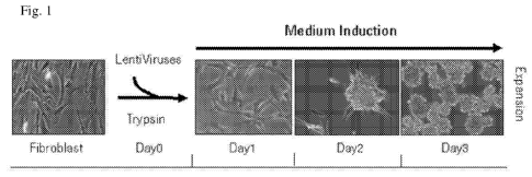
Figure 1: Schematic representation of the method for converting human
fibroblast to
irNSCs. Day 0: human fibroblasts were trypsinized and transfected in a small
volume with a
combination of genes and the nestin GFP reporter using the induction medium
(N2B27 with FGF,
EGF 30 ng/ml; BDNF 20 ng/ml; Fasudil 10 i.t.M, Polybrene 4 ig/m1). Fibroblasts
were plated in
a normal tissue culture plate at a concentration of 10000 ¨ 30000 cells/cm2.
Day 1: Media change
with fresh induction medium. GFP / nestin positive (GFP+) irNSCs started to
appear with very
low frequency (-50 irNSC GFP+ out of 100000). Day 2: The GFP+ irNSCs were
increasing in
number and they started to move together forming cell clusters. Day 3: The
cell clusters are
organized in a clear spheroid structure that lifts off and starts floating as
a GFP+ neurospheres.
The neurospheres bigger than 20 p.m were counted and harvested for further
expansion.
Figure 2: Schematic representation of the human nestin GFP reporter
lentivirus. The
fluorescent protein copGFP and the zeocin selectable marker were cloned under
the expression
control of a 1.8 kb enhancer fragment from the human nestin intron 2 linked to
a minimal CMV
promoter.
Figure 3: irNSCs at day 1 of the reprogramming induction method. Upper panel:
human
untransformed fetal lung fibroblasts IMR90 (phase contrast). Lower panel:
generation of irNSC
GFP+ cells (phase contrast and GFP channel).
Figure 4: irNSCs at day 2 of the reprogramming induction method. The cells
tend to
migrate close together and start to form a spheroid structure with a core of
irNSCs GFP+ (phase
contrast and GFP channel).
Figure 5: irNSCs at day 3 of the reprogramming induction method. The spheroid
structures formed at day 2 are now completely mature appear as neurospheres
floating in the
medium. The neurospheres have a dimension that ranges from 20 - 100 p.m in
diameter with a
high density of cells. The irNSCs are labeled by the nestin GFP expression and
can be
indentified in almost all the neurospheres, although not all the neurospheres
have the same
proportion of irNSC GFP+ (phase contrast and GFP channel).
CA 02807226 2013-01-31
WO 2012/022725 PCT/EP2011/064051
-11-
Figure 6: Number of neurospheres generated with different combinations of
genes.
Figure 7: Attached neurospheres after transduction with Sox2 ¨ Bmil. The
attached
neurospheres show a characteristic morphology of elongated bipolar cells.
Lower panel: higher
magnification of the irNSC GFP+ neuropsheres.
Figure 8: Differentiated cells after 1 week EGF and FGF withdrawal. The irNSCs
upon
withdrawal of the proliferative growth factors give rise to cells with very
thin protrusions stained
positive for the neuronal marker tujl.
Figure 9: Generation of a batch of irNSC neurospheres for expansion and
characterization.
3.6 million human fibroblasts IMR90 were trypsinized and infected in a small
volume with:
Sox2, Bmil, nestin GFP reporter using the induction medium (N2B27 with FGF,
EGF 30 ng/ml
BDNF 20 ng/ml) supplemented with Fasudil 10 i.t.M and Polybrene 4 .t.g/ml.
From Day 4 to Day
8: The GFP+ neurospheres bigger than 50 p.m were harvested and further used
for expansion.
Half of the neurospheres have been expanded using the expansion medium (N2B27
with FGF,
EGF 30 ng/ml BDNF 20 ng/ml) with Fasudil and the other half without Fasudil.
Day15:
Neurospheres grown in the expansion medium with Fasudil have a better
morphology and clear
and sharp borders (a hallmark of well-formed neurospheres, panel B); without
Fasudil the
neurospheres have bleary borders (panel A).
Figure 10: Immunocytochemistry characterization of irNSC neurospheres for the
expression of the NSCs markers Sox2 and Nestin. Day15 irNSC neurospheres
expanded with
Fasudil have been plated on PO/Lam coated plates and after 48h stained for
Sox2 and Nestin
expression. The irNSC neurospheres attached and irNSCs spread from the
spheres. The irNSCs
have a typical NSC morphology and were Sox2 and Nestin positive. Panel A:
Merge and single
channels DAPI, Sox2, Nestin; 20x magnification; Panel B: Merge channels DAPI,
Sox2, Nestin;
10x magnification.
Figure 11: Comparison Fasudil versus Balanol-like-324 compound stimulation to
generate
irNSC neurospheres. Human fibroblasts IMR90 were trypsinized and infected in a
small volume
with: Sox2, Bmil, nestin GFP reporter using the induction medium (NeuroCult
NS-A
Proliferation Kit (Human, StemCells Technologies) with FGF, EGF BDNF 20 ng/ml;
Heparin 2
i.t.g/m1; Polybrene 4 jig/ml) supplemented with Fasudil 10i.tM (hatched graph)
or Balanol-like-
324 compound 24.I.M (black graph). Fibroblasts were plated in a normal tissue
culture plate at a
concentration of 10000 ¨ 30000 cells/cm2. Day 1: Media change with fresh
induction medium.
Day 4: The GFP+ neurospheres bigger than 50 p.m were counted. The Balanol-like-
324 small
compound increased the efficiency of the neurospheres generation approximately
twofold (1.9)
and has a better reproducibility (STDEV, n=3).
WO 2012/022725 CA 02807226 2013-01-31PCT/EP2011/064051
-12-
Figure 12: Pre-treatment of human fibroblasts with Valproic Acid (VPA)
improves the
yield of GFP+ irNSC neurospheres. Human fibroblasts IMR90 were pre-treated for
48 hours
with or without the HDAC inhibitor Valproic Acid (2-propyl-pentanoic acid,
monosodium salt)
(1mM) prior infection with: Sox2, Bmil, nestin GFP reporter. Induction medium
(NeuroCult
NS-A Proliferation Kit (Human, StemCells Technologies) with FGF, EGF BDNF 20
ng/ml;
Heparin 2 iig/m1; Balanol-like-324 204). Day 7: The Neurospheres bigger than
50 p.m were
counted (Panel A) and the average number of GFP+ irNSC per neurosphere is
reported (Panel B).
Representative pictures for the irNSCs neuropheres generated with the VPA pre-
treatment (Panel
C). The VPA pre-treatment did not significantly affect the number of
neurospheres at day 7;
although the VPA treatment increased (2.1 fold) the number of GFP+ irNSCs
(STDEV, n=3).
Figure 13: Defining a minimal pool of genes in combination with Sox2 and Bmil
for
efficient induction of irNSCs neurospheres. Human fibroblasts IMR90 were pre-
treated for 48
hours with VPA (1mM) prior infection with: Sox2, Bmil, nestin GFP reporter
plus different
candidate genes to address their synergism. Induction medium: NeuroCult NS-A
Proliferation
Kit (Human, StemCells Technologies) with FGF, EGF BDNF 20 ng/ml; Heparin 2
jig/ml and
Balanol-like-324 compound 241M. Quantification at Day 7 of irNSCs neurospheres
bigger than
50 p.m. Mashl, Emx2, Foxgl, Pax6 and Soxll synergize with Bmil and Sox2 to
generate irNSC
neuropheres.
Figure 14: Generation of irNSC neurospheres from adult human dermal
fibroblasts
(HDFa). The adult human dermal fibroblasts are provided by the GIBCO (Cat.
Number: C-013-
5C). The adult human dermal fibroblasts were trypsinized and infected in a
small volume with:
Sox2, Bmil, nestin GFP reporter using the induction medium (NeuroCult NS-A
Proliferation
Kit (Human, StemCells Technologies) with FGF, EGF BDNF 20 ng/ml; Heparin 2
jig/ml)
supplemented with Fasudil 10i.tM. Day 8: irNSCs neurospheres are detected
(representative
pictures 2.5 and 10X magnification).
Figure 15: Expansion of irNSC neurospheres using a combination of Ascorbic
Acid, Sonic
Hedgehog (Shh), Jaggedl, DLL4 and FGF8 to obtain a monolayer culture of irNSCs
GFP+.
Human fibroblasts 1MR90 were infected with: Sox2, Bmil, Mashl and nestin GFP
reporter
using the induction medium (NeuroCult NS-A Proliferation Kit (Human,
StemCells
Technologies) with FGF, EGF BDNF 20 ng/ml; Heparin 2 jig/ml; Balanol-like-324
204). Day 7:
The neurospheres bigger than 50 p.m were harvested and further expanded with
the expansion
medium (NeuroCult NS-A Proliferation Kit (Human, StemCells Technologies) with
FGF, EGF
BDNF 20 ng/ml; Heparin 2 jig/ml; Balanol-like-324 24.tM; Ascorbic Acid 0.2mM,
SHH
(Recombinant Human Sonic Hedgehog, Catalog Number: 18455H) 500 ng/ml, FGF8
(Recombinant Human FGF8a Isoform, Catalog Number: 4745F8) 100 ng/ml, DLL4
(Recombinant Human DLL4, Catalog Number: 1506D4) 500 ng/ml, Jaggedl
(Recombinant
CA 02807226 2013-01-31
WO 2012/022725 PCT/EP2011/064051
-13-
Human Jagged 1 Fc Chimera, Catalog Number: 1277JG) 500 ng/ml, conditioned
media 1/10
from the hESC-derived NSCs cultured for two days in NeuroCult NS-A
Proliferation Kit
(Human, StemCells Technologies) with FGF, EGF BDNF 20 ng/ml; Heparin 2
.t.g/ml.
Representative pictures for the irNSCs neuropheres at day14 expanded with
expansion medium
reported above (Panel A). The neuropheres have defined borders and it is
possible to observe the
protrusion of spines from the neurospheres (Panel B, zoom-in). At day 21 the
expanded irNSC
neurospheres were dissociated and plated on PO/Lam coated plates to obtain a
homogenous
monolayer culture of irNSCs GFP+ (Panel C, phase contrast and GFP channel of
the irNSCs
monolayer after 4 days in culture on the monolayer).
Figure 16: Immunocytochemistry characterization of irNSC neurospheres for the
expression of the NSC markers Nestin and the early neuronal marker Tujl. Day21
irNSC
neurospheres generated as described in Figure 15 were dissociated and plated
in NSC self-
renewal conditions (NeuroCult NS-A Proliferation Kit (Human, StemCells
Technologies) with
FGF, EGF BDNF 20 ng/ml; Heparin 2 jig/m1) to test the expression of the Nestin
marker (Panel
A after 48h, all the cells are Nestin+ and Tuj 1-) or plated in
differentiation conditions
(NeuroCult NS-A differentiation Kit (Human, StemCells Technologies) with BDNF
20 ng/ml)
and stained for Tujl and Nestin at day7 (Panel B, all the cells are Tujl+ and
few cells are
Nestin+).
Examples
The method can be illustrated by reference to Figure 1 herein, which depicts a
method
according to the invention being used for converting human fibroblasts to
neural stem cells
(NSCs). In this method human fibroblast were trypsinized at day 0, counted and
their viability
determined. Between 1.0 x 10⁵ ¨ 3.0 x 10⁵ trypsinized fibroblasts were then
resuspended in the
induction medium and the combination of genes delivered as lentiviruses. At
the induction
medium polybrene (hexadimethrine bromide) was added to increase the efficiency
of the
lentiviruses transduction. The infection was performed for 15 minutes in an
eppendorf tube. In
combination with the genes, a human Nestin GFP reporter was used. Nestin is a
well known
marker expressed specifically in NSCs. In the nestin reporter the fluorescent
protein GFP is
under expression of the human nestin promoter (Figure 2), therefore it allows
an easy screen for
induced reprogrammed neural stem cells (irNSCs) GFP+.
The infected cells were plated in tissue culture plates using a concentration
of 10000 ¨
30000 cells/cm2 in the appropriate volume of induction medium. At day 1 the
total induction
medium was renewed. It was possible to identify some irNSC GFP+ (Figure 3)
with a clear
change in morphology compared to the human fibroblasts. The irNSCs GFP+
acquired a bipolar
CA 02807226 2013-01-31
WO 2012/022725 PCT/EP2011/064051
-14-
and elongated morphology with a more condensed cytoplasm, typical of NSCs.
Moreover, the
irNSCs are growing in a packed monolayer culture that resembles the typical
cell-to-cell
interaction acquired in traditional NSC cultures for activating the pro-
proliferative signal of the
Notch pathway. At day 2 the irNSCs GFP+ were in a more mature state and
started to form very
packed clusters of cells. These clusters of irNSCs started to form a spheroid
structure with a
dense core containing irNSCs GFP+ (Figure 4). At day 3 the spheroid structures
were completely
formed and started to lift off from the tissue culture plate floating as
neurospheres in the medium.
The neurospheres have a dimension of approximately 20-100 p.m with clear
borders and a core
with a high density of cells, where it is possible to identify irNSCs GFP+
(Figure 5).
To achieve the inventive breakthrough, it was necessary to use a specific
combination of
genes. The following list of genes involved in the maintenance of the NSC
property in vivo and
in vitro were retrieved from literature knowledge: Sox2 (Sox transcription
factor and important
marker for NSC), Brn2 (POU domain protein known to bind to Sox proteins.
Reported binding
of Sox2 and Brn2 on the nestin promoter. Brn2 KO mice have impairment of CNS
development),
Bmil (Protein involved in the regulation of the cell cycle, reported to
increase expression of the
p21 and p27 inhibitors of the cyclinE/cdk2 complex. CyclinE/cdk2 inhibition
determines the lost
of the retinoblastoma protein control on the cell cycle that results in a fast
cell cycle during the
self-renewal state of NSCs), Mashl (described to be an important regulator for
the proliferation
of neural precursors in vivo), Sox 11 (Sox protein reported to be expressed in
SGZ in vivo),
NCam (NSC marker in Flow Cytometry and expressed in different regions of the
CNS), Kpnal
(better known as importin alpha5 responsible together with importin beta for
the protein nuclear
import in ectoderm derived tissues).
All genes were cloned as cDNAs into lentiviruses plasmids, and subsequently
packaged
into lentiviruses. The lentiviruses packaged particles for Sox2, Bmil, Mashl,
Soxll, NCam,
Kpnal, nestin GFP reporter were transduced directly into human fibroblasts.
Different
combinations of genes were tested in the method described above. At day 3 it
was possible to
evaluate the success of the production of the irNSCs by counting the
neurospheres generated.
Only neurospheres bigger than 50 p.m were taken into account.
As represented in Figure 6 the transduction of the nestin reporter lentivirus
without
addition of Fasudil to the induction medium did not reprogram the human
fibroblasts to irNSCs.
With the addition of Fasudil to the induction medium the generation of
neurospheres (around
50i.tm) and some smaller (around 20 p.m, not counted) were reported.
Neurospheres generated with our innovative method using the genes combination:
Sox2 ¨
Bmil were harvested at day 3 and expanded for further 14 days. Expansion of
the irNSCs
neurospheres was a critical step. The neurospheres were cultured using the
N2B27 medium
WO 2012/022725 CA 02807226 2013-01-31PCT/EP2011/064051
-15-
supplemented with FGF, EGF, BDNF in special ultra-low non adherent plates
(Corning). In
order to achieve a homogenous population of irNSCs GFP+ neurospheres a
cleaning procedure
every 2-3 days was applied. During 14 days of expansion some neurospheres with
low density of
irNSCs GFP+ were not able to proliferate properly, most probably due to a
contamination by not
converted fibroblasts. Such kind of contaminated neurospheres were fallen
apart in single cells
that needed to be removed. At day 14, the neurospheres were tested for:
attachment on poly-
ornithine/laminin coated plates and generation of neuronal-like cells. For the
attachment, 20-40
neurospheres/cm2 were plated on poly-ornithine/laminin coated plates in the
expansion medium
supplemented just for the first day with Fasudil 10 i.t.M, in order to improve
cell attachment and
spreading. At day 1 of culture was possible to show the attachment and
spreading of the
neurospheres (Figure 7). At the centre of the spreading neurospheres we
identified irNSCs GFP+
with a typical NSC morphology. The neurospheres were grown for additional
three days and
then just BDNF was added to the N2B27 (neuronal differentiating conditions).
Upon EGF and
FGF withdrawal the irNSCs changed morphology. They became more elongated and
started to
form neurite-like cellular protrusions. At day 7 of the differentiating
conditions cells were fixed
and stained for the neuronal marker tujl (Figure 8).
Neurospheres expanded with Fasudil have a better morphology and clear and
sharp borders
(a hallmark of well-formed neurospheres, Figure 9, panel B); without Fasudil
the neurospheres
have bleary borders (Figure 9, panel A). The irNSCs have a typical NSC
morphology and were
Sox2 and Nestin positive (Figure 10).
Figure 11 shows that Rock kinase inhibitor Balanol-like-324 compound increases
the yield
of GFP+ neurospheres.
These evidences show that the method was able to convert human fibroblasts to
irNSCs
based on linked steps of genes transduction (best combinations: Sox2-Bmil,
Sox2-Bmil-Mashl,
Sox2-Bmil-Sox11, Sox2-Bmil-Emx2, Sox2-Bmil-Foxg1 and Sox2-Bmil-Pax6 , see also
Figure
13) and chemically defined medium induction.
To increase the yield of irNSCs, human fibroblasts were pretreated with or
without the
HDAC inhibitor Valproic Acid (VPA, 2-propyl-pentanoic acid, monosodium salt).
Towards this
end, the human fibroblasts were incubated in DMEM/F12 supplemented with FBS
10% and L-
glutamine supplemented with 1mM VPA prior to infection (Figure 12).
Figure 14 shows the generation of irNSC Neurospheres from adult human dermal
fibroblasts (HDFa).
Figure 15 shows the expansion irNSC Neurospheres using a combination of
Ascorbic Acid,
Sonic Hedgehog (Shh), Jaggedl, DLL4 and FGF8. Figure 16 shows
Immunocytochemistry
WO 2012/022725 CA 02807226 2013-01-31PCT/EP2011/064051
-16-
characterization of irNSC Neurospheres for the expression of the NSCs markers
Nestin and the
early neuronal marker Tujl.
Materials and Methods
Cell Culture:
Induction Medium: N2B27 (N2B27 is a 1:1 mixture of DMEM/F12 (Gibco, Paisley,
UK)
supplemented with N2 and B27 (both from Gibco) supplemented with human EGF
(Peprotech)
30 ng/ml, human FGF2 3Ong/m1 (Peprotech), human BDNF (Roche) 20 ng/ml and
Fasudil
(Calbiochem) 10 i.t.M or Balanol-like-324 compound (N-1(3R,4R)-444-(2-Fluoro-6-
hydroxy-3-
methoxy-benzoy1)-benzoylamino] -azepan-3 - yl } -4-hydroxy-3 ,5-dimethyl-benz
amide) 241M.
Expansion Medium: N2B27 supplemented with human EGF (Peprotech) 30 ng/ml,
human
FGF2 30 ng/ml (Peprotech), human BDNF (Roche) 20 ng/ml, or
NeuroCult NS-A Proliferation Kit (Human, StemCells Technologies) with FGF,
EGF
BDNF 20 ng/ml; Heparin 2 i.t.g/m1; Balanol-like-324 24.tM; Ascorbic Acid
0.2mM, SHH
(Recombinant Human Sonic Hedgehog, Catalog Number: 18455H) 50Ong/ml, FGF8
(Recombinant Human FGF8a Isoform, Catalog Number: 4745F8) 10Ong/ml, DLL4
(Recombinant Human DLL4, Catalog Number: 1506D4) 50Ong/ml, Jaggedl
(Recombinant
Human Jagged 1 Fc Chimera, Catalog Number: 1277JG) 50Ong/ml.
Differentiation Medium: N2B27 supplemented with human BDNF (Roche) 20 ng/ml,
Laminin 2 i.t.g/m1 (Invitrogen).
Human fibroblasts: IMR90 foetal lung fibroblasts (ATCC Lot . Num. 580229699)
or adult
human dermal fibroblasts (GIBCO, Cat. Number: C-013-5C).
Lentiviruses: Prepackaged, ready-to use lentivirus particles were obtained
from Sigma
(Stemgent Reprogramming Lentivirus human Sox2, Catalog No. 5T070012),
Genecopeia
(human Bmil Lentifect Lentiviral Particles, Catalog Nr. LP-B0015-Lv105; Soxll
Lentifect
Lentiviral Particles, Catalog Nr. LP-M0425-LV105; Mashl Lentifect Lentiviral
Particles,
Catalog Nr. LP-Z0740-LV105; human KpnalLentifect Lentiviral Particles, Catalog
Nr. LP-
U1286-Lv105; NCaml Lentifect Lentiviral Particles, Catalog Nr. LP-Z2645-Lv105)
and SBI
Systems Biosciences (Nestin GFP Reporter: pGreenZeoTm-hNestin Transcriptional
Reporter
Virus, SR10035VA-1)
WO 2012/022725 CA 02807226 2013-01-31
PCT/EP2011/064051
-17-
Titers NestinGFP 1.45*10 5/0, BMI1 4.3*10 5/0, Sox2 1.07*10 4/0, Soxl 1 3.2*10
6/0,
Mashl 4.7*10 6/0, NCam 3.3*10 4/0, Kpnal 1.8*10 5/0.
Protocols:
1. Generation of the irNSCs:
- 200.000 IMR90 human fibroblasts infected with the lentiviruses for different
genes
combination (multiplicity of infection (M.O.I.) used for each single
lentivirus 30) and the
reporter nestin GFP lentivirus (M.O.I. used 10) in an eppendorf with 300 ill
induction medium
with polybrene (hexadimethrine bromide, Sigma) 4 jig/ml.
- Incubate at room temperature for 15 min.
- Plate the 300 ill in 1.7 ml induction medium in a well of a 6-well-plate
tissue treated
- Day 1, renew the 2 ml of induction medium/each well
- Day 3, harvest of the neurospheres collecting carefully the 2 ml with the
floating spheres
- Expand the neurospheres2. Expansion of the neurospheres:
- Collect the medium with the floating neurospheres in 15 ml tubes from 3
wells of a 6-
well-plate
- Let the spheres seed down for 10 min
- Remove the supernatant very carefully (single cells will not seed down and
are aspirated
with the supernatant)
- Resuspend the spheres in 4 ml final volume expansion medium
- Plate in a B6 plate ultra low adherent plate (Corning)
- Incubate 2-3 days
- Repeat the expansion procedure every 2-3 days till day 14 from the
generation irNSCs
WO 2012/022725 CA 02807226 2013-01-31
PCT/EP2011/064051
-18-
3. Differentiation neurospheres:
- Day 14 generation of the irNSCs and after the expansion procedure select
under the
stereo microscope round neurospheres with clear borders and rich in irNSCs
GFP+
- Plate 40 spheres in a well 24-well-plate previously coated with poly-
ornhitine/laminin
using the expansion medium with addiction of Fasudil 10i.tM or Balanol-like-
324
compound 241M.
- The day after renew the expansion medium without Fasudil / Balanol-like-324
compound
241M.
- Incubate for three days
- Remove expansion medium and add differentiating medium
- Renew differentiating medium after 3 - 4 days
- Incubate for 3 ¨ 4 days
- Fix cells with PFA 4% and perform immunostainings
Protocol staining irNSCs Neurospheres: Neurospheres at day 15 were stained for
characterization with the following antibodies: Mouse Nestin and Rabbit Sox2
0/N and then the
secondary anti-mouse 488 and anti-rabbit 555 for one hour.
Primary antibodies:
Nestin Mouse, monoclonal, 1/500 dilution (MAB5326 Millipore)
Sox2 Rabbit, polyclonal, 1/500 dilution (AB5603MI Millipore)
Secondary antibodies:
Alexa fluor 488,IgG, 1/1 000 dilution, Goat anti mouse (A11029 Invitrogen)
Alexa fluor 555,IgG 1/1 000 dilution, Goat anti rabbit (A21429 Invitrogen)