Note: Descriptions are shown in the official language in which they were submitted.
Devices For Harvesting Skin Grafts
Field of the Invention
The present invention generally relates to methods for applying a skin graft.
Background
Skin is the largest organ of the human body, representing approximately 16% of
a
person's total body weight. Because it interfaces with the environment, skin
has an important
function in body defense, acting as an anatomical barrier from pathogens and
other
environmental substances. Skin also provides a semi-permeable barrier that
prevents
excessive fluid loss while ensuring that essential nutrients are not washed
out of the body.
Other functions of skin include insulation, temperature regulation, and
sensation. Skin tissue
may be subject to many forms of damage, including bums, trauma, disease, and
depigmentation (e.g., vitiligo).
Skin grafts are often used to repair such skin damage. Skin grafting is a
surgical
procedure in which a section of skin is removed from one area of a person's
body (autograft),
removed from another human source (allograft), or removed from another animal
(xenograft),
and transplanted to a recipient site of a patient, such as a wound site. As
with any surgical
procedure, skin grafting includes certain risks. Complications may include:
graft failure;
rejection of the skin graft; infections at donor or recipient sites; or
autograft donor sites oozing
fluid and blood as they heal. Certain of these complications (e.g., graft
failure and rejection
of the skin graft) may be mitigated by using an autograft instead of an
allograft or a xenograft.
One of the causes of graft failure is that a skin graft is applied to a
recipient site having
an improper orientation, i.e., the graft is applied such that the stratum
corneum layer of the
graft contacts the recipient site instead of the basal layer of the graft.
This is a particular
problem with an epidermal graft, because an epidermal graft has no blood
vessels; thus, it
must receive nutrients by diffusion from the underlying dermis through the
basement
membrane. A graft
CA 2807413 2019-05-17
CA 02807413 2013-02-01
WO 2012/019096 PCT/US2011/046739
applied with an improper orientation will not receive nutrients and the cells
of the graft will die,
leading to graft failure.
Summary
The present invention provides methods that allow for proper orientation of a
skin graft
on a recipient site. In particular, methods of the invention allow application
of a graft to a
recipient site in an orientation that allows preferred interaction between the
basal layer of the
graft and the donor site. In a particular embodiment, a graft or grafts are
prepared and applied to
a recipient site such that the proper orientation is preserved. This results
in the graft being
applied in an orientation that is closest to the natural orientation of the
skin. Preferred grafts
comprise all or substantially all epidermal layer, but grafts that have some
dermal layer
component also benefit from methods described herein. According to the
invention an epidermal
graft or an epidermal layer may comprise only or substantially only the
epidermal layer (i.e., the
graft may include some portion of dermal material). Methods of the invention
decrease graft
failure, and are particularly useful in preparing and applying epidermal
grafts.
Brief Description of the Drawings
Figure 1 is a drawing showing the anatomy of skin.
Figure 2 panels A-C are schematics showing a device for generating and
harvesting a
plurality of micrografts. Panel A provides an exploded view of the device.
Panel B provides a
top view of the assembled device. Panel C provides a side view of the
assembled device.
Figure 3 provides a schematic of an exemplary process for preparing a skin
graft
according to methods of the invention. Panel A shows an excised epidermal
blister sitting on a
sterile cutting surface with a sterile cutter tool above. Panel B shows the
cutter tool cutting the
epidermal blister to generate an array of micrografts. Panel C shows the array
of micrografts that
has been produced by the cutting tool sitting on a first substrate. Panel D
shows the first
substrate placed into an expansion device. A second substrate is placed into
the assembly cap
above. Panel E shows the expansion process. As the first substrate expands,
the micrografts
move apart. Panel F shows that as the first substrate flattens against the
assembly cap, the
micrografts are transferred to the second substrate. Panel G shows the
completed expansion
process and that the micrografts have been transferred to the second
substrate. Panel H shows
2
CA 02807413 2013-02-01
WO 2012/019096 PCT/US2011/046739
removal of the assembly cap having the second substrate and expanded
micrografts from the
expansion device. Panel I shows removal of the second substrate having the
expanded
micrografts from the assembly cap of the expansion device.
Figure 4 panels A-B are drawings showing a device of the invention for raising
a suction
blister.
Figure 5 panels A-D show different devices of the invention for raising a
suction blister.
Figure 6 is a process chart showing steps for treating vitiligo using methods
of the
invention.
Detailed Description
The skin consists of 2 layers. The outer layer, or epidermis, is derived from
ectoderm,
and the thicker inner layer, or dermis, is derived from mesoderm. The
epidermis constitutes
about 5% of the skin, and the remaining 95% is dermis. Figure 1 provides a
diagram showing
the anatomy of skin. The skin varies in thickness depending on anatomic
location, gender, and
age of the individual. The epidermis, the more external of the two layers, is
a stratified
squamous epithelium consisting primarily of melanocytes and keratinocytes in
progressive stages
of differentiation from deeper to more superficial layers. The epidermis has
no blood vessels;
thus, it must receive nutrients by diffusion from the underlying dermis
through the basement
membrane, which separates the 2 layers.
The dermis is a more complex structure. It is composed of 2 layers, the more
superficial
papillary dermis and the deeper reticular dermis. The papillary dermis is
thinner, including loose
connective tissue that contains capillaries, elastic fibers, reticular fibers,
and some collagen. The
reticular dermis includes a thicker layer of dense connective tissue
containing larger blood
vessels, closely interlaced elastic fibers, and coarse, branching collagen
fibers arranged in layers
parallel to the surface. The reticular layer also contains fibroblasts, mast
cells, nerve endings,
lymphatics, and some epidermal appendages. Surrounding the components of the
dermis is the
gel-like ground substance composed of mucopolysaccharides (primarily
hyaluronic acid),
chondroitin sulfates, and glycoproteins.
Methods of the invention are directed to preparing and applying skin grafts
such that the
basal layer of the graft is in direct contact with the recipient site to which
the graft is being
applied. Maintaining the proper (i.e., natural) orientation of the graft
increases the chance that
3
CA 02807413 2013-02-01
WO 2012/019096 PCT/US2011/046739
the graft will survive and be accepted at the recipient site. According to the
invention, graft
orientation can be maintained in any manner that is desirable. For example,
one can harvest a
graft or grafts on a first substrate in which the basal layer of the graft(s)
is oriented toward the
substrate. The graft is then transferred to a second substrate, either with or
without stretching on
the first substrate, such that the basal layer is exposed for direct
application to the recipient site.
In the case in which multiple grafts or an array of grafts is used,
orientation is maintained in the
same manner, with the result being that the majority of grafts will be
oriented properly for
application to the recipient site.
In other embodiments, orientation is maintained mechanically using a single
substrate for
application to the recipient site. Thus, a graft is harvested as described
below and placed on a
substrate with the basal layer being exposed for application to a recipient
site. Other methods of
preserving orientation will be apparent to the skilled artisan based upon the
description below.
In certain embodiments, grafts are applied directly to a recipient site in
proper orientation
with out the use of culturing or application of biologics or other active
agents (e.g., antibiotics,
growth factors, etc).
In certain embodiments, methods of the invention involve harvesting a
plurality of skin
grafts from a subject. applying the grafts to a first substrate, stretching
the first substrate, and
transferring the grafts from the first substrate to a second substrate for
application to a patient
recipient site.
Harvesting of the skin grafts may be accomplished by any technique known in
the art,
and the technique employed will depend on the type of graft required (e.g.,
epidermal graft, split
thickness graft, or full thickness graft). An epidermal graft refers to a
graft that consists of
substantially epidermal skin and does not include any substantial portion of
the dermal layer. A
split thickness graft refers to a graft that includes sheets of superficial
(epithelial) and some deep
layers (dermal) of skin. A full-thickness graft refers to a graft that
includes all of the layers of
the skin including blood vessels.
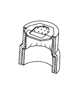
In certain embodiments, a device as shown in Figure 2 panels A-C is used to
obtain the
plurality of skin grafts. Device 200 includes a frame 201 and a lid 202.
Fitted into the frame is
a bottom plate 203, a cutter grid plate 204, a cutter plate 205, and a top
plate 206. The bottom
plate 203, the cutter plate 205, and the top plate 206, each include a hole
array 211. Once
assembled, the hole array 211 of each of plates 203, 205, and 206 are aligned.
The size of the
4
CA 02807413 2013-02-01
WO 2012/019096 PCT/US2011/046739
holes in the hole array will depend on the size of the graft needed, with
larger holes being used to
produce larger grafts. A first substrate 207 interacts with the top plate 206
and will receive the
harvested grafts.
Device 200 further includes an actuation block 208, actuation bar 209, and
actuation
block guides 210. Actuation components 208, 209, and 210 control movement of
the cutter plate
205. The frame 201 includes a vacuum stop 212 and the lid 202 includes a
suction hole barb
213. Once assembled, the frame 201 and lid 202 are arranged such that the
vacuum stop 212 and
the suction hole barb 213 are aligned with each other (Figure 1 panel B). A
vacuum source is
then connected to the device 200 such that negative pressure can be generated
within the device.
The device 200 can be held together by clamp screws 214. Device 200 may also
include a
heating element.
To produce and harvest the plurality of skin grafts, device 200 is placed on a
donor site,
such as an inner thigh of a patient. The vacuum source is turned on, producing
negative pressure
within device 200. The negative pressure causes the skin to be pulled toward
lid 202, with a
plurality of different portions of skin being pulled through each hole array
211 in each of plates
203, 205, and 206. Such action results in generation of many microblisters.
The blisters may or
may not be fluid-filled. Any type of raised blister may be used with methods
of the invention.
Once the microblisters are raised, actuation components 208, 209, and 210 are
engaged to
move cutter plate 205. The movement of cutter plate 205 disrupts the alignment
of the hole
arrays 211 in each of plates 203, 205, and 206, and results in cutting of the
microblisters. The
cut microblisters are captured on the first substrate 207 that is above top
plate 206. In this
manner, there is provided a spaced apart array of micrografts. The amount of
negative pressure
applied, the amount of time the vacuum is maintained, and/or the depth of the
holes above the
cutting surface (plate 206) determines what type of graft will be harvested,
e.g., epidermal graft,
split thickness graft, or full thickness graft. Generally, each micrograft
will have a lateral
dimension of less than about 2 mm e.g., 100 to 2000 microns.
Once the grafts have been harvested and applied to the first substrate, the
first substrate is
stretched or expanded, resulting in increased distance between the individual
micrografts,
moving them apart and resulting in production of a skin graft that can repair
a recipient site that
is larger than the donor site from which the grafts were obtained. In methods
of the invention,
the individual grafts themselves are not expanded, i.e., the graft tissue is
not stretched; rather,
stretching of the substrate increases the space or distance between each
individual micrograft.
Methods of the invention thus minimize tissue manipulation.
The purpose of such processing is to use tissue from a donor site to cover a
wound area
that is larger than the donor site. The stretching of the substrate may be
done manually, i.e., by
hand, or may be done with the help of a machine. The stretching may be
substantially uniform in
all directions or may be biased in a certain direction. In a particular
embodiment, the stretching is
substantially uniforin in all directions. Stretching of the substrate may be
performed
mechanically or may be accomplished by application of a pressurized fluid or
gas. In certain
embodiments, air pressure is used to expand the first substrate. Exemplary
devices and methods
are described in Korman (U.S. 5,914,264).
Any minimum distance can be provided between micrografts after the first
substrate is
stretched. The amount of stretching can be large enough to provide a
sufficiently large area of
substrate containing micrografts to allow a larger area of damaged tissue to
be repaired using a
particular amount of graft tissue removed from the donor site, i.e., the area
of the stretched first
substrate containing the separated micrografts can be much larger than the
total area of the donor
site. For example, the distance between adjacent micrografts on the stretched
first substrate can
be greater than about 0.5 mm, although small separation distances may also be
used. For
repigmentation of skin tissue, an amount of stretching can be applied to the
first substrate such
that the distance between adjacent micrografts is less than about 4 mm,
because it is known that
melanocytes, when grafted to a depigmented region, can migrate up to about 2
mm from each
micrograft to repigment regions between the micrografts. This average distance
can be larger if
keratinocyte migration is involved with the tissue being treated because
keratinocytes typically
migrate greater distances compared to melanocytes.
The ratio of the wound area to the donor site area is referred to as the
expansion ratio. A
higher expansion ratio is desirable to minimize the trauma of the donor site,
and to aid patients
who have only a small amount of tissue available for grafting purposes. The
amount of area
expansion, e.g., the ratio of an area of damaged tissue that can be repaired
compared to an area of
graft tissue removed from a donor site, may be 500x or more. In particular
embodiments, the
area of expansion may be from about 10x to about 100x, which provides a more
uniform
coverage and/or repigmentation of the recipient site. For repairing burns or
ulcerated tissue, the
6
CA 2807413 2018-03-05
micrografts may be smaller than those used to repair other types of damaged
tissue, and thus the
distances between adjacent micrografts may be greater after stretching of the
first substrate. In such an
exemplary application, an area expansion of about 1000x or more may be used.
In other embodiments and depending on the material of the first substrate,
maintaining the first
substrate in a stretched configuration may result in stress on the substrate
that is not optimal.
Additionally, the stretched first substrate may not retain the same properties
as the unstretched
configuration of the first substrate, i.e., technological characteristics,
such as physical, environmental
and performance characteristics could be affected by the stretching of the
substrate. Additionally,
methods used to maintain the substrate in its stretched condition may be
physically cumbersome and
prevent uniform application of the micrografts to uneven skin surfaces. Thus
in certain embodiments,
once the first substrate has been stretched, the spaced apart micrografts are
transferred to a second
substrate. By transferring the micrografts to a second substrate, methods of
the invention minimize
manipulation and stress of the substrate that holds the graft to the recipient
site.
After stretching the first substrate, the second substrate is brought into
contact with the grafts
on the stretched first substrate. Transfer is facilitated by the second
substrate having greater affinity or
more adhesive force toward the micrografts than the first substrate. In
certain embodiments, the
second substrate is coated with a hydrocolloid gel. In other embodiments, the
first substrate is wetted
with a fluid such as water or a saline solution. Wetting the micrografts and
the first substrate provides
lubrication between the grafts and the first substrate and allows for easy
transfer of the grafts from the
first substrate to the second substrate. After wetting the first substrate,
the grafts have greater affinity
for the second substrate than the first substrate. The wetted first substrate
is then removed from the
second substrate and the grafts remain attached to the second substrate. The
distance between the
micrografts is maintained after transfer of the micrografts from the stretched
first substrate to the
second substrate.
The first substrate may be made from any material that is biocompatible and
capable of being
stretched upon application of a moderate tensile force. The second substrate
may be made from any
material known in the art that is compatible with biological tissue. The
second substrate may also be
capable of being stretched upon application of a moderate tensile force.
Exemplary materials for the
first and/or second substrates include medical dressings, such as TEGADERMTm
(medical dressing,
commercially available from 3M, St. Paul, MN) or
7
CA 2807413 2019-05-17
DUODERMTm (medical dressing, commercially available from 3M, St. Paul, MN).
The first and/or
second substrates may also be gas permeable.
In certain embodiments, the first and/or second substrates include an adhesive
on one side that
facilitates attachment of the grafts to the substrates. The substrate material
may have intrinsic
adhesive properties, or alternatively, a side of the substrate may be treated
with an adhesive material,
e.g., an adhesive spray. In certain embodiments, the first and second
substrates are the same material.
In other embodiments, the first and second substrates are different materials.
In certain embodiments,
the materials of the first and second substrates are chosen to facilitate
transfer of the micrografis from
the first substrate to the second substrate. For example, in certain
embodiments, the material chosen
for the first substrate has a weaker adhesive than the material chosen for the
second substrate.
In certain embodiments, the material of the first substrate is a deformable
non-resilient
material. A deformable non-resilient material refers to a material that may be
manipulated, e.g.,
stretched or expanded, from a first configuration to a second configuration,
and once in the second
configuration, there is no residual stress on the substrate. Such materials
may be stretched to an
expanded configuration without returning to their original size, and thus in
these embodiments it is not
necessary to transfer the micrografts from a first substrate to a second
substrate. Instead, the expanded
first substrate including the micrografts is applied to a recipient site.
Such deformable non-resilient materials tend to be soft, stiff or both soft
and stiff. Softness is
measured on the durometer scale. An example of such a material is a soft
polyurethane. A soft
polyurethane is produced is as follows. Polyurethanes in general usually have
soft and hard segments.
The hard segments are due to the presence of phenyl bridges. In a soft
polyurethane, the phenyl bridge
is switched out for an aliphatic, which is more flexible as its 6 carbon ring
has no double bonds.
Therefore, all the segments are soft. On the Durometer Scale, a soft
polyethylene is rated about Shore
80A. Other materials suitable for use with methods of the invention include
low density polyethylene,
linear low density polyethylene, polyester copolymers, polyamide copolymers,
and certain silicones.
In these embodiments, the expanded first substrate having the micrografts
retains its expanded position
without any residual stress, and the expanded first substrate is applied to a
recipient site.
8
CA 2807413 2019-05-17
CA 02807413 2013-02-01
WO 2012/019096 PCT/US2011/046739
Ultimately, the grafts and substrate are applied to a recipient of site of a
patient. Prior to
applying the grafts to the recipient site, the site is prepared to receive the
grafts using any
technique known in the art. Necrotic, fibrotic or avascular tissue should be
removed. The
technique used to prepare the site will depend on damage to the recipient
site. For example,
epidermal tissue, if present at the recipient site, can be removed to prepare
the area for receiving
the micrografts. Burned or ulcerated sites may not need removal of epidermal
tissue, although
some cleaning of the site or other preparation of the site may be performed.
Wounds should be
debrided and then allowed to granulate for several days prior to applying the
graft. Most of the
granulation tissue should be removed since it has a tendency to harbor
bacteria. Applying silver
sulfadiazine to the wound for 10 days prior to grafting reduces the bacterial
count greatly.
The size of the area at the recipient site can be about the same size as the
area of the
stretched first substrate having micrografts adhered thereto. This size
generally will be greater
than the area of the original graft tissue that was removed from the donor
site to form the
micrografts. The depigmented or damaged skin can be dermabraded with sandpaper
or another
rough material. Alternatively, the epidermal tissue can be removed from the
recipient site by
forming one ore more blisters over the area to be treated, e.g., a suction
blister or a freezing
blister, and the raised epidermal blister tissue can then be removed by
cutting or another
procedure.
The substrate having the micrografts can be placed over the area to be treated
to form a
dressing. A portion of the substrate having the micrografts can be positioned
over the area to be
repaired, e.g., the area from which the epidermal tissue has been abraded or
removed for
repigmentation. The substrate can be fixed in place over the treatment area,
e.g., using tape or
the like. The substrate can be removed after sufficient time has elapsed to
allow attachment and
growth of the micrografts in the treatment area, e.g., several days to a few
weeks.
Another aspect of the invention provides harvesting a single graft from a
donor site, such
as an epidermal graft, generating an array of micrografts from the single
graft, placing the graft
on a first substrate, expanding a distance between the micrografts on a first
substrate, transferring
the micrografts from the first substrate to a second substrate, and applying
the micrografts to a
recipient site. Figure 3 provides a schematic of an exemplary process for
preparing a skin graft
according to methods of the invention.
9
Methods of the invention involve harvesting a single graft from a donor site,
such as an
epidermal graft. Harvesting of the skin grafts may be accomplished by any
technique known in
the art, and the technique employed will depend on the type of graft required
(e.g., epidermal
graft, split thickness graft, or full thickness graft). In certain
embodiments, harvesting a skin graft
involves raising a blister and cutting the blister. In certain embodiments,
the blister may be a
fluid-filled blister (e.g. a suction blister). In other embodiments, the
blister is not fluid-filled.
Any type of raised blister may be used with methods of the invention.
In certain embodiments, suction blister grafting is used. Suction blister
grafting involves
raising a blister, and then cutting off the raised blister. An exemplary
suction blister grafting
technique is shown in Awad, (Dermatol Surg, 34(9):1186-1193, 2008). This
article also shows
various devices used to form suction blisters. A suction blister device is
also described in
Kennedy et al. (U.S. 6,071,247). An exemplary device is commercially available
from Electronic
Diversities (Finksburg, MD).
A device for raising a suction blister typically operates by use of suction
chambers that
are attached to a patient's skin. An instrument typically contains a power
source, a vacuum
pump, temperature controls and all related controls to operate multiple
suction chambers. The
suction chambers are connected to the console by a flexible connection. Each
of the chambers is
controlled by a preset temperature control to provide an optimal skin warming
temperature. Both
chambers share an adjustable common vacuum source that affects all chambers
equally.
Blister formation is accomplished by attaching the suction blister device to a
patient's
skin. Typically hook & loop fastener straps are used to keep the device in
place. The chamber
heating system provides a slight warming of an orifice plate of the device,
which is in direct
contact with the patient's skin surface. The application of a moderate
negative pressure from
the instrument console, to the chamber interior, causes the patients skin to
be gently drawn
through the opening(s) in the orifice plate. The results are typical suction
blisters, approximately
the size of the opening(s) in the orifice plate. The skin and blister area is
generally not damaged
and patient discomfort is minimal.
The negative pressure chamber is fabricated of mostly plastic components, with
two
removable threaded caps. The upper cap is fitted with a clear viewing lens so
that the actual
blister formation can be observed. The opposite end of the chamber is fitted
with a removable
CA 2807413 2018-03-05
CA 02807413 2013-02-01
WO 2012/019096 PCT/US2011/046739
orifice plate that is placed on the patient's skin. Since this plate is simply
threaded onto the
chamber end, multiple plates with different opening patterns can be
interchanged as desired.
The interior of the device is warmed and illuminated by an array of low
voltage
incandescent lamps. This lamp array is controlled from the instrument console
temperature
controller, cycling as needed, to maintain the set point temperature. The heat
from these lamps is
radiated and conducted to the orifice plate, which then warms the patient's
skin. The chamber is
connected to the console via a composite vacuum and low voltage electrical
system. Quick
connections are used for the vacuum and electrical system to facilitate
removal and storage.
The Negative Pressure Instrument console is a self-contained fan cooled unit
which is
designed to operate on 120 VAC 60 Hz power. Vacuum is supplied by an
industrial quality
diaphragm type vacuum pump, capable of a typical vacuum of 20 in Hg (0-65 kpa)
at 0 CFM.
An analog controller that is preset to 40 C provides the temperature control
for each suction
chamber. This provides accurate control of the orifice plate temperature. The
instrument console
has internal adjustments that allow the user to recalibrate the temperature
setting if desired. Other
temperatures can be preset if desired. The front panel includes a vacuum gauge
and vacuum
bleeder adjustment to regulate the vacuum to both chambers. The console front
panel also
contains the connections for the chamber assemblies.
Once the suction blister is raised, it is cut by methods known in the art (see
e.g., Awad,
Dermatol Surg, 34(9):1186-1193, 2008), and placed on the first substrate. Once
on the first
substrate, an array of micrografts are generated from the single graft. Figure
3 panel A shows an
excised skin graft on a first substrate, with a sterile cutting tool above the
graft. In certain
embodiments, rather than being applied directly to the first substrate, the
cut blister is placed
onto a sterile surface, such as a glass slide, and the array of micrografts is
generated on the sterile
surface prior to transfer to the first substrate. In other embodiments, the
cut blister is trapped
between two aligned metal screens. The screens are pushed together to cut the
blister into an
array of micrografts. The micrografts are then pushed out of the screens and
deposited onto the
first substrate using an array of pushers whose size and spacing correspond to
the metal screens.
In certain embodiments, the cut blister is harvested directly between the two
screens for
generation of the array of micrografts.
In other embodiments, the cut blister is harvested directly into a shear or
punch and die
device for generation of micrografts. A shear or punch die includes an array
of flat-faced piston-
11
CA 02807413 2013-02-01
WO 2012/019096 PCT/US2011/046739
like components that fit closely into the openings in a metal screen/mesh. In
this embodiment,
the cut graft is harvested onto the array of pistons, and sits between the
array of pistons and the
screen/mesh. The screen/mesh is closed over the cut blister and force is
applied to the array of
pistons. The pistons push through the holes in the screen/mesh and in the
process, portions of
tissue are punched out from the openings of the screen/mesh and deposited on a
substrate,
producing an array of micrografts on a substrate. Such embodiments allow for
simultaneous
generation of the array of micrografts and deposition of the array of
micrografts onto the
substrate.
The array of micrografts can be generated by making cuts or using other
protocols to
form the array of micrografts from the single graft. The cuts may pass
partially or completely
through the graft tissue. For example, for repigmenting skin tissue, the
micrografts used may
have a presence of melanocytes. Accordingly, a lateral dimension of such
micrografts can be
between less than about 1 mm, e.g., 200 to 1000 microns. Other exemplary sizes
are between
400 and 800 microns. The area of the micrografts can be between about 0.04 mm2
and about 1
mm2. The exemplary sizes can provide micrografts large enough such that each
micrograft is
likely to contain some melanocytes, yet small enough to provide a large number
of micrografts
from a particular piece of graft tissue, which can facilitate a significant
degree of expansion on
the graft site.
For treating bums or ulcers, where presence and proliferation of keratinocytes
is
important, the micrograft sizes may be smaller. For example, a lateral
dimension of micrografts
containing keratinocytes can be between about 50 microns and about 1000
microns, or between
100 microns and about 800 microns. The area of such micrografts can be between
about 0.0025
mm2 and about 1 mm2. The exemplary size ranges provide micrografts large
enough to contain
viable and undamaged keratinocytes, and small enough to facilitate repair of a
larger area of
damaged skin.
Figure 3 panel B shows an exemplary cutting tool. The cutting tool may be
configured in
any manner, and such configuration will depend upon the size of the
micrografts to be produced
and the desired array pattern. The cutting tool includes a plurality of
adjacent blades. The
arrangement of the blades will depend upon the desired pattern for the array
of micrografts. The
tool shown in Figure 3 panel B is configured to produce a square grid of
micrografts (See Figure
3 panel C). The spacing of the blades in the cutting tool will depend on the
desired size of the
12
CA 02807413 2013-02-01
WO 2012/019096 PCT/US2011/046739
micrografts. For example, the blades may be spaced about 100 to 2000 microns
apart, or about
500 to 1000 microns apart. The cutting tool is pressed at least once into the
skin graft on the first
substrate to produce the array of micrografts (See Figure 3 panels B and C).
Other exemplary devices for producing an array of micrografts include mesh
devices.
Such mesh devices include rigid, biocompatible material, such as stainless
steel. The mesh
includes a plurality of openings. The openings are sized to provide an array
of micrografts of a
desired size, such as lateral sizes between about 100 microns and about 1000
microns or about
300 microns to about 500 microns. Similar to the cutting tool described above,
the mesh is
pressed at least once into the skin graft to produce the array of micrografts.
Figure 3 panels D-I show remaining steps of the method. Once the array of
micrografts
are on the first substrate, the distance between the micrografts is expanded.
Expansion results in
increased distance between the individual micrografts, moving them apart and
resulting in
production of a skin graft that can repair a recipient site that is larger
than the donor site from
which the grafts were obtained. Expansion may be performed as described above.
After
expansion of the first substrate, the second substrate is brought into contact
with the grafts on the
stretched first substrate for transfer of the micrografts from the expanded
first substrate to the
second substrate. Transfer may be performed as described above. The distance
between the
micrografts is maintained after transfer of the micrografts from the stretched
first substrate to the
second substrate. Once the grafts have been transferred to the second
substrate, the grafts and
substrate are applied to a recipient of site of a patient. Preparation of the
recipient site and
application of the array of micrografts to the prepared recipient site may be
performed as
described above.
In other embodiments, transfer to a second substrate is not necessary because
the material
of the first substrate is a deformable non-resilient material. A deformable
non-resilient material
refers to a material that may be manipulated, e.g., stretched or expanded,
from a first
configuration to a second configuration, and once in the second configuration,
there is no
residual stress on the substrate. Such materials may be stretched to an
expanded configuration
without returning to their original size. Exemplary materials are described
above. In these
embodiments, the expanded first substrate having the micrografts retains its
expanded position
without any residual stress, and the expanded first substrate is applied to a
recipient site.
13
CA 02807413 2013-02-01
WO 2012/019096 PCT/US2011/046739
Preparation of the recipient site and application of the array of micrografts
to the prepared
recipient site may be performed as described above.
In certain aspects, methods of the invention maintain a proper orientation of
a skin graft.
Epidermal skin includes a stratum comeum layer and a basal layer. The stratum
corneum refers
to the outermost layer of the epidermis, composed of large, flat, polyhedral,
plate-like envelopes
filled with keratin, which is made up of dead cells that have migrated up from
the stratum
granulosum. This layer is composed mainly of dead cells that lack nuclei. The
thickness of the
stratum corneum varies according to the amount of protection and/or grip
required by a region of
the body. In general, the stratum corneum contains 15 to 20 layers of dead
cells, and has a
thickness between 10 and 40 rim.
The basal layer (or stratum germinativum or stratum basale) refers to the
deepest layer of
the 5 layers of the epidermis. The basal layer is a continuous layer of live
cells and can be
considered the stem cells of the epidermis. These cells are undifferentiated
and proliferative, i.e.,
they create daughter cells that migrate superficially, differentiating during
migration.
Keratinocytes and melanocytes are found in the basal layer.
For a graft to become integrated at a recipient site, the graft must be able
to receive
nutrients. Since the cells of the basal layer are live cells, orienting an
epidermal graft such that
the basal layer interacts with the recipient site allows the graft to receive
nutrients, and thus
remain viable. In contrast, since the cells of the stratum corneum are dead
cells, orienting an
epidermal graft such that the stratum comeum layer interacts with the
recipient site prevents the
graft from receiving nutrients, resulting in death of the graft tissue and
graft failure. Methods of
the invention ensure that during the grafting process, the basal layer of a
graft interacts with the
recipient site of a patient, allowing for the graft to receive nutrients and
thus remain viable.
Certain methods involve harvesting an epidermal skin graft, and applying the
epidermal
skin graft to a recipient site such that the basal layer of the skin graft
makes direct contact with
the recipient site. Harvesting may be accomplished by creating a blister, such
as a suction
blister. Suction blister grafting is described above.
In one embodiment, a vacuum is used to hold the stratum corneum side of the
blister,
which can be released when the blister is deposited onto the cutting surface.
In other
embodiments, after the blister has been raised and prior to cutting the
blister, an adhesive side of
a substrate is placed in contact with the stratum comeum layer of the raised
blister. Upon cutting
14
CA 02807413 2013-02-01
WO 2012/019096 PCT/US2011/046739
the blister, the stratum corneum layer of the graft becomes adhered to the
substrate, and the basal
layer is orientated away from the substrate. Such a technique ensures that the
basal layer of the
graft is oriented away from the substrate and is thus available to interact
with the recipient site of
a patient.
Other methods of the invention involve harvesting a skin graft from a donor
site, placing
the skin graft on a first substrate such that basal cells of the graft make
direct contact with the
first substrate, transferring the graft from the first substrate to a second
substrate such that the
basal cells do not directly contact the second substrate, and applying the
second substrate to a
recipient site. Harvesting may be accomplished by creating a blister, such as
a suction blister.
Suction blister grafting is described above. The blister is cut and the basal
layer of the graft is
contacted to an adhesive side of a first substrate. The basal layer of the
graft becomes adhered to
the first substrate and the stratum corneum layer is orientated away from the
first substrate, and
is available for interaction with a second substrate.
An adhesive side of a second substrate is brought into contact with the
stratum corneum
layer of the graft that is adhered to the first substrate. Transfer to the
second substrate is
accomplished as described above. Briefly, in one embodiment, the first
substrate is wetted with
a fluid such as water or a saline solution. Wetting the graft and the first
substrate provides
lubrication between the graft and the first substrate and allows for easy
transfer of the graft from
the first substrate to the second substrate. After wetting the first
substrate, the graft has a greater
affinity for the second substrate than the first substrate. The wetted first
substrate is then
removed from the second substrate and the grafts remain adhered to the second
substrate.
Upon transfer, the stratum corneum layer of the graft becomes adhered to the
second
substrate, and the basal layer is orientated away from the second substrate.
Such a technique
ensures that the basal layer of the graft is oriented away from the second
substrate and is thus
available to interact with the recipient site of a patient.
Another aspect of the invention provides a devices for obtaining a skin graft.
Devices of
the invention include a hollow body having a distal end configured for
placement on skin, a
mechanism for raising a blister, and a cutter integrated in the body for
cutting the blister
produced on the skin.
In certain embodiments, a device as shown in Figure 4 panel A is used to
obtain a skin
graft. Device 400 includes a hollow body 401 and a mechanism for raising a
blister 402.
CA 02807413 2013-02-01
WO 2012/019096 PCT/US2011/046739
Hollow body 401 includes a distal end 403 that is configured for placement on
the skin. Such a
distal end may include an orifice plate 404. Orifice plate 404 determines the
size and the shape
of the blister or blisters that will be raised. Orifice plate 404 may be any
shape or size and will
depend on the blister or blisters to be raised. Generally, the diameter or
lateral dimension of the
blister may be from about 6 mm to about 12 mm, although larger or smaller
blister sizes may be
used.
The mechanism for raising a blister may be a vacuum component, a heating
component,
or a combination thereof. An exemplary heating component is a light source. In
a particular
embodiment, mechanism 402 is a combination of a vacuum component and a heating
component.
The hollow body 401 further includes a cutter 405, which includes cutter plate
406 and a
hole 407 (Figure 4 panel B). Device 400 further includes an actuation block
408, actuation bar
409, and actuation block guides 410. Actuation components 408, 409, and 410
control
movement of the cutter 405.
Blister formation is accomplished by attaching the distal end 403 of hollow
body 401 to
donor site of a patient, such as an inner thigh of a patient. Hook and loop
fastener straps may be
used to keep the device in place. The heating component of blister raising
mechanism 402
provides a slight warming of orifice plate 404, which is in direct contact
with the patient's skin
surface. The application of a moderate negative pressure to the chamber
interior from the
vacuum component of blister raising mechanism 402, results in the patient's
skin being gently
drawn through the opening in orifice plate 404. The result is a blister or
blisters, approximately
the size of the opening in orifice plate 404. The produced blister may be
fluid-filled or may not
contain any fluid, i.e., a blister having air within. The skin and blister
area is generally not
damaged and patient discomfort is minimal.
The cutter 405 is positioned in hollow body 401 such that upon raising the
blister, at least
a portion of the blister protrudes through hole 407 in cutter plate 406. The
actuation components
408, 409, and 410 are engaged to move cutter plate 406. The movement of cutter
plate 406
disrupts the alignment of hole 407 with the other components of device 400,
and results in
cutting of the raised blister.
Figure 5 panel A shows a device 500 that further includes a chamber 511 for
capturing
the cut blister. Chamber 511 is positioned in hollow body 501 and above cutter
505. Chamber
16
CA 02807413 2013-02-01
WO 2012/019096 PCT/US2011/046739
511 may be removable from device 500. Chamber 511 may include multiple
configurations.
For example, chamber 511 may include a retractable bottom. The bottom is in an
open position
when chamber 511 is inserted into hollow body 501. In the open position,
chamber 511 is able
to receive the cut blister. Once the cut blister is in chamber 511, the bottom
of the chamber is
closed, capturing the blister in chamber 511. Chamber 511 may then be removed
from device
500.
In another embodiment, chamber 511 includes a substrate 512 (Figure 5 panel
C). In this
embodiment, device 500 is configured such that substrate 512 is positioned in
chamber 511 so
that upon raising the blister, a portion of the blister contacts the substrate
and becomes attached
to the substrate. Cutter 505 then cuts the blister, and the cut blister
becomes attached to the
substrate 512 in chamber 511. Chamber 511 is then removed from device 500, and
substrate 512
may be removed from chamber 511. In other devices, a vacuum, instead of a
substrate, is used to
hold the cut blister within the chamber.
In certain embodiments, device 500 does not use a chamber, rather a substrate
512 is
directly integrated with device 500 in order to capture the cut blister
(Figure 5, panel D). Once
captured. substrate 512 having an attached cut blister may be removed from
device 500.
Methods of the invention may be used to prepare a skin graft to repair
numerous different
types of skin damage. For example, methods of the invention may be used to
prepare grafts to
treat bums (e.g., both thermal and chemical bums), blistering, dermatological
conditions (e.g.,
epidermolysis bullosa or pyoderma gangrenosum), radiation therapy ulcers,
diabetic ulcers,
ischemic ulcers, trophic ulcers, trauma, or depigmentation (e.g., vitiligo).
In particular embodiments, methods of the invention are used to prepare a skin
graft(s) to
treat vitiligo. Vitiligo is a chronic disorder that causes depigmentation of
patches of skin. It
occurs when melanocytes, the cells responsible for skin pigmentation, die or
are unable to
function. Although patches are initially small, they often enlarge and change
shape. When skin
lesions occur, they are most prominent on the face, hands and wrists. Some
lesions have hyper-
pigmentation around the edges. Depigmentation is particularly noticeable
around body orifices,
such as the mouth, eyes, nostrils, genitalia and umbilicus.
Vitiligo is generally classified into two categories, non-segmental vitiligo
and Segmental
vitiligo. In non-segmental vitiligo (NSV), there is usually some form of
symmetry in the
location of the patches of depigmentation. New patches also appear over time
and can be
17
CA 02807413 2013-02-01
WO 2012/019096 PCT/US2011/046739
generalized over large portions of the body or localized to a particular area.
Vitiligo where little
pigmented skin remains is referred to as vitiligo universalis. Non-segmental
vitiligo can come
about at any age, unlike segmental vitiligo which is far more prevalent in
teenage years.
Segmental vitiligo (SV) differs in appearance, aetiology and prevalence from
associated
illnesses. Its treatment is different from that of non-segmental vitiligo. It
tends to affect areas of
skin that are associated with dorsal roots from the spine. It spreads much
more rapidly than non-
segmental vitiligo and, without treatment, it is much more stable/static in
course and not
associated with auto-immune diseases.
Figure 6 is a process chart showing steps for treating vitiligo using methods
of the
invention. To treat vitiligo, an autograft is provided to the site of
depigmented skin. The graft
includes melanocytes, and thus upon the recipient site accepting the graft,
the graft will produce
pigmented skin at the recipient site. As shown in figure 6, a donor site of
pigmented skin is
aseptically cleaned prior to harvesting of a skin graft. Standard methods are
used to clean the
donor site. A typical donor site is an inner thigh, but any area of pigmented
skin may be used.
After cleaning, a skin grafted is harvested by raising a blister, such as a
suction blister,
and cutting the blister. Devices described herein may be used to raise and cut
the blister.
Alternatively, commercially available blister devices may be used. Once cut,
the epidermal
blister is placed onto a sterile cutting apparatus and divided into an array
of micrografts. The
micrografts are transferred to a first substrate for expansion. Transfer may
occur as described
above. In certain embodiments, the cut blister is placed directly onto the
first substrate and the
array of micrografts are generated directly on the first substrate. The
micrografts are expanded
as the surface area of the first substrate is expanded. The expanded
micrografts are transferred to
a second substrate. Figure 6 shows an exemplary substrate, TEGADERM (medical
dressing,
commercially available from 3M, St. Paul, MN). However, any biocompatible
substrate may be
used.
The area of depigmented skin (i.e., the recipient site), is prepared through
aseptic
cleaning and dermabrasion. The second substrate including the expanded
micrografts is applied
to the dermabraded recipient site. The donor site and the recipient site are
dressed and wound
care is provided.
18
Equivalents
The invention may be embodied in other specific forms without departing from
the spirit
or essential characteristics thereof. The foregoing embodiments are therefore
to be considered in
all respects illustrative rather than limiting on the invention described
herein. Scope of the
invention is thus indicated by the appended claims rather than by the
foregoing description, and
all changes which come within the meaning and range of equivalency of the
claims are therefore
intended to be embraced therein.
19
CA 2807413 2018-03-05