Note: Descriptions are shown in the official language in which they were submitted.
CA 02807870 2013-02-08
WO 2011/073619
PCT/GB2010/002274
Assay
The invention relates to a diagnostic/prognostic assay for prostate cancer and
including
kits used in said assay.
Cancer is an abnormal disease state in which uncontrolled proliferation of one
or more
cell populations interferes with normal biological function. The proliferative
changes are
usually accompanied by other changes in cellular properties, including
reversion to a
less organised state. Cancer cells are typically referred to as "transformed".
Transformed cells generally display several of the following properties:
spherical
morphology, expression of foetal antigens, growth-factor independence, lack of
contact
inhibition, anchorage-independence, and growth to high density. Cancer cells
form
tumours and are referred to as "primary" or "secondary" tumours. A primary
tumour
results in cancer cell growth in an organ in which the original transformed
cell develops.
A secondary tumour results from the escape of a cancer cell from a primary
tumour and
the establishment of a secondary tumour in another organ. The process is
referred to as
metastasis and this process may be aggressive, for example as in the case of
hepatoma
or lung cancer. Prostate cancer can be relatively harmless or extremely
aggressive.
Some prostate tumours are slow growing and cause few clinical symptoms.
Aggressive
prostate tumours spread rapidly to the lymph nodes and other organs,
especially bone.
It is known that the growth of prostate cancer can be inhibited by blocking
the supply of
male hormones such as testosterone. However, prostate cancers eventually
develop
and become independent of male sex hormones (i.e. they become androgen-
independent prostate cancer cells). These cells are linked with aggressive,
malignant
prostate cancer. Only two species suffer from prostate cancer; dogs and
humans.
Previous studies have identified minichromosome maintenance proteins (MCM) as
key
regulators in the cell cycling process of epithelial tissue (see W099/21014
and Gonzalez
et al; Nature Reviews/Cancer, Vol 5: pp 135-141, Feb 2005). MCMs were
identified as
useful biomarkers of "cell cycle state", i.e. whether a cell is capable of
proliferating rather
than being quiescent or senescent. Expression of all 6 MCMs (MCM 2-7) is seen
throughout all phases of the cell cycle and is down regulated following exit
from the cell
cycle into quiescence, differentiation or senescence. Androgen Receptor [AR]
is a
nuclear protein that binds the androgens testosterone and dihydrotestosterone.
AR is a
transcription factor and is involved controlling genes involved male sex
determination.
-The-cloning- and-sequencing of-AR-is-disclosed _in_W089/0979_1_which
describes the
CA 02807870 2013-02-08
WO 2011/073619
PCT/GB2010/002274
2
expression of AR in prokaryotic and eukaryotic expression systems and its use
in the
development of monoclonal antibodies. The detection of PSA as a diagnostic
test to
detect prostate cancer is well established. PSA is a protease secreted by the
cells of the
prostate gland. The detection of PSA in a blood sample is considered to be
indicative of
prostate cancer in a subject. This has considerable problems associated with
clearly
identifying whether a subject requires further investigation. Some subjects
that present
with high serum levels of PSA are eventually found not to have disease. This
causes
both psychological stress and unnecessary physical investigation.
We disclose an assay that provides a reliable test for the detection of
prostate cancer
cells in an isolated cell sample from a biological sample and removes the
current
problems associated with the detection of prostate specific antigen in serum
samples of
male subjects.
According to an aspect of the invention there is provided a diagnostic or
prognostic
method for the detection of prostate cancer cells in a biological sample
comprising the
steps:
i) obtaining an isolated cell sample from said biological sample and
contacting the isolated cell sample with a binding agent that specifically
binds at least one MCM polypeptide;
ii) contacting the isolated cell sample with a further binding agent that
specifically binds a nuclear receptor polypeptide; optionally
iii) contacting the isolated cell sample with a still further binding agent
that
specifically binds prostate specific antigen [PSA];
iv) detecting the binding of two or more binding agents; and
v) determining the number of cells in said isolated cell sample that
positively
bind two or more binding agents, wherein the number of positive cells is
an indicator of the presence of prostate cancer cells in the biological
sample.
In a preferred assay of the invention said nuclear receptor polypeptide is
Androgen
receptor polypeptide (AR).
In a preferred assay of the invention said MCM polypeptide is selected from
the group
consisting of: MCM2, MCM3, MCM4, MCM5, MCM6 or an MCM7 polypeptide.
CA 02807870 2013-02-08
WO 2011/073619
PCT/GB2010/002274
3
In a preferred assay of the invention said MCM polypeptide is MCM2 and/or
MCM7.
In a preferred assay of the invention said MCM polypeptide is MCM2.
In an alternative preferred assay of the invention said MCM polypeptide is
MCM7.
In a further preferred assay of the invention said MCM polypeptide is MCM2 and
MCM7.
In preferred assay of the invention said cell sample is contacted with a
binding agent that
specifically binds AR and a binding agent that specifically binds PSA.
In a preferred assay of the invention said assay detects at least one MCM
polypeptide,
AR and PSA.
In a preferred assay of the invention said assay detects MCM2 and/or MCM7, AR
and
PSA.
In a preferred assay of the invention steps (ii) and (iii) are reversed.
In a preferred assay of the invention said binding agent is a monoclonal
antibody, or
active binding fragment thereof.
It will be apparent that the assay provides a diagnostic tool to determine
whether a
subject has or has not a predisposition to prostate cancer. It also provides a
means to
determine whether a subject diagnosed and undergoing therapy, for example
chemotherapy or radiotherapy, is responding to the treatment or requires and
additional
or alternative treatment regime. The invention therefore provides a treatment
regime for
a subject to better manage his disease.
In a preferred method of the invention said biological sample is a sample of
isolated
bodily fluid, for example urine, semen or seminal fluid.
Biological samples are typically rapidly processed to reduce degradation of
the sample
and provide a reliable measure of expression of selected markers. For example
samples
are chilled [e.g. 4 C] and processed within at least 1 hour of being
obtained.
CA 02807870 2013-02-08
WO 2011/073619
PCT/GB2010/002274
4
In a preferred assay of the invention said biological sample is a filtered
wherein said
filtered sample provides said isolated cell sample.
An example of a filtration device useful in the operation of the invention is
disclosed by
the applicant in W02009/087375, which is incorporated by reference. The
filtration
device comprises a filter adapted to collect cells of a predetermined size; a
fluid pathway
arranged to transmit fluid to and from the filter; and a pump which provides a
positive
pressure which urges the fluid to the filter along the fluid pathway and a
negative
pressure which draws the fluid from the filter along the fluid pathway.
According to a further aspect of the invention there is provided a diagnostic
or prognostic
assay for the determination of prostate cancer in a subject comprising the
steps:
i)
obtaining a first isolated biological sample and contacting the sample with
a binding agent that specifically binds PSA; optionally
ii) detecting the presence of PSA in said sample;
iii) obtaining a second biological sample from the subject, preparing
an
isolated cell sample and contacting said cell sample with first and second
binding agents that specifically bind at least one MCM polypeptide and an
androgen receptor polypeptide;
iv) detecting
the presence of said MCM polypeptide[s] and androgen
receptor; and
v) determining the number of cells in said isolated cell sample that
positively
bind both MCM and androgen receptor, wherein the number of positive
cells is an indicator of the presence of prostate cancer cells.
In a preferred assay of the invention said first biological sample comprises
serum.
In a further preferred assay of the invention said second biological sample is
urine or
semen.
In a preferred assay of the invention said MCM polypeptide is MCM2 and/or
MCM7.
In a preferred assay of the invention said MCM polypeptide is MCM2.
In an alternative preferred assay of the invention said MCM polypeptide is
MCM7.
CA 02807870 2013-02-08
WO 2011/073619
PCT/GB2010/002274
In a preferred assay of the invention said detection of binding agents is by
fluorescence
emission.
In an alternative preferred assay of the invention said detection of binding
agents is by
5 enzymic means.
The binding of a specific binding agent such as an antibody on normal and test
samples
may be determined by any appropriate means. Labels include fluorochromes,
phosphor
or laser dye with spectrally isolated absorption or emission characteristics.
Suitable
fluorochromes include fluorescein, rhodamine, phycoerythrin and Texas Red.
Suitable
chromogenic dyes include diaminobenzidine. Other labels include macromolecular
colloidal particles or particulate material such as latex beads that are
coloured, magnetic
or paramagnetic, and biologically or chemically active agents that can
directly or
indirectly cause detectable signals to be visually observed, electronically
detected or
otherwise recorded. These molecules may be enzymes which catalyse reactions
that
develop or change colours or cause changes in electrical properties, for
example. They
may be molecularly excitable, such that electronic transitions between energy
states
result in characteristic spectral absorptions or emissions. They may include
chemical
entities used in conjunction with biosensors. In the examples described below,
alkaline
phophatase or horseradish peroxidise have been employed.
According to a further aspect of the invention there is provided a kit
comprising first
second and third binding agents wherein said agents bind a MCM polypeptide,
androgen
receptor polypeptide and prostate specific antigen polypeptides respectively,
In a preferred embodiment of the invention said binding agents are monoclonal
antibodies.
In a further preferred embodiment of the invention said kit further comprises
secondary
antibodies that bind said first second and third monoclonal antibodies wherein
each of
said secondary antibodies are provided with fluorescence labels that
facilitate the
detection of said polypeptides.
Throughout the description and claims of this specification, the words
"comprise" and
"contain" and variations of the words, for example "comprising" and
"comprises", means
CA 02807870 2013-02-08
WO 2011/073619
PCT/GB2010/002274
6
"including but not limited to", and is not intended to (and does not) exclude
other
moieties, additives, components, integers or steps.
Throughout the description and claims of this specification, the singular
encompasses
the plural unless the context otherwise requires. In particular, where the
indefinite article
is used, the specification is to be understood as contemplating plurality as
well as
singularity, unless the context requires otherwise.
Features, integers, characteristics, compounds, chemical moieties or groups
described
in conjunction with a particular aspect, embodiment or example of the
invention are to be
understood to be applicable to any other aspect, embodiment or example
described
herein unless incompatible therewith.
Definitions
As used herein, a "binding agent" is an agent of a pair of molecules which
have binding
specificity for one another. The members of a specific binding pair may be
naturally
derived or wholly or partially synthetically produced. One member of the pair
of
molecules has an area on its surface, which may be a protrusion or cavity,
which
specifically binds to and is therefore complementary to a particular spatial
and polar
organisation of the other member of the pair of molecules. Thus, the members
of the
pair have the property of binding specifically to each other.
Examples of types of specific binding agent pairs are antigen-antibody, biotin-
avidin,
hormone-hormone receptor, receptor-ligand, enzyme-substrate, DNA-DNA
(e.g.oligonucleotide).
The present invention is generally concerned with antigen-
antibody type reactions, although it also concerns small molecules which bind
to the
antigen defined herein.
The term "antibody" as used herein refers to immunoglobulin molecules and
immunologically active portions of immunoglobulin molecules, i. e., molecules
that
contain an antigen binding site that specifically binds an antigen, whether
natural or
partly or wholly synthetically produced. The term also covers any polypeptide
or protein
having a binding domain which is, or is homologous to, an antibody binding
domain.
These can be derived from natural sources, or they may be partly or wholly
synthetically
produced-Examples_of_antibodies are_the_immunoglobulin isotypesje_g.,_IgG_IgE,
IgM,_
CA 02807870 2013-02-08
WO 2011/073619
PCT/GB2010/002274
7
IgD and IgA) and their isotypic subclasses; fragments which comprise an
antigen binding
domain such as Fab, scFv, Fv, dAb, Fd; and bivalent. Antibodies may be
polyclonal or
monoclonal.
As antibodies can be modified in a number of ways, the term "antibody" should
be
construed as covering any specific binding agent or substance having a binding
domain
with the required specificity. Thus, this term covers antibody fragments,
derivatives,
functional equivalents and homologues of antibodies, humanised antibodies,
including
any polypeptide comprising an immunoglobulin binding domain, whether natural
or
wholly or partially synthetic.
Antibodies which are specific for a target of interest, such as MCM or AR or
PSA, may
be obtained using techniques which are standard in the art. Methods of
producing
antibodies include immunising a mammal (e.g. mouse, rat, rabbit) with the
protein or a
fragment thereof or a cell or virus which expresses the protein or fragment.
Antibodies
may be obtained from immunised animals using any of a variety of techniques
known in
the art, and screened, for example using binding of antibody to antigen of
interest.
An antigen binding domain is the part of an antibody which comprises the area
which
specifically binds to and is complementary to part or all of an antigen. Where
an antigen
is large, an antibody may only bind to a particular part of the antigen, which
part is
termed an epitope. An antigen binding domain may be provided by one or more
antibody
variable domains. An antigen binding domain may comprise an antibody light
chain
variable region (VL) and an antibody heavy chain variable region.
"Specific" is generally used to refer to the situation in which one member of
a specific
binding pair will not show any significant binding to molecules other than its
specific
binding partner(s), e. g., has less than about 30%, preferably 20%, 10%, or 1%
cross-
reactivity with any other molecule.
The specific binding agents of the invention will preferably be, in accordance
with the
present invention, in "isolated" form. Members will generally be free or
substantially free
of material with which they are naturally associated such as other
polypeptides with
which they are found in their natural environment, or the environment in which
they are
prepared (e. g. cell culture) when such preparation is by recombinant DNA
technology
practised in vitro or in vivo.
CA 02807870 2013-02-08
WO 2011/073619
PCT/GB2010/002274
8
Thus the specific binding agent of the invention is preferably an antibody, or
fragment
thereof. Thus, for example the specific binding agent may be an antibody, or
fragment
thereof, having an antigen binding domain specific for PSA or AR. For example,
the
specific binding agent may be an antibody, or fragment thereof, having an
antigen
binding domain specific for MCM e.g. MCM2 or MCM7. The antibody may be a
polyclonal antibody, monoclonal antibody, single chain antibody or fragment of
any of the
foregoing. Preferably the specific binding agent is a monoclonal antibody.
Monoclonal
antibodies specific for MCM, AR and PSA are known in the art.
An embodiment of the invention will now be described by example only and with
reference to the following figures:
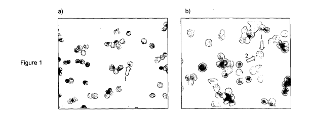
Figure 1: DAKO colourimetric staining of cultured C4-2b prostate cancer cell
line a) with
anti-PSA (brown, cytoplasmic), b). with anti-PSA (brown, cytoplasmic) and anti-
MCM2
(red, nuclear). Plate a) shows that anti-PSA antibody labels prostate cell
cytoplasm (1),
and that the nucleus is greatly enlarged (blue haematoxylin stain). Plate b)
shows dual
staining of cell cytoplasm brown with anti-PSA (1) and nucleus red with anti-
MCM2
antibodies (2).
Figure 2: Colourimetric single-staining of C4-2b cell line with Vector Stain
ImmPRESS
reagents. Plates are labelled as follows:- a) DAB staining of nucleus only (1)
with N-
terminal specific anti-AR antibody, with counter-stained cytoplasm (2); b) non-
localised
DAB staining of cells with C-terminal specific anti-AR antibody; c) DAB
staining of
cytoplasm (arrows) with anti-PSA antibody; d) novaRED staining of nucleus
(arrows) with
anti-MCM2 antibody; and e) novaRED staining of nucleus (arrows) with anti-MCM2
antibody.
Figure 3: Colourimetric dual-staining of C4-2b cell line with Vector Stain
ImmPRESS
reagents. Plate shows DAB-Ni (grey) labelled anti-PSA cytoplasm (1) with
novaRED
(red) anti-MCM2 antibody labelled nucleus (2).
Figure 4: Fluorescent (Alexafluor) staining of cultured C4-2b prostate cancer
cell line.
Plate a) with anti-PSA antibody (green, cytoplasmic); b) anti-MCM7 (red,
nuclear); c)
merged, showing distinct separate staining of the two cellular locations and
d) merged
CA 02807870 2013-02-08
WO 2011/073619
PCT/GB2010/002274
9
image using anti-PSA (green, cytoplasmic) and anti-MCM2 (red, nuclear)
antibodies
showing separate localisation.
Figure 5: Fluorescent (Alexafluor) staining of cultured C4-2b prostate cancer
cell line.
Plate a) with anti-AR antibody (green, nuclear); b) anti-MCM7 (red, nuclear);
and c)
merged, showing nuclear co-localisation of AR and MCM7 antigens.
Figure 6: Dual fluorescent staining of C4-2b prostate cancer cells isolated
from a clinical
urine sample from patients with confirmed prostatic cancer, with anti-AR
(green) and
anti-MCM7 (red) antibodies.
Figure 7: Removal of injured cells and cellular debris. Haemotoxylin stained
bladder
endothelial cells isolated from urine samples a) without pre-treatment with
lmmunosolv
DeadCert particles and b) after treatment to remove dead or dying cells and
other
cellular debris.
Figure 8: Survival/health of cultured prostatic C4-2b cell nuclei in urine
over 4 hours.
Clear red nuclear staining indicates that cells are in good condition. Less
intense red
staining, lack of staining, and increasing cytoplasmic staining indicate that
cells are
losing viability and would not be clearly identified as prostate cancer cells
in patient
samples.
Figure 9: Dual fluorescent staining of prostate cancer cells isolated from a
clinical urine
sample from patients with confirmed prostatic cancer, with (a) anti-AR (green)
and (b)
anti-MCM7 (red) antibodies and (c) Merged image of a double staining with AR
and
MCM.
Example 1
Colourimetric labelling of cultured prostate cancer cells with DAKO double-
stain kit
against PSA and MCM antigens.
C4-2b cells were cultured in T75 flasks in RPMI media containing 10% FBS and
2%
penstrep at 37 C and 5% CO2 to 90% confluence. The media was removed by
pipetting,
and the cells were washed twice with warmed PBS. They were detached from the
flask
surface by incubating for 2-3 min in 10 ml trypsin solution at 37 C, followed
by gentle
agitation. The cell_suspension_was transferred to a 50 ml falcon tube, and the
number of
CA 02807870 2013-02-08
WO 2011/073619
PCT/GB2010/002274
cells present determined by counting a sample on a haemocytometer. They were
then
pelleted by centrifugation and resuspended in warm PBS to 1 x 10⁶ cells/ml.
Aliquots of
150 pl were added to vials containing 20 ml of PreservCyt solution and
transferred to the
Thinprep-T2000 processor. This gently breaks up any cell debris, and provides
a
5 uniform suspension of cells. The suspension was drawn through a filter
which collects a
uniform layer of cells. By monitoring the resistance to flow, the instrument
prevents
collection of layers containing too few or too many cells. The entrapped cells
were
transferred to a UroCyte glass slide in a 10 mm diameter circle. The slide was
then
immersed in spirit (Cell Path Ltd. EGK-019500A), drained, and sprayed once
with
10
Surgipath coating fixative solution from a distance of 10 cm and placed on a
flat surface
to air dry.
Cell staining:- Cells were stained using a DAKO G/2 Double-stain kit (K5631)
as follows:
Slides were immersed in 50% methanol for 5 min, and rinsed in distilled water
before
rinsing in TBS (Tris-HCI ¨buffered saline, DAKO reagent) They were then rinsed
with
distilled water followed by DAKO wash buffer (S3006), and transferred to the
Autostainer
(DAKO). Slides were blocked with 200 pl DAKO dual endogenous enzyme block
(product code (K5361) for 5 min, then rinsed twice with TBS. Two hundred pl
primary
antibody A0562 rabbit anti-human PSA polyclonal antibody (or control rabbit
antibody)
was applied at 0.3 pg/ml and incubated for 30 min. All incubation steps were
carried out
at room temperature unless otherwise stated. Slides were rinsed twice with
TBS, and
200 pl Envision (anti-rabbit) Polymer Horse Radish Peroxidase (HRP) conjugate
(DAKO
K5361) added and incubated for 30 min. Slides were rinsed twice again with
TBS,
DAKO DAB substrate was applied and slides incubated for 5 min. Slides were
removed
from the auto-stainer and rinsed in tap water for 3 min.
A heat retrieval step was performed next in order to make the MCM antigen
accessible
to antibody:-
Slides were immersed 1 mM EDTA heat retrieval buffer, pH7.8 (DAKO) and micro-
waved on full power (800 W, 10 min.). Heat retrieval buffer was replenished,
and micro-
waving repeated for 10 min. Slides were prepared in this way in batches of 10.
Where a
smaller number of samples were being prepared, blank slides were included to
bring the
total number to 10 for consistency of heat treatment. Slides were allowed to
cool for 5
_min_and_returned_to_the auto-stainer.
CA 02807870 2013-02-08
WO 2011/073619
PCT/GB2010/002274
11
Slides were blocked again for 3 min with DAKO double-stain block (auxiliary
reagent)
and rinsed twice with TBS buffer. The 2nd primary antibody, mouse anti-MCM2
D1.12A3
(MRC, Cambridge, UK) or control mouse antibody was added (200 pl at 1 pg/ml)
and
incubated for 60 min. Slides were rinsed twice with TBS, and Envision Polymer-
alkaline
phosphatase conjugate (DAKO K5361) added (200 pl) for 15 min. After two
further
rinses with TBS, 200 pl liquid permanent red alk-phos substrate (vol, DAKA
code) was
added for 7 min. This reagent was prepared freshly and added to the
autostainer 30 min
before use. It was supplemented with 1 drop/ml of DAKO X3021 levamisole (DAKO
X3021) to block endogenous alk-phos activity.
After staining, slides were rinsed with distilled water and immersed in CuSO4
solution (10
g CuSO4, 17 g NaCI in 2 L water) for 5 min, then rinsed again with water. They
were
then counterstained with Haematoxylin for 10 s and rinsed with water. Finally
slides
were immersed in xylene for 5 min, air dried and cover-slipped ready for
analysis.
Antibodies were diluted from stock solutions in DAKO antibody diluent (S0809
or S2022)
to desired concentration. Slides were visualised under white light using a
Zeiss
AxioSKOP 40 microscope fitted with a Prog Res C14 colour camera. In addition
to white
light visualisation, the alkaline phosphatase substrate used to label MCM
antigens emits
fluorescence. This was observed using a Zeiss- Imager M2 with black and white
camera
Axiocam MRm and a red filter. Images were analysed with Axiovision software
(Figure
1).
In alternative assay formats, the DAKO rabbit anti-PSA polyclonal antibody
A0562 at 0.3
pg/ml was substituted with one of the following:
i) Insight Biotechnology N-20 rabbit anti-androgen receptor N-terminus
specific
polyclonal antibody (sc816) at 0.3 pg/ml.
ii) Genetex anti-PSGR antibody (GTX72749) at 1/50 dilution
iii) Abcam mouse monoclonal anti-PSA epitope 3 antibody PS2 (ab10189) at 0.3
pg/ml.
iv) Abcam mouse monoclonal anti-PSA epitope 4 antibody 5G6 (ab10186) at 0.3
pg/ml.
v) Abcam rabbit anti-PSA polyclonal antibody (ab9537) at 0.3 pg/ml.
In addition, the D1.12A3 mouse anti-human MCM2 monoclonal antibody at 1.0
pg/ml
was replaced with one of the following:
CA 02807870 2013-02-08
WO 2011/073619
PCT/GB2010/002274
12
i) Santacruz 141.2 mouse anti-MCM7 monoclonal antibody sc-9966 at 3.0 pg/ml.
(Antibodies raised specifically to either the N-terminus or C-terminus label
MCM7 in the
cytoplasm rather than the nucleus, and are therefore only suitable for use in
conjunction
with anti-AR antibodies in colourimetric assays).
ii) Vision Biosystems (Novocastra) mouse anti-human MCM3 monoclonal antibody
(NCL-
MCM3) from cell line JCCO7 at 1/200 dilution
iii) Vision Biosystems (Novocastra) mouse anti-human MCM5 monoclonal antibody
(NCL-MCM5) from cell line CRCT5.1 at 1/20 dilution
For single antigen stain assays to assess cytoplasmic antigens (PSA), the heat
retrieval
and secondary staining steps were omitted. For single stain assays to assess
nuclear
staining antigens (AR, MCM's), the primary staining step was omitted.
It was found that cytoplasmic PSA could be labelled in cultured prostate cells
with the
D1.12.A3 antibody (Figure la). PSA was also detected with antibodies against
'epitopes' 3 and 4, but not with antibodies against 'epitope' 1 (not shown).
Similarly,
antibodies to prostate specific acid phosphatase labelled prostate cell
cytoplasm,
whereas antibodies to prostate specific G-coupled receptor (PSGR) were not
effective.
In addition, nuclear antigens mini-chromosome maintenance proteins MCM2 and
MCM7,
and androgen receptor (AR) provided strong labelling, whereas MCM3 and MCM5
were
less effective (not shown). When used in combination in dual staining assays,
both PSA
and MCM5 (Figure lb) and PSA and MCM7 (not shown) could label prostate cells
and
be individually identified due their different cellular locations.
Example 2
Colourimetric labelling of cultured C4-2b prostate cancer cell line with
Vector
Laboratories ImmPRESS reagents against PSA Androgen receptor (AR) and MCM
antigens.
Slides were prepared as described in example 1 using a Thinprep T2000
instrument. All
labelling and washing steps were performed manually. The slides were first
immersed in
50% methanol for 5 min, and 200 pl DAKO dual endogenous enzyme block (product
code (K5361) added for 5 min. The slides were then rinsed 3 times with
distilled water
and 5 times with DAKO wash buffer (S3006). Ready-to-use ImmPRESS normal horse
serum (NHS) at 2.5% was applied to the slides for 20 min to block, followed by
200 pl
DAKO A0562 rabbit anti-human PSA polyclonal antibody (or control rabbit
antibody) at
0.3 pg/ml and incubated-for 10 min. All_incubation _steps were_carried out at
room
CA 02807870 2013-02-08
WO 2011/073619
PCT/GB2010/002274
13
temperature unless otherwise stated. Slides were rinsed 5 times with DAKO wash
buffer
and 200 pl ImmPRESS Reagent (universal HRP labelled anti-rabbit and anti-
mouse)
added for 30 min. Slides were rinsed 5 times with DAKO wash buffer and 200 pl
ImmPRESS substrate added for 5 min. Substrate used was any one of Vector VIP
(SK-
4600, purple), DAB (SK-4100, brown), DAB-Ni (SK-4100, grey/black) or Vector
NovaRED (SK-4800, red). Slides were washed 5 times with wash buffer.
A heat retrieval step was performed next in order to make the MCM antigens
accessible
to antibody:-
Slides were immersed 1 mM EDTA heat retrieval buffer, pH7.8 (DAKO) and micro-
waved on full power (800 W, 10 min.). Heat retrieval buffer was replenished,
and micro-
waving repeated for 10 min. Slides were prepared in this way in batches of 10.
Where a
smaller number of samples were being prepared, blank slides were included to
bring the
total number to 10 for consistency of heat treatment. Slides were allowed to
cool for 5
min.
Slides were blocked again for 20 min with 2.5% normal horse serum. The 2nd
primary
antibody, mouse anti-MCM2 D1.12A3 (MRC, Cambridge, UK) or control mouse
antibody
was added (200 pl at 1 pg/ml) and incubated for 10 min. They were rinsed 5
times with
wash buffer, and 200 pl ImmPRESS Reagent (as above) added for 30 min. After
five
further rinses with wash buffer, 200 pl ImmPRESS substrate was added for 5
min.
Substrate was any one of the four listed above, but not the same one as used
in the
earlier step.
After staining, slides were rinsed with distilled water and immersed in CuSO4
solution (10
g CuSO4, 17 g NaCI in 2 L water) for 5 min, then rinsed again with water. They
were
then counterstained with either Haematoxylin or Methyl green for 10 seconds
and rinsed
with water. Finally slides were immersed in xylene for 5 min, air dried and
cover-slipped
ready for analysis.
Antibodies were diluted from stock solutions in DAKO antibody diluent (S0809
or S2022)
with diluted 2.5% NHS to desired concentration. Slides were visualised under
white light
using a Zeiss AxioSKOP 40 microscope fitted with a Prog Res C14 colour camera.
For single stain assays to assess nuclear staining antigens (AR, MCM's), the
primary
staining step was omitted. For single antigen stain assays to assess
cytoplasmic
antigens_(PSA),_the_heat retrieval and_secondary staining steps were omitted.
CA 02807870 2013-02-08
WO 2011/073619
PCT/GB2010/002274
14
In alternative assay formats, the DAKO rabbit anti-PSA polyclonal antibody
A0562 at 0.3
pg/ml was substituted with the Insight Biotechnology N-20 rabbit anti-androgen
receptor
N-terminus specific polyclonal antibody (sc816) at 0.3 pg/ml or anti-androgen
receptor
C-terminal specific polyclonal antibody (sc815) at 0.3 pg/ml. The D1.12A3
mouse anti-
human MCM2 monoclonal antibody at 1.0 pg/ml was replaced with the Santacruz
141.2
mouse anti-MCM7 monoclonal antibody sc-9966 at 3.0 pg/ml. Staining was
achieved
variously using the following ImmPRESS substrates:- DAB (brown), DAB-Ni
(grey.black),
novaRED (red), or VIP (purple), either singly or in combination, and with or
without
counterstain.
It was found that, while the AR antibody specific for the N-terminus was able
to clearly
label the nucleus (Figure 2a), a C-terminus specific antibody did not show any
clear
cellular localisation and is therefore not suitable (Figure 2b). The PSA
antigen was also
detected in the cytoplasm of cells (Figure 2c). Antibodies against the cell
cycle antigens
MCM2 and MCM7 were both able to label the nuclei (Figure 2 d and e). All of
the
different substrates were found be effective when used for single staining
(not shown).
For dual staining, some combinations were less suitable than others due to
their similar
appearance. DAB-Ni with novaRED and DAB with vecta VIP were found to produce
good images (Figure 3). In this assay format, PSA must be used as a prostate
cell-
specific marker in combination with one or more MCM antigens. Due to it's
nuclear
localisation, AR is not suitable in this format.
Example 3
Fluorescent labelling of prostatic cell line C4-2b.
Prostatic cancer cell line C4-2b was cultured and Thinprep' slides prepared as
detailed
in example 1 above.
Cell staining:
Slides were immersed in 50% methanol for 5 min, washed in distilled water for
5 min and
rinsed once with DAKO wash buffer. Two hundred microlitres 0.1% Triton X-100
was
applied to the slide for 5 min in order to permeabilise the cells. They were
then rinsed 3
times with wash buffer and once with water. One hundred microlitres of Image-
1T FX
signal enhancer (Invitrogen 136933) was applied and incubated at room
temperature for
CA 02807870 2013-02-08
WO 2011/073619
PCT/GB2010/002274
30 min. Slides were rinsed 3 times with wash buffer followed by one rinse with
distilled
water.
Primary antibody mix or control antibody (rabbit anti-PSA at 0.3 pg/ml and
mouse anti-
5 MCM2 at 1 pg/ml) was applied to the slides (100 pl each) and incubated
for 90 min.
Mixes comprised one of the following combinations: i) DAKO rabbit anti-PSA
polyclonal
antibody A0562 (0.3pg/m1) and mouse anti-MCM2 D1.12A3 (1.0 pg/ml); ii) DAKO
rabbit
anti-PSA polyclonal antibody A0562 and Santacruz 141.2 mouse anti-MCM7
monoclonal
antibody sc-9966 (1.0 pg/ml); iii) Insight Biotechnology N-20 rabbit anti-
androgen
10 receptor N-terminus specific polyclonal antibody sc816 (0.3 pg/ml) and
mouse anti-
MCM2 D1.12A3; or iv) Insight Biotechnology N-20 rabbit anti-androgen receptor
N-
terminus specific polyclonal antibody sc816 and Santacruz 141.2 mouse anti-
MCM7
monoclonal antibody sc-9966.
15 Slides were rinsed 3 times with wash buffer and once with water. A 200
pl mix of two
Alexafluor secondary antibodies (1/1000 dilution) were applied to slides and
incubated
for 30 min. The mixes used were as follows, and in relation to the primary
antibody
mixes described above: i) and iii) Alexafluor 488 (green) anti-rabbit antibody
(Invitrogen
A11034) and Alexafluor 594 (red) anti-mouse IgG2b antibody (Invitrogen
A21145); ii)
and iv) Alexafluor 488 (green) anti-rabbit antibody (Invitrogen A11034) and
Alexafluor
594 (red) anti-mouse IgG1 antibody (Invitrogen A21125).
Example 4
Fluorescent labelling of prostate cells from patient urine.
Urine samples were provided from two patients with confirmed prostatic cancer,
and
were processed as follows immediately. Samples were centrifuged in 50 ml
falcon tubes
at 600 g for 5 min to pellet cells, and the supernatant disposed of. The cell
pellet was
resuspended in a small volume of Cytolyt (Cytyc Corp) by gentle agitation and
the tube
topped up 30 ml with CytoLyt. The suspension was vortexed for 5 min and re-
centrifuged. The cell pellet was resuspended in 100 pl PreservCyt solution,
and
transferred to a vial containing 20 ml PreservCyt. The vial was placed in the
Thinprep
T2000 and slides prepared as described in Example 3 above.
Cells were labelled with Santacruz 141.2 mouse anti-MCM7 monoclonal antibody
sc-
9966 at 1.0 pg/ml and either DAKO rabbit anti-PSA p_olyclonal antibody A0562
at 0.3
CA 02807870 2013-02-08
WO 2011/073619
PCT/GB2010/002274
16
pg/ml or Insight Biotechnology N-20 rabbit anti-androgen receptor N-terminus
specific
polyclonal antibody (sc816) at 0.3 pg/ml (Figure 6). Prostate cells were
identified in both
samples. They were all of irregular morphology, and exhibited varying degrees
of
cytoplasmic staining due to the poor condition of the cells after prolonged
exposure to
urine.
Example 5
Effect of removal of debris from urine samples.
Urine samples were centrifuged at 600 g for 10 min to pellet cells. Cells were
re-
suspended in 1 ml PBS. Total cell counts were determined using a
haemocytometer.
The volume of cell suspension was adjusted such that 100 pl contained 5 x10⁶
cells
(either by addition of PBS, or re-pelleting and re-suspending in an
appropriate volume
PBS).
A stock vial of Immunosolv Dead Cert nanoparticles (Imunosolv Ltd., Edinburgh,
UK)
was vortexed for 30 s. Twenty five microlitres of nanoparticles were
transferred to an
Eppendorf tube containing 1 ml PBS. The tube was placed in the provided
magnetic
separator for 3-5 min, until particles had collected against the magnet. The
buffer was
removed by pipetting, and the tube removed. Particles were resuspended in 100
pl PBS
by vortexing. One hundred microlitres cells (5 x 10⁶ cells) were added to the
particle
suspension, and the mixture incubated at 4 C for 40 min. During this
incubation, the
magnetic nanoparticles bind to antigens selectively expressed on the surface
of any
dead or dying cells. A further 800 pl PBS was added to the tube and mixed
gently, and
the tube then placed back into the magnetic separator for 5 min. The
supernatant
containing viable healthy cells was removed by pipetting, and added to a vial
of
PreservCyt (20 ml). A second 100 pl aliquot of cells that had not been pre-
depleted with
DeadCert particles was added to another vial of PreservCyte. Both samples were
then
processed in a Thinprep T200 as described in example 1. Slides were stained
with
haemotoxylin for visualisation Figure 7.
There were large numbers of particles of debris arising primarily from non-
viable cells in
the untreated samples. These can often be non-specifically labelled, and are
often not
distinguishable from genuine cancer cells due to the variable cellular
morphology of the
latter that is frequently seen. In contrast, treated samples contain far fewer
particles of
debris,enabling a more accurate and robust assessment of the p_atient to be
made.
CA 02807870 2013-02-08
WO 2011/073619
PCT/GB2010/002274
17
Example 6
Survival of prostatic cancer cells in urine.
Prostatic cell line C4-2b was cultured as described in example 1. When culture
flasks
had reached 80% confluence, cells were harvested by centrifugation,
resuspended in
PBS (phosphate-buffered saline) and counted. Approximately 2 x 10⁷ cells in 5
ml PBS
were added to 25 ml urine provided by healthy donors and stored at 4 C. The
samples
were removed from the fridge after 1 h, 2 h, 3 h, and 4 h and slides prepared
as detailed
in example 1 using a Thinprep T2000 instrument. The slides were stained using
mouse
anti-MCM2 D1.12A3 antibody and DAKO anti-mouse alkaline phosphatase reagents
as
in example1, and viewed under both white light (Figure 8). At 1 h and 2 h the
cells
showed clear nuclear staining of strong intensity. By 3 h staining was less
clearly
nuclear and less intense. At this point some cells did not stain positive for
MCM. By 4 h
the proportion of healthy cells had reduced significantly (data not shown).
It is of particular importance that cells stained with MCM indicated that
nuclear integrity
remained high to 4 hours with a gradual deterioration to 72 hours thereafter
(data not
shown. It was shown that the disappearance/dissolution of the protective cell
membrane
began from 1 hour onward in which PSA, as an index of cellular integrity
staining the
cytoplasm of prostate specific cells, deteriorated sharply from 1 hour onwards
(data not
shown).
Example 7
Staining of Prostate Cancer cell line with Androgen Receptor (AR) and Mini
Chromosome Maintenance Protein (MCM) antibodies
Early studies on patients with prostate cancer indicated that prostate
epithelial cells
could be captured in the urine of patients presenting with high serum prostate
specific
antigen (PSA) values. The cells were few in number and had to be processed
within one
hour of void. We have already established a method for prostate cell detection
in urine
using MCM as a nuclear marker and PSA as a cytoplasmic marker. The rapid
deterioration of these cells in urine, however, particularly the cell membrane
and hence
the cytoplasm of the cell required a more robust antibody marker.
It was noted that the nucleus of the prostate epithelial cell remained intact
for up to 4
hours-post_void_(see _Example nuclear_marker_with_specific _affinity
to_ prostate_
CA 02807870 2013-02-08
WO 2011/073619
PCT/GB2010/002274
18
tissue was selected and investigated. Good definition of prostate cells was
achieved
using androgen receptor antibodies using both prostate cancer cell lines and
in the
urines of patients with prostate cancer or those with high serum PSA values.
Normal urines spiked with an established prostate cancer cell line (C42b) were
prepared
in the laboratory using urocyte slides on an Hologic T2000 machine. The slides
were
immersed for 5 minutes in 50% methanol, washed with deionised water and 200u1
0.2%Triton X-100 added for 5 minutes to permeabilise the cells. A further
rinse x 3 with
DAKO buffer and x 1 with deionised water is followed with 2 drops (100u1)
Image-1T Fx
signal enhancer or sufficient volume to cover each coverslip or section and
then
incubated for 30 minutes at room temperature. A further rinse x 3 with buffer
and x 1
with water is followed by incubation with 200u1 of primary antibodies AR
(0.3ug/m1 AR-
N20 sourced from Santa Cruz catalogue no sc-816) and 1.15ug/m1 MCM (MCM7
sourced from Santa Cruz catalogue no sc9966) in 0.5% milk powder for 60
minutes. The
slides are rinsed x 3 with buffer and x 1 with deionised water, and then
incubated with
the relevant secondary Alexafluor antibodies (alexafluor 488 goat anti- rabbit
IgG (H + L;
lnvitrogen cat no A-11034, and Alexafluor 594 goat anti-mouse IgG1 (y1);
lnvitrogen cat
no A-21125) in the dark for 30 minutes. The slides are again rinsed x 3 with
buffer and x
1 with water, prior to the application of 1 drop of Prolong Gold Anti-fade
reagent at room
temperature followed by coverslipping of all slides. The slides are cured by
placing the
mounted sample on a flat dry surface and incubating at room temperature in the
dark for
24 hours. Fluorescence was detected using a fluorescent microscope with
different
fluorescence filters at 10 seconds (Fig 9).
The experiment showed a clear and defined nuclear staining both for Androgen
Receptor and Mini Chromosome Maintenance Protein.
An identical experiment was performed using MCM7 (at 1:100 dilution) and a
rabbit
monoclonal androgen receptor antibody (AR) (sourced from Epitomics Inc.).
Moreover
another experiment was performed using MCM2 at a 1:50 dilution and a rabbit
monoclonal androgen receptor antibody (AR) (sourced from Epitomics Inc.).
Slides were
stained in two different combinations using a mixed population of C42b
prostate
epithelial cells and EJ28 bladder cancer cells as the negative control for
evaluation of the
specificity and fixation of prostate nuclei by the MCM2 or MCM7 and androgen
receptor
combination. The methodology adopted was identical to that used in the C42b
cell line
incorporating the_androgen_ receptor_polyclonal antibody Jabove) and MCM7. In
this
CA 02807870 2013-02-08
WO 2011/073619
PCT/GB2010/002274
19
experiment the C42b cells were stained with MCM2 at 1:50 dilution and RabMAb
AR at
1:100 dilution. The individual nuclear staining components were clearly
identified under
fluorescent microscopy using AF488-1/1000 with the androgen receptor at 1:100
dilution
and AF594-1/1000 in combination with MCM2 at 1:50 dilution (data not shown).
Individual images of prostate cell lines stained in these circumstances showed
clear
identification of staining properties in the nuclei of the identified prostate
cells. When the
androgen receptor and MCM2 antibodies combined with the relevant Alexafluor
fluorescent antibody were combined there was good evidence of a clear and
defined
nuclear staining pattern whereby the green colouration of the androgen
receptor
together with the red colour of the MCM2 nuclear stain produced an orange
coloured
combination identifying prostate epithelial cells from a standardised prostate
malignant
cell line with dual nuclear staining characteristics (data not shown). Further
work using
both mixed populations of C42b and EJ28 prostate and bladder malignant cell
lines
respectively, when combined with androgen receptor and MCM7 showed good
evidence
of prostate cells with a stained yellow nucleus. EJ28 bladder cancer cells
were not
stained at all. Likewise, a mixed population of C42b and EJ28 cell lines when
stained
with androgen receptor at 1:100 and MCM2 at 1:50 dilutions showed a merged
image of
mixed populations C42b and EJ28 stained cells in which EJ28 stained cells
showed no
evidence of androgen receptor stain.
These experiments confirm that there was clear and defined nuclear staining
with
combinations of MAb AR plus MCM7 and likewise staining activity for MAb AR and
MCM2. These data confirm that epithelial prostate cancer cell lines can be
stained at a
nuclear level using both MCM2 and MCM7 in association with an androgen
receptor
antibody, in this case MAb AR.