Note: Descriptions are shown in the official language in which they were submitted.
GAL295-1CA
1
METHODS OF GENERATING OLIGODENDROCYTES AND CELL
POPULATIONS COMPRISING SAME
FIELD AND BACKGROUND OF THE INVENTION
[0001] The present invention, in some embodiments thereof, relates to methods
of generating
oligodendrocytes or oligodendrocytes progenitors from mesenchymal stem cells
and cell
populations comprising same.
[0002] Oligodendrocytes are important cells in the CNS that synthesize
multilamellar myelin
membranes that ensheath axons and therefore play an important role in the
development and
function of the CNS. Demyelination disrupts nerve conduction and leads to
nerve degeneration
which is associated with various disorders including Multiple Sclerosis (MS).
[0003] Oligodendrocytes are derived from multipotent neural progenitor cells.
Various
transcription factors and signaling pathways have been associated with this
process, including
Olig 1, NKX2.2, SHH, Wnt and Notch (2).
[0004] For example, early oligodendrogenesis is regulated by the basic helix-
loop-helix
transcription factors Oligl and 01ig2. The expression of these transcription
factors persists as
oligodendrocyte progenitors leave the ventricular zone and become mature
oligodendrocytes.
During the time when oligodendrocytes migrate into the white matter, they
acquire the
expression of two additional transcription factors, Sox 10 and Nkx2.2. The
expression of these
two transcription factors directly regulates the expression of the myelin gene
and the
differentiation of oligodendrocytes.
[0005] Multiple Sclerosis is a disease caused by chronic autoimmune
inflammatory process
resulting in patches of demyelination that affects the central nervous system
(11).
Remyelination, a regenerative process in which axons in the CNS are reinvested
with new
myelin sheaths and pre-lesion architecture and functions are restored, is
mainly mediated by a
population of cell specific adult stem/progenitor cells that are called
oligodendrocyte
precursor/progenitor cells (OPC) or glial precursor/progenitor cells. These
cells are distributed
in the white and grey matter throughout adulthood. Failure of remyelination
predisposes axons
to degeneration, a reversible process which is associated with the progressive
deterioration of
the disease. Therefore, remyelination is considered an important clinical
objective in MS in
order to slow or prevent axonal degradation and to preserve long-term axonal
survival in the
brain and spinal cord.
CA 2808372 2018-12-11
GAL295- ICA
2
[0006] Mesenchymal stem cells (MSCs) are a heterogeneous population of stromal
cells
isolated from multiple species, residing in most connective tissues including
bone marrow,
adipose, umbilical cord, placenta, amniotic fluid and perivascular tissues.
MSC can differentiate
into cells of the mesenchymal lineage, such as bone, cartilage and fat but,
under certain
circumstances, have been reported to acquire the phenotype of cells of the
endodermal and
neuroectodermal lineage, suggesting some potential for "transdifferentiation".
Within the bone
marrow these cells are tightly intermingled with and support hematopoiesis and
the survival of
hematopoietic stem cells in acquiescent state (7). In addition, MSCs derived
from the bone
marrow, adipose tissue or the cord/placenta have unique properties after
expansion in culture
including their ability to modulate innate and adaptive immunity (8).
Furthermore, MSCs
migrate to sites of inflammation and protect damaged tissues, including the
CNS, properties that
supported their use as new immunosupprcssive or rather immunoregulatory or
anti-
inflammatory agents for the treatment of inflammatory and immune-mediated
diseases
including autoimmune disorders (9).
[0007] Recent reports have demonstrated that MSCs also have the potential to
differentiate into
functional neuronal cells. MSCs have been shown to exert therapeutic effects
in a variety of
neurological diseases and dysfunctions in experimental animal models and more
recently in pilot
clinical trials. Their effects have been mainly attributed to
immunosuppressive and
neuroprotective functions. However, some studies demonstrated that neural
differentiation of
these cells increased their therapeutic effect in various instances.
Therefore, the use of MSC-
derived neuronal cells has a great potential as an easily accessible source of
autologous cells for
treatment of inflammatory and neurodegenerative disorders including Multiple
Sclerosis, ALS
and Parkinson's disease aiming for both cell mediated control of disease
activity as well as
regeneration of damaged or lost functions.
[0008] In experimental autoimmune encephalitis (EAE), an animal model of MS,
treatment of
mice with bone marrow derived MSCs resulted in significant suppression of
disease
manifestations in parallel with down-regulation of cell-mediated anti-
selfreactivity (9). The
migration of bone marrow derived MSCs paralleled improvement of the clinical
outcome of
treated recipients (9). Using genetically transduced green fluorescent donors
in these animal
models, donor derived cells migrating into the brain acquired phenotypic
markers of neurons,
astrocytes and oligodendrocytes in parallel with improvement of clinical signs
of disease as was
also confirmed by histopathological evaluation of treated as compared with
untreated controls.
CA 2808372 2018-12-11
GAL295-1CA
3
[0009] Interestingly, transplantation of glial committed progenitor into a
viral model of MS
resulted in some degree of remyelination (12), suggesting that the strategy of
transplantation of
oligodendrocytic progenitors is worthwhile pursuing.
[0010] Studies using injection of enriched and unmodified autologous bone
marrow derived and
more recently also adipose tissue derived MSC which can be prepared from
liposuction
intrathecally and intravenously suggests that some patients with otherwise
resistant MS may
benefit from treatment with autologous MSCs; however, complete restoration of
all neurological
deficits in patients with advanced and long-lasting disease has not yet been
achieved (13). Iron
nanoparticle (FeridexTM) labeled MSCs injected intrathecally and intravenously
could be
documented in the brain by MRI, thus confirming that these cells can actively
migrate into the
central nervous system.
[0011] Liu et al [Dev Biol. 302:683-693, 2007] have reported oligodendrocytic
differentiation
of bone marrow derived mesenchymal cells. This study employed fetal cells and
used
transfection with the transcription factors 011g2 and N10(.2. U.S. Patent
Application No.
20100021434 teaches oligodendrocytic differentiation of bone marrow derived
mesenchymal
cells by incubation in N2 supplement and fibroblast growth factor (FGF).
[0012] International Patent Application W02010111522 teaches mesenchymal stem
cells which
secrete and deliver microRNAs for the treatment of diseases. International
Patent Application
W02010144698 teaches expression of miRNAs in mesenchymal stem cells to induce
neuronal
differentiation thereof.
SUMMARY OF THE INVENTION
[0013] According to an aspect of some embodiments of the present invention
there is provided
a method of generating a population of cells useful for treating a nerve
disease or disorder in a
subject, the method comprising contacting mesenchymal stem cells (MSCs) with
at least one
exogenous miRNA selected from the group consisting of miR-145, miR-30d, miR-
125b, miR-
128, miR-181c, miR-26a, miR-196, miR-10b, miR-25, miR-424, miR19 and miR149,
thereby
generating the population of cells.
[0014] According to an aspect of some embodiments of the present invention
there is provided
a method of generating a population of cells useful for treating a nerve
disease or disorder in a
subject, the method comprising expressing in mesenchymal stem cells (MSCs)
exogenous
NKX2.2 and/or Olig2, thereby generating the population of cells.
CA 2808372 2018-12-11
GAL295-1CA
4
[0015] According to an aspect of some embodiments of the present invention
there is provided
a method of generating a population of cells useful for treating a central
nervous system (CNS)
disorder in a subject, the method comprising contacting mesenchymal stem cells
(MSCs) with
an agent that downregulates an amount and/or activity of connective tissue
growth factor
(CTGF), thereby generating the population of cells.
[0016] According to an aspect of some embodiments of the present invention
there is provided
an isolated population of cells generated according to the method of the
present invention having
an oligodendrocyte phenotype.
[0017] According to an aspect of some embodiments of the present invention
there is provided
a method of treating a nerve disease or disorder in a subject in need thereof,
the method
comprising administering to the subject a therapeutically effective amount of
the isolated
population of cells of the present invention, thereby treating the brain
disease or disorder.
[0018] According to an aspect of some embodiments of the present invention
there is provided
a pharmaceutical composition comprising the isolated population of cells of
the present
invention and a pharmaceutically acceptable carrier.
[0019] According to an aspect of some embodiments of the present invention
there is provided
a cell culture comprising mesenchymal stem cells which comprise at least one
miRNA selected
from the group consisting ofmiR-128, miR-9, miR-9*,miR124, miR137 andmiR218
and a
culture medium, said culture medium not being a differentiating medium.
[0020] According to an aspect of some embodiments of the present invention
there is provided
a method of treating a nerve disease or disorder in a subject in need thereof,
the method
comprising:
[0021] (a) contacting a population of mesenchymal stem cells with at least one
therapeutic
miRNA, wherein said contacting is effected for less than 5 days; and
[0022] (b) transplanting a therapeutically effective amount of said
mesenchymal stem cells
which have been modified to comprise said therapeutic miRNA to the brain of
the subject, said
miRNA being selected from the group consisting of miR-128, miR-9, miR-9*, miRl
24, miR137
and miR218, thereby treating the nerve disease or disorder.
[0023] According to an aspect of some embodiments of the present invention
there is provided
a method of treating a brain tumor in a subject in need thereof, the method
comprising
cA 2808372 2018-12-11
GAL295-1CA
transplanting a therapeutically effective amount of mesenchymal stem cells
which have been
modified to express at least one exogenous miRNA selected from the group
consisting of miR-
9, miR-124, miR-137, miR-218 and miR-212, thereby treating the brain tumor.
[0024] According to some embodiments of the invention, the at least sequence
is selected from
the group consisting of miR-145, miR-30d, miR-125b, miR-128, miR-181c.
[0025] According to some embodiments of the invention, the MSCs are isolated
from a tissue
selected from the group consisting of bone marrow, adipose tissue, placenta,
cord blood and
umbilical cord.
[0026] According to some embodiments of the invention, the MSCs are autologous
to said
subject.
[0027] According to some embodiments of the invention, the MSCs are non-
autologous to said
subject.
[0028] According to some embodiments of the invention, the MSCs are semi-
autologous to said
subject.
[0029] According to some embodiments of the invention, the contacting is
effected by
transfecting said MSCs with said at least one miRNA.
[0030] According to some embodiments of the invention, the contacting is
effected by
transfecting said MSCs with an expression vector which comprises a
polynucleotide sequence
which encodes a pre-miRNA of said at least one miRNA.
[0031] According to some embodiments of the invention, the contacting is
effected by
transfecting said MSCs with an expression vector which comprises a
polynucleotide sequence
which encodes said at least one miRNA.
[0032] According to some embodiments ofthe invention, at least 50% of the
population of cells
express at least one marker selected from the group consisting ofGalC, 04, 01,
CNPase, MOG
and MBP.
[0033] According to some embodiments of the invention, the MSCs are incubated
in a medium
comprising at least one agent selected from the group consisting of insulin,
hydrocortisone,
transferrin, pyruvate, ciliary neurotrophic factor (CNTF), neurotrophin 3 (NT-
3), heregulin,
CA 2808372 2018-12-11
GAL295-1CA
6
erythropoietin, PDGF-AA and tri-iodothyronine following, prior to or
concomitant with said
contacting.
[0034] According to some embodiments of the invention, the method further
comprises
expressing in said MSCs an exogenous differentiation factor selected from the
group consisting
of CNTF, NT-3, erythropoietin, NKX2.2 and 01ig2 following, prior to or
concomitant with said
contacting.
[0035] According to some embodiments of the invention, the MSCs are isolated
from a tissue
selected from the group consisting of bone marrow, adipose tissue, placenta,
cord blood and
umbilical cord.
[0036] According to some embodiments of the invention, the MSCs are autologous
to said
subject.
[0037] According to some embodiments of the invention, the MSCs are non-
autologous to said
subject.
[0038] According to some embodiments of the invention, the MSCs are semi-
autologous to said
subject.
[0039] According to some embodiments of the invention, the agent is a
polynucleotide agent.
[0040] According to some embodiments of the invention, the agent is an
antibody.
[0041] According to some embodiments of the invention, the polynucleotide
agent comprises
an siRNA agent.
[0042] According to some embodiments of the invention, the MSCs are incubated
in a medium
comprising at least one agent selected from the group consisting of insulin,
hydrocortisone,
transferrin, pyruvate, ciliary neurotrophic factor (CNTF), neurotrophin 3 (NT-
3), heregulin,
erythropoietin, PDGF-AA and tri-iodothyronine following, prior to or
concomitant with said
contacting.
[0043] According to some embodiments of the invention, the isolated population
of cells are
genetically modified.
[0044] According to some embodiments of the invention, the isolated population
of cells
comprises an exogenous miRNA selected from the group consisting ofmiR-145, miR-
30d, miR-
CA 2808372 2018-12-11
GAL295-1CA
7
125b, miR-128, miR-181c, miR-26 a, miR- 196, miR-10b, miR-25, miR-424,
miR19andmiR149.
[0045] According to some embodiments of the invention, the isolated population
of cells are for
use in treating a brain disease or disorder.
[0046] According to some embodiments of the invention, the brain disease or
disorder is a
neurodegenerative disorder.
[0047] According to some embodiments of the invention, the neurodegenerative
disorder is
selected from the group consisting of multiple sclerosis, Parkinson's,
epilepsy, amyotrophic
lateral sclerosis (ALS), stroke, autoim mune encephalomyelitis, diabetic
neuropathy,
glaucomatous neuropathy, Alzheimer's disease and Huntingdon's disease.
[0048] According to some embodiments of the invention, the brain disease of
disorder is
multiple sclerosis.
[0049] According to some embodiments of the invention, the nerve disease or
disorder is a
neurodegenerative disorder.
[0050] According to some embodiments of the invention, the neurodegenerative
disorder is
selected from the group consisting of multiple sclerosis, Parkinson's,
epilepsy, amyotrophic
lateral sclerosis (ALS), stroke, autoimmune encephalomyelitis, diabetic
neuropathy,
glaucomatous neuropathy, Alzheimer's disease and Huntingdon's disease.
[0051] According to some embodiments of the invention, the neurodegenerative
disease is
multiple sclerosis. According to some embodiments of the invention, the nerve
disease or
disorder comprises a spinal cord injury.
[0052] According to some embodiments of the invention, the mesenchymal stem
cells have
been genetically modified to express said at least one therapeutic miRNA.
[0053] According to some embodiments of the invention, the the nerve disease
or disorder is a
brain tumor.
[0054] According to some embodiments of the invention, the brain tumor is a
glioma.
[0055] According to some embodiments of the invention, the method further
comprises
expressing in the mesenchymal stem cells a pro-apoptotic agent.
CA 2808372 2018-12-11
GAL295-1CA
8
[0056] According to some embodiments of the invention, the pro-apoptotic agent
comprises
soluble TNF-related apoptosis-inducing ligand (sTRAIL).
[0057] Unless otherwise defined, all technical and/or scientific terms used
herein have the same
meaning as commonly understood by one of ordinary skill in the art to which
the invention
pertains. Although methods and materials similar or equivalent to those
described herein can be
used in the practice or testing of embodiments of the invention, exemplary
methods and/or
materials are described below. In case of conflict, the patent specification,
including definitions,
will control. In addition, the materials, methods, and examples are
illustrative only and are not
intended to be necessarily limiting.
BRIEF DESCRIPTION OF THE DRAWINGS
[0058] Some embodiments of the invention are herein described, by way of
example only, with
reference to the accompanying drawings. With specific reference now to the
drawings in detail,
it is stressed that the particulars shown are by way of example and for
purposes of illustrative
discussion of embodiments of the invention. In this regard, the description
taken with the
drawings makes apparent to those skilled in the art how embodiments of the
invention may be
practiced.
[0059] In the drawings:
[0060] FIG. 1 is a diagram of an exemplary vector used to transfect
mesenchymal stromal stem
cells in order to analyze its differentiation status.
[0061] FIGS. 2A-B illustrate that incubation of BM-derived MSCs in G5 medium
induces
changes in the morphology of the cells to OPC characteristics.
[0062] FIG. 3 illustrates that incubation of BM-derived MSCs in G5 medium
induces the
expression of the OPC markers, 01ig2 and NKX2.2, compared to incubation in
DMEM.
[0063] FIGS. 4A-F are photographs illustrating differentiation of MSCs
transfected with miR-
145 for 12 days in G5 medium. Cells were transfected with miR-145 and
maintained in G5
medium. Cells were stained with anti-MOG antibody. The results are
representative of five
similar experiments.
[0064] FIGS. 5A-D are photographs illustrating that miR-145 induces the
expression of GalC
in BM-MSCs. Cells were transfected with miR-145 and maintained in G5 medium.
Cells were
stained with anti-GalC antibody. The results are representative of five
similar experiments.
CA 2808372 2018-12-11
GAL295-1CA
9
[0065] FIG. 6 illustrates that miR-145 induces the expression of CNPase in BM-
MSCs. Cells
were transfected with miR-145 mimic and were then maintained in NM or GS
medium for 12
days. The expression of CNPase was determined using Western blot analysis.
Actin expression
was determined to demonstrate equal protein loading. The results are
representative of five
similar experiments.
[0066] FIGS. 7A-F are photographs illustrating induction of04 in BM-MSCs by
miR-145. Cells
were transfected with miR-145 and maintained in G5 medium. Cells were stained
with anti-04
antibody. The results are representative of five similar experiments.
[0067] FIG. 8 is a graph illustrating expression of oligodendrocyte markers in
control MSCs
and MSCs transfected with miR-145 (i.e., treatment). The expression of various
oligodendrocytic markers was examined 12 days following transfection using qRT-
PCR. The
results are representative of four similar experiments. NG2 ¨ proteoglycan
(developing and adult
oligodendrocyte precursor cells); PlPs ¨myelin proteolipid protein; NKX2.1 ¨
transcription
factor, oligodendrocyte progenitors; CNP¨ development and differentiation of
oligodendrocytes; MBP ¨ myelin basic protein, oligodendrocytes.
[0068] FIGS. 9A-B are photographs illustrating induction of MBP in BM-MSCs.
Cells were
transfected with miR-145 and maintained in medium supplemented with
oligodendrocytic
promoting medium for 12 days. The induction of the oligodendrocyte reporter,
MBP-GFP was
analyzed using a fluorescent microscope. The results are representative of
five similar
experiments.
[0069] FIGS. 10A-B are graphs illustrating that miR-145 induces the expression
of MBP-GFP
in MSCs. BM-derived MSCs were transfected with MBP-GFP and with miR-145 for 12
days in
G5 medium. The fluorescence of the MBP-GFP was determined using PACS analysis.
The
results represent three different experiments.
[0070] FIG. 11 is a graph illustrating that miR-145 decreases the expression
of CTGF.
[0071] Two different preparations of BM-MSCs were transfected with miR-145.
mRNA was
extracted after 3 days and the expression of CTGF was then examined using
realtime PCR. The
results represent the means SD of three separate experiments.
[0072] FIG. 12 is a graphical illustration of an expression construct used to
determine whether
miR-145 binds to the 3' UTR of CTGF.
cA 2808372 2018-12-11
1`
GAL295-1CA
[0073] FIG. 13 is a graph illustrating target validation of miR-145. 3'-UTR-
CTGF and a
scrambled control were cloned into a luciferase reporter plasmid (FIG. 12) and
co-transfected
with miR-145 mimic into MSCs. The luciferase activity of these cells was
measured 72 hours
thereafter. As presented in FIG. 12, miR-145 significantly decreased the
luciferase activity of
the 3' -UTR-CTGF, whereas it did not affect that of the CV. Likewise, a
control miR did not
alter the luciferase activity of cells co-transfected with the 3'-UTRCTGF. The
results represent
the means SD of three separate experiments.
[0074] FIG. 14 is a graph illustrating that the decrease in CTGF expression
plays a role in the
oligodendrocytic differentiation induced by miR-145. MSCs were transfected
with a CTGF
construct that lacks the 3' UTR followed by transfection with a miR-145 mimic.
The expression
of CNPase mRNA was examined 12 days later using real-time PCR. The results are
representative of five similar experiments.
[0075] FIGS. 15A-B illustrates bone marrow (BM)-MSCs transfer miRs to co-
cultured glioma
cells. BM-derived MSCs were transfected with a control miR or with a miR-124
mimic labeled
with FAM (A). BM-MSCs and AD-MSCs were transfected with miR-145-FITC (B).
Following
24 hr, U87 cells (A) or Al 72 cells (B) labeled with CellTracker were added to
the MSC culture
and the expression of the fluorescent miR-124 or miR-145 was analyzed 24 hours
later using a
confocal microscope. The results are representative of three different
experiments that gave
similar results.
[0076] FIG. 16 illustrates in situ hybridization ofmiR-145 in gliomas cells.
BM-MSCs were
transfected with a miR-145 mimic and were co-cultured with U87 cells labeled
with CellTracker
for additional 24 hr. In situ hybridization of miR-145 was then performed and
the labeled cells
were visualized for the presence of labeled miR-145.
[0077] FIGS. 17A-B are graphs illustrating that transferred miR-124
downregulates the
expression of SCP-1 in glioma cells. U87 cells were transfected with a miR-124
mimic and the
expression of SCP-1 was examined using qRT-PCR after 3 days (A). U87 cells
were transfected
with a construct expressing SCP-1 3'-UTR conjugated to luciferase. The cells
were then co-
cultured with BM-MSCs or AD-MSCs that were transfected with either a control
miRNA or
miR-124 mimic for 24 hr. The luciferase activity of the cells was determined
after 72 hr of co-
culture (B). The results the mean SE of three different experiments.
*p<0.001.
CA 2808372 2018-12-11
GAL295-1CA
11
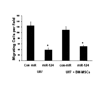
[0078] FIGS. 18A-D illustrate that transferred miR-124 decreases the migration
of glioma cells.
U87 cells were transfected with a miR-124 mimic and cell migration was
determined 48 hr later
using transwell migration (A). U87 cells (A,B) or cells labeled with
CellTracker (C,D) were
cultured with BM-MSCs expressing either a control miRNA or miR-124 mimic The
migration
of the U87 cells (A,B) or the labeled U87 cells (C.D) was determined after 48
hr using transwell
migration assay. The results are representative of three different experiments
that gave similar
results. *p<0. 001.
[0079] FIGS. 19A-C illustrate that MSCs transfer miR mimics to glioma stem
cells (GSCs) and
decrease their self-renewal. BM-MSCs or AD-MSCs were transfected with
fluorescent miR-
124 or miR-145or with miR124 and 145 mimics. After 24 hr, HF 2584 GSCs labeled
with
CellTracker were added to the cultured MSCs for additional 24 hr. The
expression of the
fluorescent miRs was analyzed using a confocal microscope (A). HF-2584 or
HF2587 GSCs
cocultured with BM-MSCs or AD-MSCs transfected with either a control miR or
miR-145
mimic were collected after 24 hr of co-culture and were analyzed for self-
renewal for 10 days
(B). BM-MSC and AD-MSCs were transfected with a control miR or with a miR-124
mimic.
After 24 hr, HF2587 GSCs transfected with a plasmid of 3' -UTR SCP-1 tagged to
luciferase
were added to the cultured MSCs. The luciferase activity of SCP-1-3'UTR
expressed in the
GSCs was analyzed after 48 hour (C). The results are representative of three
different
experiments that gave similar results. *p<0.001.
[0080] FIGS. 20A-B illustrate that MSCs transfer neuronal miR mimics to neural
progenitor
cells and promote their neuronal differentiation. BM-MSCs or AD-MSCs (data not
shown) were
transfected with a miR 124 mimics or a control miR. After 24 hr, the RenCell
neural progenitor
cells labeled with CellTracker were added to the cultured MSCs for additional
24 hr. The
percentage of-3-tubulin+cells out of the CellTracker -labeled cells were
determined for both
REN cells co-cultured with MSCs transfected with a control miR or with MSCs
transfected with
miR-124 using a fluorescent microscope (A). BM-MSC and AD-MSCs (data not
shown) were
transfected with a control miR or with a miR-124 mimic. After 24 hr, REN cells
transfected
with a plasmid of 3'-UTR SCP-1 tagged to luciferase were added to the cultured
MSCs. The
luciferase activity of SCP-1-31UTR expressed in the REN cells was analyzed
after 48 hr (C).
The results are representative of three different experiments that gave
similar results. *p<0.001.
CA 2808372 2018-12-11
GAL295-1CA
12
[0081] FIG. 21 is a bar graph illustrating the expression of oligodendrocyte
markers 01igo2,
PDGER-alpha and CNP in MSCs transfected with miR-145, miR-30d, miR-125b, miR-
128 and
miR-181 maintained in G5medium.
[0082] FIG. 22 is a bar graph illustrating the expression of oligodendrocyte
markers
PDGFRalpha, CNPase and PLP in MSCs genetically modified to express NI0(2.2
and/or 01ig2.
DESCRIPTION OF SPECIFIC EMBODIMENTS OF THE INVENTION
[0083] The present invention, in some embodiments thereof, relates to methods
of generating
oligodendrocytes from mesenchymal stem cells and cell populations comprising
same.
[0084] Before explaining at least one embodiment of the invention in detail,
it is to be
understood that the invention is not necessarily limited in its application to
the details set forth
in the following description or exemplified by the Examples. The invention is
capable of other
embodiments or of being practiced or carried out in various ways including the
use of MSCs as
carriers for delivery of miRs into adjacent normal or malignant target cells.
[0085] The importance of myelination is demonstrated by the demyelinating
disease multiple
sclerosis, in which myelin sheaths in some regions of the central nervous
system are destroyed
by an unknown mechanism. The significance of myelination is also demonstrated
in many other
neurodegenerative diseases, in which myelinated neurons are injured. Where
this happens, the
propagation of nerve impulses is greatly slowed, often with devastating
neurological
consequences.
[0086] Restoration of myelin has been proposed as a treatment therapy in order
to address the
underlying cause of such diseases. However, obtaining large numbers of
myelinating cells for
transplantation remains a major stumbling block.
[0087] Whilst reducing the present invention to practice, the present
inventors have found that
a number of micro RNAs (miRNAs) including miR-145, miR-125b, miR128 and miR-
30d
induce oligodendrocytic differentiation of bone marrow, adipose-derived,
amniotic fluid and
cord/placenta derived mesenchymal stem cells (MSCs) and propose that such
differentiated
MSCs may be used to treat patients with brain diseases or disorders.
[0088] Specifically, the present inventors have shown that transfection of
MSCs with the
miRNAs listed above change the morphological appearance of the cells and
further increase
CA 2808372 2018-12-11
GAL295-1CA
13
expression of various oligodendrocytic markers therein, as assessed by RT-PCR,
Western Blot
and immunohistochemistry (FIGS. 4A-F, 5A-D, 6 7A-F, 8, and 9A-B).
[0089] The present inventors further identified CTGF as a novel target of miR-
145 and as an
important mediator of the effect of this miRNA on the oligodendrocytic
differentiation ofmiR-
145. Therefore, the present inventors propose blocking anti-CTGF antibodies or
silencing of
CTGF in order to differentiate MSCs towards an oligodendrocytic phenotype.
[0090] Thus, according to one aspect of the present invention there is
provided a method of
generating a population of cells useful for treating a nerve disease or
disorder in a subject, the
method comprising contacting (either ex vivo or in vivo) mesenchymal stem
cells (MSCs) with
at least one miRNA selected from the group consisting of miR-145, miR-30d, miR-
125b, miR-
128, miR-181c, miR-26a, miR-196, miR-10b, miR-25, miR-424, miR19 and miR149,
thereby
generating the population of cells.
[0091] Mesenchymal stem cells give rise to one or more mesenchymal tissues
(e.g., adipose,
osseous, cartilaginous, elastic and fibrous connective tissues, myoblasts) as
well as to tissues
other than those originating in the embryonic mesoderm (e.g., neural cells)
depending upon
various influences from bioactive factors such as cytokines. Although such
cells can be isolated
from embryonic yolk sac, placenta, umbilical cord, fetal and adolescent skin,
blood and other
tissues, their abundance in the easily accessible fat tissue and BM far
exceeds their abundance
in other tissues and as such isolation from BM and fat tissue is presently
preferred.
[0092] Methods of isolating, purifying and expanding mesenchymal stem cells
(MSCs) are
known in the arts and include, for example, those disclosed by Caplan and
Haynesworth in U.S.
Pat. No. 5,486,359 and Jones E. A. et al., 2002, Isolation and
characterization of bone marrow
multipotential mesenchymal progenitor cells, Arthritis Rheum. 46(12):3349-60.
[0093] Mesenchymal stem cells may be isolated from various tissues including
but not limited
to bone marrow, peripheral blood, blood, placenta (e.g. fetal side of the
placenta), cord blood,
umbilical cord, amniotic fluid, placenta and from adipose tissue.
[0094] A method ofisolating mesenchymal stem cells from peripheral blood is
described by
Kassis et al [Bone Marrow Transplant. 2006 May; 37(10):967-76]. A method of
isolating
mesenchymal stem cells from placental tissue is described by Zhang et al
[Chinese Medical
Journal, 2004, 117 (6):882-887]. Methods of isolating and culturing adipose
tissue, placental
F CA 2808372 2018-12-11
GAL295-1CA
14
and cord blood mesenchymal stem cells are described by Kern et al [Stem Cells,
2006; 24:1294-
1301]1.
[0095] According to a preferred embodiment of this aspect of the present
invention, the
mesenchymal stem cells are human.
[0096] According to another embodiment of this aspect of the present
invention, the
mesenchymal stem cells are isolated from newborn humans.
[0097] Bone marrow can be isolated from the iliac crest of an individual by
aspiration. Low-
density BM mononuclear cells (BMMNC) may be separated by a FICOL -PAQUE
density
gradient or by elimination of red blood cells using Hetastarch (hydroxyethyl
starch). Preferably,
mesenchymal stem cell cultures are generated by diluting BM aspirates (usually
20 ml) with
equal volumes of Hank's balanced salt solution (HBSS; GIBCO Laboratories,
Grand Island,
N.Y., USA) and layering the diluted cells over about 10 ml of a Ficoll column
(Ficoll -Paque;
Pharmacia, Piscataway, N.J., USA). Following 30 minutes of centrifugation at
2,500xg, the
mononuclear cell layer is removed from the interface and suspended in HB SS.
Cells are then
centrifuged at 1,500xg for 15 minutes and resuspended in a complete medium
(MEM, a medium
without deoxyribonucleotides or ribonucleotides; GIBCO); 20% fetal calf serum
(FCS) derived
from a lot selected for rapid growth of MSCs (Atlanta Biologicals, Norcross,
GA); 100 units/ml
penicillin (GIBCO), 100 ug/m1 streptomycin (GIBCO); and 2 mM L-glutamine
(GIBCO).
Resuspended cells are plated in about 25 ml of medium in a 10 cm culture dish
(Corning Glass
Works, Corning, N.Y.) and incubated at 37 C. with 5% humidified CO2.
Following 24 hours in
culture, nonadherent cells are discarded, and the adherent cells are
thoroughly washed twice
with phosphate buffered saline (PBS). The medium is replaced with a fresh
complete medium
every 3 or 4 days for about 14 days. Adherent cells are then harvested with
0.25% trypsin and 1
mM EDTA (Trypsin/EDTA, GIBCO) for 5 min at 37 C., re-plated in a 6-cm plate
and cultured
for another 14 days. Cells are then trypsinized and counted using a cell
counting device such as
for example, a hemocytometer (Hausser Scientific, Horsham, Pa.). Cultured
cells are recovered
by centrifugation and resuspended with 5% DMSO and 30% FCS at a concentration
of 1 to
2x10⁶ cells per ml. Aliquots of about 1 ml each are slowly frozen and stored
in liquid nitrogen.
[0098] Adipose tissue-derived MSCs can be obtained by liposuction and
mononuclear cells can
be isolated manually by removal of the fat and fat cells, or using the
Celution System (Cytori
Therapeutics) following the same procedure as described above for preparation
of MSCs.
P CA 2808372 2018-12-11
GAL295-1CA
[0099] According to one embodiment the populations are plated on polystyrene
plastic surfaces
(e.g. in a flask) and mesenchymal stem cells are isolated by removing non-
adherent cells.
Alternatively mesenchymal stem cell may be isolated by FACS using mesenchymal
stem cell
markers.
[0100] Preferably the MSCs are at least 50% purified, more preferably at least
75% purified and
even more preferably at least 90% purified.
[0101] To expand the mesenchymal stem cell fraction, frozen cells are thawed
at 37 C., diluted
with a complete medium and recovered by centrifugation to remove the DMSO.
Cells are
resuspended in a complete medium and plated at a concentration of about 5,000
cells/cm".
Following 24 hours in culture, nonadherent cells are removed and the adherent
cells are
harvested using Trypsin/EDTA, dissociated by passage through a narrowed
Pasteur pipette, and
preferably re-plated at a density of about 1.5 to about 3.0 cells/cm2. Under
these conditions,
MSC cultures can grow for about 50 population doublings and be expanded for
about 2000-fold
[Colter DC., et al. Rapid expansion of recycling stem cells in cultures of
plastic-adherent cells
from human bone marrow. Proc Natl Acad Sci USA. 97: 3213-3218, 2000].
[0102] MSC cultures utilized by some embodiments of the invention preferably
include three
groups of cells which are defined by their morphological features: small and
agranular cells
(referred to as RS-1, herein below), small and granular cells (referred to as
RS-2, hereinbelow)
and large and moderately granular cells (referred to as mature MSCs,
hereinbelow). The
presence and concentration of such cells in culture can be assayed by
identifying a presence or
absence of various cell surface markers, by using, for example,
immunofluorescence, in situ
hybridization, and activity assays.
[0103] When MSCs are cultured under the culturing conditions of some
embodiments of the
invention they exhibit negative staining for the hematopoietic stem cell
markers CD34, CD1 in,
CD43 and CD45. A small fraction of cells (less than 10%) are dimly positive
for CD31 and/or
CD38 markers. In addition, mature MSCs are dimly positive for the
hematopoietic stem cell
marker, CD11 7 (c-Kit), moderately positive for the osteogenic MSCs marker,
Stro-1 [Simmons,
P. J. & 'Iorok-Storb, B. (1991). Blood 78, 5562] and positive for the
thymocytes and peripheral
T lymphocytes marker, CD90 (Thy-1). On the other hand, the RS-1 cells are
negative for the
CD 117 and Strol markers and are dimly positive for the CD90 marker, and the
RS-2 cells are
negative for all of these markers.
CA 2808372 2018-12-11
GAL295-1CA
16
[0104] The mesenchymal stem cells of the present invention may be of a
syngeneic or allogeneic
source, as further described herein below.
[0105] Differentiation of the mesenchymal stem cells can be induced by
incubating the MSCs
in differentiating media such as those described in U.S. Pat. No. 6,528,245
and by Sanchez-
Ramos et al. (2000); Woodburry et al. (2000); Woodbuny et al. (J. Neurisci.
Res. 96:908-917,
2001); Black and Woodbury (Blood Cells Mol. Dis. 27:632-635, 2001); Deng et
al. (2001),
Kohyama et al. (2001), Reyes and Verfatile (Ann. N.Y. Acad. Sci. 938:231-
235,2001) and Jiang
etal. (Nature 418:47-49, 2002).
[0106] The differentiating media may be DMEM or DMEM/F 12, OptiMEMTm or any
other
medium that supports neuronal growth. According to a preferred embodiment of
this aspect of
the present invention, the medium comprises neurobasal medium (e.g. Cat. No.
21103049,
Invitrogen, Calif., U.S.A.).
[0107] According to another embodiment of this aspect of the present
invention, the medium is
supplemented with at least one of insulin, hydrocortisone, transferring,
pyruvate and
nicotinamide. According to another embodiment, the medium comprises GSTM
supplement
(Catalogue No. F001-003, PAA Laboratories).
[0108] As mentioned, the mesenchymal stem cells are contacted (either ex vivo
or in vivo) with
at least one of the following miRNAs in order to induce differentiation into
oligodendrocyte-
like cells-miR-145 (SEQ ID NO: 15), miR-30d (SEQ ID NO: 16), miR-125b (SEQ ID
NO: 17),
miR-128 (SEQ ID NO: 18), miR-181c (SEQ ID NO: 19), miR-26a (SEQ ID NO: 27),
miR-196
(SEQ ID NO: 28), miR-10b (SEQ ID NO: 31), miR-25 (SEQ ID NO: 32), miR-424 (SEQ
ID
NO: 33), miR19 (SEQ ID NO: 34) and miR149 (SEQ ID NO: 35).
[0109] It will be appreciated that prior to contacting with one of the above-
mentioned miRNAs,
the MSCs may be contacted with additional miRNAs that serve to induce
dedifferentiation of
the cells into pluripotent cells. Such miRNAs include transfecting with
amicroRNA-
302bcad/367 (SEQ ID NOs: 42, 44, 36, 48 and 50).
[0110] The term "microRNA", "miRNA", and "miR" are synonymous and refer to a
collection
of non-coding single stranded RNA molecules of about 19-28 nucleotides in
length, which
regulate gene expression. miRNAs are found in a wide range of organisms and
have been shown
to play a role in development, homeostasis, and disease etiology.
r cA 2808372 2018-12-11
GAL295- I CA
17
[0111] Below is a brief description of the mechanism of miRNA activity.
[0112] Genes coding for miRNAs are transcribed leading to production of an
miRNA precursor
known as the pri-miRNA. The pri-miRNA is typically part of a polycistronic RNA
comprising
multiple pri-miRNAs. The pri-miRNA may form a hairpin with a stem and loop.
The stem may
comprise mismatched bases.
[0113] The hairpin structure of the pri-miRNA is recognized by Drosha, which
is an RNase III
endonuclease. Drosha typically recognizes terminal loops in the pri-miRNA and
cleaves
approximately two helical turns into the stem to produce a 60-70 nt precursor
known as the pre-
miRNA. Drosha cleaves the pri-miRNA with a staggered cut typical of RNase III
endonucleases
yielding a pre-miRNA stem loop with a 5' phosphate and-2 nucleotide 3'
overhang. It is
estimated that approximately one helical turn of stem (-10 nucleotides)
extending beyond the
Drosha cleavage site is essential for efficient processing. The pre-miRNA is
then actively
transported from the nucleus to the cytoplasm by Ran-GTP and the export
receptor exportin-5.
[0114] The double-stranded stem of the pre-miRNA is then recognized by Dicer,
which is also
an RNase III endonuclease. Dicer may also recognize the 5' phosphate and 3'
overhang at the
base of the stem loop. Dicer then cleaves off the terminal loop two helical
turns away from the
base of the stem loop leaving an additional 5' phosphate and -2 nucleotide 3'
overhang. The
resulting siRNA-like duplex, which may comprise mismatches, comprises the
mature miRNA
and a similar-sized fragment known as the miRNA*. The miRNA and miRNA* may be
derived
from opposing arms of the pri-miRNA and pre-miRNA. miRNA*sequences may be
found in
libraries of cloned miRNAs but typically at lower frequency than the miRNAs.
[0115] Although initially present as a double-stranded species with miRNA*,
the miRNA
eventually become incorporated as a single-stranded RNA into a
ribonucleoprotein complex
known as the RNA-induced silencing complex (RISC). Various proteins can form
the RISC,
which can lead to variability in specificity for miRNA/miRNA* duplexes,
binding site of the
target gene, activity of miRNA (repress or activate), and which strand of the
miRNA/miRNA*
duplex is loaded in to the RISC.
[0116] When the miRNA strand of the miRNA:miRNA* duplex is loaded into the
RISC, the
miRNA* is removed and degraded. The strand of the miRNA:miRNA* duplex that is
loaded
into the RISC is the strand whose 5' end is less tightly paired. In cases
where both ends of the
CA 2808372 2018-12-11
GAL295-1CA
18
miRNA:miRNA* have roughly equivalent 5' pairing, both miRNA and miRNA* may
have gene
silencing activity.
[0117] The RISC identifies target nucleic acids based on high levels of
complementarity
between the miRNA and the mRNA, especially by nucleotides 2-7 of the miRNA.
[0118] A number of studies have looked at the base-pairing requirement between
miRNA and
its mRNA target for achieving efficient inhibition of translation (reviewed by
Bartel 2004, Cell
116-281). In mammalian cells, the first 8 nucleotides of the miRNA may be
important (Doench
& Sharp 2004 GenesDev 2004-504). However, other parts of the microRNA may also
participate in mRNA binding. Moreover, sufficient base pairing at the 3' can
compensate for
insufficient pairing at the 5' (Brennecke et al, 2005 PLoS 3-e85). Computation
studies, analyzing
miRNA binding on whole genomes have suggested a specific role for bases 2-7 at
the 5' of the
miRNA in target binding but the role of the first nucleotide, found usually to
be "A" was also
recognized (Lewis et at 2005 Cell 120-15). Similarly, nucleotides 1-7 or 2-8
were used to
identify and validate targets by Krek et al (2005, Nat Genet 37-495). The
target sites in the
mRNA may be in the 5' UTR, the 3' UTR or in the coding region. Interestingly,
multiple
miRNAs may regulate the same mRNA target by recognizing the same or multiple
sites. The
presence of multiple miRNA binding sites in most genetically identified
targets may indicate
that the cooperative action of multiple RISCs provides the most efficient
translational inhibition.
MiRNAs may direct the RISC to downregulate gene expression by either of two
mechanisms:
mRNA cleavage or translational repression. The miRNA may specify cleavage of
the mRNA if
the mRNA has a certain degree of complementarity to the miRNA. When a miRNA
guides
cleavage, the cut is typically between the nucleotides pairing to residues 10
and 11 of the
miRNA. Alternatively, the miRNA may repress translation if the miRNA does not
have the
requisite degree of complementarity to the miRNA. Translational repression may
be more
prevalent in animals since animals may have a lower degree of complementarity
between the
miRNA and binding site.
[0119] It should be noted that there may be variability in the 5' and 3' ends
of any pair of miRNA
and miRNA*. This variability may be due to variability in the enzymatic
processing of Drosha
and Dicer with respect to the site of cleavage. Variability at the 5' and 3'
ends of miRNA and
miRNA* may also be due to mismatches in the stem structures of the pri-miRNA
and pre-
miRNA. The mismatches of the stem strands may lead to a population of
different hairpin
CA 2808372 2018-12-11
GAL295-1CA
19
structures. Variability in the stem structures may also lead to variability in
the products of
cleavage by Drosha and Dicer. The term "microRNA mimic" refers to synthetic
non-coding
RNAs that are capable of entering the RNAi pathway and regulating gene
expression. miRNA
mimics imitate the function of endogenous microRNAs (miRNAs) and can be
designed as
mature, double stranded molecules or mimic precursors (e.g., or pre-miRNAs).
miRNA mimics
can be comprised of modified or unmodified RNA, DNA, RNA-DNA hybrids, or
alternative
nucleic acid chemistries (e.g., LNAs or 2?-0,4'-C-ethylene-bridged nucleic
acids (ENA)). For
mature, double stranded miRNA mimics, the length of the duplex region can vary
between 13-
33, 18-24 or 21-23 nucleotides. ThemiRNA may also comprise a total of at least
5, 6, 7, 8, 9,
10, 11, 12, 13, 14,15,16, 17, 18, 19, 20, 21, 22, 23, 24, 25, 26, 27, 28, 29,
30, 31, 32, 33, 34, 35,
36, 37, 38, 39 or 40 nucleotides. The sequence of the miRNA may be the first
13-33 nucleotides
of the pre-miRNA. The sequence of the miRNA may also be the last 13-
33nucleotides of the
pre-miRNA. The sequence of the miRNA may comprise any of the sequences of SEQ
ID NOS:
15-19 or 27-39, or variants thereof.
[0120] It will be appreciated from the description provided herein above, that
contacting
mesenchymal stem cells may be affected in a number of ways:
[012111. Transiently transfecting the mesenchymal stem cells with the mature
double stranded
miRNA;
[0122] 2. Stably, or transiently transfecting the mesenchymal stem cells with
an expression
vector which encodes the mature miRNA (SEQ ID NOs: 15-19 or 27-39).
[0123] = 3. Stably, or transiently transfecting the mesenchymal stem cells
with an expression
vector which encodes the pre-miRNA (SEQ ID NOs: 20-24 and 52-71). The pre-
miRNA
sequence may comprise from 45-90, 60-80 or 60-70 nucleotides. The sequence of
the prc-
miRNAmay comprise a miRNA and a miRNA* as set forth herein. The sequence of
the pre-
miRNA may also be that of a pri-miRNA excluding from 0-160 nucleotides from
the 5' and 3'
ends of the primiRNA. The sequence of the pre-miRNA may comprise the sequence
of the
miRNA -i.e. SEQ ID NOs: 15-19 or 27-39 or variants thereof.
[0124] 4. Stably, or transiently transfecting the mesenchymal stem cells with
an expression
vector which encodes the pri-miRNA. The pri-miRNA sequence may comprise from
45-30,000,
50-25,000, 100-20,000, 1,000-1,500 or 80-100 nucleotides. The sequence of the
pri-miRNA
may comprise a pre-miRNA, miRNA and miRNA*, as set forth herein, and variants
thereof.
r CA 2808372 2018-12-11
GAL295-1CA
Preparation of miRNAs mimics can be effected by chemical synthesis methods or
by
recombinant methods.
[0125] To express miRNAs in mesenchymal stem cells, a polynucleotide sequence
encoding
the miRNA (or pre-miRNA, or pri-miRNA) is preferably ligated into a nucleic
acid construct
suitable for mesenchymal stem cell expression. Such a nucleic acid construct
includes a
promoter sequence for directing transcription of the polynucleotide sequence
in the cell in a
constitutive or inducible manner.
[0126] It will be appreciated that the nucleic acid construct of some
embodiments of the
invention can also utilize miRNA homologues which exhibit the desired activity
(i.e.,
oligodendrocytic differentiating ability). Such homologues can be, for
example, at least 80%, at
least 81 %, at least 82%, at least 83%, at least 84%, at least 85%, at least
86%, at least 87%, at
least 88%, at least 89%, at least 90%, at least 91 %, at least 92%, at least
93%, at least 94%, at
least 95%, at least 96%, at least 97%, at least 98%, at least 99% or 100%
identical to any of the
sequences SEQ ID NOs:15-19 or 27-39, as determined using the BestFit software
of the
Wisconsin sequence analysis package, utilizing the Smith and Waterman
algorithm, where gap
weight equals 50, length weight equals 3, average match equals 10 and average
mismatch equals
-9.
[0127] in addition, the homologues can be, for example, at least 60%, at least
61 %, at least
62%, at least 63%, at least 64%, at least 65%, at least 66%, at least 67%, at
least 68%, at least
69%, at least 70%, at least 71 %, at least 72%, at least 73%, at least 74%, at
least 75%, at least
76%, at least 77%, at least. 78%, at least 79%, at least 80%, at least 81 %,
at least 82%, at least
83%, at least 84%, at least 85%, at least 86%, at least 87%, at least 88%, at
least 89%, at least
90%, at least 91 %, at least 92%, at least 93%, at least 94%, at least 95%, at
least 96%, at least
97%, at least 98%, at least 99% or 100% identical to SEQ ID NOs: 20-24 and 27-
39, as
determined using the BestFit software of the Wisconsin sequence analysis
package, utilizing the
Smith and Waterman algorithm, where gap weight equals 50, length weight equals
3, average
match equals 10 and average mismatch equals -9.
[0128] Constitutive promoters suitable for use with some embodiments of the
invention are
promoter sequences which are active under most environmental conditions and
most types of
cells such as the cytomegalovirus (CMV) and Rous sarcoma virus (RSV).
Inducible promoters
F CA 2808372 2018-12-11
GAL295-1CA
21
suitable for use with some embodiments of the invention include for example
tetracycline-
inducible promoter (Zabala M, et al., Cancer Res. 2004, 64(8): 2799-804).
[0129] Eukaryotic promoters typically contain two types of recognition
sequences, the
[0130] TATA box and upstream promoter elements. The TATA box, located 25-30
base pairs
upstream of the transcription initiation site, is thought to be involved in
directing RNA
polymerase to begin RNA synthesis. The other upstream promoter elements
determine the rate
at which transcription is initiated.
[0131] Preferably, the promoter utilized by the nucleic acid construct of some
embodiments of
the invention is active in the specific cell population transformed=-i.e.
mesenchymal stem cells.
[0132] Enhancer elements can stimulate transcription up to 1,000-fold from
linked homologous
or heterologous promoters. Enhancers are active when placed downstream or
upstream from the
transcription initiation site. Many enhancer elements derived from viruses
have a broad host
range and are active in a variety of tissues. For example, the SV40 early gene
enhancer is suitable
for many cell types. Other enhancer/promoter combinations that are suitable
for some
embodiments of the invention include those derived from polyoma virus, human
or murine
cytomegalovirus (CMV), the long-term repeat from various retroviruses such as
murine
leukemia virus, murine or Rous sarcoma virus and HIV. See, Enhancers and
Eukaryotic
Expression, Cold Spring Harbor Press, Cold Spring Harbor, N.Y. 1983.
[0133] In the construction of the expression vector, the promoter is
preferably positioned
approximately the same distance from the heterologous transcription start site
as it is from the
transcription start site in its natural setting. As is known in the art,
however, some variation in
this distance can be accommodated without loss of promoter function.
[0134] In addition to the elements already described, the expression vector of
some
embodiments of the invention may typically contain other specialized elements
intended to
increase the level of expression of cloned nucleic acids or to facilitate the
identification of cells
that carry the recombinant DNA. For example, a number of animal viruses
contain DNA
sequences that promote the extra chromosomal replication of the viral genome
in permissive
cell types. Plasmids bearing these viral replicons are replicated episomally
as long as the
appropriate factors are provided by genes either carried on the plasmid or
with the genome of
the host cell. The vector may or may not include a eukaryotic replicon. If a
eukaryotic replicon
is present, then the vector is amplifiable in eukaryotic cells using the
appropriate selectable
CA 2808372 2018-12-11
GAL295-1CA
22
marker. If the vector does not comprise a eukaryotic replicon, no episomal
amplification is
possible. Instead, the recombinant DNA integrates into the genome of the
engineered cell, where
the promoter directs expression of the desired nucleic acid.
[0135] Examples for mammalian expression vectors include, but are not limited
to, pcDNA3,
pcDNA3.1(+/-), pGL3, pZeoSV2(+/-), pSecTag2, pDisplay, pEF/myc/cyto,
pCMV/myc/cyto,
pCR3.1, pSinRep5, DH26S, DHBB, pNMT1, pNMT41,pNMT81, which are available from
Invitrogen, pCI which is available from Promega, pMbac, pPbac, pBK-RSV and pBK-
CMV
which are available from Strategene, pTRES which is available from Clontech,
and their
derivatives.
[0136] Expression vectors containing regulatory elements from eukaryotic
viruses such as
retroviruses can be also used. SV40 vectors include pSVT7 and pMT2. Vectors
derived from
bovine papilloma virus include pBV-1MTHA, and vectors derived from Epstein Bar
virus
include pHEBO, and p205. Other exemplary vectors include pMSG, pAV009/A+,
pMT010/A+,
pMAMneo-5, baculovirus pDSVE, and any other vector allowing expression of
proteins under
the direction of the SV-40 early promoter, SV-40 later promoter,
metallothionein promoter,
murine mammary tumor virus promoter, Rous sarcoma virus promoter, polyhedrin
promoter, or
other promoters shown effective for expression in eukaryotic cells.
[0137] As described above, viruses are very specialized infectious agents that
have evolved, in
many cases, to elude host defense mechanisms. Typically, viruses infect and
propagate in
specific cell types. The targeting specificity of viral vectors utilizes its
natural specificity to
specifically target predetermined cell types and thereby introduce a
recombinant gene into the
infected cell. Thus, the type of vector used by some embodiments of the
invention will depend
on the cell type transformed. The ability to select suitable vectors according
to the cell type
transformed is well within the capabilities of the ordinary skilled artisan
and as such no general
description of selection consideration is provided herein. For example, bone
marrow cells can
be targeted using the human T cell leukemia virus type I (HTLV-I) and kidney
cells may be
targeted using the heterologous promoter present in the baculovirus Autographa
califomica
nucleopolyhedrovirus (AcMNPV) as described in Liang CY et al., 2004 (Arch
Viral. 149: 51-
60).
[0138] According to one embodiment, a lentiviral vector is used to transfect
the mesenchymal
stem cells.
CA 2808372 2018-12-11
GAL295-1CA
23
[0139] Various methods can be used to introduce the expression vector of some
embodiments
of the invention into mesenchymal stem cells. Such methods are generally
described in
Sambrook et al., Molecular Cloning: A Laboratory Manual, Cold Springs Harbor
Laboratory,
New York (1989, 1992), in Ausubel et al., Current Protocols in Molecular
Biology, John Wiley
and Sons, Baltimore, Md. (1989), Chang et al., Somatic Gene Therapy, CRC
Press, Ann Arbor,
Mich. (1995), Vega et al., Gene Targeting, CRC Press, Ann Arbor Mich. (1995),
Vectors: A
Survey of Molecular Cloning Vectors and Their Uses, Butterworths, Boston Mass.
(1988) and
Gilboa et at [Bliotechniques 4 (6): 504-512, 1986] and include, for example,
stable or transient
transfection, lipofection, electroporation and infection with recombinant
viral vectors. In
addition, see U.S. Pat. Nos. 5,464,764 and 5,487,992 for positive-negative
selection methods.
[0140] Introduction of nucleic acids by viral infection offers several
advantages over other
methods such as lipofection and electroporation, since higher transfection
efficiency can be
obtained due to the infectious nature of viruses.
[0141] Other vectors can be used that are non-viral, such as cationic lipids,
polylysine, and
dendrimers. Nanoparticles are also contemplated.
[0142] Other modes of transfection that do not involved integration include
the use of minicircle
DNA vectors or the use of PiggyBac transposon that allows the transfection of
genes that can
be later removed from the genome.
[0143] As mentioned hereinabove, a variety of prokaryotic or eukaryotic cells
can be used as
host-expression systems to express the miRNAs of some embodiments of the
invention. These
include, but are not limited to, microorganisms, such as bacteria transformed
with a recombinant
bacteriophage DNA, plasmid DNA or cosmid DNA expression vector containing the
coding
sequence; yeast transformed with recombinant yeast expression vectors
containing the coding
sequence; plant cell systems infected with recombinant virus expression
vectors (e.g.,
cauliflower mosaic virus, CaMV; tobacco mosaic virus, TMV) or transformed with
recombinant
plasmid expression vectors, such as Ti plasmid, containing the coding
sequence. Mammalian
expression systems can also be used to express the miRNAs of some embodiments
of the
invention.
[0144] Examples of bacterial constructs include the pET series of E. coli
expression vectors
[Studier et al. (1990) Methods in Enzymol. 185:60-89).
CA 2808372 2018-12-11
GAL295-1CA
24
[0145] In yeast, a number of vectors containing constitutive or inducible
promoters can be used,
as disclosed in U.S. patent application Ser. No: 5,932,447. Alternatively,
vectors can be used
which promote integration of foreign DNA sequences into the yeast chromosome.
[0146] By determining the targets of the miRNAs of the present invention, it
will be appreciated
that the scope of the present invention may be broadened to include down-
regulation of the
targets by means other than contacting with miRNA.
[0147] For example, the present inventors have shown that one of the targets
of miR-145 is
connective tissue growth factor (CTGF). Thus, the present invention
contemplates that
differentiation towards the oligodendrocytic lineage may be affected by down-
regulation of this
protein.
[0148] Thus, according to another aspect of the invention, there is provided a
method of
generating a population of cells useful for treating a CNS disorder in a
subject, the method
comprising contacting mesenchymal stem cells (MSCs) with an agent that
downregulates an
amount and/or activity of connective tissue growth factor (CTGF) or a receptor
thereof, thereby
generating the population of cells.
[0149] CTGF is a cysteine-rich monomeric peptide of Mr 38,000. It is a member
of the CCN
family of growth regulators which includes the mouse (also known as fisp-12 or
betalGM2) and
human CTGF, Cyr61 (mouse), CeflO (chicken), and Nov (chicken). Based on
sequence
comparisons, it has been suggested that the members of this family all have a
modular structure,
consisting of (1) an insulin-like growth factor domain responsible for
binding, (2) a von
Willebrand factor domain responsible for complex formation, (3) a
thrombospondin type I
repeat, possibly responsible for binding matrix molecules, and (4) a C-
terminal module found
in matrix proteins, postulated to be responsible for receptor binding.
[0150] The cDNA for human CTGF (hCTGF) has been reported to contain an open
reading
frame of 1047 nucleotides with an initiation site at position 130 and a TGA
termination site at
position 1177. The cDNA encodes a peptide of 349 amino acids. See, U.S. Patent
Publ. US
2002/0115156A1. The cDNA sequence is also available at GenBank No.: NM-001901,
which
is also reproduced as SEQ ID NO: 25. The gene is reported to contain 2358
nucleotides with the
open reading frame represented by nucleotides 207 through 1256. The 349-amino
acid
polypeptide expressed from this sequence is available under GenBank No.:
NP001892.1, which
is also reproduced as SEQ ID NO: 26.
CA 2808372 2018-12-11
CiAL295-1CA
[0151] Downregulation of CTGF (or any of the other miRNA targets of the
present invention)
can be obtained at the genomic and/or the transcript level using a variety of
molecules which
interfere with transcription and/or translation (e.g., RNA silencing agents,
Ribozyme,
DNAzyme and antisense), or on the protein level using e.g., antagonists,
enzymes that cleave
the polypeptide and the like.
[0152] Following is a list of agents capable of downregulating expression
level and/or activity
of CTGF.
[0153] One example of an agent capable of downregulating CTGF is an antibody
or antibody
fragment capable of specifically binding thereto. Preferably, the antibody is
capable of being
internalized by the cell and entering the nucleus.
[0154] The term "antibody" as used in this invention includes intact molecules
as well as
functional fragments thereof, such as Fab, F(ab')2, and Fv that are capable of
binding to
macrophages. These functional antibody fragments are defined as follows: (1)
Fab, the fragment
which contains a monovalent antigen-binding fragment of an antibody molecule,
can be
produced by digestion of whole antibody with the enzyme papain to yield an
intact light chain
and a portion of one heavy chain; (2) Fab', the fragment of an antibody
molecule that can be
obtained by treating whole antibody with pepsin, followed by reduction, to
yield an intact light
chain and a portion of the heavy chain; two Fab' fragments are obtained per
antibody molecule;
(3) (Fab')2, the fragment of the antibody that can be obtained by treating
whole antibody with
the enzyme pepsin without subsequent reduction; F(ab')2 is a dimer of two Fab'
fragments held
together by two disulfide bonds; (4) Fv, defined as a genetically engineered
fragment containing
the variable region of the light chain and the variable region of the heavy
chain expressed as two
chains; and (5) Single chain antibody ("SCA"), a genetically engineered
molecule containing
the variable region of the light chain and the variable region of the heavy
chain, linked by a
suitable polypeptide linker as a genetically fused single chain molecule.
[0155] Downregulation of CTGF can be also achieved by RNA silencing. As used
herein, the
phrase "RNA silencing" refers to a group of regulatory mechanisms [e.g. RNA
interference
(RNAi), transcriptional gene silencing (TGS), posttranscriptional gene
silencing (PTGS),
quelling, co-suppression, and translational repression] mediated by RNA
molecules which result
in the inhibition or "silencing" of the expression of a corresponding protein-
coding gene. RNA
silencing has been observed in many types of organisms, including plants,
animals, and fungi.
CA 2808372 2018-12-11
GAL295-1CA
26
[0156] As used herein, the term "RNA silencing agent" refers to an RNA which
is capable of
inhibiting or "silencing" the expression of a target gene. In certain
embodiments, the RNA
silencing agent is capable of preventing complete processing (e.g, the full
translation and/or
expression) of an mRNA molecule through a post-transcriptional silencing
mechanism. RNA
silencing agents include noncoding RNA molecules, for example RNA duplexes
comprising
paired strands, as well as precursor RNAs from which such small non-coding
RNAs can be
generated. Exemplary RNA silencing agents include dsRNAs such as siRNAs,
miRNAs and
shRNAs. In one embodiment, the RNA silencing agent is capable of inducing RNA
interference.
In another embodiment, the RNA silencing agent is capable of mediating
translational
repression.
[0157] RNA interference refers to the process of sequence specific post-
transcriptional gene
silencing in animals mediated by short interfering RNAs (siRNAs). The
corresponding process
in plants is commonly referred to as post-transcriptional gene silencing or
RNA silencing and is
also referred to as quelling in fungi. The process of post-transcriptional
gene silencing is thought
to be an evolutionarily-conserved cellular defense mechanism used to prevent
the expression of
foreign genes and is commonly shared by diverse flora and phyla. Such
protection from foreign
gene expression may have evolved in response to the production of double-
stranded RNAs
(dsRNAs) derived from viral infection or from the random integration of
transposon elements
into a host genome via a cellular response that specifically destroys
homologous single-stranded
RNA or viral genomic RNA.
[0158] The presence of long dsRNAs in cells stimulates the activity of a
ribonuclease III enzyme
referred to as dicer. Dicer is involved in the processing of the dsRNA into
short pieces of dsRNA
known as short interfering RNAs (siRNAs). Short interfering RNAs derived from
dicer activity
are typically about 21 to about 23 nucleotides in length and comprise about 19
base pair
duplexes. The RNAi response also features an endonuclease complex, commonly
referred to as
an RNA induced silencing complex (RISC), which mediates cleavage of single-
stranded RNA
having sequence complementary to the antisense strand of the siRNA duplex.
Cleavage of the
target RNA takes place in the middle of the region complementary to the
antisense strand of the
siRNA duplex.
[0159] Accordingly, the present invention contemplates use of dsRNA to
downregulate protein
expression from mRNA.
CA 2808372 2018-12-11
GAL295-1CA
27
[0160] According to one embodiment, the dsRNA is greater than 30 bp. The use
of long dsRNAs
(i.e. dsRNA greater than 30 bp) has been very limited owing to the belief that
these longer
regions of double stranded RNA will result in the induction of the interferon
and PKR response.
However, the use of long dsRNAs can provide numerous advantages in that the
cell can select
the optimal silencing sequence alleviating the need to test numerous siRNAs;
long dsRNAs will
allow for silencing libraries to have less complexity than would be necessary
for siRNAs; and,
perhaps most importantly, long dsRNA could prevent viral escape mutations when
used as
therapeutics.
[0161] Various studies demonstrate that long dsRNAs can be used to silence
gene expression
without inducing the stress response or causing significant off-target effects-
c-see for example
[Strat et al., Nucleic Acids Research, 2006, Vol. 34, No. 13 3803-3810;
Bhargava A et al. Brain
Res. Protoc. 2004;13:115-125; Diallo M., et al., Oligonucleotides. 2003;13:381-
392; Paddison
P. J., et al., Proc. Natl Acad. Sci. USA. 2002;99:1443-1448; Tran N., et al.,
FEBS Lett.
2004;573 :127-134].
[0162] In particular, the present invention also contemplates introduction of
long dsRNA (over
30 base transcripts) for gene silencing in cells where the interferon pathway
is not activated (e.g.
embryonic cells and oocytes) see for example Billy et al., PNAS 2001, Vol 98,
pages 14428-
14433. and Diallo et al, Oligonucleotides, Oct. 1, 2003, 13(5): 381-392. doi:
10.1089/154545703322617069.
[0163] The present invention also contemplates introduction of long dsRNA
specifically
designed not to induce the interferon and PKR pathways for down-regulating
gene expression.
For example, Shinagwa and Ishii [Genes & Dev. 17 (11): 1340-1345, 2003] have
developed a
vector, named pDECAP, to express long double-strand RNA from an RNA polymerase
II (Pol
II) promoter. Because the transcripts from pDECAP lack both the 5'-cap
structure and the 3'-
poly(A) tail that facilitate ds-RNA export to the cytoplasm, long ds-RNA from
pDECAP does
not induce the interferon response.
[0164] Another method of evading the interferon and PKR pathways in mammalian
systems is
by introduction of small inhibitory RNAs (siRNAs) either via transfection or
endogenous
expression.
[0165] The term "siRNA" refers to small inhibitory RNA duplexes (generally
between 18-30
base pairs) that induce the RNA interference (RNAi) pathway. Typically, siRNAs
are
CA 2808372 2018-12-11
GAL295-1CA
28
chemically synthesized as 21mers with a central 19 by duplex region and
symmetric 2-base 3'-
overhangs on the termini, although it has been recently described that
chemically synthesized
RNA duplexes of 25-30 base length can have as much as a 100-fold increase in
potency
compared with 21mers at the same location. The observed increased potency
obtained using
longer RNAs in triggering RNAi is theorized to result from providing Dicer
with a substrate
(27mer) instead of a product (21mer) and that this improves the rate or
efficiency of entry of the
siRNA duplex into RISC.
[0166] It has been found that position of the 3'-overhang influences potency
of an siRNA and
asymmetric duplexes having a 31-overhang on the antisense strand are generally
more potent
than those with the 3'-overhang on the sense strand (Rose et al., 2005). This
can be attributed to
asymmetrical strand loading into RISC, as the opposite efficacy patterns are
observed when
targeting the antisense transcript.
[0167] The strands of a double-stranded interfering RNA (e.g., an siRNA) may
be connected to
form a hairpin or stem-loop structure (e.g., an shRNA). Thus, as mentioned the
RNA silencing
agent of the present invention may also be a short hairpin RNA (shRNA).
[0168] The term "shRNA", as used herein, refers to an RNA agent having a stem-
loop structure,
comprising a first and second region of complementary sequence, the degree of
complementarity and orientation of the regions being sufficient such that base
pairing occurs
between the regions, the first and second regions being joined by a loop
region, the loop resulting
from a lack of base pairing between nucleotides (or nucleotide analogs) within
the loop region.
The number of nucleotides in the loop is a number between and including 3 to
23, or 5 to 15, or
7 to 13, or 4 to 9, or 9 to 11. Some of the nucleotides in the loop can be
involved in base pair
interactions with other nucleotides in the loop. Examples of oligonucleotide
sequences that can
be used to form the loop include 5'-UUCAAGAGA-3'; (Brummelkamp, T. R. et al.
(2002)
Science 296: 550) and 5'-UUUGUGUAG-3' (Castanotto, D. et al. (2002) RNA
8:1454). It will
be recognized by one of skill in the art that the resulting single chain
oligonucleotide forms a
stem-loop or hairpin structure comprising a double-stranded region capable of
interacting with
the RNAi machinery.
[0169] According to another embodiment the RNA silencing agent may be a miRNA,
as further
described herein above.
CA 2808372 2018-12-11
GAL295-1CA
29
[0170] Synthesis of RNA silencing agents suitable for use with the present
invention can be
effected as follows. First, the miRNA target mRNA sequence (e.g. CTGF
sequence) is scanned
downstream of the AUG start codon for AA dinucleotide sequences. Occurrence of
each AA
and the 3' adjacent 19 nucleotides is recorded as potential siRNA target
sites. Preferably, siRNA
target sites are selected from the open reading frame, as untranslated regions
(UTRs) are richer
in regulatory protein binding sites. UTR-binding proteins and/ or translation
initiation
complexes may interfere with binding ofthe siRNA endonuclease complex 1 Tuschl
ChemBiochem. 2:239-2451. It will be appreciated though, that siRNAs directed
at untranslated
regions may also be effective, as demonstrated for GAPDH wherein siRNA
directed at the 5'
UTR mediated about 90% decrease in cellular GAPDH mRNA and completely
abolished
protein level (www.ambion.comitechlib/tn/91/912 .html).
[0171] Second, potential target sites are compared to an appropriate genomic
database (e.g.,
human, mouse, rat etc.) using any sequence alignment software, such as the
BLAST software
available from the NCBI server (www.ncbi.nlm.nih gov/BLAST/). Putative target
sites which
exhibit significant homology to other coding sequences are filtered out.
[0172] Qualifying target sequences are selected as template for siRNA
synthesis. Preferred
sequences are those including low G/C content as these have proven to be more
effective in
mediating gene silencing as compared to those with G/C content higher than
55%. Several target
sites are preferably selected along the length of the target gene for
evaluation. For better
evaluation of the selected siRNAs, a negative control is preferably used in
conjunction. Negative
control siRNA preferably includes the same nucleotide composition as the
siRNAs but lack
significant homology to the genome. Thus, a scrambled nucleotide sequence of
the siRNA is
preferably used, provided it does not display any significant homology to any
other gene.
[0173] The RNA silencing agents of the present invention may comprise nucleic
acid analogs
that may have at least one different linkage, e.g., phosphoramidate,
phosphorothioate,
phosphorodithioate, or 0-methylphosphoroamidite linkages and peptide nucleic
acid backbones
and linkages. Other analog nucleic acids include those with positive
backbones; nonionic
backbones, and non-ribose backbones, including those described in U.S. Pat.
Nos. 5,235,033
and 5,034,506. Nucleic acids containing one or more non-naturally occurring or
modified
nucleotides are also included within one definition of nucleic acids. The
modified nucleotide
analog may be located for example at the 5'-end and/or the 3'-end of the
nucleic acid molecule.
P' CA 2808372 2018-12-11
GAL295-1CA
Representative examples of nucleotide analogs may be selected from sugar- or
backbone-
modified ribonucleotides. It should be noted, however, that also nucleobase-
modified
ribonucleotides, i.e. ribonucleotides, containing a non-naturally occurring
nucleobase instead of
a naturally occurring nucleobase such as uridines or cytidines modified at the
5-position, e.g. 5-
(2-amino) propyl uridine, 5-bromo uridine; adenosines and guanosines modified
at the 8-
position, e.g. 8-bromo guanosine; deaza nucleotides, e.g. 7-deaza-adenosine; 0-
and N-alkylated
nucleotides, e.g. N6-methyl adenosine are suitable. The 2'-0H-group may be
replaced by a group
selected from H, OR, R, halo, SH, SR, NH2, NHR, NR2 or CN, wherein R is Cl -C6
alkyl,
alkenyl or alkynyl and halo is F, Cl, Br or I. Modified nucleotides also
include nucleotides
conjugated with cholesterol through, e.g., a hydroxyprolinol linkage as
described in Krutzfeldt
et al., Nature 438:685-689 (2005), Soutschek etal., Nature 432:173-178 (2004),
and U.S. Patent
Publication No. 20050107325. Additional modified nucleotides and nucleic acids
are described
in U.S. Patent Publication No.20050182005. Modifications of the ribose-
phosphate backbone
may be done for a variety of reasons, e.g., to increase the stability and half-
life of such molecules
in physiological environments, to enhance diffusion across cell membranes, or
as probes on a
biochip. The backbone modification may also enhance resistance to degradation,
such as in the
harsh endocytic environment of cells. The backbone modification may also
reduce nucleic acid
clearance by hepatocytes, such as in the liver and kidney. Mixtures of
naturally occurring nucleic
acids and analogs may be made; alternatively, mixtures of different nucleic
acid analogs, and
mixtures of naturally occurring nucleic acids and analogs may be made.
[0174] In some embodiments, the RNA silencing agent provided herein can be
functionally
associated with a cell penetrating peptide." As used herein, a "cell-
penetrating peptide" is a
peptide that comprises a short (about 12-30residues) amino acid sequence or
functional motif
that confers the energy-independent (i.e., non-endocytotic) translocation
properties associated
with transport of the membrane-permeable complex across the plasma and/or
nuclear
membranes of a cell. The cell-penetrating peptide used in the membrane
permeable complex of
the present invention preferably comprises at least one non-functional
cysteine residue, which
is either free or derivatized to form a disulfide link with a double-stranded
ribonucleic acid that
has been modified for such linkage. Representative amino acid motifs
conferring such properties
are listed in U.S. Pat. No. 6,348,185. The cell-penetrating peptides of the
present invention
preferably include, but are not limited to, penetratin, transportan, pis!,
TAT(48-60), pVEC,
MTS, and MAP.
CA 2808372 2018-12-11
GAL295-1CA
31
[0175] Another agent capable of downregulating CTGF is a DNAzyme molecule
capable of
specifically cleaving an mRNA transcript or DNA sequence of CTGF. DNAzymes are
single-
stranded polynucleotides which are capable of cleaving both single and double
stranded target
sequences (Breaker, R.R. and Joyce, G. Chemistry and Biology 1995; 2:655;
Santoro, S. W. &
Joyce, G. F. Proc. Natl, Acad. Sci. USA 1997;943:4262) A general model (the
"10-23" model)
for the DNAzyme has been proposed. "10-23" DNAzymes have a catalytic domain of
15
deoxyribonucleotides, flanked by two substrate-recognition domains of seven to
nine
deoxyribonucleotides each. This type of DNAzyme can effectively cleave its
substrate RNA at
purine:pyrimidine junctions (Santoro, S. W. & Joyce, G. F. Proc. Natl, Acad.
Sci. USA 199; for
rev of DNAzymes see Khachigian, LM [Curr Opin Mo! Ther 4: 119-21(2002)].
[0176] Examples of construction and amplification of synthetic, engineered
DNAzymes
recognizing single and double-stranded target cleavage sites have been
disclosed in U.S. Pat.
No. 6,326,174 to Joyce et al.
[0177] Downregulation of CTGF can also be obtained by using an antisense
polynucleotide
capable of specifically hybridizing with an mRNA transcript encoding CTGF.
[0178] Design of antisense molecules which can be used to efficiently
downregulate to CTGF
should take into consideration two aspects important to the antisense
approach. The first aspect
is delivery of the oligonucleotide into the cytoplasm of the appropriate
cells, while the second
aspect isdesign of an oligonucleotide which specifically binds the designated
mRNA within
cells in a way which inhibits translation thereof.
[0179] The prior art teaches of a number of delivery strategies which can be
used to efficiently
deliver oligonucleotides into a wide variety of cell types [see, for example,
Luft J Mo! Med 76:
75-6 (1998); Kronenwett et al. Blood 91: 852-62 (1998); Rajur et al. Bioconjug
Chem 8: 935-
40 (1997); Lavigne et al. Biochem Biophys Res Commun 237: 566-71 (1997) and
Aoki et al.
(1997) Biochem Biophys Res Commun 231: 540-5 (1997".
[0180] In addition, algorithms for identifying those sequences with the
highest predicted
binding affinity for their target mRNA based on a thermodynamic cycle that
accounts for the
energetics of structural alterations in both the target mRNA and the
oligonucleotide are also
available [see, for example, Walton et al. Biotechnol Bioeng 65: 1-9 (1999)].
[0181] Such algorithms have been successfully used to implement an antisense
approach in
cells. For example, the algorithm developed by Walton et al. enabled
scientists to successfully
CA 2808372 2018-12-11
GAL295-1CA
32
design antisense oligonucleotides for rabbit beta-globin (RBG) and mouse tumor
necrosis
factor-alpha (INF alpha) transcripts. The same research group has more
recently reported that
the antisense activity of rationally selected oligonucleotides against three
model target mRNAs
(human lactate dehydrogenase A and B and rat gpl 30) in cell culture as
evaluated by a kinetic
PCR technique proved effective in almost all cases, including tests against
three different targets
in two cell types with phosphodiester and phosphorothioate oligonucleotide
chemistries.
[0182] In addition, several approaches for designing and predicting efficiency
of specific
oligonucleotides using an in vitro system were also published (Matveeva et
al., Nature
Biotechnology 16: 1374 - 1375 (1998)].
[0183] Another agent capable of downregulating CTGF is a ribozyme molecule
capable of
specifically cleaving an mRNA transcript encoding CTGF. Ribozymes are being
increasingly
used for the sequence-specific inhibition of gene expression by the cleavage
of mRNAs
encoding proteins of interest [Welch et al., Curr Opin Biotechnol. 9:486-96
(1998)]. The
possibility of designing ribozymes to cleave any specific target RNA has
rendered them valuable
tools in both basic research and therapeutic applications.
[0184] An additional method of regulating the expression of a CTGF gene in
cells is via triplex
forming oligonuclotides (TF0s). Recent studies have shown that TFOs can be
designed which
can recognize and bind to polypurine/ polypirimidine regions in double-
stranded helical DNA
in a sequence-specific manner. These recognition rules are outlined by Maher
III, L. J., et al.,
Science,1989;245:725-730; Moser, H. E., et al., Science, 1987;238:645-630;
Beal, P.A., et al,
Science, 1992;251:1360-1363; Cooney, M., et al., Science,1988;241:456-459; and
Hogan, M.
E., et al., EP Publication 375408. Modification of the oligonuclotides, such
as the introduction
of intercalators and backbone substitutions, and optimization of binding
conditions (pH and
cation concentration) have aided in overcoming inherent obstacles to TFO
activity such as
charge repulsion and instability, and it was recently shown that synthetic
oligonucleotides can
be targeted to specific sequences (for a recent review see Seidman and Glazer,
J Clin Invest
2003;112:487-94).
[0185] In general, the triplex-forming oligonucleotide has the sequence
correspondence:
oligo 3'--A
duplex 5'--A
duplex 3'--T C G A
CA 2808372 2018-12-11
GAL295- 1 CA
33
[0186] However, it has been shown that the A-AT and G-GC triplets have the
greatest triple
helical stability (Reither and Jeltsch, BMC Biochem, 2002, Sep. 12, Epub). The
same authors
have demonstrated that TFOs designed according to the A-AT and G-GC rule do
not form non-
specific triplexes, indicating that the triplex formation is indeed sequence
specific.
[0187] Triplex-forming oligonucleotides preferably are at least 15, more
preferably 25, still
more preferably 30 or more nucleotides in length, up to 50 or 100bp.
Transfection of cells (for
example, via cationic liposomes) with TFOs, and formation of the triple
helical structure with
the target DNA induces steric and functional changes, blocking transcription
initiation and
elongation, allowing the introduction of desired sequence changes in the
endogenous DNA and
resulting in the specific downregulation of gene expression. Examples of such
suppression of
gene expression in cells treated with TFOs include knockout of episomal supFG1
and
endogenous HPRT genes in mammalian cells (Vasquez et al., Nucl Acids Res.
1999;27:1176-
81, and Puri, et al, J Biol Chem, 2001;276:28991-98), and the sequence- and
target specific
down regulation of expression of the Ets2 transcription factor, important in
prostate cancer
etiology (Carbone, et al, Nucl Acid Res. 2003;31:833-43), and the pro-
inflammatory ICAM-1
gene (Besch et al, J Biol Chem, 2002;277:32473-79). In addition, Vuyisich and
Beal have
recently shown that sequence specific TFOs can bind to dsRNA, inhibiting
activity of dsRNA-
dependent enzymes such as RNA-dependent kinases (Vuyisich and Beal, Nuc. Acids
Res
2000;28:2369-74).
[0188] Additionally, TFOs designed according to the abovementioned principles
can induce
directed mutagenesis capable of effecting DNA repair, thus providing both
downregulation and
upregulation of expression of endogenous genes (Seidman and Glazer, J Clin
Invest
2003;112:487-94). Detailed description of the design, synthesis and
administration of effective
TFOs can be found in U.S. Patent Application Nos. 2003 017068 and 2003 0096980
to Froehler
et al, and 2002 0128218 and2002 0123476to Emanuele et al, and U.S. Pat. No.
5,721,138 to
Lawn.
[0189] Other agents which may be used to down-regulate CTGF are disclosed for
example in
US Patent Application No. 20080193443.
[0190] The conditions used for contacting the mesenchymal stem cells are
selected for a time
period/concentration of cells/concentration of miRNA/ratio between cells and
miRNA which
enable the miRNA to induce differentiation thereof. Likewise, the conditions
used for contacting
R.
CA 2808372 2018-12-11
GAL295-1CA
34
the mesenchymal stem cells are selected for a time period/concentration of
cells/concentration
of CTGF down-regulatory agent/ ratio between cells and CTGF down-regulatory
agent which
enable the CTGF down-regulatory agent to induce differentiation thereof.
[0191] The present invention further contemplates incubation of the
mesenchymal stem cells
with a differentiation factor which promotes differentiation towards an
oligodendrocytic lineage.
The incubation with such differentiation factors may be affected prior to,
concomitant with or
following the contacting with the miRNA.
[0192] Alternatively, or additionally, the mesenchymal stem cells may be
genetically modified
so as to express such differentiation factors, using expression constructs
such as those described
herein above.
[0193] The present inventors showed that co-expression of at least one of the
miRNAs disclosed
herein and ciliary neurotrophic factor (CNTF), neurotrophin 3 (NT-3) or
erythropoietin,
increased the effects of the miRs beyond that effects of the miRs alone.
[0194] Additional contemplated differentiation factors include, but are not
limited to heregulin,
platelet derived growth factor (PDGF-AA) and tri-iodothyronine.
[0195] The differentiating factor may be a transcription factor, such as for
example NKX2.2
and/or 01ig2. The present inventors have shown that over-expression of one or
both these
transcription factors induce expression of oligodendrocyte markers (see FIG.
22).
[0196] The differentiating media may also comprise other agents such as
neurotrophic factors
(e.g. BDNF, GDNF, NTN, NT3 or LIF), hormones, growth factors (e.g. TGF-beta,
TGF-alpha,
and FGF), vitamins, hormones e.g., insulin, progesterone and other factors
such as sonic
hedgehog, bone morphogenetic proteins, forskolin, retinoic acid, ascorbic
acid, putrescin,
selenium and transferrin.
[0197] During or following the differentiation step the mesenchymal stem cells
may be
monitored for their differentiation state. Cell differentiation can be
determined upon examination
of cell or tissue-specific markers which are known to be indicative of
differentiation. For
example, the differentiated cells may express the following markers: GalC, 04,
01, CNPase,
MOG and MBP.
[0198] Tissue/cell specific markers can be detected using immunological
techniques well
known in the art [Thomson J A et al., (1998). Science 282: 1145-7]. Examples
include, but are
CA 2808372 2018-12-11
GAL295-1CA
not limited to, flow cytometry for membrane-bound markers,
immunohistochemistry for
extracellular and intracellular markers and enzymatic immunoassay, for
secreted molecular
markers. In addition, cell differentiation can be also followed by specific
reporters that are
tagged with GFP or RFP and exhibit increased fluorescence upon
differentiation. Isolated cell
populations obtained according to the methods describe herein are typically
non-homogeneous.
[0199] The term "isolated" as used herein refers to a population of cells that
has been removed
from its in-vivo location (e.g. bone marrow, neural tissue). Preferably the
isolated cell
population is substantially free from other substances (e.g., other cells)
that arc present in its in-
vivo location.
[0200] Cell populations may be selected such that more than about 50% of the
cells express at
least one at least two at least three, at least four, at least five or all of
the following markers:
GalC, 04, 01, CNPase, MUG and MBP.
[0201] Cell populations may be selected such that more than about 60% of the
cells express at
least one at least two at least three, at least four, at least five or all of
the following markers:
GalC, 04, 01, CNPase, MUG and MBP.
[0202] Cell populations may be selected such that more than about 70% of the
cells express at
least one at least two at least three, at least four, at least five or all of
the following markers:
GalC, 04, 01, CNPase, MUG and MBP. Cell populations may be selected such that
more than
about 80% of the cells express at least one, at least two, at least three, at
least four, at least five
or all of the following markers: GalC, 04, 01, CNPase, MUG and MBP.
[0203] Cell populations may be selected such that more than about 90% of the
cells express at
least one at least two at least three, at least four, at least five or all of
the following markers:
GalC, 04, 01, CNPase, MUG and MBP.
[0204] Cell populations may be selected such that more than about 50% of the
cells express at
least one at least two at least three, at least four, at least five or all of
the followingmarkers:
GalC, 04, 01, CNPase, MUG and MBP.
[0205] The cells of the populations of this aspect of the present invention
may comprise
structural oligodendrocyte phenotypes including a cell size, a cell shape, an
organelle size and
an organelle number. Thus, mature oligodendrocyte structural phenotypes
include, a branched
and ramified phenotype and formation of myelin membranes. Examples of
oligodendrocyte
CA 2808372 2018-12-11
GAL295-1CA
36
progenitor cell (OPC) structural phenotype include, but are not limited to
elongated, bipolar or
multipolar morphology. For example, only OPCs, but not mature
oligodendrocytes, incorporate
bromodeoxyuridine (BUdR), a hallmark of mitosis.
[0206] These structural phenotypes may be analyzed using microscopic
techniques (e.g.
scanning electron microscopy). Antibodies or dyes may be used to highlight
distinguishing
features in order to aid in the analysis.
[0207] The cells and cell populations of the present invention may be useful
for a variety of
therapeutic purposes. Diseases and conditions of the nervous system that
result from the
deterioration of, or damage to, the myelin sheathing generated by myelin
producing cells are
numerous. Myelin may be lost as a primary event due to direct damage to the
myelin or as a
secondary event as a result of damage to axons and neurons. Primary events
include
neurodegenerative diseases such as multiple sclerosis (MS), human
immunodeficiency MS-
associated myelopathy, transverse myelopathy/ myelitis, progressive multi
focal
Ieukoencepholopathy, central pontine myelinolysis and lesions to the myelin
sheathing (as
described below for secondary events). Secondary events include a great
variety oflesions to the
axons or neurons caused by physical injury in the brain or spinal cord,
ischemia diseases,
malignant diseases, infectious diseases (such has HIV, Lyme disease,
tuberculosis, syphilis, or
herpes), degenerative diseases (such as Parkinson's, Alzheimer's,
Huntington's, ALS, optic
neuritis, postinfectious encephalomyelitis, adrenoleukodystrophy and
adrenomyeloneuropathy),
schizophrenia, nutritional diseases/disorders (such as folic acid and Vitamin
B12 deficiency,
Wemicke disease), systemic diseases (such as diabetes, systemic lupus
erthematosis,
carcinoma), and toxic substances (such as alcohol, lead, ethidium bromide);
and iatrogenic
processes such as drug interactions, radiation treatment or neurosurgery.
[0208] The use of differentiated MSCs may be also indicated for treatment of
traumatic lesions
of the nervous system including spinal cord injury and also for treatment of
stroke caused by
bleeding or thrombosis or embolism because of the need to induce neurogenesis
and provide
survival factors to minimize insult to damaged neurons.
[0209] Since differentiation of MSCs by miRs also induced the expression of
various potent
neurotrophic factors, the use of such cells may be indicated for treatment of
all neurological
diseases where providing neurotrophic factors may improve regeneration of
injured neurons or
enhance survival of damaged neurons.
CA 2808372 2018-12-11
GAL295-1CA
37
[0210] In any of the methods described herein the cells may be obtained from
an autologous,
semi-autologous or nonautologous (i.e., allogeneic or xenogeneic) human donor
or embryo or
cord/placenta. For example, cells may be isolated from a human cadaver or a
donor subject.
[0211] The term semi-autologous refers to donor cells which are partially-
mismatched to
recipient cells at a major histocompatibility complex (MHC) class I or class
II locus.
[0212] The cells of the present invention can be administered to the treated
individual using a
variety of transplantation approaches, the nature of which depends on the site
of implantation.
[0213] The term or phrase "transplantation", "cell replacement" or "grafting"
are used
interchangeably herein and refer to the introduction of the cells of the
present invention to target
tissue. As mentioned, the cells can be derived from the recipient or from an
allogeneic, semi-
allogeneic or xenogeneic donor.
[0214] The cells can be injected systemically into the circulation,
administered intrathecally or
grafted into the central nervous system, the spinal cord or into the
ventricular cavities or
subdurally onto the surface of a host brain. Conditions for successful
transplantation include: (i)
viability of the implant; (ii) retention of the graft at the site of
transplantation; and (iii) minimum
amount of pathological reaction at the site of transplantation. Methods for
transplanting various
nerve tissues, for example embryonic brain tissue, into host brains have been
described in:
"Neural grafting in the mammalian CNS", Bjorklund and Stenevi, eds. (1985);
Freed et al.,
"Transplantation of Embryonic Dopamine Neurons for Severe Parkinson's
Disease", New
England Journal of Medicine vol. 344: pages 710-719 (8 March 2001); Olanow et
al., "A
double-blind controlled trial of bilateral fetal nigral transplantation in
Parkinson's disease",
Annals of Neurology vol. 54: pages 403-414 (28 August 2003). These procedures
include
intraparenchymal transplantation, i.e. within the host brain (as compared to
outside the brain or
extraparenchymal transplantation) achieved by injection or deposition of
tissue within the brain
parenchyma at the time of transplantation.
[0215] Intraparenchymal transplantation can be performed using two approaches:
(i) injection
of cells into the host brain parenchyma or (ii) preparing a cavity by surgical
means to expose
the host brain parenchyma and then depositing the graft into the cavity. Both
methods provide
parenchymal deposition between the graft and host brain tissue at the time of
grafting, and both
facilitate anatomical integration between the graft and host brain tissue.
This is of importance if
it is required that the graft becomes an integral part of the host brain and
survives for the life of
the host.
[0216] Alternatively, the graft may be placed in a ventricle, e.g. a cerebral
ventricle or
subdurally, i.e. on the surface of the host brain where it is separated from
the host brain
parenchyma by the intervening pia mater or arachnoid and pia mater. Grafting
to the ventricle
CA 2808372 2019-12-03
GAL295-1CA
38
may be accomplished by injection of the donor cells or by growing the cells in
a substrate such
as 3% collagen to form a plug of solid tissue which may then be implanted into
the ventricle to
prevent dislocation of the graft. For subdural grafting, the cells may be
injected around the
surface of the brain after making a slit in the dura. Injections into selected
regions of the host
brain may be made by drilling a hole and piercing the dura to permit the
needle of a microsyringe
to be inserted. The microsyringe is preferably mounted in a stereotaxic frame
and three
dimensional stereotaxic coordinates are selected for placing the needle into
the desired location
of the brain or spinal cord. The cells may also be introduced into the
putamen, nucleus basalis,
hippocampus cortex, striatum, substantia nigra or caudate regions of the
brain, as well as the
spinal cord.
[0217] The cells may also be transplanted to a healthy region of the tissue.
In some cases, the
exact location of the damaged tissue area may be unknown and the cells may be
inadvertently
transplanted to a healthy region. In other cases, it may be preferable to
administer the cells to a
healthy region, thereby avoiding any further damage to that region. Whatever
the case, following
transplantation, the cells preferably migrate to the damaged area.
[0218] For transplanting, the cell suspension is drawn up into the syringe and
administered to
anesthetized transplantation recipients. Multiple injections may be made using
this procedure.
[0219] The cellular suspension procedure thus permits grafting of the cells to
any predetermined
site in the brain or spinal cord, is relatively non-traumatic, allows multiple
grafting
simultaneously in several different sites or the same site using the same cell
suspension, and
permits mixtures of cells from different anatomical regions. Multiple grafts
may consist of a
mixture of cell types, and/or a mixture of transgenes inserted into the cells.
Preferably from
approximately 104 to approximately 109 cells are introduced per graft. Cells
can be
administered concomitantly to different locations such as combined
administration intrathecally
and intravenously to maximize the chance of targeting into affected areas.
[0220] For transplantation into cavities, which may be preferred for spinal
cord grafting, tissue
is removed from regions close to the external surface of the central nerve
system (CNS) to form
a transplantation cavity, for example as described by Stenevi et al. (Brain
Res. 114:1-20., 1976),
by removing bone overlying the brain and stopping bleeding with a material
such a gelfoam.
Suction may be used to create the cavity. The graft is then placed in the
cavity. More than one
transplant may be placed in the same cavity using injection of cells or solid
tissue implants.
CA 2808372 2018-12-11
ir
GAL295-1CA
39
Preferably, the site of implantation is dictated by the CNS disorder being
treated. Demyelinated
MS lesions are distributed across multiple locations throughout the CNS, such
that effective
treatment of MS may rely more on the migratory ability of the cells to the
appropriate target
sites.
[0221] MSCs typically down regulate MHC class 2 and are therefore less
immunogenic.
Embryonal or newborn cells obtained from the cord blood, cord's
[0222] Warton's jelly or placenta are further less likely to be strongly
immunogenic and
therefore less likely to be rejected, especially since such cells are
immunosuppressive and
immunoregulatory to start with.
[0223] Notwithstanding, since non-autologous cells may induce an immune
reaction when
administered to the body several approaches have been developed to reduce the
likelihood of
rejection of non-autologous cells. Furthermore, since diseases such as
multiple sclerosis are
inflammatory based diseases, the problem of immune reaction is exacerbated.
These include
either administration of cells to privileged sites, or alternatively,
suppressing the recipient's
immune system, providing anti-inflammatory treatment which may be indicated to
control
autoimmune disorders to start with and/or encapsulating the non-
autologous/semi-autologous
cells in immuno-isolating, semipermeable membranes before transplantation.
[0224] As mentioned herein above, the present inventors also propose use of
newborn
mesenchymal stem cells to limit the immune reaction.
[0225] The following experiments may be performed to confirm the potential use
of new born's
MSCs isolated from the cord/placenta for treatment of neurological disorders:
1) Differentiated
MSCs (to various neural cells or neural progenitor cells) may serve as
stimulators in one way
mixed lymphocyte culture with allogeneic T cells and proliferative responses
in comparison
with T cells responding against allogeneic lymphocytes isolated from the same
donor may be
evaluated by 3H-Thymidine uptake to document hyporesponsivness. 2)
Differentiated MSCs
may be added/co-cultured to one-way mixed lymphocyte cultures and to cell
cultures with T
cell mitogens (phytohemmaglutinin and concanavalinA) to confirm the
immunosuppressive
effects on proliferative responses mediated by T cells. 3) Cord and placenta
cells cultured from
Brown Norway rats (unmodified and differentiated), may be enriched for MSCs
and these cells
may be infused into Lewis rats with induced experimental autoimmune
encephalomyelitis
(EAE). Alternatively, cord and placenta cells cultured from BALB/c mice,
CA 2808372 2018-12-11
GAL295-1CA
(BALB/cxC57BL/6)F1 of xenogeneic cells from Brown Norway rats (unmodified and
differentiated), may be enriched for MSCs and these cells may be infused into
C57BL/6 or SJL/j
recipients with induced experimental autoimmune encephalomyelitis (EAE). The
clinical
effects against paralysis may be investigated to evaluate the therapeutic
effects of xenogeneic,
fully MHC mismatched or haploidentically mismatched MS Cs. Such experiments
may provide
the basis for treatment of patients with a genetic disorder or genetically
prone disorder with
family member's haploidentical MSCs. 4) BALB/c MSCs cultured from cord and
placenta may
be transfused with pre-miR labeled with GFP or RFP, which will allow the
inventors to follow
the migration and persistence of these cells in the brain of C57BL/6
recipients with induced
EAE. The clinical effects of labeled MHC mismatched differentiated MSCs may be
evaluated
by monitoring signs of disease, paralysis and histopathology. The migration
and localization of
such cells may be also monitored by using fluorescent cells from genetically
transduced GFP
"green" or Red2 "red" donors. As mentioned, the present invention also
contemplates
encapsulation techniques to minimize an immune response.
[0226] Encapsulation techniques are generally classified as
microencapsulation, involving
small spherical vehicles and macroencapsulation, involving larger flat-sheet
and hollowfiber
membranes (Uludag, H. et al. Technology of mammalian cell encapsulation. Adv
Drug Deliv
Rev. 2000; 42:29-64).
[0227] Methods of preparing microcapsules are known in the arts and include
for example those
disclosed by Lu M Z, et al., Cell encapsulation with alginate and alpha-
phenoxycinnamylidene-
acetylated poly(allylamine ). Biotechnol Bioeng. 2000, 70: 479-83, Chang TM
and Prakash S.
Procedures for microencapsulation of enzymes, cells and genetically engineered
microorganisms. Mol. Biotechnol. 2001, 17:249-60, and Lu M Z, et al., A novel
cell
encapsulation method using photosensitive poly(allylamine alpha-
cyanocinnamylideneacetate).
J. Microencapsul. 2000, 17: 245-51.
[0228] For example, microcapsules are prepared by complexing modified collagen
with a ter-
polymer shell of 2-hydroxyethyl methylacrylate (HEMA), methacrylic acid (MAA)
and methyl
methacrylate (MMA), resulting in a capsule thickness of 2-5 µm. Such
microcapsules can be
further encapsulated with additional 2-5 µm ter-polymer shells in order to
impart a negatively
charged smooth surface and to minimize plasma protein absorption (Chia, S. M.
et al. Multi-
layered microcapsules for cell encapsulation Biomaterials. 2002 23: 849-56).
CA 2808372 2018-12-11
GAL295-1CA
41
[0229] Other microcapsules are based on alginate, a marine polysaccharide
(Sannbanis, A.
Encapsulated islets in diabetes treatment. Diabetes Technol. Ther. 2003, 5:
665-8) or its
derivatives. For example, microcapsules can be prepared by the polyelectrolyte
complexation
between the polyanions sodium alginate and sodium cellulose sulphate with the
polycation
poly(methylene-co-guanidine) hydrochloride in the presence of calcium
chloride.
[0230] It will be appreciated that cell encapsulation is improved when smaller
capsules are used.
Thus, the quality control, mechanical stability, diffusion properties, and in
vitro activities of
encapsulated cells improved when the capsule size was reduced from 1 mm to 400
µm
(Canaple L. et al, Improving cell encapsulation through size control. J
Biomater Sci Polym Ed.
2002; 13:783-96). Moreover, nanoporous biocapsules with well-controlled pore
size as small as
7 rim, tailored surface chemistries and precise microarchitectures were found
to successfully
immuno-isolate microenvironments for cells (Williams D. Small is beautiful:
microparticle and
nanoparticle technology in medical devices. Med Device Technol. 1999, 10: 6-9;
Desai, T. A.
Microfabrication technology for pancreatic cell encapsulation. Expert Opin
Biol Ther. 2002, 2:
633-46).
[0231] Examples of immunosupprcssive agents include, but are not limited to,
methotrexate,
cyclophosphamide, cyclosporine, cyclosporin A, chloroquine,
hydroxychloroquine,
sulfasalazine (sulphasalazopyrine ), gold salts, D-penicillamine, leflunomide,
azathioprine,
anakinra, infliximab (REMICADETm), etanercept, TNF alpha blockers, a
biological agent that
targets an inflammatory cytokine, and NonSteroidal Anti-Inflammatory Drug
(NSAIDs).
Examples of NSAIDs include, but are not limited to acetyl salicylic acid,
choline magnesium
salicylate, diflunisal, magnesium salicylate, salsalate, sodium salicylate,
diclofenac, etodolac,
fenoprofen, flurbiprofen, indomethacin, ketoprofen, ketorolac, meclofenamate,
naproxen,
nabumetone, phenylbutazone, piroxicam, sulindac, tolmetin, acetaminophen,
ibuprofen, Cox-2
inhibitors and tramadol.
[0232] In any of the methods described herein, the cells can be administered
either per se or,
preferably as a part of a pharmaceutical composition that further comprises a
pharmaceutically
acceptable carrier.
[0233] As used herein a "pharmaceutical composition" refers to a preparation
of one or more of
the cell compositions described herein, with other chemical components such as
CA 2808372 2018-12-11
GAI,295-1CA
42
pharmaceutically suitable carriers and excipients. The purpose of a
pharmaceutical composition
is to facilitate administration of the cells to a subject
[0234] Hereinafter, the term "pharmaceutically acceptable carrier" refers to a
carrier or a diluent
that does not cause significant irritation to a subject and does not abrogate
the biological activity
and properties of the administered compound. Examples, without limitations, of
carriers are
propylene glycol, saline, emulsions and mixtures of organic solvents with
water.
[0235] Herein the term "excipient" refers to an inert substance added to a
pharmaceutical
composition to further facilitate administration of a compound. Examples,
without limitation,
of excipients include calcium carbonate, calcium phosphate, various sugars and
types of starch,
cellulose derivatives, gelatin, vegetable oils and polyethylene glycols.
[0236] Techniques for formulation and administration of drugs may be found in
"Remington's
Pharmaceutical Sciences," Mack Publishing Co., Easton, Pa., latest edition.
[0237] Suitable routes of administration include direct administration into
the circulation
(intravenously or intraarterial), into the spinal fluid or into the tissue or
organ of interest. Thus,
for example the cells may be administered directly into the brain.
[0238] For any preparation used in the methods of the invention, the
therapeutically effective
amount or dose can be estimated initially from in vitro and cell culture
assays. Preferably, a dose
is formulated in an animal model to achieve a desired concentration or titer.
Such information
can be used to more accurately determine useful doses in humans.
[0239] Toxicity and therapeutic efficacy of the active ingredients described
herein can be
determined by standard pharmaceutical procedures in vitro, in cell cultures or
experimental
animals. For example, animal models of demyelinating diseases include shiverer
(shi/shi, MBP
deleted) mouse, MD rats (PLP deficiency), Jimpy mouse (PLP mutation), dog
shaking pup (PLP
mutation), twitcher mouse (galactosylceramidase defect, as in human Krabbe
disease), trembler
mouse (PMP-22 deficiency). Virus induced demyelination model comprise use if
Theiler's virus
and mouse hepatitis virus. Autoimmune EAE is a possible model for multiple
sclerosis.
[0240] The data obtained from these in vitro and cell culture assays and
animal studies can be
used in formulating a range of dosage for use in human. The dosage may vary
depending upon
the dosage form employed and the route of administration utilized. The exact
formulation, route
of administration and dosage can be chosen by the individual physician in view
of the patient's
CA 2808372 2018-12-11
GAL295-1CA
43
condition, (see e.g., Fingl, et al., 1975, in "The Pharmacological Basis of
Therapeutics", Ch. I
p. 1). For example, a multiple sclerosis patient can be monitored
symptomatically for improved
motor functions indicating positive response to treatment.
[0241] For injection, the active ingredients of the pharmaceutical composition
may be
formulated in aqueous solutions, preferably in physiologically compatible
buffers such as
Hank's solution, Ringer's solution, or physiological salt buffer.
[0242] Dosage amount and interval may be adjusted individually to levels of
the active
ingredient which are sufficient to effectively treat the brain
disease/disorder. Dosages necessary
to achieve the desired effect will depend on individual characteristics and
route of
administration. Detection assays can be used to determine plasma
concentrations.
[0243] Depending on the severity and responsiveness of the condition to be
treated, dosing can
be of a single or a plurality of administrations, with course of treatment
lasting from several
days to several weeks or diminution of the disease state is achieved.
[0244] The amount of a composition to be administered will, of course, be
dependent on the
individual being treated, the severity of the affliction, the manner of
administration, the
judgment of the prescribing physician, etc. The dosage and timing of
administration will be
responsive to a careful and continuous monitoring of the individual changing
condition. For
example, a treated multiple sclerosis patient will be administered with an
amount of cells which
is sufficient to alleviate the symptoms of the disease, based on the
monitoring indications.
[0245] The cells of the present invention may be co-administered with
therapeutic agents useful
in treating neurodegenerative disorders, such as gangliosides; antibiotics,
neurotransmitters,
neurohormones, toxins, neurite promoting molecules; and antimetabolites and
precursors of
neurotransmitter molecules such as L-DOPA. Additionally, the cells of the
present invention
may be co-administered with other cells capable of myelination- e.g. Schwarm
cells, such as
those described in U.S. Pat No. 6,989,271.
[0246] In addition to the ability of the different miRNAs to induce
oligodendrocytic
differentiation of MSCs, the present inventors have also found that the
transfected MSCs can
deliver the expressed miRs or pre-miRs to both glioma and neural stem cells,
thus enabling their
use in delivering miRs to endogenous cells in the brain.
CA 2808372 2018-12-11
GAL295-1CA
44
[0247] Contemplated endogenous brain cells include neural cell, neural
progenitor cell and/or
cancer cells.
[0248] Thus, according to still another aspect of the present invention, there
is provided a
method of treating a nerve disease or disorder in a subject in need thereof,
the method
comprising:
[0249] (a) contacting a population of mesenchymal stem cells with at least one
therapeutic
miRNA, wherein said contacting is effected for less than 5 days; and
[0250] (b) transplanting a therapeutically effective amount of said
mesenchymal stem cells
which have been modified to comprise said therapeutic miRNA to the brain of
the subject, said
miRNA being selected from the group consisting of SEQ ID NOs: miR-128, miR-9,
miR9*,
miR-124, miR137 and miR-218, thereby treating the nerve disease or disorder.
[0251] According to this aspect of the present invention the contacting is
effected under
conditions that does not allow neuronal or oligodendrocyte differentiation
ofthe cells. Thus, for
example the contact is effected in a medium that does not induce
differentiation (e.g. DMEM
(with fetal calf serum)) and for an amount of time that does not induce
differentiation (e.g. less
than 5 days, more preferably less than 4 days, more preferably less than 3
days, more preferably
less than 2 and more preferably for about 1 day. The medium typically should
not comprise
additional factors which bring about the differentiation of the MSCs to
neuronal or
oligodendrocyte like cclls-s-i.c. differentiation factors.
[0252] Thus, according to another aspect of the present invention there is
provided a method of
treating a brain tumor in a subject, the method comprising administering to
the subject a
therapeutically effective amount of mesenchymal stem cells which express (e.g.
genetically
modified to express) at least one of the following miRNAs: miR-145 (SEQ ID NO:
15),miR-
124 (SEQ ID NO: 36),miR-137 (SEQ ID NO: 37), miR-9 (SEQ ID NO: 29), miR-218
(SEQ ID
NO: 38) and miR212 (SEQ ID NO: 39).
[0253] According to some embodiments the miRNA which is transported from MSCs
to neural
progenitor cells causes differentiation thereof. Such miRNAs include miRNA-124
(SEQ ID NO:
36), miR-9 (SEQ ID NO: 29), miR-9* (SEQ ID NO: 30), miR-137 (SEQ ID NO: 37)
and miR
128 (SEQ ID NO: 18) and miR 218 (SEQ ID NO: 38).
CA 2808372 2018-12-11
GAL295-1CA
[0254] The term "brain tumor" is not limited to any stage, grade,
histomorphological feature,
invasiveness, agressivity or malignancy of an affected tissue or cell
aggregation. In particular
grade I, grade II, grade III or grade IV brain tumors, and all other types of
cancers, malignancies
and transformations associated with the brain are included. A preferred brain
tumor to be treated
by the method of the present invention is a glionta. Preferred are anaplastic
astrocytomas,
anaplastic oligoastrocytomas and anaplastic oligodendrogliomas, in particular
fibrillary
astrocytoma WHO grade IT, oligoastrocytoma WHO grade II, oligodendroglioma
grade II,
anaplastic astrocytoma WHO grade III, anaplastic oligoastrocytoma WHO grade
III, anaplastic
oligodendroglioma grade III or glioblastoma.
[0255] The present inventors have found that co-expression of at least one of
the miRNAs listed
above and soluble TRAIL had a synergistic effect on apoptosis of the cancer
cells. Thus, the
present inventors contemplate co-expression of the miRNA and a pro-apoptotic
agent in
mesenchymal stem cells for the treatment of cancers, such as brain tumors.
[0256] As used herein, the phrase "pro-apoptotic agent'' refers to an agent
(e.g. chemical or
polypeptide) capable of promoting programmed cell death.
[0257] Exemplary pro-apoptotic agents that may be used in accordance with the
present
invention include, but are not limited to TNF-a, Fasl., Trail (Apo2 ligand)
and Tweak (Apo3
ligand). Such pro-apoptotic agents may be recombinant polypeptides,
biochemically
synthesized or purified from cell extracts. Recombinant TNF-a, Fasl., Trail
and Tweak are all
commercially available from Companies such as R&D Systems (Minneapolis, Minn.)
and
Abnova Corporation (Taiwan). Those skilled in the art are aware that many
pharmaceutical
agents exist that enhance apoptosis. Among such agents are bis-
indolylmaleimide-8 and
quabain. If desired, these agents may be used in conjunction with the
proapoptotic agents of this
invention.
[0258] As used herein the term "about" refers to +1-10%.
[0259] Throughout this application, various embodiments of this invention may
be presented in
a range format. It should be understood that the description in range format
is merely for
convenience and brevity and should not be construed as an inflexible
limitation on the scope of
the invention. Accordingly, the description of a range should be considered to
have specifically
disclosed all the possible subranges as well as individual numerical values
within that range.
For example, description of a range such as from Ito 6 should be considered to
have specifically
CA 2808372 2018-12-11
GAL295-1CA
46
disclosed subranges such as from 1 to 3, from 1 to 4, from 1 to 5, from 2 to
4, from 2 to 6, from
3 to 6 etc., as well as individual numbers within that range, for example, 1,
2, 3, 4, 5, and 6. This
applies regardless of the breadth of the range.
[0260] As used herein the term "method" refers to manners, means, techniques
and procedures
for accomplishing a given task including, but not limited to, those manners,
means, techniques
and procedures either known to, or readily developed from known manners,
means, techniques
and procedures by practitioners of the chemical, pharmacological, biological,
biochemical and
medical arts.
[0261] As used herein, the term "treating" includes abrogating, substantially
inhibiting, slowing
or reversing the progression of a condition, substantially ameliorating
clinical or aesthetical
symptoms of a condition or substantially preventing the appearance of clinical
or aesthetical
symptoms of a condition.
[0262] It is appreciated that certain features of the invention, which are,
for clarity, described in
the context of separate embodiments, may also be provided in combination in a
single
embodiment. Conversely, various features of the invention, which are, for
brevity, described in
the context of a single embodiment, may also be provided separately or in any
suitable
subcombination or as suitable in any other described embodiment of the
invention. Certain
features described in the context of various embodiments are not to be
considered essential
features of those embodiments, unless the embodiment is inoperative without
those elements.
[0263] Various embodiments and aspects of the present invention as delineated
hereinabove and
as claimed in the claims section below find experimental support in the
following examples.
EXAMPLES
[0264] Reference is now made to the following examples, which together with
the above
descriptions illustrate some embodiments of the invention in a non-limiting
fashion.
[0265] Generally, the nomenclature used herein and the laboratory procedures
utilized in the
present invention include molecular, biochemical, microbiological and
recombinant DNA
techniques. Such techniques are thoroughly explained in the literature. See,
for example,
"Molecular Cloning: A laboratory Manual" Sambrook et al., (1989); "Current
Protocols in
Molecular Biology" Volumes I-III Ausubel, R. M., ed. (1994 ); Ausubel et al.,
"Current
Protocols in Molecular Biology", John Wiley and Sons, Baltimore, Md. (1989);
Perbal, "A
Pr CA 2808372 2018-12-11
GAL295-1CA
47
Practical Guide to Molecular Cloning", John Wiley & Sons, New York (1988);
Watson et al.,
"Recombinant DNA", Scientific
[0266] American Books, New York; Birren et al. (eds) "Genome Analysis: A
Laboratory
Manual Series", Vols. 1-4, Cold Spring Harbor Laboratory Press, New York
(1998);
methodologies as set forth in U.S. Pat. Nos. 4,666,828; 4,683,202; 4,801,531;
5,192,659 and
5,272,057; "Cell Biology: A Laboratory Handbook", Volumes I-III Cellis, J. E.,
ed. (1994);
"Culture of Animal Cclls-e-A Manual of Basic Technique" by Freshney, Wiley-
Liss, N. Y.
(1994), Third Edition; "Current Protocols in Immunology" Volumes I-III Coligan
J. E., ed.
(1994); Stites et al. (eds), "Basic and Clinical Immunology" (8th Edition),
Appleton & Lange,
Norwalk, Conn. (1994); Mishell and Shiigi (eds), "Selected Methods in Cellular
Immunology",
W. H. Freeman and Co., New York (1980); available immunoassays are extensively
described
in the patent and scientific literature, see, for example, U.S. Pat. Nos.
3,791,932; 3,839,153;
3,850,752; 3,850,578; 3,853,987; 3,867,517; 3,879,262; 3,901,654; 3,935,074;
3,984,533;
3,996,345; 4,034,074; 4,098,876; 4,879,219; 5,011,771 and 5,281,521;
"Oligonucleotide
Synthesis" Gait, M. J., ed. (1984); "Nucleic Acid Hybridization" Hames, B. D.,
and Higgins S.
J., eds. (1985); "Transcription and Translation" Hames, B. D., and Higgins S.
J., eds. (1984);
"Animal Cell Culture" Freshney, R. I., ed. (1986); "Immobilized Cells and
Enzymes" IRL Press,
(1986); "A Practical Guide to Molecular Cloning" Perbal, B., (1984) and
"Methods in
Enzymology" Vol. 1-317, Academic Press; ''PCR Protocols: A Guide To Methods
And
Applications", Academic Press, San Diego, Calif. (1990); Marshak et al.,
"Strategies for Protein
Purification and Characterization---A Laboratory Course Manual" CSHL Press
(1996). Other
general references are provided throughout this document. The procedures
therein are believed
to be well known in the art and are provided for the convenience of the
reader.
General Materials and Methods
[0267] Mesenchymal stromal stem cells: Adult MSCs were obtained from 4
different sources,
bone marrow, adipose tissue, umbilical cord (Wharton's jelly) and placenta.
[0268] Bone marrow aspiration: After appropriate screening, painless bone
marrow aspiration
was performed under epidural anesthesia or systemic sedation and local
infiltration with
lignocaine 2% with puncture from the posterior superior iliac bone with the
patient lying in left
or right lateral position.
CA 2808372 2018-12-11
GAL295-1CA
48
[0269] Obtaining adipose tissue derived mesenchymal stem cells: Adipose tissue
derived
mesenchymal stem cells were isolated from liposuction either manually
following separation of
liquid fat followed by isolation of mononuclear cells from the fat tissue, or
using Cytori cell
separator using collagenase.
[0270] Preparation of MSCs: A culture of purified mesenchymal stromal cells
was prepared
under aseptic conditions (positively pressurized "clean rooms") using filtered
sterilized low
glucose DMEM medium (Biological Industries) supplemented with 10% fetal bovine
serum
(Biological Industries), 1 % L-glutamine (Biological Industries) and 1 %
penstrep-nystatin
solution (Biological Industries). Mesenchymal cells were cultured for 24-48
days, until they
reached confluence, and were then harvested and cryopreserved in 10% DMSO
containing
medium in liquid nitrogen (-196 C.). Most samples were harvested at passage
0, but cells
maintained all the properties up to passage 4 with stable karyotype. A sample
was taken for a 2
week sterility testing in the microbiological laboratory and for quality
control. FACS analysis
of the cells demonstrated that they consistently (more than 98%) expressed the
characteristic
MSC surface markers, CD29+, CD90+, CD105+, CD166+, and were negative for CD34,
CD45
and CD14.
[0271] Neural differentiation: The cells were differentiated to the different
neural cells using
the protocols detailed below with and without the addition of various growth
factors. Following
the different treatments, the morphology of the cells, their growth pattern
and survival are
monitored daily using phase contrast microscopy, cell count, MTT and LDH
assays. Different
autophagy and apoptosis assays (e.g., LC3-II, acridine orange, Annexin/P1,
active caspase 3)
were also employed to detect cell death. No cell death was observed using any
of the approaches
used.
[0272] The differentiation of the cells was monitored by measuring the
expression of various
neural markers using immunofluorescence staining, Western blot analysis and
realtime PCR.
The following markers were tested:
[0273] Neural progenitor cells: Nestin.
[0274] Neuronal: ¨HI tubulin, MAP2, NeuroN.
[0275] The cells were also evaluated for the expression of neuronal
excitability by the
expression of the sodium channels NAV.1 and by assessing the
electrophysiological
characteristics of the cells.
CA 2808372 2018-12-11
GAL295-1CA
49
[0276] Glial: Astrocytic differentiation was characterized by the expression
of GFAP and
Oligodendrocytic differentiation was characterized by the expression of
various markers
indicative of the various stages of oligodendrocytes differentiation. Glial
progenitors (GP)
produce a bipolar morphology and begin to express Olig 1, PDGFRa and NG2. Upon
further
culture and the addition of PDGF-AA, GPs begin to exhibit multiple filopodial
extensions and
begin to express 04 and later 01, GalC and CNPase. These OP cells were further
characterized
as early, mid- and late OP cells. Specifically, cells at the early OP stage
began to express 04,
while cells of the mid OP stage expressed 01 and GalC, and the late OP stage
expressed CNPase.
MOG and MBP were used as markers to indicate fully mature oligodendrocytes.
Mature
oligodendrocytes may be characterized by structural phenotype¨ large cell
bodies and extensive
filopodial branching.
[0277] In a second approach, neural reporters were used for the high
throughput analysis of
MSC differentiation. Lentivirus vectors (see FIG.1) expressing Nestin promoter-
DsRed2,
MAP2 promoter-GFP, GFAP promoter-GFP and MBP promoter-GFP were used, which
allowed for the concomitant infection of the cells with markers of neural stem
cells. The MSCs
were infected with two reporters, (e.g. Nestin-DsRed2 and MAP2-GFP to assess
neuronal
differentiation, NestinDsRed2 and GFAP-GFP for astrocytic differentiation or
Nestin-DsRed2
and MBP-GFP for oligodendrocytic differentiation) or the dsRed2 plasmid under
the tyrosine
hydroxylase promoter to assess dopaminergic differentiation. The level of
differentiation was
assessed by FACS analysis or confocal microscopy. This approach allows for the
analysis of
spatial and temporal differentiation in live cells and for the identification
and purification of
subpopulations of the differentiated MSCs.
[0278] Immunofluorescence staining: Cells were analyzed by immunofluorescence
staining and
were examined using an LSM510 Meta confocal microscope equipped with
ultraviolet, argon,
and helium/ neon lasers (Nikon). The following primary antibodies were used:
rabbit MAP2
(DAKO, Carpinteria, Calif.), mouse anti-ji-111-tubulin (Covance, Richmond,
Calif., 1:500) and
rabbit anti-ji-111-tubulin (Covance; 1:2000), rabbit anti-nestin and anti-04
(Chemicon, 1:200) and
anti-MOG (Chemicon 1 :200). The secondary antibodies utilized were Alexa Fluor
488, 568,
and 660 conjugated donkey immunoglobulin (MolecularProbesinc, Eugene, Oreg.).
[0279] Western blot analysis: Cell pellets (106 cells/mL) were resuspended in
100 L lysis
buffer [25 mmol/L TrisHC1 (pH 7.4), 50 mmol/L NaCl, 0.5% Na deoxycholate, 2%
NP40, 0.2%
Fr CA 2808372 2018-12-11
GAL295-1CA
SDS, 1 mmol/L PMSF, 50 ug/mL aprotinin, 50 mon leupeptin, and 0.5 mmol/L
Na3VO4] on
ice for 15 minutes. Sample buffer (2x) was added and the samples were boiled
for 5 minutes.
Lysates (30 ug protein) were resolved by SDS-PAGEand transferred to
nitrocellulose
membranes. The membranes were blocked with 5% dry milk in PBS and subsequently
stained
with the primary antibody. Specific reactive bands were detected using a goat
anti-rabbit or goat
anti-mouse IgG conjugated to horseradish peroxidase (BioRad, Hercules, Calif.)
and the
immunoreactive bands were visualized by the ECL Western blotting detection kit
(Amersham,
Arlington Heights, Ill.). Equal loading was verified by Ponceau S staining or
by using anti-actin
antibody.
[02801 Cell transfection: miRIDIAN microRNA mimics were obtained from Thermo
Scientific.
MSCs were transfected with miR-145, miR-125b or mir-128 or control miRNA using
siMPORTER and after two days were transferred to NM medium containing GS.
Similar results
were obtained using pre-microRNA-145 expression vector (lentivirus vectors
expressing pre-
miR-145, System Bioseiences).
[0281] Real-time PCR: Total RNA was extracted from the tissue samples by
RNeasy (Qiagen)
according to manufacturer's instructions. One microgram of total RNA was
transcribed into
cDNA using the Reverse Transcriptase System (Promega) and pd(N)6 random
nucleotides.
Relative levels of the different oligodendrocyte-related mRNA were estimated
by a semi-
quantitative polymerase chain reaction (PCR) as compared to the mRNA levels of
the ribosomal
protein S-12. PCR amplification was performed using Taq DNA Polym erase
(Takara, Japan).
Amplification step consisted of 95 C. for2 minand26 or30 (for S-12) cycles of
95 C. for30
sec, 65 C. for 30 sec and 72 C. for 90 sec. In a preliminary study, each
cDNA was amplified
in serial of 20-40 cycles to obtain data within the linear-range of the assay.
PCR products were
size-fractionated by electrophoresis in 2% agarose gels and stained with
ethidium bromide. The
specificity of the PCR product was verified by DNA sequencing. Bands from
RTPCR using the
specific oligo-related genes and S12 primers were scanned and quantified by
Scion Image. The
oligorelated gene products were normalized to 512 products to control for
differences in loading
and sample integrity. The following primers were used: NKX2.2; forward S' -
GATGAAGTCTACCAAAGCTC(SEQ ID NO: 1) and reverse S'
AACTCCTTCTCCAGCTCTAG (SEQ ID NO: 2); OLIG2; forward S'
TTCAAGTCATCCTCGTCCAGC(SEQ ID NO: 3) and reverse S'
CTCGCGGCTGTTGATCTTGA (SEQ ID NO: 4); NG2; forward S'
CA 2808372 2018-12-11
GAL295-1CA
51
TCTGACGGCGAGCACACTTC (SEQ ID NO: S) and reverse S'
TCTGACTGCTGAGTGGCTGG (SEQ ID NO: 6); CNPase; forward S'
TCAAGAAGGAGCTGCGACAAT (SEQ ID NO: 7) and reverse S'
AGCCTTCCCGTAGTCACAAA (SEQ ID NO: 8); PLP forward S'
TGATGCCAGAATGTATGGTGT(SEQ ID NO: 9) and reverse S'
GCAGCAATAAACAGGTGGAA(SEQ ID NO: 10) MBP; forward S'
AAGAACTGCTCACTACGGCTC (SEQ ID NO: 11) and reverse S'
AATCCTGGTCTCTGGCCTTC (SEQ ID NO: 12). For S12 the following primers were
employed: forward primer S'-GGAAGGCATTGCTGCTGG (SEQ ID NO: 13), reverse
primer:
S'-CCTCAATGACATCCTTGG (SEQ ID NO: 14; 28S by product). Primers for S-12 and
the
different oligo-related genes span exon-intron junctions in order to avoid
amplification of
contaminating genomic DNA.
[0282] Luciferase reporter assay: The 3' UTRs of CTGF in the pEZK-M01 plasmid
was
transfecled into BM-MSC followed by transfection with miR-145. After 72 hours,
cell extract
was obtained and firefly and Renilla luciferase activities were measured with
the dual-luciferase
reporter system (Promega) according to the manufacturer's instructions.
Example 1
Induction of Oligodendrocytic Differentiation by GS Medium and miR-145
[0283] Results
[0284] GS medium contains a mixture of insulin, hydrocortisone, transferrin
and pyruvate.
Incubation of the MSCs in GS medium induced the generation of oligodendrocyte
progenitor
cells after 10-12 days in culture. After 6-8 days the cells started to exhibit
bipolar morphology
and to express markers of oligodendrocyte progenitor cells such as Oligl ,
01ig2 and NG2,
whereas after 10-12 days the cells expressed higher levels of these markers
(FIG. 3).
[0285] As presented in FIGS. 2A-B, the cells acquired bipolar morphology
characteristics of
early OPC.
[0286] To determine the effect ofmiR-145 on the differentiation of MSCs, three
different
preparations of the cells at passages 4-9 were employed. The cells were plated
in DMEM+10%
FCS for 24 hours and were then transfected with double-stranded RNA
oligonucleotide of the
CA 2808372 2018-12-11
GAL295- ICA
52
mature sequence of miR-145 and with a negative control oligonucleotide.
Following 2 days,
cells were transferred to Neurobasal Medium (NB) supplemented with GS. Cell
morphology
was monitored every 24 hr and analysis of oligodendrocytic markers was
determined following
12 days of transfection.
[0287] As presented in FIG. 4, transfection of the cells with miR-145
decreased cell
proliferation and induced morphological differentiation of the cells already
after 4 days of
transfection. The cells acquired a typical oligodendrocytic phenotype with
round cell bodies and
multiple processes. Cells transfected with the control miRNA resembled the
control untreated
cells. About 80% of the miR-145 transfected cells exhibited oligodendrocytic
morphology.
[0288] It was further found that transfection of the MSCs with miR-145 induced
differentiation
of the cells to more mature oligodendrocytic cells. After 12 days in cultures,
the cells expressed
markers such as GalC, 04, 01, high levels of CNPase mRNA and protein,
expression of MOG
and MBP mRNA. miR-145 induced oligodendrocytic differentiation in the majority
of the
treated MSCs.
[0289] Expression of GalC was detected by immunofluorescence staining in the
treated cells
(FIGS. SA-D) and CNPase (FIG. 6) by Western blot analysis. Growing the cells
in GS medium
(without miRNA transfection) induced a small increase in CNPase, as compared
to the NM
(neuronal) medium and the effect of miR-145 was more significant in the GS
medium.
[0290] Real-time PCR analysis of oligodendrocytic markers: The expression of
various
oligodendrocytic markers was analyzed using real-time PCR analysis. BM-MSCs
were either
incubated in oligodendrocytic medium (GS) or transfected with miR-145 and
maintained in the
same medium. As presented in FIG. 8, cells transfected with miR-145 in OS
medium induced
the expression of different oligodendrocytic marker, in accordance with the
results that are
presented in FIG. 4.
[0291] Additional miRNAs were also analyzed for their effect on the expression
of
oligodendrocytic markers in MSCs maintained in GS medium. The results are
presented in FIG.
21. Similar results albeit to a different degree were observed with adipose
MSCs (a similar or
stronger effect), cord and placenta MSCs (data not shown). In addition to
these miRs, it was
also found that miR-26a, miR-196, miR9 and miR9* miR-10b, miR-2S, miR-424,
miR19 and
miR149 induced oligodendrocytic markers when added in either GS or NM media.
CA 2808372 2018-12-11
GAL295-1CA
53
[0292] Overexpression ofNKX2,2 and/or 01ig2 were overexpressed in mesenchymal
stem cells
incubated in GS medium. As presented in FIG. 22, overexpression ofNKX2.2
increased the
expression of the PDGFR alpha and induced a modest increase in the expression
of CNPase.
Overexpression of 011g2 induced an increase in the expression of PDGI'Ralpha,
CNPase and
proteolipid (PLP). In contrast, a larger increase was observed in the
expression of all these
markers by overexpression of 01ig2 and NKX2.2 as well as in the staining of
the immature
oligodendrocyte marker 01.
[0293] MSCs differentiated to oligodendrocytes lose their mesenchymal
characteristics: MSCs
differentiate into osteoblasts, chondrocytes and adipocytes in response to
appropriate stimuli.
To examine the mesenchymal characteristics of the miR-differentiated MSCs two
approaches
were employed. In the first, the induced differentiation of these cells using
specific staining for
adipocytes, chondrocytes and osteoblasts was examined. A significant
inhibition of
differentiation towards the mesenchymal phenotypes was found in the miR-145,
miR-12Sb or
mir-128 transfected MSCs.
Example 2
miR-145 Induces Oligodendrocytic Differentiation also in Adipose-derived MSCs
[0294] The effect ofmiR-145 on the oligodendrocytic differentiation of adipose
derived MSCs
was examined. Cells were transfected with 100 nM miR-145 or control miR and
the cells were
transferred to GS medium. The morphological differentiation of the cells was
determined
following 12 days in culture. Similar to the BM-MSC, the adipose-derived MSCs
also exhibited
an oligodendrocytic differentiation following transfection with miR-145 (FIGS.
7A-F).
Example 3
Analysis of MSC Differentiation Using Specific Neural Reporters
[0295] Oligodendrocytic differentiation of BM-MSCs was analyzed using a
specific fluorescent
neural reporter, MBPGFP. In this reporter the GFP is under the MBP promoter
[0296] As presented in FIGS. 9A-B, transfection of the cells with miR-145 and
incubation with
GS resulted in a oligodendrocytic differentiation and a large number of the
treated MSCs were
fluorescent indicating the induction of MBP in these cells.
CA 2808372 2018-12-11
GAL295-1CA
54
[0297] Addition ofT3 (tri-iodothyronine) or PDGF-AA to the miR-145 transfected
cells,
induced a more mature phenotype of the cells and some of them expressed MOG
and MBP
immunoreactivity.
Example 4
Connective Tissue Growth Factor (CTGF) is a Target of miR145 and Mediates its
Effect on the
Oligodendrocytic Differentiation of MS C s
[0298] Targets of miR-145 were identified using several different sources of
publicly-available
software as each program uses its own unique algorithms to measure
complementarity. To filter
this extensive set of predicted targets, an Entrez Gene database search was
conducted to only
return proteins with reported roles in myelination and oligodendrocyte
differentiation.
[0299] Using this approach, CTGF (connective tissue growth factor) was
identified as a putative
target of miR-145. To examine this possibility, the expression of CTGF mRNA
and protein
levels in MSC s transfected with miR-145 was examined. Cells were transfected
with either miR-
145 or control miR and the expression of CTGF was examined 3 days thereafter
using real-time
PCR. As presented in FIG. 11, miR-145 significantly decreased the expression
of CTGF mRNA
and protein.
[0300] In addition to demonstrating that miR-145 decreased the expression of
CTGF the binding
of miR-145 to the 3' UTR of CTGF was examined using a luciferase reporter
assay. In this assay,
the 3 UTR of CTGF was cloned into a luciferase reporter gene (FIG. 12).
[0301] This plasmid was transfected into MSCs and luciferase activity was
quantified after 3
days. The co-transfection of miR-145 with the plasmid suppressed luciferase
activity by about
70% (P<0.01) in comparison to a scrambled-duplex-co-transfected control (FIG.
13). These data
indicate that the transfected miR-145 binds the target 3-UTR and repressed the
expression of
luciferase.
[0302] To examine the role of CTGF in the effect of miR-145 on
oligodendrocytic
differentiation, a CTGF construct that lacks the 3' -UTR of this gene was
used. This CTGF
construct partially abolished the oligodendrocytic differentiation induced by
miR-145
suggesting that CTGF mediates, at least in part the oligodendrocytic
differentiation induced by
miR-145 (FIG. 14).
CA 2808372 2018-12-11
GAL295-1CA
Example 5
[0303] Additional miRNAs induce oligodendrocytic differentiation. In addition
to miR-145 the
present inventors have uncovered additional miRNAs that can induce
oligodendrocytic
differentiation.
[0304] Transfcction of cord blood and BM-MSCs with miR-30d induced a 2.8
increase in
CNPase mRNA and about S-fold increase in MBP mRNA.
[0305] Similarly, miR-12Sb, miR-128 and miR-18 1 c also increased the
expression of various
oligodendrocytic markers in BM-MSC and cord-MSC in GS medium.
[0306] These miRs were also able to induce some neuronal differentiation in
cells maintained in
NM medium or in OptiMEM medium.
Example 6
MSCs can Deliver miRs to Neuronal Cells
[0307] Recent studies suggested that various cells, including MCSs can secrete
miRs and that
secreted miRs can be taken up by different cells. Since MSCs have been
reported to migrate to
sites of tumors and metastases in general and lesions including lesions in the
brain and to areas
of brain tumors, the present inventors examined whether MSCs can deliver
exogenous pre-miRs
and miRs to glioma cells and to neural stern cells. For these experiments MSCs
were infected
with lentivirus vector expressing pre-miR-145-GFP or with miR-145 as well as
their respective
controls, Con-pre-miRGFP and Control miRNA. Transwells were used in which U87
glioma
cells or human neural stem cells were plated in the lower wells and
transfected MSCs were plated
in the higher wells. After 24 and 48 hours, the supernatant and cells were
collected and the levels
of miRs and pre-miRs were determined. High levels of both pre-miR 1 4S and miR-
145 were
detected in the supernatants of the MSCs, suggesting that both the pre-MiR and
miR can be
secreted by the MSCs.
[0308] To further explore the ability of MSCs to deliver miRs, their ability
to transfer miRs to
glioma cells by coculturing the two cell types together. For these
experiments, U87 glioma cells
were stained with a dye and were cocultured with MSCs transfected with a
fluorescent miR-145
and miR-124. Following 24-48 hr, the cells were viewed by a fluorescent
microscope and the
CA 2808372 2018-12-11
GAL295- I CA
56
presence of the fluorescent miR-124 and miR-145 was monitored in the dyed U87
cells. Since
U87 cells do not express miR-145 or miR-124, the presence of these miRs in
these cells resulted
from their delivery by the co-cultured MSCs. Moreover, it was found that the
level of CTGF, a
target ofmiR-145 was decreased.
[0309] Finally, it was found that MSCs transfected with miR-124 and miR-145
significantly
decreased the migration of U87 cells, when co-cultured together, as compared
to MSCs
transfected with a control miR. These results suggest that MSCs can secrete
miRs, deliver it to
adjacent cells and affect the function of the cells in a target-specific
manner.
[0310] Similar results were obtained in the human neural stem cells. These
results suggest that
following transfection into MSCs, miR-145 and miR-124 can serve to control
differentiation of
MSCs and the transfected cells themselves can be used to deliver these miRs to
endogenous
neural stem cells or oligodendrocyte precursor cells to induce their
differentiation as well or to
tumor cells to inhibit their growth and migration. [0312] To examine the
ability of MSCs to
deliver miRNA to gliomas cells, MSCs from two different tissues were used bone
marrow and
adipose, and two types of glioma cell lines, U87 and Al 72 were also used. In
addition, two
glioma stem cells (GSCs) derived from GBM specimens were also employed. In
these
experiments miRNAs that are not expressed in either the glioma cell lines or
the GSCs were
used. Recent studies indicated that miRNA-124 is expressed in low levels in
GBMs. The present
inventors therefore first examined the expression of this miRNA in glioma cell
lines as compared
to human astrocytes and in GSCs as compared to NSCs. Using qRT-PCR, it was
found that miR-
124 was not expressed in the different glioma cell lines or GSCs examined,
whereas it was highly
expressed in two types of NSCs and in human astrocytes. Similarly, it was
found thatmiR-145
was not expressed in GSCs and in the glioma cells U87 and Al 72 (data not
shown).
[0311] To examine the ability of MSCs to transfer exogenous miRNAs to glioma
cells and
GSCs, miR-124 and miR-145 mimics labeled with FAM or F1TC were employed. The
MSCs
were transfected with themiR-124-FAM ormiR-145- FITC and co-cultured with the
specific
glioma cell lines that were stained with CellTracker. Following 24 hour the
cells were viewed
under a confocal microscope.
[0312] Results
[0313] As presented in FIG. 15A, miR-124-FAM was observed in MSCs and in some
U87 cells
labeled with the CellTracker. The same experiment was repeated with MSCs
transfected with
CA 2808372 2018-12-11
GAL295-1CA
57
miR-145-F1TC and similar results were obtained. The transfected MSCs
efficiently transferred
the miR-145 mimic into the adjacent cocultured Al 72 glioma cells that were
labeled with
CellTracker (FIG. 15B).
[0314] To further demonstrate the delivery of miR mimics BM-MSCs were
transfected with a
non-fluorescent miR-145 mimic and these cells were co-cultured with
CellTracker - labeled Al
72 cells. Following 24 hours, in situ hybridization of miR-145 in the glioma
cells was performed.
As presented in FIG. 16, the Al 72 cells that were co-cultured with MSCs
expressing a control
miRNA did not show expression of miR-145, whereas many of the Al 72 cells that
were co-
cultured with MSCs expressing the miR-145 mimic expressed this miR, further
indicating that
MSCs transfer exogenous miRs to neighboring glioma cells.
Example 7
Transferred MSC-Derived miR-124 Downregulates Gene Expression in Glioma Cells
[0315] The present inventors then examined if the transferred miR-124 was
functional in glioma
cells. miR-124 has been shown to target SCP-1 in various cells. qRT-PCR and a
luciferase
reporter assay was performed in order to determine whether the miR-124 mimic
down-regulated
expression of this gene in U87 cells. To examine the ability of the MSC
derived miR-124 mimic
to target SCP-1 in the recipient glioma cells, the SCP-1 3'-UTR-luciferase
plasmid was expressed
in the U87 cells and luciferase activity in these cells co-cultured with MSCs
transfected with a
control miR or with miR-124 mimic was examined.
[0316] Results
[0317] Using qRT-PCR it was found that the miR-124 mimic down-regulated the
expression of
SCP-1 in U87 cells (FIG. 17A). The luciferase reporter assay showed that the
miR-124 mimic
significantly decreased the luciferase activity of this construct in these
cells (FIG. 17B).
[0318] It was found that co-culture ofU87 cells with BMMSCs expressing a
control miR did not
affect the luciferase activity of the SCP-1 3'-UTR, whereas a co-culture ofU87
with BM-MSCs
expressing a miR-124 mimic resulted in a significant decrease (FIG. 17B).
Similar results were
observed with U87 cultured with AD-MSC expressing amiR-124 mimic (FIG. 17B).
These
results indicate that miR mimics are efficiently transferred by MSCs to the
glioma cells and can
downregulate the expression of their respective target genes. Similar results
were obtained using
MSCs infected with pre-miR-124 plasmid tagged to GFP. The pre-miR was
successfully
CA 2808372 2018-12-11
GAL295-1CA
58
transferred by the MSCs to the glioma cells, as evident by the significant
decrease in the
luciferase activity of the SCP-1 3'-UTR (data not shown).
Example 8
Transferred miR-124 Decreases the Migration of Glioma Cells
[0319] The present inventors next examined if the transferred miR-124 mimic
can modulate the
function of the glioma cells by analyzing their migration.
[0320] Results
[0321] It was found that transfection of glioma cells with a miR-124 mimic
decreased the
migration of these cells (FIG. 18A). Similarly, it was found that co-culture
ofU87 cells with
MSCs transfected with a miR-124 mimic significantly decreased the migration of
the cells as
determined by a transwell migration assay and as compared with U87 cells
cultured with MSCs
expressing a control miR (FIGS. 18A, 4B).
[0322] Since the co-culture consisted of both MSCs and U87 cells, the present
inventors further
examined the specific migration of the U87 cells by analyzing only the tracker
labeled cells using
a fluorescent microscope. As presented in FIGS. 18C and 18D, the U87 that were
cultured with
MSCs expressing a miR-124 mimic exhibited a significantly decreased cell
migration as
compared to cells cultured with MSCs expressing a control miR.
[0323] Similar results were obtained with AD-MSCs and with MSCs expressing a
non-
fluorescent miR-124 (data not shown).
Example 9
MSCs Transfer miRs to GSCs and Regulate their Self-Renewal
[0324] Glioma stem cells (GSCs) are a rare population of cancer cells that
play a role in the
migration, resistance to therapy and recurrence of GBM. Therefore, targeting
these cells is
extremely important.
[0325] Results
[0326] It was found that BM-MSC and AD-BMCs successfully transferred miR-145-
FITC to
the HF-2584 GSCs, as evident by the localization of the fluorescent miR in the
labeled GSCs
(FIG. 19A). In addition, it was found that miR-145 mimic decreased the self-
renewal of the
CA 2808372 2018-12-11
GAL295-1CA
59
HF2587 GSCs (FIG.19B). Similarly, GSCs that were co-cultured with MSCs
expressing a miR-
145 mimic exhibited a significant decrease in their self-renewal as compared
to GSCs that were
co-cultured with MSCs expressing a control miR (FIG. 19B).
[0327] Furthermore, it was found that both BM-MSCs and AD-MSCs were able to
transfer miR-
124 mimic to the cocultured HF-2584 GSCs as evident by the decrease luciferase
activity of
GSCs expressing the SCP-1 3-UTR tagged to luciferase (FIG. 19C).
[0328] Additional miRs and pre-miRs that could inhibit the growth of gliomas
cells and the self-
renewal of gliomas stem cells following transport in MSCs include miR-13 7
(SEQ ID NO: 37),
miR-9 (SEQ ID NO: 29), miR-218 (SEQ ID NO: 38) and miR-212 (SEQ ID NO: 39).
[0329] It was found that some of the mi Rs transferred by the MSCs sensitized
the gliomas cells
and the gliomas stem cells to the apoptotic effect of TRAIL. Thus, MSCs
transfected with either
miR-212 or miR 218 mimics or pre-miR 212 or pre miR-218, transferred the miR
mimics or the
mature miRs to co-cultured U87 and U251 glioma cells and to HF2684 and HF2303
GSCs and
sensitized the cells 100 ng/ml TRAIL as compared to MSCs that expressed a
control miR mimic
or control pre-miR (data not shown).
[0330] Since MSCs can transfer miRs that sensitize glioma cells and glioma
stem cells (GSCs)
to TRAIL, lentivirus vectors were generated expressing both soluble TRAIL
(sTRAIL) and pre-
miR 212 or sTRAIL and pre-miR-218. When the MSCs were infected with lentivirus
vectors
expressing both sTRAIL and the specific pre-miRs they were both secreted. Co-
culture of MSCs
infected with lentivirus vector expressing either sTRAIL and pre-miR-212 or
sTRAIL and pre-
miR-218 significantly increased the apoptosis of the co-cultured U87, U251 and
11E2303 and
HF2584 GSCs as compared to MSCs infected with a control lentivirus vector or
with lentivirus
vectors expressing sTRAIL, premiR-212 or pre-miR-218 alone. These results
suggest that MSCs
can transfer efficiently both sTRAIL and specific premiRs to induce cell
apoptosis in glioma
cells and GSCs.
Example 10
MSCs Transfer Neuronal miRs to Neural Progenitor Cells and Promotes their
Neuronal
Differentiation
CA 2808372 2018-12-11
GAL295-1CA
[0331] In addition to transferring anti-cancer miR mimics to cancer cells, it
was also found that
MSCs were able to transfer neural miRs to neural progenitor cells. miR-124 has
been shown to
induce neuronal differentiation in neural progenitor cell and MSCs
(W02010144698).
[0332] The present inventors have now transfected MSCs with a miR-124 mimic
and co cultured
them with the neural progenitor cells RenCell labeled with CellTracker.
Following 12 days in
the co-culture the cells were stained for ¨3 tubulin-F1TC and the percentage
of the 133-tubulin
positive cells was determined as compared to REN cells co-cultured with MSCs
expressing a
control miR.
[0333] Results
[0334] As presented in FIG. 20A, co-culturing of REN cells with MSCs
expressing a miR-124
mimic significantly increased their neuronal differentiation as compared to
REN cells co-
cultured with MSCs expressing a control miR. In addition to the neuronal
differentiation, it was
found that the transferred miR-124 mimic decreased the luciferase activity of
the SCP-1 3 -
UTR-luciferase that was expressed in the RenCell (FIG. 20B).
[0335] Additional miR mimics and pre-miRs that were transferred successfully
by MSCs to the
neural progenitor cells which induced their neuronal differentiation, as
indicated by an increase
in ¨3-tubulin expression, include miR 9 (SEQ ID NO: 29), miR-9* (SEQ ID NO:
30), miR-137
(SEQ ID NO: 3 7) and miR 128 (SEQ ID NO: 18) and miR 218 (SEQ ID NO: 38).
[0336] In addition, it was also found that the MSCs transferred miR-145 mimic
or premiR-145
to neural progenitor cells and induced their oligodendrocytic differentiation
as indicated by the
increased expression of CNPase and 01 (data not shown).
Example 11
Cord and Placenta-Derived MSCs Transfer miRs to Neighboring Cells
[0337] It was found that in addition to BM- and AD-derived MSCs, MSCs that are
derived from
cord or placenta were also able to transfer miR mimics and pre-miR to glioma
cells and neural
progenitor cells (data not shown).
[0338] Furthermore, it was found that the transfer of the miR mimics and pre-
miRs by the
different types of MSCs was mediated by exosomes (data not shown).
CA 2808372 2018-12-11
GAL295-1CA
61
[0339] Although the invention has been described in conjunction with specific
embodiments
thereof, it is evident that many alternatives, modifications and variations
will be apparent to those
skilled in the art. Accordingly, it is intended to embrace all such
alternatives, modifications and
variations that fall within the spirit and broad scope of the appended claims.
[0340] Citation or identification of any reference in this application shall
not be construed as an
admission that such reference is available as prior art to the present
invention. To the extent that
section headings are used, they should not be construed as necessarily
limiting.
1 CA 2808372 2018-12-11