Note: Descriptions are shown in the official language in which they were submitted.
CA 02808577 2013-02-15
WO 2012/024650
PCT/US2011/048518
ANTI-NGF ANTIBODIES AND THEIR USE
Technical Field
The disclosure relates to anti-NGF antibodies and polynucleotides encoding the
same, and use
of such antibodies and/or polynucleotides in the treatment and/or prevention
of pain, including but not
limited to post-surgical pain, rheumatoid arthritis pain, cancer pain, and
osteoarthritis pain.
Background
Nerve growth factor (NGF) is a secreted protein that was discovered over 50
years ago as a
molecule that promotes the survival and differentiation of sensory and
sympathetic neurons. (See
Levi-Montalcini, Science 187: 113 (1975), for a review). The crystal structure
of NGF and NGF in
complex with the tyrosinekinase A (TrkA) receptor has been determined
(McDonald et al., Nature
354: 411(1991); Wiesmann et al., Nature 401: 184-188 (1999)).
The role of NGF in the development and survival of both peripheral and central
neurons has
been well characterized. NGF has been shown to be a critical survival and
maintenance factor in the
development of peripheral sympathetic and embryonic sensory neurons and of
basal forebrain
cholinergic neurons (see, e.g., Smeyne et al., Nature 368: 246-9 (1994); and
Crowley et al., Cell,
76:1001-11(1994)). It has been shown to inhibit amyloidogenesis that leads to
Alzheimer's disease
(Calissano et al., Cell Death and Differentiation, 17: 1126-1133 (2010)). NGF
up-regulates
expression of neuropeptides in sensory neurons (Lindsay et al., Nature,
337:362-364 (1989)) and its
activity is mediated through two different membrane-bound receptors, the TrkA
receptor and the p75
common neurotrophin receptor (Chao et al., Science, 232:518-521 (1986); Huang
et al.õ4nnu. Rev.
Neurosci., 24:677-736 (2001); Bibel et al., Genes Dev., 14:2919-2937 (2000)).
NGF is produced by a number of cell types including mast cells (Leon, et al.,
Proc. Natl.
Acad. Sci., 91: 3739-3743 (1994)), B-lymphocytes (Torcia, et al., Cell, 85:
345-356 (1996),
keratinocytes (Di Marco, et al., J. Biol. Chem., 268: 22838-22846)), smooth
muscle cells (Ueyama, et
al., J. Hypertens., 11: 1061-1065 (1993)), fibroblasts (Lindholm, et al.. Eur.
.7. Neurosci., 2: 795-801
(1990)), bronchial epithelial cells (Kassel, et al., Clin, Exp. Allergy,
31:1432-40 (2001)), renal
mesangial cells (Steiner, et al., Am. J. Physiol., 261:F792-798 (1991)) and
skeletal muscle myotubes
(Schwartz, et al., J Photochem. Photobiol., B66: 195-200 (2002)). In addition,
NGF receptors have
been found on a variety of cell types outside of the nervous system.
NGF has been implicated in processes outside of the nervous system, e.g., NGF
has been
shown to enhance vascular permeability (Otten, et al., Eur J Pharmacol., 106:
199-201 (1984)).
enhance T- and B-cell immune responses (Wen, et al., Proc. Natl. Acad Sci.,
USA 86: 10059-10063
(1989)), induce lymphocyte differentiation and mast cell proliferation and
cause the release of soluble
biological signals from mast cells (Matsuda, et al., Proc. Natl. Acad. Sci.,
85: 6508-6512 (1988);
1
CA 02808577 2013-02-15
WO 2012/024650
PCT/US2011/048518
Pearce, et al., J. Physiol., 372:379-393 (1986); Bischoff, et al., Blood, 79:
2662-2669 (1992);
Horigome, et al., J. Biol. Chem., 268: 14881-14887 (1993)).
Both local and systemic administrations of NGF have been shown to elicit
hyperalgesia and
allodynia (Lcwin, G.R. et al., Eur. J. Neurosci. 6: 1903-1912 (1994)).
Intravenous infusion of NGF in
humans produces a whole body myalgia while local administration evokes
injection site hyperalgesia
and allodynia in addition to the systemic effects (Apfel, S.C. et al.,
Neurology, 51: 695-702(1998)).
Furthermore, in certain forms of cancer, excess NGF facilitates the growth and
infiltration of nerve
fibers with induction of cancer pain (Zhu, Z. et al., J. Clin. Oncol., 17: 241-
228 (1999)). Although
exogenously added NGF has been shown to be capable of having all of these
effects, it is important to
note that it has only rarely been shown that endogenous NGF is important in
any of these processes in
vivo (Torcia, et al., Cell, 85(3): 345-56 (1996)).
An elevated level of NGF has been implicated in certain inflammatory
conditions in humans and
animals, e.g., systemic lupus erythematosus (Bracci-Laudiero, et al.,
Neuroreport, 4: 563-565 (1993)),
multiple sclerosis (Bracci-Laudiero, et al., Neurosci. Lett., 147:9-12
(1992)), psoriasis (Raychaudhuri,
et al., Acta Derrn. Penereol., 78: 84-86 (1998)), arthritis (Falcim, et al.,
Ann. Rheum. Dis., 55: 745-748
(1996)), interstitial cystitis (Okragly, et al., J. Urology,161: 438-441
(1999)) and asthma (Braun, et
al., Eur. J Itnmunol., 28:3240-3251 (1998)). The synovium of patients affected
by rheumatoid
arthritis expresses high levels of NGF while in non-inflamed synovium NGF has
been reported to be
undetectable (Aloe, et al., Arch. Rheum., 35:351-355 (1992)). Similar results
were seen in rats with
experimentally induced rheumatoid arthritis (Aloe, et al., Clin. Exp.
Rheumatol.,10: 203-204 (1992)).
Elevated levels of NGF have been reported in transgenic arthritic mice along
with an increase in the
number of mast cells (Aloe, et al., Int. J. Tissue Reactions-Exp. Clin.
Aspects, 15: 139-143 (1993)).
Additionally, elevated levels of expression of canine NGF has been shown in
lame dogs (Isola, M.,
Ferrari, V., Stabile, F., Bernardini, D., Carnier, P., Busetto, R. Nerve
growth factor concentrations in
the synovial fluid from healthy dogs and dogs with secondary osteoarthritis.
Vet. Comp. Orthop.
Traumatol. 4: 279 (2011)). PCT Publication No. WO 02/096458 discloses use of
anti-NGF
antibodies of certain properties in treating various NGF related disorders
such as inflammatory
condition (e.g., rheumatoid arthritis). It has been reported that a purified
anti-NGF antibody injected
into arthritic transgenic mice carrying the human tumor necrosis factor (TNF)
gene caused reduction
in the number of mast cells, as well as a decrease in histamine and substance
P levels within the
synovium of arthritis mice (Aloe et al., Rheumatol. Int., 14: 249-252 (1995)).
It has been shown that
exogenous administration of a NGF antibody reduced the enhanced level of TNF
occurring in arthritic
mice (Manni et al., Rheumato/. Int., 18: 97-102 (1998)).
Increased expression of NGF and high affinity NGF receptor (TrkA) was observed
in human
osteoarthritis chondrocytes (Iannone et al., Rheumatology, 41: 1413-1418
(2002)). Rodent anti-NGF
antagonist antibodies have been reported (Hongo et al., Hybridoma, 19(3):215-
227 (2000); Ruberti et
2
CA 02808577 2013-02-15
WO 2012/024650
PCT/US2011/048518
al., Cell. Molee. Neurobiol., 13(5): 559-568 (1993)). However, when rodent
antibodies are used
therapeutically in non-rodent subjects, an anti-murine antibody response
develops in significant
numbers of treated subjects.
The involvement of NGF in chronic pain has led to considerable interest in
therapeutic
approaches based on inhibiting the effects of NGF (Saragovi, et al., Trends
Pharmaeol Sei. 21: 93-98
(2000)). For example, a soluble form of the TrkA receptor was used to block
the activity of NGF,
which was shown to significantly reduce the formation of neuromas, responsible
for neuropathic pain,
without damaging the cell bodies of the lesioned neurons (Kryger, et al., J.
Hand Surg. (Am.), 26:
635-644 (2001)).
Certain anti-NGF antibodies have been described (PCT Publication Nos. WO
2001/78698,
WO 2001/64247, WO 2002/096458, WO 2004/032870, WO 2005/061540, WO 2006/131951,
WO
2006/110883; U.S. Publication Nos. US 20050074821, US 20080033157, US
20080182978 and US
20090041717; and U.S. Patent No. 7,449,616). In animal models of neuropathic
pain (e.g., nerve
trunk or spinal nerve ligation) systemic injection of neutralizing antibodies
to NGF prevents both
allodynia and hyperalgesia (Ramer et al., Eur. I Neurosei.,11: 837-846 (1999);
Ro et al., Pain, 79:
265-274 (1999)). Furthermore, treatment with a neutralizing anti-NGF antibody
produces significant
pain reduction in a murine cancer pain model (Sevcik et al., Pain, 115: 128-
141 (2005)). Thus, there
is a serious need for anti-NGF antagonist antibodies for humans and animals.
Summary of the Invention
The present disclosure provides a novel family of binding proteins, CDR
grafted antibodies,
mammalized (such as bovanized, camelized, caninized, equinized, felinized,
humanized etc.)
antibodies, and fragments thereof, capable of binding and neutralizing NGF.
The disclosure provides
a therapeutic means with which to inhibit NGF and provides compositions and
methods for treating
disease associated with increased levels of NGF, particularly inflammatory
disorders.
In one aspect, the present disclosure provides a binding protein, or fragment
thereof,
comprising hypervariable region sequences wholly or substantially identical to
sequences from an
antibody from a donor species; and constant region sequences wholly or
substantially identical to
sequences of antibodies from a target species, wherein the donor and target
species are different.
The binding protein may for example specifically bind NGF and have a heavy
chain having a heavy
chain variable region and a light chain having a light chain variable region.
In another aspect, the present disclosure provides a binding protein that
specifically binds
NGF and which has a heavy chain having a heavy chain variable region and a
light chain having a
light chain variable region, wherein the heavy chain variable region comprises
an amino acid
sequence having at least 90% identity with a sequence selected from the group
consisting of SEQ ID
NO: 2, SEQ ID NO: 6, SEQ ID NO: 10, SEQ ID NO:14, SEQ ID NO: 18, SEQ ID NO:
22, SEQ ID
3
CA 02808577 2013-02-15
WO 2012/024650
PCT/US2011/048518
NO: 25, SEQ ID NO: 27, SEQ ID NO: 29, SEQ ID NO: 31, SEQ ID NO: 33, SEQ ID NO:
35, SEQ
ID NO: 37, SEQ ID NO: 39, SEQ ID NO: 41, SEQ ID NO: 43, SEQ ID NO: 165, SEQ ID
NO: 166,
SEQ ID NO: 167, SEQ ID NO: 168, SEQ ID NO: 169, SEQ ID NO: 170, SEQ ID NO:
177, SEQ ID
NO: 179, SEQ ID NO: 180, SEQ ID NO: 182, SEQ ID NO: 184, SEQ ID NO: 185, SEQ
ID NO: 187,
SEQ ID NO: 189, SEQ ID NO: 190, SEQ ID NO: 192, SEQ ID NO: 195, SEQ ID NO:
197, SEQ ID
NO: 199, SEQ ID NO: 201, SEQ ID NO: 203, SEQ ID NO: 206, SEQ ID NO: 207, or an
antigen-
binding or an immunologically functional irnmunoglobulin fragment thereof.
In another aspect, the present disclosure provides a binding protein that
specifically binds
NGF and which has a heavy chain having a heavy chain variable region and a
light chain having a
light chain variable region, wherein the light chain variable region comprises
an amino acid sequence
having at least 90% identity with a sequence selected from the group
consisting of SEQ ID NO: 4,
SEQ ID NO: 8, SEQ ID NO: 12, SEQ ID NO: 16, SEQ ID NO: 20, SEQ ID NO: 24, SEQ
ID NO: 26,
SEQ ID NO: 28, SEQ ID NO: 30, SEQ ID NO: 32, SEQ ID NO: 34, SEQ ID NO: 36, SEQ
ID NO:
38, SEQ ID NO: 40, SEQ ID NO: 42, and SEQ ID NO: 44, SEQ ID NO: 171, SEQ ID
NO: 172, SEQ
ID NO: 173, SEQ ID NO: 174, SEQ ID NO: 175, SEQ ID NO: 176, SEQ ID NO: 181,
SEQ ID NO:
183, SEQ ID NO: 186, SEQ ID NO: 188, SEQ ID NO: 191, SEQ ID NO: 193, SEQ ID
NO: 194, SEQ
ID NO: 196, SEQ ID NO: 198, SEQ ID NO: 200, SEQ ID NO: 202, or an antigen-
binding or an
immunologically functional immunoglobulin fragment thereof
A binding protein of the present disclosure may comprise at least one CDR
comprising an
amino acid sequence selected from: a) heavy chain CDRs consisting of SEQ ID
NO: 55, 56, 57, 61,
62, 63, 67, 68, 69, 73, 74, 75, 79, 80, 81, and modified CDR amino acid
sequences having a sequence
identity of at least 50% to one of said sequences; and b) light chain CDRs
consisting of SEQ ID NO:
58, 59, 60, 64, 65, 66, 70, 71, 72, 76, 77, 78, 82, 83, 84, and modified CDR
amino acid sequences
having a sequence identity of at least 50% to one of said sequences.
Alternatively, the binding protein
of the present disclosure may comprise at least one CDR comprising an amino
acid sequence selected
from: a) heavy chain CDRs consisting of SEQ ID NO: 55, 56, 57, 61, 62, 63, 67,
68, 69, 73, 74, 75,
79, 80, 81, and modified CDR amino acid sequences having a sequence identity
of at least 70% to one
of said sequences; and b) light chain CDRs consisting of SEQ ID NO: 58, 59,
60, 64, 65, 66, 70, 71,
72, 76, 77, 78, 82, 83, 84, and modified CDR amino acid sequences having a
sequence identity of at
least 70% to one of said sequences. The binding protein of the present
disclosure may comprise at
least one CDR comprising an amino acid sequence selected from: a) heavy chain
CDRs consisting of
SEQ ID NO: 55, 56, 57, 61, 62, 63, 67, 68, 69, 73, 74, 75, 79, 80, 81, and
modified CDR amino acid
sequences having a sequence identity of at least 80% to one of said sequences;
and b) light chain
CDRs consisting of SEQ ID NO: 58, 59, 60, 64, 65, 66, 70, 71, 72, 76, 77, 78,
82, 83, 84, and
modified CDR amino acid sequences having a sequence identity of at least 80%
to one of said
sequences. binding protein of the present disclosure may comprise at least one
CDR comprising an
4
CA 02808577 2013-02-15
WO 2012/024650
PCT/US2011/048518
amino acid sequence selected from: a) heavy chain CDRs consisting of SEQ ID
NO: 55, 56, 57, 61,
62, 63, 67, 68, 69, 73, 74, 75, 79, 80, 81, and modified CDR amino acid
sequences having a sequence
identity of at least 90% to one of said sequences; and b) light chain CDRs
consisting of SEQ ID NO:
58, 59, 60, 64, 65, 66, 70, 71, 72, 76, 77, 78, 82, 83, 84, and modified CDR
amino acid sequences
having a sequence identity of at least 90% to one of said sequences.
A binding protein of the present disclosure may comprise a heavy chain human
immunoglobulin constant domain selected from the group consisting of IgM
constant domain, Ig04
constant domain, IgG1 constant domain, IgE constant domain, Ig02 constant
domain, IgG3 constant
domain, and IgA constant domain. A binding proteins of the present disclosure
may alternatively
comprise a heavy chain canine immunoglobulin constant domain selected from the
group consisting
of IgM constant domain, IgG4 constant domain, IgG1 constant domain, IgE
constant domain, IgG2
constant domain, IgG3 constant domain, and IgA constant domain. A binding
protein of the present
disclosure may alternatively comprise a heavy chain feline immunoglobulin
constant domain. A
binding protein of the present disclosure may alternatively comprise a heavy
chain equine
immunoglobulin constant domain. A binding protein of the present disclosure
may further comprise
a constant region having an amino acid sequence selected from the group
consisting of SEQ ID
NO:52 and SEQ ID NO:54.
Any of the above binding proteins may be selected from the group consisting
of; an
immunoglobulin molecule, disulfide linked Fv, monoclonal antibody, scFv,
chimeric antibody,
single domain antibody, CDR-grafted antibody, diabody, humanized antibody,
caninized mAb,
canine mAb, feline mAb, felinized mAb, equine mAb, equinized mAb, a
multispecific antibody, a
Fab, a dual specific antibody, a DVD-Ig, a Fab', a bispecific antibody, a
F(ab')2, and a Fv.
Any of the above binding proteins may be capable of modulating a biological
function of
NGF. or neutralizing NGF.
Any of the above binding proteins may be capable of neutralizing NGF with a
potency (IC50)
of at least about 10 nM, at least about 5 nM, at least about 1 nM, at least
about 0.5 nM, at least about
0.1 nM, at least about 0.05 nM, at least about 0.01 nM, or at least about
0.001 nM, as measured in the
TF-1 cell proliferation assay or the pERK and Pathhunter assays.
Any of the above binding proteins may have an on rate constant (Kon) for NGF
of: at least
about 1 02M-1s-1, at least about 103M-1s-1, at least about 1 04M-1s-1, at
least about 105M-1s-1, or at least
about 106M-1s-1, or at least about 107M-1s-1, as measured by surface plasmon
resonance.
Any of the above binding proteins may have an off rate constant (Koff) for NGF
selected from
the group consisting of: at most about 10-3s-1, at most about 10-4s-1, at most
about 10-5s-1, at most about
1t16s-1, and at most about 10-7s-1, as measured by surface plasmon resonance.
Any of the above binding proteins may have a dissociation constant (KD) for
NGF selected
from the group consisting of: at most about 10-7 M, at most about 104 M, at
most about 10-9 M, at
5
CA 02808577 2013-02-15
WO 2012/024650
PCT/US2011/048518
most about 10-10 M, at most about 10-11 M at most about 10-12 M, at most about
10-13 M and at most
about 10-14M. The dissociation constant (1(0) may be, for example, about lx10-
9M, about 1 x10⁻¹ M,
about 3.14x10⁻¹ M, about lx10-11 M, about 2.37x10⁻¹¹ M, about 1x10¹² M, about
1x10-13 M, and about
3.3x10⁻¹⁴ M.
Any of the above binding proteins may further comprise an agent selected from
the group
consisting of; an immunoadhension molecule, an imaging agent, a therapeutic
agent, and a cytotoxic
agent. The agent may be, for example, an imaging agent selected from the group
consisting of a
radiolabel, an enzyme, a fluorescent label, a luminescent label, a
bioluminescent label, a magnetic
label, and biotin. The imaging agent may be a radiolabel selected from the
group consisting of: 311,
14C, 35S, 90Y, 99Tc, 111In, 1251, 1311, 177Lu, 166Ho, and 153Sm.
Alternatively, the agent may be
a therapeutic or cytotoxic agent, such as, for example, an anti-metabolite, an
alkylating agent, an
antibiotic, a growth factor, a cytokine, an anti-angiogenic agent, an anti-
mitotic agent, an
anthracycline, toxin, and an apoptotic agent.
Any of the binding proteins may possess a murine, canine, feline, human or
equine
glycosylation pattern.
Any of the binding proteins may be a crystallized binding protein. The
crystallized binding
protein may be a carrier-free pharmaceutical controlled release crystallized
binding protein.
In another aspect, the present disclosure provides an isolated nucleic acid
encoding any of the
above binding proteins. The isolated nucleic acid may comprise RNA or DNA.
In another aspect, the present disclosure provides an isolated nucleic acid
comprising or
complementary to a nucleic acid sequence that encodes a binding protein that
specifically binds NGF
having a heavy chain having a heavy chain variable region and a light chain
having a light chain
variable region, wherein the heavy chain variable region is encoded by a
nucleotide sequence having
at least 90% sequence identity with a sequence selected from the group
consisting of SEQ ID NOS:
1, 5, 9, 13, 17, and 21.
In another aspect, the present disclosure provides an isolated nucleic acid
comprising or
complementary to a nucleic acid sequence that encodes a binding protein that
specifically binds NGF
having a heavy chain having a heavy chain variable region and a light chain
having a light chain
variable region, wherein the light chain variable region is encoded by a
nucleotide sequence having at
least 90% sequence identity with a sequence selected from the group consisting
of SEQ ID NOS: 3, 7,
11, 15, 19 and 23.
In another aspect, the present disclosure provides a recombinant vector
comprising an isolated
nucleic acid encoding a binding protein that specifically binds NGF as
described herein. A
recombinant vector according to the present disclosure may comprise pcDNA,
pTT, pTT3, pEFBOS,
pBV, pJV or pBJ. Also provided is a host cell comprising such a recombinant
vector. The host cell
may be for example a eukaryotic cell, or a prokaryotic cell. The host cell may
be a protist cell; an
6
CA 02808577 2013-02-15
WO 2012/024650
PCT/US2011/048518
animal cell such as but not limited to a mammalian cell, avian cell; an insect
cell, such as but not
limited to an insect Sf9 cell; a plant cell; or a fungal cell. The host cell
may be for example an E. coli
cell. The host cell may be a CHO cell, or a COS cell. Also provided is an
isolated cell line that
produces a binding protein that specifically binds NGF as described herein.
In another aspect, the present disclosure provides a pharmaceutical or
diagnostic composition
comprising a binding protein that specifically binds NGF as described herein,
and a pharmaceutically
acceptable carrier, diluent or excipient. A pharmaceutical composition may
comprise a therapeutically
effective amount of the NGF binding protein.
In another aspect, the present disclosure provides a composition for the
release of a binding
protein, the composition comprising: (a) a composition comprising a binding
protein that specifically
binds NGF as described herein, and a pharmaceutically acceptable carrier,
excipient or diluent, and
(b) at least one polymeric carrier.
In another aspect, the present disclosure provides a method for reducing NGF
activity in a
subject (for example, a dog, cat, horse, ferret, etc.) suffering from a
disorder in which NGF activity is
detrimental, comprising administering to the subject a therapeutically
effective amount of a binding
protein that specifically binds NGF as described herein.
In another aspect, the present disclosure provides a method for making anti-
NGF antibodies
comprising: (a) production of murine monoclonal antibodies; (b) screening
hybridoma supernatants;
(c) grafting of donor CDRs into target frameworks; and (d) introducing
backmutations in the
framework region of the target antibodies, wherein the anti-NGF antibodies
comprise hypervariable
region sequences wholly or substantially identical to sequences from an
antibody from the donor
species and constant region sequences wholly or substantially identical to
sequences of an antibody
from the target species, wherein the donor and the target species are
different. In the method, the
donor may be, for example, a mouse and the target a non-murine mammal, such as
but not limited to a
bovine, canine, equine, or feline mammal, or a camel goat, human or sheep.
In another aspect, the present disclosure provides a method for detecting the
presence or
amount of NGF in a sample, comprising: providing a reagent comprising any of
the above binding
proteins that specifically bind NGF; combining the binding protein with the
sample for a time and
under conditions sufficient for the binding protein to bind to any NGF in the
sample; and determining
the presence or amount of NGF in the sample based on specific binding of the
binding protein to
NGF. In the method, the binding protein may be immobilized or may be capable
of being immobilized
on a solid support. In the method, the binding protein may be coupled to a
detectable label, such as,
for example, an imaging agent such as but not limited to a radiolabel, an
enzyme, a fluorescent label, a
luminescent label, a bioluminescent label, a magnetic label, and biotin. The
imaging agent may be for
example a radiolabel selected from the group consisting of: 3H, 14C, 35S, 90Y,
99Tc, 111111, 1251,
1311, 177Lu, 166Ho, and 153Sm.
7
CA 02808577 2013-02-15
WO 2012/024650
PCT/US2011/048518
In another aspect, the present disclosure provides an immunoassay device for
detecting the
presence or amount of NGF in a sample, the device comprising any of the above
binding proteins that
specifically bind NGF, immobilized on a solid support.
In another aspect, the present disclosure provides a kit for detecting the
presence or amount of
NGF in a sample, the kit comprising: an immunoregamet comprising any of the
above binding
proteins that specifically bind NGF, and instructions for determining the
presence or amount of NGF
in the sample based on specific binding of the immunoreagent to NGF. In the
kit, the binding protein
may be immobilized on a solid support.
In still yet another aspect, the present disclosure relates to an antibody or
antigen binding
fragment thereof comprising:
a heavy chain variable region comprises an amino acid sequence having at least
90% identity
with a sequence selected from the group consisting of SEQ ID NO: 2, SEQ ID NO:
6, SEQ ID NO:
10, SEQ ID NO: 14, SEQ ID NO: 18, SEQ ID NO: 22, SEQ ID NO: 25, SEQ ID NO: 27,
SEQ ID
NO: 29, SEQ ID NO: 31, SEQ ID NO: 33, SEQ ID NO: 35, SEQ ID NO: 37, SEQ ID NO:
39, SEQ
ID NO: 41, SEQ ID NO: 43, SEQ ID NO: 165, SEQ ID NO: 166, SEQ ID NO: 167, SEQ
ID NO:
168, SEQ ID NO: 169, SEQ ID NO: 170, SEQ ID NO: 177, SEQ ID NO: 179, SEQ ID
NO: 180, SEQ
ID NO: 182, SEQ ID NO: 184, SEQ ID NO: 185, SEQ ID NO: 187, SEQ ID NO: 189,
SEQ ID NO:
190, SEQ ID NO: 192, SEQ ID NO: 195, SEQ ID NO: 197, SEQ ID NO: 199, SEQ ID
NO: 201, SEQ
ID NO: 203, SEQ ID NO: 206, SEQ ID NO: 207, or an antigen-binding or an
immunologically
functional immunoglobulin fragment thereof; and
a light chain variable region comprises an amino acid sequence having at least
90% identity
with a sequence selected from the group consisting of SEQ ID NO: 4, SEQ ID NO:
8, SEQ ID NO:
12, SEQ ID NO: 16, SEQ ID NO: 20, SEQ ID NO: 24, SEQ ID NO: 26, SEQ ID NO: 28,
SEQ ID
NO: 30, SEQ ID NO: 32, SEQ ID NO: 34, SEQ ID NO: 36, SEQ ID NO: 38, SEQ ID NO:
40, SEQ
ID NO: 42, and SEQ ID NO: 44, SEQ ID NO: 171, SEQ ID NO: 172, SEQ ID NO: 173,
SEQ ID NO:
174, SEQ ID NO: 175, SEQ ID NO: 176, SEQ ID NO: 181, SEQ ID NO: 183, SEQ ID
NO: 186, SEQ
ID NO: 188, SEQ ID NO: 191, SEQ ID NO: 193, SEQ ID NO: 194, SEQ ID NO: 196,
SEQ ID NO:
198, SEQ ID NO: 200, SEQ ID NO: 202, or an antigen-binding or an
immunologically functional
immunoglobul in fragment thereof.
More specifically, the above-described antibody may comprise at least one CDR
comprising
an amino acid sequence selected from: a) heavy chain CDRs consisting of SEQ ID
NO: 55, 56, 57,
61, 62, 63, 67, 68, 69, 73, 74, 75, 79, 80, 81, and modified CDR amino acid
sequences having a
sequence identity of at least 50% to one of said sequences; and b) light chain
CDRs consisting of SEQ
ID NO: 58, 59, 60, 64, 65, 66, 70, 71, 72, 76, 77, 78, 82, 83, 84, and
modified CDR amino acid
sequences having a sequence identity of at least 50% to one of said sequences.
Alternatively, the
above-described antibody may comprise at least one CDR comprising an amino
acid sequence
selected from: a) heavy chain CDRs consisting of SEQ ID NO: 55, 56, 57, 61,
62, 63, 67, 68, 69, 73,
8
CA 02808577 2013-02-15
WO 2012/024650
PCT/US2011/048518
74, 75, 79, 80, 81, and modified CDR amino acid sequences having a sequence
identity of at least
70% to one of said sequences; and b) light chain CDRs consisting of SEQ ID NO:
58, 59, 60, 64, 65,
66, 70, 71, 72, 76, 77, 78, 82, 83, 84, and modified CDR amino acid sequences
having a sequence
identity of at least 70% to one of said sequences. Alternatively, the above-
described antibody may
comprise at least one CDR comprising an amino acid sequence selected from: a)
heavy chain CDRs
consisting of SEQ ID NO: 55, 56, 57, 61, 62, 63, 67, 68, 69, 73, 74, 75, 79,
80, 81, and modified CDR
amino acid sequences having a sequence identity of at least 80% to one of said
sequences; and b) light
chain CDRs consisting of SEQ ID NO: 58, 59, 60, 64, 65, 66, 70, 71, 72, 76,
77, 78, 82, 83, 84, and
modified CDR amino acid sequences having a sequence identity of at least 80%
to one of said
sequences. Alternatively, the above-described antibody may comprise at least
one CDR comprising
an amino acid sequence selected from: a) heavy chain CDRs consisting of SEQ ID
NO: 55, 56, 57,
61, 62, 63, 67, 68, 69, 73, 74, 75, 79, 80, 81, and modified CDR amino acid
sequences having a
sequence identity of at least 90% to one of said sequences; and b) light chain
CDRs consisting of SEQ
ID NO: 58, 59, 60, 64, 65. 66, 70, 71, 72, 76, 77, 78, 82, 83, 84, and
modified CDR amino acid
sequences having a sequence identity of at least 90% to one of said sequences.
The above-described antibody may comprise a heavy chain human immunoglobulin
constant
domain selected from the group consisting of IgM constant domain, IgG4
constant domain, IgG1
constant domain, IgE constant domain, IgG2 constant domain, IgG3 constant
domain, and IgA
constant domain. More specifically, the antibody may comprise a heavy chain
canine
immunoglobulin constant domain selected from the group consisting of IgM
constant domain, IgG4
constant domain, IgG1 constant domain, IgE constant domain, Ig02 constant
domain, IgG3 constant
domain, and IgA constant domain. Alternatively, the antibody comprises a heavy
chain feline
immunoglobulin constant domain. Still further alternatively, the antibody
comprises a heavy chain
equine immunoglobulin constant domain. Moreover, the above-described antibody
may comprise a
constant region having an amino acid sequence selected from the group
consisting of SEQ ID NO:52
and SEQ ID NO:54. Still further, the above-described antibody is selected from
the group consisting
of: an immunoglobulin molecule, disulfide linked Fv, monoclonal antibody,
scFv, chimeric
antibody, single domain antibody, CDR-grafted antibody, diabody, humanized
antibody, caninized
mAb, canine mAb, feline mAb, felinized mAb, equine mAb, equinized mAb, a
multispecific
antibody, a Fab, a dual specific antibody, a DVD-Ig, a Fab', a bispecific
antibody, a F(ab'),, and a Fv.
In another aspect, the above-identified antibody is capable of modulating a
biological function
of NGF.
In still yet another aspect, the present disclosure relates to an isolated
nucleic acid encoding
the above-described antibody.
In another aspect, the present invention relates to an antibody or antigen
binding fragment
thereof having a heavy chain variable region that comprises an amino acid
sequence having at least
9
CA 02808577 2013-02-15
WO 2012/024650
PCT/US2011/048518
90% identity with a sequence of SEQ ID NO:37 and a light chain variable region
that comprises an
amino acid sequence having at least 90% identity with a sequence of SEQ ID
NO:38. The above-
described antibody may comprise a heavy chain human immunoglobulin constant
domain selected
from the group consisting of IgM constant domain, IgG4 constant domain, IgG1
constant domain, IgE
constant domain, Ig02 constant domain, IgG3 constant domain, and IgA constant
domain. More
specifically, the antibody may comprise a heavy chain canine immunoglobulin
constant domain
selected from the group consisting of IgM constant domain, Ig04 constant
domain, IgG1 constant
domain, IgE constant domain, IgG2 constant domain, IgG3 constant domain, and
IgA constant
domain. Alternatively, the antibody comprises a heavy chain feline
immunoglobulin constant
domain. Still further alternatively, the antibody comprises a heavy chain
equine immunoglobulin
constant domain. Moreover, the above-described antibody may comprise a
constant region having an
amino acid sequence selected from the group consisting of SEQ ID NO:52 and SEQ
ID NO:54. Still
further, the above-described antibody is selected from the group consisting
of: an immunoglobulin
molecule, disulfide linked Fv, monoclonal antibody, scFv, chimeric antibody,
single domain
antibody, CDR-grafted antibody, diabody, humanized antibody, caninized mAb,
canine mAb, feline
mAb, felinized mAb, equine mAb, equinized mAb, a multispecific antibody, a
Fab, a dual specific
antibody, a DVD-Ig, a Fab', a bispecific antibody, a F(ab'),,, and a Fv.
In another aspect, the above-identified antibody is capable of modulating a
biological function
of NGF.
In still yet another aspect, the present disclosure relates to an isolated
nucleic acid encoding
the above-described antibody.
In another aspect, the present invention relates to an antibody or antigen
binding fragment
thereof having a heavy chain variable region comprises an amino acid sequence
having at least 90%
identity with a sequence of SEQ ID NO: 192 and the light chain variable region
comprises an amino
acid sequence having at least 90% identity with a sequence of SEQ ID NO: 193.
The above-described
antibody may comprise a heavy chain human immunoglobulin constant domain
selected from the
group consisting of IgM constant domain, IgG4 constant domain, IgG1 constant
domain, IgE constant
domain, IgG2 constant domain, IgG3 constant domain, and IgA constant domain.
More specifically,
the antibody may comprise a heavy chain canine immunoglobulin constant domain
selected from the
group consisting of IgM constant domain, IgG4 constant domain, IgG1 constant
domain, IgE constant
domain, IgG2 constant domain, IgG3 constant domain, and IgA constant domain.
Alternatively, the
antibody comprises a heavy chain feline immunoglobulin constant domain. Still
further alternatively,
the antibody comprises a heavy chain equine immunoglobulin constant domain.
Moreover, the
above-described antibody may comprise a constant region having an amino acid
sequence selected
from the group consisting of SEQ ID NO: 52 and SEQ ID NO: 54. Still further,
the above-described
antibody is selected from the group consisting of: an immunoglobulin molecule,
disulfide linked Fv,
CA 02808577 2013-02-15
WO 2012/024650
PCT/US2011/048518
monoclonal antibody, scFv, chimeric antibody, single domain antibody, CDR-
grafted antibody,
diabody, humanized antibody, caninized rriAb, canine rnAb, feline rnAb,
felinized mAb, equine
mAb, equinized mAb, a multispecific antibody, a Fab, a dual specific antibody,
a DVD-Ig, a Fab', a
bispecific antibody, a F(ab'),, and a Fv.
In another aspect, the above-identified antibody is capable of modulating a
biological function
of NGF.
In still yet another aspect, the present disclosure relates to an isolated
nucleic acid encoding
the above-described antibody.
In still yet another aspect, the present disclosure relates to a
pharmaceutical or diagnostic
composition comprising at least one of the above-described antibodies, and a
pharmaceutically
acceptable carrier, diluent or excipient. More specifically, the
pharmaceutical or diagnostic
composition may comprise a therapeutically effective amount of at least one of
the above-described
antibodies. In addition, the pharmaceutical or diagnostic composition may
comprise at one
preservative. Examples of at least one preservative that may be used is
methylparaben,
propylparaben, benzyl alcohol, chlorobutanol or benzalkonium chloride.
The pharmaceutical composition can have a pH of greater than about 7Ø
Alternatively, the
pharmaceutical composition can have a pII of between about 6.8 and about 8.2.
Alternatively, the
pharmaceutical composition can have a pH of between about 7.2 and about 7.8.
Still further
alternatively, the pH of the pharmaceutical composition can be between about
7.4 and about 7.6. Still
further alternatively, the pH of the pharmaceutical composition can be about
6.8, 6.9, 7.0, 7.1, 7.2,
7.3, 7.4, 7.5, 7.6, 7.7, 7.8, 7.9, 8.0, 8.1 or 8.2.
The pharmaceutical composition of the present disclosure may have a half-life
of from about
8.0 days to about 15.0 days when dosed intravenously or subcutaneously.
Alternatively, the
pharmaceutical composition of the present invention may have a half-life of
from about 10.0 days to
about 13.0 days. Still further alternatively, the pharmaceutical composition
of the present invention
may have a half-life of about 8.0 days, about 8.5 days, about 9.0 days, about
9.5 days, about 10.0
days, about 10.5 days, about 11.0 days, about 11.5 days, about 12.0 days,
about 12.5 days, about 13.0
days, about 13.5 days, about 14.0 days, about 14.5 days or about 15.0 days.
In another aspect, the present disclosure relates to a method for reducing NGF
activity in a
subject suffering from a disorder in which NGF activity is detrimental,
comprising administering to
the subject a therapeutically effective amount of an antibody of antigen
binding fragment thereof of at
least one of the above-described antibodies or antigen-binding fragments
thereof.
Brief Description of Fiaures
3s
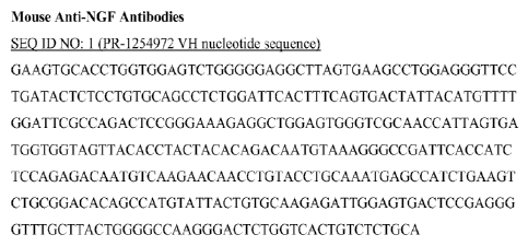
Figure 1 illustrates PR-1254972 VH nucleotide sequence (SEQ ID NO: 1) of mouse
anti-NGF
antibody.
11
CA 02808577 2013-02-15
WO 2012/024650
PCT/US2011/048518
Figure 2 illustrates PR-1254972 VH amino acid sequence (SEQ ID NO: 2) of mouse
anti-
NGF antibody.
Figure 3 illustrates PR-1254972 VL nucleotide sequence (SEQ ID NO: 3) of mouse
anti-NGF
antibody.
Figure 4 illustrates PR-1254972 VL amino acid (SEQ ID NO: 4) of mouse anti-NGF
antibody.
Figure 5 illustrates PR-1254973 VH nucleotide sequence (SEQ ID NO: 5) of mouse
anti-NGF
antibody.
Figure 6 illustrates PR-1254973 VH amino acid (SEQ ID NO: 6) of mouse anti-NGF
antibody.
Figure 7 illustrates PR-1254973 VL nucleotide sequence (SEQ ID NO: 7) of mouse
anti-NGF
antibody.
Figure 8 illustrates PR-1254973 VL amino acid (SEQ ID NO: 8) of mouse anti-NGF
antibody.
Figure 9 illustrates PR-1254977 VH nucleotide sequence (SEQ ID NO: 9) of mouse
anti-NGF
antibody.
Figure 10 illustrates PR-1254977 VH amino acid (SEQ ID NO: 10) of mouse anti-
NGF
antibody.
Figure 11 illustrates PR-1254977 VL nucleotide sequence (SEQ ID NO: 11) of
mouse anti-
NGF antibody.
Figure 12 illustrates PR-1254977 VL amino acid (SEQ ID NO: 12) of mouse anti-
NGF
antibody.
Figure 13 illustrates PR-1254980 VH nucleotide sequence (SEQ ID NO: 13) of
mouse anti-
NGF antibody.
Figure 14 illustrates PR-1254980 VH amino acid (SEQ ID NO: 14) of mouse anti-
NGF
antibody.
Figure 15 illustrates PR-1254980 VL nucleotide sequence (SEQ ID NO: 15) of
mouse anti-
NGF antibody.
Figure 16 illustrates PR-1254980 VL amino acid (SEQ ID NO: 16) of mouse anti-
NGF
antibody.
Figure 17 illustrates PR-1254981 VH nucleotide sequence (SEQ ID NO: 17) of
mouse anti-
NGF antibody.
12
CA 02808577 2013-02-15
WO 2012/024650
PCT/US2011/048518
Figure 18 illustrates PR-1254981 VH amino acid (SEQ ID NO: 18) of mouse anti-
NGF
antibody.
Figure 19 illustrates PR-1254981 VL nucleotide sequence (SEQ ID NO: 19) of
mouse anti-
NGF antibody.
Figure 20 illustrates PR-1254981 VL amino acid (SEQ ID NO: 20) of mouse anti-
NGF
antibody.
Figure 21 illustrates PR-1254982 VH nucleotide sequence (SEQ ID NO: 21) of
mouse anti-
NGF antibody.
Figure 22 illustrates PR-1254982 VH amino acid (SEQ ID NO: 22) of mouse anti-
NGF
antibody.
Figure 23 illustrates PR-1254982 VL nucleotide sequence (SEQ ID NO: 23) of
mouse anti-
NGF antibody.
Figure 24 illustrates PR-1254982 VL amino acid (SEQ ID NO: 24) of mouse anti-
NGF
antibody.
Figure 25 illustrates mouse anti-NGF mAb caninized by CDR grafting onto canine
Ig
frameworks (CDRs are underlined), SEQ ID NO: 25 (72.1 VH amino acid).
Figure 26 illustrates mouse anti-NGF mAb caninized by CDR grafting onto canine
Ig
frameworks (CDRs are underlined), SEQ ID NO: 26 (72.1 VL amino acid).
Figure 27 illustrates mouse anti-NGF mAb caninized by CDR grafting onto canine
Ig
frameworks (CDRs are underlined) SEQ ID NO: 27 (73.1 VH amino acid).
Figure 28 illustrates mouse anti-NGF mAb caninized by CDR grafting onto canine
Ig
frameworks (CDRs are underlined) SEQ ID NO: 28 (73.1 VL amino acid).
Figure 29 illustrates mouse anti-NGF mAb caninized by CDR grafting onto canine
Ig
frameworks (CDRs are underlined), SEQ ID NO: 29 (77.1 VII amino acid).
Figure 30 illustrates mouse anti-NGF mAb caninized by CDR grafting onto canine
Ig
frameworks (CDRs are underlined), SEQ ID NO: 30 (77.1 VL amino acid).
Figure 31A illustrates mouse anti-NGF mAb caninized by CDR grafting onto
canine Ig
frameworks (CDRs are underlined), SEQ ID NO: 31 (81.1 VH amino acid).
Figure 31B illustrates mouse anti-NGF mAb caninized by CDR grafting onto
canine Ig
frameworks (CDRs are underlined), SEQ ID NO: 177 (81.1B VII amino acid).
Figure 32 illustrates mouse anti-NGF mAb caninized by CDR grafting onto canine
Ig
frameworks (CDRs are underlined), SEQ ID NO: 32 (81.1 VL amino acid)
13
CA 02808577 2013-02-15
WO 2012/024650
PCT/US2011/048518
Figure 33 illustrates mouse anti-NGF mAb caninized by CDR grafting onto canine
Ig
frameworks (CDRs are underlined), SEQ ID NO: 33 (82.1 VH amino acid)
Figure 34 illustrates mouse anti-NGF mAb caninized by CDR grafting onto canine
Ig
frameworks (CDRs are underlined), SEQ ID NO: 34 (82.1 VL amino acid).
Figure 35 illustrates caninized anti-NGF antibodies containing back mutation
residues
(backmutation residues shown in bold), SEQ ID NO: 35 (72.2 VH amino acid).
Figure 36A illustrates caninized anti-NGF antibodies containing back mutation
residues
(backmutation residues shown in bold), SEQ ID NO: 36 (72.2 VL amino acid).
Figure 36B illustrates caninized anti-NGF antibodies containing back mutation
residues
(backmutation residues shown in bold), SEQ ID NO: 179 (72.3 VH amino acid).
Figure 36C illustrates caninized anti-NGF antibodies containing back mutation
residues
(backmutation residues shown in bold), SEQ ID NO: 180 (72.4 VII amino acid).
Figure 36D illustrates caninized anti-NGF antibodies containing back mutation
residues
(backmutation residues shown in bold), SEQ ID NO: 181 (72.4 VL amino acid).
Figure 37 illustrates caninized anti-NGF antibodies containing back mutation
residues
(backmutation residues shown in bold), SEQ ID NO: 37 (73.2 VH amino acid).
Figure 38A illustrates caninized anti-NGF antibodies containing back mutation
residues
(backmutation residues shown in bold), SEQ ID NO: 38 (73.2 VL amino acid).
Figure 38B illustrates caninized anti-NGF antibodies containing back mutation
residues
(backmutation residues shown in bold), SEQ ID NO: 182 (73.4 VII amino acid).
Figure 38C illustrates caninized anti-NGF antibodies containing back mutation
residues
(backmutation residues shown in bold), SEQ ID NO: 183 (73.4 VL amino acid).
Figure 39 illustrates caninized anti-NGF antibodies containing back mutation
residues
(backmutation residues shown in bold), SEQ ID NO: 39 (77.2 VII amino acid).
Figure 40A illustrates caninized anti-NGF antibodies containing back mutation
residues
(backmutation residues shown in bold), SEQ ID NO: 40 (77.2 VL amino acid).
Figure 40B illustrates caninized anti-NGF antibodies containing back mutation
residues
(backmutation residues shown in bold), SEQ ID NO: 184 (77.3 VII amino acid).
Figure 40C illustrates caninized anti-NGF antibodies containing back mutation
residues
(backmutation residues shown in bold), SEQ ID NO: 185 (77.4 VII amino acid).
Figure 40D illustrates caninized anti-NGF antibodies containing back mutation
residues
(backmutation residues shown in bold), SEQ ID NO: 186 (77.4 VL amino acid).
14
CA 02808577 2013-02-15
WO 2012/024650
PCT/US2011/048518
Figure 41 illustrates caninized anti-NGF antibodies containing back mutation
residues
(backmutation residues shown in bold), SEQ ID NO: 41(81.2 VH amino acid).
Figure 42A illustrates caninized anti-NGF antibodies containing back mutation
residues
(backmutation residues shown in bold), SEQ ID NO: 42 (81.2 VL amino acid).
Figure 42B illustrates caninized anti-NGF antibodies containing back mutation
residues
(backmutation residues shown in bold), SEQ ID NO: 187 (81.4 VH amino acid).
Figure 42C illustrates caninized anti-NGF antibodies containing back mutation
residues
(backmutation residues shown in bold), SEQ ID NO: 188 (81.4 VL amino acid).
Figure 42D illustrates caninized anti-NGF antibodies containing back mutation
residues
(backmutation residues shown in bold), SEQ ID NO: 189 (81.2B VH amino acid).
Figure 42E illustrates caninized anti-NGF antibodies containing back mutation
residues
(backmutation residues shown in bold), SEQ ID NO: 190 (81.4B VII amino acid).
Figure 42F illustrates caninized anti-NGF antibodies containing back mutation
residues
(backmutation residues shown in bold, SEQ ID NO:206 (81.5B VH amino acid).
Figure 420 illustrates caninized anti-NGF antibodies containing back mutation
residues
(backmutation residues shown in bold, SEQ ID NO:207 (81.6B VH amino acid).
Figure 43 illustrates caninized anti-NGF antibodies containing back mutation
residues
(backmutation residues shown in bold), SEQ ID NO: 43 (82.2 VH amino acid).
Figure 44A illustrates caninized anti-NGF antibodies containing back mutation
residues
(backmutation residues shown in bold), SEQ ID NO: 44 (82.2 VL amino acid).
Figure 44B illustrates caninized anti-NGF antibodies containing back mutation
residues
(backmutation residues shown in bold), SEQ ID NO: 191 (82.3 VL amino acid).
Figure 44C illustrates caninized anti-NGF antibodies containing back mutation
residues
(backmutation residues shown in bold), SEQ ID NO: 192 (82.4 VII amino acid).
Figure 44D illustrates caninized anti-NGF antibodies containing back mutation
residues
(backmutation residues shown in bold), SEQ ID NO: 193 (82.4 VL amino acid).
Figure 45 illustrates primer sequence to clone canine NGF, SEQ ID NO: 45 (NGF-
Dog-S
primer).
Figure 46 illustrates primer sequence to clone canine NGF, SEQ ID NO: 46 (NGF-
Dog-AS
primer).
Figure 47 illustrates primer sequence to clone canine NGF, SEQ ID NO: 47 (NGF-
d-Ec-S
primer).
= CA 02808577 2013-02-15
Figure 48 illustrates primer sequence to clone canine NGF, SEQ ID NO: 48 (NGF-
d-Ec-AS
primer).
Figure 49 illustrates canine NGF C-terminal 6His (SEQ ID NO: 208) fusion
nucleotide
< sequence, SEQ ID NO: 49.
Figure 50 illustrates canine NGF C-terminal 6-His ,(SEQ ID NO: 208) amino acid
sequence,
SEQ ID NO: 50.
Figure 51 illustrates canine IgG constant region nucleotide sequence, SEQ ID
NO: 51.
Figure 52 illustrates canine IgG constant region amino acid sequence, SEQ ID
NO: 52.
Figure 53 illustrates canine kappa constant region nucleotide sequence, SEQ ID
NO: 53
Figure 54 illustrates canine kappa constant region amino acid sequence, SEQ ID
NO: 54.
Figure 55 illustrates complementarity determining region, SEQ ID NO: 55 (72.1
VH amino
acid; CDR1).
Figure 56 illustrates complementarity determining region, SEQ ID NO: 56 (72.1
VH amino
acid; CDR2).
Figure 57 illustrates complementarity determining region, SEQ ID NO: 57 (72.1
VH amino
acid; CDR3).
Figure 58 illustrates complementarily determining region, SEQ ID NO: 58 (72.1
VL amino
acid; CDR I ).
Figure 59 illustrates complementarity determining region, SEQ ID NO: 59 (72.1
VL amino
acid; CDR2).
Figure 60 illustrates complementarity determining region, SEQ ID NO: 60 (72.1
VL amino
acid; CDR3).
Figure 61 illustrates complementarity determining region, SEQ ID NO: 61(73.1
VII amino
acid; CDR I ).
Figure 62 illustrates complementarity determining region, SEQ ID NO: 62 (73.1
VH amino
acid; CDR2).
Figure 63 illustrates complementarity determining region, SEQ ID NO: 63 (73.1
VII amino
acid; CDR3).
Figure 64 illustrates complementarity determining region, SEQ ID NO: 64 (73.1
VL amino
acid; CDR1).
Figure 65 illustrates complementarily determining region, SEQ ID NO: 65 (73.1
VL amino
acid; CDR2).
16
CA 02808577 2013-02-15
WO 2012/024650
PCT/US2011/048518
Figure 66 illustrates complementarity determining region, SEQ ID NO: 66 (73.1
VL amino
acid; CDR3).
Figure 67 illustrates complementarity determining region, SEQ ID NO: 67 (77.1
VII amino
acid; CDR1).
Figure 68 illustrates complementarity determining region, SEQ ID NO: 68 (77.1
VH amino
acid; CDR2).
Figure 69 illustrates complementarily deleunining region, SEQ ID NO: 69 (77.1
VH amino
acid; CDR3).
Figure 70 illustrates complementarity determining region, SEQ ID NO: 70 (77.1
VL amino
acid; CDR1).
Figure 71 illustrates complementarity determining region, SEQ ID NO: 71(77.1
VL amino
acid; CDR2).
Figure 72 illustrates complementarity determining region, SEQ ID NO: 72 (77.1
VL amino
acid; CDR3).
Figure 73 illustrates complementarity determining region, SEQ ID NO: 73 (81.1
VII amino
acid; CDR1).
Figure 74 illustrates complementarity determining region, SEQ ID NO: 74 (81.1
VH amino
acid; CDR2).
Figure 75 illustrates complementarily deleunining region, SEQ ID NO: 75 (81.1
VH amino
acid; CDR3).
Figure 76 illustrates complementarity determining region, SEQ ID NO: 76 (81.1
VL amino
acid; CDR1).
Figure 77 illustrates complementarity determining region, SEQ ID NO: 77 (81.1
VL amino
acid; CDR2).
Figure 78 illustrates complementarity determining region, SEQ ID NO: 78 (81.1
VL amino
acid; CDR3).
Figure 79 illustrates complementarity determining region, SEQ ID NO: 79 (82.1
VII amino
acid; CDR1).
Figure 80 illustrates complementarity determining region, SEQ ID NO: 80 (82.1
VH amino
acid; CDR2).
Figure 81 illustrates complementarity determining region, SEQ ID NO: 81(82.1
VH amino
acid; CDR3).
17
CA 02808577 2013-02-15
WO 2012/024650
PCT/US2011/048518
Figure 82 illustrates complementarity determining region, SEQ ID NO: 82 (82.1
VL amino
acid; CDR1).
Figure 83 illustrates complementarity determining region, SEQ ID NO: 83 (82.1
VL amino
acid; CDR2).
Figure 84 illustrates complementarity determining region, SEQ ID NO: 84 (82.1
VL amino
acid; CDR3).
Figure 85 illustrates the sequence of human I3NGF (SEQ ID NO: 85).
Figure 86 illustrates the sequences shown in Table 12.
Figure 87 illustrates the sequences shown in Table 13.
Figure 88 illustrates the sequences shown in Table 14.
Figure 89 illustrates the sequences shown in Table 15.
Figure 89 illustrates the sequences shown in Table 15A.
Detailed Description of the Disclosure
The disclosure describes NGF binding proteins, particularly anti-NGF
antibodies, or antigen-
binding portions thereof that bind NGF. Various aspects of the disclosure
relate to antibodies and
antibody fragments, and pharmaceutical compositions thereof as well as nucleic
acids, recombinant
expression vectors and host cells for making such antibodies and fragments.
Methods of using the
antibodies of the disclosure to detect human and canine NGF, to inhibit human
and canine NGF
activity, either in vitro or in vivo; and to regulate gene expression are also
encompassed by the
disclosure.
Unless otherwise defined herein, scientific and technical terms used in
connection with the
present disclosure shall have the meanings that are commonly understood by
those of ordinary skill
in the art. The meaning and scope of the terms should be clear, however, in
the event of any latent
ambiguiy, definitions provided herein take precedent over any dictionary or
extrinsic definition.
Further, unless otherwise required by context, singular terms shall include
pluralities and plural terms
shall include the singular. In this application, the use of "or" means
"andior" unless stated otherwise.
Furthermore, the use of the term "including", as well as other forms, such as
"includes" and
"included", is not limiting. Also, terms such as "element" or "component"
encompass both elements
and components comprising one unit and elements and components that comprise
more than one
subunit unless specifically stated otherwise.
Generally, nomenclatures used in connection with, and techniques of, cell and
tissue culture,
molecular biology, immunology, microbiology, genetics and protein and nucleic
acid chemistry and
hybridization described herein are those well known and commonly used in the
art. The methods and
techniques of the present disclosure are generally performed according to
conventional methods well
18
CA 02808577 2013-02-15
WO 2012/024650
PCT/US2011/048518
known in the art and as described in various general and more specific
references that are cited and
discussed throughout the present specification unless otherwise indicated.
Enzymatic reactions and
purification techniques are performed according to manufacturer's
specifications, as commonly
accomplished in the art or as described herein. The nomenclatures used in
connection with, and the
laboratory procedures and techniques of, analytical chemistry, synthetic
organic chemistry, and
medicinal and pharmaceutical chemistry described herein are those well known
and commonly used
in the art. Standard techniques are used for chemical syntheses, chemical
analyses, pharmaceutical
preparation, formulation, and delivery, and treatment of patients.
That the present disclosure may be more readily understood, select terms and
phrases as used
herein are defined below.
A. Defintions
The terms "acceptor" and "acceptor antibody" refer to the antibody or nucleic
acid sequence
providing or encoding at least 80%, at least 85%, at least 90%, at least 95%,
at least 98% or 100% of
the amino acid sequences of one or more of the framework regions. The term
"acceptor" encompasses
an antibody amino acid or nucleic acid sequence providing or encoding the
constant region(s). The
term also encompasses the antibody amino acid or nucleic acid sequence
providing or encoding one or
more of the framework regions and the constant region(s). For example, the
term "acceptor" may
refer to a human antibody amino acid or nucleic acid sequence that provides or
encodes at least 80%,
at least 85%, at least 90%, at least 95%, at least 98%, or 100% of the amino
acid sequences of one or
more of the framework regions. Such an acceptor may contain at least 1, at
least 2, at least 3, least 4,
at least 5, or at least 10 amino acid residues that does (do) not occur at one
or more specific positions
of a human antibody. An acceptor framework region and/or acceptor constant
region(s) may be, e.g.,
derived or obtained from a germline antibody gene, a mature antibody gene, a
functional antibody
(e.g., antibodies well-known in the art, antibodies in development, or
antibodies commercially
available).
The term "agonist" refers to a modulator that, when contacted with a molecule
of interest,
causes an increase in the magnitude of a certain activity or function of the
molecule compared to the
magnitude of the activity or function observed in the absence of the agonist.
Particular agonists of
interest may include, but are not limited to. NGF polypeptides or
polypeptides, nucleic acids,
carbohydrates, or any other molecules that bind to NGF.
The term "antagonist" or "inhibitor" refer to a modulator that, when contacted
with a molecule
of interest causes a decrease in the magnitude of a certain activity or
function of the molecule
compared to the magnitude of the activity or function observed in the absence
of the antagonist.
Particular antagonists of interest include those that block or modulate the
biological or immunological
activity of NGF. Antagonists and inhibitors of NGF may include, but are not
limited to, proteins,
nucleic acids, carbohydrates, or any other molecules, which bind to NGF.
19
CA 02808577 2013-02-15
WO 2012/024650
PCT/US2011/048518
The term "antibody" broadly refers to any immunoglobulin (Ig) molecule
comprised of four
polypeptide chains, two heavy (H) chains and two light (L) chains, or any
functional fragment,
mutant, variant, or derivation thereof which retains the essential epitope
binding features of an Ig
molecule. Such mutant, variant, or derivative anitbody formats are known in
the art. Nonlimiting
examples are discussed herein below.
In a full-length antibody, each heavy chain is comprised of a heavy chain
variable region
(abbreviated herein as HCVR or VH) and a heavy chain constant region. The
heavy chain constant
region is comprised of three domains, CH1, CH2 and CH3. Each light chain is
comprised of a light
chain variable region (abbreviated herein as LCVR or VL) and a light chain
constant region. The
light chain constant region is comprised of one domain, CL. The VH and VL
regions may be further
subdivided into regions of hypervariability, termed complementarity
determining regions (CDR),
interspersed with regions that are more conserved, termed framework regions
(FR). Each VH and VL
is composed of three CDRs and four FRs, arranged from amino-terminus to
carboxy-terminus in the
following order: FR1, CDR1, FR2, CDR2, FR3, CDR3, FR4. Immunoglobulin
molecules may be of
any type (e.g., IgG, IgE, IgM, IgD, IgA and IgY), class (e.g., IgG 1, IgG2,
IgG 3, Ig04, IgAl and
IgA2) or subclass.
The term "antibody conjugate" refers to a binding protein, such as an
antibody, chemically
linked to a second chemical moiety, such as a therapeutic or cytotoxic agent.
The term "agent" is used
herein to denote a chemical compound, a mixture of chemical compounds, a
biological
macromolecule, or an extract made from biological materials. In one aspect the
therapeutic or
cytotoxic agents include, but are not limited to, pertussis toxin, taxol,
cytochalasin B, gramicidin D,
ethidium bromide, emetine, mitomycin, etoposide, tenoposide, vincristine,
vinblastine, colchicin,
doxorubicin, daunorubicin, dihydroxy anthracin dione, mitoxantrone,
mithramycin, actinomycin D, 1-
dehydrotestosterone, glucocorticoids, procaine, tetracaine, lidocaine,
propranolol, and puromycin and
analogs or homologs thereof.
The term "antibody construct" refers to a polypeptide comprising one or more
the antigen
binding portions linked to a linker polypeptide or an immunoglobulin constant
domain. Linker
polypeptides comprise two or more amino acid residues joined by peptide bonds
and are used to link
one or more antigen binding portions. Such linker polypeptides are well known
in the art (Holliger, et
al., Proc. Nad. Acad. Sci., 90: 6444-6448 (1993); Poljak, et al., Structure 2:
1121-1123 (1994)). An
immunoglobulin constant domain refers to a heavy or light chain constant
domain. Human IgG heavy
chain and light chain constant domain amino acid sequences are known in the
art; canine, equine, and
feline are rarer.
The term "antibody fragments" or "antigen-binding moiety" comprises a portion
of a full
length antibody, generally the antigen binding or variable domain thereof.
Examples of antibody
CA 02808577 2013-02-15
WO 2012/024650
PCT/US2011/048518
fragments include Fab, Fab', F(ab)2, Fv , sFy fragments, diabodies, linear
antibodies, single-chain
antibody molecules.
The term "antigen-binding portion" of an antibody (or simply "antibody
portion") refers to
one or more fragments of an antibody that retain the ability to specifically
bind to an antigen (e.g.,
NGF). It has been shown that the antigen-binding function of an antibody may
be performed by
fragments of a full-length antibody. These may also be bispecific, dual
specific, or multi-specific
formats; specifically binding to two or more different antigens. Examples of
binding fragments
encompassed within the term "antigen-binding portion" of an antibody include
(i) a Fab fragment, a
monovalent fragment consisting of the VL, VH, CL and CH1 domains; (ii) a
F(a1:02 fragment, a
bivalent fragment comprising two Fab fragments linked by a disulfide bridge at
the hinge region; (iii)
a Fd fragment consisting of the VH and CH1 domains; (iv) a PV fragment
consisting of the VL and
VH domains of a single arm of an antibody, (v) a dAb fragment (Ward et al.,
Nature, 341: 544-546
(1989); PCT publication WO 90/05144), which comprises a single variable
domain; and (vi) an
isolated complementarity determining region (CDR). Furthermore, although the
two domains of the
Fv fragment, VL and VH, are coded for by separate genes, they may be joined,
using recombinant
methods, by a synthetic linker that enables them to be made as a single
protein chain in which the VL
and VH regions pair to form monovalent molecules (known as single chain Fv
(scFv) (Bird et al.,
Science, 242: 423-426 (1988); and Huston et al., Proc. Natl. Acad. Sci., 85:
5879-5883 (1988)). Such
single chain antibodies are also intended to be encompassed within the term
"antigen-binding portion"
of an antibody. Other forms of single chain antibodies, such as diabodies are
also encompassed.
Diabodies are bivalent, bispecific antibodies in which VH and VL domains are
expressed on a single
polypeptide chain, but using a linker that is too short to allow for pairing
between the two domains on
the same chain, thereby forcing the domains to pair with complementary domains
of another chain
and creating two antigen binding sites (Holliger, et al., Proc. Nall. Acad.
Sci., 90: 6444-6448 (1993);
Poljak, et al., Structure 2: 1121-1123 (1994)). Such antibody binding portions
are known in the art
(Kontennann and Dubel eds., Antibody Engineering (2001) Springer-Verlag. New
York. 790 pp.
(ISBN 3-540-41354-5).
Still further, an antibody or antigen-binding portion thereof may be part of a
larger
immunoadhesion molecule, formed by covalent or noncovalent association of the
antibody or
antibody portion with one or more other proteins or peptides. Examples of such
immunoadhesion
molecules include use of the streptavidin core region to make a tetrameric
scFv molecule (Kipriyanov,
S.M., et al., Human Antibodies and Hybridomas, 6: 93-101 (1995)) and use of a
cysteine residue, a
marker peptide and a C-terminal polyhistidine tag to make bivalent and
biotinylated scFv molecules
(Kipriyanov, et al., 'VIOL Imtnunol., 31: 1047-1058 (1994)). Antibody
portions, such as Fab and
F(ab')1 fragments, may be prepared from whole antibodies using conventional
techniques, such as
papain or pepsin digestion, respectively, of whole antibodies. Moreover,
antibodies, antibody
21
CA 02808577 2013-02-15
WO 2012/024650
PCT/US2011/048518
portions and immunoadhesion molecules may be obtained using standard
recombinant DNA
techniques, as described herein.
The term "anti-NGF antibody" refers to an antibody which is able to bind to
nerve growth
factor (NGF) and inhibit NGF biological activity and/or downstream pathway(s)
mediated by NGF
signaling. An anti-NGF antibody encompasses antibodies that block, antagonize,
suppress or reduce
(including significantly) NGF biological activity, including downstream
pathways mediated by NGF
signaling, such as receptor binding and/or elicitation of a cellular response
to NGF. Anti-NGF
antibodies encompass those that neutralize NGF biological activity, bind NGF
and prevent NGF
dimerization and/or binding to an NGF receptor (such as p75 and/or trkA),
and/or bind NGF and
prevent trkA receptor dimerization and/or trkA autophosphorylation. Examples
of anti-NGF
antibodies are provided herein.
The term "binding protein" refers to a natural or synthetic polypeptide that
specifically binds
to any portion of a target such as an antigen. The term "binding
proteieencompasses antibodies as
described herein, including an isolated antibody, antigen-binding portion
thereof, or immunologically
functional fragment therof
The term "canine antibody" refers to a naturally-occuring or recombinantly
produced
immunoglobulin composed of amino acid sequences representative of natural
antibodies isolated from
canines of various breeds. Canine antibodies are antibodies having variable
and constant regions
derived from canine germline immunoglobulin sequences. The canine antibodies
of the disclosure
may include amino acid residues not encoded by canine germline immunoglobulin
sequences (e.g.,
mutations introduced by random or site-specific mutagenesis in vitro or by
somatic mutation in vivo),
for example in the CDRs and in particular CDR3. however, the term "canine
antibody" is not
intended to include antibodies in which CDR sequences derived from the
germline of another
mammalian species, such as a mouse, have been grafted onto canine framework
sequences.
The term "caninization" is defined as a method for transferring non-canine
antigen-binding
amino acids from a donor antibody to a canine antibody acceptor framework to
generate protein
therapeutic treatments useful in dogs.
The term "caninized antibody" refers to antibodies which comprise heavy and
light chain
variable region sequences from a non-canine species (e.g., a mouse) but in
which at least a portion of
the VH and/or VL sequence has been altered to be more "canine-like", i.e.,
more similar to canine
germline variable sequences. One type of caninized antibody is a CDR-grafted
antibody, in which
non-canine CDR sequences are introduced into canine VII and VL sequences to
replace the
corresponding canine CDR sequences.
Caninized forms of non-canine antibodies provided herein are canine antibodies
that contain
sequence derived from a non-canine antibody. For the most part, caninized
antibodies are canine
antibody sequences ("acceptor" or "recipient" antibody) in which hypervariable
region residues of the
CA 02808577 2013-02-15
WO 2012/024650
PCT/US2011/048518
recipient are replaced by hypervariable region residues from a non-canine
species ("donor" antibody)
such as mouse, rat, rabbit, cat, goat, chicken, bovine, horse, llama, camel,
dromedaries, sharks, non-
human primates, human, humanized, recombinant sequence, or an engineered
sequence having the
desired properties. In some instances, framework region (FR) residues of the
canine antibody are
replaced by corresponding non-canine FR residues. Furthermore, caninized
antibodies may include
residues that are not found in the recipient antibody or in the donor
antibody. These modifications are
made to further refine antibody performance. The caninized antibody may also
comprise at least a
portion of an immunoglobulin constant region (Fe) of a canine antibody.
Strategies for caninization
of antibodies include, but are not limited to, the strategies disclosed in WO
2003/060080.
The caninized antibody is an antibody or a variant, derivative, analog or
fragment thereof
which immunospecifically binds to an antigen of interest and which comprises a
framework (FR)
region having substantially the amino acid sequence of a canine antibody and a
complementary
determining region (CDR) having substantially the amino acid sequence of a non-
canine antibody. A
caninized antibody comprises substantially all of at least one, and typically
two, variable domains
(Fab, Fab', F(ab') 2, FabC, Fv) in which all or substantially all of the CDR
regions correspond to those
of a non-canine immunoglobulin (i.e., donor antibody) and all or substantially
all of the framework
regions are those of a canine immunoglobulin consensus sequence. A caninized
antibody also
comprises at least a portion of an immunoglobulin constant region (Fe),
typically that of a canine
immunoglobulin. A canine or caninized antibody may contain both the light
chain as well as at least
the variable domain of a heavy chain. The antibody also may include the CH1,
hinge, CH2, CH3, and
CH4 regions of the heavy chain. A caninized antibody may only contain a
caninized light chain, or
may only contain a caninized heavy chain. An exemplary caninized antibody
contains a caninized
variable domain of a light chain and a caninized variable domain of a heavy
chain.
The term "canonical" residue refers to a residue in a CDR or framework that
defines a
particular canonical CDR structure as defined by Chothia et al. (J. MoL Biol.,
196:901-907 (1987);
Chothia et al., J. MoL Biol., 227:799 (1992). According to Chothia et al.,
critical portions of the CDRs
of many antibodies have nearly identical peptide backbone conformations
despite great diversity at
the level of amino acid sequence. Each canonical structure specifies primarily
a set of peptide
backbone torsion angles for a contiguous segment of amino acid residues
forming a loop.
The term "CDR" refers to the complementarity determining region within
antibody variable
sequences. There are three CDRs in each of the variable regions of the heavy
chain and the light
chain, which are designated CDR1, CDR2 and CDR3, for each of the variable
regions. The term
"CDR set" refers to a group of three CDRs that occur in a single variable
region capable of binding
the antigen. The exact boundaries of these CDRs have been defined differently
according to different
systems. The system described by Kabat (Kabat et al., Sequences of Proteins of
Immunological
Interest (National Institutes of Health, Bethesda, Md. (1987) and (1991)) not
only provides an
23
CA 02808577 2013-02-15
WO 2012/024650
PCT/US2011/048518
unambiguous residue numbering system applicable to any variable region of an
antibody, but also
provides precise residue boundaries defining the three CDRs. These CDRs may be
referred to as
Kabat CDRs. Chothia and coworkers (Chothia & Lesk, J. Mol. Biol. 196:901-917
(1987) and Chothia
et al., Nature 342:877-883 (1989)) found that certain sub- portions within
Kabat CDRs adopt nearly
identical peptide backbone conformations, despite having great diversity at
the level of amino acid
sequence. These sub-portions were designated as LI, L2 and L3 or HI, H2 and H3
where the "L" and
the "H" designates the light chain and the heavy chains regions, respectively.
These regions may be
referred to as Chothia CDRs, which have boundaries that overlap with Kabat
CDRs. Other boundaries
defining CDRs overlapping with the Kabat CDRs have been described by Padlan
(FASEB J. 9:133-
139 (1995)) and MacCallum (J Mol Biol 262(5):732-45 (1996)). Still other CDR
boundary definitions
may not strictly follow one of the above systems, but will nonetheless overlap
with the Kabat CDRs,
although they may be shortened or lengthened in light of prediction or
experimental findings that
particular residues or groups of residues or even entire CDRs do not
significantly impact antigen
binding. The methods used herein may utilize CDRs defined according to any of
these systems,
although certain methods described herein use Kabat or Chothia defined CDRs.
The term "CDR-grafted antibody" refers to antibodies which comprise heavy and
light chain
variable region sequences from one species but in which the sequences of one
or more of the CDR
regions of VH and/or VL are replaced with CDR sequences of another species,
such as antibodies
having murine heavy and light chain variable regions in which one or more of
the murine CDRs (e.g.,
CDR3) has been replaced with human CDR sequences.
The term "chimeric antibody" refers to antibodies which comprise heavy and
light chain
variable region sequences from one species and constant region sequences from
another species, such
as antibodies having murine heavy and light chain variable regions linked to
human, canine, equine,
or feline constant regions. Chimeric antibodies comprise a portion of the
heavy and/or light chain that
is identical to or homologous with corresponding sequences from antibodies
derived from a particular
species or belonging to a particular antibody class or subclass, while the
remainder of the chain(s) is
identical to or homologous with corresponding sequences in antibodies from
another species or
belonging to another antibody class or subclass, as well as fragments of such
antibodies, exhibiting
the desired biological activity (See e.g., U.S. Pat. No. 4,816,567; and
Morrison et al., Proc. Natl.
Acad. Sci. USA 81:6851-6855 (1984)).
The terms "crystal" and "crystallized" refer to an antibody, or antigen
binding portion
thereof, that exists in the form of a crystal. Crystals are one form of the
solid state of matter, which
is distinct from other forms such as the amorphous solid state or the liquid
crystalline state. Crystals
are composed of regular, repeating, three-dimensional arrays of atoms, ions,
molecules (e.g.,
proteins such as antibodies), or molecular assemblies (e.g., antigen/antibody
complexes). These
three-dimensional arrays are arranged according to specific mathematical
relationships that are well-
24
CA 02808577 2013-02-15
WO 2012/024650
PCT/US2011/048518
understood in the field. The fundamental unit, or building block, that is
repeated in a crystal is called
the asymmetric unit. Repetition of the asymmetric unit in an arrangement that
conforms to a given,
well-defined crystallographic symmetry provides the "unit cell" of the
crystal. Repetition of the unit
cell by regular translations in all three dimensions provides the crystal. See
Giege, R. and Ducruix,
A. Barrett, Crystallization of Nucleic Acids and Proteins, a Practical
Approach, 2nd ea., pp. 20 1-16,
Oxford University Press, New York, New York, (1999).
The term "diabodies" refers to small antibody fragments with two antigen-
binding sites,
which fragments comprise a heavy chain variable domain (VH) connected to a
light chain variable
domain (VL) in the same polypeptide chain (VH-VL). By using a linker that is
short to allow pairing
between the two domains on the same chain, the domains are forced to pair with
the complementary
domains of another chain and create two antigen-binding sites.
The terms "donor" and "donor antibody" refer to an antibody providing one or
more CDRs.
A donor antibody may be an antibody from a species different from the antibody
from which the
framework regions are obtained or derived. In the context of a humanized
antibody, the term "donor
antibody" refers to a non-human antibody providing one or more CDRs. In the
context of a caninized
antibody, the term "donor antibody" refers to a non-canine antibody providing
one or more CDRs. In
the context of a felinized antibody, the term "donor antibody" refers to a non-
feline antibody
providing one or more CDRs. In the context of a equinized antibody, the term
"donor antibody"
refers to a non-equine antibody providing one or more CDRs.
The term "epitope" includes any polypeptide determinant capable of specific
binding to an
immunoglobulin or T-cell receptor. Epitope determinants may include chemically
active surface
groupings of molecules such as amino acids, sugar side chains, phosphoryl, or
sulfonyl, and may have
specific three dimensional structural characteristics, and/or specific charge
characteristics. An epitope
is a region of an antigen that is bound by an antibody. An antibody is said to
specifically bind an
antigen when it preferentially recognizes its target antigen in a complex
mixture of proteins andior
macromolecules.
The term "equine antibody" refers to a naturally-occuring or recombinantly
produced
immunoglobulin composed of amino acid sequences representative of natural
antibodies isolated from
equines of various breeds. Equine antibodies are antibodies having variable
and constant regions
derived from equine gemiline immunoglobulin sequences. The equine antibodies
of the disclosure
may include amino acid residues not encoded by equine germline immunoglobulin
sequences (e.g.,
mutations introduced by random or site-specific mutagenesis in vitro or by
somatic mutation in vivo),
for example in the CDRs and in particular CDR3. However, the term "equine
antibody" is not
intended to include antibodies in which CDR sequences derived from the
germline of another
mammalian species, such as a mouse, have been grafted onto equine framework
sequences.
CA 02808577 2013-02-15
WO 2012/024650
PCT/US2011/048518
The term "equinization" is defined as a method for transferring non-equine
antigen-binding
amino acids from a donor antibody to an equine antibody acceptor framework to
generate protein
therapeutic treatments useful in horses.
The term "equinized antibody" refers to antibodies which comprise heavy and
light chain
variable region sequences from a non-equine species (e.g., a mouse) but in
which at least a portion of
the VH and/or VL sequence has been altered to be more "equine-like", i.e.,
more similar to equine
gen-nline variable sequences. One type of equinized antibody is a CDR-grafted
antibody, in which
non-equine CDR sequences are introduced into equine VH and VL sequences to
replace the
corresponding equine CDR sequences.
Equinized forms of non-equine antibodies provided herein are equine antibodies
that contain
sequence derived from a non-equine antibody. For the most part, equinized
antibodies are equine
antibody sequences ("acceptor" or "recipient" antibody) in which hypervariable
region residues of the
recipient are replaced by hypervariable region residues from a non-equine
species ("donor" antibody)
such as mouse, rat, rabbit, cat, dogs, goat, chicken, bovine, horse, llama,
camel, dromedaries, sharks,
non-human primates, human, humanized, recombinant sequence, or an engineered
sequence having
the desired properties. In some instances, framework region (FR) residues of
the equine antibody are
replaced by corresponding non-equine FR residues. Furthermore, equinized
antibodies may include
residues that are not found in the recipient antibody or in the donor
antibody. These modifications are
made to further refine antibody performance. The equinized antibody may also
comprise at least a
portion of an immunoglobulin constant region (Fe) of an equine antibody.
The equinized antibody is an antibody or a variant, derivative, analog or
fragment thereof
which immunospecifically binds to an antigen of interest and which comprises a
framework (FR)
region having substantially the amino acid sequence of a equine antibody and a
complementary
determining region (CDR) having substantially the amino acid sequence of a non-
equine antibody.
An equinized antibody comprises substantially all of at least one, and
typically two, variable domains
(Fab, Fab', F(ab') 2, FabC, Fv) in which all or substantially all of the CDR
regions correspond to those
of a non-equine immunoglobulin (i.e., donor antibody) and all or substantially
all of the framework
regions are those of an equine immunoglobulin consensus sequence. An equinized
antibody also
comprises at least a portion of an immunoglobulin constant region (Fe),
typically that of an equine
immunoglobulin. An equine or equinized antibody for example may contain both
the light chain as
well as at least the variable domain of a heavy chain. The antibody also may
include the CH1, hinge,
CH2, CH3, and CH4 regions of the heavy chain. An equinized antibody may only
contain an
equinized light chain, or an equinized heavy chain. An exemplary equinized
antibody contains an
equinized variable domain of a light chain an equinized variable domain of a
heavy chain. Equine
isotypes include, for example, IgGa, IgGb, IgGc, IgG(T), IgM, IgA
26
CA 02808577 2013-02-15
WO 2012/024650
PCT/US2011/048518
The term "Fab" refers to antibody fragments. Papain digestion of antibodies
produces two
identical antigen-binding fragments, called "Fab" fragments, each with a
single antigen-binding site,
and a residual "Fc" fragment, whose name reflects its ability to readily
crystallize. Pepsin treatment
yields a binding cross-linking antigen. The Fab fragment also contains the
constant domain of the
light chain and the first constant domain (CH1) of the heavy chain. Fab'
fragments differ from Fab
fragments by the addition of a few residues at the carboxyl terminus of the
heavy chain CHI domain
including one or more cysteine(s) from the antibody hinge region. Fab'-SH is
the designation herein
for Fab' in which the cysteine residue(s) of the constant domains bear a free
thiol group. F(ab')2
antibody fragments originally were produced as pairs of Fab' fragments which
have hinge cysteines
between them. Other chemical couplings of antibody fragments are also known.
The term "feline antibody" refers to a naturally-occuring or recombinantly
produced
immunoglobulin composed of amino acid sequences representative of natural
antibodies isolated from
felines of various breeds. Feline antibodies are antibodies having variable
and constant regions
derived from feline germline immunoglobulin sequences. The feline antibodies
of the disclosure may
include amino acid residues not encoded by feline getinline immunoglobulin
sequences (e.g.,
mutations introduced by random or site-specific mutagenesis in vitro or by
somatic mutation in vivo),
for example in the CDRs and in particular CDR3. however, the term "feline
antibody" is not intended
to include antibodies in which CDR sequences derived from the germline of
another mammalian
species, such as a mouse, have been grafted onto feline framework sequences.
The term "felinization" is defined as a method for transferring non-feline
antigen-binding
amino acids from a donor antibody to a feline antibody acceptor framework to
generate protein
therapeutic treatments useful in cats.
The term "felinized antibody" refers to antibodies which comprise heavy and
light chain
variable region sequences from a non-feline species (e.g., a mouse) but in
which at least a portion of
the VH and/or VL sequence has been altered to be more "feline-like", i.e.,
more similar to feline
germline variable sequences. One type of felinized antibody is a CDR-grafted
antibody, in which
non-feline CDR sequences are introduced into feline VU and VL sequences to
replace the
corresponding feline CDR sequences.
Felinized forms of non-feline antibodies provided herein are feline antibodies
that contain
sequence derived from a non-feline antibody. For the most part, felinized
antibodies are feline
antibody sequences ("acceptor" or "recipient" antibody) in which hypervariable
region residues of the
recipient are replaced by hypervariable region residues from a non-feline
species ("donor" antibody)
such as mouse, rat, rabbit, cat, dogs, goat, chicken, bovine, horse, llama,
camel, dromedaries, sharks,
non-human primates, human, humanized, recombinant sequence, or an engineered
sequence having
the desired properties. In some instances, framework region (FR) residues of
the feline antibody are
replaced by corresponding non-feline FR residues. Furthermore, felinized
antibodies may include
27
CA 02808577 2013-02-15
WO 2012/024650
PCT/US2011/048518
residues that are not found in the recipient antibody or in the donor
antibody. These modifications are
made to further refine antibody performance. The felinized antibody may also
comprise at least a
portion of an immunoglobulin constant region (Fc) of a feline antibody.
The felinized antibody is an antibody or a variant, derivative, analog or
fragment thereof
which immuno specifically binds to an antigen of interest and which comprises
a framework (FR)
region having substantially the amino acid sequence of a feline antibody and a
complementary
determining region (CDR) having substantially the amino acid sequence of a non-
feline antibody. A
felinized antibody comprises substantially all of at least one, and typically
two, variable domains
(Fab, Fab', F(ab') 2, FabC, Fv) in which all or substantially all of the CDR
regions correspond to those
of a non-feline immunoglobulin (i.e., donor antibody) and all or substantially
all of the framework
regions are those of a feline immunoglobulin consensus sequence. A felinized
antibody also
comprises at least a portion of an immunoglobulin constant region (Fe),
typically that of a feline
immunoglobulin. A feline or felinized antibody may contain both the light
chain as well as at least
the variable domain of a heavy chain. The antibody also may include the CHL
hinge. CH2, CH3, and
CH4 regions of the heavy chain. A felinized antibody may only contain a
felinized light chain or a
felinized heavy chain. An exemplary felinized antibody only contains a
felinized variable domain of a
light chain and a felinized variable domain of a heavy chain.
The term "framework" or "framework sequence" refers to the remaining sequences
of a
variable region minus the CDRs. Because the exact definition of a CDR sequence
may be determined
by different systems, the meaning of a framework sequence is subject to
correspondingly different
interpretations. The six CDRs (CDR-L1, -L2, and -L3 of light chain and CDR-H1,
-H2, and -H3 of
heavy chain) also divide the framework regions on the light chain and the
heavy chain into four sub-
regions (FR1, FR2, FR3 and FR4) on each chain, in which CDR1 is positioned
between FR1 and
FR2, CDR2 between FR2 and FR3, and CDR3 between FR3 and FR4. Without
specifying the
particular sub-regions as FR1, FR2, FR3 or FR4, a framework region, as
referred by others, represents
the combined FR's within the variable region of a single, naturally occurring
immunoglobulin chain.
An FR represents one of the four sub- regions, and FRs represents two or more
of the four sub-
regions constituting a framework region. Human heavy chain and light chain
acceptor sequences are
known in the art. Canine heavy chain and light chain acceptor sequences are
also known (patent
application publication W003/060080 and US patent 7261890B2).
The term "germline antibody gene" or "gene fragment" refers to an
immunoglobulin sequence
encoded by non- lymphoid cells that have not undergone the maturation process
that leads to genetic
rearrangement and mutation for expression of a particular immunoglobulin
(Shapiro et al., Crit Rev.
Immunol. 22(3): 183-200 (2002); Marchalonis et al., Adv Exp Med Biol. 484:13-
30 (2001)). One of
the advantages provided by the binding proteins of the present disclosure
stems from the recognition
that germline antibody genes are more likely than mature antibody genes to
conserve essential amino
28
CA 02808577 2013-02-15
WO 2012/024650
PCT/US2011/048518
acid sequence structures characteristic of individuals in the species, hence
less likely to be recognized
as from a foreign source when used therapeutically in that species.
The term "Fv" refers to the minimum antibody fragment that contains a complete
antigen-
recognition and binding site. This region consists of a dimer of one heavy
chain and one light chain
variable domain.
The term "human antibody" refers to antibodies having variable and constant
regions derived
from human germline immunoglobulin sequences. The human antibodies of the
disclosure may
include amino acid residues not encoded by human germline immunoglobulin
sequences (e.g.,
mutations introduced by random or site-specific mutagenesis in vitro or by
somatic mutation in vivo),
for example in the CDRs and in particular CDR3. However, the term "human
antibody" is not
intended to include antibodies in which CDR sequences derived from the
germline of another
mammalian species, such as a mouse, have been grafted onto human framework
sequences.
The term "humanized antibody" refers to antibodies which comprise heavy and
light chain
variable region sequences from a non-human species (e.g., a mouse) but in
which at least a portion of
the VH and/or VL sequence has been altered to be more "human-like", i.e., more
similar to human
germline variable sequences. One type of humanized antibody is a CDR-grafted
antibody, in which
non-human CDR sequences are introduced into human VII and VL sequences to
replace the
corresponding human CDR sequences.
The humanized antibody is an antibody or a variant, derivative, analog or
fragment thereof
which immunospecifically binds to an antigen of interest and which comprises a
framework (FR)
region having substantially the amino acid sequence of a human antibody and a
complementary
determining region (CDR) having substantially the amino acid sequence of a non-
human antibody. A
humanized antibody comprises substantially all, or at least one, and typically
two, variable domains
(Fab, Fab', F(ab') 2, FabC, Fv) in which all or substantially all of the CDR
regions correspond to those
of a non-human immunoglobulin (i.e., donor antibody) and all or substantially
all of the framework
regions are those of a human immunoglobulin consensus sequence. A humanized
antibody also
comprises at least a portion of an immunoglobulin constant region (Fe),
typically that of a human
immunoglobulin. A humanized or caninized antibody may contain both the light
chain as well as at
least the variable domain of a heavy chain. The antibody also may include the
CHL hinge, CH2,
CH3, and CH4 regions of the heavy chain. Alternatively, a humanized antibody
may only contain a
humanized light chain, or a humanized heavy chain. An exemplary humanized
antibody contains a
humanized variable domain of a light chain and a humanized variable domain of
a heavy chain.
The bovanized, camelized, caninized, equinized, felinized, or humanized
antibody may be
selected from any class of immunoglobulins, including IgM, IgG, IgD, IgA and
IgE, and any isotype,
including without limitation IgG 1, IgG2, IgG3 and IgG4. The bovanized,
camelized, caninized,
equinized, felinized, or humanized antibody may comprise sequences from more
than one class or
29
WO 20121024650
PCOUS21111/1148518
isotype, and particular constant domains may be selected to optimize desired
effector functions using
techniques well- known in the art.
The framework and CDR regions of a bovanized, cainelized, caninized,
equinized,
or humanized antibody- need not correspond precisely to the parental
sequences, e.g., the donor
antibody CDR of the consensus framework may be mutagenized by substitution,
insertion and/or
deletion of at least one amino acid residue so that the CDR or framework
residue at that site does not
correspond to either the donor antibody or the consensus framework. Such
mutations, however, will
not be extensive. Usually, at least 50%, at least 60%, at least 70%. at least
80%, at least 90% and at
least 95% oldie bovanized. eamelized. caninized, equinized, felinized, or
humanized antibody
residues will correspond to those of the parental FR and CDR sequences. The
term "consensus
framework" refers to the framework region in the consensus immunoglobulin
sequence. The term
"consensus immunoglobulin sequence' refers to the sequence formed from the
most frequently
occurring amino acids (or nucleotides) in a family of related immunoglobulin
sequences (See e.g.,
Winnakcr, From Genes to Clones (Vcrlagsgcsellschaft, Weinhcim. Germany 1987).
In a family of
immunoglobolins, each position in the consensus sequence is occupied by the
amino acid occurring
most frequently at that position in the family. If two amino acids occur
equally frequently, either 'nay
be included in the consensus sequence.
The term "hypervariable region" refers to the amino acid residues of an
antibody which are
responsible for antigen-binding. The hypervariable region comprises amino acid
residues from a
21) "emnplementarity determining -region" or "CDR" irt the light chain
variable domain and in. the heavy
chain variable domain as defined by Kabat et al., 5th Ed. Public Health
Service, National Institutes of
Health, Bethesda, Md. (1991) and/or as defined by (Chothia and Lesk, Mol.
Biol. I 96:901-917 (1987)
and/or as defined as "AbM loops" by Martin, et tn., .Proc.. Nall. Acad. Set.
USA, 86:9268-9272 (1989)
and/or as defined by 1.ellanc et al., Nucleic Acids Res, 27:209-217 (1999) in
the international
hnMunoCieneTiWinfom-tation systems database. "Framework." or "FR" residues are
those variable
domain residues other than the hypervariabIe region residues as herein
defined.
The term "identity" refers to a relationship between the sequences of two or
more potypeptide
molecules or two or more nucleic acid molecules, as determined by comparing
their sequences
thereof, wherein "identity" refrs more specifically to the degree of sequence
relatedness between
nucleic acid molecules or polypeptides, as determined by the match between
strings of two or more
nucleotide or two or more amino acid sequences. "Identity" measures the
percent of identical matches
between the smaller of two or more sequences with gap alignments if any)
addressed by a particular
mathematical model or computer program (i.e., "algorithms"). The term
"similarity" is used to refer
to a related concept with respect to the relationship oftwo or more nucleic
acid molcies or two or more
polypeptide molcules. In contrast to "identity," "similarity" refers to a
measure of relatedness, which
includes both identical matches and conservative substitution matches. For
example, for two
CA 28 08 577 2 01 7-10 ¨1 8
CA 02808577 2013-02-15
WO 2012/024650
PCT/US2011/048518
polypeptide sequences that have 50/100 identical amino acids, and the
remainder are all non-
conservative substitutions, then the percent identity and similarity would
both be 50%. With respect to
the same two sequences, if 25 more positions had conservative substitutions,
then the percent identity
remains 50%, while percent similarity would be 75% (75/100). Identity and
similarity of related
nucleic acids and polypeptides may be readily calculated by methods well known
and readily
available in the art, including but are not limited to, those described in
COMPUTATIONAL
MOLECULAR BIOLOGY, (Lesk, A. M., ed.), 1988, Oxford University Press, New
York;
BIOCOMPUTING: INFORMATICS AND GENOME PROJECTS, (Smith, D. W., ed.), 1993,
Academic Press, New York; COMPUTER ANALYSIS OF SEQUENCE DATA, Part 1,
(Griffin, A.
M., and Griffin, H. G., eds.), 1994, Humana Press, New Jersey; von Heinje, G.,
SEQUENCE
ANALYSIS IN MOLECULAR BIOLOGY, 1987, Academic Press; SEQUENCE ANALYSIS
PRIMER, (Gribskov, M. and Devereux, J., eds.), 1991, M. Stockton Press, New
York; Carillo et al.,
1988, SIAM J. Applied Math., 48:1073; and Durbin et al., 1998, BIOLOGICAL
SEQUENCE
ANALYSIS, Cambridge University Press.
Preferred methods to determine identity are designed to provide the highest
match between
the compared sequences, and are well described in readily publicly available
computer programs.
Preferred such computerized methods for determining identity between two
sequences include, but are
not limited to, the GCG program package, including GAP (Devereux et al., 1984,
Nucl. Acid. Res.,
12:387; Genetics Computer Group. University of Wisconsin, Madison, Wis.),
BLASTP, BLASTN,
and FASTA (Altschul et al., 1990, J. Mol Biol., 215:403-410). The BLASTX
program is publicly
available from the National Center for Biotechnology Information (NCBI) and
other sources (BLAST
Manual, Altschul et al. NC13/NLMINIII Bethesda, Md. 20894; Altschul et al.,
1990, supra). The well-
known Smith Waterman algorithm may also be used to determine identity.
The terms "individual," "patient," and "subject" are used interchangeably
herein, to refer to
mammals, including, but not limited to, humans, murines, simians, felines,
canines, equines, bovines,
porcines, ovines, caprines, mammalian farm and agricultural animals, mammalian
sport animals, and
mammalian pets. Exemplary subjects companion animals, such as a dog, cat or
horse.
An "isolated antibody" refers to an antibody that is substantially free of
other antibodies
having different antigenic specificities (e.g., an isolated antibody that
specifically binds NGF is
substantially free of antibodies that specifically hind antigens other than
NGF). An isolated antibody
that specifically binds NGF may, however, have cross-reactivity to other
antigens, such as NGF
molecules from other species. Moreover, an isolated antibody may be
substantially free of other
cellular material and/or chemicals.
The terms "isolated polynucleotide" and "isolated nucleic acid" as used
interchangeably
herein refer to a polynucleotide of genomic, cDNA, or synthetic origin or some
combination thereof,
which by virtue of its origin is not associated with all or a portion of a
polynucleotide in which the
31
CA 02808577 2013-02-15
WO 2012/024650
PCT/US2011/048518
isolated polynucleotide is found in nature, or is linked to a another
polynucleotide to which it is not
linked in nature, or is not found in nature within a larger sequence.
The term "isolated protein" or "isolated polypeptide" is a protein or
polypeptide that by virtue
of its origin or source of derivation is not associated with naturally
associated components that
accompany it in its native state; is substantially free of other proteins from
the same species; is
expressed by a cell from a different species; or does not occur in nature.
Thus, a polypeptide that is
chemically synthesized or synthesized in a cellular system different from the
cell from which it
naturally originates will be "isolated" from its naturally associated
components. A protein may also be
rendered substantially free of naturally associated components by isolation,
using protein purification
techniques well known in the art.
The term "Kd" refers to the dissociation constant of a particular antibody-
antigen interaction
as is known in the art.
The term "Kon" is refers to the on rate constant for association of an
antibody to the antigen
to form the antibody/antigen complex as is known in the art.
ls The term "Koff refers to the off rate constant for dissociation of an
antibody from the
antibody/antigen complex as is known in the art.
The terms "Kabat numbering", "Kabat definitions and "Kabat labeling" are used
interchangeably herein. These terms, which are recognized in the art, refer to
a system of numbering
amino acid residues which are more variable (i.e. hypervariable) than other
amino acid residues in the
heavy and light chain variable regions of an antibody, or an antigen binding
portion thereof (Kabat et
al., Ann. A' Y Acad, Sci., 190:382-391 (1971); and Kabat et al., Sequences of
Proteins of
Immunological Interest, Fifth Edition, U.S. Department of Health and Human
Services, NIH
Publication No. 91-3242 (1991)). For the heavy chain variable region, the
hypervariable region
ranges from amino acid positions 31 to 35 for CDR1, amino acid positions 50 to
65 for CDR2, and
amino acid positions 95 to 102 for CDR3. For the light chain variable region,
the hypervariable
region ranges from amino acid positions 24 to 34 for CDR1, amino acid
positions 50 to 56 for CDR2,
and amino acid positions 89 to 97 for CDR3.
The term "key residue" refers to certain residues within the variable region
that have more
impact on the binding specificity and/or affinity of an antibody, in
particular a mammalized antibody
such as humanized, caninized, equinized or felinized antibody. A key residue
includes, but is not
limited to, one or more of the following: a residue that is adjacent to a CDR,
a potential glycosylation
site (may be either N- or 0-glycosylation site), a rare residue, a residue
capable of interacting with the
antigen, a residue capable of interacting with a CDR, a canonical residue, a
contact residue between
heavy chain variable region and light chain variable region, a residue within
the Vernier zone, and a
residue in the region that overlaps between the Chothia definition of a
variable heavy chain CDR1 and
the Kabat definition of the first heavy chain framework.
CA 02808577 2013-02-15
WO 2012/024650
PCT/US2011/048518
The term "labeled binding protein" refers to a protein with a label
incorporated that provides
for the identification of the binding protein. In one aspect, the label is a
detectable marker, e.g.,
incorporation of a radiolabeled amino acid or attachment to a polypeptide of
biotinyl moieties that
may be detected by marked avidin (e.g., streptavidin containing a fluorescent
marker or enzymatic
activity that may be detected by optical or colorimetric methods). Examples of
labels for
polypeptides include, but are not limited to, the following: radioisotopes or
radionuclides (e.g., 3H,
14C, 35s, 90y, 99Tc, 111/n, 121, 131/, 177Lu, , 166,m
n or 153Sm); fluorescent labels (e.g., FITC,
rhodamine,
lanthanide phosphors), enzymatic labels (e.g., horseradish peroxidase,
luciferase, alkaline
phosphatasc); chemiluminescent markers; biotinyl groups; predetermined
polypeptide epitopes
recognized by a secondary reporter (e.g., leucine zipper pair sequences,
binding sites for secondary
antibodies, metal binding domains, epitope tags); and magnetic agents, such as
gadolinium chelates.
The term "marnmalization" refers to a method for transferring donor antigen-
binding
information to a mammalian antibody acceptor to generate useful therapeutic
treatments. More
specifically, the invention provides methods for felinization, equinization
and caninization of
antibodies.
The term "mammalized antibody" refers to antibodies which comprise heavy and
light chain
variable region sequences from a mammal species (e.g., a mouse) but in which
at least a portion of
the VH and/or VL sequence has been altered to be more like "mammal of
interest," see for example,
humanized, caninized, equinized or felinized antibodies defined herein. Such
mammalized antibodies
include, but are not limited to, bovanized, carnelized, can inized, equinized,
fel in ized, or humanized
antibodies.
The terms "modulate" and "regulate" are used interchangeably and refer to a
change or an
alteration in the activity of a molecule of interest (e.g., the biological
activity of NGF). Modulation
may be an increase or a decrease in the magnitude of a certain activity or
function of the molecule of
interest. Exemplary activities and functions of a molecule include, but are
not limited to, binding
characteristics, enzymatic activity, cell receptor activation, and signal
transduction.
The term "modulator" is a compound capable of changing or altering an activity
or function
of a molecule of interest (e.g., the biological activity of NGF). For example,
a modulator may cause
an increase or decrease in the magnitude of a certain activity or function of
a molecule compared to
the magnitude of the activity or function observed in the absence of the
modulator. A modulator may
be an inhibitor, which decreases the magnitude of at least one activity or
function of a molecule.
Exemplary inhibitors include, but are not limited to, proteins, peptides,
antibodies, peptibodies,
carbohydrates or small organic molecules. Peptibodies are described, e.g., in
W001/83525.
The term "monoclonal antibody" refers to an antibody that is derived from a
single clone,
including any eukaryotic, prokaryotic, or phage clone; and not the method by
which it is produced
and is not limited to antibodies produced through hybridoma technology.
33
The term "multivalent binding protein" is used in this specification to denote
a binding
protein comprising two or more antigen binding sites. The multivalent binding
protein is engineered
to have the three or more antigen binding sites, and is generally not a
naturally occurring antibody.
The term "multispecific binding protein" refers to a binding protein capable
of binding two or more
related or unrelated targets. Dual variable domain (DVD) binding proteins are
binding proteins that
comprise two or more antigen binding sites and are tetravalent or multivalent
binding proteins. Such
DVDs may be monospeeitle, i.e capable of binding one antigen or multispecifie,
i.e. capable of
binding two or more antigens. DVD binding proteins comprising two heavy chain
DVD polypeptides
and two light chain DVD polypcptides arc refered to a -DVD Ig. Each half of a
DVD 1g comprises a
in heavy chain DVD polypcplide, and a light chain DVD polypeptide, and two
antigen binding sites.
Each binding site comprises a heavy chain variable domain and a light chain
variable domain with a
total off CDRs involved in antigen binding per antigen binding site. DVD
binding proteins and
methods of making DVD binding proteins are disclosed in US. Patent Application
No. 11/507,050.
Ore aspect of thedisclosure pertains to a DVD binding protein comprising
binding proteins
capable ofbinding NOE. In another aspect, the DVD binding protein is capable
of binding NGF and
a second target.
The terms "nerve growth factor" and "NGF" refer to nerve growth factor and
variants thereof
that retain at least part of the biological activity NGF. NCF includes all
mammalian species of
native sequence NCI', including 111U E:iie, rat, human, rabbit, canine,
feline, equine, or bovine.
Table 1: Sequence of NGF
Protein Sequence Identifier
Canine N017 C-terminal 6-11is(SEQ ID NO:208) SEQ ID NO: 50
Human NGF
SEQ ID NO: 85
The term "NGF receptor" refers to it polypepticie that is bound by or
activated by NGF. NGF
receptors include the TrkA receptor and thc p75 receptor of any mammalian
species, includine, but
are not limited to, human, =ire, feline, equine, primate, or bovine.
The terms "NGF-related disease" and -NUE-related disorder" encompass any
disease or
disorder in which the activity ofNGF in a subject suffering from the disease
or disorder has been
shown to be or is suspected of being either responsible for the
pathophysiology of the disease or
disorder, or a factor that contributes to a worsening of the disease or
disorder, which may occur as a
result of increased levels of -NCH' or increased sensitivity ofthe subject to
NGF. Accordingly, an
NGF-relateci disease or NGF-relatcd disorder is a disease or disorder in which
reduction of NGF
activity is expected to alleviate the symptoms arid/or progression of the
disease or disorder. Such
diseases and disorders may be evidenced, for example, by an increase in the
concentration of NGF
34
CA 2808577 2017-10-18
CA 02808577 2013-02-15
WO 2012/024650
PCT/US2011/048518
in a biological fluid of a subject suffering from the disorder (e.g., an
increase in the concentration of
NGF in serum, plasma, synovial fluid, etc. of the subject), which may be
detected, for example,
using an anti- NGF antibody as described above. Non-limiting examples of
diseases and disorders
that may be treated with the antibodies of the disclosure include those
diseases and disorders
discussed in the section below pertaining to pharmaceutical compositions of
the antibodies of the
disclosure, and encompass acute pain resulting for example from surgery or
other trauma, and
chronic pain.
The term "neutralizing" refers to neutralization of biological activity of a
NGF when a
binding protein specifically binds NGF. A neutralizing binding protein is a
neutralizing antibody,
whose binding to NGF results in inhibition of a biological activity of NGF.
The neutralizing binding
protein binds NGF and reduces a biologically activity of NGF by at least about
20%, 40%, 60%, 80%,
85% or more. Inhibition of a biological activity of NGF by a neutralizing
binding protein may be
assessed by measuring one or more indicators of NGF biological activity well
known in the art,
including cell proliferation, cell morphology changes, cell signaling, or any
detectable cellular
response.resulting from binding of NGF to the TrkA receptor.
The term "operably linked" refers to a juxtaposition wherein the components
described are in
a relationship permitting them to function in their intended manner. A control
sequence "operably
linked" to a coding sequence is ligated in such a way that expression of the
coding sequence is
achieved under conditions compatible with the control sequences. "Operably
linked" sequences
include both expression control sequences that are contiguous with the gene of
interest and expression
control sequences that act in trans or at a distance to control the gene of
interest. The term "expression
control sequence" refers to polynucleotide sequences which are necessary to
effect the expression and
processing of coding sequences to which they are ligated. Expression control
sequences include
appropriate transcription initiation, termination, promoter and enhancer
sequences; efficient RNA
processing signals such as splicing and polyadenylation signals; sequences
that stabilize cytoplasmic
mRNA; sequences that enhance translation efficiency (i.e., Kozak consensus
sequence); sequences
that enhance protein stability; and when desired, sequences that enhance
protein secretion. The nature
of such control sequences differs depending upon the host organism; in
prokaryotes, such control
sequences generally include promoter, ribosomal binding site, and
transcription termination sequence;
in eukaryotes, generally, such control sequences include promoters and
transcription termination
sequence. The term "control sequences" is intended to include components whose
presence is
essential for expression and processing, and may also include additional
components whose presence
is advantageous, for example, leader sequences and fusion partner sequences.
The term "pharmaceutically acceptable carrier" includes any and all solvents,
dispersion
media, coatings, antibacterial and antifungal agents, isotonic and absorption
delaying agents, and the
like that are physiologically compatible. Examples of pharmaceutically
acceptable carriers include
CA 02808577 2013-02-15
WO 2012/024650
PCT/US2011/048518
one or more of water, saline, phosphate buffered saline, dextrose, glycerol,
ethanol and the like, as
well as combinations thereof. In many cases, it will be preferable to include
isotonic agents, for
example, sugars, polyalcohols such as mannitol, sorbitol, or sodium chloride
in the composition.
Pharmaceutically acceptable carriers may further comprise minor amounts of
auxiliary substances
such as wetting or emulsifying agents, preservatives or buffers, which enhance
the shelf life or
effectiveness of the antibody or antibody portion.
The term "polynucleotide" means a polymeric form of two or more nucleotides,
either
ribonucleotides or deoxvnucleotides or a modified form of either type of
nucleotide. The term
includes single and double stranded forms of DNA. The term "isolated
polynucleotide" shall mean a
polynucleotide (e.g., of genomic, cDNA, or synthetic origin, or some
combination thereof) that, by
virtue of its origin, the "isolated polynucleotide" is not associated with all
or a portion of a
polynucleotide with which the "isolated polynucleotide" is found in nature; is
operably linked to a
polynucleotide that it is not linked to in nature; or does not occur in nature
as part of a larger
sequence.
The tem' "polypeptide refers to any polymeric chain of amino acids. The terms
"peptide"
and "protein" are used interchangeably with the term polypeptide and also
refer to a polymeric chain
of amino acids. The term "polypeptide" encompasses native or artificial
proteins, protein fragments
and polypeptide analogs of a protein sequence. A polypeptide may be monomeric
or polymeric.
The term "prophylactically effective amount" refers to an amount effective, at
dosages and for
periods of time necessary, to achieve the desired prophylactic result.
The term "recombinant host cell" (or simply "host cell") is intended to refer
to a cell into
which exogenous DNA has been introduced. It should be understood that such
terms are intended to
refer not only to the particular subject cell, but, to the progeny of such a
cell. Because certain
modifications may occur in succeeding generations due to either mutation or
environmental
influences, such progeny may not, in fact, be identical to the parent cell,
but are still included within
the scope of the term "host cell". In one aspect, host cells include
prokaryotic and eukaryotic cells
selected from any of the Kingdoms of life. Eukaryotic cells include protist,
fungal, plant and animal
cells. In another aspect host cells include, but are not limited to, the
prokaryotic cell line E.Coli;
mammalian cell lines CHO, HEK 293 and COS; the insect cell line Sf9; and the
fungal cell
Saccharomyces cerevisiae.
Standard techniques may be used for recombinant DNA, oligonucleotide
synthesis, and tissue
culture and transformation (e.g., electroporation, lipofection). Enzymatic
reactions and purification
techniques may be performed according to manufacturer's specifications or as
commonly
accomplished in the art or as described herein. The foregoing techniques and
procedures may be
generally performed according to conventional methods well known in the art
and as described in
various general and more specific references that are cited and discussed
throughout the present
36
WO 2012/024650 'PC:I/USN
1/04/1511;
specification. See c.a., Sambrook et al, Molecular Cloning: A Laboratory
Manual (2d ed.., Cold
Spring Ilarbor Laboratory Press, Cold Spring Harbor, N.Y. (1989)).
The term "recombinant antibody" refers to all species of antibodies or
immuneglobulins that
are prepared, expressed, created or isolated by recombinant means, such as
antibodies expressed using
a recombinant expression vector transfected into a host cell. antibodies
isolated from a recombinant.
combinatorial human antibody library aloogenboom, =71B Tech., 15: 62-70(1997);
A:m.2,y et al..
Riochem., 35: 425-445 (2002); Gavilondo et al., Rio:Techniques, 29: 128-145
(2002);
Ilooeenboom et al, Immunology Today, 21: 371-378 (2000)), antibodies isolated
from an animal
(e.g., a mouse) that is transgenic for human immunoglobulin genes (see e.g.,
Taylor* L. D., et al.
(1992) Nucl. Acids Res. 20:6287-6295; Kell-el-mann et al., Current Opinion in
Biotechnology, 13:
593-597 (2002); Little et at, Immunology Today, 21: 364-370 (2000)) or
antibodies prepared,
expressed, created or isolated by any other means that involves splicing of
immunoglobulin gene
sequences to other DNA. sequences. Such recombinant antibodies have variable
and constant regions
derived from species-specific gemiline immunoglobulin sequences. Such
recombinant antibodies may
be subjected to in vitro .mutagenesis (or. when an animal transgenic for human
tg sequences is used, in
vivo somatic trutagenesis) and thus the amino acid sequences of the VII and
VI. regions of the
recombinant antibodies are sequences that, while derived from and related to
species-specific
germline V.11 and VL sequences, may not naturally exist within the antibody
germline repertoire in
vivo.
The term "recovering" refers to the process of rendering a chemical species
such as a
poly peptide substantially free of naturally associated components by
isolation, e.g., using protein
purification techniques well known in the art.
The term "sample" is used. in its broadest sense. A "biological sample"
includes, but. is not
limited to, any quantity of a substance from a living thing or formerly living
thing. Such living things
include, but are not limited to, humans, mice, rats, monkeys, dogs. rabbits
and other animals. Such
substances include, but are not limited to, blood, scrum, urine, synovia I
fluid, cells, organs, tissues,
bone marrow, lymph nodes and spleen.
The term "single-chainFvn or "scFv" refers to antibody fragments comprising
the VII and VL
domains of antibody, wherein these domains arc present in a single polypeptide
chain. Generally, the
Fv polypeptide flirt her comprises a polypeptide linker between the VII and VL
domains which
enables the si'v to form the desired structure for antigen binding. For a
review of scFv, see Pluckthun
in The Pharmacology of Monoclonal Antibodies, vol. 113, Rosenburg and Moore
eds. Sprineer-
Verlag, New York, pp. 269-315 (1994).
The terms "specific binding' or "specifically binding" in reference to the
interaction of an
antibody, a protein, or a peptide with a second chemical species, mean that
the interaction is
37
CA 2808577 2017-10-18
CA 02808577 2013-02-15
WO 2012/024650
PCT/US2011/048518
dependent upon the presence of a particular structure (e.g., an antigenic
determinant or epitope) on the
chemical species; for example, an antibody recognizes and binds to a specific
protein structure rather
than to proteins generally. If an antibody is specific for epitope "A", the
presence of a molecule
containing epitope A (or free, unlabeled A), in a reaction containing labeled
"A" and the antibody.
will reduce the amount of labeled A bound to the antibody.
The term "substantially" in the context of a CDR refers to a CDR having an
amino acid
sequence at least 80%, at least 85%, at least 90%, at least 95%, at least 98%
or at least 99% identical
to the amino acid sequence of a non-human antibody CDR.
The term "surface plasmon resonance" refers to an optical phenomenon that
allows for the
analysis of real-time biospecific interactions by detection of alterations in
protein concentrations
within a biosensor matrix, for example using the BIAcore system (Pharmacia
Biosensor AB, Uppsala,
Sweden and Piscataway, NJ). For further descriptions (Jonsson, et al. Ann.
Biol. Clin. 51: 19-26
(1993); Jonsson, et al., Biotechniques 11: 620-627 (1991); Johnsson, et al.,
.1. Recognit. 8:125-
131 (1995); and Johnnson, B., et al., Anal. Biochem., 198: 268-277 (1991)).
The term "therapeutically effective amount" refers to an amount effective, at
dosages and for
periods of time necessary, to achieve the desired therapeutic result. A
therapeutically effective
amount may be the amount and/or duration of a therapy which is sufficient to
reduce or ameliorate the
severity and/or duration of a disorder or one or more symptoms thereof,
prevent the advancement of a
disorder, cause regression of a disorder, prevent the recurrence, development,
onset or progression of
one or more symptoms associated with a disorder, detect a disorder, or enhance
or improve the
prophylactic or therapeutic effect(s) of another therapy (e.g. , prophylactic
or therapeutic agent). A
therapeutically effective amount of the antibody or antibody portion may be
determined by a person
skilled in the art and may vary according to factors such as the disease
state, age, sex, and weight of
the individual, and the ability of the antibody or antibody portion to elicit
a desired response in the
individual. A therapeutically effective amount is also one in which any toxic
or detrimental effects of
the antibody, or antibody portion, are outweighed by the therapeutically
beneficial effects.
The term "transformation" refers to any process by which exogenous DNA enters
a host cell.
Transformation may occur under natural or artificial conditions using various
methods well known in
the art. Transformation may rely on any known method for the insertion of
foreign nucleic acid
sequences into a prokaryotic or eukaryotic host cell. The method is selected
based on the host cell
being transformed and may include, but is not limited to, viral infection,
electroporation, lipofection,
and particle bombardment. Such "transformed" cells include stably transformed
cells in which the
inserted DNA is capable of replication either as an autonomously replicating
plasmid or as part of the
host chromosome. They also include cells which transiently express the
inserted DNA or RNA for
limited periods of time.
38
WO 2012/011650 -PCT/US20 I
1/04851.8
The term "transgenic organism" refers to an organism having cells that contain
a transgene,
wherein the transgene introduced into the organism (or an ancestor of the
organism) expresses a
polynept i de not naturally expressed in the organism. A. "transgene" is a DNA
construct, which is
stably and, operably integrated into the genome of a cell from which a
transgenic organism develops,
directing the expression of an encoded gene product in one or more cell types
or tissues of the
transgenic organism.
The term "vector" is intended to refer to a nucleic acid molecule capable of
transporting
another nucleic acid to which it has been linked. One type of vector is a
"plasmid", which refers to a
circular double stranded DNA loop into which additional DNA segments may be
ligated. Another
type of vector is a viral vector, wherein additional DNA segments may be
ligated into the viral
genome. Certain vectors are capable of autonomous replication in a host cell
into which they axe
introduced (e.g., bacterial vectors havin 2 a bacterial origin of replication
and episomal mammalian
vectors). Other vectors (e.g., non-episomal mammalian vectors) may be
integrated into the genome of
a host cell upon introduction into the host cell, and thereby arc replicated
along with the host gcnomc.
IS Moreover, certain vectors are capable of directing the expression of
genes to Which they are
operatively linked. Such vectors are referred to herein as "recombinant
expression vectors" (or simply,
"expression vectors"). In general, expression, vectors of utility in
recombinant DNA techniques are
often in the fonn of plasmids. In the present specification, "plasmid" and
"vector" may be used.
interchangeably as the plasmid is the most commonly used form of vector.
However, the disclosure is
211 intended to include such other forms of expression vectors, such as
viral vectors (e.g., replication
defective retro vi ruses, adenoviruses and adeno-associated viruses), which
serve equivalent functions.
The term "Vernier zone" refers to a subset of framework residues that may
adjust CDR
structure and Ellie-tune the fit to antigen as described by Foote and Winter
(1992, ./. Mot. Biol.
224:487-499). Vernier zone residues limn a -layer
25 underlying the ('Ohs and may impact on the structure of CDRs and the
affinity of the antibody.
13. Anti NW; Rindine Proteins
The present disclosure p:-ovides a mivc1 family a binding proteins, aturine
antibodies, CDR.
grafted antibodies, nrammaiized (bovaniztat, camelized, eaninized, equiniied,
felinized, or
30 humanized) antibodies, and fragments thereof, capable of binding and
modulating the biological
activity or function of NCR including the capability of neutralizing NOF, The
disclosure thus also
provides a therapeutic means with which to inhibit NOP and provides
compositions and methods for
treating disease associated with increased levels of NGF, particularly a
disease, condition or disorder
where increased levels orN(11', as compared to NCIF levels observed in
comparable normal subjects,
35 is detrimental.
39
CA 2808577 2017-10-18
CA 02808577 2013-02-15
WO 2012/024650
PCT/US2011/048518
Binding proteins of the present disclosure may be made by any of a number of
techniques
known in the art and as described herein, including culturing a host cell
described herein in culture
medium under conditions sufficient to produce a binding protein capable of
binding NGF.
Monoclonal antibodies may be prepared using a wide variety of techniques known
in the art
including the use of hybridoma, recombinant, and phage display technologies,
or a combination
thereof. For example, monoclonal antibodies may be produced using hybridoma
techniques including
those known in the art and taught, for example, in Harlow et al. , Antibodies:
A Laboratory Manual,
(Cold Spring Harbor Laboratory Press, 2nd ed. 1988); Hammerling, et al., in:
Monoclonal Antibodies
and T-Cell Hybridomas 563-681 (Elsevier, N.Y., 1981).
Methods for producing and screening for specific antibodies using hybridoma
technology
are well known in the art. Such methods include, for example, culturing a
hybridoma cell secreting
an antibody of the disclosure wherein the hybridoma is generated by fusing
splenocytes isolated
from a mouse immunized with an antigen of the disclosure with myeloma cells
and then screening
the hybridomas resulting from the fusion for hybridoma clones that secrete an
antibody able to bind
a polypeptide of the disclosure. Briefly, or example, mice may be immunized
with an NGF antigen.
The NGF antigen may be administered, with or without an adjuvant, to stimulate
the immune
response. Such adjuvants include complete or incomplete Freund's adjuvant,
RIBI (muramyl
dipeptides) or ISCOM (immunostimulating complexes). Such adjuvants may protect
the polypeptide
from rapid dispersal by sequestering it in a local deposit, or they may
contain substances that
stimulate the host to secrete factors that are chemotactic for macrophages and
other components of
the immune system. If a polypeptide is being administered, the immunization
schedule will involve
two or more administrations of the polypeptide, spread out over several weeks.
After immunization of an animal with an NGF antigen, antibodies and/or
antibody-producing
cells may be obtained from the animal. An anti- NGF antibody-containing serum
may be obtained
from the animal by bleeding or sacrificing the animal. The serum may be used
as it is obtained from
the animal, an immunoglobulin fraction may be obtained from the serum, or the
anti- NGF antibodies
may be purified from the serum. Serum or immunoglobulins obtained in this
manner are polyclonal,
thus having a heterogeneous array of properties.
Once an immune response is detected, e.g., antibodies specific for the antigen
NGF are
detected in the mouse serum, the mouse spleen may be harvested and splenocytes
isolated. The
splenocytes are then fused by well-known techniques to any suitable myeloma
cells, such as, for
example, cells from cell line SP20 available from the ATCC. Hybridomas may be
selected and
cloned by limited dilution. The hybridoma clones may then be assayed by
methods known in the art
for cells that secrete antibodies capable of binding NGF. Ascites fluid, which
generally contains high
levels of antibodies, may be generated by immunizing mice with positive
hybridoma clones.
CA 02808577 2013-02-15
WO 2012/024650
PCT/US2011/048518
Antibody-producing immortalized hybridomas may be prepared from the immunized
animal.
After immunization, the animal may be sacrificed and the splenic B cells fused
to immortalized
myeloma cells as is well known in the art (Harlow et al., supra).
Alternatively, the myeloma cells may
be from a non-secretory cell line and do not secrete immunoglobulin
polypeptides. After fusion and
antibiotic selection, the hybridomas may be screened using NGF, or a portion
thereof, or a cell
expressing NGF. Initial screening may be performed, for example, using an
enzyme-linked
immunoassay (ELISA) or a radioirnmunoassay (RIA). An example of ELISA
screening is provided in
WO 00/37504.
Anti-NGF antibody-producing hybridomas may be selected, cloned and further
screened for
desirable characteristics, including robust hybridoma growth, high antibody
production and desirable
antibody characteristics, as discussed further below. Hybridomas may be
cultured and expanded in
vivo in syngeneic animals, in animals that lack an immune system, e.g., nude
mice, or in cell culture in
vitro. Methods of selecting, cloning and expanding hybridomas are well known
to those of ordinary
skill in the art.
An exemplary animal system for preparing hybridomas is the mouse. Hybridoma
production
in the mouse is very well established, and immunization protocols and
techniques for isolation of
immunized splenocytes for fusion are well known. Fusion partners (e.g., murine
myeloma cells) and
fusion procedures are also known. Alternatively, the hybridomas may be
produced in a non-human,
non-mouse species such as a rat, sheep, pig, goat, cattle or horse.
Alternatively, human hybridomas
may be produced, in which a human non-secretory myeloma is fused with a human
cell expressing an
anti- NGF antibody.
Antibody fragments that recognize specific epitopes may be generated by known
techniques.
For example, Fab and F(ab')2 fragments of the disclosure may be produced by
proteolytic cleavage of
immunoglobulin molecules, using enzymes such as papain (to produce Fab
fragments) or pepsin (to
produce F(ab')2 fragments). F(ab')2 fragments contain the variable region, the
light chain constant
region and the CHI domain of the heavy chain.
Recombinant antibodies may be generated from single, isolated lymphocytes
using a
procedure referred to in the art as the selected lymphocyte antibody method
(SLAM), as described in
U.S. Patent No. 5,627,052, PCT Publication WO 92/02551 and Babcock et al.,
Proc. Natl. Acad. ,S'ci.,
93: 7843-7848 (1996). In this method, single cells secreting antibodies of
interest, e.g., lymphocytes
derived from any one of the immunized animals described in Section 1, are
screened using an antigen-
specific hemolytic plaque assay, wherein the antigen NGF, or a fragment
thereof, is coupled to sheep
red blood cells using a linker, such as biotin, and used to identify single
cells that secrete antibodies
with specificity for NGF. Following identification of antibody-secreting cells
of interest, heavy- and
light-chain variable region cDNAs may be rescued from the cells by reverse
transcriptase-PCR and
these variable regions may then be expressed, in the context of appropriate
immunoglobulin constant
41
CA 02808577 2013-02-15
WO 2012/024650
PCT/US2011/048518
regions (e.g., human constant regions), in mammalian host cells, such as COS
or CHO cells. The host
cells transfected with the amplified immunoglobulin sequences, derived from in
vivo selected
lymphocytes, may then undergo further analysis and selection in vitro, for
example by panning the
transfected cells to isolate cells expressing antibodies to NGF. The amplified
immunoglobulin
sequences further may be manipulated in vitro, such as by in vitro affinity
maturation methods such as
those described in PCT Publication WO 97/29131 and PCT Publication WO
00/56772.
Antibodies may be produced by immunizing a non-human animal comprising some or
all of
the human immunoglobulin loci with an NGF antigen. For example, human
monoclonal antibodies
directed against NGF may be generated using transgenic mice carrying parts of
the human immune
system rather than the mouse system, referred to in the literature and herein
as "HuMalf mice,
contain a human immunoglobulin gene minilocus that encodes unrearranged human
heavy (tt and y)
and K light chain immunoglobulin sequences, together with targeted mutations
that inactivate the
endogenous js and lc chain loci (Lonberg et al., 1994, Nature 368:856-859).
These mice exhibit
reduced expression of mouse IgM or x and in response to immunization, and the
introduced human
heavy chain and light chain transgenes undergo class switching and somatic
mutation to generate
high affinity human IgG ic monoclonal antibodies. The preparation of HuMab
mice is well
described in the literature. (See, e.g., Lonberg et al., 1994, Nature 368:856-
859; Lonberg, 1994,
Handbook of Exp. Pharmacology 113:49-101; Taylor et al., 1994, International
Immunology 6:579-
591; Lonberg & Huszar, 1995, Intern. Rev. Immunol. 13:65-93; and Harding &
Lonberg, 1995,
Ann. N.Y. Acad. Sci 764:536-546). Alternatively, other known mouse strains
such as tile HCo7,
HCo12, and KM transgenic mice strains may be used to generate human anti- NGF
antibodies.
Another suitable, though non-limiting example of a transgenic mouse is the
XENOMOUSE
transgenic mouse, which is an engineered mouse strain that comprises large
fragments of the human
immunoglobulin loci and is deficient in mouse antibody production. See, e.g.,
Green et al. Nature
Genetics, 7:13-21 (1994); and United States Patents 5,916,771, 5,939,598,
5,985,615, 5,998,209,
6,075,181, 6,091,001, 6,114,598 and 6,130,364; WO 91/10741, WO 94/02602, WO
96/34096, WO
96/33735, WO 98/16654, WO 98/24893, WO 98/50433, WO 99/45031, WO 99/53049, WO
00
09560, and WO 00/037504. The XENOMOUSE transgenic mouse produces an adult-
like human
repertoire of fully human antibodies, and generates antigen-specific human
Mabs. The
XENOMOUSE transgenic mouse contains approximately 80% of the human antibody
repertoire
through introduction of megabase sized, germline configuration YAC fragments
of the human heavy
chain loci and light chain loci (Mendez et al., Nature Genetics 15:146-156
(1997), Green et al., .J.
Exp. Med., 188: 483-495 (1998)).
In vitro methods also may be used to make the antibodies of the disclosure,
wherein an
antibody library is screened to identify an antibody having the desired
binding specificity. Methods
for such screening of recombinant antibody libraries are well known in the art
and include methods
CA 02808577 2013-02-15
WO 2012/024650
PCT/US2011/048518
described in, for example, U.S. Patent No. 5,223,409; PCT Publication Nos. WO
92/18619; WO
91/17271; WO 92/20791; WO 92/15679; WO 93/01288; WO 92/01047; WO 92/09690;
Fuchs et al.
Bio/Technology, 9:1370-1372 (1991); Hay et al., Ifitm Antibod Hybridomas, 3:
81-85 (1992); Huse et
al., Science, 246: 1275-1281 (1989); McCaffcrty et al., Nature, 348: 552-554
(1990); Griffiths et al.,
EMBO J., 12: 725-734 (1993); Hawkins et al., J Mol Biol., 226: 889-896 (1992);
Clackson et al.,
Nature, 352: 624-628 (1991); Gram et al., PNAS, 89: 3576-3580 (1992); Garrad
et al.,
Bio/Technology, 9:1373-1377 (1991); Hoogenboom et al., Nuc Acid Res, 19: 4133-
4137 (1991); and
Barbas et al., PNAS, 88: 7978-7982 (1991), US patent application publication
20030186374, and PCT
Publication No. WO 97/29131.
The recombinant antibody library may be from a subject immunized with NGF, or
a portion
of NGF. Alternatively, the recombinant antibody library may be from a naïve
subject that has not
been immunized with NGF, such as a canine antibody library from a canine
subject that has not been
immunized with canine NGF. Antibodies of the disclosure are selected by
screening the recombinant
antibody library with the peptide comprising canine NGF to thereby select
those antibodies that
recognize NGF. Methods for conducting such screening and selection are well
known in the art, such
as described in the references in the preceding paragraph. To select
antibodies of the disclosure
having particular binding affinities for hNGF, such as those that dissociate
from canine NGF with a
particular koff rate constant, the art-known method of surface plasmon
resonance may be used to select
antibodies having the desired koff rate constant. To select antibodies of the
disclosure having a
particular neutralizing activity for hNGF, such as those with a particular an
IC50, standard methods
known in the art for assessing the inhibition of hNGF activity may be used.
For example, the antibodies of the present disclosure may also be generated
using various
phage display methods known in the art. In phage display methods, functional
antibody domains are
displayed on the surface of phage particles which carry the polynucleotide
sequences encoding them.
In a particular aspect, such phage may be utilized to display antigen-binding
domains expressed from
a repertoire or combinatorial antibody library (e. g., canine, human or
murine). Phage expressing an
antigen binding domain that binds the antigen of interest may be selected or
identified with antigen,
e.g., using labeled antigen or antigen bound or captured to a solid surface or
bead. Phage used in these
methods are typically filamentous phage including fd and M13 binding domains
expressed from
phage with Fab, Fv or disulfide stabilized Fv antibody domains recombinantly
fused to either the
phage gene III or gene VIII protein. Examples of phage display methods that
may be used to make the
antibodies of the present disclosure include those disclosed in Brinkman et
al., .J. Immunol. Methods,
182: 41-50 (1995); Ames et al., ,1. Immunol. Methods, 184: 177-186 (1995);
Kettleborough et al., Eur.
J. Immunol., 24:952-958 (1994); Persic et al., Gene. 187: 9-18 (1997); Burton
et al., Advances in
Immunology, 57: 191-280 (1994); PCT application No. PCTIGB91/01134; PCT
publications WO
90/02809; WO 91/10737; WO 92/01047; WO 92/18619; WO 93/11236; WO 95/15982; WO
43
WO 2012/024650
PCT1tIS20.11104851.8
95/20401; and U.S.' Pat. Nos, 5,698,426: 5,223,409; 5,403,484; 5,580,717;
5,427,908: 5,750,753;
5,821,047; 5,571,698: 5,427,908; 5,516,637; 5.780, 225; 5.658,727, 5,733,743
and 5,969,108.
As desetibed in the above references, after phage selection, the antibody
coding regions from
the phage may be isolated and used to generate whole antibodies including
human antibodies or any
other desired antigen binding fragment, and. expressed in any desired host,
including mammalian cells,
insect cells, plant cells, yeast. and bacteria, e.g., as described in detail
below. For example, techniques
recombinantly produce Fah. Fab and F(ab')2 fragments may also be employed
using methods
known in the art such as those disclosed in PC'T publication WO 92/22324;
MuIlinax et al.,
Bic?Techniques, 12(6):864-869 (1992); and Sawai et A, MR.I. 34;26-34 (1995);
and 13ctter et al.,
Science, 240:1041-1043 (1988).
Examples of techniques which may be used to produce single-chain EVS and
antibodies include those
described in U.S. Pat. 4,946,778 and 5,258, 498; Huston el al., ,iefethods= in
EnKHriology, 203:46-88
(1991); Sim et al.. P.V.,a, 90:7995-7999 (1993); and Skerra etal., Science,
240:1038-1040 (1988).
Alternatives to screening of recombinant antibody libraries by phagc display
arc known and
include other methodologies for screening large coinbinatorial libraries which
may be applied to (he
identification of dual specificity antibodies of the disclosure. One type of
alternative expression
system is one in which the recombinant antibody library is expressed as RNA-
protein fusions, as
described PCT Publication No. WO 98/3170(1, and in Roberts et al., Proc. Natl.
Acad. Sci.,
94:12297-12302 ((997). In this system, a covalent fusion is created between a
irRNA and the peptide
or protein that it encodes by in vitro translation of synthetic mRNAs that
carry puromycin, a peptidyl
acceptor antibiotic, at their 3' end. Thus, a specific in RNA may be enriched
from is complex mixture
of mRNAs (e.g., a combinatorial library) based on the properties of the
encoded peptide or protein,
e.g., antibody, or portion thereof, such as binding oftbe antibody, or portion
thereof, to the dual
specificity antigen. Nucleic acid sequences encoding antibodies, or portions
thereof, recovered from
screening of such libraries may be expressed by recombinant means as described
above (e.g., in
mammalian host cells) and, moreover, may be subjected to further affinity
maturation by either
additional rounds of screening of tn,RNA-peptide fusions in Which mutations
have been introduced
into the originally selected sequence(s), or by other methods for affinity
maturation in vitro of
recombinant antibodies, as described above.
In another approach the antibodies of the present disclosure may also be
generated or affinity
matured using yeast display methods known in the art. In yeast display
methods, genetic methods are
used to tether antibody domains to the yeast cell wall and display them on the
surface of yeast. In
particular, such yeast tray be utilized to display antigen-binding domains
expressed from a repertoire
or combinatorial antibody library (e.g., human or marine). Examples of yeast
display methods that
may be used to make the ant:bodies of the pre-sent disclosure include those
disclosed Wittrup, et al.
U.S. Patent No. 6,699,658.
44
CA 2808577 2017-10-18
CA 02808577 2013-02-15
WO 2012/024650
PCT/US2011/048518
The antibodies or antigen binding fragments described herein may also be
produced by
genetic engineering. For example, the technology for expression of both heavy
and light chain genes
in E. coli is the subject of the PCT patent applications: publication number
WO 901443, W0901443,
and WO 9014424 and in Huse et al., 1989 Science 246:1275-81. The present
disclosure thus also
encompasses the isolated nucleic acids encoding any of the binding proteins
described herein, as well
as a recombinant vector comprising such a nucleic acid molecule, and a a host
cell comprising such a
recombinant vector.
A vector is a nucleic acid molecule, which may be a construct, capable of
transporting another
nucleic acid to which it has been linked. A vector may include any preferred
or required operational
elements. Preferred vectors are those for which the restriction sites have
been described and which
contain the operational elements needed for transcription of the nucleic acid
sequence. Such
operational elements include for example at least one suitable promoter, at
least one operator, at least
one leader sequence, at least one terminator codon, and any other DNA
sequences necessary or
preferred for appropriate transcription and subsequent translation of the
nucleic acid sequence. Such
vectors contain at least one origin of replication recognized by the host
organism along with at least
one selectable marker and at least one promoter sequence capable of initiating
transcription of the
nucleic acid sequence. A vector may be a plasmid into which additional DNA
segments may be
ligated. A vector may be a viral vector, wherein additional DNA segments may
be ligated into the
viral genome. Certain vectors are capable of autonomous replication in a host
cell into which they are
introduced (e.g., bacterial vectors having a bacterial origin of replication
and episomal mammalian
vectors). Other vectors (e.g., non-episomal mammalian vectors) may be
integrated into the genome of
a host cell upon introduction into the host cell, and thereby are replicated
along with the host genome.
Moreover, certain vectors are capable of directing the expression of genes to
which they are
operatively linked. Such vectors are referred to herein as "recombinant
expression vectors" (or
simply, "expression vectors"). In general, expression vectors of utility in
recombinant DNA
techniques are often in the form of plasmids. In the present specification,
"plasmid" and "vector" may
be used interchangeably as a plasmid is the most commonly used form of vector.
However, the
present disclosure is intended to include such other forms of expression
vectors, such as viral vectors
(e.g., replication defective retroviruses. adenoviruses and adeno-associated
viruses), which serve
equivalent functions. By way of example and not limitation, suitable vectors
include pcDNA, pTT
(Durocher et al., Nucleic Acids Research, Vol 30, No.2 (2002)); pTT3 (pTT with
additional multiple
cloning site, pEFBOS (Mizushima et al., Nucleic acids Research, Vol 18, No. 17
(1990)), pBV,
pBJ, or pHybE (patent publication no.: US 2009/0239259 Al) .
Sequences that are operably linked are in a relationship permitting them to
function in their
intended manner. A control sequence operably linked to a coding sequence is
ligated in such a way
that expression of the coding sequence is achieved under conditions compatible
with the control
CA 02808577 2013-02-15
WO 2012/024650
PCT/US2011/048518
sequences. Operably linked sequences include both expression control sequences
that are contiguous
with the gene of interest and expression control sequences that act in trans
or at a distance to control
the gene of interest. Expression control sequences are polynucleotide
sequences that are necessary to
effect the expression and processing of coding sequences to which they are
ligated. Expression
control sequences include appropriate transcription initiation, termination,
promoter and enhancer
sequences; efficient RNA processing signals such as splicing and
polyadenylation signals; sequences
that stabilize cytoplasmic mRNA; sequences that enhance translation efficiency
(i.e., Kozak
consensus sequence); sequences that enhance protein stability; and when
desired, sequences that
enhance protein secretion. The nature of such control sequences differs
depending upon the host
organism; in prokaryotes, such control sequences generally include promoter,
ribosomal binding site,
and transcription termination sequence; in eukaryotes, such control sequences
generally include
promoters and transcription termination sequence. Control sequences may
include components whose
presence is essential for expression and processing, and may also include
additional components
whose presence is advantageous, for example, leader sequences and fusion
partner sequences.
A host cell may be transformed with a vector that introduces exogenous DNA
into a host cell.
Transformation may occur under natural or artificial conditions using various
methods well known in
the art. Transformation may rely on any known method for the insertion of
foreign nucleic acid
sequences into a prokaryotic or eukaryotic host cell. The method is selected
based on the host cell
being transformed and may include, but is not limited to, viral infection,
electroporation, lipofection,
calcium-phosphate precipitation, DEAE-dextran transfection, particle
bombardment and the like.
Transformed cells include stably transformed cells in which the inserted DNA
is capable of
replication either as an autonomously replicating plasmid or as part of the
host chromosome, and cells
which transiently express the inserted DNA or RNA for limited periods of time.
Host organisms such as host cells are cultured under conditions appropriate
for amplification
of the vector and expression of the protein, as well known in the art.
Expressed recombinant proteins
may be detected by any of a number of methods also well known in the art.
Suitable host organisms include for example a prokaryotic or eukaryotic cell
system. A
eukaryotic cell may be a protist cell, animal cell, plant cell or fungal cell.
A eukaryotic cell is for
example an animal cell which may be a mammalian cell, avian cell, or an insect
cell such as an insect
Sf9 cell. Cells from established and readily available may be used, such as
but not limited to HeLa,
MRC-5 or CV-1. The host cell may be an E.coli cell, or a yeast cell such as
but not limited to
Saccharomyces cerevisiae. Mammalian host cells for expressing the recombinant
antibodies of the
disclosure also include Chinese Hamster Ovary (CHO cells) (including dhfr- CHO
cells, described in
Lirlaub et al., Proc. Natl. Acad. S'ci., 77: 4216-4220 (1980), used with a
DHFR selectable marker, e.g.,
as described in Kaufman et al., Mol. Biol., 159: 601-621 (1982)), NSO myeloma
cells, COS cells and
SP2 cells. When recombinant expression vectors encoding antibody genes are
introduced into
46
CA 02808577 2013-02-15
WO 2012/024650
PCT/US2011/048518
mammalian host cells, the antibodies are produced by culturing the host cells
for a period of time
sufficient to allow for expression of the antibody in the host cells, or by
secretion of the antibody into
the culture medium in which the host cells are grown. Antibodies may be
recovered from the culture
medium using standard protein purification methods.
Host cells may also be used to produce functional antibody fragments, such as
Fab fragments
or scFv molecules. It will be understood that variations on the above
procedure are within the scope
of the present disclosure. For example, it may be desirable to transfect a
host cell with DNA encoding
functional fragments of either the light chain and/or the heavy chain of an
antibody of this disclosure.
Recombinant DNA technology may also be used to remove some, or all, of the DNA
encoding either
or both of the light and heavy chains that is not necessary for binding to the
antigens of interest. The
molecules expressed from such truncated DNA molecules are also encompassed by
the antibodies of
the disclosure. In addition, bifunctional antibodies may be produced in which
one heavy and one light
chain are an antibody of the disclosure and the other heavy and light chain
are specific for an antigen
other than the antigens of interest by crosslinking an antibody of the
disclosure to a second antibody
by standard chemical crosslinking methods.
In a system for recombinant expression of an antibody, or antigen-binding
portion thereof, of
the disclosure, a recombinant expression vector encoding both the antibody
heavy chain and the
antibody light chain is introduced into dhfr- CHO cells by calcium phosphate-
mediated transfection.
Within the recombinant expression vector, the antibody heavy and light chain
genes are each
operatively linked to CMV enhancer/AdMLP promoter regulatory elements to drive
high levels of
transcription of the genes. The recombinant expression vector also carries a
DHFR gene, which
allows for selection of CII0 cells that have been transfected with the vector
using methotrexate
selection/amplification. The selected transformant host cells are cultured to
allow for expression of
the antibody heavy and light chains and intact antibody is recovered from the
culture medium.
Standard molecular biology techniques are used to prepare the recombinant
expression vector,
transfect the host cells, select for transformants, culture the host cells and
recover the antibody from
the culture medium. Still further the disclosure provides a method of
synthesizing a recombinant
antibody of the disclosure by culturing a host cell of the disclosure in a
suitable culture medium until
a recombinant antibody of the disclosure is synthesized. The method may
further comprise isolating
the recombinant antibody from the culture medium.
The present disclosure thus provides anti NGF binding proteins that are
specific for and
substantially neutralize NGF polypeptides, including active human NGF. Also
provided are antibody
heavy and light chain amino acid sequences which are substantially specific
for and substantially
neutralize NGF polypeptides when they are bound to them. This specificity
enables the anti-human
NGF human antibodies, and human monoclonal antibodies with like specificity,
to be effective
immunotherapy for NGF associated diseases.
47
CA 02808577 2013-02-15
WO 2012/024650
PCT/US2011/048518
The present disclosure encompasses anti NGF binding proteins comprising at
least one of the
amino acid sequences selected from the group consisting of: SEQ ID NO: 2, SEQ
ID NO: 6, SEQ ID
NO: 10, SEQ ID NO: 14, SEQ ID NO: 18, SEQ ID NO: 22, SEQ ID NO: 25, SEQ ID NO:
27, SEQ
ID NO: 29, SEQ ID NO: 31, SEQ ID NO: 33, SEQ ID NO: 35, SEQ ID NO: 37, SEQ ID
NO: 39,
SEQ ID NO: 41, SEQ ID NO: 43, SEQ ID NO: 165, SEQ ID NO: 166, SEQ ID NO: 167,
SEQ ID
NO: 168, SEQ ID NO: 169, SEQ ID NO: 170, SEQ ID NO: 177, SEQ ID NO: 179, SEQ
ID NO: 180,
SEQ ID NO: 182, SEQ ID NO: 184, SEQ ID NO: 185, SEQ ID NO: 187, SEQ ID NO:
189, SEQ ID
NO: 190, SEQ ID NO: 192, SEQ ID NO: 195, SEQ ID NO: 197, SEQ ID NO: 199, SEQ
ID NO: 201,
SEQ ID NO: 203, SEQ ID NO: 206, SEQ ID NO: 207 and SEQ ID NO: 4. SEQ ID NO: 8,
SEQ ID
NO: 12, SEQ ID NO: 16, SEQ ID NO: 20, SEQ ID NO: 24, SEQ ID NO: 26, SEQ ID NO:
28, SEQ
ID NO: 30, SEQ ID NO: 32, SEQ ID NO: 34, SEQ ID NO: 36, SEQ ID NO: 38, SEQ ID
NO: 40,
SEQ ID NO: 42, and SEQ ID NO: 44, SEQ ID NO: 171, SEQ ID NO: 172, SEQ ID NO:
173, SEQ ID
NO: 174, SEQ ID NO: 175, SEQ ID NO: 176, SEQ ID NO: 181, SEQ ID NO: 183, SEQ
ID NO: 186,
SEQ ID NO: 188, SEQ ID NO: 191, SEQ ID NO: 193, SEQ ID NO: 194, SEQ ID NO:
196, SEQ ID
NO:198, SEQ ID NO: 200, SEQ ID NO: 202, and which binds an NGF polypeptide
epitope with
substantially high affinity as described herein and has the capacity to
substantially modulate,
including substantially reduce, NGF polypeptide activity.
Examples of such binding proteins include binding proteins comprising a
variable heavy
chain polypeptide comprising an amino acid sequence selected from the group
consisting of SEQ ID
NO:2, SEQ ID NO: 6, SEQ ID NO: 10, SEQ ID NO: 14, SEQ ID NO: 18, SEQ ID NO:
22, SEQ ID
NO: 25, SEQ ID NO: 27, SEQ ID NO: 29, SEQ ID NO: 31, SEQ ID NO: 33, SEQ ID NO:
35, SEQ
ID NO: 37, SEQ ID NO: 39, SEQ ID NO: 41, SEQ ID NO: 43, SEQ ID NO: 165, SEQ ID
NO: 166,
SEQ ID NO: 167, SEQ ID NO: 168, SEQ ID NO: 169, SEQ ID NO: 170, SEQ ID NO:
177, SEQ ID
NO: 179, SEQ ID NO: 180, SEQ ID NO: 182, SEQ ID NO: 184, SEQ ID NO: 185, SEQ
ID NO: 187,
SEQ ID NO: 189, SEQ ID NO: 190, SEQ ID NO: 192, SEQ ID NO: 195, SEQ ID NO:
197, SEQ ID
NO: 199, SEQ ID NO: 201, SEQ ID NO: 203, SEQ ID NO: 206 and SEQ ID NO: 207;
and a variable
light chain polypeptide comprising an amino acid sequence selected from the
group consisting of SED
ID NO: 4, SEQ ID NO: 8, SEQ ID NO: 12, SEQ ID NO: 16, SEQ ID NO: 20, SEQ ID
NO: 24, SEQ
ID NO: 26, SEQ ID NO: 28, SEQ ID NO: 30, SEQ ID NO: 32, SEQ ID NO: 34, SEQ ID
NO: 36,
SEQ ID NO: 38, SEQ ID NO: 40, SEQ ID NO: 42, and SEQ ID NO: 44, SEQ ID NO:
171, SEQ ID
NO: 172, SEQ ID NO: 173, SEQ ID NO: 174, SEQ ID NO: 175, SEQ ID NO: 176, SEQ
ID NO: 181,
SEQ ID NO: 183, SEQ ID NO: 186, SEQ ID NO: 188, SEQ ID NO: 191, SEQ ID NO:
193, SEQ ID
NO: 194, SEQ ID NO: 196, SEQ ID NO: 198, SEQ ID NO: 200 and SEQ ID NO: 202.
Exemplary pairings of a variable heavy chain polypeptide and a variable light
chain
polypeptide are represented by the following pairings: SEQ ID NO: 2 and SEQ ID
NO: 4; SEQ ID
NO: 6 and SEQ ID NO: 8; SEQ ID NO: 10 and SEQ ID NO: 12; SEQ ID NO: 14 and SEQ
ID NO:
48
CA 02808577 2013-02-15
WO 2012/024650
PCT/US2011/048518
16; SEQ ID NO: 18 and SEQ ID NO: 20; SEQ ID NO: 22 and SEQ ID NO: 24; SEQ ID
NO: 25 and
SEQ ID NO: 26; SEQ ID NO: 27 and SEQ ID NO: 28; SEQ ID NO: 29 and SEQ ID NO:
30; SEQ ID
NO: 31 and SEQ ID NO: 32; SEQ ID NO: 177 and SEQ ID NO: 32; SEQ ID NO: 33 and
SEQ ID
NO: 34, SEQ ID NO: 35 and SEQ ID NO:36; SEQ ID NO: 37 and SEQ ID NO: 38; SEQ
ID NO: 39
and SEQ ID NO: 40; SEQ ID NO: 41 and SEQ ID NO: 42; SEQ ID NO: 43 and SEQ ID
NO: 44;
SEQ ID NO: 180 and SEQ ID NO: 181, SEQ ID NO: 182 and SEQ ID NO: 183; SEQ ID
NO: 185
and SEQ ID NO: 186; SEQ ID NO: 187 and SEQ ID NO: 188; and SEQ ID NO: 192 and
SEQ ID
NO: 193.
Also encompassed in the disclosure are binding proteins that specifically bind
NGF as
described herein and comprise a heavy chain variable region having at least
90%, at least 91%, at least
92%, at least 93%, at least 94%, at least 95%, at least 96%, at least 97%, at
least 98%, or at least 99%
identity with any of SEQ ID NO: 2, SEQ ID NO: 6, SEQ ID NO: 10, SEQ ID NO: 14,
SEQ ID NO:
18, SEQ ID NO: 22, SEQ ID NO: 25, SEQ ID NO :27, SEQ ID NO: 29, SEQ ID NO:31,
SEQ ID
NO:33, SEQ ID NO:35, SEQ ID NO: 37, SEQ ID NO: 39, SEQ ID NO: 41, SEQ ID NO:
43, SEQ ID
NO: 165, SEQ ID NO: 166, SEQ ID NO: 167, SEQ ID NO: 168, SEQ ID NO: 169, SEQ
ID NO: 170,
SEQ ID NO: 177, SEQ ID NO: 179, SEQ ID NO: 180, SEQ ID NO: 182, SEQ ID NO:
184, SEQ ID
NO: 185, SEQ ID NO: 187, SEQ ID NO: 189, SEQ ID NO: 190, SEQ ID NO: 192, SEQ
ID NO: 195,
SEQ ID NO: 197, SEQ ID NO: 199, SEQ ID NO: 201, SEQ ID NO: 203, SEQ ID NO:
206, SEQ ID
NO: 207. Also encompassed are binding proteins that specifically bind NGF as
described herein and
comprise a light chain variable region having at least 90%, at least 91%, at
least 92%, at least 93%, at
least 94%, at least 95%, at least 96%, at least 97%, at least 98%, or at least
99% identity with any of
SED ID NO: 4, SEQ ID NO: 8, SEQ ID NO: 12, SEQ ID NO: 16, SEQ ID NO: 20, SEQ
ID NO: 24,
SEQ ID NO: 26, SEQ ID NO: 28, SEQ ID NO: 30, SEQ ID NO: 32, SEQ ID NO: 34, SEQ
ID NO:
36, SEQ ID NO: 38, SEQ ID NO: 40, SEQ ID NO: 42, and SEQ ID NO: 44, SEQ ID NO:
171, SEQ
ID NO: 172, SEQ ID NO: 173, SEQ ID NO: 174, SEQ ID NO: 175, SEQ ID NO: 176,
SEQ ID NO:
181, SEQ ID NO: 183, SEQ ID NO: 186, SEQ ID NO: 188, SEQ ID NO: 191, SEQ ID
NO: 193, SEQ
ID NO: 194, SEQ ID NO: 196, SEQ ID NO: 198, SEQ ID NO: 200, SEQ ID NO: 202.
Exemplary binding proteins that specifically bind NGF as described herein
preferably
comprise a heavy chain variable region and a light chain variable region as
follows:
a heavy chain variable region comprising an amino acid sequence having at
least 90% identity
with SEQ ID NO: 2, or an antigen-binding or an immunologically functional
immunoglobulin
fragment thereof and the light chain variable region comprising an amino acid
sequence having at
least 90% identity with SEQ ID NO: 4 or an antigen-binding or an
immunologically functional
immunoglobulin fragment thereof;
a heavy chain variable region comprising an amino acid sequence having at
least 90% identity
with SEQ ID NO: 6 or an antigen-binding or an immunologically functional
immunoglobulin
49
CA 02808577 2013-02-15
WO 2012/024650
PCT/US2011/048518
fragment thereof, and a light chain variable region comprising an amino acid
sequence having at least
90% identity with SEQ ID NO: 8 or an antigen-binding or an immunologically
functional
immunoglobulin fragment thereof;
a heavy chain variable region comprising an amino acid sequence having at
least 90% identity
with SEQ ID NO: 10 or an antigen-binding or an immunologically functional
immunoglobulin
fragment thereof, and a light chain variable region comprising an amino acid
sequence having at least
90% identity with SEQ ID NO: 12 or an antigen-binding or an immunologically
functional
immunoglobulin fragment thereof;
a heavy chain variable region comprising an amino acid sequence having at
least 90% identity
with SEQ ID NO: 14 or an antigen-binding or an immunologically functional
immunoglobulin
fragment thereof; and a light chain variable region comprising an amino acid
sequence having at least
90% identity with SEQ ID NO: 16 or an antigen-binding or an immunologically
functional
immunoglobulin fragment thereof;
a heavy chain variable region comprising an amino acid sequence having at
least 90% identity
with SEQ ID NO: 18 or an antigen-binding or an immunologically functional
immunoglobulin
fragment thereof, and a light chain variable region comprising an amino acid
sequence having at least
90% identity with SEQ ID NO: 20 or an antigen-binding or an immunologically
functional
immunoglobulin fragment thereof;
a heavy chain variable region comprising an amino acid sequence having at
least 90% identity
with SEQ ID NO: 22 or an antigen-binding or an immunologically functional
immunoglobulin
fragment thereof, and a light chain variable region comprising an amino acid
sequence having at least
90% identity with SEQ ID NO: 24 or an antigen-binding or an immunologically
functional
immunoglobulin fragment thereof;
a heavy chain variable region comprising an amino acid sequence having at
least 90% identity
with SEQ ID NO: 25 or an antigen-binding or an immunologically functional
immunoglobulin
fragment thereof; and a light chain variable region comprising an amino acid
sequence having at least
90% identity with SEQ ID NO: 26 or an antigen-binding or an immunologically
functional
immunoglobulin fragment thereof;
a heavy chain variable region comprising an amino acid sequence having at
least 90% identity
with SEQ ID NO: 27 or an antigen-binding or an immunologically functional
immunoglobulin
fragment thereof; and a light chain variable region comprising an amino acid
sequence having at least
90% identity with SEQ ID NO: 28 or an antigen-binding or an immunologically
functional
immunoglobulin fragment thereof;
a heavy chain variable region comprising an amino acid sequence having at
least 90% identity
with SEQ ID NO: 29 or an antigen-binding or an immunologically functional
immunoglobulin
fragment thereof; and a light chain variable region comprising an amino acid
sequence having at least
CA 02808577 2013-02-15
WO 2012/024650
PCT/US2011/048518
90% identity with SEQ ID NO: 30 or an antigen-binding or an immunologically
functional
immunoglobulin fragment thereof;
a heavy chain variable region comprising an amino acid sequence having at
least 90% identity
with SEQ ID NO: 31 or an antigen-binding or an immunologically functional
immunoglobulin
fragment thereof; and a light chain variable region comprising an amino acid
sequence having at least
90% identity with SEQ ID NO: 32 or an antigen-binding or an immunologically
functional
immunoglobulin fragment thereof;
a heavy chain variable region comprising an amino acid sequence having at
least 90% identity
with SEQ ID NO: 177 or an antigen-binding or an immunologically functional
immunoglobulin
fragment thereof; and a light chain variable region comprising an amino acid
sequence having at least
90% identity with SEQ ID NO: 32 or an antigen-binding or an immunologically
functional
immunoglobulin fragment thereof;
a heavy chain variable region comprising an amino acid sequence having at
least 90% identity
with SEQ ID NO: 33 or an antigen-binding or an immunologically functional
immunoglobulin
fragment thereof; and a light chain variable region comprising an amino acid
sequence having at least
90% identity with SEQ ID NO: 34 or an antigen-binding or an immunologically
functional
immunoglobulin fragment thereof;
a heavy chain variable region comprising an amino acid sequence having at
least 90% identity
with SEQ ID NO: 35 or an antigen-binding or an immunologically functional
immunoglobulin
fragment thereof, and a light chain variable region comprising an amino acid
sequence having at least
90% identity with SEQ ID NO: 36 or an antigen-binding or an immunologically
functional
immunoglobulin fragment thereof;
a heavy chain variable region comprising an amino acid sequence having at
least 90% identity
with SEQ ID NO: 37 or an antigen-binding or an immunologically functional
immunoglobulin
fragment thereof; and a light chain variable region comprising an amino acid
sequence having at least
90% identity with SEQ ID NO: 38 or an antigen-binding or an immunologically
functional
immunoglobulin fragment thereof;
a heavy chain variable region comprising an amino acid sequence having at
least 90% identity
with SEQ ID NO: 39 or an antigen-binding or an immunologically functional
immunoglobulin
fragment thereof; and a light chain variable region comprising an amino acid
sequence having at least
90% identity with SEQ ID NO: 40 or an antigen-binding or an immunologically
functional
immunoglobulin fragment thereof;
a heavy chain variable region comprising an amino acid sequence having at
least 90% identity
with SEQ ID NO: 41 or an antigen-binding or an immunologically functional
immunoglobulin
fragment thereof; and a light chain variable region comprising an amino acid
sequence having at least
51
CA 02808577 2013-02-15
WO 2012/024650
PCT/US2011/048518
90% identity with SEQ ID NO: 42 or an antigen-binding or an immunologically
functional
immunoglobulin fragment thereof;
a heavy chain variable region comprising an amino acid sequence having at
least 90% identity
with SEQ ID NO: 43 or an antigen-binding or an immunologically functional
immunoglobulin
fragment thereof; and a light chain variable region comprising an amino acid
sequence having at least
90% identity with SEQ ID NO: 44 or an antigen-binding or an immunologically
functional
immunoglobulin fragment thereof;
a heavy chain variable region comprising an amino acid sequence having at
least 90% identity
with SEQ ID NO: 180 or an antigen-binding or an immunologically functional
immunoglobulin
fragment thereof; and a light chain variable region comprising an amino acid
sequence having at least
90% identity with SEQ ID NO: 181 or an antigen-binding or an immunologically
functional
immunoglobulin fragment thereof;
a heavy chain variable region comprising an amino acid sequence having at
least 90% identity
with SEQ ID NO: 182 or an antigen-binding or an immunologically functional
immunoglobulin
fragment thereof; and a light chain variable region comprising an amino acid
sequence having at least
90% identity with SEQ ID NO: 183 or an antigen-binding or an immunologically
functional
immunoglobulin fragment thereof;
a heavy chain variable region comprising an amino acid sequence having at
least 90% identity
with SEQ ID NO: 185 or an antigen-binding or an immunologically functional
immunoglobulin
fragment thereof, and a light chain variable region comprising an amino acid
sequence having at least
90% identity with SEQ ID NO: 186 or an antigen-binding or an immunologically
functional
immunoglobulin fragment thereof;
a heavy chain variable region comprising an amino acid sequence having at
least 90% identity
with SEQ ID NO: 187 or an antigen-binding or an immunologically functional
immunoglobulin
fragment thereof; and a light chain variable region comprising an amino acid
sequence having at least
90% identity with SEQ ID NO: 188 or an antigen-binding or an immunologically
functional
immunoglobulin fragment thereof;
a heavy chain variable region comprising an amino acid sequence having at
least 90% identity
with SEQ ID NO: 189 or an antigen-binding or an immunologically functional
immunoglobulin
fragment thereof; and a light chain variable region comprising an amino acid
sequence having at least
90% identity with SEQ ID NO: 42 or an antigen-binding or an immunologically
functional
immunoglobulin fragment thereof;
a heavy chain variable region comprising an amino acid sequence having at
least 90% identity
with SEQ ID NO: 190 or an antigen-binding or an immunologically functional
immunoglobulin
fragment thereof; and a light chain variable region comprising an amino acid
sequence having at least
CA 02808577 2013-02-15
WO 2012/024650
PCT/US2011/048518
90% identity with SEQ ID NO: 188 or an antigen-binding or an immunologically
functional
immunoglobulin fragment thereof
a heavy chain variable region comprising an amino acid sequence having at
least 90% identity
with SEQ ID NO: 206 or an antigen-binding or an immunologically functional
immunoglobulin
fragment thereof, and a light chain variable region comprising an amino acid
sequence having at least
90% identity with SEQ ID NO: 42 or an antigen-binding or an immunologically
functional
immunoglobulin fragment thereof
a heavy chain variable region comprising an amino acid sequence having at
least 90% identity
with SEQ ID NO: 207 or an antigen-binding or an immunologically functional
immunoglobulin
fragment thereof, and a light chain variable region comprising an amino acid
sequence having at least
90% identity with SEQ ID NO: 188 or an antigen-binding or an immunologically
functional
immunoglobulin fragment thereof and
a heavy chain variable region comprising an amino acid sequence having at
least 90% identity
with SEQ ID NO: 192 or an antigen-binding or an immunologically functional
immunoglobulin
fragment thereof and a light chain variable region comprising an amino acid
sequence having at least
90% identity with SEQ ID NO: 193 or an antigen-binding or an immunologically
functional
immunoglobulin fragment thereof
Exemplary binding proteins as disclosed herein may include at least one CDR
comprising an
amino acid sequence selected from: a) heavy chain CDRs consisting of SEQ ID
NOS: 55, 56, 57, 61,
62, 63, 67, 68, 69, 73, 74, 75, 79, 80, 81; or modified CDR amino acid
sequences having a sequence
identity of at least 50%, at least 60%, at least 70%, at least 80%, or at
least 90% to one of said
sequences; b) light chain CDRs consisting of SEQ ID NOS: 58, 59, 60, 64, 65,
66, 70, 71, 72, 76, 77,
78, 82, 83, 84; or modified CDR amino acid sequences having a sequence
identity of at least 50% %,
at least 60%, at least 70%, at least 80%, or at least 90% to one of said
sequences.
It should be understood that variations are contemplated in any of the nucleic
acid and amino
acid sequences described herein. Such variations include those that will
result in a nucleic acid
sequence that is capable of directing production of analogs of the
corresponding NGF binding
proteins. It will be understood that due to the degeneracy of the genetic
code, many substitutions of
nucleotides may be made that will lead to a DNA sequence that remains capable
of directing
production of the corresponding protein or its analogs. All such variant DNA
sequences that are
functionally equivalent to any of the sequences described herein, are
encompassed by the present
disclosure.
A variant of any of the binding proteins described herein means a protein (or
polypeptide)
that differs from a given protein (e.g., an anti-NGF antibody) in amino acid
sequence by the addition
(e.g., insertion), deletion, or conservative substitution of amino acids, but
that retains the biological
activity of the given protein. A conservative substitution of an amino acid,
i.e., replacing an amino
53
WO 2012/02-1650
.PCT/LS2011/048518
acid with a different amino acid of similar properties (e.g., hydrophilicity
and degree and distribution
of charged regions) is recognized in the art as typically involving a minor
change. These minor
changes may be identified, in part, by considering the hydropathic index of
innino acids. as
understood in the art (see, e.g., Kyte et al., J. Mot. Biol. 137: 105-132
(1982)). The hydropathic
index of an amino acid is based on a consideration of its hydrophobicity and
charge. It is known in
the art, that amino acids of similar hydropathic indexes may be substituted
and still retain protein
function in one aspect, amino acids having hydropathic indexes of 4: 2 are
substituted. The
hydrophilicity of amino acids also may be used to reveal substitutions that
would result in proteins
retaining biological function. A consideration of the hydrophilicity of amino
acids in the context of a
I 0 peptide permits calculation of the greatest local average
hythopiallicity of that peptide, a useful
measure that has been reported to correlate well with antigenicity and
immunogenicity (see, e.g., U.S.
Patent No. 4,554,101).
Substitution of ammo acids having
similar hydrophilicity values may result in peptides retaining biological
activity, for example
immunogenicity, as is understood in the art. hi one aspect, substitutions are
performed with amino
I 5 acids having hydrophilicity values within 2 of each other. Both the
hydrophobicity index and the
hydrophilicity value of amino acids are influenced by the particular side
chain ni that amino acid.
Consistent with that observation, amino acid substitutions that are compatible
with biological function
are understood to depend on the relative similarity of the amino acids, and
particularly the side chains
of those amino acids, as revealed by the hydrophobicity, hydrophilicity,
charge, size, and other
20 properties. "Variant' also may be used to describe a polypeptide or
fragment thereof that has been
differentially processed, such as by prot eolys is, phospliorylation, or other
post-translational
modification, yet retains its biological activity or antigen reactivity, e.g.,
the ability to bind to NCIF.
Use Or "variant" herein is intended to encompass fragments of a variant unless
otherwise contradicted
by context.
The binding proteins described herein encompass an immunoelobulin molecule,
di6;ullide
Fv, scFv. monoclonal antibody, marine antibody, chimeric antibody, single
domain antibody.
CDR-grafted antibody, diabody, .mammalized (bovanized, eamelized, eaninized,
cq Ili 11 ized, felinized,
or humanized) antibody, a canine antibody, feline antibody, equine antibody,
murine antibody.
mult specific antibody. Fab, dual specific antibody, DVD, Fab', bispecific
antibody, F(ab')2. or F.v
30 including a single chain Fv fragment.
A binding protein may comprise a particular heavy chain constant region. such
as an 1Kil,
Ig02,1g03, IgA, IgE, Iglvl or EgD constant region. An exemplary binding
protein includes an
10.1 heavy chain constant region or an 1gG4 heavy chain constant region.
Furthermore, the antibody
may comprise a light chain constant region, such as a kappa light chain
constant region or a lambda
35 light chain constant region. An exemplary binding protein comprises a
kappa light chain constant
region.
54
CA 2808577 2017-10-18
CA 02808577 2013-02-15
WO 2012/024650
PCT/US2011/048518
Replacements of amino acid residues in the Fc portion to alter antibody
effector function are
known in the art (Winter, et al. US PAT NOS 5,648,260; 5624821). The Fe
portion of an antibody
mediates several important effector functions e.g. cytokine induction, ADCC,
phagocytosis,
complement dependent cytotoxicity (CDC) and half-life/ clearance rate of
antibody and antigen-
antibody complexes. In some cases these effector functions are desirable for
therapeutic antibody but
in other cases might be unnecessary or even deleterious, depending on the
therapeutic objectives.
Certain human IgG isotypes, particularly IgG1 and Ig03, mediate ADCC and CDC
via binding to
FcyRs and complement Clq, respectively. Neonatal Fe receptors (FcRn) are the
critical components
determining the circulating half-life of antibodies. At least one amino acid
residue may be replaced in
the constant region of the antibody, for example the Fe region of the
antibody, such that effector
functions of the antibody are altered.
Binding proteins according to the present disclosure may comprise a heavy
chain
immunoglobulin constant domain such as, for example, a human or canine or
equine or feline IgM
constant domain, a human or canine or equine or feline IgG4 constant domain, a
human or canine or
equine or feline IgG1 constant domain, a human or canine or equine or feline
IgE constant domain, a
human or canine or equine or feline IgG2 constant domain, a human or canine or
equine or feline
Ig03 constant domain, and a human or canine or equine or feline IgA constant
domain. A binding
protein as described herein may comprise a light chain immunoglobulin constant
domain such as but
not limited to any of human, canine, equine or feline, kappa or lambda
constant domains, or any of
canine, equine or feline kappa or lambda equivalent constant domains. An
exemplary such binding
protein has a constant region having an amino acid sequence of SEQ ID NO: 52
or SEQ ID NO: 54.
Binding proteins as described herein may also encompass an NGF anti-idiotype
antibody
relative to at least one NGF binding protein of the present disclosure. The
anti-idiotype antibody
includes any protein or peptide containing molecule that comprises at least a
portion of an
immunoglobulin molecule such as, but not limited to, at least one
complimentarity determining region
(CDR) of a heavy or light chain or a ligand binding portion thereof, a heavy
chain or light chain
variable region, a heavy chain or light chain constant region, a framework
region, or; any portion
thereof, which may be incorporated into a binding protein of the present
disclosure.
The binding proteins of the disclosure are capable of binding to human and
canine NGF with
high specificity, and additionally are capable of modulating the biological
activity or function of NGF
in an organism or a subject, including substantially neutralizing human and
canine NGF. Also
encompassed by the present disclosure are isolated murine monoclonal
antibodies, or antigen-binding
portions thereof, that bind to NGF with a substantially high affinity, have a
slow off rate and/or have a
substantially high neutralizing capacity. An exemplary binding protein as
disclosed herein is capable
of neutralizing NGF with a potency (IC50) of at least about 10 nM, at least
about 5 nM, at least about 1
nM, at least about 0.5 nM, at least about 0.1 nM, at least about 0.05 nM, at
least about 0.01 nM, or at
CA 02808577 2013-02-15
WO 2012/024650
PCT/US2011/048518
least about 0.001 nM, as measured in the TF-1 cell proliferation assay or the
pERK and Pathhunter
assays. Binding proteins as described herein may have an on rate constant
(Kon) to NGF of at least
about 102m_1s_1; at least about 103M-1s-1; at least about 104M-1s-1; at least
about 105M-ls-1; at least
about 106M-1s-1; or at least about 107M-1s-l' as measured by surface plasmon
resonance. Binding
proteins as described herein may have an off rate constant (Koff) to NGF of at
most about 10-3s-1; at
most about 10-4s-1; at most about 10-5s-1; at most about 10-6s-1 or at most
about 10-7s-1, as measured by
surface plasmon resonance. Binding proteins as described herein may have a
dissociation constant
(KD) to NGF of at most about 10-7 M; at most about 10-8 M; at most about 10-9
M; at most about 10-19
M; at most about 10-11 M; at most about 10-12 M; at most about 10-13 M, or at
most about 10-14 M. For
example, a binding protein as described herein may have a dissociation
constant (KD) of about lx10-
õ,
9M, about lx10io
-m about 3.14x10- m about 1x101' M, about 2.37x10⁻¹¹ M, about lx10-12 M, about
lx10-13 M, or about 3.3x10⁻¹⁴ M.
Binding proteins as described herein including an isolated antibody, or
antigen-binding
portion thereof, or immunologically functional fragment therof, may bind NGF
and dissociate from
NGF with a koff rate constant of about 0.1s-1 or less, as determined by
surface plasmon resonance, or
may inhibit NGF activity with an IC50 of about 1 x 10⁻⁶M or less.
Alternatively, the antibody, or an
antigen-binding portion thereof, may dissociate from NGF with a koff rate
constant of about 1 x 10⁻²s-
1
or less, as determined by surface plasmon resonance, or may inhibit NGF
activity with an 1050 of
about 1 x 10⁻⁷ M or less. Alternatively, the antibody, or an antigen-binding
portion thereof, may
dissociate from NGF with a koff rate constant of about 1 x 10⁻³s-1 or less, as
determined by surface
plasmon resonance, or may inhibit NGFwith an 1050 of about 1 x 10⁻⁸M or less.
Alternatively, the
antibody, or an antigen-binding portion thereof, may dissociate from NGF with
a koff rate constant of
about 1 x 10⁻⁴s-1 or less, as determined by surface plasmon resonance, or may
inhibit NGF activity
with an IC 50 of about 1 x 10⁻⁹M or less. Alternatively, the antibody, or an
antigen-binding portion
thereof, may dissociate from NGF with a koff rate constant of about 1 x 10⁻⁵s-
1 or less, as determined
by surface plasmon resonance, or may inhibit NGF activity with an 1050 of
about 1 x 10-19M or less.
Alternatively, the antibody, or an antigen-binding portion thereof, may
dissociate from NGF with a
koff rate constant of about 1 x 10-'s-lor less, as determined by surface
plasmon resonance, or may
inhibit NGF activity with an IC50 of about 1 x 10-11M or less.
A binding protein as described herein may bind canine NGF, wherein the
antibody, or
antigen-binding portion thereof, may dissociate from canine NGF with a koff
rate constant of about
0.1s-1 or less, as determined by surface plasmon resonance, or may inhibit
canine NGF activity with
an IC50 of about 1 x 10⁻⁶M or less. Alternatively, the antibody, or an antigen-
binding portion thereof,
may dissociate from canine NGF with a koff rate constant of about 1 x 10⁻²s-
1or less, as determined by
surface plasmon resonance, or may inhibit canine NGF activity with an 1050 of
about 1 x 10⁻⁷M or
less. Alternatively, the antibody, or an antigen-binding portion thereof, may
dissociate from canine
56
CA 02808577 2013-02-15
WO 2012/024650
PCT/US2011/048518
NGF with a koff rate constant of about 1 x 10⁻³s-1 or less, as determined by
surface plasmon resonance,
or may inhibit canine NGFwith an 1050 of about 1 x 10⁻⁸M or less.
Alternatively, the antibody, or an
antigen-binding portion thereof, may dissociate from canine NGF with a k0 rate
constant of about 1 x
1es-1 or less, as determined by surface plasmon resonance, or may inhibit
canine NGF activity with
an IC50 of about 1 x 10⁻⁹M or less. Alternatively, the antibody, or an antigen-
binding portion thereof,
may dissociate from canine NGF with a Ica rate constant of about 1 x 10-'s-1
or less, as determined by
surface plasmon resonance, or may inhibit canine NGF activity with an 1050 of
about 1 x 10⁴ M or
less. Alternatively, the antibody, or an antigen-binding portion thereof, may
dissociate from canine
NGF with a kat rate constant of about 1 x 10⁻⁵s-1or less, as determined by
surface plasmon resonance,
or may inhibit canine NGF activity with an 1050 of about 1 x 10⁻¹IM or less.
The binding proteins of the disclosure further encompass binding proteins
coupled to an
immunoadhesion molecule, imaging agent, therapeutic agent, or cytotoxic agent.
Non-limiting
examples of suitable imaging agents include an enzyme, fluorescent label,
luminescent label,
bioluminescent label, magnetic label, biotin or a radiolabel including, but
not limited to, 3H, 14C,
35S, 90Y, 99Tc, 111In, 1251, 1311, 177Lu, 166Ho, and 153Sm. The therapeutic or
cytotoxic agent
may be an anti-metabolite, alkylating agent, antibiotic, growth factor,
cytokine, anti-angiogenic agent,
anti-mitotic agent, anthracycline, toxin, or apoptotic agent. Also provided
herein is a labeled binding
protein wherein an antibody or antibody portion of the disclosure is
derivatized or linked to another
functional molecule (e.g., another peptide or protein). For example, a labeled
binding protein of the
disclosure may be derived by functionally linking an antibody or antibody
portion of the disclosed
binding protein (by chemical coupling, genetic fusion, noncovalent association
or otherwise) to one or
more other molecular entities, such as another antibody (e.g., a bispecific
antibody or a diabody), a
detectable agent, a cytotoxic agent, a pharmaceutical agent, and/or a protein
or peptide that may
mediate associate of the antibody or antibody portion with another molecule
(such as a streptavidin
core region or a polyhistidine tag).
Useful detectable agents with which an antibody or antibody portion of the
disclosure may be
derivatized, may include fluorescent compounds. Exemplary fluorescent
detectable agents include, for
example, fluorescein, fluorescein isothiocyanate, rhodamine, 5-dimethylamine-1-
napthalenesulfonyl
chloride, phycoerythrin and the like. An antibody may also be derivatized with
detectable enzymes,
such as alkaline phosphatase, horseradish peroxidase, glucose oxidase and the
like. When an antibody
is derivatized with a detectable enzyme, it is detected by adding additional
reagents that the enzyme
uses to produce a detectable reaction product. For example, when the
detectable agent horseradish
peroxidase is present, addition of hydrogen peroxide and diaminobenzidine
leads to a colored reaction
product, which is detectable. An antibody may also be derivatized with biotin,
and detected through
indirect measurement of avidin or streptavidin binding.
57
CA 02808577 2013-02-15
WO 2012/024650
PCT/US2011/048518
The binding proteins described herein may be in crystallized form.
Crystallized binding
proteins according to the present disclosure may be produced according to
methods known in the art,
as disclosed for example in WO 02072636. Preferably the crystallized binding
protein retains
biological activity after crystallization. The binding proteins may thus be
provided as crystals of
whole anti-NGF antibodies or portions or fragments thereof as disclosed
herein. Such crystals may
be used to prepare formulations and compositions incorporating anti NGF
binding proteins, including
diagnostic and therapeutic compositions. An exemplary such crystallized
binding protein is a carrier-
free, controlled release crystallized binding protein. An exemplary
crystallized binding protein
demonstrates a greater half-life in vivo than the soluble counterpart of the
binding protein.
Anti NGF binding proteins as described herein may be glycosylated. The
glycosylation may
demonstrate, for example, a bovine, camel, canine, murine, equine, feline, or
human glycosylation
pattern. Glycosylated binding proteins as described herein include the
antibody or antigen-binding
portion coupled to one or more carbohydrate residues. Nascent in vivo protein
production may
undergo further processing, known as post-translational modification. Sugar
(glycosyl) residues may
be added enzymatically, a process known as glycosylation. The resulting
proteins bearing covalently
linked oligosaccharide side chains are known as glycosylated proteins or
glycoproteins. Protein
glycosylation depends on the amino acid sequence of the protein of interest,
as well as the host cell in
which the protein is expressed. Different organisms may produce different
glycosylation enzymes
(eg., glycosyltransferases and glycosidases), and have different substrates
(nucleotide sugars)
available. Due to such factors, protein glycosylation pattern, and composition
of glycosyl residues,
may differ depending on the host system in which the particular protein is
expressed. Glycosyl
residues useful in the disclosure may include, but are not limited to,
glucose, galactose, mannose,
fucose, n-acetylglucosamine and sialic acid. The glycosylated binding protein
comprises glycosyl
residues such that the glycosylation pattern is human, murine. canine, feline,
bovine or equine.
It is known to those skilled in the art that differing protein glycosylation
may result in
differing protein characteristics. For instance, the efficacy of a therapeutic
protein produced in a
microorganism host, such as yeast, and glycosylated utilizing the host
endogenous pathway may be
reduced compared to that of the same protein expressed in a mammalian cell,
such as a CHO cell line.
Such glycoproteins may also be immunogenic in humans and show reduced half-
life in vivo after
administration. Specific receptors in humans and other animals may recognize
specific glycosyl
residues and promote the rapid clearance of the protein from the bloodstream.
Other adverse effects
may include changes in protein folding, solubility, susceptibility to
proteases, trafficking, transport,
compartmentalization, secretion, recognition by other proteins or factors,
antigenicity, or allergenicity.
Accordingly, a practitioner may prefer a therapeutic protein with a specific
composition and pattern of
glycosylation, such as a glycosylation composition and pattern identical, or
at least similar, to that
produced in human cells or in the species-specific cells of the intended
subject animal.
58
CA 02808577 2013-02-15
WO 2012/024650
PCT/US2011/048518
Expressing glycosylated proteins different from that of a host cell may be
achieved by
genetically modifying the host cell to express heterologous glycosylation
enzymes. Using techniques
known in the art, a practitioner may generate antibodies or antigen-binding
portions thereof exhibiting
human protein glycosylation. For example, yeast strains have been genetically
modified to express
non-naturally occurring glycosylation enzymes such that glycosylated proteins
(glycoproteins)
produced in these yeast strains exhibit protein glycosylation identical to
that of animal cells,
especially human cells (U.S patent applications 20040018590 and 20020137134).
Further, it will be appreciated by those skilled in the art that a protein of
interest may be
expressed using a library of host cells genetically engineered to express
various glycosylation
enzymes such that member host cells of the library produce the protein of
interest with variant
glycosylation patterns. A practitioner may then select and isolate the protein
of interest with
particular novel glycosylation patterns. The protein having a particularly
selected novel glycosylation
pattern exhibits improved or altered biological properties.
Anti NGF Chimeric Antibodies
A chimeric antibody is a molecule in which different portions of the antibody
are derived
from different animal species, such as antibodies having a variable region
derived from a murine
monoclonal antibody and a non-murine immunoglobulin constant region. Methods
for producing
chimeric antibodies are known in the art, see e.g., Morrison, Science, 229:
1202 (1985); Oi et al.,
BioTechniques, 4: 214 (1986); Gillies et al., 1 Innininol. Methods, 125: 191-
202 (1989); U.S. Pat.
Nos. 5,807,715; 4,816,567; and 4,816,397. In addition, techniques developed
for the production of
"chimeric antibodies" (Morrison et al., Proc. Natl. Acad. Sci. 81: 851-855
(1984); Neuberger et al.,
Nature, 312:604-608 (1984); Takeda et al., Nature, 314: 452-454 (1985)) by
splicing genes from a
mouse antibody molecule of appropriate antigen specificity together with genes
from a human
antibody molecule of appropriate biological activity may be used.
Anti NGF CDR Grafted Antibodies
CDR-grafted antibodies of the disclosure may comprise heavy and light chain
variable region
sequences from a non-murine antibody wherein one or more of the CDR regions of
Vll and/or VL are
replaced with CDR sequences of the murine antibodies of the disclosure. A
framework sequence
from any non-murine antibody may serve as the template for CDR grafting.
However, straight chain
replacement onto such a framework often leads to some loss of binding affinity
to the antigen. The
more homologous a non-murine antibody is to the original murine antibody, the
less likely the
possibility that combining the murine CDRs with the non-murine framework will
introduce
distortions in the CDRs that could reduce affinity.
A non-murine variable framework that is chosen to replace the murine variable
framework
apart from the CDRs may have at least 50%, at least 60%, at least 70%, at
least 80% or at least 90%
sequence identity with the murine antibody variable region framework. The non-
murine variable
59
WO 2012/02-1650
PCT/1152011/0.4851.8
framework, apart from the CDR,s, that is chosen to replace the aniline
variable framework, apart from
the CDRs, may be a bovine, camel, canine, equine, feline or human variable
framework. For example,
the non-murine variable framework that is chosen to replace the murine
variable framework, apart
from the CDRs, is a canine variable framework and has at least 50%, at least
60%, at least 70%, at
least 80%, at least 90%, at. least 91%, at least 92%, at least 93%, at least
94%, at least 95%, at least
96%, at least 97%, at least 98% or at least 99% sequence identity with the
murine antibody variable
region framework.
Methods for producing CDR-grafted antibodies are known in the art (see EP
239,400, P(.71
publication WO 91/09967; U.S. Pat. Nos. 5,225,539; 5,530.101: and 5,585,089),
and. include
it) veneering or resurfacing (EP 592,106; EP 519,596; Padlan, Molecular
immunology 28(4/5): 489-498
(1991); Studnicka et at., Protein Engineering, 7(6):805-814 (1994); Roguska et
al., PNAS, 91:969-973
(1994)), and chain shuffling (U.S. Pat. No. 5,565,352).
Anti NCI" Humanized Antibodies
The process of modifying a monoclonal antibody from an animal to render it
less
immunogenic for therapeutic administration to humans (humanization) has been
aggressively pursued
and has been described in a number of publicatons (Antibody Engineering: A.
practical Guide. Carl
A.K. Born:buck ed. WI!. Freeman and Company, 1992; and references cited
above). Humanized
antibodies are antibody molecules limn non-human species antibody that binds
the desired antigen
having one or more complementarity determining regions (CDR.$) from the non-
human species and
framework regions from a human immunoglobulin molecule. Known human ig
sequences are
disclosed in a variety of wehshes which are available on the Internet (such as
the NM website,
Antibody Resource, and known to those skilled in the art as well as in Kabat
el al., Sequences of
Proteins of Immunological Interest, U.S. Dept. Health (1983),
Additional sequences are shown in Table IA below. Such imported sequences may
be used
to reduce immunogenicity or reduce. enhance or modify binding, affinity, on-
rate. off-rate, avidity,
specificity, hali-life, or any other suitable characteristic oft:le antibody,
as known in the art.
CA 2808577 2017-10-18
CA 02808577 2013-02-15
WO 2012/024650
PCT/US2011/048518
Table 1A: Mouse Anti-NGF mAb CDRs Grafted onto Human Ig Frameworks (CDR-
Grafted
Anti-NGF Abs (This Table 1A is identical to Table 15 in the Examples)
Name Sequence (CDRs are underlined)
HU72 VH EVQLVESGGGLVQPGGSLRLSCAASGFTFSDYYMFWVRQATGKGLE
WVSTISDGGSYTYYTDNVKGRFTISRENAKNSLYLQMNSLRAGDT
(CDR-GRAFT
AVYYCARDWSDSEGFAYWGQGTLVTVSS (SEQ ID NO: 165)
VH3-13/JH5)
Hu73 VH QVQLVQSGAEVKKPGASVKVSCKASGYTFTNYWMHWVRQAPGQGL
EWMGRIDPYGGGTKHNEKFKRRVTMTTDTSTSTAYMELRSLRSDDTA
(CDR-GRAFT
VYYCARSGYDYYFDVWGQGTTVTVSS (SEQ ID NO: 166)
VH1 -18/JH6)
HU77 VH QVQLVQSGAEVKKPGSSVKVSCKASGFNIKDTYIYWVRQAPGQGLEW
MGRIDPANGNTIYASKFQGRVTITADKSTSTAYMELSSLRSEDTAVYY
(CDR-GRAFT
CARYGYYAYWGQGTTVTVSS (SEQ ID NO: 167)
VH1-69/JH6)
HU80 VH QVQLVQSGAEVKKPGASVKVSCKASGFNIKDTYIYWVRQAPGQGLE
WMGRIDPANGNTIYASKFQGRVTMTTDTSTSTAYMELRSLRSDDTAV
(CDR-GRAFT
YYCARYGYYAYWGQGTTVTVSS (SEQ ID NO: 168)
VH1-18/1116)
HU81 VH EVQLVESGGGLVKPGGSLRLSCAASGFTFSNHYMYWVRQAPGKGLE
WVGSISDGCiAYTFYPDTVKCiRFTISRDDSKNTLYLQMNSLKTEDTAV
(CDR-GRAFT
YYCTTEESANNGFAFWGQGTLVTVSS (SEQ ID NO: 169)
VH3-15/1111)
HU82 VH QVTLKESGPVLVKPTETLTLTCTVSGFSLTGYNTNWIRQPPGKALEWL
AMIWGYGDTDYNSALKSRLTISKDTSKSQVVLTMTNMDPVDTATYY
(CDR-GRAFT
CARDIIYGGNDWYFDVWGQGTTVTVSS (SEQ ID NO: 170)
VH2-26/JE6)
HU72 VL DIVMTQTPLSLPVTPGEPASISCRSSQSIVQSNGNTYLEWYLQKPGQSP
QLLIYKVSNRFSGVPDRFSGSGSGTDFTLKISRVEAEDVGVYYCFQGS
(CDR -GR AFT
HVPFTFGQCiTKLEIKR (SEQ ID NO: 171)
01/JK2)
HU73 VL DIQMIQSPSFLSASVGDRVSIICRASENIYSFLAWYLQKPGKSPKLFLYN
61
WO 2012/024650 PCT/U S20 1
in-18518
(CDR-GRAFT A.NTLA.EGVSSRFSGRGSGTDFTLTIISLKPEDFAAYYCQIIIIF(ffPFTFG
1.22/1K2) QGIKLEIKR (SEQ ID NO: 172)
VL
DI \TM TOTI'LSLPVTPGEPASISCKSTKSIL LNDDGFTYLDWYLQKPGQSP
QLLIYLVSNRFSGVPDRFSGSGSGTDFTLKISRVEA.EDVGVYYCFESNY
(CD.R-C,R.AFT
LFTFGQGTK.LLIKR (SEQ ID NO: 173)
0 IIJK2)
HUN VL DIVMTQTPLSLPVTPGEPASISCKSTKS1LNGDC1FTYLDWYLQKPGOSP
OLLIYLVSNRFSGVPDRFSGSGSGT.DFTLKISRVEALDVOVYYCFESNY
(CDR-GRAFT
LfTFOQGTKLEIKR. (SEQ 11) NO: 1740
1./JK2)
II1_18 I VI DIVMTQTPLSLPVTPC F PASISC RSS S II HSGN TYLEWYLQKPGQSP
QLLIYRVSNRFSGVPDRFSGSGSGTDFTLKISRVEAEDVOVYYCFOGA
(CDR-GRAFT
IIVPITliGQGTKLE.IKR.(SEQ ID NO: 175)
01/1IK2)
11U82 \ft DIQMTQSPSSLSASVGDRVTITCR.ASODITNYLNWYQQKPGKAPKW
YYTSRLI lsuvPiRl:SGsUsurDFTFTISSLOPEDIATYYCQQGKTLPRIT
(CDR-GRAFT
OQGTKLEIKR (SEQ ID NO: 176)
08/JK2)
Framework residues in the human framework regions may be substituted with the
corresponding residue from the CDR donor antibody to alter or improve, antigen
binding. These
framework substitutions are. identified by methods well known in the art,
e.g., by modeling of the
interactions of the C.DR and framework. residues to identify framework.
residues important for antigen
binding and sequence comparison to identify unusual framework residues at
particular positions. (See,
e.g., Queen et: al., U.S. Pat. No. 5,585,089: Riechmann et al.. Nature 332:323
(1988).
Three-dimensional immunoglohulin models are
commonly available and are familiar to those skilled in the art. Computer
programs are available
It) which illustrate and display probable three-dimensional conformational
structures of selected
candidate immtmoglobulin sequences. Inspection of these displays permits
analysis of the likely role
of the residues in the functioning of the candidate immunoglobulin sequence,
i.e., the analysis of
residues that influence the ability of the candidate immunoglobulin to bind
its antigen. In this way, FR
residues may be selected and combined. from the consensus and import sequences
so that the desired
1 5 antibody characteristic, such as increased affinity for the target
antigen(s), is achieved. In general, the
CDR residues are directly and most substantially involved in influencing
antigen binding. Antibodies
may be hu numized using a variety of techniques known in the art, such as but
not limited to those
62
CA 2808577 2017-10-18
CA 02808577 2013-02-15
WO 2012/024650
PCT/US2011/048518
described in Jones et al., Nature 321:522 (1986); Verhoeyen et al., Science
239:1534 (1988)), Sims et
al., J. Immunol. 151: 2296 (1993); Chothia and Lesk, J. Mol. Biol. 196:901
(1987), Carter et al., Proc.
Natl. Acad. Sci. U.S.A. 89:4285 (1992); Presta et al., J. Immunol. 151:2623
(1993), PadIan,
Molecular Immunology 28(4/5):489-498 (1991); Studnicka et al., Protein
Engineering 7(6):805-814
(1994); Roguska. et al. , PNAS 91:969-973 (1994); PCT publication WO 91/09967,
PCT/:
U598/16280, US96/18978, U591/09630, US91/05939, U594/01234, GB89/01334,
GB91/01134,
GB92/01755; W090/14443, W090/14424, W090/14430, EP 229246, EP 592,106; EP
519,596, EP
239,400, U.S. Pat. Nos. 5,565,332, 5,723,323, 5,976,862, 5,824,514, 5,817,483,
5814476, 5763192,
5723323, 5,766886, 5,714,352, 6,204,023, 6,180,370, 5,693,762, 5,530,101,
5,585,089, 5,225,539;
and 4,816,567.
Anti NGF Caninized Antibodies
The process of modifying a monoclonal antibody from an animal to render it
less
immunogenic for therapeutic administration to canines (caninization) has been
described in US
7,261,890 B2 2007). The amino acid sequence of canine IgG1 is provided in
GenBank (AF354264).
Determination of the amino acid sequence of the variable regions of both a
canine IgM and a canine
IgA heavy chain (Wasserman et al., Biochetn., 16, 3160 (1977), determination
of the amino acid
sequence of the x light chain from a canine IgA (Wasserman et al.,
Immunochem., 15, 303 (1978)),
complete amino-acid sequence of a canine t chain was disclosed (McCumber et
al., 11/161. ImmunoL,
16, 565 (1979)), a single canine IgG-Ay chain cDNA and four canine IgG-Ay
chain protein sequences
were disclosed (Tang et al., Vet. Immunology Immztnopathology, 80, 259
(2001)). It describes PCR
amplification of a canine spleen cDNA library with a degenerate
oligonucleotide primer designed
from the conserved regions of human, mouse, pig, and bovine IgGs. Canine
immunoglobulin variable
domains, caninized antibodies, and methods for making and using them are
disclosed in US Patent
Application No. 2004/0181039 and US Patent Nos. 7,261,890; 6,504,013;
5,852,183; 5,5225,539.
Table 2 below is a list of amino acid sequences of VH and VL regions of
selected caninized
anti-NGF antibodies of the disclosure.
63
CA 02808577 2013-02-15
WO 2012/024650 PCT/U
S2011/048518
Table 2
SEQ ID NO: Region
25 72.1 VH
26 72.1 VL
27 73.1 VH
28 73.1 VL
29 77.1 VH
30 77.1 VL
31 81.1 VH
32 81.1 VL
33 82.1 VH
34 82.1 VL
35 72.2 VH
36 72.2 VL
37 73.2 VH
38 73.2 VL
39 77.2 VH
40 77.2 VL
41 81.2 VH
42 81.2 VL
43 82.2 VH
44 82.2 VL
177 81.1B VH
179 72.3 VH
180 72.4 VH
181 72.4 VL
182 73.4 VH
183 73.4 VL
184 77.3 VH
185 77.4 VH
186 77.4 VL
187 81.4 VH
188 81.4 VL
189 81.2B VH
190 81.4B VH
64
CA 02808577 2013-02-15
WO 2012/024650
PCT/US2011/048518
191 82.3 VL
192 82.4 VH
193 82.4 VL
206 81.5B VH
207 81.6B VH
C. Uses of Anti-NGF Antibodies
Binding proteins as described herein may be used in a method for detecting the
presence of
NGF in a sample in vivo or in vitro (e.g., in a biological sample, such as
serum, plasma, tissue,
biopsy). The in vitro method may be used for example to diagnose a disease or
disorder, e.g., an NGF-
associated disorder. The method includes (i) contacting the sample or a
control sample with the anti-
NGF antibody or fragment thereof as described herein; and (ii) detecting
formation of a complex
between the anti-NGF antibody or fragment thereof, and the sample or the
control sample, wherein a
statistically significant change in the formation of the complex in the sample
relative to the control
sample is indicative of the presence of the NGF in the sample.
Binding proteins as described herein may be used in a method for detecting the
presence of
NGF in vivo (e.g., in vivo imaging in a subject). The method may be used to
diagnose a disorder, e.g.,
an NGF-associated disorder. The method includes: (i) administering the anti-
NGF antibody or
fragment thereof as described herein to a subject or a control subject under
conditions that allow
binding of the antibody or fragment to NGF; and (ii) detecting formation of a
complex between the
antibody or fragment and NGF, wherein a statistically significant change in
the formation of the
complex in the subject relative to the control subject is indicative of the
presence of NGF.
Given the ability to bind to NGF, the anti-NGF antibodies, or portions
thereof, or
combinations thereof, as described herein may be used as immunoreagent(s) to
detect NGF (e.g., in a
biological sample, such as scrum or plasma), in a conventional immunoassay,
such as an enzyme
linked immunosorbent assays (ELISA), a radioimmunoassay (RIA) or tissue
immunohistochemistry.
A method for detecting NGF in a biological sample involves contacting a
biological sample with an
antibody, or antibody portion, of the disclosure and detecting either the
antibody (or antibody portion)
bound to NGF or unbound antibody (or antibody portion), to thereby detect NGF
in the biological
sample. The binding protein may be directly or indirectly labeled with a
detectable substance to
facilitate detection of the bound or unbound antibody. Suitable detectable
substances include various
enzymes, prosthetic groups, fluorescent materials, luminescent materials and
radioactive materials.
Examples of suitable enzymes include horseradish peroxidase, alkaline
phosphatase, p-galactosidase,
or acetylcholinesterase; examples of suitable prosthetic group complexes
include streptavidin/biotin
and avidin/biotin; examples of suitable fluorescent materials include
umbelliferone, fluorescein,
fluorescein isothiocyanate, rhodamine, dichlorotriazinylamine fluorescein,
dansyl chloride or
CA 02808577 2013-02-15
WO 2012/024650
PCT/US2011/048518
phycoerythrin; an example of a luminescent material includes luminol; and
examples of suitable
14C, S,
90y 99Tc 111/11 125j, 131/, 131u,-0
166,
radioactive material include 3H,, , , , , H or 153Sm.
NGF may alternatively be assayed in biological fluids by a competition
immunoassay
utilizing recombinant NGF standards labeled with a detectable substance and an
unlabeled anti-NGF
antibody. In this assay, the biological sample, the labeled recombinant NGF
standards and the anti-
NGF antibody are combined and the amount of labeled rNGF standard bound to the
unlabeled
antibody is determined. The amount of NGF in the biological sample is
inversely proportional to the
amount of labeled rNGF standard bound to the anti-NGF antibody. Similarly, NGF
may also be
assayed in biological fluids by a competition immunoassay utilizing rNGF
standards labeled with a
detectable substance and an unlabeled anti-NGF antibody.
The disclosure thus also contemplates immunoassay reagents, devices and kits
including one
or more of the presently disclosed binding proteins for detecting the presence
or amount of NGF in a
sample. It is contemplated for example that an immunoreagent comprising one or
more of the
presently disclosed binding proteins may be provided in the form of a kit with
one or more containers
such as vials or bottles, with each container containing a separate reagent
such as an anti-NGF binding
protein, or a cocktail of anti-NGF binding proteins, detection reagents and
washing reagents employed
in the assay. The immunoregaent(s) may be advantageously provided in a device
in which the
immunoreagents(s) is immobilized on a solid support, such as but not limited
to a cuvette, tube,
microtiter plates or wells, strips, chips or beads. The kit may comprise at
least one container for
conducting the assay, and/or a buffer, such as an assay buffer or a wash
buffer, either one of which
may be provided as a concentrated solution, a substrate solution for the
detectable label (e.g., an
enzymatic label), or a stop solution. Preferably, the kit comprises all
components, i.e., reagents,
standards, buffers, diluents, etc., which are necessary to perform the assay.
The kit may contain
instructions for determining the presence or amount of NGF in the sample based
on specific binding
of the immunoreagent to NGF, in paper form or computer-readable form, such as
a disk, CD, DVD, or
the like, and/or may be made available online.
The binding proteins in the kit may be labeled with a detectable label such as
those described
above including a fluorophore, a radioactive moiety, an enzyme, a
biotin/avidin label, a chromophore,
a chemiluminescent label, or the like; or the kit may include reagents for
carrying out detectable
labeling. The antibodies, calibrators and/or controls can be provided in
separate containers or pre-
dispensed into an appropriate assay format, for example, into microtiter
plates.
Optionally, the kit includes quality control components (for example,
sensitivity panels,
calibrators, and positive controls). Preparation of quality control reagents
is well-known in the art and
is described on insert sheets for a variety of immunodiagnostic products.
Sensitivity panel members
optionally are used to establish assay performance characteristics, and
further optionally are useful
indicators of the integrity of the immunoassay kit reagents, and the
standardization of assays.
66
CA 02808577 2013-02-15
WO 2012/024650
PCT/US2011/048518
The kit can also optionally include other reagents required to conduct a
diagnostic assay or
facilitate quality control evaluations, such as buffers, salts, enzymes,
enzyme co-factors, enzyme
substrates, detection reagents, and the like. Other components, such as
buffers and solutions for the
isolation and/or treatment of a test sample (e.g., pretreatment reagents),
also can be included in the kit.
The kit can additionally include one or more other controls. One or more of
the components of the kit
can be lyophilized, in which case the kit can further comprise reagents
suitable for the reconstitution
of the lyophilized components.
The various components of the kit optionally are provided in suitable
containers as necessary,
e.g., a microtiter plate. The kit can further include containers for holding
or storing a sample (e.g., a
container or cartridge for a urine sample). Where appropriate, the kit
optionally also can contain
reaction vessels, mixing vessels, and other components that facilitate the
preparation of reagents or the
test sample. The kit can also include one or more instruments for assisting
with obtaining a test
sample, such as a syringe, pipette, forceps, measured spoon, or the
like.Instructions:
It will be appreciated that the antibodies and antibody portions of the
disclosure are capable of
substantially neutralizing NGF activity both in vitro and in vivo.
Accordingly, such antibodies and
antibody portions of the disclosure can also be used to substantially inhibit
NGF activity, e.g., in a cell
culture containing NGF, in mammalian subjects having NGF with which an
antibody of the disclosure
cross-reacts. The disclosure thus provides a method for inhibiting NGF
activity comprising
contacting NGF with an antibody or antibody portion of the disclosure such
that NGF activity is
substantially inhibited. For example, in a cell culture containing, or
suspected of containing NGF, an
antibody or antibody portion of the disclosure can be added to the culture
medium to inhibit NGF
activity in the culture.
Accodingly, the disclosure also provides a method for inhibiting NGF. activity
comprising
contacting NGF with a binding protein such that NGF activity is substantially
inhibited. In another
aspect, the disclosure provides a method for inhibiting NGF activity in a
subject suffering from a
disorder in which NGF activity is detrimental, comprising administering to the
subject a binding
protein disclosed above such that NGF activity in the subject is substantially
inhibited and treatment is
achieved.
The disclosure also provides a method for reducing NGF activity in a subject,
such as a
subject suffering from a disease or disorder in which NGF activity is
detrimental. The disclosure
provides methods for reducing NGF activity in a subject suffering from such a
disease or disorder,
which method comprises administering to the subject an antibody or antibody
portion of the
disclosure such that NGF activity in the subject is reduced. The subject can
be a mammal expressing
an NGF to which an antibody of the disclosure is capable of binding. Still
further the subject can be a
mammal into which NGF has been introduced (e.g., by administration of NGF or
by expression of an
67
CA 02808577 2013-02-15
WO 2012/024650
PCT/US2011/048518
NGF transgene). An antibody of the disclosure can be administered to a subject
in need thereof for
therapeutic purposes.
An antibody of the disclosure can be administered for veterinary purposes to a
non-human
mammal expressing an NGF with which the antibody is capable of binding. For
example, an antibody
of the disclosure can be administered for veterinary purposes to a non-human
mammal such as a dog,
horse, cat, or livestock (beef and dairy cattle, swine, sheep, goats, poultry,
etc.) expressing an NGF
with which the antibody is capable of binding.
In another aspect, the disclosure provides a method of treating (e.g., curing,
suppressing,
ameliorating, delaying or preventing or decreasing the risk of the onset,
recurrence or relapse of) or
preventing an NGF associated disorder, in a subject. The method includes:
administering to the
subject a disclosed NGF binding protein (particularly an antagonist), e.g., an
anti-NGF antibody or
fragment thereof as described herein, in an amount sufficient to treat or
prevent the NGF associated
disorder. The NGF antagonist, e.g., the anti-NGF antibody or fragment thereof,
can be administered
to the subject, alone or in combination with other therapeutic modalities as
described herein.
An antibody of the disclosure can be administered to a non-human mammal
expressing an
NGF with which the antibody is capable of binding as an animal model of human
disease. Such
animal models may be useful for evaluating the therapeutic efficacy of
antibodies of the disclosure
(e.g., testing of dosages and time courses of administration).
In another aspect, the antibodies and binding proteins of the disclosure are
useful for treating
NGF-related diseases and disorders including or involving acute or chronic
pain. Non-limiting
examples of NGF-related diseases and disorders include general inflammation,
surgical and post-
surgical pain including pain from amputation, dental pain, pain from trauma,
fracture pain, pain from
abscess, neuropathic pain, hyperalgesia and allodynia, neuropathic pain, post-
herpetic neuralgia,
diabetes including, but not limited to, diabetic neuropathy pain, stroke,
thalamic pain syndrome, gout
joint pain, osteoarthritis or rheumatoid arthritis pain, rheumatic diseases,
lupus, psoriasis, sciatica,
pain associated with musculoskeletal diseases including, but not limited to,
chronic low back pain,
fibromyalgia, sprains, pains associated with sickle cell crises, general
headache, migraine, cluster
headache, tension headache, trigeminal neuralgia, dysmenorrhea, endometriosis,
ovarian cysts,
visceral pain, prostatitis, cystitis, interstitial cystitis, erythromelalgia
or pain caused by pancreatitis or
kidney stones, general gastrointestinal disorders including, but not limited
to, colitis, gastric ulceration
and duodenal ulcers, gastroesophageal reflux, dyspepsia, inflammatory bowel
disorders, irritable
bowel syndrome, inflammatory bladder disorders, incisional pain, pain from
burns and/or wounds,
ankylosing spondilitis, periarticular pathologies, cancer pain including, but
not limited to, pain from
bone metastases and pain from cancer treatment, and pain from HIV or AIDS.
Other examples of
NGF-related diseases and conditions include malignant melanoma, Sjogren's
syndrome, rhinitis,
bronchial disorders, and asthma, such as uncontrolled asthma with severe
airway hyper-
68
CA 02808577 2013-02-15
WO 2012/024650
PCT/US2011/048518
responsiveness, intractable cough; and pain from skin diseases or disorders
with an inflammatory
component such as, but not limited to, sunburn, allergic skin reactions,
dermatitis, pruritis, and
vitiligo.
The disclosure also provides a method of treating a subject suffering from a
disorder in
which NGF is detrimental comprising administering a binding protein before,
concurrent, or after the
administration of a second agent. In another aspect, the additional
therapeutic agent that can be
coadministered and/or cofonnulated with one or more NGF antagonists, (e.g.,
anti-NGF antibodies or
fragments thereof) include, but are not limited to, TNF antagonists; a soluble
fragment of a TNF
receptor; ENBRELO; TNF enzyme antagonists; TNF converting enzyme (TACE)
inhibitors;
muscarinic receptor antagonists; TGF-beta antagonists; interferon gamma;
perfenidone;
chemotherapeutic agents, methotrexate; leflunomide; sirolimus (rapamycin) or
an analog thereof,
CCI-779; COX2 or cPLA2 inhibitors; NSAIDs; inununomodulators; p38 inhibitors;
TPL-2, MK-2
and NFKB inhibitors; budenoside; epidermal growth factor; corticosteroids;
cyclosporine;
sulfasalazine; aminosalicylates; 6-mercaptopurine; azathioprine;
metronidazole; lipoxygenase
inhibitors; mesalamine; olsalazine; balsalazide; antioxidants; thromboxane
inhibitors; anti-IL-6
antibodies; growth factors; elastasc inhibitors; pyridinyl-imidazole
compounds; antibodies or agonists
of TNF, CGRP, substance P, bradykinin, MMP-2, MMP-9, MMP-13, LT, IL-la, IL-
113, IL-2, IL-3,
IL-4, IL-5, IL-6, IL-7, IL-8, IL-9, IL-10, IL-11, IL-12, IL-14, IL-15, IL-16,
IL-17, IL-18, IL-19, IL-
20, IL-21, IL-22, IL-23, IL-24, IL-25, IL-26, IL-27, IL-28, IL-29, IL-30, IL-
31, IL-32, IL-33, EMAP-
II, GM-CSF, FGF, or PDGF; antibodies of CD2, CD3, CD4, CD8, CD25, CD28, CD30,
CD40,
CD45, CD69, CD90 or their ligands; FK506; rapamycin; mycophenolate mofetil;
ibuprofen;
prednisolone; phosphodiesterase inhibitors; adensosine agonists;
antithrombotic agents; complement
inhibitors; adrenergic agents; IRAK, NIK, IKK, p38, or MAP kinase inhibitors;
IL-113 converting
enzyme inhibitors; TNFcc converting enzyme inhibitors; T-cell signaling
inhibitors; metalloproteinase
inhibitors; 6-mercaptopurines; angiotensin converting enzyme inhibitors;
soluble cytokine receptors;
soluble p55 TNF receptor; soluble p75 TNF receptor; sIL-1RI; sIL-1RII; sIL-6R;
anti-inflammatory
cytokines; IL-4; IL-10; IL-11; and TGF13.
D. Pharmaceutical Compositions
The antibodies and antibody-portions of the disclosure can be incorporated
into
pharmaceutical compositions suitable for administration to a subject.
Typically, the pharmaceutical
composition comprises at least one antibody or antibody portion of the
disclosure and a
pharmaceutically acceptable carrier. Such compositions can be used for example
in a method for
treating a mammal for a disease or disorder involving increased levels of NGF
by administering to the
mammal an effective amount of the composition. A pharmaceutical compositon may
include a
therapeutically effective amount of the antibody or antibody portion. The
pharmaceutical
69
CA 02808577 2013-02-15
WO 2012/024650
PCT/US2011/048518
compositions as described herein may be used for diagnosing, detecting, or
monitoring a disorder or
one or more symptoms thereof; preventing, treating, managing, or ameliorating
a disorder or one or
more symptoms thereof; and/or research. As used herein, the phrase "increased
levels of NGF" refers
to a level of NGF in a subject, such as a mammal, that is greater or higher
than an established or
predetermined baseline level of NGF such as, for example, a level previously
established for said
subject or averaged from a group of subjects.
A pharmaceutical composition may comprise, for example, a binding protein and
a
pharmaceutically acceptable carrier, excipient or diluent. For example,
pharmaceutical compositions
may comprise a therapeutically effective amount of one or more of the binding
proteins as disclosed
herein, together with a pharmaceutically acceptable diluent, carrier,
solubilizer, emulsifier,
preservative and/or adjuvant. The pharmaceutical composition may contain one
or more various
formulation materials for modifying, maintaining or preserving the composition
or properties of the
composition, for example, the color, consistency, isotonicity, odor,
osmolarity, pH, sterility, stability,
viscosity and other properties of the composition. Such formulation materials
are generally well
known and many suitable formulation materials are described for example in
REMINGTON'S
PHARMACEUTICAL SCIENCES, 18t1 Ed. (A. R. Gennaro, ed.) 1990, Mack Publishing
Company.
Non-limiting examples of suitable formulation materials include amino acids
(such as glycine,
glutamine, asparagine, arginine or lysine); antimicrobials; antioxidants (such
as ascorbic acid, sodium
sulfite or sodium hydrogen-sulfite); buffers (such as borate, bicarbonate,
Tris-HC1, citrates,
phosphates or other organic acids); bulking agents (such as mannitol or
glycine); chelating agents
(such as ethylenediamine tetraacetic acid (EDTA)); complexing agents (such as
caffeine,
polyvinylpyrrolidone, beta-cyclodextrin or hydroxypropyl-beta-cyclodextrin);
fillers;
monosaccharides; disaccharides; and other carbohydrates (such as glucose,
mannose or dextrins);
proteins (such as serum albumin, gelatin or immunoglobulins); coloring,
flavoring and diluting
agents; emulsifying agents; hydrophilic polymers (such as
polyvinylpyrrolidone); low molecular
weight polypeptides; salt-forming counterions (such as sodium); preservatives
(such as benzalkonium
chloride, benzoic acid, salicylic acid, thimerosal, phenethyl alcohol,
methylparaben, propylparaben,
chlorhexidine, sorbic acid or hydrogen peroxide); solvents (such as glycerin,
propylene glycol or
polyethylene glycol); sugar alcohols (such as mannitol or sorbitol);
suspending agents; surfactants or
wetting agents (such as pluronics, PEG, sorbitan esters, polysorbates such as
polysorbate 20,
polysorbate 80, triton, tromethamine, lecithin, cholesterol, tyloxapal);
stability enhancing agents (such
as sucrose or sorbitol); tonicity enhancing agents (such as alkali metal
halides, preferably sodium or
potassium chloride, mannitol sorbitol); delivery vehicles; diluents;
excipients and/or pharmaceutical
adjuvants. In addition, the pharmaceutical composition can also contain one or
more preservatives.
Examples of suitable preservatives that can be used include, but are not
limited to, methylparaben,
propylparaben, benzyl alcohol, chlorobutanol, and benzalkonium chloride.
Optimal pharmaceutical
CA 02808577 2013-02-15
WO 2012/024650
PCT/US2011/048518
formulations can be readily determined by one skilled in the art depending
upon, for example, the
intended route of administration, delivery format and desired dosage.
The pharmaceutical composition may comprise at least one additional
therapeutic agent for
treating a disorder in which NGF activity is detrimental. The additional agent
can be, for example, a
therapeutic agent, imaging agent, cytotoxic agent, angiogenesis inhibitors,
kinase inhibitors, co-
stimulation molecule blockers, adhesion molecule blockers, anti-cytokine
antibody or functional
fragment thereof methotrexate, cyclosporine, rapamycin, FK506, detectable
label or reporter, TNF
antagonist, anti-rheumatic, muscle relaxant, narcotic, non-steroid anti-
inflammatory drug (NSAID),
analgesic, anesthetic, sedative, local anesthetic, neuromuscular blocker,
antimicrobial, antipsoriatic,
corticosteriod, anabolic steroid, erythropoietin, immunoglobulin,
immunosuppressive, growth
hormone, hormone replacement drug, radiopharmaceutical, antidepressant,
antipsychotic, stimulant,
asthma medication, beta agonist, inhaled steroid, oral steroid, epinephrine or
analog, cytokine, or a
cytokine antagonist.
The pharmaceutical composition of the present disclosure may have a pH greater
than about
7.0 or between about 7.0 and about 8Ø Alternatively, the pharmaceutical
composition may have a
pH of between about 7.2 to about 7.8. Still further alternatively, the pH of
the pharmaceutical
composition may be between about 7.4 to about 7.6. Still further
alternatively, the pII of the
pharmaceutical composition may be about 7.0, 7.1, 7.2, 7.3, 7.4, 7.5, 7.6 7.7,
7.8, 7.9 or 8Ø With
respect to the pharmaceutical compositions of the present disclosure, there is
an increase in
degradation, an increase in fragmentation or an increase in degradation and an
increase in
fragmentation at a pH of 6.0 or less. This finding was surprising as many
pharmaceutical
compositions comprising humanized antibodies exhibit an increase in
degradation, an increase
fragmentation or an increase in degradation and an increase in fragmentation
at a pH lower than 5.0
and again at a pH higher than about 6Ø Accordingly, most pharmaceutical
compositions containing
humanized antibodies are stable at a pH between about 5.0 to about 6Ø
A composition for the release of a binding protein may comprise, for example,
a formulation
including an amount of a crystallized binding protein, crystallized antibody
construct or crystallized
antibody conjugate as disclosed above. The composition may further comprisean
additional
ingredient, such as carrier, excipient or diluent, and at least one polymeric
carrier. The polymeric
carrier can comprise one or more polymers selected from the following: poly
(acrylic acid), poly
(cyanoacrylates), poly (amino acids), poly (anhydrides), poly (depsipeptide),
poly (esters), poly (lactic
acid), poly (lactic-co-glycolic acid) or PLGA, poly (b-hydroxybutryate), poly
(caprolactone), poly
(dioxanone); poly (ethylene glycol), poly ((hydroxypropyl) methacrylamide,
poly
[(organo)phosphazene], poly (ortho esters), poly (vinyl alcohol), poly
(vinylpyrrolidone), maleic
anhydride- alkyl vinyl ether copolymers, pluronic polyols, albumin, alginate,
cellulose and cellulose
derivatives, collagen, fibrin, gelatin, hyaluronic acid, oligosaccharides,
glycaminoglycans, sulfated
71
CA 02808577 2013-02-15
WO 2012/024650
PCT/US2011/048518
polyeaccharides, blends and copolymers thereof. The additional ingredient may
be, for example,
albumin, sucrose, trehalose, lactitol, gelatin, hydroxypropy1-13-cyclodextrin,
methoxypolyethylene
glycol and polyethylene glycol.
The polymeric carrier may be capable of affecting the release of the binding
protein from the
composition as described further herein below. Polymeric materials can be used
in the formulation of
pharmaceutical compositions comprising the disclosed binding proteins to
achieve controlled or
sustained release of the disclosed binding proteins (Medical Applications of
Controlled Release,
Langer and Wise (eds.), CRC Pres., Boca Raton, Fla. (1974); Controlled Drug
Bioavailability, Drug
Product Design and Perfbrmance, Smolen and Ball (eds.), Wiley, New York
(1984); Rangeret al., J.
Macromol. Set. Rev. Macromol. Chem., 23:61 (1983); Levy et al., Science, 228:
190 (1985); During
et al., Ann. Neurol., 25: 351(1989); Howard et al., J. Neurosurg.,7 1:105
(1989); U.S. Pat. Nos.
5,679,377; 5, 916,597; 5,912,015; 5,989,463; 5,128,326; PCT Publication Nos.
WO 99/15154; and
WO 99/20253). Examples of polymers used in sustained release formulations
include, but are not
limited to, poly(2-hydroxy ethyl methacrylate), poly(methyl methacrylate),
poly(acrylic acid),
poly(ethylene-co-vinyl acetate), poly(methacrylic acid), polyglycolides (PLG),
polyanhydrides,
poly(N- vinyl pyn-olidone), poly(vinyl alcohol), polyacrylamide, poly(ethylene
glycol), polylactides
(PLA), poly(lactide-co-glycolides) (PLGA), and polyorthoesters. The polymer
used in a sustained
release formulation may be inert, free of leachable impurities, stable on
storage, sterile, and
biodegradable. A controlled or sustained release system can be placed in
proximity of the
prophylactic or therapeutic target, thus requiring only a fraction of the
systemic dose (see, e.g.,
Goodson, in Medical Applications of Controlled Release, supra, vol. 2, pp. 115-
138 (1984)).
Controlled release systems are discussed in the review by Langer (Science,
249:1527-1533
(1990)). Any technique known to one of skill in the art can be used to produce
sustained release
formulations comprising one or more therapeutic agents of the disclosure (U.
S. Pat. No. 4,526. 938;
PCT publication Nos. WO 91/05548 and WO 96/20698; Ning et al., Radiotherapy &
Oncology, 39:
179-189 (1996), Song et al., FDA Journal of Pharmaceutical Science
&Technology, 50: 372-397
(1995); Cleek et al., Pro. Int?. Symp. Control. Rel. Bioact. Mater., 24: 853-
854 (1997); and Lam et al.,
Proc. Intl Symp. Control Rel. Bioact. Mater., 24: 759- 760 (1997)).
The binding proteins of the present disclosure can be administered by a
variety of methods
known in the art. For example, the binding proteins of the present disclosure
may be administered by
subcutaneous injection, intravenous injection or infusion. Administration can
be systemic or local.
As will be appreciated by the skilled artisan, the route and/or mode of
administration may vary
depending upon the desired results. The active compound may be prepared with a
carrier that will
protect the compound against rapid release, such as a controlled release
formulation, including
implants, transdenrial patches, and microencapsulated delivery systems.
Biodegradable,
biocompatible polymers can be used, such as ethylene vinyl acetate,
polyanhydrides, polyglycolic
CA 02808577 2013-02-15
WO 2012/024650
PCT/US2011/048518
acid, collagen, polyorthoesters, and polylactic acid. Many methods for the
preparation of such
formulations are known to those skilled in the art. See, e.g., Sustained and
Controlled Release Drug
Delivery Systems, J.R. Robinson, ed., Marcel Dekker, Inc., New York, 1978.
For example, such pharmaceutical compositions may be administered to a subject
by
parenteral, intradermal, subcutaneous, intramuscular, intravenous,
intrarticular, intrabronchial,
intraabdominal, intracapsular, intracartilaginous, intracavitary, intracelial,
intracerebellar,
intracerebroventricular, intracolic, intracervical, intragastric,
intrahepatic, intramyocardial, intraosteal,
intrapelvic, intrapericardiac, intraperitoneal, intrapleural, intraprostatic,
intrapulmonary, intrarectal,
intrarcnal, intrarctinal, intraspinal, intrasynovial, intrathoracic,
intrauterine, intravesical, bolus,
vaginal, rectal, buccal, sublingual, intranasal, or transdermal. Methods of
administering a
prophylactic or therapeutic agent of the disclosure also include, but are not
limited to, epidural
administration, intratumoral administration, and mucosal administration (e.g.,
intranasal and oral
routes). In addition, pulmonary administration can be employed, e.g., by use
of an inhaler or
nebulizer, and formulation with an aerosolizing agent (U.S. Pat. Nos. 6.
019,968; 5,985, 320;
5,985,309; 5,934, 272; 5,874,064; 5,855,913; 5,290, 540; and 4,880,078; and
PCT Publication Nos.
WO 92/19244, WO 97/32572, WO 97/44013, WO 98/31346, and WO 99/66903). The
antibodies and
antibody portions described herein can be administered for example using
Alkermes AIR pulmonary
drug delivery technology (Alkermes, Inc., Cambridge, Mass.). The prophylactic
or therapeutic agents
may be administered by any convenient route, and may be administered together
with other
biologically active agents.
Various delivery systems are known and can be used to administer one or more
disclosed
binding proteinsor the combination of one or more disclosed binding
proteinsand a prophylactic agent
or therapeutic agent useful for preventing, managing, treating, or
ameliorating a disorder or one or
more symptoms thereof e.g., encapsulation in liposomes, microparticles,
microcapsules, recombinant
cells capable of expressing the antibody or antibody fragment, receptor-
mediated endocytosis (see, e.
g., Wu et al., J. Biol. Chem., 262: 4429-4432 (1987)), construction of a
nucleic acid as part of a
retroviral or other vector, etc. It may be desirable to administer the
disclosed binding proteins locally
to the area in need of treatment, which may be achieved by, for example, and
not by way of limitation,
local infusion, by injection, or by means of an implant, said implant being of
a porous or non-porous
material, including membranes and matrices, such as sialastic membranes,
polymers, fibrous matrices
(e.g., Tissue18), or collagen matrices. An effective amount of one or more
disclosed binding
proteinscan be administered locally to the affected area to a subject to
prevent, treat, manage, and/or
ameliorate a disorder or a symptom thereof. Alternatively, an effective amount
of one or more of the
disclosed binding proteinsis administered locally to the affected area in
combination with an effective
amount of one or more therapies (e. g., one or more prophylactic or
therapeutic agents) other than
73
CA 02808577 2013-02-15
WO 2012/024650
PCT/US2011/048518
disclosed binding proteinsof a subject to prevent, treat, manage, and/or
ameliorate a disorder or one or
more symptoms thereof.
The disclosed binding proteins can be delivered in a controlled release or
sustained release
system such as, for example, an infusion pump device operable to achieve
controlled or sustained
release of the disclosed binding proteins (see Langer, supra; Sefton, CRC
Crit. Ref Biomed. Eng.
14:20 (1987); Buchw-ald et al., Surgery, 88: 507 (1980); Saudek et al., N
Engl. J. Med., 321: 574
(1989)).
When a composition as described herein comprises a nucleic acid encoding a
binding protein
as described herein as a prophylactic or therapeutic agent, the nucleic acid
can be administered in vivo
to promote expression of its encoded prophylactic or therapeutic agent, by
constructing it as part of an
appropriate nucleic acid expression vector and administering it so that it
becomes intracellular, e.g.,
by use of a retroviral vector (see U. S. Pat. No. 4,980,286), or by direct
injection, or by use of
microparticle bombardment (e.g., a gene gun; Biolistic, Dupont), or coating
with lipids or cell-surface
receptors or transfecting agents, or by administering it in linkage to a
homeobox-like peptide which is
known to enter the nucleus (Joliot et al., Proc. Natl. Acad. Sci., 88: 1864-
1868 (1991)). Alternatively,
a nucleic acid can be introduced intracellularly and incorporated within host
cell DNA for expression
by homologous recombination.
A pharmaceutical composition of the disclosure is formulated to be compatible
with its
intended route of administration. Examples of routes of administration
include, but are not limited to,
parenteral, e.g., intravenous, intradermal, subcutaneous, oral, intranasal
(e.g., inhalation), transderrnal
(e.g., topical), transmucosal, and rectal administration. For example, a
composition may be formulated
in accordance with routine procedures as a pharmaceutical composition adapted
for intravenous,
subcutaneous, intramuscular, oral, intranasal, or topical administration to
human beings and
companion animals. Typically, compositions for intravenous administration are
solutions in sterile
isotonic aqueous buffer. Where necessary, the composition may also include a
solubilizing agent and
a local anesthetic such as lignocamne to ease pain at the site of the
injection.
If the compositions of the disclosure are to be administered topically, the
compositions can be
formulated in the form of an ointment, cream, transdermal patch, lotion, gel,
shampoo, spray, aerosol,
solution, emulsion, or other form well-known to one of skill in the art
(Remington's Pharmaceutical
Sciences and Introduction to Pharmaceutical Dosage Forms, 19th ed., Mack Pub.
Co., Easton, Pa.
(1995)). For non- sprayable topical dosage forms, viscous to semi-solid or
solid forms comprising a
carrier or one or more excipients compatible with topical application and
having a dynamic viscosity
greater than water are typically employed. Suitable formulations include,
without limitation, solutions,
suspensions, emulsions, creams, ointments, powders, liniments, salves, and the
like, which are, if
desired, sterilized or mixed with auxiliary agents (e.g., preservatives,
stabilizers, wetting agents,
buffers, or salts) for influencing various properties, such as, for example,
osmotic pressure. Other
74
CA 02808577 2013-02-15
WO 2012/024650
PCT/US2011/048518
suitable topical dosage forms include sprayable aerosol preparations wherein
the active ingredient, in
combination with a solid or liquid inert carrier, is packaged in a mixture
with a pressurized volatile
(e.g., a gaseous propellant, such as Freon) or in a squeeze bottle.
Moisturizers or humectants can also
be added to pharmaceutical compositions and dosage forms if desired. Examples
of such additional
ingredients are well-known in the art.
The pharmaceutical composition of the present disclosure can have a half-life
of from about 8
days to about 15 days when dosed intravenously or subcutaneously.
Alternatively, the pharmaceutical
composition of the present invention can have a half-life of from about 10
days to about 13 days. Still
further alternatively, the pharmaceutical composition of the present invention
can have a half-life of
about 8 days, such as about 8.5 days, about 9 days, such as about 9.5 days,
about 10 days, such as
about 10.5 days, about 11 days, such as about 11.5 days, about 12 days, about
12.5 days, about 13
days, such as about 13.5 days, about 14 days, such as about 14.5 days, or
about 15 days.
If the method of the disclosure comprises intranasal administration of a
composition, the
composition can be formulated in an aerosol form, spray, mist or in the form
of drops. In particular,
prophylactic or therapeutic agents for use according to the present disclosure
can be conveniently
delivered in the form of an aerosol spray presentation from pressurized packs
or a nebuliser, with the
use of a suitable propellant (e.g., dichlorodifluoromethane,
trichlorofluoromethane,
dichlorotetrafluoroethane, carbon dioxide or other suitable gas). In the case
of a pressurized aerosol
the dosage unit may be determined by providing a valve to deliver a metered
amount. Capsules and
cartridges (composed of, e.g., gelatin) for use in an inhaler or insufflator
may be formulated
containing a powder mix of the compound and a suitable powder base such as
lactose or starch.
If the method of the disclosure comprises oral administration, compositions
can be formulated
orally in the form of tablets, capsules, cachets, gelcaps, solutions,
suspensions, and the like. Tablets or
capsules can be prepared by conventional means with pharmaceutically
acceptable excipients such as
binding agents (e.g., pregelatinised maize starch, polyvinylpyrrolidone, or
hydroxypropyl
methylcellulose); fillers (e.g., lactose, microcrystalline cellulose, or
calcium hydrogen phosphate) ;
lubricants (e.g., magnesium stearate, talc, or silica); disintegrants (e.g.,
potato starch or sodium starch
glycolate) ; or wetting agents (e.g., sodium lauryl sulphate). The tablets may
be coated by methods
well-known in the art. Liquid preparations for oral administration may take
the form of, but not
limited to, solutions, syrups or suspensions, or they may be presented as a
dry product for constitution
with water or other suitable vehicle before use. Such liquid preparations may
be prepared by
conventional means with pharmaceutically acceptable additives such as
suspending agents (e.g.,
sorbitol syrup, cellulose derivatives, or hydrogenated edible fats);
emulsifying agents (e.g., lecithin or
acacia); non-aqueous vehicles (e.g., almond oil, oily esters, ethyl alcohol,
or fractionated vegetable
oils); and preservatives (e.g., methyl or propyl-p- hydroxybenzoates or sorbic
acid). The preparations
may also contain buffer salts, flavoring, coloring, and sweetening agents as
appropriate. Preparations
CA 02808577 2013-02-15
WO 2012/024650
PCT/US2011/048518
for oral administration may be suitably formulated for slow release,
controlled release, or sustained
release of a prophylactic or therapeutic agent(s).
The method of the disclosure may comprise pulmonary administration, e.g., by
use of an
inhaler or nebulizer, of a composition formulated with an aerosolizing agent
(U.S. Pat. Nos. 6,019,
968, 5,985, 320, 5, 985,309, 5,934,272, 5,874,064, 5,855,913, 5,290,540, and
4,880,078; and PCT
Publication Nos. WO 92/19244, WO 97/32572, WO 97/44013, WO 98/31346, and WO
99/66903).
For example, an antibody of the disclosure, combination therapy, and/or
composition of the disclosure
may be administered using Alkermes AIR pulmonary drug delivery technology
(Alkermes, Inc.,
Cambridge, Mass.).
The method of the disclosure may comprise administration of a composition
formulated for
parenteral administration by injection (e.g., by bolus injection or continuous
infusion). Formulations
for injection may be presented in unit dosage form (e.g., in ampoules or in
multi-dose containers) with
an added preservative. The compositions may take such forms as suspensions,
solutions or emulsions
in oily or aqueous vehicles, and may contain formulatory agents such as
suspending, stabilizing
and/or dispersing agents. Alternatively, the active ingredient may be in
powder form for constitution
with a suitable vehicle (e.g., sterile pyrogen-free water) before use.
The methods of the disclosure may additionally comprise administration of
compositions
formulated as depot preparations. Such long acting formulations may be
administered by implantation
(e.g., subcutaneously or intramuscularly) or by intramuscular injection. Thus,
for example, the
compositions may be formulated with suitable polymeric or hydrophobic
materials (e.g., as an
emulsion in an acceptable oil) or ion exchange resins, or as sparingly soluble
derivatives (e.g., as a
sparingly soluble salt).
The methods of the disclosure encompass administration of compositions
formulated as
neutral or salt forms. Pharmaceutically acceptable salts include those formed
with anions, such as
those derived from hydrochloric, phosphoric, acetic, oxalic, tartaric acids,
etc., and those formed with
cations such as those derived from sodium, potassium, ammonium, calcium,
ferric hydroxides,
isopropylamine, triethylamine, 2-ethylamino ethanol, histidine, procaine, etc.
The ingredients of the disclosed compositions may be supplied either
separately or mixed
together in unit dosage form, for example, as a dry lyophilized powder or a
substantially water-free
concentrate in a hermetically sealed container such as an ampoule or sachette
which may indicate the
quantity of active agent. Where the mode of administration is infusion, the
disclosed compositions can
be dispensed with an infusion solution containing sterile pharmaceutical grade
solution such as water
or saline. Where the mode of administration is by injection, an ampoule of
sterile solution such as
water or saline can be provided so that the ingredients may be mixed prior to
administration.
In particular, the disclosure also provides that one or more of disclosed
binding proteins or
pharmaceutical compositions thereof is packaged in a hermetically sealed
container such as an
76
CA 02808577 2013-02-15
WO 2012/024650
PCT/US2011/048518
ampoule or sachette which may indicate the quantity of the agent. One or more
of the disclosed
binding proteins or pharmaceutical compositions thereof may be supplied as a
dry sterilized
lyophilized powder or substantially water-free concentrate in a hermetically
sealed container and can
be reconstituted (e.g., with water or saline) to the appropriate concentration
for administration to a
subject. One or more of the disclosed binding proteins or pharmaceutical
compositions thereof may
be supplied as a dry sterile lyophilized powder in a hermetically sealed
container at a unit dosage of at
least about 0.5 mg, 1 mg, 2 mg, 4 mg, 5 mg, 10 mg, 15 mg, 25 mg, 35 mg, 45 mg,
50 mg, 75 mg, or
100 mg. The lyophilized disclosed binding proteins or pharmaceutical
compositions thereof may be
stored at any suitable temperature, such as, for example, between about 2 C
and about 8 C and may
be stored in its original container. The disclosed binding proteins or
pharmaceutical compositions
thereof may be administered within about 1 week, within about 5 days, within
about 72 hours, within
about 48 hours, within about 24 hours, within about 12 hours, within about 6
hours, within about 5
hours, within about 3 hours, or within about 1 hour after being reconstituted.
Alternatively, one or
more of the disclosed binding proteins or pharmaceutical compositions thereof
may be supplied in
liquid foul' in a hermetically sealed container which may indicate the
quantity and concentration of
the agent. The liquid form of the administered composition may be supplied in
a hermetically sealed
container at concentrations of at least about 0.01 mg/mL, at least about 0.05
mg/mL, at least about 0.1
mg/mL, at least about 0.2 mg/mL, at least about 0.25 mg/ml, at least about 0.5
mg/ml, at least about 1
mg/ml, at least about 2.5 mg/ml, at least about 5 mg/ml, at least about 8
mg/ml, at least about 10
mg/ml, at least about 15 mg/kg, at least about 25 mg/ml, at least about 50
mg/ml, at least about 75
mg/ml, or at least about 100 mg/ml. The liquid form may be stored at any
suitable temperature such as
between about 2 C and about 8 C and may be stored in its original container.
The binding proteins of the disclosure can be incorporated into a
pharmaceutical composition
suitable for parenteral administration. In one aspect, binding proteins are
prepared as an injectable
solution containing between about 0.1 and about 250 mg/ml antibody. The
injectable solution can be
composed of either a liquid or lyophilized dosage form in a flint or amber
vial, ampoule or pre-filled
syringe. The buffer can be any suitable buffer such as L-histidine or a
phosphate buffer saline at a
concentration of about 1-50 mM, or about 5-10mM Other suitable buffers
include, but are not limited
to, sodium succinate, sodium citrate, sodium phosphate and potassium
phosphate. Buffers may be
used to modify the toxicity of the pharmaceutical composition. For example,
sodium chloride can be
used to modify the toxicity of the binding protein solution at a concentration
of from about 0.1 and
about 300 mM, such as aboutl 50 mM saline to modify the toxicity of a liquid
dosage form.
Cryoprotectants, such as sucrose, can be included in a lyophilized dosage form
at a concentration of
about 0.1 to about 10% or from about 0.5 to about 1.0% may be used. Other
suitable cryoprotectants
include, but are not limited to, trehalose and lactose. Bulking agents, such
as mannitol, can be
included in a lyophilized dosage form at a concentration of about Ito about
10%, or from about 2 to
77
CA 02808577 2013-02-15
WO 2012/024650
PCT/US2011/048518
about 4%. Stabilizers, such as L-Methionine, can be used in both liquid and
lyophilized dosage forms
at a concentration of about Ito about 50 mM, or about 5 to about 10 mM). Other
suitable bulking
agents include, but are not limited to, glycine and arginine. Surfactants,
such as polysorbate-80, can
be included in both liquid and lyophilized dosage forms at a concentration of
about 0.001 to about
0.05% or about 0.005 to about 0.01%. Additional surfactants include, but are
not limited to,
polysorbate 20 and BRIJ surfactants.
An exemplary pharmaceutical formulation or composition of the present
disclosure may be a
liquid pharmaceutical composition having a pH between about 7.4 to about 8Ø
The liquid
pharmaceutical composition comprises about 5 mg/ml to about 50 mg/ml of an
antibody comprising a
heavy chain variable region comprising an amino acid sequence having a
sequence of SEQ ID NO: 37
and a light chain variable region comprising an amino acid sequence comprising
a sequence of SEQ
ID NO: 38. The liquid pharmaceutical composition further comprises at least
one buffer (such as,
phosphate buffer saline, tris or histidine). The molarity of buffer that can
be used can be from about 1
mM to about 60 mM. Optionally, said pharmaceutical composition or formulation
can also contain at
least one preservative, such as, methylparaben, propylparaben, benzyl alcohol,
chlorobutanol or
benzalkonium chloride. The amount of preservative that can be used can be from
about 0.01 percent
by volume to about 5.0% by volume depending on the preservative used.
Another exemplary pharmaceutical formulation or composition of the present
disclosure may
be a liquid pharmaceutical composition comprising a pH between about 7.4 to
about 8Ø The liquid
pharmaceutical composition comprises about 5 mg/rnl to about 50 rng/rn1 of an
antibody comprising
a heavy chain variable region comprising an amino acid sequence having a
sequence of SEQ ID NO:
192 and a light chain variable region comprising an amino acid sequence
comprising a sequence of
SEQ ID NO: 193. The liquid pharmaceutical composition further comprises at
least one buffer (such
as, phosphate buffer saline, tris or histidine). The molarity of buffer that
can be used can be from
about 1 mM to about 60 mM. Optionally, said pharmaceutical composition or
forinulation can also
contain at least one preservative, such as, methylparaben, propylparaben,
benzyl alcohol,
chlorobutanol or benzalkonium chloride. The amount of preservative that can be
used can be from
about 0.01 percent by volume to about 5.0% by volume depending on the
preservative used.
The compositions of this disclosure may be in a variety of forms. These
include, for example,
liquid, semi-solid and solid dosage forms, such as liquid solutions (e.g.,
injectable and infusible
solutions), dispersions or suspensions, tablets, pills, powders, liposomes and
suppositories. The form
of the disclosed composition may depend on the intended mode of administration
and therapeutic
application. The disclosed compositions may be in the form of injectable or
infusible solutions, such
as compositions similar to those used for passive immunization of humans with
other antibodies. The
mode of administration may be parenteral (e.g., intravenous, subcutaneous,
intraperitoneal,
78
CA 02808577 2013-02-15
WO 2012/024650
PCT/US2011/048518
intramuscular). The disclosed binding proteins may be administered by
intravenous infusion or
injection, or by intramuscular or subcutaneous injection.
Therapeutic compositions typically must be sterile and stable under the
conditions of
manufacture and storage. The composition can be formulated as a solution,
microcmulsion, dispersion,
liposome, or other suitable ordered structure such as those suitable to high
drug concentration. Sterile
injectable solutions can be prepared by incorporating the active compound
(i.e., antibody or antibody
portion) in the required amount in an appropriate solvent with one or a
combination of ingredients
enumerated above, as required, followed by filtered sterilization. Dispersions
may be prepared by
incorporating the active compound into a sterile vehicle that contains a basic
dispersion medium and the
required other ingredients from those enumerated above. In the case of
sterile, lyophilized powders for
the preparation of sterile injectable solutions, the methods of preparation
include, but are not limited to,
vacuum drying and spray-drying that yield a powder of the active ingredient
plus any additional desired
ingredient from a previously sterile-filtered solution thereof. The proper
fluidity of a solution can be
maintained, for example, by the use of a coating such as lecithin, by the
maintenance of the required
particle size in the case of dispersion and by the use of surfactants.
Prolonged absorption of injectable
compositions can be brought about by including in the composition an agent
that delays absorption such
as, for example, monostearate salts and gelatin.
An antibody or antibody portion of the disclosure may be orally administered,
for example,
with an inert diluent or an assimilable edible carrier. The compound (and
other ingredients, if
desired) may also be enclosed in a hard or soft shell gelatin capsule,
compressed into tablets, or
incorporated directly into the subject's diet. For oral therapeutic
administration, the compounds may
be incorporated with excipients and used in the form of ingestible tablets,
buccal tablets, troches,
capsules, elixirs, suspensions, syrups, wafers, and the like. To administer a
compound of the
disclosure by other than parenteral administration, it may be necessary to
coat the compound with, or
co-administer the compound with, a material to prevent its inactivation.
The disclosed binding proteins may be co-administered withother active
compounds which
may also be incorporated into the disclosed compositions. An antibody or
antibody portion of the
disclosure may be coformulated with and/or coadministered with one or more
additional therapeutic
agents that are useful for treating disorders in which NGF activity is
detrimental. For example, an
anti-NGF antibody or antibody portion of the disclosure may be coformulated
andlor coadministered
with one or more additional antibodies that bind other targets (e.g.,
antibodies that bind other
cytokines or that bind cell surface molecules). Furthermore, one or more
disclosed binding proteins
may be used in combination with two or more of the foregoing therapeutic
agents. Such combination
therapies may, for example, enable the use of lower dosages of the
administered therapeutic agents,
thus avoiding possible toxicities or complications associated with the various
monotherapies.
79
WO 2012/1124650
.PC111.1S2011/048518
An antibody to NC1F or fragment thereof may be formulated with a vehicle that
extends the
half-life of the binding protein. Suitable vehicles known in the art include.
but are not limited to, the
Fe domain, polyethylene glycol, and dextran, Such vehicles are described,
e.g., in U.S. Patent
No. 6,660,843 and published PCT Application No. WO 99/25044.
Isolated nucleic acid sequences comprising nucleotide sequences encoding
disclosed binding
proteins or another prophylactic or therapeutic agent of the disclosure may be
administered to treat,
prevent, manage, or ameliorate a disorder or one or more symptoms thereof by
way of gene therapy.
Gene therapy refers to therapy performed by the administration to a subject of
an expressed or
expressible nucleic acid, Wherein the nucleic acids produce their encoded
antibody or prophylactic or
in therapeutic agent of the disclosure that mediates a prophylactic or
therapeutic effect.
Any of the methods for gene therapy available in the art can he used according
to the present
disclosure. For general reviews of the methods of gene therapy, see Goldspiel
et al., Clinical
Pharmacy, 12: 488-505 (1993); Wu et al., Biotherapy, 1 87-95 (1991);
Tolstoshev, Ann. Rer.
Pharmacol. Toxicol., 32: 573-596 (1993); Mulligan, Science, 260: 926- 932
(1993); and Morgan et
AWL Rev. Biochem., 62: 191-217 (1993); TIBTFCII, 11(5):155-215 (1993). Methods
commonly
known in the art of recombinant DNA technology which can be used are described
in, for example,
Ausubel et at. (eds.), Current Protocols' in Molecular Biology, John Wiley
&Sons, NY (1993); and
Kriegler, Gene Transfer and .Expression, A Laboratory Manual. Stockton Press;
NY (1990). Detailed
descriptions of various methods of gene therapy are disclosed in
.U.S20050042664A1.
20 Antibodies of the disclosure, or antigen binding portions thereof,
can be used alone or in
combination to treat NGF related discuses. It should be understood that the
antibodies of the
disclosure or antigen binding portion thereof can be used alone or in
combination with an additional
agent. e.g., a therapeutic agent. said additional agent being selected by the
skilled artisan tbr its
intended purpose. For example, the additional agent. can he a therapeutic
agent art-recognized as
25 being useful to treat the disease or condition being treated by the
antibody of the present disclosure.
The additional agent also can be an agent that imparts a beneficial attribute
to the therapeutic
composition cat.. an agent which affects the viscosity of the composition.
It shoukl further be understood that the combinations which are to he included
within this
disclosure arc those combinations useful for their intended purpose. The
agents set forth below are
30 illustrative for purposes and not intended to be limited. The
combinations, which are part of this
disclosure, can he the antibodies of the present disclosure and at least t.me
additional agent selected
from the lists below. The combination can also include more than one
additional agent, e.g., two or
three additional agents if the conibination. is such that the formed
composition can. perform its
intended function,
35 Combinations include non-steroidal anti-inflammatory drug(s) also
referred to as NS.AIDS
which include drugs like ibuprofen. Other combinations are corticosteroids
including prednisolone;
CA 2808577 2017-10-18
CA 02808577 2013-02-15
WO 2012/024650
PCT/US2011/048518
the well known side-effects of steroid use can be reduced or even eliminated
by tapering the steroid
dose required when treating patients in combination with the anti-NGF
antibodies of this disclosure.
Non-limiting examples of therapeutic agents for rheumatoid arthritis or pain
with which an antibody,
or antibody portion, of the disclosure can be combined include the following:
cytokine suppressive
anti-inflammatory drug(s) (CSAIDs); antibodies to or antagonists of cytokines
or growth factors, for
example, TNF, LT, IL-1, IL-2, IL-3, IL-4, IL-5, IL-6, IL-7, IL-8, IL-15, IL-
16, IL-18, IL-21,
interferons, EMAP-II, GM-CSF, FGF, and PDGF. Antibodies of the disclosure, or
antigen binding
portions thereof, can be combined with antibodies to cell surface molecules
such as CD2, CD3, CD4,
CD8, CD25, CD28, CD30, CD40. CD45, CD69, CD80 (B7.1), CD86 (B7.2), CD90. CTLA
or their
ligands including CD154 (gp39 or CD4OL).
Combinations of therapeutic agents may interfere at different points in the
autoimmune and
subsequent inflammatory cascade; examples include TNF antagonists like
chimeric, humanized or
human TNF antibodies, D2E7, (PCT Publication No. WO 97/29131), CA2
(Remicaderm), CDP 571,
and soluble p55 or p75 TNF receptors, derivatives, thereoL (p75TNFR1gG
(Enbrelrm) or
p55TNFR1gG (Lenercept), and also TNFa converting enzyme (TACE) inhibitors;
similarly other IL-
1 inhibitors (Interleukin-l-converting enzyme inhibitors, IL-1RA etc.) may be
effective for the same
reason. Other combinations include Interleukin 11.
The antibodies of the disclosure, or antigen binding portions thereof may also
be combined
with agents, such as methotrexate, 6-MP, azathioprine sulphasalazine,
mesalazine, olsalazine
chloroquinine/hydroxychloroquine, pencillamine, aurothiomalate (intramuscular
and oral),
azathioprine, cochicinc, corticostcroids (oral, inhaled and local injection).
beta-2 adrcnoreceptor
agonists (salbutamol, terbutaline, salmeteral), xanthines (theophylline,
aminophylline), cromoglyeate,
nedocromil, ketotifen, ipratropium and oxitropium, cyclosporin, FK506,
rapamycin, mycophenolate
mofetil, leflunomide, NSAIDs, for example, ibuprofen, corticosteroids such as
prednisolone,
phosphodiesterase inhibitors, adensosine agonists, antithrombotic agents,
complement inhibitors,
adrenergic agents, agents which interfere with signalling by proinflammatory
cytokines such as TNFcc
or IL-1 (e.g. IRAK, NIK, IKK , p38 or MAP kinase inhibitors), IL-113
converting enzyme inhibitors,
TNFa converting enzyme (TACE) inhibitors, T-cell signalling inhibitors such as
kinase inhibitors,
metalloproteinase inhibitors, sulfasalazine, azathioprine, 6-mercaptopurines,
angiotensin converting
enzyme inhibitors, soluble cytokine receptors and derivatives thereof (e.g.
soluble p55 or p75 TNF
receptors and the derivatives p75TNFRIgG (EnbrelTm and p55TNFRIgG
(Lenercept)), sIL-1RI, sIL-
1RH, sIL-6R), anti inflammatory cytokines (e.g. 1L-4, TL-10, IL-11, IL-13 and
TGF13), celecoxib, folic
acid, hydroxychloroquine sulfate, rofecoxib, etanercept, infliximab, naproxen,
valdecoxib,
sulfasalazine, methylprednisolone, meloxicam, methylprednisolone acetate, gold
sodium thiomalate,
aspirin, triamcinolone acetonide, propoxyphene napsylate/apap, folate,
nabumetone, diclofenac,
piroxicam, ctodolac, diclofenac sodium, oxaprozin, oxycodone hcl, hydrocodone
bitartrate/apap,
81
CA 02808577 2013-02-15
WO 2012/024650
PCT/US2011/048518
diclofenac sodium/misoprostol, fentanyl, anakinra, human recombinant, tramadol
hcl, salsalate,
sulindac, cyanocobalaminla/pyridoxine, acetaminophen, alendronate sodium,
prednisolone, morphine
sulfate, lidocaine hydrochloride, indomethacin, glucosamine sulf/chondroitin,
amitriptyline hcl,
sulfadiazine, oxycodonc hcl/acetaminophen, olopatadinc hcl, misoprostol,
naproxen sodium,
omeprazole, cyclophosphamide, rituximab, IL-1 TRAP, MRA, CTLA4-IG, IL-18 BP,
anti-IL-18,
Anti-1L15, BIRB-796, SC10-469, VX-702, AMG-548, VX-740, Roflumilast, IC-485,
CDC-801, and
Mesopram. Other combinations include methotrexate or leflunomide and in
moderate or severe
rheumatoid arthritis cases, cyclosporine. The antibodies of the disclosure, or
antigen binding portions
thereof, may also be combined with agents, such as cancer chemotherapeutics,
antimicrobials, anti-
inflammatories, and anthelmintics used in animals.
The NSAID may be any non-steroidal anti-inflammatory compound. NSAIDs are
categorized
by virtue of their ability to inhibit cyclooxygenase. Cyclooxygenase 1 and
cyclooxygenase 2 are two
major isoforms of cyclooxygenase and most standard NSAIDs are mixed inhibitors
of the two
isoforms. Most standard NSAIDs fall within one of the following five
structural categories: (1)
propionic acid derivatives, such as ibuprofen, naproxen, naprosyn, diclofenac,
and ketoprofen; (2)
acetic acid derivatives, such as tolmetin and slindac; (3) fenamic acid
derivatives, such as mefenamic
acid and meclofenamic acid; (4) biphenylcarboxylic acid derivatives, such as
diflunisal and flufenisal;
and (5) oxicams, such as piroxim, sudoxicam, and isoxicam. Another class of
NSAID has been
described which selectively inhibit cyclooxygenase 2. Cox-2 inhibitors have
been described (U.S. Pat.
Nos. 5,616,601; 5,604,260; 5,593,994; 5,550,142; 5,536,752; 5,521,213;
5,475,995; 5,639,780;
5,604,253; 5,552,422; 5,510,368; 5,436,265; 5,409,944; and 5,130,311). Certain
exemplary COX-2
inhibitors include celecoxib (SC-58635), rofecoxib, DUP-697, flosulide (CGP-
28238), meloxicam, 6-
methoxy-2 naphthylacetic acid (6-MNA), MK-966, nabumetone (prodrug for 6-MNA),
nimesulide,
NS-398, SC-5766, SC-58215, T-614; or combinations thereof
The NGF antagonist and/or an additional therapeutic agent, such as NSAID, can
be
administered to a subject via any suitable route. For example, they can be
administered together or
separately, and/or simultaneously and/or sequentially, orally, intravenously,
sublingually,
subcutaneously, intraarterially, intramuscularly, rectally, intraspinally,
intrathoracically,
intraperitoneally, intraventricularly, sublingually, transdermally or by
inhalation. Administration can
be systemic, e.g., intravenous, or localized. The nerve growth factor
antagonist and the additional
therapeutic agent may be present together with one or more pharmaceutically
acceptable carriers or
excipients, or they may be present in separate compositions. In another
aspect, the invention provides
a synergistic composition of an NGF antagonist and an NSAID.
The pharmaceutical compositions of the disclosure may include a
"therapeutically effective
amount" or a "prophylactically effective amount" of an antibody or antibody
portion of the disclosure.
Dosage regimens may be adjusted to provide the optimum desired response (e.g.,
a therapeutic or
CA 02808577 2013-02-15
WO 2012/024650
PCT/US2011/048518
prophylactic response). For example, a single bolus may be administered,
several divided doses may
be administered over time or the dose may be proportionally reduced or
increased as indicated by the
exigencies of the therapeutic situation. It is especially advantageous to
formulate parenteral
compositions in dosage unit form for ease of administration and uniformity of
dosage. Dosage unit
form refers to physically discrete units suited as unitary dosages for the
mammalian subjects to be
treated; each unit containing a predetermined quantity of active compound
calculated to produce the
desired therapeutic effect in association with the required pharmaceutical
carrier. The specification
for the dosage unit forms of the disclosure are dictated by and directly
dependent on (a) the unique
characteristics of the active compound and the particular therapeutic or
prophylactic effect to be
achieved, and (b) the limitations inherent in the art of compounding such an
active compound for the
treatment of sensitivity in individuals.
An exemplary, non-limiting range for a therapeutically or prophylactically
effective amount
of an antibody or antibody portion of the disclosure is about 0.001 to about
20 mg/kg or about 0.001
to about 10 mg/kg. It is to be noted that dosage values may vary with the type
and severity of the
condition to be alleviated. It is to be further understood that for any
particular subject, specific dosage
regimens should be adjusted over time according to the individual need and the
professional judgment
of the person administering or supervising the administration of the
compositions, and that dosage
ranges set forth herein are exemplary only and are not intended to limit the
scope or practice of the
claimed composition.
It will be readily apparent to those skilled in the art that other suitable
modifications and
adaptations of the methods of the disclosure described herein are obvious and
may be made using
suitable equivalents without departing from the scope of the disclosure or the
embodiments disclosed
herein. Having now described the present disclosure in detail, the same will
be more clearly
understood by reference to the following examples, which are included for
purposes of illustration
only and are not intended to be limiting of the disclosure.
E. Examples
The following examples are provided for exemplary guidance to make and use the
disclosed
binding proteins and pharmaceutical compositions thereof according to the
inventive subject matter.
However, it should be recognized that numerous modifications may be made
without departing from
the inventive concept presented herein.
Example 1: Immunization of mice with NGF
To generate mouse anti-NGF mAbs, female AIJ mice were immunized subcutaneously
with 25
p..g of human 13 NGF (R&D Systems catalog # 256-GF/CF) in CFA. Animals were
boosted every
three weeks with 25 ug human 13 NGF in IFA. Four days prior to fusion, the
mice were boosted with
10 ug of human 13 NGF in sterile saline intravenously. Spleen cells from the
immunized mouse were
83
CA 02808577 2013-02-15
WO 2012/024650 PCT/U S2011/048518
fused with SP2/0-Ag14 myeloma cells at a 5:1 ratio spleen to SP2/0 cells,
using standard techniques.
Seven to ten days post fusion, when macroscopic colonies were observed,
supernatants were tested in
a capture ELISA format for binding to biotinylated human or rat 13 NGF. ELISA-
positive wells were
expanded to 24 well plates and tested for binding to biotinylated rat 13 NGF.
Supernatants from
hybridoma cell lines testing positive for both human and rat NGF were
evaluated in a bioassay format.
Cell lines of interest were cloned by limiting dilution to isolate an NGF-
specific mouse monoclonal
antibody.
Example 2: Screening Hybridoma Supernatants to identify secreted Anti-NGF MAbs
A. Indirect Binding ELISA
To determine if anti-NGF mAbs were present in hybridoma supernatants, ELISA
plates were
coated with goat anti-murine IgG Fe (Jackson ImmunoResearch, cat # 115-005-
164) and incubated
overnight at 4 C. The plates were washed three times with wash buffer. The
plates were blocked
with 200 1.1.1 of 2% milk and incubated for 1 hour at room temperature. The
plates were washed as
above. Hybridoma supernatants were diluted 5-fold, 25-fold, 125-fold and 1625-
fold with PBS and
then added to the plate wells and incubated for 1 hour at room temperature.
The positive control was
crude sera (diluted 1:500 with PBS) isolated from a 13 NGF immunized mouse and
the negative
control was hybridoma supernatant derived from a mouse immunized with an
antigen other than NGF.
The plates were washed and then 50 jil of biotinylated human or rat 13 NGF at
50 ng/m1 was added and
incubated for 1 hour at room temperature. The plates were washed. Streptavidin-
HRP (Thermo, cat
#21126) conjugate was diluted at 10,000 and added to the plates. The plates
were incubated for 30
minutes at room temperature. The plates were washed and then TMB substrate
(Invitrogen, catalog
#00-2023) was added. The reaction was stopped using 2N I12SO4 (VWR, catalog #
BDI13500-1).
The absorbance at 450 nm was read on a Spectromax 2E plate reader (Molecular
Devices); these
absorbance readings are shown in Tables 1 and 2. The numerical value indicates
binding of mouse
anti-NGF antibodies to biotinylated human or rat 13 NGF. This data indicates
that several hybridoma
supernatants contained anti-NGF antibodies.
Table 1: Biotinylated Human NGF Indirect Binding ELISA data
Supernatant 30F1
dilution (fold) 1
23F1 22E1 3C3 16B9 17G6 23H2 25E5 29E6 7H1 19C1 30A1
5
1.066 1.143 1.288 1.137 0.821 1.122 0.913 1.299 1.196 1.155 0.936 1.090
25
1.005 1.171 1.255 1.108 0.644 1.127 0.529 1.254 1.127 1.159 0.555 0.926
125
0.873 0.979 0.772 0.948 0.340 1.017 0.191 0.988 0.889 1.002 0.234 0.507
625
0.436 0.696 0.296 0.571 0.107 0.713 0.085 0.512 0.426 0.673 0.100 0.223
84
CA 02808577 2013-02-15
WO 2012/024650
PCT/US2011/048518
Supernatant
dilution 26H1
(fold) 29A7 27A5 26D5 2 23D7 22A9 22G3 21D4 3E9 3F9 2G11 1D6
1.198 1.116 0.954 0.943 1.087 0.707 0.662 1.154 1.167 0.974 1.038 0.545
25 1.092 0.887 0.903 0.794 1.060 0.549 0.498 1.042 0.996 0.694 0.992
0.457
125 0.762 0.395 0.823 0.381 0.857 0.348 0.240 0.899 0.655 0.323 0.819
0.164
625 0.293 0.174 0.542 0.135 0.489 0.168 0.126 0.543 0.298 0.145 0.486
0.066
Supernatant
dilution
(fold) 4B6 8E4 9E2 9H2 20B10 14G6 12H12 11D1
5 1.252 1.294 1.126 1.167 1.098 1.274 1.222
0.642
25 1.131 1.076 1.085 0.915 0.997 1.206 1.083
0.497
125 0.768 0.595 0.938 0.395 0.576 0.956 0.741
0.275
625 0.341 0.250 0.605 0.171 0.143 0.598 0.363
0.117
Supernatant Po sitiv
dilution
(fold) control 4E2 12D6 1D10 2D8 3F7 4F11 4112 5D8 5G9 6B2 6F10
3 1.018
1.078 0.985 1.105 1.046 1.282 1.192 1.013 0.790 1.052 1.231 1.096
0.981 0.991 0.844 0.963 0.868 1.166 1.016 0.800 0.654 0.919 0.939 1.045
75 1.020
0.705 0.501 0.655 0.436 1.049 0.702 0.447 0.420 0.534 0.505 0.999
Supernatant
dilution 10A1 Negative
(fold) 6H2 7C10 7G1 8G9 2 10B6
11A9 12A5 12F6 13E3 14A9 control
3 1.322 0.745 0.233 0.849 0.192 1.135 0.056 0.725 1.003 1.003 1.107
0.054
15 1.221 0.378 0.106 0.548 0.089 1.088 0.051 0.401 0.944 0.881 1.082
0.053
75 0.791 0.151 0.066 0.220 0.060 0.872 0.050 0.183 0.681 0.463 0.951
0.051
CA 02808577 2013-02-15
WO 2012/024650 PCT/US2011/048518
Table 2: Biotinylated Rat NGF Indirect Binding ELISA data
Supernatant
dilution 30F1
(fold) 1 23F1 22E1 3C3 16B9 17G6 23H2 25E5 29E6 7H1 19C1 30A1
0.694 0.764 1.054 0.698 0.443 0.749 0.670 1.091 0.677 0.733 0.660 0.690
25 0.734 0.767 0.936 0.729 0.350 0.758 0.412 1.099 0.655 0.664 0.462 0.681
125 0.603 0.737 0.557 0.628 0.218 0.751 0.176 0.803 0.523 0.603 0.197
0.445
625 0.361 0.528 0.229 0.520 0.094 0.567 0.083 0.396 0.261 0.401 0.088
0.180
Supernatant
dilution 26H1
(fold) 29A7 27A5 26D5 2 23D7 22A9 22G3 21D4 3E9 3F9 2G11 1D6
5 0.967 0.610 0.611 0.538 0.684 0.508 0.521 0.787 1.098 0.633 0.705 0.327
25 0.907 0.514 0.571 0.368 0.775 0.417 0.384 0.760 0.945 0.502 0.669 0.278
125 0.441 0.236 0.516 0.169 0.654 0.240 0.209 0.671 0.530 0.264 0.588 0.132
625 0.224 0.113 0.413 0.082 0.396 0.117 0.107 0.453 0.219 0.117 0.353 0.063
Supernatant
dilution
(fold) 4B6 8E4 9E2 9112 20B10 14G6 121112 11D1
5 0.607 0.685 0.632 0.453 0.472 0.755 0.676 0.122
25 0.508 0.518 0.559 0.310 0.431 0.739 0.571 0.095
125 0.438 0.317 0.529 0.157 0.261 0.665 0.357 0.076
625 0.234 0.150 0.382 0.085 0.108 0.424 0.173 0.060
Supernatant
dilution Positive
(fold) control 4E2 12D6 1D10 2D8 3F7 4F11 4H2 5D8 5G9 6B2 6F10
3 0.773 0.777 0.459 1.023 0.590 1.097 0.952 0.945 0.565 0.952 1.122 0.937
0.736 0.651 0.379 0.877 0.599 1.125 0.690 0.684 0.467 0.767 0.876 1.005
75 0.760 0.471 0.210 0.548 0.323 1.044 0.576 0.348 0.294 0.406 0.453 0.849
5
Supernatant
dilution 10A1
Negative
(fold) 6H2 7C10 7G1 8G9 2
10B6 11A9 12A5 12F6 13E3 14A9 control
3 1.108 0.541 0.197 0.681 0.145 0.440 0.058 0.521 0.904 0.786 0.845 0.055
15 0.860 0.275 0.093 0.396 0.077 0.784 0.052 0.334 0.810 0.737 0.777 0.053
75 0.603 0.115 0.061 0.155 0.060 0.727 0.051 0.153 0.565 0.413 0.582 0.052
86
CA 02808577 2013-02-15
WO 2012/024650 PCT/US2011/048518
B. TrkA Binding ELISA
To determine if anti-NGF mAbs in hybridoma supernatants blocked NGF from
binding to the
TrkA receptor, ELISA plates were coated with goat anti-human IgG Fe (Jackson
ImmunoResearch,
cat # 109-005-008) at 2 1.1.g/m1 in PBS and incubated over night at 4 C. The
plates were washed three
times with PBS/Tween. The plates were blocked with 200 p3/well of 2% milk in
PBS for 1 hour at
room temperature. The plates were washed three times as above. Rat TrkA/Fc
chimera (R&D
Systems, catalog #1056-TK) was added at 1 g/m1 (50 1.11/well) in PBS/0.1% BSA
and then incubated
for 1 hour at room temperature. Biotinylated human NGF was titered and pre-
incubated with anti-
NGF antibody supernatants diluted 1-fold, 5-fold, and 25-fold, or purified
anti-NGF mAbs diluted to
0.08, 0.4, 2, or 10 for 1 hour at room temperature on a plate shaker. The
negative control was
unrelated conditioned supernatant. The positive control was sera from a mouse
immunized with NGF.
The plates were washed and then 50 [1,1 of each biotinylated NGF/Ab mix was
added to the
appropriate wells. The plates were incubated for 1 hour at room temperature.
The plates were
washed. 50 Ill of streptavidin-HRP (Thermo, cat # 21126) was added at 10.000
dilution. The plates
were incubated for 30 min at room temperature. The plates were washed. 50 ul
of TMB (Invitrogen,
cat # 00-2023) was added and the reaction was stopped using 2N H2SO4 (VWR, cat
# BDH3500-1).
The absorbance at 450 nm was read on a Spectromax 2E plate reader (Molecular
Devices), and the
absorbance readings are shown in Table 3. The numerical value indicates
binding of biotinylated
human 13 NCiF to rat TrkA/Fc chimera. This data indicates that several
hybridoma supernatants
contained anti-NGF receptor-blocking antibodies.
Table 3: Rat TrkA Inhibition Binding ELISA Data for Anti-NGF IIybridoma
Supernatants
Supernatant
dilution Negative Positive 30F1
(fold) control control 1 23F1 22E1 3C3 16B9 17G6 23H2 25E5 29E6 7H1
1 0.465 0.050 0.158 0.108 0.357 0.146 0.142 0.091 0.379 0.304
0.291 0.217
5 0.456 0.055 0.210 0.140 0.429 0.195 0.249 0.123 0.622 0.354
0.600 0.419
0.462 0.102 0.331 0.276 0.558 0.345 0.409 0.210 0.418 0.505 0.881 0.758
Supernatant
dilution
(fold) 19C1 30A1 29A7 27A5 26D5 26H12 23D7 22A9 22G3 21D4 3E9 3F9
0.285 0.148 0.427 0.444 0.063 0.344 0.131 0.322 0.150 0.133 0.328 0.186
5 0.567 0.281 0.462 0.800 0.076 0.621 0.212 0.362 0.211 0.242 0.416
0.295
25 0.686 0.464 0.502 0.680 0.101 0.665 0.393 0.453 0.337 0.404 0.682
0.498
87
CA 02808577 2013-02-15
WO 2012/024650 PCT/U S2011/048518
Supernatant
dilution
(fold) Neg Pos 2G11 1D6 4B6 8E4 9E2 9H2
1 0.372 0.052 0.138 0.226 0.169 0.273 0.103 0.380
0.336 0.073 0.205 0.281 0.287 0.669 0.125 0.604
25 0.318 0.228 0.328 0.343 0.424 0.693 0.151 0.521
Supernatant
dilution
(fold) 20B10 14G6 12H12 11D1 19Al2 2B12 PBS PBS
1 0.166 0.113 0.060 0.101 0.100 0.065 0.295 0.315
5 0.200 0.192 0.099 0.152 0.170 0.070 0.334 0.297
25 0.289 0.334 0.190 0.264 0.295 0.095 0.306 0.289
Supernatant
dilution -ye +ye +ye -I-ye
(fold) contrl contrl contrl contrl 13E3 14A9 4E2 12D6 1D10 2D8 3F7 4F11
1 0.386 0.112 0.145 0.104 0.400 0.121 0.283 0.145 0.248 0.359 0.056 0.286
5 0.388 0.164 0.234 0.140 0.383 0.208 0.290 0.211 0.312 0.588 0.083 0.356
25 0.386 0.308 0.488 0.216 0.497 0.376 0.334 0.364 0.447 0.497 0.149 0.541
Supernatant
dilution
(fold) 4112 5D8 5G9 6B2 6F10 6112 7C10 8G9 10B6 12A5 12F6 PBS
1 0.396 0.363 0.344 0.096 0.206 0.400 0.230 0.409 0.329 0.306 0.172 0.436
5 0.457 0.398 0.387 0.215 0.212 0.523 0.489 0.473 0.364 0.328 0.227 0.351
25 0.606 0.504 0.473 0.451 0.242 0.738 0.487 0.413 0.399 0.406 0.324 0.338
5
C. SureFire Cellular Phospho-ERK (pERK) Assay
To determine if anti-NGF mAbs in hybridoma supernatants blocked downstream
signaling as a
result of blocking NGF from binding to TrkA, Neuroscreen-1 cells (Thermo
Fisher Scientific) were
grown on collagen I-coated flasks in RPMI medium supplemented with 10% horse
serum, 5% FBS,
100 units/ml penicillin/streptomycin, 2 mM L-glutamine, and 10 mM HEPES at 37
C in a humidified
atmosphere at 95% air and 5% CO2. For the ERK phosphorylation assay, 5 x 10⁴
cells were seeded in
each well of a 96-well plate coated with collagen I (Becton Dickinson). Cells
were then serum starved
88
CA 02808577 2013-02-15
WO 2012/024650
PCT/US2011/048518
for 24 hours before stimulation. 130 pM human 13 NGF (R&D Systems catalog #256-
GF/CF) was
mixed into diluted hybridoma supernatants (to achieve a final supernatant
dilution (fold) of 10-fold,
100-fold, 500-fold or 1,000-fold) and mixtures were pre-incubated for 15 min
at 37 C before being
added to the cells. Each diluted hybridoma supernatant was tested in
quadruplicate. After 5 min of
stimulation, the medium was removed and replaced with SureFireTm AlphaScreen
cell lysis
(PerkinElmer). Cell lysates were then processed according to the
manufacturer's instructions and
fluorescence signals quantified using an EnVision plate reader (PerkinElmer);
the fluorescence data is
summarized in Table 4. The numerical value indicates ERK phosphorylation due
to TrkA signaling
in the presence of human 13 NGF and is expressed as the percentage of signal
vs. maximum signal.
The maximum signal is defined as 100% response from cells showing ERK
phosphorylation in the
presence of only p NGF (no hybridoma supernatant). This data indicates that
several hybridoma
supernatants contained neutralizing anti-NGF antibodies.
Table 4: SureFire pERK Assay Data Generated with Anti-NGF mAb Hybridoma
Supernatants
Supernatant
dilution
(fold) 23F1 17G6 30F11 3C3
100 5 2 4 2 2 0 1 1 5 2 2 0 5
2 5 3
1000 5 3 4 3 2 2 2 1 4 2 4 3 6
3 5 4
5000 8 7 8 8 13 7 15 8 30 26 28 25 24 25 22 23
10000 35 33 32 32 44 25 43 23 65 45 56 42 57 52 68 62
Supernatant
dilution
(fold) 2B12 21D4 4B6 22G3
100 0 0 -1 0 4 3 5 2 5 4 1 0 13
7 7 2
1000 0 0 0 0 5 3 5 3 1 0 2 1 3
2 4 2
5000 18 16 21 17 11 7 12 8 8 8 7 8 23 18 25 20
10000 51 43 49 41 38 23 37 23 30 34 30 35 51 45 47 43
Supernatant
dilution
(fold) 2G11 14G6 16B9 19Al2
100 5 2 6 3 4 3 0 -1 3 3 3 2 2
1 2 1
1000 6 3 6 3 -1 0 0 0 65 57 70 60 3 2 3 2
5000 14 8 14 8 7 7 8 7 72 63 73 62 47 36 46 32
10000 44 30 74 48 38 36 36 36 76 62 77 62 69 55 81 65
89
WO 2012/4124650 PCPUS2011/0-
48518
= --,
Supernatant
. dilution
(fold) 30A1 26115 231)7 23112
100 0 0 1 0 0 0 0 0 2 2
-1 0 47 41 ,L4 44
= 1000 1 2 2 2 1
1 1 1 1 0 1 1 86 80 79 82
5000 40 41 42 40 37 30
35 30 59 48 56 54 85 80 85 83
10000 62 07 63 64 64 52 80
71 74 63 70 00 85 84 82 84
Supernatant
dilution 11E/1
(fold) 0E2 20.1110 121112
100 3 3 3 4 2 1 1 1 3 3 3 2 69 75
70 78
1000 5 7 5 8 1 I J 0 10 30 30 32 71 84 72 85 1
5000 37 57 36 55 20 28
19 29 62 64 60 61 76 78 80 76
1001111 55 69 56 67 55 71 73 JL 09 78 76 77 89 95 102 101
D. Pathilunter Assay
To determine if anti-NGF mAbs in hybridoma supernatants blocked downstream
signaling as
a result of blocking NC& from binding to TrkA, the Path! lunter U2OS stable
cell line stably
expressing the NCiF receptor TrIKA. and the co-activator protein SIICI fused
to complementing
fragments of P-galactosidase was purchased from DiscoveRi.mCells were grown in
MEM media
supplemented with 10% EDS, 100 units/nil penicillin/streptomycin, 2 triM
',glutamine, 500 pgiml.
Cieneticia 0418, and 250 pgiml Hygromycin at 37 C in a humidified atmosphere
at 95% air and 5%
CO. Sixteen hours before the assay. 2 x 10⁴ cells were seeded in each well of
a 96-well half-volume
black plate in 40 .1.1 of lvIEM media supplemented with 0.5% horse serum. 440
pM human 13 NOF (R
and D Systems catalog # 256-(JFICE) was mixed into diluted hybridoma
supernatants (to achieve a
final supernatant dilution of 10-fold, 100-fold, 500-fold or 1,000-fold) and
mixtures were pre-
incubated for 15 min at 37 C before being added to the cells. Cell plates were
incubated for 5 min at
room temperature before stimulation with 10 pl per well of NCiFlantibody
mixture. After 3 hours of
cell induction at room. temperature, 25 pl of PathIlurter detection reagent
was added to each well
according to the manufacturer's instructions. The chemilwrinescent signal was
detected 1 hour later
using a TopCount plate reader (Perkiniilmer): the ehernithmineseenee signal
data is shown in Table S.
The numerical value indicates P-galactosidase generation due to IrkA signaling
in the presence of
human 1 NGF and is expressed as the percentage of signal vs. maximum signal.
The maximum signal
is defined as 100% response from cells showing in the presence of il-
galactositlase generation in the
CA 2808577 2017-10-18
CA 02808577 2013-02-15
WO 2012/024650
PCT/US2011/048518
presence of only 13 NGF (no hybridoma supernatant). This data indicates that
several hybridoma
supernatants contained neutralizing anti-NGF antibodies.
Table 5: PathHunter Data Generated with Hybridoma Supernatants
Supernatant
dilution
(fold) 30F11 23F1 3C3 16B9 17G6 19Al2
100 21 22 5 5 16 29 19 24 10 14 25
28
1000 42 37 23 13 40 46 117 114 23 24 21 30
5000 97 99 69 70 71 81 120 127 100 115 93 92
10000 94 93 92 91 78 84 114 129 115 120 89 98
Supernatant
dilution
(fold) 2B12 30A1 26D5 23D7 23H2 22G3
100 89 90 21 24 83 84 31 28 142 176 16 16
1000 64 70 50 57 49 51 54 53 88 134 20 23
5000 128 127 126 139 92 96 112 131 117 120 89 99
10000 128 133 133 129 95 84 124 148 111 136 86 101
Supernatant
dilution
(fold) 21D4 2G11 4B6 9E2 20B10
100 9 10 22 22 24 31 102 104 6 5
1000 16 17 29 27 46 49 77 91 0 4
5000 107 100 66 72 88 94 137 152 25 31
10000 108 112 66 72 102 109 137 143 52 58
Supernatant
dilution Unrelated
(fold) 14G6 12H12 11D1 26H12 hybridoma
100 18 17 26 23 117 101 106 119 156 185
1000 27 27 46 56 127 118 124 115 136 144
5000 105 83 145 137 109 99 137 166 126 132
10000 113 109 122 145 91 98 151 167 138 137
91
WO 2012/024651)
PC1/11$2011/04851.8
Example 3: flybridomit Sub-cloning
Ilybridoma cell linos were subcloned using standard limiting dilution
techniques. Cells were
diluted to a concentration o r 50, 5, or 0.5 cells/mi._ 200 uL of the diluted
cell suspensions were plated
into 96 well tissue culture plates. The plates were incubated at 37 C. with 5%
CO", and -90% relative
humidity. The growth was visually checked at day 7 for macroscopic colonies.
Supernatants from
wells were screened for antibody production when colony growth was visible.
Table 6 Shows the
subclone identification nomenclature and monikers. This data indicates that
several anti-NGF
antibodies could be isolated from a clonal population of cells.
I a Table 6: 11.y ri d onia Subclone Identification and -Monik ers
Litt lridoina
Subcioned
Supernatant Moniker Let #
trybridcina.NamName
14G6 ML129-14G6.3113 PR-1254970 1734671
2011 M1129-2011.301 PR-1254971 1734673
201310 MIA 29-201110.3E4 PR-1254972. 1734675
2012 MLI29-21312.509 PR-1254973 1734676
1766 - ML129-17(i6.3E7 PR-1254974 1734677
211)4 =Ml.,129-211)4.4A11 PR-1254977 1734678
4136 ML129-4136.4113 PR-1254978 1734679
2203 ML129-2203.3F3 PR.-1254979 1734680
23F1 WA29-23E1AM PR-I254980 1734681 1
14A9 ML130-14A9.51312 PR-1254981 1734682 I
3E7 ML130-3 F7.4A8 . PR-1254982 1734683
Example 4: Scale Up and Purification of Monoclonal Antibodies
Subcioncd hybridoma cell lines were expanded into Hybridoma SEM (Invitrogen
catalog #
12045) with 5% Low IgCl Fetal bovine serum (Invitrogen catalog # 16250-078).
Supernatants were
I 5 harvested, centrifuged and filtered to remove cellular debris, and
concentrated. Antibodies were
mixed with Pierce binding buffer A (Thermo, catalog # 21001) in a I ratio. The
antibodies were
loaded onto ti recombinant ProteinA sepharose.m (GE Healthcare, catalog N 17-
1279-04)
chromatography column, eluted using! Pierce elution huller (Thermo, catalog 4
2 I 0(4), neutralized
using 2M Tris pH 7.5, and then dialyzed into PBS. This work allowed the
isolation of anti-NGF
20 tnAbs for characterization studies.
92
CA 2808577 2017-10-18
Example 5: Cloning of Canine NGF
The coding region of canine NW' was amplified from canine universal cDNA
(Biochain
Institute, catalog #4734565) using primers of SEQ ID NO: 45 and SEQ ID NO: 46
or primers of SEQ
ID NO: 47 and SEQ ID NO: 48 and cloned into a mammalian or bacterial
expression vector,
respectively. The PCR reactions were set up as recommended by the manufacturer
(Novagen, KOD
Hot Start Master Mix, catalog # 71842-3). The mammalian clone was made as a C-
terminal 6-his
fusion protein (SEQ ID NO: 208) by ligating the PCR product with p116 vector
(Abbott) at the
Kpnl/Xbal restriction
sites. The bacterial clone was made with the pro-NGF sequence using the
mammalian clone as a
template and ligated with pET1513 (Novagen) at the NdeI/Xhol restriction
sites. The DNA sequence
and amino acid sequence of the canine NGF isolated are listed as SEQ ID NO: 49
and SEQ ID NO:
50, respectively. This work allowed expression of canine NGF protein for
purification.
Example 6: Expression of Canine NW,'
The canine NGF clone in the bacterial expression vector was grown at 37 C in
overnight express auto
inducing Terrific Broth (Novagen) in Rosetta2 (0E3) E. coli host (EN1D
Biosciences) in 2 L non-
baffled flasks. The cells were centrifuged down and the cell paste was
resuspended in 100 mL oilysis
buffer (25 inM Iris, 300 mM NaC1, 10% glycerol, 0.1% Triton X lOompll 8,0)
with lysonasc and
sonicated for 2 min on ice. The sample was centrifuged at 15000 RPM and the
pellet was solubilized
in 50 mh of 25 mM Iris, 6 M OdlICI plI 8Ø The sample was centrifuged at
15000 rpm for 30 min
and the supernatant was loaded on to a 10 nil IN1AC resin.
A. IMAC chromato yapliv
A ID ml GE-Ni FF column was prepared. Buffer A: 25 rriNI Tris, 6 M GdHCI pH
8.0, Buffer
B: A + 500 mM Imidazoic. The resin was equilibrated and loaded with
recirculation to allow for
complete binding (-10 passes) overnight at 4 C. The column was washed with
Buffer A. Batch
elution was carried out with 40 tn1 Buffer A, followed by 30 ml Buffer A. 5 ml
fractions were
collected and pooled.
B. Refoldine, by Rapid Dilution
The pooled fraction was reduced by adding 50 mM DTI, and EDIA was added to 10
inM,
and incubated for lb at RT. The sample was acidified by adding 6M !ICI to pH
4.0 and dialyzed into
GM CidlICI pH 5.0 to remove excess OTT. Refolding was
performed by diluting the
reduced/acidified sample in t L of 100 mM Tns, I M Arginine, 5 mM EDTA, 5 mM
OS!!, 1 niM
GSSG (SEC! ID NO: 209) pH 9.5 for 4 h at 4 C. The refolded protein was
dialyzed against 25mM
Iris, 200 rriM NaCI,
10% Glycerol pH 8Ø Precipitation was cleared by filtration. The clarified
sample was concentrated
and diafiltered into 25 rriM Iris, 200 mM NaC1, 10% Glycerol pll 8.0 using a
10K membrane.
93
CA 2808577 2017-10-18
WO 2012/024650
PCT/1,S2011/048518
C. Ni-IMAC
Refolded pro-NGF was loaded on a 3 ml Ni-IM.AC. Buffer A: 25 inM Tris, 300
iniV1 NaCi,
10% Glycerol 1)11 8Ø Buffer B: A + 500 inM ImidazoIe. The column was washed
with Buffer A. 8
ml fractions were collected. Elution was performed with a linear gradient 0-
100% Buffer 13. 5 ml
fractions were collected. Samples of each fraction were mixed with lion-
reducing NuPage SLB
(Ins itrogen) and separated on a 4-12% NUPAGE Novex Bis-Tris Midi gel for
analysis. Fractions
containing protein were pooled and dialyzed against 20 mM Na Phosphate, 50 mM
NaC1, 10%
glycerol till 7.4.
D. Trypsin Digestion
Pro-13 NUE was mixed with trypsin in resuspension buffer and incubated on ice
for 30 min.
Immobilized inhibitor was added and incubated for 15 min and then filtered.
E. Sepharose Cation Exchange Chromatography.
The sample was loaded on a 5 ml SP Sepharose high performance chromatography
column
(GE Healthcare). Buffer A: 20 mM Na Phosphate, 50 iriM NaC1, 10% Glycerol pH
7.4, Buffer B: A +
I 5 1 M NaCl. The column was washed with Buffer A. Elution was performed
with linear gradient 0-
100% Buffer B. 5 ml fractions were collected. The fractions were separated on
a 4-12% Criterion XT
Bis-Tris Midi gel kir analysis. Fractions containing protein were pooled.
dialyzed in PBS pII 7.4, and
concentrated.
This work resulted in the production of several milligrams of purified canine
NCiF for
20 characterization studies and for studies of anti-NGF canine antibodies.
Example 7: Characterization of Subleoned and Purified Hybridorna Antibodies
A. Canine NGF Direct Binding ELISA.
To determine if purified mouse anti-NGF mAbs bind to canine 13NGb. ELISA
plates were
25 coated with 50 p1/well or canine \ GE (Abbot( Laboratories) al 1 ughill
in PBS and incubated over
night at 4'C. The plates were washed three times with PBS + Tweeirbuffer. The
plates were blocked
with 200 ullwell of 2% milk in PBS for I hour at loom temperature. The plates
were washed three
times as above. Purified antibodies were diluted to (1.4, 2, or 10 tigimi. 50
gl of each concentration of
purified antibody was added to the plates. The plates were incubated for I
hour at room temperature.
30 The plates were washed. 50 pl or a 5000-fold diluted goat anti-mouse IgG
Fc-IIRP (Thermo, catalog
31439) was added, The plates were incubated for 1 hour at room temperature. 50
al of -MB
(liwitrogen, catalog # 00-2023) was added and the reaction was slopped using
2N II2SO4 (VWK,
catalog # BDII3500-1). The absorbance at 450 rim was mad on a Spectromax 2E
plate reader
(Molecular Devices). The results are shown in Table 7, and the numerical value
indicates binding of
35 mouse anti-NGF antibodies to canine p NO.F.
94
CA 2808577 2017-10-18
CA 02808577 2013-02-15
WO 2012/024650 PCT/US2011/048518
Table 7: Canine NGF Direct Binding ELISA Data Using Purified Anti-NGF mAbs
PR- PR- PR- PR- PR- PR- PR- PR- PR- PR- PR-
lug/m1 125497 125497 125497 125497 125497 125497 125497 125497 125498 125498
125498
Mab 0 1 2 3 4 7 8 9 0 1 2
0.530 0.497 0.154 0.905 0.552 0.552 0.579 0.683
0.491 0.610 0.208
2 0.342 0.324 0.091 0.836 0.383 0.414 0.458 0.566
0.334 0.458 0.142
0.4 0.176 0.165 0.071 0.769 0.209 0.223 0.253
0.313 0.168 0.229 0.095
B.TF-1 Cell Proliferation Potency Assay
5 TF-1 is a human
erythroleukaernic cell line that expresses human TrkA and proliferates in
response
to recombinant 13 NGF. To determine if purified anti-NGF mAbs blocked NGF-
induced proliferation,
TF-1 cells (ATCC# CRL-2003) were maintained at 37 C and 5% CO2 in RPMI (Gibco,
cat # 11875-
093) media containing recombinant human GM-CSF at 2 ng/mL (R&D Systems, cat #
215-GM) and
fetal bovine serum (FBS, Hyclone, cat # SH 30070.03). GM-CSF and FBS was
removed 24 hours
10 before the
assay. On day one of the assay each anti-NGF mAb was titrated (concentrations
ranging
from 33.3 nM to 1.7 fM) and added to a fixed concentration of recombinant
canine NGF (70 pM) and
TF-1 cells (2.5 x 10⁴ cells/ well) in RPMT + 4% FBS for 72 hours. Cell
proliferation was measured
using Cell Titer-glo (Promega, cat # G7571). The IC50 values of each anti-NGF
mAb on canine NGF-
induced TF-1 cell proliferation is shown in Table 8, and the data shows that
in the presence of 70 pM
canine NGF, most of the anti-NGF
antibodies display sub-nM potencies, and some display potencies
of less than 50 pM.
Table 8: Potency of Mouse Anti-NGF Antibodies on Canine NGF-Induced TF-1 Cell
Proliferation
Moniker Lot IC50 (nM)
PR-1254970 1734671 0.662
PR-1254971 1734673 1.088
PR-1254972 1734675 0.303
PR-1254973 1734676 0.039
PR-1254974 1734677 0.230
PR-1254977 1734678 0.217
PR-1254978 1734679 0.978
PR-1254979 1734680 0.288
PR-1254980 1734681 0.343
PR-1254981 1734682 0.046
PR-1254982 1734683 0.025
WO 2012/02-1650
PC1/ES2011/0-1851ii
C.SureFiiitCellular_pERK and PathHunter Assays
To ,..c.ermine if purified mouse anti-NGF mAbs blocked canine NGF-induced
cellular
responses, purified antibodies were characterized by titration in the Sure
Fire cellular pERK (using
128 IN canine (3 .NGE in each lest well) and Pathi limier assays (using 441 pM
canine -NGF in each
test well) as described in Example 2 Sections C and D. The 1CR, of each anti-
Nil inAh on canine p
NGE-induced cellular responses is summarized in Table 9, and the data shows
that in the presence of
128 Oil canine NOE all the anii-NOF antibodies display sub-nM potencies, and
sonic display
potencies of less than 50 pM (pERK assay). Also, in the presence of 441 pM
canine NGE, all the
anti-NGP antibodies display sub-tAl potencies, and some display potencies of
Less than 150 01
(Path! unter assay).
Table 9: Summary of pERK and. Path Hunter Assay Data for Purified Anti-NGF
m.Abs
SureFiresERK Pat:hi:limier
Antibody
ICidn11)
PR-1254970 0.02711 0.3346
PR-1254971 0.04750 0.4986
PR-1254972 0.2282 0.3133
PR4254973 0.01876 0.1428
PR-1254974 0.01561 0.2464
PR-I254977 0.01759 0.1810
PR-I254978 0.02466 0.3559
PR-1254979 0.01627 0.2414
PR-1254980 0.01371 0.3812
PR-1254981 0.02135 0.2794
PR-1254982 0.005804 0.1505
Example 8: Characterization of Purified Anti-NGF Antibodies Following
Hybridonia
Solaloning
A. -Mass Spectrophotometrv (MS) and. Size Exclusion Chromatography (SEC)
Analysis on Anti-
NGF Antibodies
The mouse anti-NGE mAbs were reduced using IM DTI' and analyzed using ITPLUMS
on it
.20 6224 TOE mass spectrometer and a. 1200 11PLC: (Agilent technologies)
using a .Vydite4C4,
IMMx150min column (CN* 214115115, the Nest Group) at a flow rate of 50 ul/min.
Buffer A:
99.9% .1111C water +0.1% EA + 0.01% TEA and buffer 13: 99.9% ACN -0.1% FA +
0.01% TEA,
The LC equilibrium and sample desalting was performed using 5% buffer B for 7
min. TI-112
separation gradient was performed using 30% to 50% Buffer 13 for 10 min and a
washing step was
96
CA 2808577 2017-10-18
CA 02808577 2013-02-15
WO 2012/024650
PCT/US2011/048518
performed at 95% buffer B for 10mins. The TOF acquisition parameters were: gas
temperature at
350C and OCT/RF at 750V. The mass range was from 600-3200rnlz and the rate
specified was 1.03
spectra/s. Qualitative analysis software (Agilent) was used to deconvolute
antibody molecular
weights.
The antibodies were analyzed on Shimadzu LC-10AVP system (Shimadzu
Scientific). The SEC
column used was a Superdex-200 10/300L (GE Healthcare). The flow rate was 0.75
ml/min and
1.JV280 was used to monitor peaks. The buffer used was Na2SO4 +92mM NaPO4+5mM
NaZ3, pH 7Ø
The reagent antibody was injected in 10 p.t (10 !ig). The gel protein markers
on SEC were from Bio-
Rad (CN#151-1901). The MS and SEC results arc summarized in Table 10. This
data determined the
hybridoma-derived antibodies were highly monomeric following purification. In
addition, the
molecular weights of the the.heavy and light chains comprising the hybridoma-
derived antibodies
were determined.
B. Antibody Isotype Determination
The isotype of the anti-NGF mAbs was determined using the Zymed Mouse MonoAb-
ID Kit
(Inyitrogen catalog# 90-6550 lot# 1407589). The isotyping results are
summarized in Table 10. This
data indicates that murine IgGl/k, IgG2a/k, and IgG2b/k mouse antibodies are
capable of binding and
neutralizing NGF.
Table 10: Isotyping, Size Exclusion Chromatography, and Mass Spectrometry
Analysis of Anti-NGF
Antibodies
Molecular
Molecular
weight (Dal)
Hybridoma Name Moniker Lot Isotype % Monomer weight (Dal)
Heavy
Light Chain
Chain
ML129-14G6.3H3 PR-1254970 1734671 IgG1 Kappa 96.9
24221.43 49479.67
ML129-2G11.3B1 PR-1254971 1734673 IgG1 Kappa 96.8
24156.26 49491.69
ML129-20B10.3F4 PR-1254972 1734675 IgG2b Kappa 99.0
24159.34 50329.24
ML129-2B12.5G9 PR-1254973 1734676 IgG2b Kappa 99.4
23539.38 51102.21
ML129-17G6.3E7 PR-1254974 1734677 IgG1 Kappa 98.8
24221.43 49479.45
ML129-21D4.4A1 1 PR-1254977 1734678 IgG1 Kappa 98.4
24221.46 49479.70
ML129-4B6.4H3 PR-1254978 1734679 IgG1 Kappa 96.7
24170.40 49533.92
ML129-2263.3143 PR-1254979 1734680 IgG2a Kappa 99.0
24221.42 50123.17
ML129-23F1.4G3 PR-1254980 1734681 IgG1 Kappa 99.5
24221.42 49493.95
ML130-14A9.5B12 PR-1254981 1734682 IgG1 Kappa 99.1
24180.28 50241.85
ML130-3F7.4A8 PR-1254982 1734683 IgG1 Kappa 99.4
23708.54 50289.13
97
WO 2012/024650 PCT/C.S20
11/04851s
Example 9: Binding Kinetics of Anti-NGF Antibodies
A bioniolecular protein interaction analysis was used to evaluate the binding
kinetics of the
interaction between the purified auti-NGF Itybridoma antibodies and
recombinant canine r, NOE. The
antibodies were captured using a goat anti-mouse IgG FC (10000 RU) surface
Which was directly
immobilized to a CM5 chip using an amine coupling procedure according to the
manufacturer's
instructions (13iacor. A sample size of 5 ul of antibody at a concentration of
1 figinil was captured
at 10 41..1 minute. Recombinant canine NCiE was used as the antigen. Canine
NGF was injected at 75
ultmin (concentration range: 5-0.039nM) Er mouse antibodies. The association
rate was monitored
for 3.3 minutes and the dissociation rate was monitored for 10 minutes.
Aliquots of canine NGE were
also simultaneously injected over a reference reaction CM surface to record
any nonspecific binding
background. The instrument sensitivity :for on-rate is I xl O. such that any
on-rate that is faster than
lx1 0? may not be accurately measured; the instrument sensitivity for off-rate
is 1x10.6, such that any
off-rate that is slower than 1x10m may not be accurately measured. Therefore,
an on-rate that is faster
than lx.1(f is recorded as >1x10: and an off-rate that is slower than lx10`6
is recorded as <ix ie. The
binmolecular protein interaction analysis results are summarized in Table 11.
This data indicates that
the isolated murine anti-NGF inAbs have fast on-rates (from greater than
7x10⁶) and slow off-rates
(from less than lx.! 0 ). The overall K Ds of the marine anti-NGF InA.bs range
from about 300 pM to
0.1 pM demonstrating efficient binding of the purified anti-NGF hybrido:ma
antibodies to recombinant
canine [I N(iE
Table 11: Binding Kinetics of Anti-NOE niAbs to Canine NCiF
Antibody On rate Off rate Overall affinity
t= Ms) 1;s) (M
PR-1254972 Ex at I >ix107
lot: 1734675 Expt 2 >lx.107 3.21x1ff <3.21x10''"
Average >1x10' 3.18x 10-' <3.18x10
PR-1254973 Expt 1.21x10⁻⁴ <1.21x10"'
lot: 1734676 Expt 2 >1x10⁷ 1.38x10⁴ <1.38x10.''
Average >1x10⁷ 1 30x104
PR-1254977 Expt 1 Ix107 1.39x10.4
Ica: 1734678 Expt 2 >1x10⁷ ' L60x104 <1.600'1
Average >1x10' ' 1.50 x10⁻⁴
PR-1254980 Expt 1 >Ix 107 2.17 x10⁴ <2.37x (F
lot: 1734681 Expt2.:,1x107 2.25x 10⁻⁴
Average >1x-107 2.31x1)4 <2.31x10
PR-1254981 Expt 1 8.67x10⁶ 1.27x10⁴ 1.47x111Y
! lot: 1734682 Expt 2 , 7.48x10⁶ 1.40x10⁻⁴ 1.87x10.
98
CA 2808577 2017-10-18
WO 20121024650 s24)
E1/o48518
Average 8.08x10' 1.34x10⁻⁴ 1.67x10¨
PR-1254982 Expt J :x10-6 eixt0-13
lot: 1734683 Ex pt 2 =",1x10
Average ->Ix10 <lex10 = e1x10-11
Example 10: Method fir Identifying Anti-GE Antibody Sequences from Ilybridomas
by
Cloning and Sequencing
To identify the nucleotide and imino acid sequence of the six subcloned
hybridoma mAbs
shown in Table I the RNA from individual hybridoma cultures was extracted with
QiagennitNeasy
kit (Qiagenr,meat # 74104). RNA was reverse-transcribed and cDNA antibody
sequences amplified
using the QiagetImOne-Step RT-PCR kit (Qiageni,mcatalog # 210212). Forward
primers were
degenerate and designed to anneal to the variable regions (heavy chain
primers: 11-1A, 1H13, 111C,
111D, 111E. 111E; and light chain primers: ILA, I LB, I LC, 1ED, ILL. 1LF,
1LG) (EMD4
In Biosciences
catalog 4 69896). Reverse primers were also degenerate and made to constant
regions of
gamma (heavy chains) and kappa (light chains). PCR products of approximately
400-450 base pairs
were gel isolated and purified with Qiage.liGel Extraction kit (Qiager?õmeat #
28706).
Purified PCR products were cloned into TOPO TA cloning vectors (Invitrogen,
cat # K4500-
01SC). Faun topoisomerase reaction mixture was used to transform TOP 10
chemically competent
bacteria and plated on LB plates with 75 ug/m1 Ampicillin and 60 il 2% Bluo-
Gal (Invitrogen, cat #
15519-028). Isolated colonies were picked from the LB plate to inoculate 20 01
LB broth/100 )iginil
carbenieillin. One ul of this mini-culture was used in a PCR reaction with MI3
forward and reverse
primers to amplify the insert in the TOPO vector. PCR products were separated
on 2% agarose gels;
samples indicating an appropriately-sized insert in the vector were sequenced
using an Applied
Biosystems model 3730S DNA sequencer. DNA sequences derived from the
identification of all
murine inAb heavy and light chain variable domains were translated into
protein sequence and arc
shown in Figure 1 to Figure 24.
Example It: Homology Modeling of Murine Anti-NGF Antibodies
The sequences of the heavy and light chain variable regions of each anti=NG.F
antibody
were imported into Insightll (Aceelrys, San Diego, CA). Each sequence was used
as a template for
BLAST to find the Xray crystal structures from the Protein Data Bank which
were
closest ia identity. One structure for each of the heavy and light chains was
selected based both on
percent identity and on matching the exact length of all CDR bops. The
sequences of each template
and each query sequence were aligned and standard homology modeling techniques
used to construct
homology models of each chain. The complex of both modeled chains was then
minimized for 50
cycles of restrained (500 Kcal/Angstrom for all heavy atoms) conjugate
gradient minimization using
the CVFF Ibrcefield in the DISCOVER. program (Accelrys. San Diego, CM.
99
CA 2808577 2017-10-18
CA 02808577 2013-02-15
WO 2012/024650
PCT/US2011/048518
The likelihood that a given framework residue would impact the binding
properties of the
antibody depends on its proximity to the CDR residues. Therefore, using the
model structures,
residues that fell within 5 A of any CDR atom were identified as most
important and were
recommended to be candidates for retention of the murinc residue in the
caninized antibody
sequences. A change in nucleotide(s) in a mutant gene that restores the
original sequence and hence
the original phenotype is often referred to as a back mutation. Therefore, we
refer to residues that are
candidates for retention of the murine residue in the caninized antibody
sequences as backmutations.
Example 12: Identification of Canine Heavy and Light Chain Antibody Sequences
from Canine
PBMCs
To identify canine Ig heavy and lamda light chain antibody variable domain
amino acid
sequences, RNA was isolated from mongrel canine peripheral blood mononuclear
cells (PBMCs)
using an RNEasy kit (Qiagen #74104). Canine PBMC mRNA was reverse transcribed
(RT) with
SuperScript III reverse transcriptasc (Invitrogen catalog #18080-093) and
cDNAs were amplified
using the 5' RACE System (Rapid Amplification of cDNA Ends) (Invitrogen #18374-
058). RT and
PCR primers (RK323, RK324, RK122, LGO10, LG011, LGO12) are described in patent
publication
number: US 7,261,890 B2 entitled Methods for Using Canine Immunoglobulin
Variable Domains and
Caninized Antibodies). Primers RK323 and RK324 were used for canine IgG
reverse transcription
followed by nested PCR with RK326 and the Abridged Anchor Primer (AAP)
(Invitrogen). LGO11
was used for canine lambda light chain RT PCR, followed by nested PCR with
LGO10 and LGO12
and AAP.
The resulting PCR products were separated by agarose gel electrophoresis. The
600 base pair
(canine lambda and kappa light chains) and 800 base pair (canine Ig heavy
chain) PCR products were
purified from the agarosc using a Gel Extraction kit (Qiagen #28706) and
cloned into the TA site of
the pCR2.1 TOPO vectors using the TOPO-TA Cloning system (Invitrogen #K4500-
01SC).
Transformed TOP10 bacteria were selected and plasmid DNA was isolated using
Qiaprep Spin Mini-
Prep Kit (Qiagen #27104). Plasmid DNA from 25 heavy chain, 38 kappa light
chain and 23 lambda
light chain colonies was sequenced to identify the nucleotide and
corresponding amino acid
sequences. Complete variable domain sequence data were obtained from 25 heavy
chain, 38 kappa
light chain and 19 lambda light chain clones. Variable domain sequence data
including the leader
peptide (when identified) are shown in Tables 12, 13 and 14. All derived heavy
chain and light chain
sequence are unique compared to those disclosed in patent publication number:
US 7,261,890 B2.
3s
100
CA 02808577 2013-02-15
WO 2012/024650
PCT/US2011/048518
Table 12: Canine Heavy Chain Variable Domain Sequences Derived from Canine
PBMC RNA
Name Sequence
Ca-1005
EVQLEE SG GDLVKPG G SLRL S CVAS G F SIG SYGM SWVRQ SP GKG L QWVAWIK
YDG SRTFYADAVKGRFTISRDNAKNTLFLQMNSLRAEDTAVYF CVKGPNS SW
LPSTYFASWGQGTLVTVSS (SEQ ID NO:178)
C a-2301
EMQLVESGGDLVRPGGSLRLSCVASGFTF STYGMTWVRQ SP GKGLQWVATI G
PGGRNTYYADAVKGRFTISRDDAENTLFLQMNSLRAEDTAVYYCAQAFDATY
YTSFDCWGRGSLVAVSS (SEQ ID NO: 86)
Ca-2302
MESVLSWVFLVALLQGIQGEIRLVESGGDLVKPGGSLRLSCVASGFIFGNYDM
SWVRQAPGKGLQWVAAVRYDGS STYYSDAVKGRITISRDDPGNTVYLQLD SL
RAEDTATYYCVRGGYYSSSFYIGGAFGHWGPGTLITVSS (SEQ ID NO: 87)
Ca-2303
MECVLGWVFLVAILRGVQGEVQLVESGGDLVKPGGSLRLSCVASGFTFSDYY
MSWIRQAPGKGLQWVADISDGGDGTGYAGAVKGRFTVSRENVKNTLYLQMN
DLRAEDTAIYYCTKAREMYGYRDFDSWGPGTLVTVSS (SEQ ID NO: 88)
Ca-2304
ME SVLGLVALLTILKGVQ GEVQLVE SG GDLVKP GG SLRL S CVAS GFTF SNYYM
TWVRQAPGKGLEWVGYIHNGGTYTYYADAVKGRFTISRDDAKNTLYLEMNS
LRAEDTAVYYCGKMIFDYWGQGTLVTVSS (SEQ ID NO: 89)
Ca-2305
MESALSWVFLVTILKGVQGEVLLVESGGDLVKPGGSLRLSCLTSGFTFNTYDW
GWVRQAPGKGLQWIAYIKKGGSDVRYADAVKGRFTISRDDAKNTLYLQMNS
LRAEDTAVYYCARSAWDSFDYWGQGTLVTVSS (SEQ ID NO: 90)
Ca-2306
MESVFCWVFLVAILKGVRGVQGEVQLVESGGDLVKPAGSLRLSCVASGFTFT
DYSMNWVRQAPGKGLQWVATISNDGTSTDYTDAVKCiRFTVSRDSARNTVYL
QMTSLRADDTATYYCVSRHSYSLLADYIATGQGTLVTVSS (SEQ ID NO: 91)
Ca-2307
MQMPWSLLCLLAAPLGVL SEVTL QE S GP GLVKP SQTL SLTCAVSGGSVIRNYY
WHWIRQRPGRGLEWMGCWSETTYYSPAFRGRISITIDAATDQF SLHLNSMTTD
DTAVYYCARALYPTSSWYDGMDYWGHGASVVVSS (SEQ ID NO: 92)
Ca-2308
EVQLVESGGDLVKPGG SLRLSCE S SGFIF SQYAMNWVRQAPGKGLQWVAYIG
GAGFITYHADDVKGRFTISRDNAKNTLYLQMNSL TINDTAVYYCVRSNSRIPD
YWGQGTLVAVSS (SEQ ID NO: 93)
101
CA 02808577 2013-02-15
WO 2012/024650
PCT/US2011/048518
Ca-2309
ME SVF CWVFLVA ILK GVQGEVQLVES GGDLVKP GGSLR L SCVA SGFTF SSVY
MSWVRQAPGKGLQWVARITTDGTDTFYADAVKGRFTISRDNVKNMLYLEMN
SLRAEDTAIYYCGDPWQPAYPDLWGQGTMVTVSS (SEQ ID NO: 94)
C a-2310
MESVLCWVFLVAILKGVQGEVHLVESGGDLVKPGGTLRLSCVASGFTFSQYD
MSWVRQ SP GKGL QWVALSRYHGGGTYYADAVKGRFTI SRDNAKNMLYL QM
NSLRAEDTAVYYCVKEGSRWDLRGDYDYWGQGTLVTVSS (SEQ ID NO: 95)
C a-2311
MQMPWSLLCLLAAPLGVLSELTLQESGPGLVKPSQTLSLTCVVSGGSVTSSHY
WNWIRQRPGRGLEWMGYWTGNVNYNPAFQGRISIIGDAAKNQFSLHLS SMTT
DDTAVYYCARCGIVAPGFLPIGDFDFWGQGTLVTVSS (SEQ ID NO: 96)
C a-2312
ME SVF CWVFLVAILKGVQGEVQLVES GGDLVKP GGSLRL S CVAS GF SF SNYFM
FWGRQAPGKGLQWVARIRSDGGSTYYADAVKGRFTISRDNARNTLYL QMNSL
RAEDTATYYCAKADIIKLPEYRGQGTLVTVSS (SEQ ID NO: 97)
C a-2401
ESVLGWIFLATILKGVQGEVQLVESGGDLVKPGGSLRLSCVGSGFTFSSSWMN
WVRQAPGKCiLQW1AEISGTGS STN YADAVKGRFTI SRD N DKN TLYL QMN SLR
AEDTAMYYCARAAYYGNYRNDLDYWGQGTLVTVSS (SEQ ID NO: 98)
Ca-2402
KPAGSLRLSCVASGFTFSSHSVTWVRQAPGKGLQFVAGITSGGNNRYYTDAVR
GRFTLSRDNAKNTVYLQMNSLRAEDTAMYFCALGSYEWL SGEFDYWGQGTL
VTVSS (SEQ ID NO: 99)
Ca-2403
MESVFCWVELVAILKGVQGEVQLVESGGDLVKPGGSLRLSCVASGFTLNNYF
MYWVRQAPGKGLQWVARLNSNGD STFYADAVKGRFTISRDNAKNTLYLQM
NSLRAEDTSMYYCAKDLIYGYTLWGQGTLVTVSS (SEQ ID NO: 100)
Ca-2404
MASVL SWVFLVAIVKGVQGEVQLVESGGDLVKPGGSLRL SCVASGFIFNKYEV
YWVRQAPGKGLEWVARILE S GNPTYYAEAVE GRFTISRDNAKNMAYL QMNS
LRADDTAVYYCATPSVSSTVAIDYWGQGALVTVSS (SEQ ID NO: 101)
Ca-2405
MQMPWSLLCLLATPLGVLSELTLQESGPGLVKPSQTLSLTCVVSRGSVTSDYY
WNWIRQRP GRG L EWMG HWIG STAYNPAF QG RI SITADTAKNQL SLQLRSMTT
EDTAVYFCARGS SWTP SGD SWGQGTLVTVS S (SEQ ID NO: 102)
Ca-2406
MASVLKLGF SCRYCKKVSRVRCNXVESGGDLVKPGGSLRL SCVASGFIFNKYE
VYWVRQAPGKGLEWVARTLESGNPTYYAEAVEGRFTISRDNAKNMAYLQMN
SLRADDTAVYYCATPSVSSTVAIDYWGQGALVTVSS (SEQ ID NO: 103)
102
CA 02808577 2013-02-15
WO 2012/024650
PCT/US2011/048518
Ca-2407
MDCSWRIFFLL AL A TGVHSEVQLVQ SA AEVKKP GA SVKVSCKTSGYTLTDYYT
HWVQ QAPGTGLHWMGWIDPEXGTTDYAQKFQGXVTLTADTSTNTAYMELS
GLRAEDTAVYYCARFPRSLDYGSFPFDYWGQGTLVTVSS (SEQ ID NO: 104)
Ca-2408
MESVLCWVELVAILKGVQGEVRLVESGGDLVKPGGSLRLSCVASGFTFRNYG
MSWVRQRPGKGLQWVAAIRSDGVTYYADDLKVRFTVSRDDARNTLYLQLNS
LGAEDTAVYYCAKAPWGLYDAWGQGTLVTVSS (SEQ ID NO: 105)
Ca-2409
MESVLSWVFLVAILQGVQGEVQVVESGGDLVKPAGSLRLSCVASGYSISTYTM
TWVRQVPGKGLQLVAGINGDGSSTYYTDAVKGRFTISRDNARNTVYLQMNSL
RAEDTAMYYCLGEYSWFYYWGQGTLVTVSS (SEQ ID NO: 106)
Ca-2410
MQMPWSLLCLLAAPLGVL SELTLQE SGPRLVKP S QTL SLTCAVSGG SVTTT SY
WSWIRQRPGRGLEWVGYWTGTTNYSPAF Q GRI SI SADTAKNQF SLHLSSVTTE
DTALYFCASKSASTSWYFSLFESWGQGTLVTVSS (SEQ ID NO: 107)
Ca-2411
MESVLGLVFLLTILKGVQGEVQLVESGGDLVKPGGSLRLSCVASGFTFSSYSM
SWVRQAPCiKGLQWVGYIDN CiGTSTYYADAVKCiRFTISRDNAKNTLYLQMN S
LRAEDTAVYYCGRGSYGMEYWGHGTSLFVSS (SEQ ID NO: 108)
Ca-2412
MESVLGLLFLVAILKGVQGEIQLVESGGDLLKPGGSLRLSCVASGFTFSGSDMN
WIRQAPGKGLQWVAHITHEGIGT SYVGSVKGRFTISRDNAKNTLYLQMNDLR
AEDTAMYYCAYSPWNYYSFDSWGQGTLVTVSS (SEQ ID NO: 109)
103
CA 02808577 2013-02-15
WO 2012/024650
PCT/US2011/048518
Table 13: Canine Lambda Light Chain Variable Domain Sequences Derived from
Canine PBMC
RNA
Name Sequence
Ca-1001 MT STMAW SPLLLTLLTHCTVSWAQTVLTQ SP SVSAVLGRRVTISCTGSDT
NIG SI IRDVQWYQLVPGKSPKTLIYGTDNRP SGIPVRF SG SKS GNS GTLTIT
GIQAEDEADYYCQSYDDDLSMNVFGGGTHLTVLG (SEQ ID NO: 110)
Ca-1002 MDWVPFYILPFIF STGFCALPVLTQPTNASASLEESVKLTCTLSSEHSNYIV
RWYQQ QPGKAPRYLMYVRSD G SYKRGD GIP SRFSGSSSGADRYLTISNIK
SEDEDDYYYCGADYTISGQYGSVFGGGTHLTVLG (SEQ ID NO:111)
Ca-1003 LWISGGSALGTPTMAWTHLLLPVLTLCTGSVASSVLTQPPSVSVSLGQTA
TI SC SGE SL SKYYAQWF Q QKAGQVPVLVIYKD TERP SGIPDRFSGSSSGNT
HTLTISRARAEDEADYYCESEVSTGTYCVRRRHP SNRPRSAQGLPLGHTLP
ALL (SEQ ID NO:204)
Ca-1006 MTSTMAWSPLLLTLLTHCTGSWAQSVLTQPASLSGSLGQRVTISCTGSSS
NIGGYSVNWLQQLPGTGPRTIIYNNSNRP SGVPDRF SGSRSGTTATLTISGL
QAEDEADYYCSTWDSNLRTIVFGGGTHLTVLG (SEQ ID NO: 112)
Ca-1007 MTSTMDWSPLLLTLLAHCTGSWAQSVLTQPASVSGSLGQRVTISCTGSTS
NLGTYNVGWLQQVPGTGPRTVIYTNIYRP SGVPDRF S G SE S G STATLTI SD
LQAEDEAEYYCTAWDSSLNAYVFGSGTQLTVLG (SEQ ID NO: 113)
Ca-1008 MTSNMAWCPFLLTLLAYCTGSWAQSVLTQPTSVSGSLGQRVTISCSGSTN
NIGIVGASWYQQLPGKAPKLLVYSDGDRP SGVPDRF SGSNSGNSDTLTITG
LQAEDEADYYCQSFDTTLDAAVFGGGTHLTVLG (SEQ ID NO: 114)
Ca-1009 MTSTMAWSPLLLTLLAHCIVSWAQAVLTQPPSVSAALGQRVTISCTGSDT
NIG S GYEVHWYRQVPGKSPAIIIYGNSNRP SGVPVRF S G SKS G STATLTITG
IEAEDEADYHCQSYDGNLDGGVFGGGTHLTVLG (SEQ ID NO: 115)
Ca-1010 MT STMGWFPLILTLLAHCAG SWAQ SVLTQPASVSG SL GQRVTISCTG S SP
NVGYGDFVAWYQ QVP GT SPRTLIYNTRSRP SGVPDRF SA SRS GNTATLTI S
GLQAEDEADYYCSSYDNTLIGIVFGGGTHLTVLG (SEQ ID NO: 116)
Ca-1011 MTSTMGWSPLLLTLLAHCTGSWAQSVLTQPASVSGSLGQRVTITCTGSSS
NIGRANVAWF Q QVP GT GPRTVIYT SVKRP SGVPDRFSGSKSGSTATLTISG
LQAEDEADYYCSSWDNSLDAGVFGGGTHLTVLG (SEQ ID NO: 117)
Ca-1012 MTSTMGWFPLLLTLLAHSTGSWAQSVLTQPASVSGSLGQRVTITCTGGTS
NIGRGFVSWFQQVPGIGPKILIFDAYRRPSGVPDRFSGSRSGNTATLTISGL
QAEDEADYYCAVYDSRLDVGVFGSGSQLTVLS (SEQ ID NO: 118)
104
CA 02808577 2013-02-15
WO 2012/024650 PCT/US2011/048518
Ca-1202 MTSNMAWCPFLLTLLTYCTGSWARSVLTQPASVSCiSPGQKVT1YCSGTM
SDIGVLGANWYQQLPGK APKLLVDNDGDRP SGVPDRFS A SKSGHSDTLTI
TGLQPEDEGDYYCQSFDSSLDAAIFGEGTHLTVLG (SEQ ID NO: 119)
Ca-1203 SVASYVLTQ SP S QNVTLRQAAHITCEGHNIGTKSVHWYQ QKQ GQAPVLII
YDDK SRP S GIPERF S GANS GNTATLTIS GALAEDEADYYCLVWD S SAIWV
FGEGTHLTVLG (SEQ ID NO: 120)
Ca-1204 MTSTMAWSPLLLTLLAHFTGSWAQSVLTQPTSVSGSLGQRVTISCTASSS
N1DRDYVAWYQQLPCiTRPRALIYAN SNRPSGVPDRFSCiSKSGSTATLTISG
LQAEDEADYYCSTWDNSLTYVFGSGTQLTVLG (SEQ ID NO: 121)
Ca-1205 SVASYVLTQVP SVSVNLGKTATIT CE GDNVGEKYTHWYQ QEYGQAPVLII
YEDSRRPSGIPERFSGSNSGNTATLTISGARAEDETDYYCQVWDDSGNVF
GGGTHLTVLG (SEQ ID NO: 122)
Ca-1206 MTSTMGWFPLILTLLAIICAGSWAQSVLTQPASVSGSLGQRVTISCTGSDS
NVGYGD SIAYGD SVAWYQ QVPGT SPRTLIYDVT SRP S GVPDRF SGSRS GT
TATLTISGLQAEDEADYYCSSFDKTLNGLIVGGGTHLTVLG (SEQ ID NO:
123)
Ca-1207 MTSNMAWSPLLLTLLAYCTGSWAQSALTQPTSVSGSLGQRVSISCSGGIH
NIG SVGATWYQ QLPG KAPKLLVS SD GDRP S G IPDRF S G SRSGNSVTLTITG
LQAEDEAEYYCQSFDSTLGVHVVFGGGTHLTVLG (SEQ ID NO: 124)
Ca-1208 LC SAVGPPKTE SVMT STMGW SPLLLTLLAI ICTG SWAQ SVLT QPASVS G SL
GQRVTIP CTG S S SNIDRYNVAWF Q QLP GT GPKP S SIVLLTDP Q G SLID SLAP
SQAA (SEQ ID NO:205)
Ca-1209 MTSTMAWFPLLLTLLAHYTGSWARSDLTQPASVSGSLGQRITISCTGSSSN
I GRNYVGWYQQLP GRGPRTVVYGINSRP S GVPDRF SGSKSG STVTLTISGL
QAEDEADYYCSTWDDSLSVVVFGGGTHLTVLG (SEQ ID NO: 125)
Ca-1210 MTSTMGWSPLLLTLTHWTGSWAQ SVL SQPASMSGSL GLRITICCTGKNSN
INNSYVDWNQPLAGTGPRTVIHDDGDRPSGVPDQFSGSKSGNTATLTISRL
QAEDEADYNGASFETSFNAVFGGGTHVTVLG (SEQ ID NO: 126)
Table 14: Canine Kappa Light Chain Variable Domain Sequences Derived from
Canine PBMC RNA
Ca Ka016-Al LSWLRQKPGHSPQRLIHQVSSRDPGVPDRFSGSGSGTDFTLTISRVEADDGGV
YYCGQGSQSIPTFG QGTKVEIKR (SEQ. ID NO. 127)
105
CA 02808577 2013-02-15
WO 2012/024650 PCT/US2011/048518
Ca Ka016-A2 MREPSQLLGLLMLWIPGSAGDIVMTQTPLSLSVSPGEPASISCKASQSLLHSN
GNTYLYWFRQKP GQ SP QRLIYKVSNRDP GVPDRF S G SG S GTDFTLRI SRVET
DDAGVYYCGQVIQDPWTFGVGTKLELKR (SEQ. ID NO. 128)
Ca Ka016-A3 MREPSQLLGLLMLWIPGSSGDIVMTQTPLSLSVSPGETASISCRASQTLLYSN
GKNYLFWYRQKP GQ SP QRLIDLASNRDP GVPDRF SG SG S GTDFTLRI SRVEA
DDAGVYYCGQGMEIPWTFGAGTKVELKR (SEQ. ID NO. 129)
Ca Ka016-A4 MICEPSLLLGLLMLWIPGSTGEAVMTQTPLSLAVTPGEVATISCRASQSLLHSD
GKSYLN WYL QKPCIQTPRPLIYEASKRF SCIVSDRF SGSGSGTDFTLKINRVEAE
DVGVYYCQQSLHEPPTEGPGTKVELKR (SEQ. ID NO. 130)
Ca Ka016-A5 PDRF S GS G S GTDFTLTI SRVEAD DAGIYYC GQATQTPVIT GAGTKLDLKR
(SEQ. ID NO. 131)
Ca Ka016-A6 MREPSQLLGLLMLWIPGSSGDTVMTQTPLSLSVRPGESASISCKASQSLLHSG
GGTYLNWERQRPGQSPQRLIYEVSKRDTGVPDRFSGSGSGTDFTLRITRVEA
DDTGIYYCGQNTQLPLITGQGTKVEIKR (SEQ. ID NO. 132)
Ca Ka016-A7 MREPSQLLGLLMLWIPGSTGDIVMTQTPLSLSVSPGEPASISCKASQSLLHSN
GNTYLFWLRQKP GQ SP QRLIYRVSNRDPGVPDRFSGSGSGTDFTLRISRVEA
DDAGVYYCGQRVRSPWTFGAGTKVEVKR (SEQ. ID NO. 133)
Ca Ka016-A8 MREPSQLLGLLMLWIPGSAGDIVMTQTPLSLSVSPGEPASISCKASQSLLHSN
GNTYLYWFRQKP GQ SP QRLIYKVSNRDP GVPDRF S G SG S GTDFTLRI SRVET
DDAGVYYCGQVIQDPWTEGVGTKLELKR (SEQ. ID NO. 134)
Ca Ka016-A9 MREPSQLLGLLMLWIPGSSGDVVMAQTPLSLSVSPGETASISCRASQSLLHSN
GNTELFWFRQKPCIQSPQRLINFLSNRDPGVPDRFSGSGSCITDFTLRINRVEAD
DAGLYYCGQGLQAPLTFGQGTKLEIKR (SEQ. ID NO. 135)
Ca Ka016-Al 0 MREPSQLLGLLMLWIPGSNGDDVLTQTPLSLSYRPGETVSILCKASESLLHSD
GNTYLSWVRQKAGQ SP QRLMYRVSDRDTGVPDRF SGSGSGTDFTLTISGVE
ADDAGIYYCGQATHYPLEFGQGTRVEIKR (SEQ. ID NO. 136)
Ca Ka016-Al 1 LMLWIPGSTGEIVLTQTPL SL SVSP GEPASI SCKAS Q SLLHPNGVTYLYWFRQ
KPGQSPQRLIYKVSNRDPGVPDRFSGSGSEIDFTLIISRVEADDGGIYYCGQGI
QNPFTFGQGTKLEIKR (SEQ. ID NO. 137)
106
CA 02808577 2013-02-15
WO 2012/024650 PCT/US2011/048518
Ca Ka016-Al2 MRFPSQLLGLLMLWIPGSTGDIVMTQTPLSLSVSPGESASISCKASQSLLHSNG
NTYLYWFRQKPGHSPQRLIHQVSSRDPGVPDRFSGSGSGTDFTLRISRVEAD
DAGLYYCGQGTQFPFTFGQGTKVEIKR (SEQ. ID NO. 138)
Ca Ka016-B1 MRFPSQLLGLLMLWIPGSIGDIVMTQTPLSLSVSPGESASISCKASQSLLHSNG
NTYLYWFRQKPGH
SP QRLIHQVS SRDP GVPDRF S G SGS GTDFTLRI SRVEADDAGLYYCGQ GTQFP
FTFGQGTKVEIKR (SEQ. ID NO. 139)
Ca Ka016-B2 MRFPSQLLGLLMLWIPGSSGDIVMTQTPLSLSVSPGETASISCRASQSLLHSN
GNTYSFWFRQKPGQ SPQRLINLVSSRGP GVPDRF SGSGSGTDFTLIISRVEAD
DAGVYYCGHGKEAPYTFSQGTKLEIKR (SEQ. ID NO. 140)
Ca Ka016-B3 MRFP S QLL GLLMLWIPG SVGD IVMTQ SPM SLSVGP GE SASM SCKANQ SLLYS
DGITYLSWFLQRPGQSPQRLIYEVSKRDTGVPGRFIGSGAGTDFTLRISRVEA
DDAGVYYCGQALQFPLTFSQGAKLEIER (SEQ. ID NO. 141)
Ca Ka016-B4 MRFPSQLLGLLMLWIPGSSGDVVMTQTPLSLSVRPGETASISCRASQSLLHSS
GITKLFWYRQKPGQSPQRLVYWVSNRDPGVPDRFTGSGSGTDFTLRISRLEA
DDAGIYYCGHAIGFPLTFGQGTKVEIKR (SEQ. ID NO. 142)
Ca Ka016-B5 MRFPSQLLGLLMLWIPGSSGDIVMTQTPLSLSVRPGESASISCKASQSLLHSG
GGTYLNWFRQRPGQSPQRLIYEVSKRDTGVPDRFSGSGSGTDFTLIUTRVEA
DDTGWYCGQNTQFPLTFGQGTKVETKR (SEQ. ID NO. 143)
Ca Ka016-B6 MRFPSQLLGLLMLWIPGSSGGIVMTQTPLSLSVRPGETASISCRASQSLLYSD
GNTYLFWFRQKPGQSPQRLMYRVSDRDTGVPDRFSGSGSGTDFTLTISGVEA
DDAGIYYCGQATHYPLEFGQGTXVEIKR (SEQ. ID NO. 144)
Ca Ka016-B7 MRFPSQLLGLLMLWIPGSSGDIVMTQTPLSLSVRPGESASISCKASQSLLHSG
GGTYLNWFRQRPGQSPQRLIYEVSKRDTGVPDRFIGSGAGTDFTLRISRVEA
DDAGVYYCGQGVQGPWTIGAGTKLELQR (SEQ. ID NO. 145)
Ca Ka016-B 8 MRFPSQLLGLLMLWIPGSSGDIVMTQTPLSVSVSPGETASISCKASQSLLSHD
GNTYLHWFRQKP G Q SP QRLIYKVSNRDTGVPDRF SG SG SG TDFTLKISRVEA
DDTGVYYCGQITQDPFTFGQGTKLEIKR (SEQ. ID NO. 146)
107
CA 02808577 2013-02-15
Ca Ka016-B9 MRFP SQLLGLLMLWIPGS SGDIVMTQTPLSLSVSPGETASISCRASQSLLHSN
GNTYLFWFRQKPGQSPQRLINWVSNRDPGVPDRFGGSGSGTDFTLRISRVEA
DDAGIYYCGQGIQGPYTFSQGTKLEIKR (SEQ. ID NO. 147)
Ca Ka016-B10 MRFTSQFLGLLMLWIPGSSGDIAMTQTPLSLSVGPGETASITCKASQSLLHSN
GNTYLFWFRQKPGQSPQRLIYLVSNRDPGVPDRFSGSGSGTDFTLTISRVEAD
DAGIYYCGQATQTPPTFGAGTKLDLKR (SEQ. ID NO. 148)
Ca Ka016-B 11 MRFP S QLLG LLMLWIP G S SGDIVMAQTPLSLSVSPGEPASISCKASQSLLHSD
GRTCLS WFRQ KS G QS P QRLIYEV SNRDTGVPDRFSG SG SGTDFTLRI SRVEAD
DTGIYYCGQTVQFPLTFGQGTKLEIKR (SEQ. ID NO. 149)
Ca Ka016-B12 G Q SPQRLIYKVSNRDPGVPDRF SG SGSGTDFTLRISRVEPEDVGVYYCGQGT
LNPWTFGAGTKVELK R (SEQ. ID NO. 150)
Ca Ka017-1 MRFPSQLLGLLMLWIPGS SGDVVMTQTPLSLSVSPGETASISCRASQSLLHSN
GNITLFWFRQ*PGQSPQRLINFVSNRDPGVPDRFSGSGSGTDFTLRISRVEAD
DAGIYYCGQGLLAPPTFGQGTKVEIRR (SEQ. ID NOS 151 and 210,
respectively)
NOTE: * INDICATES A STOP CODON
Ca Ka017-2 MRFP S QLL GLLMLWIP GS G GDIVMTQTPP SLSVSPREPASIS CKASQ SLLRSN
GNTYLYWFRQKPGQ SPEGLIYRVSNRFTGVSDRFS GS GSGTDFTLRISTVEAD
DAGVYYCGQATQFPSTFSQGTKLEIKR (SEQ. ID NO. 152)
Ca Ka017-3 MRFPSQLLGLLMLWIPGSXGDIVLTQTPLSLSVSPGEPASISCKASQSLLHSNG
ITYLNWYRQRPGQSPQXLIYKVSNRDTGVPDRFSGSGSGTDFTLRXSKVEAD
DTGIYYCGQDTQFPLTLGXGTHXEIKR (SEQ. ID NO. 153)
Ca Ka017-5 MRFPSQLLGLLMLWIPGSTGDIVMTQTPLSLSVSPGEPASIYCICASQSLLHSN
GKTFLYWFRQKPGQS
PQRLIYRVSNRDPGVPDRFSGSGSGTDFTLRISRVEADDAGIYYCGQGIQDPT
FGQGTKVEIKR (SEQ. ID NO. 154)
Ca Ka017-6 MRFPSQLLGLLMLWIPGSGGDIVMTQTPPSLSVSPREAASISCICASQSLLKSN
GNTYFYWFRQKPG
QVSEGLIYKVS SRFTGVSDRFSGSGSGTDFTLRISRVEADDAGVYFCGQALQF
PYTFSQGTKLDIKR (SEQ. ID NO. 155)
108
CA 02808577 2013-02-15
WO 2012/024650 PCT/US2011/048518
Ca Ka017-10 MRFP SQLLGLLMLWIPESGGDVVLTQTPP SLSLSPGET A STSCK A SR SLLNSD
G STYLDWYLQKP GQ SPRLLIYLVSNRF S GVSDRF S G SG S GTDFTLTISRVEAD
DAGVYYCGQGSRVPLTFGQGTKVEIKR (SEQ. ID NO. 156)
Ca Ka017-11 MRFPSQLLGLLMLWIPGSSGDIVMTQTPLSLSVSPGETASISCRASQSLLHRN
GITYLSWFRQRPGQSP
QRLINLVSNRDPGVPDRFSGSGSGTDFTLRISRVEADDVGVYYCGHGLQTPY
TFGQGTSLEIER (SEQ. ID NO. 157)
Ca Ka017-12 MRFPSQLLGLLVLWIPGSSGDIVMTQTPLSLSVSPGETVSISCRASQSLLYSDG
NIYLFWFRRKPGQSP
QHLINLVSNRDPGVPDRFSGSGSGTDFTLRISRVEADDAGVYYCGQGTQPPY
TFSQGTKVEIKR (SEQ. ID NO. 158)
Ca Ka017-13 MRFPSQLLGLLMLWIPESGGDVVLTQTPPSLSLSPGETASISCKASRSLLNSD
GSTYLDWYLQKPGQS
PRLLIYLVSNRFSGVSDRFSGSGSGTDFTLTISRVEADDAGVYYCGQGSRVPL
TFGQGTKVEIKR (SEQ. ID NO. 159)
Ca Ka017-14 MRFPSQLLGLLMLWIPG SSGDIVMAQTPLSLSVSPGETASISCRASQSLLHSN
GITYLFWYRQKPGQS
PQRLISMVFNRDPGVPDRFGGSGSGTDFTLRISRVEADDAGLYFCGHGTQIPY
SFSQGTKLEIKR (SEQ. ID NO. 160)
Ca Ka017-16 MRFPSQLLGLLMLWIPGSSGDIVMTQTPLSLSISPGEPASISCKASQSLLHSGG
DTYLNWFRQRPGQS
PQLLINRVSSRKKGVPDRFSGSGSGTEFTLRISRVEADDAGIYFCGQGTQFPY
TFSQGTKLEIKR (SEQ. ID NO. 161)
Ca Ka017-20 MRFP SQLLGLLMLWIPGSGGDIVMTQTPP SLSVSPGEPASM SCKASQ SLLIISN
GNTYLYWFRQKP
GQSPEALIYKVSNRFTGVSDRFSGSCiSCiTDFTLRINRVEADDVGVYYCGQGI
QIPYTFSQGTKLEIKR (SEQ. ID NO. 162)
109
WO 2012/024650 PC1.71
S2011 /04X51.8
Ca Ka017-23 NIREPSQLLOLLMLWIPOSTGEIVLTQTPLSLSVSPOESASISCKASQSLLYSNG
NTYLYWFRQKAGQSP
Q.RVIYRVSNRDPGVPDR.FSCi SUSOTDFILRISSVEN D D AGVYYC(IQCiS ED PP
TP(MOTKVELK8 (SEQ. ID NO. t 63)
Ca Ka017-24 MR.F.1) SQLLG LLTLWIPCi STG DI VIVITQTPLSLSVSPG SISC KA
SO SLL G
NTYLYWERQKPOQS
PQRLIYKVSNRDPCIVPXRFSGSGSGTDFILAVSXVEADDACIVYYCOQGVQD
PETTOQGTKLEIKR. (SEQ. ID NO. 164)
Example 13: CDR-Cirafting to Create Caninized Monoclonal Antibodies
To generate caninized antibody sequences from mouse anti-N(3F antibodies, each
murine
variable heavy chain antibody gene sequence was separately aligned against 36
canine 1g germlite
variable heavy chain sequences using Vector NTI software. Eleven canine Itt
gemiline variable heavy
chain sequences were derived from US Patent No. 7,261.890 B2, (Methods for
Using Canine
hninnirogloindin Variable Domains and Caninized Antibodies),
and 25 canine Ig gemiline variable heavy chain sequences were derived
from Table 12 (Canine Heavy Chain Variable Domain Sequences Derived from
Canine PBMC RNA).
to Each =rine variable light chain gene sequence was separately aligned
against 68 gennline variable
light chain sequences (derived fromUS Patent No. 7,261,890 32) using Vector
NTT software. Canine
variable domain sequences having the highest overall homology to the original
murine sequences
were selected for each heavy chain and light chain sequence to provide the
framework sequence. fa
Alija) construction of complete CDR grafted antibodies was accomplished by
substitution of canine
variable domain CDR sequences with murine CDR sequences (derived from the
subcloned anti-NOF
antibody hybridoma mAbs). To identify residues in each sequence, the first
amino acid in each listed
sequence was defined as I, and all remaining residues numbered consecutively
thereafter using Kahat
numbering system. .
The heavy chain CDR sequences from PR-12549'72 were grafted in vilieo onto
canine 894 as
tiillows: (1) One N-linked elycosyiation pattern (N- -SIT) was found in
these proposed constructs.
(2) Six back-mutations (Q31I, V371, Q46E, D73N, T77N, R83K) were introduced to
make the 72.2
VII sequence. (3) One, two, three, four, five, or six of the hack-mutations
disclosed above could be
introduced into 72.2 VII to maintain antibody affinity to NOF after
caninization of inAh 72.2. (4)
One, two, three, four, five, or six of these back-mutations may be substituted
during subsequent
affinity maturation of 72.2 VH. 72.3 VII was generated by introducing the back-
mutations M 72.2
VII with the addition of 1139Q back-mutation. 72.4 VII was venerated by
introducing back-mutations
Q311, 1139Q, Q46E, D73N. The light chain CDR sequences from PR-I254972 were
grafted msilico
110
CA 2808577 2017-10-18
CA 02808577 2013-02-15
WO 2012/024650
PCT/US2011/048518
onto canine 1001 as follows: (1) No N-linked glycosylation pattern (N- {PI -
S/T) was found in these
proposed constructs. (2) Four back-mutations (I2V, V3L, Q45K, S59P) were
introduced to make the
72.2 VL sequence. (3) One, two, three, or four of these back-mutations could
be introduced into 72.2
VL to maintain antibody affinity to NGF after caninization of mAb 72.2. (4)
One, two, three, or four
of these back-mutations may be substituted during subsequent affinity
maturation of 72.2 VL. 72.4
VL was generated by introducing back-mutations Q45K, and S59P.
The heavy chain CDR sequences from PR-1254973 were grafted in silico onto
canine 894 as
follows: (1) No N-linked glycosylation pattern (N- {P} -S/T) was found in
these proposed constructs.
(2) Eight back-mutations (T24A, M48I, V67A, L69V, T73K, N76S, V78A, A93T) were
introduced to
make the 73.2 VH sequence. (3) One, two, three, four, five, six, seven, or
eight of these back-
mutations could be introduced into 73.2 VH to maintain antibody affinity to
NGF after caninization of
mAb 73.2. (4) One, two, three, four, five, six, seven, or eight of these eight
back-mutations may be
substituted during subsequent affinity maturation of 73.2 VH. 73.4 VH was
generated by introducing
back-mutations T24A, T73K, A93T. The light chain CDR sequences from PR-1254973
were grafted
in silico onto canine 1034 as follows: (1) No N-linked glycosylation pattern
(N- {P} -S/T) was found in
these proposed constructs. (2) Eight back-mutations (I 1D, V3Q, S22T, F36H,
R46L, I48V, D6OS,
D70Q) were introduced to make the 73.2 VL sequence. (3) One, two, three, four,
five, six, seven, or
eight of these back-mutations could be introduced into 73.2 VL to maintain
antibody affinity to NGF
after caninization of mAb 73.2. (4) One, two, three, four, five, six, seven,
or eight of these eight back-
mutations may be substituted during subsequent affinity maturation of 73.2 VL.
73.4 VL was
generated by introducing back-mutations IlD, V3Q, F36H, R46L, D6OS, D70Q.
The heavy chain CDR sequences from PR-1254977 were grafted in silico onto
canine 894 as
follows: (1) No N-linked glycosylation pattern (N- {P} -S/T) was found in
these proposed constructs.
(2) Eight back-mutations (T24A, Q38K, M48I. R66K, V67A, T688, L69I, V78A) were
introduced to
make the 77.2 VH sequence. (3) One, two, three, four, five, six, seven, or
eight of these back-
mutations could be introduced into 77.2 VH to maintain antibody affinity to
NGF after caninization of
mAb 77.2. (4) One, two, three, four, five, six, seven, or eight of these back-
mutations may be
substituted during subsequent affinity maturation of 77.2 VH. 77.3 VH was
generated by introducing
the back-mutations in 77.2 VH with the addition of R94G back-mutation. 77.4 VH
was generated by
introducing back-mutations T24A, Q38K, and R94G. The light chain CDR sequences
from PR-
1254977 were grafted in silico onto canine 997 as follows: (1) No N-linked
glycosylation pattern (N-
{PI -SIT) was found in these proposed constructs. (2) Four back-mutations
(L2V, F36Y, R46L, S98G)
were introduced to make the 77.2 VL sequence. (3) One, two, three, or four of
these back-mutations
could be introduced into 77.2 VL to maintain antibody affinity to NGF after
caninization of mAb
77.2. (4) One, two, three, or four of these back-mutations may be substituted
during subsequent
111
CA 02808577 2013-02-15
WO 2012/024650
PCT/US2011/048518
affinity maturation of 77.2 VL. 77.4 VL was generated by introducing back-
mutations F36Y and
R46L.
The heavy chain CDR sequences from PR-1254981 were grafted in silico onto
canine 876 as
follows: (1) No N-linked glycosylation pattern (N-IPI-S/T) was found in these
proposed constructs.
(2) Six back-mutations (Q46E, G49A, T77N, R83K, L91Y, E93T) were introduced to
make the 81.2
VH sequence. (3) One, two, three, four, five, or six of these back-mutations
could be introduced into
81.2 VH to maintain antibody affinity to NGF after caninization of mAb 81.2.
(4) One, two, three,
four, five, or six of these six back-mutations may be substituted during
subsequent affinity maturation
of 81.2 VII. 81.4 VII was generated by introducing back-mutations Q46E, G49A,
L91Y, and E93T.
The light chain CDR sequences from PR-1254981 were grafted in silk onto
canine 1011 as
follows: (1) No N-linked glycosylation pattern (N-RI-S/T) was found in these
proposed constructs.
(2) Four back-mutations (V3L, A7T, F36Y, R46L) were introduced to make the
81.2 VL sequence.
(3) One, two, three, or four of these back-mutations could be introduced into
81.2 VL to maintain
antibody affinity to NCiF after caninization of mAb 81.2. (4) One, two, three,
or four of these back-
mutations may be substituted during subsequent affinity maturation of 81.2 VL.
81.4 VL was
generated by introducing back-mutations A7T, F36Y, and R46L.
Alternatively, the heavy chain CDR sequences from PR-1254981 were grafted in
silico onto
canine 1005 VH as follows: (1) No N-linked glycosylation pattern (N-{P} -S/T)
was found in these
proposed constructs. (2) Seven back-mutations (Q46E, T77N, F79Y, R83K, F91Y,
V93T, K94R)
were introduced to make the 81.5B VH sequence. (3) One, two, three, four,
five, six, or seven of
these back-mutations could be introduced into 81.5B VH to maintain antibody
affinity to NGF after
caninization of mAb 81.5B. (4) One, two, three, four, five, six, or seven of
these seven back-
mutations may be substituted during subsequent affinity maturation of 81.5B
VH. 81.6B was
generated by introducing back-mutations Q46E, F79Y, F91Y, and V93T. Variants
81.2B and 81.4B
were generated by introducing A84K mutation to 81.5B and 81.6B, respectively.
The heavy chain CDR sequences from PR-1254982 were grafted in silico onto
canine 892 as
follows: (1) No N-linked glycosylation pattern (N- IP I-S/T) was found in
these proposed constructs.
(2) Twelve back-mutations (13Q, I37V, M48L, I67L, T705, A71K, G73N, N765,
H77Q, L78V, 579F,
T93A) were introduced to make the 82.2 VH sequence. (3) One, two, three, four,
five, six, seven,
eight, nine, ten, eleven, or twelve of these back-mutations could be
introduced into 82.2 VH to
maintain antibody affinity to NGF after caninization of mAb 82.2. (4) One,
two, three, four, five, six,
seven, eight, nine, ten, eleven, or twelve of these back-mutations may be
substituted during
subsequent affinity maturation of 82.2 VH. 82.4 VH was generated by
introducing back-mutations
I3Q, A71K, H77Q, S79F, and T93A. The light chain CDR sequences from PR-1254982
were grafted
in silico onto canine 1034 as follows: (1) No N-linked glycosylation pattern
(N- {P}-5/T) was found in
these proposed constructs. (2) Ten back-mutations (I1D, V3Q, 522T, F36Y, Q45K,
R46L, D605,
112
CA 02808577 2013-02-15
WO 2012/024650 PCT/U
S2011/048518
F71Y, T72S, Y87F) were introduced to make the 82.2 VL sequence. (3) One, two,
three, four, five,
six, seven, eight, nine, or ten of these back-mutations could be introduced
into 82.2 VL to maintain
antibody affinity to NGF after caninization of mAb 82.2. (4) One, two, three,
four, five, six, seven,
eight, nine, or ten of these back-mutations may be substituted during
subsequent affinity maturation of
82.2 VL. 82.3 VH was generated by introducing the back-mutations in 82.2 VH
with the addition of
P44V back-mutation. 82.4 VL was generated by introducing back-mutations HD,
V3Q, F36Y,
Q45K, R46L, D6OS, F71Y, and Y87F.
Example 14: Isoelectric point of canine framework amino acids
The heavy chain framework amino acids (i.e. non-CDR amino acids) of the
caninized IgG1
kappa antibodies yield a calculated isoelectric point of less than 8Ø The
light chain framework amino
acids, when the light chain is kappa, yield a calculated isoelectric point of
less than 6.5. The
isoelectric point of the caninized antibodies as a whole, i.e. heavy and light
chain combined, due to
the framework amino acids, and when the light chain is kappa, is less than
8Ø In comparision, the
framework amino acids of human IgG1 heavy chains typically yield isoelectric
points of greater than
8Ø The framework amino acids of human kappa light chains typically yield
isoelectric points of
greater than 6.5. The framework amino acids of whole human IgG1 /lc antibodies
typically yield
isoelectric points of greater than 8Ø
Example 15: CDR-Grafting to Create Humanized Monoclonal Antibodies
Each murine variable heavy and variable light chain antibody gene sequence (as
set forth in
Table 14) was separately aligned against 44 human immunoglobulin gettnline
variable heavy chain or
46 germline variable light chain sequences (derived from NCBI Ig Blast website
which is well known
to those skilled in the art) using Vector NTI software. Human variable domain
sequences having the
highest overall homology to the original marine sequences were selected for
each heavy chain and
light chain antibody sequence to provide the framework (FW) 1, 2 and 3
sequences for CDR-grafting
purposes. Identification of a suitable human variable heavy and light chain
FW4 region (also known
as the "joining" region) was accomplished by separately aligning each murine
heavy chain and light
chain FW4 region with 6 human immunoglobulin germline joining heavy chain and
5 germline
joining light chain sequences in the NCBI database. In silico construction of
complete CDR grafted
variable domains was accomplished by substitution of human variable domain CDR
sequences
(derived from the NCBT website) with murine CDR sequences (derived from the
murine antibodies)
with addition of a FW4 region (derived from the NCBI website) to each 3' end.
Further humanization
may be accomplished by identification of back-mutations. Full length human Igs
may be produced by
expressing the variable domains of each CDR-grafted mAb with an in-frame human
IgG constant
113
CA 02808577 2013-02-15
WO 2012/024650
PCT/US2011/048518
domain. Mouse Anti-NGF mAb CDRs grafted onto human Ig framworks (CDR-grafted
Anti-NGF
Abs) produced are those listed in Table 15.
Table 15: Mouse Anti-NGF nriAb CDRs Humanized by CDR Grafting onto Human Ig
Frameworks
Name Sequence (CDRs are underlined)
HU72 VH EVQLVESGGGLVQPGGSLRLSCAASGFTFSDYYMFWVRQATGKGLE
WVSTISDGGSYTYYTDNVKGRFTISRENAKNSLYLQMNSLRAGDT
(CDR-GRAFT
AVYYCARDWSDSEGFAYWGQGTLVTVSS (SEQ ID NO: 165)
VH3-13/JH5)
Hu73 VH QVQLVQSGAEVKKPGASVKVSCKASGYTFTNYWMHWVRQAPGQGL
EWMGRIDPYGGGTKHNEKFKRRVTMTTDTST STAYMELRSLRSDDTA
(CDR-GRAFT
VYYCARSGYDYYFDVWGQGTTVTVSS (SEQ ID NO: 166)
1-1-18/JH6)
HU77 VH QVQLVQSGAEVKKPGSSVKVSCKASGFNIKDTYIYWVRQAPGQGLEW
MGRIDPANGNTIYASKFQGRVTITADKSTSTAYMELSSLRSEDTAVYY
(CDR-GRAFT
CARYGYYAYWGQGTTVTVSS (SEQ ID NO: 167)
VH1-693116)
HU80 VH QVQLVQSGAEVKKPGASVKVSCKASGFNIKDTYIYWVRQAPGQGLE
WMGRIDPANGNTIYASKFQGRVTMTTDTSTSTAYMELRSLRSDDTAV
(CDR-GRAFT
YYCARYGYYAYWGQGTTVTVSS (SEQ ID NO: 168)
VH1-1831-16)
HU81 VH EVQLVESGGGLVKPGGSLRLSCAASGFTFSNHYMYWVRQAPGKGLE
WVGSISDGGAYTFYPDTVKGRFTISRDDSKNTLYLQMNSLKTEDTAV
(CDR-GRAFT
YYCTTEESANNGFAFWGQGTLVTVSS (SEQ ID NO: 169)
VH3-15/1111)
HU82 VH QVTLKESGPVLVKPTETLTLTCTVSGFSLTGYNTNTWIRQPPGKALEWL
AMIWGYGDTDYNSALKSRLTISKDTSKSQVVLTMTNMDPVDTATYY
(CDR-GRAFT
CARDHYGGNDWYFDVWGQGTTVTVSS (SEQ ID NO: 170)
VH2-26/JH6)
IIU72 VL DIVMTQTPL SLPVTPGEPASISCRSSQSIVQSNGNTYLEWYLQKPGQSP
QLLIYKVSNRFSGVPDRFSGSGSGTDFTLKISRVEAEDVGVYYCFQGS
(CDR -GR AFT
HVPFTFGQCiTKLEIKR (SEQ ID NO: 171)
01/JK2)
114
CA 02808577 2013-02-15
WO 2012/024650
PCT/US2011/048518
HU73 VL DIQMIQSPSFLSASVGDRVSTICRASENIYSFLAWYLQKPGKSPKLFLYN
ANTLAEGVSSRFSGRGSGTDFTLTIISLKPEDFAAYYCQHHFGTPFTFG
(CDR-GRAFT
QGTKLEIKR (SEQ ID NO: 172)
L22/JK2)
11U77 VL DIVMTQTPLSLPVTPGEPASISCKSTKSLLNGDGFTYLDWYLQKPGQSP
QLLIYLVSNRFSGVPDRFSGSGSGTDFTLKISRVEAEDVGVYYCFESNY
(CDR-GRAFT
LFTFGQGTKLE1KR (SEQ ID NO: 173)
01/JK2)
HU80 VL DIVMTQTPLSLPVTPGEPASISCKSTKSLLNGDGFTYLDWYLQKPGQSP
QLLIYLVSNRFSGVPDRFSGSGSGTDFTLKISRVEAEDVGVYYCFESNY
(CDR-GRAFT
LFTFGQGTKLEIKR (SEQ ID NO: 174)
01/JK2)
HU8 1 VL DIVMTQTPLSLPVTPGEPASISCRSSQSILHSNGNTYLEWYLQKPGQSP
QLLIYRVSNRFSGVPDRFSGSGSGTDFTLKISRVEAEDVGVYYCFQGA
(CDR-GRAFT
HVPFTFGQGTKLEIKR (SEQ ID NO: 175)
01/JK2)
HU82 VL DIQMTQSPSSLSASVGDRVTITCRASQDITNYLNWYQQKPGKAPKLLI
YYTSRLHSGVPSRFSGSGSGTDFTFTISSLQPEDIATYYCQQGKTLPRTF
(CDR-GRAFT
GQGTKLEIKR (SEQ ID NO: 176)
08/JK2)
Example 16: Method for Constructing Full-Length Mouse/Canine Chimeric and
Caninized
Antibodies
Using conventional molecular biology techniques, a cDNA fragment encoding the
canine
IgGl constant region (which was obtained from the IMGT , the International
ImMunoGeneTics
information system, which is the global reference in immunogenetics and
immunoinformatics, created
in by Marie-Paule Lefranc (Universite Montepellier 2 and CNRS)) was
synthesized and ligated to the
3' end of each of the heavy chain variable domains derived from marine anti-
NGF monoclonal
antibodies PR-1254972, PR-1254973, PR-1254977, PR-1254981, PR-1254982. For
these same anti-
NGF mAbs, a cDNA fragment encoding the canine kappa constant region obtained
from US Patent
No. 5,852,183A, (Sequence ID No. 54) was synthesized and ligated to the 3' end
of each of the light
chain variable domains. The complete canine IgG heavy chain constant domain
nucleotide sequence
and amino acid sequence is shown as SEQ ID NO: 51 and SEQ ID NO: 52,
respectively. The
complete canine kappa light chain constant domain nucleotide sequence and
amino acid sequence is
shown as SEQ ID NO: 53 and SEQ ID NO: 54, respectively. Complete heavy chain
and light chain
115
CA 02808577 2013-02-15
WO 2012/024650
PCT/US2011/048518
chimeric cDNAs were ligated into the pHybE expression plasmid; the sequences
of these chimeric
rnAbs are in Table 15A below.
Table 15A: Mouse/Canine Chimeric Antibody Sequences
Name Sequence (CDRs are underlined)
PR-1290646 light
DVLMTQTPLSLPVSLGDQASISCRSSQSIVQSNGNTYLEWYLQKPGQS
chain amino acid
PKWYKVSNRFSGVPDRFSGSGSGTDFTLKISREAEDLGVYYCFQGSH
sequence
VPFTFGSGTKLEIKRNDAQPAVYLFQPSPDQLHTGSASVVCLLNSFYPK
DINVKWKVDGVIQDTGIQESVTEQDKDSTYSLSSTLTMSSTEYLSIIEL
YSCEITHKSLPSTLIKSFQRSECQRVD (SEQ ID NO:194)
PR-1290646 heavy
EVHLVESGGGLVKPGGFLIL SCAASGFTF SDYYMFWIRQTPGKRLEWV
chain amino acid
ATI SDG G SYTYYTDNVKG RFTI SRDNVKNNLYL QM SHLKSADTAMYY
sequence
CARDWSD SE GFAYWGQGTLVTVSAASTTAP SVFPLAP S CG ST SGSTV
ALACLVSGYFPEPVTVSWN SCiSLTSCiVHTFPSVLQS SCiLHSLS SMVTV
PS SR WP SE TFTCNVVHP A SNTKVDKPVFNECR CTDTPP CPVPEPLG GP S
VLIFPPKPKDILRITRTPEVTCVVLDLGREDPEVQISWFVDGKEVHTAK
T Q SRE Q QFNGTYRVVSVLPIEI IQDWLT GKEFKCRVNI IIDLP SPIERTIS
KARGRAHKP SVYVLPPSPKELS SSDTVSITCLIKDFYPPDIDVEWQ SNG
QQEPERKHRMTPPQLDEDGSYFLYSKLSVDKSRWQQGDPFTCAVMH
ETLQNHYTDLSLSHSPGK (SEQ ID NO:195)
PR-1290654 light
DI QMTQ SPA SL SASVGETVTVTCRASENIYSFLAWHQQKQGKSPQLLV
chain amino acid
YNANTLAEGVPSRFSGSGSGTQFSLKINSLQPEDFGSYYCQHHFGTPFT
sequence
FGSGTKLEIKRNDAQPAVYLFQPSPDQLHTGSASVVCLLNSFYPKDIN
VKWKVDGVIQDTGIQESVTEQDKDSTYSL SSTLTMSSTEYL SHELYSC
EITIIKSLPSTLIKSFQR SECQRVD (SEQ ID NO:196)
PR-1290654 heavy QvQLQQPGAELVKPGASVKLSCKASGYTFTNYWMHWVKQRPGQGL
chain amino acid
EWIGRIDPYGGGTKHNEKFKRKATVTADKSSSTAYILLSSLTSEDSAV
sequence
YYCTRSGYDYYFDVWGTGTTVTVSSASTTAPSVFPLAP S CG ST SGSTV
ALACLVSGYFPEPVTVSWNSGSLTSGVHTFPSVLQSSGLHSLSSMVTV
PS SRWP SE TFTCNVVHPASNTKVDKPVFNECRCTDTPP CPVPEPLGGP S
VLIFPPKPKDILRITRTPEVTCVVLDLGREDPEVQISWFVDGKEVHTAK
TQSREQQFNGTYRVVSVLPIEHQDWLTGKEFKCRVNHIDLP SPIERTIS
KARGRAHKP SVYVLPPSPKELS SSDTVSITCLIKDFYPPDIDVEWQ SNG
QQEPERKHRMTPPQLDEDGSYFLYSKLSVDKSRWQQGDPFTCAVMH
116
CA 02808577 2013-02-15
WO 2012/024650
PCT/US2011/048518
ETLQNHYTDLSLSHSPGV (SEQ ID NO:197)
PR-1290656 light
DVVLTQTPLSLPVNIGDQASISCKSTKSLLNGDGFTYLDWYLQKPGQS
chain amino acid
PQLLIYLVSNRF S GVPDRF SGSGSGTDFTLKISRVEAEDLGVYYCFESN
sequence
YLFTFGSGTKLEMKRNDAQPAVYLFQPSPDQLHTGSASVVCLLNSFYP
KDINVKWKVDGVIQDTGIQE SVTEQDKD STYSL S STL TM S STEYL SHE
LYSCEITHKSLPSTLIKSFQRSECQRVD (SEQ ID NO:198)
PR-1290656 heavy
EVQLQQSGAELVKPGASVKLSCTASGFNIKDTYIYWVKQRPEQGLEWI
chain amino acid
GRIDPANGNTIYASKFQGKASITADT SSNTAYMQLS SLTSGDTAVYYC
sequence
AGYGYYAYWGQ GTTL TVS SASTTAP SVFPLAP SCG ST S G STVALACLV
SGYFPEPVTVSWNSGSLTSGVHTFP SVLQ S SGLHSLS SMVTVP SSRWP S
ETFTCNVVHPASNTKVDKPVFNECRCTDTPPCPVPEPLGGP SVLIFPPK
PKDILRITRTPEVTCVVLDLGREDPEVQISWFVDGKEVHTAKTQ SREQ
QFNGTYRVV SVLPIEHQDIATL TGKEFKCRVNHIDLP SPIERTISKARGRA
HKP SVYVLPP SPKEL SS SDTVSITCLIKDFYPPDIDVEWQ SNGQQEPERK
HRMTPPQLDEDG SYFLYSKLSVDKSRWQQGDPFTCAVMHETLQNHY
TDLSLSHSPGK (SEQ ID NO:199)
PR-1290657 light
DVLMTQTPLSLPVSLGDQASISCRSSQSILHSNGNTYLEWYLQKPGQSP
chain amino acid
NLLTYRVSNRFSGVPDRFSGSGSGTDFTLKISRVEAEDLGVYYCFQGA
sequence
HVPFTFGSGTKLEIKRNDAQPAVYLFQPSPDQLHTGSASVVCLLNSFYP
KDINVKWKVDGVIQDTGIQE SVTEQDKD STYSL S STL TM S STEYL SHE
LYSCEITHKSLPSTLI KSFQRSECQRVD (SEQ ID NO:200)
PR-1290657 heavy
EVQLVESGGGAVKPGGSLTL SCAASGFTF SNHYMYWVRQTPEKRLE
chain amino acid
WVASISDGGAYTFYPDTVKGRFTISRDNVNNNLYLQMRIILKSEDTAM
sequence
YYCTREESANNGFAFWGQGTLVTVSAASTTAP SVFPLAP S C G ST S G ST
VALACLVSGYFPEPVTVSWNSGSLTSGVHTFP SVLQS SGLHSLS SMVT
VP S SR WP SETFTCNVVHP A SNTKVDKPVFNECR CTDTPPCPVPEPLGG
P SVLIFPPKPKDILRITRTPEVTCVVLDLGREDPEVQ I SWFVD GKEVHTA
KTQ SREQQFNGTYRVVSVLPIEHQDWLTGKEFKCRVNHIDLPSPIERTI
SKARGRAHKP SVYVLPPSPKELSS SDTVSITCLIKDFYPPDIDVEWQ SN
GQQEPERKHRMTPPQLDEDGSYFLYSKL SVDKSRWQQGDPFTCAVM
HETLQNHYTDLSLSHSPGV (SEQ ID NO:201)
117
CA 02808577 2013-02-15
WO 2012/024650
PCT/US2011/048518
PR-1290658 light
DIQMTQTT S SL S A SLGDRVTIT CR A SQDITNYLNWYQQKPDGTVKLLT
chain amino acid
YYTSRLHSGVPSRFSGSGSGTDYSLTISNLDQEDIATYFCQQGKTLPRT
sequence
FGGGTKLEIKRNDAQPAVYLFQPSPDQLIITGSASVVCLLNSFYPKDIN
VKWKVDGVIQDTGIQESVTEQDKDSTYSL SSTLTMSSTEYL SHELYSC
EITHKSLPSTLIKSFQRSECQRVD (SEQ ID NO :202)
PR-1290658 heavy
QVQLKESGPGLVAPSQSLSITCTVSGFSLTGYNINWVRQPPGKGLEWL
chain amino acid
GMIWGYGDTDYNSALKSRLSISKDNSKSQVFLKMNSLQTDDTARYYC
sequence
ARDHYGGNDWYFDVWGTGTTVTVSSASTTAPSVFPLAPSCGSTSGST
VALACLVSGYFPEPVTVSWNSGSLTSGVHTFPSVLQSSGLHSLSSMVT
VPSSRWPSETFTCNVVHPASNTKVDKPVFNECRCTDTPPCPVPEPLGG
PSVLIFPPKPKDILRITRTPEVTCVVLDLGREDPEVQISWFVDGKEVHTA
KTQSREQQFNGTYRVVSVLPIEHQDWLTGKEFKCRVNHIDLPSPIERTI
SKARGRAHKPSVYVLPPSPKELSSSDTVSITCLIKDFYPPDIDVEWQSN
GQQEPERKHRMTPPQLDEDGSYFLYSKLSVDKSRWQQGDPFTCAVM
HETLQNHYTDLSLSHSPGK (SEQ ID NO :203)
The canine IgG1 constant region nucleotide sequence described above was also
ligated to the
3' end of each of the cDNAs encoding heavy chain variable domains derived from
caninized anti-
NGF monoclonal antibodies 72.2 VH, 72.3 VH, 72.4 VH, 73.2 VH, 73.4 VH, 77.2
VH, 77.3 VH, 77.4
VH, 81.2 VH, 81.4 VH, 81.2B, 81.4B, 81.5B, 81.6B, 82.2 VH, 82.4 VH. The canine
kappa light
chain constant domain nucleotide sequence described above was also ligated to
the 3' end of each of
the cDNAs encoding light chain variable domains derived from caninized anti-
NGF monoclonal
antibodies 72.2 VL, 72.4, 73.2 VL, 73.4 VL, 77.2 VL, 77.4 VL, 81.2 VL, 81.4
VL, 82.2 VL.
Full-length chimeric or caninized antibodies were transiently expressed in 293-
6E cells by
co-transfection of combinations of heavy and light chain pllybE plasmids.
Table 17A highlights all
possible combination of of caninized heavy and light chains that may be
combined to produce a
caninized antibody per the name in the table (Table 17A). In Table 17A, the
heavy chain plasmids
encoding caninized versions of murine heavy chains are listed on the top line
and proceed rightward.
The light chain plasmids encoding caninized versions of murine light chains
are listed on the left-hand
column and proceed downward. At each point where these boxes intersect, a name
has been indicated
to describe a potential resulting caninized antibody.
Example 17: Caninized Monoclonal Antibody Expression and Purification
Selected heavy chain and light chain mouse/canine chimeric and caninized
antibody plasmids
were co-transfected into 293-6e cells in suspension and allowed to grow for 7-
8 days. Cell
supernatants were harvested, centrifuged, and filtered. For each expressed
antibody, supernatant was
118
CA 02808577 2013-02-15
WO 2012/024650
PCT/US2011/048518
mixed with an equal volume of Pierce binding buffer to perform Protein A
Sepharose affinity
chromatography according to manufacturers instructions (GE Healthcare #17-1279-
04). Although
according to several sources canine IgGs bind directly to Protein A moderately
well (GE Healthcare
Antibody Purification Handbook package insert; Scott, M.A., et.al., Vet
Immunol-Immunopatho,
59:205, 1997; Warr, G.1AT and Hart, I.R., Am J Vet Res, 40:922, 1979; Thermo
Scientific Pierce
Antibody Production and Purification Technical Handbook) the monoclonal canine
mAbs did not
quantitatively bind to Protein A and therefore could not be purified from
supernatants without
modification to the Protein A purification methodology.
To allow quantitative binding of canine IgGs to Protein A, supernatants were
concentrated
and mixed with an equal volume of Pierce binding buffer (Thermo #21007). To
the concentrated and
diluted supernatants, NaC1 was added to a final concentration of 2.5 M. NaCl-
adjusted supernatant
was loaded onto Protein A Sepharose by continuous over-night loading, washed
with Pierce binding
buffer, and eluted using Pierce elution buffer (Thermo #21004). The eluates
were neutralized by
dropwise addition of 1M Tris pH 8.0; following this the neutralized antibodies
were dialyzed into
PBS and amounts of antibody were quantified spectrophotometrically by 0D280.
The amount purified
was mathematically divided by the total volume of cell supernatant purified to
determine the overall
estimated expression levels in ugimL. The isolation and purification of theses
canine IgGlik mAbs
allowed analytical characterization studies of the mAbs to be completed.
For purification of large-scale cell supernatants (10-15 L), cell supernatants
were
concentrated, then mixed with Pierce binding buffer A (Thermo, catalog #
21001) in a 1 to 1 ratio. To
this mixture, 5 M NaC1 was added to 1.3 M final concentration. The pH of the
mixture was adjusted
to 8.5 with ION Na0II. The p11-adjusted cell supes were loaded onto a Protein
A MabSelect SuRe
(GE Healthcare, catalog # 17-5438-03) chromatography column and eluted using
two steps. The first
step of the elution was performed using 20 mM Tris, 25 mM NaC1, pH 8.0, 7.4
mS/cm. Fractions
containing antibodies were identified by 0D280 and size exclusion
chromatography. To quantitatively
isolate the remaining antibody bound to the Protein A column, the second step
elution was performed
using Pierce elution buffer (Thermo, catalog #21004), pH 2.7, 3.7 mS/cm, and
fractions containing
antibodies were identified by 0D280 and size exclusion chromatography. All
fractions containing
antibodies were neutralized using 2M Tris pH 8.5, and then dialyzed into PBS.
The method
employed to purify large volumes of cell supernatant containing canine
monoclonal antibodies (ex.
10-15 L) differs from the method typically employed to purify human antibodies
from large volumes.
For human antibodies, Protein A purification is typically accomplished with
cell supernatant binding
conditions of pH 7.0 to 8.3 and 15 to 20 mS/cm, washing with similar
conditions (1 x PBS) and a 1
step elution of human antibodies with 0.1 M acetic acid, 0.15 M sodium
chloride, pH 2.7 at 15 to 20
mS/cm or Thermo IgG elution buffer, pH 2.7, at 15 mS/cm.
119
CA 02808577 2013-02-15
WO 2012/024650
PCT/US2011/048518
Purified canine antibodies were analyzed by mass spectroscopy (MS) to confirm
the
expressed antibody protein molecular weight matched the expected weight based
on amino acid
sequence. In addition, canine antibodies were analyzed by size exclusion
chromatography (SEC) to
determine the percent monomer. This data indicated that mouse/canine chimierc
IgGl/k mAbs may
be expressed transiently in 293-6e cells and are 81% or greater monomeric
following purification.
This data also indicated that caninized IgGI/k mAbs may be expressed
transiently in 293-6e cells and
in most cases are 80% or greater monomeric following purification. In some
cases, expression of
protein may not be detected and in some cases purified eaninized mAb is
between 24 and 34%
monomeric. The data is summarized in tables 16 and 17B.
Table 16: Mouse/Canine Chimeric Monoclonal Antibody Characterization Data
Estimated
Moniker of
Name of Expression Level
Hybridoma Mouse/Canine
Mouse/Canine in Cell
Monomeric
Moniker Chimeric
Chimeric Version Supernatants mAb
Version
(ug/mL)
Mu72 Canine IgGlik
PR-1254972 PR-1290646 3.2 97
Chimera
Mu73 Canine IgGlik
PR-1254973 PR-1290654 7 88.3
Chimera
Mu77 Canine IgGlik
PR-1254977 PR-1290656 0.3 82.4
Chimera
Mu81 Canine IgGlik
PR-1254981 PR-1290657 0.9 81
Chimera
Mu82 Canine IgGlik
PR-1254982 PR-1290658 11.9 92.3
Chimera
120
Attorney Docket No. 10556W001
Table 17A:Production of Caninized Antibodies by Combinations of Caninized
Heavy and Light Chains 0
IJ
C
I--L
Heavy chain / 72.2 VH 72.3 VH 72.4 VH 732 VH 73.4 VH 77.2
VH 77.3 VH 77.4 VH 81.2 VH 81.4 VH 82.2 VH 82.4 VH
tv
Light chain
.6.
ch
un
722 VL 72VHv2 / 72.3 72VH v4 / 73.5 73VHv4 / 77VHv2 /
77.5 7 7VHv4 / 81.5 81VHv4 1 82.5 82VHv4 / c
.
72VLv2 C algG 1 /k 72VLv2 C al gG 1 k 72VLv2 72VLv2 Ca
I gG 1/k 72VLv2 Ca I gG1/k 72VLv2 Ca I gG 1 k 72VLv2
72.4 VL 72VHv2 I 72VHv3 / 72.4 73VHv2 / 73VHv4 /
77VHv2 / 77VH v3 / 7 7VHv4 I 81 VHv2 / 81VHv4 I 82VHv2 /
82VHv4 /
72 VL v4 72VLv4 Ca Ig G 1/k 72VLv4 72VLv4 72VLv4 72VLv4
72VLv4 72VLv4 72VLv4 72VLv4 72VLv4
732 VL 72VHv2 / 72.5 72VH v4 / 73.2 73VHv4 / 77VHv2 /
77.6 7 7VHv4 / 81.6 81VHv4 / 82.6 82VHv4 /
.
73VLv2 Ca I gG/k 73VLv2 C al gG 1 /k 73VLv2 73VLv2
Ca I gG 1/k 73VLv2 Ca I gG1/k 73VLv2 Ca I gG 1 /k 73VLv2
C)
73.4 VL 72VHv2 I 72VHv3 / 72VH v4 / 73VHv2 / 73.4
77VHv2 / 77VH v3 / 7 7VHv4 I 81 VHv2 / 81VHv4 I 82VHv2 /
82VHv4 /
73VLv4 73VLv4 73VLv4 73VLv4 Ca IgG/k 73VLv4 73VLv4
73VLv4 73VLv4 73VLv4 73VLv4 73VLv4 o
n.)
co
o
2 VL
72VHv2 / 72.6 72VH v4 / 73.6 73VHv4 / 77VHv2 /
77.3 7 7VHv4 / 81.7 81VHv4 / 82.7 82VHv4 / co
77.ul
77VLv2 Ca I gG/k 77VLv2 C al gG 1 /k 77VLv2 77VLv2
Ca I gG 1/k 77VLv2 Ca I gG1/k 77VLv2 Ca I gG 1 /k 77VLv2
-.3
n.)
72VHv2 / 72VHv3 / 72VH v4 / 73VHv2 / 73VHv4 / 77VHv2 /
77VH v3 / 774 81 VHv2 / 81VHv4 I 82VHv2 / 82VHv4 / o
77.4 VL
H
77 VL v4 77VLv4 77VLv4 77VLv4 77VLv4 77VLv4 77VLv4
CalgG 1 /k 77VLv4 77VLv4 77VLv4 77VLv4 u.)
o1
81.2 VL 72VHv2 / 72.7 72V1-1 v4 / 73.7 73VHv4 /
77VHv2 / 77.7 7 7VHv4 / 812 81VHv4 / 82.8C al
gG 1 / 82VHv4 / n.)
1
81VLv2 Ca IgG/k 81VLv2 CalgG 1 /k 81VLv2 81VLv2 Ca
IgG 1/k 81VLv2 Ca IgG1 /k 81VLv2 k 81VLv2 i-
in
81.4 VL 72VHv2 / 72VHv3 / 72VH v4 / 73VHv2 /
73VHv4 / 77VHv2 / 77VH v3 / 7 7VHv4 / 81 VHv2 / 814
82VHv2 / 82VHv4 /
81 VLv4 81 VLv4 81VLv4 81 VLv4 81 VLv4 81 VLv4
81VLv4 81 VLv4 81 VLv4 Cal gG 1/k 81 VLv4 81 VLv4
82.2 VL 72VHv2 / 72VHv3 / 72VH v4 /
73VHv2 / 73VHv4 / 77VHv2 / 77VH v3 / 7 7VHv4 / 81 VHv2 / 81VHv4 /
82VHv2 / 82VHv4 /
82 VL v2 82VLv2 82VLv2 82VLv2 82VLv2 82VLv2 82VLv2
82VLv2 82VLv2 82VLv2 82VLv2 82VLv2
IV
72VHv2 / 72.8 72VH v4 / 73.8 73VHv4 / 77VHv2 /
77.8 7 7VHv4 / 81.8 81VHv4 / 82.3 82VHv4 / n
82.3 VL
82VLv3 Ca I gG/k 82VLv3 C al gG 1 k 82VLv3 82VLv3 Ca
I gG 1/k 82VLv3 Ca I gG1/k 82VLv3 Ca I gG 1 /k 82VLv3 1-3
---.
cr
82.4 VL 72VHv2 I 72VHv3 / 72VH v4 / 73VHv2 /
73VHv4 / 77VHv2 / 77VH v3 / 7 7VHv4 I 81 VHv2 / 81VHv4 I
82VHv2 / 82.4 No
82 VL v4 82VLv4 82VLv4 82VLv4 82VLv4 82VLv4 82VLv4
82VLv4 82VLv4 82VLv4 82VLv4 Calg G1 /k 1--L
1--,
--C-'
4,
oc
ril
1--L
OC
493918.3 121
CA 02808577 2013-02-15
WO 2012/024650
PCT/U82011/048518
Table 17B: Caninized Monoclonal Antibody Characterization Data
Estimated
Expression Level in % Monomeric
Name Moniker Lot
Cell Supernatants mAb
(ng/mL)
72.3 Gmine igGl/k PR-1313524 1804091 2.63 88.3
72.4 Canine IgGl/k PR-1314949 1805928 1.6 81.5
73.2 Canine IgGl/k PR-1313520 1810546 13.4 96.5
73.4 Canine IgGl/k PR-1314950 1805932 1.8 90
77.3 Canine IgGl/k N/A N/A 0.7 24.8
77.4 Canine igGl/k N/A N/A 1 34.6
81.2 Canine IgGl/k N/A No mAb detected No mAb detected
N/A
81.4 Canine IgGl/k N/A No mAb detected No mAb detected
N/A
82.3 Canine IgGI/k PR-1313519 1810585 4.4 80.7
82.4 Canine IgGl/k PR-1313521 1816320 9.8 94.2
Example 18: Affinity Analysis of Canine Antibodies
Purified mouse/canine chimeric antibodies and caninized antibodies were
analyzed for
affinity to canine NGF using a Biacore T100 instrument. Goat anti Canine IgG
(Southern Biotech)
was immobilized at 5000-10000 RU on a CM5 chip using an amine coupling
procedure according to
the manufacturer's instructions (B aco re). Canine NGF was injected at 5 OuL/m
in at a concentration
range of 50-0.156 nM for the mouse/canine chimeric antibodies or 10-0.156 nM
for the caninized
antibodies. The association rate was monitored for 5 min and the dissociation
rate was monitored for
10-20 min. The chip surface was regenerated using 50-75 uL 10mM glycine pH 1.5
at a flow rate of
50-100 uL/min. Data was analyzed using Biaevaluation T100 software version
2Ø2, software, GE
Healthcare Life Sciences (Piscataway, NJ). Overall affinity parameters
established for mouse/canine
chimeric antibodies is summarized in Table 18 and for caninized antibodies in
Table 19. This data
indicates that the isolated mouse/canine chimeric anti-NGF mAbs have fast on-
rates (from greater
than 2x10⁶) and slow off-rates (from less than 3x10⁻³). The overall KDs of the
mouse/canine anti-NGF
mAbs range from about 1300 pM to 1.6 pM. This data also indicates that the
isolated caninized
chimeric anti-NGF mAbs have fast on-rates (from greater than 6x10⁶) and slow
off-rates (from less
than 2x10⁴). The overall KDs of the caninized anti-NGF mAbs range from about
42 pM to 1.2 pM.
Table 18: Affinity Parameters of Mouse/Canine Chimeric Monoclonal Antibodies
to Canine NGF
On-rate Off-rate Overall
Name Moniker
(1/114=S) (VS) Affinity (M)
Mu72 Canine IgGl/k Chimera PR-1290646 2.9x10⁶ 3.8x10⁻³
1.3x10⁻⁹
Mu73 Canine IgGl/k Chimera PR-1290654 6.3xI06 9x10⁻⁵ 1.4x10¹¹
Mu77 Canine IgGl/k Chimera PR-1290656 9.1x10⁶ 1.9x10⁻⁴
2.1x10¹¹
Mu81 Canine IgGl/k Chimera PR-1290657 4.2x10⁶ 3.5x10⁻⁴
8.2x10¹¹
Mu82 Canine IgGl/k Chimera PR-1290658 8.7x10⁶ 1.4x10⁻⁵ 1.6x10-
12
122
CA 02808577 2013-02-15
WO 2012/024650
PCT/US2011/048518
Table 19: Affinity Parameters of Caninized Monoclonal Antibodies to Canine NGF
On-rate Off-rate Overall affinity
Name
(1/M-s) (Vs) (M)
73.2 canine IgGl/k Expt 1 6.3x10⁶ 2.8x10⁻⁴ 4.4x10⁻¹¹
PR-13113520 Expt 2 6.9x10⁶ 2.9x10⁻⁴ 4.2x10⁻¹¹
Average 6.6x10⁶ 2.9x10⁻⁴ 4.3x10⁻¹¹
82.3 canine IgGl/k Expt 1 8.2x10⁶ 2x10⁻⁵ 2.4x10¹²
PR-13113519 Expt 2 8.5x10⁶ 1.3x10⁻⁵ 1.6x10⁻¹²
Average 8.4x10⁶ 1.7x10⁻⁵ 2x10-12
82.4 canine IgGl/k Expt 1 8.6x10⁶ 1.1x10⁻⁵ 1.2x10¹²
PR-13113521 Expt 2 7.7x10⁶ 1.2x10⁻⁵ 1.5x10¹²
Average 8.2x10⁶ 1.2x10⁻⁵ 1.4x10⁻¹²
Example 19: Characterization of Canine Antibodies by the TF-1 Cell
Proliferation Potency
Assay
Purified mouse/canine chimeric antibodies and caninized antibodies were
characterized using
the TF-1 Cell Proliferation Potency Assay (described previously) using 70 pM
canine NGF in the
assay. The summarized potency data is in Tables 20 and 21. The data shows that
in the presence of
70 pM canine NGF, all of the mouse/canine chimeric anti-NGF antibodies display
sub-nM potencies,
and all display potencies of less than 50 pM. The data shows that in the
presence of 70 pM canine
NGF, some of the caninized anti-NGF antibodies have no neutralization potency
on 70 pM canine
NGF. Some caninized mAbs have sub-nM potencies, and some have potencies of
less than 20 pM.
Table 20: Potency of Mouse/Canine Chimeric NGF Monoclonal Antibodies on Canine
NGF-Induced
TF-1 Cell Proliferation
Name Moniker Lot IC50 (nM)
Mu72 Canine IgGl/k Chimera PR-1290646 1785614 0.041
Mu73 Canine IgGl/k Chimera PR-1290654 1785658 0.008
Mu77 Canine IgGl/k Chimera PR-1290656 1785699 0.028
Mu81 Canine IgGl/k Chimera PR-1290657 1778832 0.012
Mu82 Canine IgGl/k Chimera PR-1290658 1785732 0.007
25
123
CA 02808577 2013-02-15
WO 2012/024650
PCT/US2011/048518
Table 21: Potency of Caninized NGF Monoclonal Antibodies on Canine NGF-Induced
TF-1 Cell
Proliferation (N/A = not applicable)
Name Moniker Lot IC50 (nM)
72.3 Canine IgGl/k PR-1313524 1804091 0
72.4 Canine IgGI/k PR-1314949 1805928 0
73.2 Canine IgGl/k PR-1313520 1810546 0.422
73.4 Canine IgGl/k PR-1314950 1805932 0
77.3 Canine IgGl/k N/A N/A 0.625
77.4 Canine IgGl/k N/A N/A 0
82.3 Canine IgGl/k PR-1313519 1810585 0.017
82.4 Canine IgGl/k PR-1313521 1816320 0.016
Example 20: Characterization of Solubility and Stability of Caninized Anti-NGF
Antibodies
Stock solutions of two caninized anti-NGF antibodies (73.2 canine IgGl/k and
82.4 canine
IgGl/k) were obtained. [Are these the canonized Abs produced in Ex. 131 The
antibodies were
formulated in phosphate buffer saline (PBS) at concentrations below 5 mg/ml
(PBS contains, but is
not limited to, the following ingredients: 15 mM phosphate buffer and 150 mM
sodium chloride at pH
7.4).
Solubility:
The solubility of the caninized antibodies at high concentrations in PBS were
evaluated by
concentrating the antibodies with Amicon 30K molecular weight cutoff
centrifuge spin filters. The
final concentrations were determined by UV absorbance.
At room temperature, 73.2 canine IgGl/k was soluble to at least 54 mg/ml and
82.4 canine
IgGl/k was soluble to at least 83 mg/ml. When stored at 5 C for 5 hours at
those concentrations, 73.2
canine IgGl/k formed a gel layer at the bottom of the container while 82.4
canine IgGl/k remained as
a uniform solution. When re-equilibrated to room temperature, 73.2 canine
IgGl/k became a uniform
solution. When 73.2 canine IgGl/k was diluted to 27 mg/ml, it remained as a
uniform solution at
5 C.
In comparison, adalimurnab, a human antibody, demonstrated a solubility of at
least 150
mg/m1 at 5 C and at room temperature. This was observed in a formulation with
a pH of 7 and with a
sodium chloride concentration of 150 mM. The observations are described in
Table 22.
30
124
CA 02808577 2013-02-15
WO 2012/024650
PCT/US2011/048518
Table 22: Solubility of 73.2 canine IgGl/k, 82.4 canine IgGl/k, and human
antibody adalimumab in
PBS.
Room temperature solubility
Antibody Observations when placed at 5 C
(mg/ml)
73.2 canine IgGl/k >54 Gel layer
formed at container bottom *
82.4 canine IgGl/k >83 Remained as solution
adalimumab >150 Remained as solution
* returned to uniform solution when brought back to room temperature;
when diluted to 27 mg/ml, remained as uniform solution at 5 C
The solubility of 73.2 canine IgGl/k 82.4 canine IgGl/k was also evaluated in
15 mM
histidine buffer pH 6Ø This is a buffer typically used to formulate human
therapeutic antibodies. The
PBS buffer comprising the stock solutions of 73.2 canine IgGl/k and 82.4
canine IgGl/k were
exchanged with the histidine buffer using Amicon 30K molecular weight cutoff
centrifuge spin filters.
Following buffer exchange, the antibodies exhibited white precipitation and
solubilities of less than 2
mg/m1 at room temperature, as determined by UV absorbance. In comparison, the
human antibody
adalimumab was observed to reach a concentration of at least 150 mg/ml in 15
mM histidine buffer
pH 6.0 at room temperature. These observations are summarized in Table 23.
Table 23: Solubility of anti-NGF caninized antibodies 73.2 canine IgGlik, 82.4
canine IgGl/k and
human antibody adalimumab in 15 mM histidine buffer pH 6Ø
1s
Room temperature solubility
Antibody Observations
(mg/m1)
73.2 canine
< 2 IgGl White precipitate observed
/k
82.4 canine
< 2 IgGl White precipitate observed
/k
adalimumab >150 Remained as
solution
Freeze-Thaw Stability
An assessment of the freeze-thaw (FT) stability of 73.2 canine IgGl/k and 82.4
canine igGl/k
in PBS, and after dilution with PBS to 1 mg/ml, was performed. Both antibodies
were frozen at -80 C
for at least 4 hours. They were then thawed in a 30 C water bath (this
constitutes one freeze-thaw
cycle). Stability was assessed for four freeze-thaw cycles by size exclusion
HPLC (SEC). The freeze-
thaw analysis is summarized in Table 24.
125
CA 02808577 2013-02-15
WO 2012/024650
PCT/US2011/048518
Table 24:Freeze-thaw stability of 73.2 canine IgGl/k and 82.4 canine IgGl/k at
1 mg/ml in PBS.
Percentage Species
Antibody Species P re-FT Post Post Post
FT#1 FT#2 FT#4
Monomer 97.4 97.3 97.3 97.2
73.2 canine IgGl/k Aggregate 1.7 1.8 1.8 1.8
Fragment 0.9 0.9 0.9 1
Monomer 96.6 96.6 96.6 96.1
82.4 canine IgGl/k Aggregate 2.9 2.9 2.9 3.2
Fragment 0.5 0.5 0.5 0.7
Storage stability and accelerated stability:
The stability of 73.2 canine IgGl/k and 82.4 canine IgGl/k when formulated at
a
concentration of 10 mg/mL and within a pH range of 5 to 8 and at low (-7.5 mM)
and high (-> 150
mM) ionic strengths was assessed. Stability at these conditions was assessed
by monitoring the
stability of the antibodies in the following buffers and salt concentrations:
(A) 15 mM acetate pH 5 ;
(B) 15 mM acetate pH 5 + 150 mM NaCl; (C) 15 mM histidine pH 6 + 150 mM NaC1 ;
(D) 15 mM
phosphate pH 7.4; (E) PBS pH 7.4; (F) 15 mM Tris pH 7.5; (G) 15 mM Tris pH
8Ø Sodium azide
(0.02 %) was added to all buffers as an anti-microbial agent.
Stock solutions of 73.2 canine IgGl/k and 82.3 canine IgGlIk in PBS were
concentrated up to
mg/ml using 30K molecular weight cutoff centrifuge spin filters. They were
then dialyzed against
15 the buffers listed above for 18 hours using mini-dialysis 1 kDa
molecular weight cut-off dialysis tubes
(GE Healthcare). Following dialysis, samples were diluted with the respective
buffers to a final
concentration of 10 mg/ml. 150 ttl of each sample was aliquoted to cryovials
which were then stored
at 40 C or 5 C. Samples were analysed at time = 0 hours (TO), at 7 days (T7d),
and at 21 days (T21d)
and stability was assessed by SEC.
After 21 days at 40 C, accelerated stability testing showed that 73.2 canine
IgGl/k and 82.3
canine IgGl/k have much greater fragmentation at pH values below 7.4 than at
pH values above 7.4.
In comparison, the human antibody adalimumab, exhibited less fragmentation
within the pH range 4
to 8 over 21 days at 40 C. In particular, the fragmentation of adalimumab at
pH 6 was much less than
the fragmentation of 73.2 canine IgGl/k or 82.4 canine IgGl/k at pH 6. Also,
adalimumab at the
higher stress condition of pH 4 showed equal or less fragmentation compared to
73.2 canine IgGl/k or
82.4 canine IgGI/k at the lower stress condition of pH 5. The results of the
stability analyses and
fragmentation profiles are shown, respectively, in Tables 25 and 26. These
data suggest that canine
126
CA 02808577 2013-02-15
WO 2012/024650
PCT/US2011/048518
IgGl/k monoclonal antibodies have a different degradation profile compared to
that of human IgGl/k
monoclonal antibodies. Specifically, the fragmentation appears to be more
extensive for canine
IgGlIk antibodies than for human antibodies at pH 6 and below.
127
CA 02808577 2013-02-15
WO 2012/024650 PCT/U S2011/048518
Table 25: Stability data from SEC for 73.2 canine IgGlik, 82.4 canine IgGl/k
and human antibody
adalimumab in different formulations at 7 and 21 days at 5 C and at 40 C.
73.2 canine IgGl/k at 5 C
Percentage Monomer Percentage Aggregate Percentage
Fragment
Buffer TO T7d T21d TO T7d T21d TO T7d T21d
A (pH 5) 94.6 91.4 90.3 2.9 3.1 3.4 2.6 5.5
6.2
B (pH 5) 95.2 98.2 98.2 3.4 0.4 0.5 1.4 1.4
1.3
C (pH 6) 93.9 98.0 97.8 4.4 0.5 0.7 1.8 1.5
1.5
D (pH 7.4) 94.3 97.9 97.7 4.7 0.6 0.8 1.0 1.5
1.5
E (pH 7.4) 94.5 97.9 97.8 4.5 0.5 0.8 1.0 1.6
1.5
F (pH 7.5) 94.5 98.0 97.8 4.0 0.5 0.8 1.6 1.5
1.5
G (pH 8.0) 93.7 97.8 97.6 4.6 0.7 0.9 1.7 1.5
1.5
73.2 canine IgGl/k at at 40 C
Percentage Monomer Percentage Aggregate Percentage
Fragment
Buffer TO T7d T21d TO T7d T21d TO T7d T21d
A (pH 5) 94.6 81.3 79.0 2.9 4.5 5.1 2.6 14.2
15.9
B (pH 5) 95.2 92.1 90.9 3.4 0.6 1.2 1.4 7.4
7.8
C (pH 6) 93.9 94.4 91.6 4.4 0.6 1.1 1.8 5.0
7.3
D (pII 7.4) 94.3 97.5 96.6 4.7 0.9 1.6 1.0 1.5
1.8
E (pH 7.4) 94.5 98.0 97.3 4.5 0.6 1.1 1.0 1.4
1.6
F (pH 7.5) 94.5 97.5 96.3 4.0 0.9 1.8 1.6 1.6
1.9
G (pH 8.0) 93.7 97.0 95.2 4.6 1.2 2.6 1.7 1.8
/.2
82.4 canine IgGl/k at 5 C
Percentage Monomer Percentage Aggregate Percentage
Fragment
Buffer TO T7d T21d TO T7d T21d TO T7d T21d
A (pH 5) 96.7 98.1 98.7 2.3 1.3 .8 1.0 0.5
0.5
B (pH 5) 96.3 97.2 97.3 2.4 2.3 2.3 1.3 0.5
0.4
C (pH 6) 97.0 97.1 97.1 2.6 2.3 2.3 0.4 0.6
0.6
D (pH 7.4) 96.4 97.0 97.1 2.5 2.4 2.5 1.0 0.5
0.4
E (pH 7.4) 96.7 96.8 96.8 2.8 2.5 2.6 0.5 0.7
0.6
F (pH 7.5) 96.8 97.1 96.9 2.9 /.5 1.5 0.2 0.5
0.6
G (pH 8.0) 96.6 96.9 96.9 2.5 /.5 2.5 0.9 0.6
0.6
82.4 canine IgGl/k at 40 C
Percentage Monomer Percentage Aggregate Percentage
Fragment
Buffer TO T7d T21d TO T7d T21d TO T7d T21d
A (pH 5) 96.7 93.1 87.8 2.3 2.8 4.2 1.0 4.1
8.0
B (pH 5) 96.3 93.3 91.3 2.4 2.5 3.1 1.3 4.3
5.6
C (p116) 97.0 94.1 92.5 2.6 2.3 2.7 0.4 3.5
4.8
D (pH 7.4) 96.4 93.5 93.8 2.5 3.4 3.3 1.0 3.1
/.9
E (pH 7.4) 96.7 95.1 93.6 2.8 /.4 2.6 0.5 2.5
3.8
F (pH 7.5) 96.8 93.4 923.6 2.9 /.6 3.0 0.2 4.0
0.5
G (pH 8.0) 96.6 94.8 92.7 2.5 2.8 3.5 0.9 2.4
0.4
adalimumab at 40 C
Percentage Monomer Percentage Aggregate Percentage
Fragment
pH TO T7d T21d TO T7d T21d TO T7d T21d
4 99 98 95 <1 <2 0.5 <1 <2 4.5
6 99 99 99 <1 <1 0.3 <1 <1 0.7
8 99 98 98 <1 <7 1.2 <1 <-) 0.8
128
CA 02808577 2013-02-15
WO 2012/024650
PCT/US2011/048518
Table 26: Fragmentation profile from SEC for 73.2 canine IgGl/k, 82.4 canine
IgGl/k and human
antibody adalimumab in different formulations at 21 days and at 40 C.
Increase in
Percent
Antibody Buffer Fragmentation
over 21 days at
40 C
A (pH 5) 13.3
B (pH 5) 6.4
C (pH 6) 5.5
73.2 canine IgGl/k D (pH 7.4) 0.8
E (pH 7.4) 0.6
F (pH 7.5) 0.3
G (pH 8.0) 0.5
A (pH 5) 7.0
B (pH 5) 4.3
C (pH 6) 4.4
82.4 canine IgGl/k D (pH 7.4) 1.9
E (pII 7.4) 3.3
F (pH 7.5) 0.3
G (pH 8.0) -0.5
pH 4 <4.5
adalimumab pH 6 <0.7
pH 8 <0.8
Example 21: Canine Single Dose PK Study and Antigen Bridging Assay for PK
Serum Sample
Analysis
The serum levels of 73.2 canine IgGl/k and 82.4 canine IgGl/k were analyzed
following a
single dose of 4.5 mg/kg (intravenous or subcutaneous) in mongrel dogs.
Following the injection, 13
samples of venous blood were collected over 672 hours. Blood samples were
allowed to clot and the
serum removed for antibody quantitation.
An NGF bridging assay was developed to quantitate canine anti-NGF mAbs in
serum.
Streptavidin-coated 96-well plates (MSD #L 1 ISA-1) were blocked with Blocker
A (MSD #R93BA-
4). Canine anti-NGF antibody present in serum (or in PBS) was mixed with equi-
molar ratios of
biotin-tagged NGF and Sulfo-tagged NGF (Sulfo Reagent MSD #R91AN-1) and
incubated to form an
NGF + antibody complex. The final concentration of the biotin-tagged NGF and
sulfo-tagged NGF in
the assay was between 1-2 nM. NGF-antibody complexes were added to the
streptavidin-coated plate
and allowed to bind for 60 minutes. Following incubation, plates were washed
with PBS plus 0.05%
Tween-20, and bound NGF-antibody complexes were detected in Read Buffer T (MSD
#R92TC-1)
on an MSD SECTOR Imager 6000. Data was quantitated to estimate the total
amount of antibody in
ug/mL of a sample liquid and is provided below in Tables 27-30.
129
CA 02808577 2013-02-15
WO 2012/024650 PCT/US2011/048518
Table 27: Serum Concentrations of 82.4 canine IgGl/k Following a Single
Subcutaneous Dose
Dog # 1073305 1072602 1072104
1072306 1072105
Hours post injection ug/mL
0 0.0 0.0 0 0 0.1
0.25 2.6 0.4 0.0 2.24 0.1
1 9.0 2.2 0.9 9.07 0.4
8 31.5 17.7 13.8 32.55 12.8
12 41.1 20.5 18.7 33.31 24.5
/4 4/./ 25.6 /3./ 35.73 23.8
48 52.3 37.2 36.3 41.35 35.3
72 51.2 41.1 34.4 38.91 36.8
144 47.1 42.6 35.2 33.46 36.7
240 39.0 32.4 29.0 26.03 31.7
336 30.9 28.2 24.1 19.99 26.8
504 20.1 18.3 16.0 11.03 18.6
672 16.3 12.8 10.4 5.81 5.4
Table 28: Serum Concentrations of 82.4 canine IgGl/k Following a Single
Intravenous Dose
Dog # 1072705 1073804 1073303 1073903
1074502
Hours post
ug/mL
injection
0 0 0 0 0 0
0.25 102.6 83.1 84.0 80.1 81.7
1 106.7 70.1 87.4 74.1 81.9
8 88.4 65.7 68.1 67.4 68.9
12 91.5 61.6 62.6 62.1 59.3
24 87.4 60.0 57.2 53.9 53.0
48 76.6 49.9 52.6 50.9 50.2
72 60.3 49.1 46.0 40.8 44.6
144 45.7 43.1 36.1 35.7 36.0
240 37.2 34.4 32.6 24.3 32.6
336 31.7 32.7 24.8 20.6 20.1
504 23.5 17.4 18.5 1/.1 12.8
672 15.2 10.7 12.5 7.3 8.3
Table 29: Serum Concentrations of 73.2 canine IgGl/k Following a Single
Subcutaneous Dose
Dog # 1072607 1074307 1072606
1074503
Hours post injection ug/mL
0 0 0 0 0
0.25 0 0 /.1 0
1 1.0 5.9 4.7 0.0
8 18.6 31.3 18.7 7.4
12 22.7 32.5 21.3 8.7
24 26.8 33.2 24.2 12.0
48 33.7 35.9 28.4 16.2
72 35.0 37.6 30.7 19.4
144 34.9 37.4 30.2 21.8
240 31.6 31.8 26.8 22.0
336 24.4 24.7 22.6 16.5
504 15.3 14.3 13.8 10.6
672 6.8 9.2 5.1 5.4
130
CA 02808577 2013-02-15
WO 2012/024650 PCT/US2011/048518
Table 30: Serum Concentrations of 73.2 canine IgGl/k Following a Single
Intravenous Dose
Dog # 1072804 1073304 1072604
Hours post
ug/mL
injection
0 0 0 0
0.25 93.6 33.2 108.5
1 86.3 30.8 103.1
8 76.9 22.1 85.4
12 72.1 21.4 80.5
24 63.0 17.1 68.0
48 54.6 14.6 56.8
72 49.4 13.5 50.8
144 41.4 10.2 41.4
240 35.4 8.9 30.8
336 30.5 6.3 20.9
504 22.2 4.0 3.4
672 14.8 3.4 0.0
Example 22: Pharmacokinetic analysis of serum concentration data
Pharmacokinctic parameters for both intravenous (IV) and subcutaneous (SC)
dosing routes
were calculated for each animal using WinNonlin software (Pharsight
Corporation, Mountain View,
CA) by noncompartmental analysis. Other calculations, e.g. mean, standard
deviation (SD), and
percent subcutaneous bioavailability (F: %) were carried out using Microsoft
Excel software
(Microsoft Corporation Redmond, WA). The data is shown in Table 31 and 32.
Table 31: Pharmacokinetic Analysis of 73.2 canine IgGl/k Following a Single
Intravenous Dose
IV SC
T1/2 Vss CI T1/2 Cmax Tmax
0/0F
(day) (mL/kg) (mL/h/kg) (day) (ug/mL)
(day)
14.8* 71 0.15 8.0* 31.3 4.8 51
*Harmonic Mean
Table 32: Pharmacokinetic Analysis of 82.4 canine IgGl/k Following a Single
Intravenous Dose
IV SC
T1/2 Vss CI T1/2 Cmax Tmax
0/0F
(day) (mL/kg) (mL/h/kg) (day) (ug/mL)
(day)
10.9* 73 0.19 11.6* 41.9 3.0 94
*Harmonic Mean
131
WO 2012/024650
PC111S20.11/048518
The data indicates that canine mAbs 712 and 82.4 have a half-life of about S
to about 15 days when
dosed [V or SC, suggesting that these molecules exhibit mammalian antibody-
like half-lives and
overall PK parameters.
Example 23: ELISA for Titering Canine Antibodies
To quantitate canine antibodies in cell supernatants (or other liquids), high-
binding EIA plates
(Costar #90I8) were coated with polyclonal goat anti-dog IgG antibodies
(Rockland #604-1102) at 4
uglml in PBS. After blocking with 2% non-fat milk in PBS, canine monoclonal
antibodies were
added to the plates and the plates were washed with PBS plus 0.05%Tween-20.
Bound canine in Abs
In were detected with IIRP-tagged goat anti-dog IgG antibodies (Rockland
#604-1302) at 0.1 uglinl.
Plates were washed with PBS plus 0.05% Tween-20. Canine inAbs were deteetedby
addition of
TMB substrate (Neogen #308177), and the reaction was stopped with IN IIC1.
Bound canine
antibodies were quantitated by absorption at 450nM to estimate the total
amount of antibody in ugirriL
of a sample liquid.
IS
These techniques include, but are not limited to, techniques described
in the following publications:
Ausubel. F.M. et al. eds., Short Protocols In Molecular Biology (4th Ed. 1999)
John Wiley 84
Sons, NY. (ISBN 0-471-32938-X).
20 Lu and Weiner eds., Cloning and Expression Vectors for Gene Function
Analysis (2001)
BioTechniques Press. Westborough, MA. 298 pp. (ISBN I-881299-21-X).
Komi:I-mann and Dulici eds., Antibody En tinc.cring (2001) Springer-Verlag.
New York. 790
PP . (ISBN 3-540-41354-5).
Old, R.W. & S.B. Primrose. Principles of Gene Manipulation: An Introduction To
Genetic
25 Engineering (3d Fd. .1985) Blackwell Scientific Publications, Boston.
Studies in Microbiology:
V.2:409 pp. (NUN 0-632-01318-4).
Sambrook, I. et al. eds.. Molecular (Iloning: A Laboratory Manual (2d Ed.
1989) Cold Spring
I !arbor Laboratory Press, NY, Vols. 1-3. (ISBN 0-87969-309-6).
\,Vinnacker, El, From Genes To Clones: Introduction To Gene Technology (1987)
Val
30 Publishers, NY (translated by Horst lbelgaurts I. 634 pp. (ISBN 0-89573-
614-4).
Although a number of embodiments, aspects and features have been described
above, it will
be understood by those skilled in the art that modifications and variations of
the described
35 embodiments and features may he made without departing from the present
disclosure or the
disclosure as defined in the appended claims.
132
CA 2808577 2017-10-18