Note: Descriptions are shown in the official language in which they were submitted.
r' CA 02808760 2013-02-19
Method for Isolating Urea While Removing Undesirable CO2
The present invention relates to a method for isolating urea present in blood
samples.
Urea is an organic compound, which constitutes an end product of the
metabolism of
nitrogen compounds in the human organism. In humans, urea is excreted with the
urine.
Urea is primarily produced in liver cells and to a lesser extent in the
kidneys. The production
of urea in the body is associated with a variety of diseases, some of them
congenital, which
can cause significant damage to a person's health. Determining the production
of urea is an
indicator of the function of the liver, for example during liver transplants
or the
transplantation of liver cells.
For example, on page 213, Tuchman et al., Pediatric Research 2008 (64),
describe a
deficiency of N-acetylglutamate synthase and the analysis of urea production.
So as to measure the production of urea, patients were orally administered 13C-
labeled
sodium-acetate, which in the body resulted in the production of 13C-labeled
urea;
13C-labeled acetate turns into 13CO2, which is converted to 13C carbamoyl-
phosphate and
then to 13C urea.
The majority of chemical elements exist in nature in form of mixtures of
several stable or
radioactive isotopes. Even in tracer studies using enriched compounds,
isotopic
abundance is generally indicated using the unit atom percent (atom %) or ppm.
The relative
delta scale in parts per thousand (%0) exists to describe the variations in
the range of natural
abundance. The 6 values (such as 613C, 618N, 6180) are defined as the
difference of the
respective isotope ratio R ([heavy isotope]/[light isotope], for example
R13C=c3cyrci) of
the sample compared to a standard, relative to this standard.
For example, the 613C value is calculated as follows:
Rsampie ¨R Standard Rsample
6'3c = RStandard woo = R.9andard1) = woo
The standard for carbon is limestone, Pee Dee Belemnite (PDB). The carbon that
is bound
by inserting CO2 in the photosynthesis is generally depleted of 13C. The
majority of plants
1
CA 02808760 2013-02-19
reduce CO2 to form carbohydrates according to the Calvin-Benson or C3 pathway.
This
causes the biomass of C3 plants (which include the useful plants rice,
potatoes, soy, sugar
beets, and cereals) to show 613C values in the range of -24 to -32%0. Other
plants fix CO2
according to the Hatch-Slack or C4 pathway. The 613C values of products from
C4 plants
(corn, millet, and sugar cane) have 613C values in the range of -10 to -16%0.
This allows
613C values to be used to check the origin and uniqueness of organic
substances.
The 613C value of plasma urea is usually determined by way of reacting urea to
form CO2
using an enzyme. Therefore, it is important that the urea solution that is
isolated from
plasma is free of foreign CO2. However, CO2 is generally always present in the
plasma,
either as dissolved free CO2 or as bound CO2 in the form of bicarbonate.
Freeing the
plasma entirely from CO2 is not easy to do; in addition, CO2 must be prevented
from being
introduced in the sample during the isolation.
Tuchman et al. employ a method comprising the following steps to isolate the
urea from
blood plasma:
A plasma sample of 0.5 ml was mixed with 0.5 ml H20 and 40 pl 60% perchloric
acid, and
precipitated protein was separated. Thereafter, the container rested for 30
minutes so as to
allow CO2 to be released. After transferring the mixture to a new vessel and
adjusting the
pH to the range of 6 to 7 using 300 pl KOH 1 M, precipitated potassium
perchlorate was
separated. The remaining bicarbonate was removed using an ion exchange column.
The column was washed with 1 ml HCI 10 mM and the eluate was dried in a glass
container
at 80 C. The sample rested overnight in a closed container in which also a
piece of gauze,
which was saturated with sodium hydroxide, was enclosed so as to absorb
residues of
CO2.
Thereafter, the container was rinsed with helium and the vessel was closed in
an air-tight
manner with a rubber stopper. An amount of 400 pl potassium phosphate buffer
0.5 M, pH
6.0, containing 3 mg urease enzyme/400 pl was injected through the rubber
stopper. After
one hour, 100 pl 20% phosphoric acid was added so as to release CO2 and stop
the urease
reaction. The released 13CO2 was measured using an isotope ratio mass
spectrometer
(IRMS).
2
CA 02808760 2013-02-19
The analyses conducted by the applicant showed that the method described above
is very
sensitive. With this method, several sources of foreign CO2 can distort the
measured delta
values of the CO2 that resulted from urea. In addition, the method is also
extremely
complex and protracted. Because of the low yield of urea, a larger volume (0.5
ml) of
plasma is necessary. This may cause problems in children. In addition, non-
reproducible
differences were shown in the results of a cross-validation conducted with the
USA and
Europe.
A comparison of the 613C values of CO2 generated from urea can be used to
demonstrate
that the method described in Tuchman et al. is not successful in completely
eliminating
undesirable CO2. The 613C values observed by Tuchman et al. are very low at -
25 to -26%0.
In plasma, natural urea has a 613C value of -19 to -23%o, depending on diet.
It was the object of the invention to provide a method that overcomes at least
some of the
disadvantages of the known method.
The object is achieved by a method for isolating urea and removing CO2 in
plasma
samples, comprising the following steps:
a) providing a plasma sample containing urea;
b) adding an acid so as to partially remove CO2;
c) lyophilizing the sample so as to remove CO2 and obtain a dried sample; and
d) redissolving the dried sample and neutralizing to a pH value of 4 to 7,
preferably 4 to
6.9, and in particular 4 to 6, using a buffer solution.
The starting point for the isolation of the urea in a plasma sample is a
plasma sample that
contains urea. Plasma samples can be obtained in the known manner from blood
samples.
Because of the high reproducibility of the method according to the invention,
plasma
samples having a volume in the range of 0.2 to 0.3 ml suffice, however larger
quantities can
also be used.
At the very least, the plasma sample contains urea having a natural isotope
ratio. In
addition, the sample can be mixed with urea enriched with 13C. However, it may
also be
enriched with 13C by administering 13C-labeled precursors, for example acetate
or
bicarbonate. Compatible salts thereof, for example Na or K salts, may be
administered.
3
CA 02808760 2013-02-19
According to the invention, the urea is measured by reacting the urea with
urease so as to
release CO2, and therefore initially present CO2 must be removed.
In one embodiment of the invention, a filtration step is first carried out.
For this purpose,
solvent, for example acetonitrile and/or formic acid, is added for
precipitation. As a result of
this filtration, proteins, and lipids can be separated from the plasma. So-
called
HybridSPETM columns, which are available from SUPELCO, are particularly suited
for this
purpose. In particular, these are also suited for removing phospholipids.
However, it has been shown that it is also possible to dispense with the step
of separating
the proteins and lipids from the plasma, still achieving values that can be
reproduced very
easily. Dispensing with the filtration step saves not only time but also
costs, which are
incurred for corresponding filters.
According to the invention, an acid is added. Because of the addition of acid,
a portion of
the CO2 is removed from the plasma sample. For example, phosphoric acid is
suitable for
this purpose in a concentration of approximately 20%. Relative to a plasma
sample of 0.3
ml, a quantity of acid (phosphoric acid, for example) of approximately 50 pl
is sufficient. Of
course, it is also possible to use other acids.
An essential step of the method according to the invention is the subsequent
lyophilization
of the sample. Lyophilization is a method in which a sample is frozen and the
water
contained therein is sublimed under vacuum. According to the invention, this
method is
excellently suited for removing residual amounts of CO2 in the sample.
The sample thus obtained is then dissolved again and adjusted to a pH value in
the range
of 4 to 7, preferably 4 to 6.9, in particular 4 to 6, and preferably 5 to 6.
Buffer solutions, for
example phosphate buffer solutions having a pH of 9.0, are especially suited
for adjustment
purposes. Other buffer solutions may also be used. The caustic potash solution
used in
Tuchman et al. is particularly unfavorable because this contains in part
larger quantities of
CO2.
In one embodiment of the invention, the samples are degassed after the dried
samples
have been redissolved so as to expel residues of foreign CO2, particularly
from the addition
of the buffer. Applying a vacuum in the range of 1 to 10 mbar for 2 to 3 hours
has been
found suitable.
4
CA 02808760 2013-02-19
The result of this method is a sample that still contains the urea from the
plasma, but is
substantially free of CO2 from the plasma.
So as to determine the isotope ratio of the urea, a method that is known per
se, this being
the conversion using urease, is employed:
The redissolved sample is rinsed with an inert gas, for example helium,
nitrogen, or argon,
and closed in a gas-tight manner. Thereafter, urease is added so as to produce
CO2 from
the available urea. Urease, such as that which is available from Sigma for
example, is
suitable for this purpose. A quantity of 20 to 100 units of urease for a
plasma sample of 0.3
ml has been found to be particularly suitable.
Thereafter, the solution is incubated. Incubation at a temperature of
approximately 36 C for
a period of approximately 60 minutes has proven to be suitable.
Then, an acid is added so as to stop further urease reaction. The addition of
the acid further
releases the CO2 that has been produced. With respect to the plasma sample
having a
volume of 0.3 ml, the use of 100 pl 20% phosphoric acid has proven to be
suitable. CO2that
originated from the reaction of the urea in the plasma has now been released
in the
gas-tight container. The isotope ratio of the CO2 can now be determined. IRMS
is
especially suited for this determination. With IRMS, the ratio between 13C and
12C relative to
standard is measured, as described above.
So as to check the execution, it may be useful to check whether the solution
is free from
CO2 before adding urease. This can be done by means of IRMS, for example.
In one application of the invention in order to measure the kinetics of the
urea production,
the patient is administered 13C-labeled substances so as to determine the
production of
13C-labeled urea. In a preferred embodiment of the invention, the kinetics is
determined by
first collecting a blood sample before a patient takes 13C-labeled urea
precursors (basal
value), followed by one or preferably more blood collections after the patient
has taken
13C-labeled urea precursors. This allows the production of 13C-labeled urea to
be checked.
According to the invention, plasma volumes of approximately 100 to 200, or 200
to 300 ml
can be used, which is to say a blood sample half the size compared to the
method
5
CA 02808760 2013-02-19
according to Tuchman suffices. In particular, when the kinetics is determined
and the
method is employed in children, it is advantageous if the amounts of collected
blood are
especially small. Compared to the method according to Tuchman et al., the
method
according to the invention offers better reproducibility and is less complex.
In a particularly preferred embodiment, the present invention relates to the
following
aspects:
Aspect 1: A method for isolating urea and removing CO2 from plasma samples,
comprising the following steps: a) providing a plasma sample; b) adding an
acid so as to
partially remove CO2; c) lyophilizing the sample so as to further remove CO2
and obtain a
dried sample; and d) redissolving the dried sample and neutralizing to a pH
value of 4 to 7,
preferably 4 to 6.9, and most preferably 4 to 6, using a buffer solution.
Aspect 2: The method according to aspect 1, characterized in that a filtration
step is
carried out in step b) before adding an acid.
Aspect 3: The method according to aspect 1 or 2, characterized in that, after
step d),
the sample is degassed at reduced pressure.
Aspect 4: A method according to at least one of aspects 1 to 3, characterized
in that, in
step d), the sample is adjusted to a pH value of 5 to 6.
Aspect 5: A method according to at least one of aspects 1 to 4, characterized
in that
the plasma sample originates from a subject or patient who took 13C-labeled
urea
precursors before the collection.
Aspect 6: A method for determining the 13C isotope ratio of urea in a plasma
sample,
comprising the steps of isolating urea by way of a method according to any one
of aspects
1 to 5, rinsing with inert gas, adding urease so as to produce CO2,
incubating, adding an
acid so as to release the CO2 that has been produced, and measuring the 13C
isotope ratio
of the released CO2.
Aspect 7: A method for diagnosing urea metabolism, comprising the steps of
providing
a first plasma sample of a patient, determining the 13C isotope ratio of urea
in the first
plasma sample according to aspect 6, providing at least one additional plasma
sample
6
CA 02808760 2013-02-19
originating from the patient, wherein the patient took 13C-labeled urea
precursors before the
collection, determining the 13C isotope ratio of urea in the at least one
additional plasma
sample according to aspect 6, and quantifying the amount of the urea that has
been
produced by way of the 13C isotope ratio of the urea in the first and the at
least one
additional plasma samples.
Aspect 8: The method according to aspect 7, characterized in that at least
two
additional plasma samples are used.
Aspect 9: The method according to aspect 8, characterized in that the
additional
plasma samples were collected over a period of 15 to 240 minutes after the
patient took
13C-labeled urea precursors.
Aspect 10: The method according to either aspect 8 or 9, characterized in
that the
additional plasma samples were collected at intervals of 15 minutes after the
patient took
13C-labeled urea precursors.
The present invention relates in particular also to the following aspects: A),
B), C), D), E),
F), G), H), I):
Aspect A): A method for determining urea in plasma samples, comprising the
following
steps: a) providing a plasma sample containing urea; b) adding an acid; c)
lyophilizing the
sample so as to remove CO2 and obtain a dried sample; d) redissolving the
dried sample
and neutralizing to a pH value of 5 to 7 using a buffer solution; e) rinsing
with inert gas; f)
adding urease so as to produce CO2; g) incubating; h) adding an acid so as to
release the
CO2 that has been produced; and i) determining the isotope ratio of the
released CO2.
Aspect B): The method according to aspect A), characterized in that a
filtration step is
carried out before lyophilizing.
Aspect C): The method according to aspect A) or B), characterized in that,
after step d),
the sample is heated to a temperature between 40 and 70 C.
Aspect D): A method according to at least one of aspects A) to C),
characterized in that
IRMS is employed to determine the isotope ratio of the CO2.
7
CA 02808760 2013-02-19
Aspect E): A method according to at least one of aspects A) to D),
characterized in that
13C-labeled acetate is administered before collecting a plasma sample.
Aspect F): A method according to at least one of aspects A) to E),
characterized in that,
in step d), the sample is adjusted to a pH value of 5 to 6.
Aspect G): A method for diagnosing urea metabolism, comprising the steps of
collecting
a blood sample from a patient, obtaining a plasma sample from the blood
sample,
determining the urea in the plasma sample by way of the method according to
any one of
aspects A) to F), administering a 13C-labeled urea precursor, collecting at
least two blood
samples with time lag from each other, and determining the urea after
obtaining plasma
samples from the blood samples by way of a method according to any one of
aspects A) to
F).
Aspect H): The method according to aspect G), characterized in that, after
the urea
precursors have been administered, the at least two blood samples are
collected over a
period of at least 120 minutes, preferably at least 240 minutes.
Aspect I): The method according to aspect G or H), characterized in that a
time period
of 10 to 20 minutes exists between two collections of blood samples.
Description of Figures
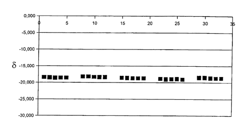
FIG. 1 shows the reproducibility of the method according Example 3.
FIG. 2 shows the calibration curve of the spiking experiment according to
Example
4.
FIG. 3 shows the reproducibility of the method according to Example 5.
FIG. 4 shows the calibration curve of the spiking experiment according to
Example
6.
FIGS. 5 and 6 show the results of the measurement of the urea production
according to Example 7.
The method will be described in more detail by way of the following examples.
8
CA 02808760 2013-02-19
Example 1: Urea isolation using filtration
Plasma was obtained from a blood sample. 300 pl plasma was diluted with 200 pl
deionized
water. 100 pl acetonitrile containing 1% formic acid was added to the sample.
A
precipitation formed in the vessel. The samples were filtered using a
HybridSPE column.
The filtrate was mixed with 50 pl 1 M phosphoric acid, frozen and lyophilized.
The sample
was adjusted to a pH value of 5.5 using a degassed 0.5 M phosphate buffer
solution, pH 9.
The sample container (Vacutainer) was rinsed with helium gas. Thereafter, 70
pl of a
solution containing 15 mg/ml Jack Bean Urease Type III from Sigma in phosphate
buffer
was added. The sample was incubated for one hour at 36 C. Thereafter, 60 pl
20%
phosphoric acid was injected through the septum so as to stop the urease
reaction and
release CO2. The released CO2 was used to determine the isotope ratio by means
of IRMS.
Example 2: Urea isolation using no filtration
The method as in Example 1 was carried out, however the addition of
acetonitrile and
formic acid, and the filtration step were dispensed with, which is to say the
plasma sample
was directly lyophilized after acid was added. The remainder of the method was
carried out
in identical fashion.
Example 3: Reproducibility of the method
A plasma sample was divided into five samples, which were each subjected
separately
from each other to the method according to Example 1. Each CO2 sample that was
ob-
tamed was measured five times. The differences are minimal; refer to FIG. 1.
Example 4: Spiking experiment
99% labeled 13C urea was added to the plasma samples from Example 1 in
quantities of
0.01 mg, 0.25 mg, 0.5 mg, 0.1 mg, 0.2 mg, and 0.3 mg, and the sample was
treated in
accordance with method 1. FIG. 2 shows the corresponding measurement values.
The
calibration curve is located on a straight line with a correlation coefficient
of 0.99923.
9
CA 02808760 2013-02-19
Example 5: Reproducibility
The measurement regarding reproducibility according to Example 3 was repeated
for the
method using no filter according to Example 2. FIG. 3 shows the results.
Again, excellent
reproducibility exists.
Example 6: Spiking experiment
The spiking experiment according to Example 4 was repeated, wherein the method
according to Example 2 was employed. The corresponding straight calibration
line is
apparent from FIG. 4. The coefficient of correlation thereof was R=0.99959.
Example 7: Measurement of the urea production
Blood was collected from two subjects, and from 300 pl of the plasma that was
obtained
from each of the blood samples, urea was isolated using the method according
to the
invention, and the 13C/12C isotope ratio of the urea was determined.
Each sample was measured 5 times and the basal value was determined by way of
the
average value. The subjects were then administered 27 mg/kg 99% 13C-labeled
Na-acetate.
The procedure of collecting blood and determining the 13C/12C isotope ratio of
the isolated
urea was repeated at 15-, 30-, 45-, 60-, 75-, 90-, 120-, 180-, and 240-minute
intervals. A
smaller portion of the 13C-labeled acetate is converted into urea in the body
and can be
detected because of the sensitivity of the measurement. The kinetics of the
newly produced
urea was determined by measuring the delta values (increasing the 13C/12C
isotope ratio).
The kinetics of the urea production is shown in FIGS. 5 and 6.
10