Note: Descriptions are shown in the official language in which they were submitted.
CA 02809464 2013-03-12
FLOWER CATHETER FOR MAPPING AND ABLATING VEINOUS AND OTHER
TUBULAR LOCATIONS
BACKGROUND OF THE INVENTION
[0001] Electrode catheters have been in common use in medical practice for
many years.
They are used to stimulate and map electrical activity in the heart and to
ablate sites of aberrant
electrical activity. In use, the electrode catheter is inserted into a chamber
of the heart. Once the
catheter is positioned, the location of aberrant electrical activity within
the heart is then located.
[0002] One location technique involves an electrophysiological mapping
procedure whereby
the electrical signals emanating from the conductive endocardial tissues are
systematically
monitored and a map is created of those signals. By analyzing that map, the
physician can
identify the interfering electrical pathway. A conventional method for mapping
the electrical
signals from conductive heart tissue is to percutaneously introduce an
electrophysiology catheter
(electrode catheter) having mapping electrodes mounted on its distal
extremity. The catheter is
maneuvered to place these electrodes in contact with or in close proximity to
the endocardium.
By monitoring the electrical signals at the endocardium, aberrant conductive
tissue sites
responsible for the arrhythmia can be pinpointed.
[0003] For mapping, it is desirable to have a relatively small mapping
electrode. It has been
found that smaller electrodes record more accurate and discrete electro grams.
Additionally, if a
bipolar mapping arrangement is used, it is desirable that the two electrodes
of the mapping
arrangement be in close proximity to each other and that they be similar in
size to produce more
accurate and useful electrograms.
[0004] Once the origination point for the arrhythmia has been located in
the tissue, the
physician uses an ablation procedure to destroy the tissue causing the
arrhythmia in an attempt to
remove the electrical signal irregularities and restore normal heart beat or
at least an improved
heart beat. Successful ablation of the conductive tissue at the arrhythmia
initiation site usually
terminates the arrhythmia or at least moderates the heart rhythm to acceptable
levels.
100051 A typical ablation procedure involves providing a reference
electrode, generally taped
to the skin of the patient. RF (radio frequency) current is applied to the tip
electrode, and current
1
CA 02809464 2013-03-12
flows through the media that surrounds it, i.e., blood and tissue, toward the
reference electrode
Alternatively, the catheter may carry bipolar electrodes, in which instance,
the current flows
from the tip electrode, through the media and toward another electrode carried
on the catheter
tip. In any case, the distribution of current depends on the amount of
electrode surface in contact
with the tissue as compared to blood, which has a higher conductivity than the
tissue. Heating of
the tissue occurs due to electrical current. The tissue is heated sufficiently
to cause cellular
destruction in the cardiac tissue resulting in formation of a lesion within
the cardiac tissue which
is electrically non-conductive.
[0006] A disadvantage with current catheters is where the aberrant activity
originates in a
vein or other tubular structure leading away from the heart chamber. In the
case of
electrophysiological triggers in such locations, a common alternative to the
ablation of the tissue
that generates the triggers involves ablating a lesion to interrupt wavelets,
for example, when
ablating a line of block. For tubular regions in or around the heart, this
procedure requires the
line of block to be made about a circumference of the tubular region. However,
it is difficult to
manipulate and control the distal end of a straight catheter so that it
effectively ablates about the
circumference. Moreover, although most vessels have circular cross-sections,
many do not and
they come in different sizes. Accordingly, a need exists for an improved
catheter that is
particularly useful for such applications
[0007] Flower mapping catheters are known; however, conventional flower
catheters carry
smaller electrodes which are not well suited for ablation. Furthermore,
existing flower catheters
were developed for atrial diagnostics, not vein mapping or ablation which pose
different
challenges.
[0008] Lasso catheters are also known. However, lasso catheters have a
generally circular
main portion which is not always adaptable to noncircular tubular structures.
Moreover, the
generally circular main portion is typically positioned along a single
circumference of the tubular
structure for forming a line of isolation. As such, testing the line of
isolation for completion
requires repositioning the catheter, or use of a second catheter, both of
which increase the
duration, complexity and/or cost of the ablation procedure.
2
CA 02809464 2013-03-12
[0009] Thus, there is a desire for a catheter adapted for mapping and
ablation in a tubular
structure, especially a tubular structure with a noncircular cross-section. It
is further desired that
the catheter be adapted for testing completeness of ablation isolation lines
without the need for
repositioning or the use of an additional catheter.
SUMMARY OF THE INVENTION
[0010] The present invention is directed to an improved catheter for
ablating tubular regions
at or near the heart. The catheter comprises a distal assembly having a
plurality of spines, each
capable of ablating and/or obtaining electrical data from the heart tissue.
The use of a plurality
of spines extending radially outward from the catheter ensures contact between
the spines and
surrounding tissue generally without regard to the size or shape of the
tubular region. Because
each spine is fixed only at its proximal end, the free distal end of each
spine can independently
adapt to the tubular region, especially if the tubular region has a
noncircular cross-section. Each
spine has a generally L-shaped configuration with a generally straight
proximal portion, and a
distal portion that is generally orthogonal to the proximal portion.
Advantageously, the generally
L-shaped configuration converts into a generally U-shaped configuration with
the distal portion
lying against the surrounding tissue for greater contact once the proximal
portion is pushed or
advanced into the tubular cavity. It is understood that the change in
configuration is enabled and
occurs where the radial size of the tubular cavity is sufficiently small
relative to the length or
"reach" of the distal portion such that the distal end of the distal portion
can contact with the
surrounding tissue of the tubular cavity. Such greater contact along the
distal portion enables a
tip electrode and at least one ring electrode, both carried on the distal
portion of each spine, to
make simultaneous contact with the surrounding heart tissue along two
different inner
circumferences of the tubular region, where a first inner circumference is
defined by contact with
the tip electrode of each spine and at least a second inner circumference
advantageously deeper
in the tubular region is defined by contact with at least one ring electrode
of each spine.
[0011] In one embodiment, the catheter comprising an elongated catheter
body having a
proximal end, a distal end and at least one lumen extending longitudinally
therethrough. The
distal assembly comprises about five spines. Each spine includes a
nonconductive covering and
3
CA 02809464 2013-03-12
a support arm with shape memory extending therein. The distal assembly
includes a spine
mounting assembly that fixes proximal ends of each spine to the distal end of
the catheter body.
Each spine has a generally L-shaped configuration with a generally straight
proximal portion,
and a distal portion that is generally orthogonal to the proximal portion and
carries a tip electrode
and at least one ring electrode. Depending on various parameters, including
length and/or
curvature of each portion of the spine, the distal end or tip electrode of the
spine when adopting a
generally U-shaped configuration within a tubular cavity defines an angle 0
with the proximal
end of the spine. The angle 0 ranges between about 45 and 135 degrees,
preferably between
about 65 and 115 degrees, and preferably about 90 degrees. Where the angle 0
is less than 90
degrees, the distal end is distal of the proximal end of the spine. Where the
angle 0 is about 90
degrees, the distal end is about even with the proximal end of the spine 14.
Where the angle 0 is
greater than 90 degrees, the distal end is proximal of the proximal end of the
spine. However,
regardless of the angle 0, the generally U-shaped configuration of the spine
when the proximal
portion is advanced into the tubular cavity ensures that the ring electrodes
are predictably and
consistently positioned deeper in the tubular region than the tip electrodes.
In alternate
embodiments, each spine may have a nonlinear distal portion with a curved
configuration or a
zig-zag configuration.
[0012] The catheter of the present invention may include a steering
mechanism for uni- or bi-
directional deflection. In one embodiment, an intermediate deflectable section
extends between
the catheter body and the distal assembly and deflection is actuated by one
puller wire or a pair
of puller wires that extend from a control handle to a distal end of the
intermediate deflectable
section. A compression coil surrounds each puller wire in the catheter body.
Mechanism for
actuating the puller wire(s) are provided in the control handle for
manipulation by the user.
[0013] The present invention is also directed to a method of ablating a
tubular region at or
near the heart. A method for ablating a tubular structure of the heart
includes introducing the
distal assembly of the above catheter having L-shaped spines into the tubular
region and
positioning the distal assembly so that the one tip electrode from each spine
is in contact with
heart tissue. The method includes advancing the distal assembly deeper into
the tubular region
such that the L-shaped spines changes to a U-shape where the tip electrode of
each spine is in
4
CA 02809464 2013-03-12
contact with heart tissue along a first inner circumference of the tubular
structure and at least one
ring electrode from each spine is in contact with heart tissue along a second
inner circumference
of the tubular structure deeper into the tubular region than the first inner
circumference. The
method includes energizing at least one electrode (tip or ring) on each spine
to ablate along the
respective circumference. The method includes sensing electrical activity of
the tubular region
by the other electrodes of each spine during, after or between the ablation to
assess the lesions
formed by the ablating electrodes. Advantageously, the sensing of electrical
activity can occur
without repositioning of the distal assembly and while the electrodes
performing the ablation are
in contact with the heart tissue.
DESCRIPTION OF THE DRAWINGS
[0014] These and other features and advantages of the present invention
will be better
understood by reference to the following detailed description when considered
in conjunction
with the accompanying drawings wherein:
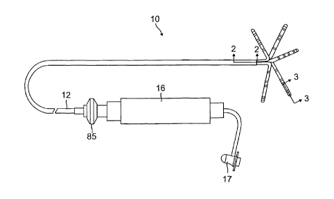
[0015] FIG. 1 is a perspective view of a catheter according to an
embodiment of the
invention.
[0016] FIG. lA is an enlarged view of a distal assembly of FIG. 1.
[0017] FIG. 2 is a side cross-sectional view of a portion of the catheter
of FIG. 1, including a
junction between a catheter body and a spine, taken along line 2-2.
[0018] FIG. 3 is a side cross-sectional view of a spine of the catheter of
FIG. 1, taken along
line 3-3.
[0019] FIG. 4 is an end cross-sectional view of the junction of FIG. 2,
taken along line 4-4.
[0020] FIG. 5 is an end cross-sectional view of the junction of FIG. 2,
taken along line 5-5.
[0021] FIG. 6A is a side view of a distal assembly advancing toward a
tubular region
according to an embodiment of the invention, with spines of the distal
assembly in a generally
relaxed L-shaped configuration.
[0022] FIG. 6B is a side view of the distal assembly of FIG. 6A entering a
tubular region,
with spines of the distal assembly.
CA 02809464 2013-03-12
[0023] FIG. 6C is a side view of the distal assembly of FIG. 6A positioned
in a tubular
region, with spines of the distal assembly in a generally U-shaped
configuration.
[0024] FIG. 6D is a schematic drawing of various embodiments of a spine in
a U-shaped
configuration in accordance with the invention.
[0025] FIG. 7 is a side cross-sectional view of a portion of a catheter in
accordance with
another embodiment of the present invention, including a junction between a
catheter body and a
spine.
[0026] FIG. 8 is an end cross-sectional view of the junction of FIG. 7,
taken along line 8-8.
[0027] FIG. 9 is a side cross-sectional view of a portion of a spine in
accordance another
embodiment of the present invention.
[0028] FIG. 10 is a side cross-sectional view of a portion of a catheter in
accordance with yet
another embodiment of the present invention, including an intermediate
deflectable section.
[0029] FIG. 11 is an end cross-sectional view of the intermediate
deflectable section of FIG.
10, taken along line 11-11.
[0030] FIG. 12 is a perspective view of a distal assembly (with spines in a
neutral state) in
accordance with another embodiment of the invention.
[0031] FIG. 13A is a perspective view of a distal assembly (with spines in
a neutral state) in
accordance with yet another embodiment of the invention.
[0032] FIG. 13B is a top plan view of the distal assembly of FIG. 13A.
[0033] FIG. 13C is a side view of the distal assembly of FIG. 13B, taken
along line C--C.
[0034] FIG. 13D is a perspective view of the distal assembly of FIG. 13A in
a tubular region.
DETAILED DESCRIPTION OF THE INVENTION
[0035] The invention is directed to a catheter 10 as shown in FIG. 1, having a
distal assembly
18 comprising a plurality of spines 14. Each spine carries at least one
electrode, preferably a tip
electrode 20 and at least one ring electrode 28, such that when the spines are
positioned in
contact with tissue of a tubular structure at or near the heart, each spine is
capable of obtaining
electrical data and ablating tissue. As shown in FIG. 1, the catheter 10
comprises an elongated
catheter body 12 having proximal and distal ends, a control handle 16 at the
proximal end of the
6
CA 02809464 2013-03-12
catheter body 12, and a distal assembly 18 comprising a plurality of spines 14
mounted at the
distal end of the catheter body 12.
[0036] As shown in FIGS. 1 and 2, the catheter body 12 comprises an elongated
tubular
construction having a single, axial or central lumen 15, but can optionally
have multiple lumens
along all or part of its length if desired. The catheter body 12 is flexible,
i.e., bendable, but
substantially non-compressible along its length. The catheter body 12 can be
of any suitable
construction and made of any suitable material. A presently preferred
construction of the
catheter body 12 comprises an outer wall 13 made of polyurethane or PEBAX
(polyether block
amide). The outer wall 13 comprises an imbedded braided mesh of stainless
steel or the like, as
is generally known in the art, to increase torsional stiffness of the catheter
body 12 so that, when
the control handle 16 is rotated, the distal end of the catheter body 12 will
rotate in a
corresponding manner.
[0037] The length of the catheter body 12 is not critical, but preferably
ranges from about 90
cm to about 120 cm, and more preferably is about 110 cm. The outer diameter of
the catheter
body 12 is also not critical, but is preferably no more than about 8 french,
more preferably about
7 french. Likewise, the thickness of the outer wall 13 is not critical, but is
preferably thin
enough so that the central lumen 15 can accommodate lead wires, sensor cables
and any other
wires, cables or tubes. If desired, the inner surface of the outer wall 13 is
lined with a stiffening
tube (not shown) to provide improved torsional stability. An example of a
catheter body
construction suitable for use in connection with the present invention is
described and depicted in
U.S. Patent No. 6,064,905, the entire disclosure of which is incorporated
herein by reference.
[0038] In the depicted embodiment, the distal assembly 18 comprises five
spines 14. Each
spine 14 has a proximal end attached at the distal end of the catheter body 12
and a free distal
end, i.e., the distal end is not attached to any of the other spines, to the
catheter body, or to any
other structure that confines movement of the distal end. Each spine 14
contains a support arm
24 comprising a metal or plastic material that has shape memory, such that the
support arm 24
forms an initial shape when no external forces are applied, forms a deflected
shape when an
external force is applied, and returns to its initial shape when the external
force is released. In a
preferred embodiment, the support arm 24 comprises a superelastic material,
for example a
7
CA 02809464 2013-03-12
nickel-titanium alloy, such as nitinol. Each spine 14 also comprises a non-
conductive covering
26 in surrounding relation to the support arm 24. In a preferred embodiment,
the non-conductive
covering 26 comprises a biocompatible plastic tubing, such as a polyurethane
or polyimide
tubing.
[0039] As will be recognized by one skilled in the art, the number of spines
14 can vary as
desired depending on the particular application, so that the catheter 10 has
at least two spines,
preferably at least three spines, more preferably at least five spines and as
many as eight, ten or
more spines. For clarity however only two spines are shown in FIG. 2. As
described in more
detail below, the spines 14 are moveable between an expanded arrangement,
wherein, for
example, each spine extends radially outwardly from the catheter body 12 in a
generally L-
shaped configuration, or the spines 14 may be arranged in a collapsed
arrangement, wherein, for
example, each spine is disposed generally along a longitudinal axis of the
catheter body 12 so
that the spines are capable of fitting within a lumen of a guiding sheath, as
discussed further
below.
[0040] With reference to FIG. 3, each spine 14 carries at least one electrode
mounted along its
length at or near its distal end. In the depicted embodiment, a tip electrode
20 is mounted on a
distal end of each non-conductive covering 26 and at least a first ring
electrode 28a is mounted
on each non-conductive covering 26 on the distal end of the non-conductive
covering 26. The
distance between the tip electrode 20 and ring electrode 28a preferably ranges
from about 0.5
mm to about 2.0 mm. Additional single or pair of ring electrodes 28b-28d may
be mounted on
each non-conductive covering 26 proximal of the first ring electrode 28a. In
the depicted
embodiment, the catheter is configured so that the tip electrode functions
with a distal-most ring
electrode as a distal electrode pair. An alternate embodiment of the catheter
may be utilize the
tip electrode for unipolar electrograms only, where the distance between the
tip electrode and the
distal-most ring electrode 28a would be greater. In the depicted embodiment,
the distance
between the first ring electrode 28a and adjacent electrode 28b ranges from
about 0.5mtn to
about 2.0 mm. The distance between adjacent pairs of electrode ranges from
about 2.0 mm to
about 8.0 mm. The distance between ring electrodes of one pair ranges from
about 0.5 mm to
about 2.0 mm. Any of the ring electrodes 28a-28d may be used for either
unipolar or bipolar
8
CA 02809464 2013-03-12
electrogram measurement. That is, the tip and ring electrodes may be used in
conjunction with
one or more reference electrodes attached to outside the body of the patient
(e.g., in the form of a
patch), or any of the ring electrodes may function as a reference electrode.
[0041] Each tip electrode 20 has an exposed length preferably ranging from
about 0.5 mm to
about 4.0 mm, more preferably from about 0.5 mm to about 2.0 mm, still more
preferably about
1.0 mm. Each ring electrode 28 has a length preferably up to about 2.0 mm,
more preferably
from about 0.5 mm to about 1.0 mm.
[0042] Each tip electrode 20 and each ring electrode 28 is electrically
connected to an
electrode lead wire 29, which in turn is electrically connected to a connector
17 (FIG. 1). The
connector 17 is connected to an appropriate mapping or monitoring system (not
shown). Each
electrode lead wire 29 extends from the connector 17, through the control
handle 16, through the
central lumen 15 in the catheter body 12, and into the non-conductive covering
26 of the spine 14
where it is attached to its corresponding tip electrode 20 or ring electrode
28. Each lead wire 29,
which includes a non-conductive coating (not shown) over almost all of its
length, is attached to
its corresponding tip electrode 20 or ring electrode 28 by any suitable method
to ensure electrical
conduction.
[0043] One method for attaching a lead wire 29 to a ring electrode 28 involves
first making a
small hole through an outer wall of the non-conductive covering 26. Such a
hole can be created,
for example, by inserting a needle through the non-conductive covering 26 and
heating the
needle sufficiently to form a permanent hole. The lead wire 29 is then drawn
through the hole by
using a microhook or the like. The end of the lead wire 29 is then stripped of
any coating and
welded to the underside of the ring electrode 28, which is then slid into
position over the hole
and fixed in place with polyurethane glue or the like. Alternatively, each
ring electrode 28 may
be formed by wrapping the lead wire 29 around the non-conductive covering 26 a
number of
times and stripping the lead wire of its own non-conductive coating on its
outwardly facing
surfaces. In such an instance, the lead wire 29 functions as a ring electrode.
[0044] Each spine 14 can also include at least one temperature sensor, e.g., a
thermocouple or
thermistor, for the tip electrode 20 or any of the ring electrodes. In the
depicted embodiment, a
thermocouple is formed by an enameled wire pair. One wire of the wire pair is
a copper wire 41,
9
CA 02809464 2013-03-12
e.g., a number "40" copper wire. The other wire of the wire pair is a
constantan wire 45. The
wires 41 and 45 of the wire pair are electrically isolated from each other
except at their distal
ends where they are twisted together, covered with a short thin piece of
plastic tubing 58, e.g.,
polyamide, and covered with epoxy with good thermal conductive coefficient.
[0045] The wires 41 and 45 extend through the central lumen 15 of the catheter
body 12 (FIG.
2). Within the central lumen 15, the wires 41 and 45 extend through a
protective sheath (not
shown) along with the lead wires 29. The wires 41 and 45 then extend out
through the control
handle 16 and to a connector (not shown) connectable to a temperature monitor
(not shown).
Alternatively, the temperature sensing means may be a thermistor. A suitable
thermistor for use
in the present invention is Model No. AB6N2-GC14KA143E/37C sold by
Thermometrics (New
Jersey).
[0046] FIG. 3 illustrates a suitable technique for mounting the tip electrode
lead wire 29, the
thermocouple wires 41 and 45 and the support arm 24 to the tip electrode 20. A
distal end of the
electrode lead wire 29 may be secured to the tip electrode 20 by drilling a
first blind hole 48 into
the tip electrode 20, stripping the lead wire 29 of any coating and placing
the lead wire 29 within
the first blind hole 48 where it is electrically connected to the tip
electrode 20 by a suitable
means, such as by soldering or welding. The lead wire 29 may then be fixed in
place, for
example, by using a polyurethane glue or the like. The support arm 24 may also
be similarly
affixed to the tip electrode 20. For example, a second blind hole 52 may be
drilled into the tip
electrode 20 such that a distal end of the support arm 24 may be inserted into
the second blind
hole 52 and affixed therein, for example, using a polyurethane glue or the
like. Moreover, a third
blind hole 53 may be drilled into the tip electrode 20 such that the plastic
tubing 58 surrounding
distal ends of the thermocouple wires 41 and 45 may be inserted into the third
blind hole and
affixed therein, using a polyurethane glue or the like. Alternatively, the
wires 41 and 45 can be
soldered into the blind hole 53.
[0047] Alternatively, a single blind hole (not shown) in the proximal end of
the tip
electrode 20 can be used for mounting the support arm 24 and thermocouple
wires 41 and 45,
and the distal end of the lead wire 29 can be wrapped around the outside
proximal end of the tip
CA 02809464 2013-03-12
=
electrode, which is not exposed and attached by solder, welding or any other
suitable technique.
Any other arrangement for mounting these components in the spine could also be
used.
100481 A suitable construction of the distal end of the catheter body 12,
having spines 14
mounted thereto, is depicted in FIGS. 2 and 4. Again, for clarity, only two
spines 14 are shown
in FIG. 2. Mounted in the distal end of the lumen 15 of the catheter body 12
is a spine mounting
assembly 31 that secures the proximal ends of the spines to the catheter. In
the illustrated
embodiment, the spine mounting assembly 31 comprises an outer mounting ring 32
disposed
within the outer wall 13 of the catheter body 12. The outer mounting ring 32
preferably
comprises a metal material, such as stainless steel, more particularly
stainless steel 303, and may
be attached at the distal end of the catheter body 12 by a variety of methods,
such as by welding
or by use of an adhesive, such as a polyurethane glue. Alternatively, the
outer mounting ring 32
may comprise a plastic material. A mounting structure 34 is provided coaxially
within the outer
mounting ring 32. In the depicted embodiment, the mounting structure 34 is
multi-sided and
comprises a metal material, such as stainless steel, more particularly
stainless steel 303. The
mounting structure 34 may also alternatively comprise a plastic material. The
outer mounting
ring 32 and the mounting structure 34 provide a channel 38 therebetween in
which the proximal
end of each support arm 24 is mounted. Specifically, each spine 14 is mounted
in the catheter
body 12 by removing a portion of the non-conductive covering 26 at the
proximal end of each
spine 14, inserting the exposed proximal end of each support arm 24 into the
channel 38 between
the outer mounting ring 32 and the multi-sided mounting structure 34 and
affixing within the
channel 38 by any suitable means, such as with a polyurethane glue or the
like. The lead wires
29 and thermocouple wires 41 and 45 also extend through the channel 38 between
the outer
mounting ring 32 and the mounting structure 34.
[0049] In one embodiment, the support arm 24 has a generally trapezoidally-
shaped end cross
section with curved sides as illustrated in FIGS. 4 and 5. In such an
arrangement, when each
support arm 24 is inserted into the channel 38, a substantially flat surface
of each support arm 24,
preferably the base of the trapezoidally-shaped end cross section, is mounted
against a
substantially flat surface on the multi-sided mounting structure 34.
Preferably the number of
substantially flat outer surfaces on the multi-sided mounting structure 34
corresponds to the
11
CA 02809464 2013-03-12
number of spines 14. In such an instance, the support arm 24 of each spine 14
may be mounted
within the channel 38 and adjacent to its corresponding side on the multi-
sided mounting
structure 34 to enable the support arms 24, and thus the spines 14, to be
equally spaced around
the multi-sided mounting structure 34. The multi-sided mounting structure 34
may be
approximately co-axial with the longitudinal axis of the catheter body 12 such
that the spines 14
are equally spaced about the catheter body 12 as well. Once each support arm
24 is properly
positioned within the channel 38, each support arm 24 may be affixed within
the channel 38 by
any suitable means, such as by use of an adhesive, such as a polyurethane
glue. Alternatively,
the mounting structure 34 can have a round outer surface, although with such
an embodiment
more care needs to be taken if the support arms 24 are to be evenly spaced
about the mounting
structure.
[0050] In the depicted embodiment, a first non-conducting tube 40 is disposed
between the
outer mounting ring 32 and the support arms 24, and a second non-conducting
tube 42 is
disposed between the support arms 24 and the mounting structure 34. The non-
conducting tubes
40 and 42, which may be polyimide tubes, ensure that each support arm 24
remains electrically
isolated.
[0051] In accordance with a feature of the invention, each spine 14 has a
generally L-shaped
configuration supported by the support arm 24. In the illustrated embodiment
of FIGS. 1 and 6,
the generally L-shaped configuration of each spine is defined by a generally
straight proximal
portion 60, a generally straight distal portion 64 that extends at a generally
orthogonal angle from
the proximal portion 60. When the distal assembly 18 is initially deployed in
a tubular cavity 71,
as illustrated in FIG. 6A, the spines 14 are in their generally neutral and
relaxed L-shaped
configuration. As the distal assembly enters the tubular cavity as illustrated
in FIG. 6B, the
distal ends of the spines come into contact with an opening or ostium 70 of
the tubular cavity,
where the L-shaped configuration of the spines 14 starts to change under a
contact force applied
to the distal ends of the spines by the ostium. When the distal assembly 18 is
advanced, the
proximal portions 60 of the spines are pushed deeper into the tubular cavity
and the distal ends of
the spines 14 come into contact with tissue lining the tubular cavity 71. The
distal assembly 18
is further advanced so that the distal portions 64 increasingly bend over the
distal ends until more
12
CA 02809464 2013-03-12
of the distal portions 64 also come into contact with deeper tissue in the
tubular cavity 71
whereupon the spines 14 are in a generally U-shaped configuration as shown in
FIG. 6C.
Depending on various parameters, the distal end or tip electrode 20 of the
spine in the generally
U-shaped configuration defines an angle 0 with the proximal end of the spine.
The angle 0
ranges between about 45 and 135 degrees, preferably between about 80 and 100
degrees, and
preferably about 90 degrees, as illustrated in FIG. 6D. Where the angle 0 is
less than 90 degrees,
the tip electrode 20 is distal of the proximal end of the spine 14 at the
spine mounting assembly
31. Where the angle 0 is about 90 degrees, the tip electrode 20 is about even
with the proximal
end of the spine 14. Where the angle 0 is greater than 90 degrees, the tip
electrode 20 is
proximal of the proximal end of the spine 14. However, regardless of the angle
0, the generally
U-shaped configuration of the support member 24 (and hence the spine 14)
provides that the
distal portion 64 is nearly parallel with the generally straight proximal
portion 60 and that the
ring electrodes 28 are consistently positioned deeper in the tubular structure
than the tip electrode
20. As illustrated in FIG. 6D, the length and/or curvature of each portion 60
and 64 can be
varied as desired or appropriate. Moreover, the length, curvature and/or angle
0 need not be
uniform for each spine throughout the distal assembly. For example, a first
set of spines may
have one length, curvature and/or angle 0 and a second set of spines may have
another length,
curvature and/or angle 0. Although the spines are equally radially spaced from
each other in the
illustrated embodiments, the radial spacing can also be varied as desired or
appropriate. In the
illustrated embodiment of FIG. 6C, the catheter is illustrated as it appears
when pushed into a
tubular cavity. The change in configuration of the spine from a relaxed state
(L-shaped) in FIG.
6A to a confined state (U-shaped) in FIB. 6C reflects how the angle 0 grows
when the distal
assembly 18 is pushed into the tubular cavity. The length of the spine 14
extending between the
exposed proximal end of the covering 26 to the distal tip end of the spine can
range between
about 1.0 cm and 5.0 cm.
[0052] In accordance with a feature of the present invention, when the distal
assembly 18 is
inserted into and confined within a tubular structure the tip electrodes 20 of
the distal assembly
18 are well adapted to make contact with the surrounding tissue of a tubular
structure 71 at
locations spanning generally along an inner circumference C of the tubular
structure. Similarly,
13
CA 02809464 2013-03-12
the first ring electrodes 28a on the spines 14 are well adapted to contact
tissue at locations
spanning generally along another or a first adjacent inner circumference Ca
deeper in the tubular
structure. Likewise, additional ring electrodes 28b-28d are well adapted to
contact tissue at
locations spanning generally along other or additional adjacent inner
circumferences Cb-Cd more
deeply in the tubular structure. Rotation of the catheter by the control
handle rotates the distal
assembly 18 so as to rotate and shift the electrodes to different contact
locations along each inner
circumference. Where, for example, the tip electrodes 20 are adapted for
ablation, an isolation
line can be created at the circumference C and the integrity or completeness
of the isolation line
at the circumference C can be sensed by the ring electrodes 28a-28d at
locations along adjacent
circumferences Ca-Cd further into the tubular structure. Alternatively, any of
the sets of ring
electrodes 28, can be adapted for ablation for creating an isolation line at
Ci, and any gaps in the
isolation line at C, can be sensed by any of the non-ablating sets of tip or
ring electrodes. Thus,
ablation and testing of the resulting lesions can be advantageously
accomplished by the catheter
without repositioning of the distal assembly 18 or use of an additional
catheter.
[0053] With reference to FIGS. 2 and 4, a main irrigation tube 44 extends,
e.g., coaxially,
through the mounting structure 34. The irrigation tube 44 comprises a non-
conductive material
such as PEBAX, polyimide or polyurethane. The irrigation tube 44 extends
through the catheter
body 12 and out through the control handle 16 or out a sidearm (not shown) as
is known in the
art and described in U.S. Patent No. 6,120,476, the disclosure of which is
incorporated herein by
reference. As discussed further below, the irrigation tube 44 is used to
introduce irrigation fluid
to the region between the spines 14 and at the tip electrodes 20 of the
spines. The region
between the spines is prone to thrombus formation and the ablating electrodes
can overheat
causing the formation of char. The distal end of the main irrigation tube 44
is preferably glued in
place between the spines 14.
[0054] As illustrated in FIGS. 4 and 5, a distal end of the main irrigation
tube 44 receives
proximal ends of a short irrigation tube 47 for the region between the spines,
and a plurality of
dedicated irrigation tubes 49, one for each spine. In the illustrated
embodiment, the short
irrigation tube 47 positioned centrally and surrounded radially by the spine
irrigation tubes 49. A
14
CA 02809464 2013-03-12
lumen of the short irrigation tube 47 provides a fluid path (arrow 61) from
the distal end of the
main irrigation tube 44 to outside of the catheter in the region between the
spines.
[0055] Each spine irrigation tube 49 arranged around the short irrigation tube
47 extends from
the distal end of the main irrigation tube 44 into a respective spine 14 of
the distal assembly 18.
As illustrated in FIGS. 2 and 3, each spine irrigation tube 49 extends through
a respective
nonconductive covering 26, along with the lead wires 29, thermocouple wires 41
and 45, support
arm 24 of the respective spine, where a distal end of the spine irrigation
tube 44 terminates in an
irrigation passage 75 that leads to a fluid chamber 76, both formed in the tip
electrode 20.
Irrigation apertures 74 are formed in distal wall 78 of the tip electrode 20
to allow fluid
communication from the fluid chamber 76 to outside the tip electrode 20.
[0056] As illustrated in FIG. 2, irrigation fluid passing through the
irrigation tube 44 travels
through the control handle 16 and the catheter shaft 12. At the distal end of
the irrigation tube
44, a portion of the fluid exits the catheter through the short irrigation
tube 47 (arrow 61) and
other portions continue into the spines (arrow 63) through the spine
irrigation tubes 49. At the
tip electrode 20, the irrigation fluid enters the fluid chamber 76 via the
irrigation passage 75 and
exits the tip electrode 20 through the irrigation apertures 74. The distal end
of the main irrigation
tube 44 is plugged by an adhesive or sealant 82 which also fixes the short
irrigation tube 47 and
proximal ends of the spine irrigation tubes 49 in the distal end of the main
irrigation tube 44. As
would be recognized by one skilled in the art, the main irrigation tube 44 can
comprise a
plurality of structures that define a continuous path through the catheter
body 12 and into the
handle 16, including a combination of one or more lumens and one or more
tubes. The distal end
of the spine irrigation tubes 49 is adhered to the spine tip electrode 20
irrigation passage 75 by an
adhesive such as EPDXY or sealant.
[0057] As previously discussed, when mounting the support arms 24 to the spine
mounting
assembly 31, a portion of the non-conductive covering 26 at the proximal end
of each spine 14 is
removed to expose the support arm 24. Removing a portion of the non-conductive
covering 26
at the proximal end of each spine 14 enables the electrode lead wires 29 and
the thermocouple
wires 41 and 45 to extend from the lumen 15 of the catheter 12, through lumen
46 of the
mounting ring 32, and into each non-conductive covering 26. As shown in FIG.
2, once inserted
CA 02809464 2013-03-12
into the non-conductive coverings 26, the electrode lead wires 29 and
thermocouple wires 41 and
45 extend within the non-conductive covering 26, where the lead wires 29 are
electrically
connected at their distal ends to their corresponding tip electrode 20 and
ring electrode 28.
[0058] An alternate embodiment is illustrated in FIGS. 7, 8 and 9 with each
spine 114
comprising a multi-lumened tube 100 with at least two lumens, including a
lumen 110 for the
lead wires 29, thermocouple wires 41 and 45 and/or the support arm 24, and a
lumen 112 for
irrigation fluid. A plurality of short connector tubes 47 extend between the
distal end of the main
irrigation tube 44 and the proximal end of a respective spine irrigation lumen
112. The lumen
112 delivers irrigation fluid to irrigated ring electrodes 128, and to the tip
electrode 20 via a short
irrigation connector tube 95 that connects the irrigation passage 75 and the
lumen 112. The
irrigated ring electrodes 128 mounted on the tube can be adapted for mapping,
sensing and/or
ablation and are configured with a raised mid-portion 114 (FIG. 9) to form an
annular fluid
chamber 116 with an outer wall 118 of the tube 100. Fluid passes from the
lumen 112 through a
hole 122 formed in the outer wall 118 and is distributed in the annular fluid
chamber 116 before
exiting the electrodes 128 through apertures 124 formed in and near the raised
mid-portion 114.
[0059] If desired, the catheter of the present invention may include a
steering mechanism for
deflection of the distal end of the catheter body 12. As illustrated in FIGS.
10 and 11, the
catheter includes an intermediate deflectable section 30 extending between the
catheter body 12
and the spines 14. The catheter body 12 of comprises an outer wall 22 made of
a polyurethane,
or PEBAX. The outer wall 22 comprises an imbedded braided mesh of high-
strength steel,
stainless steel or the like to increase torsional stiffness of the catheter
body 12 so that, when the
control handle 16 is rotated, the tip section 14 of the catheter 10 will
rotate in a corresponding
manner.
[0060] The inner surface of the outer wall 22 is lined with a stiffening tube
23, which can be
made of any suitable material, such as polyimide or nylon. The stiffening tube
23, along with the
braided outer wall 22, provides improved torsional stability while at the same
time minimizing
the wall thickness of the catheter, thus maximizing the diameter of the
central lumen 18. The
outer diameter of the stiffening tube 23 is about the same as or slightly
smaller than the inner
diameter of the outer wall 22. Polyimide tubing may be used for the stiffening
tube 23 because it
16
CA 02809464 2013-03-12
may be very thin walled while still providing very good stiffness. This
maximizes the diameter
of the central lumen 18 without sacrificing strength and stiffness.
[0061] The intermediate deflectable section 30 comprises a short length of
tubing 35, e.g., 2.0
to 4.0 inches in length, that is more flexible than the remainder of the
catheter body 12. The
tubing 35 is multi-lumened with lumens 54, 55, 56, 65 and 66. Extending
through the lumen 65
are the lead wires 29 and thermocouple wires 41 and 45. A nonconductive
protective sheath 69
may be provided to extend through the catheter body 12 and the intermediate
deflectable section
30. Extending through the lumen 66 is the main irrigation tube 44.
[0062] A suitable steering mechanism comprises one, if not two, puller wires
37 that extend
from a proximal end in the control handle 16, through the central lumen 15 in
the catheter body
12 and into diametrically opposed, off-axis lumens 54 and 55 in the short
length of tubing 35.
Within the catheter body 12, each puller wire 37 extends through a respective
closely wound
compression coil 57 that is bendable but substantially non-compressible. The
coils 57 have
proximal and distal ends that are fixed near the proximal and distal ends,
respectively, of the
catheter body 12 to prevent deflection of the catheter body 12. The distal end
of each puller 37
wire is anchored at the distal end of the short length of tubing in its
respective off- axis lumen by
means of a T-bar 59 (FIG. 10). As understood by one of ordinary skill in the
art, the proximal
end of each puller wire 37 is anchored to a movable member (e.g., a thumb
control 85 of FIG. 1)
in the handle 16 that can be moved relative to the catheter body 12. Proximal
movement of the
movable member relative to the catheter body 12 results in deflection of the
short length of
tubing to one side or another, generally within a plane, depending on the
puller wire actuated.
An example of such a steering mechanism and construction is described in more
detail in U.S.
Patent No. 6,064,905, the disclosure of which is incorporated herein by
reference.
[0063] It may also be desirable for the catheter of the present invention to
include a location
sensor, especially where the catheter includes a steering mechanism. In the
depicted
embodiment of FIGS. 10 and 11, the tube 35 of the intermediate deflectable
section 30 has a
dedicated lumen 56 for location sensor 90 and sensor cable 92. The location
sensor 90 is
positioned at or near the distal end of the tube 35 and is used to determine
the coordinates of the
17
CA 02809464 2013-03-12
distal assembly 18, for example, at each instant when the electrodes 20 and 28
are being used to
collect electrical mapping data points and electrical activity data (e.g.,
ECG) and/or to ablate.
[0064] The sensor cable 92 extends through the lumen 56 of the intermediate
section 30, the
central lumen 15 of the catheter body 12, the control handle 16 and out the
proximal end of the
control handle 16 within an umbilical cord (not shown) to a sensor control
module (not shown)
that houses a circuit board (not shown). Alternatively, the circuit board can
be housed within the
control handle 16, for example, as described in U.S. Patent No. 6,024,739, the
disclosure of
which is incorporated herein by reference. The sensor cable 92 comprises
multiple wires
encased within a plastic covered sheath. In the sensor control module, the
wires of the sensor
cable 92 are connected to the circuit board. The circuit board amplifies the
signal received from
the corresponding location sensor 90 and transmits it to a computer in a form
understandable by
the computer by means of a sensor connector at the proximal end of the sensor
control module.
[0065] The location sensor 90 may be an electromagnetic location sensor. For
example, the
location sensor 90 may comprise a magnetic-field-responsive coil, as described
in U.S. Patent
No. 5,391,199, or a plurality of such coils, as described in International
Publication WO
96/05758. The plurality of coils enables the six-dimensional coordinates (i.e.
the three positional
and the three orientational coordinates) of the location sensor 90 to be
determined. Alternatively,
any suitable location sensor known in the art may be used, such as electrical,
magnetic or
acoustic sensors. Suitable location sensors for use with the present invention
are also described,
for example, in U.S. Patent Nos. 5,558,091, 5,443,489, 5,480,422, 5,546,951,
and 5,568,809, and
International Publication Nos. WO 95/02995, WO 97/24983, and WO 98/29033, the
disclosures
of which are incorporated herein by reference. Other suitable location sensors
90 are single axis
sensors, such as that described in the U.S. Patent Application Serial No.
09/882,125 filed June
15, 2001, entitled "Position Sensor Having Core with High Permeability
Material," the
disclosure of which is incorporated herein by reference, and in U.S. Patent
Application Serial
No. 12/982,765 filed December 30, 2010, entitled "Catheter with Single Axial
Sensors," the
disclosure of which is incorporated herein by reference.
[0066] Alternatively or in addition to the foregoing single location sensor
90, a location sensor
may be mounted on each spine, for example, at or near the tip electrode 20.
Smaller sensors are
18
CA 02809464 2013-03-12
particularly desirable for that purpose because of the need to keep the
diameters of the spines 14
small enough so that they all fit within the lumen of a guiding sheath.
[0067] Distal of the intermediate deflectable section 30 is a short connector
tubing 113 has that
is comparable in structure and design to the aforementioned tubing 13 of the
catheter body 12 of
FIG. 2 or FIG. 7. It is understood that the distal end of the connector tubing
113 can be
structured similarly to the distal end of the tubing 13 of FIG. 2 or FIG. 7 so
as to allow mounting
of the proximal ends of the spines 14 in construction of the distal assembly
18 by means of the
mounting assembly 31.
[0068] To use the catheter 10 of the invention, a cardiologist or
electrophysiologist introduces a
guiding sheath and a dilator into the patient, as is generally known in the
art, so that the distal
ends of the sheath and dilator are in the region of the heart to be mapped.
Thereafter, the dilator
is removed from the guiding sheath, and the catheter 10 is introduced into the
patient through the
guiding sheath. To insert the catheter 10 into the guiding sheath, the mapping
assembly 18 must
be in its collapsed arrangement, wherein each spine 14 is disposed generally
along the
longitudinal axis of the catheter body 12. A suitable guiding sheath for use
in connection with
the catheter 10 is the PREFACETM Braided Guiding Sheath (commercially
available from
Biosense Webster, Inc., Diamond Bar, California). Such a guiding sheath has
sufficient strength
to hold each support arm 24 in the collapsed arrangement, such that the spines
14 and also the
entire remainder of the catheter 10 can travel within the guiding sheath, from
an insertion point
in the patient, through a vein or artery and to a desired location in the
heart.
[0069] Once the distal end of the catheter has reached the desired location,
such as a position
within the left ventricle of the heart, relative longitudinal movement between
the catheter 10 and
the guiding sheath is provided to allow at least a portion of each spine 14 to
protrude from the
guiding sheath. Preferably the guiding sheath is moved proximally relative to
the distal end of
the catheter to expose the spines 14. When a portion of each spine 14
protrudes from the guiding
sheath and a compression force is no longer applied by the guiding sheath on
the spines, the
shape memory of the support arms 24 allows the support arms to revert to an
expanded
arrangement. The distal assembly 18 can be advanced into a tubular structure
off the left
ventricle, such as a pulmonary vein, as illustrated in FIG. 6A. With the
distal assembly in the
19
CA 02809464 2013-03-12
expanded arrangement of FIG. 6B, at least one electrode from each spine 14 can
be placed into
contact with the heart tissue at a plurality of locations such that
electrical, locational and
mechanical information can be obtained from such locations of the tissue for
generating a 3-D
map of the tissue. Additionally, with the electrodes 20 and 28 in contact with
heart tissue, either
the tip electrodes or selected ring electrodes can be energized for ablation
in creating a lesion
isolation line along the circumference of the energized electrodes. In that
regard, the catheter
can be rotated along its longitudinal axis (e.g., by rotating the control
handle along its
longitudinal axis) so that the electrodes 20 and 28 are repositioned to ablate
different locations
along the circumferences to create a generally continuous isolation line. And
because the tip and
ring electrodes are in contact with the heart tissue along different
circumferences generally
parallel with the ablated circumference, any of the electrodes in the other
circumferences can be
used to sense electrical activity at such adjacent circumferences in detecting
errant electrical
activity that may indicate gaps in the lesion isolation line. Such sensing can
advantageously be
conducted during or between ablation without the use of another catheter or
the need to
reposition the catheter. After mapping and ablation are completed, the
catheter is moved
proximally relative to the guiding sheath to retract the spines within the
sheath.
[0070] During mapping and ablation, the region between the spines 14 and at
the electrodes 20
and 28 can be prone to thrombus formation and/or overheating. Accordingly,
irrigation fluid is
introduced through the irrigation tube 44 before, during and/or after a
mapping and ablation
procedure to flush the region between the spines 14 and the electrodes.
Preferably irrigation is
provided continuously during the procedure to minimize any potential blood
clotting in the
irrigation tube. Suitable irrigation fluids for use in connection with the
invention include saline,
heparinized saline and thrombolitica. Although the irrigation tube 44 is
preferably positioned
coaxial with the catheter body 12 so that it is mounted between all of the
spines, other positions
for the irrigation tube at or near the distal end of the catheter can be used
in accordance with the
present invention.
[0071] The expanded arrangement of spines 14 in a neutral state can take on
various shapes. In
an alternate embodiment as shown in FIG. 12, a distal assembly 18' has spines
14', each with a
generally straight proximal portion 60', and a nonlinear or curved distal
portion 64' that extends
CA 02809464 2013-03-12
radially outwardly from the catheter body 12. The spines 14' also assume a
generally U-shaped
configuration when the distal assembly 18' is advanced into a tubular region.
[0072] In yet another embodiment as shown in FIG. 13A, a distal assembly 18"
has spines 14",
each with a generally straight proximal portion 60", and a distal portion 64"
having a zig-zag
portion 68 that includes at least two generally straight sections that are
angularly offset from
adjacent sections at angles ct= of between about 45 to 90 degrees. In the
illustrated embodiment,
there are three sections: a proximal section 91 with ring electrode 28d, a
middle section 92 with
ring electrodes 28b and 28c, and a distal section 93 with ring electrode 28a
and tip electrode 20.
As illustrated in FIG. 13B, the spines 14" advantageously form a "pinwheel"
pattern such that
corners 85 (defined by offset angles of the spines point clockwise (arrow 87)
or
counterclockwise (arrow 89) when the distal assembly 18" (especially when the
spines are in a
neutral state) is viewed on-axis. In contrast, when the distal assembly is
viewed from the side as
illustrated in FIG. 13C (with spines in an neutral state), each spine 14"
generally lies within a
plane. With the "pinwheel" zig-zag configuration, the spines 14" are less
prone to slide into
radial grooves 77 of a tubular region 78 and are more prone to lie flat
against the tissue, as
illustrated in FIG. 13D.
[0073] Using the inventive catheter 10 having multiple spines 14, each having
electrical and
mechanical mapping and ablation capabilities, the cardiologist can map local
activation time and
obtain voltage maps, and ablate in a circumferential manner to create an
ablation isolation line in
a tubular region of the heart. The cardiologist can ablate at a first
circumference with the tip
electrodes while obtaining an electrocardiogram with the ring electrodes at
adjacent
circumferences to detect any gaps in the ablation isolation line at the first
circumference during
or between ablation without the need for a second catheter or repositioning of
the ablation
catheter, which lessens the cost and duration of the procedure.
[0074] The preceding description has been presented with references to
presently preferred
embodiments of the invention. Persons skilled in the art and technology to
which this invention
pertains will appreciate that alterations and changes in the described
structures can be practiced
without meaningfully departing from the principle, spirit and scope of this
invention. As also
understood by one of ordinary skill in the art, the drawings are not
necessarily to scale.
21
CA 02809464 2013-03-12
Accordingly, the foregoing description should not be read as pertaining only
to the precise
structures described and shown in the accompanying drawings, but rather should
be read as
consistent with and as support for the following claims, which are to have
their fullest and fairest
scope.
22