Note: Descriptions are shown in the official language in which they were submitted.
Agent Docket: P3683PC00
METHOD, SYSTEM AND APPARATUS FOR THE DETECTION,
CHARACTERIZATION AND CLASSIFICATION OF PARTICLES
CROSS-REFERENCE TO RELATED APPLICATION
[0001] This application claims priority to U.S. application number
61/696,455
filed on September 4, 2012.
FIELD
[0002] The specification relates generally to the detection,
characterization and
classification of particles, and specifically to a method and system for the
detection,
characterization and classification of particles using photoacoustic and
ultrasound
techniques.
BACKGROUND
[0003] The identification, characterization and classification of different
particles
is an important task in many fields and industries, including medicine,
materials science,
pharmacology and electronics. Unfortunately, many of the currently known
techniques
can have undesirable side-effects and are limited to the identification,
characterization
and classification of only certain types of particles.
SUMMARY
[0004] The present specification provides a method to identify,
characterize and
classify particles using photoacoustic (PA) and/or ultrasound (US) analysis
methods and
accompanying systems. In some cases, the particles are 1-50 um in diameter,
which give
unique PA and US spectral features in the about 100 MHz to about 1000 MHz
frequency
range. Particles can be biological-related such as cells, or microbubbles,
liquids such as
emulsions, or solids such as polymers, microbeads and plastics. When
irradiated (i.e.
interrogated) with a light beam, such as a laser or any other form of
electromagnetic
radiation, the particle emits an ultrasound wave (also referred to as a
photoacoustic wave)
that has characteristic spectral features that are unique to the shape, size
and composition
of the particle. In addition, when irradiated with an ultrasound pulse, the
resulting
1
CA 2809596 2019-06-04
CA 02809596 2013-03-15
Agent Docket: P3683PC00
scattered ultrasound waves from the particle have characteristic spectral
features that are
also unique to the shape, size and composition of the particle. The spectrum
produced by
the described PA/US methods and systems can be compared to a reference power
spectrum, such as a control measurement or a established theory (for example,
Diebold
theory for photoacoustics, or Anderson or Faran theory for ultrasound) for
particle
identification.
[00051 Particle identification can be also performed using US and/or
PA time
domain signals (such as the amplitude or intensity). The presence or absence
of a PA and
an US signal can be used to determine if a particle is present in a sample.
External
additives such as dyes, nanoparticles or micrometer-sized beads can be added
to a sample
of particles, where these additives bind to specific particles. The ultrasound
detects if a
particle is present in the target area; the presence of a both a PA and US
signal denotes
that the additive was bound to the particle, and the absence of a PA signal,
but presence
of a US signal denotes no additive was bound to the particle. In this way,
specific particle
populations within a sample can be counted. Endogenous optical absorbers (such
as
melanin in melanocyte cells, hemoglobin in RBCs, or even DNA) can be used
instead of
external additives for label-free particle counting.
[0006] Flow cytometry is a significant application for this
technology, where
particles are streamed through a target area to be identified at high speed at
rates of
thousands of particles per second or more. Current flow cytometers use optical
imaging,
electrical impedance and light scattering methods to count and determine the
size and
volume of single particles. Optical fluorescence flow cytometry is often
considered the
gold standard for biological identification where cells are stained with a
fluorescence dye.
However these dyes can introduce cytotoxic effects and are generally used with
fixed
cells only. Moreover, fluorescence-based flow cytometers must be combined with
another method to determine size, adding to their expense and complexity.
[0007] The described PA/US methods and systems may be used to rapidly
count
specific particles in a sample, and/or determine the size, morphology and
properties of
particles of many types, and examine individual live cells without staining,
which could
2
CA 02809596 2013-03-15
Agent Docket: P3683PC00
be advantageous over fluorescence-based flow cytometry methods and is a highly
desired
feature of flow cytometers. However, staining methods could be employed to
increase
sensitivity in cases where label-free methods cannot be used.
[0008] According to a first non-limiting implementation, there is
provided a
method to detect, characterize and classify a particle comprising: controlling
a light
source and an ultrasound transducer to irradiate the particle with light and
an ultrasound
pulse; determining a feature associated with the particle by processing
ultrasound data
resulting from the particle being irradiated; and comparing the feature to a
reference to
determine at least one property of the particle.
[0009] According to an aspect of the first non-limiting implementation, the
feature comprises a power spectrum of the particle. According to a related non-
limiting
implementation, the reference comprises one or more of a control power
spectrum and a
theoretical model power spectrum. According to another related non-limiting
implementation, the theoretical model power spectrum is based on one or more
of an
ultrasound scattering model, photoacoustic generation model or a finite
element model.
[00101 According to another aspect of the first non-limiting
implementation, the
feature comprises one or more of an amplitude and an intensity of a pressure
wave
received by the ultrasound transducer.
[00111 According to another aspect of the first non-limiting
implementation, the
method further comprises using a light-based analysis technique to assist in
determining
the feature of the particle. According to a related non-limiting
implementation, the light-
based analysis technique comprises one or more of photoacoustics,
fluorescence, light
scattering, spatially localized light scattering, light transmission and
absorbance.
10012] According to another aspect of the first non-limiting
implementation, the
ultrasound transducer is configured to measure one or more of a photoacoustic
wave and
a pressure wave resulting from irradiation of the particle by the light and
the ultrasound
pulse. According to a related non-limiting implementation, the ultrasound data
comprises
3
CA 02809596 2013-03-15
Agent Docket: P3683PC00
data received from the ultrasound transducer when the ultrasound transducer is
measuring
the one or more of a photoacoustic wave and a pressure wave.
[00131 According to another aspect of the first non-limiting
implementation, the
ultrasound data comprises data resulting from detecting one or more of a
photoacoustic
pulse and an ultrasound pulse.
[00141 According to an aspect of the first non-limiting
implementation, the
ultrasound data is processed to determine characteristics in a range of about
100 MHz to
about 1000 MHz of the power spectrum.
[0015] According to an aspect of the first non-limiting
implementation, the
ultrasound pulse is in a range of about 100 MHz to about 1000 MHz. According
to
another aspect of the first non-limiting implementation, the determining the
feature
comprises applying a Fast Fourier Transform (FTT) to the ultrasound data.
[0016] According to another aspect of the first non-limiting
implementation, the
ultrasound data is received from at least one transducer which in turn
measures a received
ultrasound pulse from the particle and converts the received ultrasound pulse
to the
ultrasound data and, further, the at least one transducer may comprise one or
more of the
ultrasound transducer and a further ultrasound transducer.
100171 According to another aspect of the first non-limiting
implementation, the
ultrasound data is indicative of one or more of an ultrasound wave and a
scattered
ultrasound wave produced when the particle is irradiated.
100181 According to another aspect of the first non-limiting
implementation,
controlling one or more of the light source and the ultrasound transducer to
irradiate the
particle comprises alternately irradiating the particle with one of the light
and the
ultrasound pulse and then the other of the light and the ultrasound pulse.
[0019] According to another aspect of the first non-limiting
implementation, the
particle comprises one or more of a solid particle, a solid spherical
particle, a liquid
particle, a liquid spherical particle and a gas particle.
4
CA 02809596 2013-03-15
Agent Docket: P3683PC00
[0020] According to another aspect of the first non-limiting
implementation, the
at least one property comprises one or more of a size, an orientation, a
morphology and a
composition of the particle.
[0021] According to another aspect of the first non-limiting
implementation, the
at least one property comprises one or more of a type, a count and a state of
the particle.
[0022] According to another aspect of the first non-limiting
implementation, the
light source comprises a laser.
[0023] According to a second non-limiting implementation, there is
provided a
computing device to detect, characterize and classify a particle, comprising:
a processing
unit and a memory device, the processing unit enabled to: receive the input
data and
control a light source and an ultrasound transducer to irradiate the particle
with light and
an ultrasound pulse based on the input data, determine a feature associated
with the
particle by processing ultrasound data resulting from the particle being
irradiated, and
compare the feature to a reference stored at the memory device to determine at
least one
property of the particle.
[0024] According to an aspect of the second non-limiting
implementation, the
feature comprises a power spectrum of the particle.
[0025] According to an aspect of the second non-limiting
implementation, the
feature comprises one or more of an amplitude and an intensity of a pressure
wave
received by the ultrasound transducer.
[0026] According to an aspect of the second non-limiting
implementation, the
ultrasound transducer is configured to measure one or more of a photoacoustic
wave and
a pressure wave resulting from irradiation of the particle by the light and
the ultrasound
pulse. According to a related aspect of the second non-limiting
implementation, the
ultrasound data comprises data received from the ultrasound transducer when
the
ultrasound transducer is measuring the one or more of a photacoustic wave and
a pressure
wave.
5
CA 02809596 2013-03-15
Agent Docket: P3683PC00
[0027] According to an aspect of the second non-limiting
implementation, the
ultrasound pulse is in a range of about 100 MHz to about 1000 MHz.
100281 According to another aspect of the second non-limiting
implementation,
the determination of the feature comprises applying a Fast Fourier Transform
(FTT) to
the ultrasound data.
[00291 According to another aspect of the second non-limiting
implementation,
the ultrasound data is received from at least one transducer which in turn
measures a
received ultrasound pulse from the particle and converts the received
ultrasound pulse to
the ultrasound data and, further, the at least one transducer may comprise one
or more of
.. the ultrasound transducer and a further ultrasound transducer.
[00301 According to another aspect of the second non-limiting
implementation,
control of the light source and the ultrasound transducer to irradiate the
particle comprises
alternately irradiating the particle with one of the light and the ultrasound
pulse and then
the other of the light and the ultrasound pulse.
[0031] According to another aspect of the second non-limiting
implementation,
the light source comprises a laser.
BRIEF DESCRIPTIONS OF THE DRAWINGS
[00321 For a better understanding of the various implementations
described
herein and to show more clearly how they may be carried into effect, reference
will now
be made, by way of example only, to the accompanying drawings in which:
[00331 Fig. 1 depicts the theoretical photoaeoustic power spectrum
from particles
with different compositions, including biological cells, polystyrene
microbeads and
perfluorochemical (PFC) emulsions.
100341 Fig. 2 depicts the theoretical ultrasonic backscatter power
spectrum (top
graph) and ultrasonic sidescatter power spectrum (bottom graph) from particles
with
different compositions, such as a biological cell and a polystyrene microbead,
according
to non-limiting implementations.
6
CA 02809596 2013-03-15
Agent Docket: P3683PC00
[0035] Fig. 3 depicts a system to detect and classify a particle,
according to non-
limiting implementations. Fig. 3 further depicts one aspect of the orientation
of a laser
and ultrasound transducer(s) relative to an irradiated particle used in flow
cytometry.
[00361 Fig. 4 depicts a photoacoustic power spectrum of a single 7 gm
diameter
red blood cell (RBC) based on theory (using finite element method
calculations),
according to non-limiting implementations. The RBC is oriented with the
flat/narrow
section of the RBC towards the transducer at 90 , and oriented with the long
edge of the
RBC towards the transducer at 0 .
100371 Fig. 5 depicts a flowchart of a method for detecting and
classifying
particles, according to non-limiting implementations.
[00381 Fig. 6 depicts a schematic diagram showing how particles in
suspension
were measured according to a non-limiting implementation. In this
implementation, the
culture dish was coated with a thin layer of 1% agar to prevent back
reflections from the
substrate.
[00391 Fig.7 depicts a diagram showing how irregular shaped particles (such
as
RBCs) were measured according to a non-limiting implementation. In this
implementation, the particles were immobilized in a gelatin phantom to
preserve their
position and orientation relative to the transducer.
[0040] Fig. 8 depicts an overview of a photoacoustic extinction method
to
measure a change in photoacoustic signal at the transducer as a particle
passes through
the laser light, according to non-limiting implementations.
[0041] Fig. 9 depicts a photoacoustic extinction signal of a 20 tam
polystyrene
bead passing through a laser light, according to a non-limiting
implementation. In this
implementation, the particle diameter is equal to the full width half maximum
(FWHM).
[00421 Fig. 10 depicts an optical measurement (photograph), photoacoustic
(top
graph) and ultrasound power spectra (bottom graph) of a melanoma cell compared
to
theory, according to a non-limiting implementation. In this implementation,
the cell
diameter was 21.7 gm (determined optically, see photograph) and the nucleus
diameter
was 18.0 p.m (determined from fluorescence). Melanoma cells typically have
optical
absorbing melanin particles throughout the cytoplasm, but not the nucleus. By
fitting the
7
CA 02809596 2013-03-15
Agent Docket: P3683PC00
measured data (dotted line) to theory (solid line), the sound speed was 1560
m/s and the
density was 1050 kg/m3.
100431 Fig. 11 depicts optical (photograph), photoacoustic (top
graph), and
ultrasound (bottom graph) measurements of a malignant (MCF7) cell compared to
theory,
according to non-limiting implementations, where the cell was stained with
trypan blue.
In this implementation, the cell diameter was 13.7 um (determined optically).
By fitting
the measured data to theory, the sound speed was 1565 m/s and the density was
1045
kg/m3.
100441 Fig. 12 depicts the measured (dotted line) and theoretical
(solid line)
photoacoustic pressure waveform (left graph) and spectrum (right graph) of a
2.45 um
per-fluorocarbon emulsion measured with a 750 MHz ultrasound transducer,
according to
a non-limiting implementation. In this implementation, excellent agreement
between
measured values and theory were observed, supporting the methodology. The size
was
confirmed optically.
[0045] Fig. 13 depicts the measured (dotted line) and theoretical (solid
line)
photoacoustic power spectrum of a RBC measured according to a non-limiting
implementation. The RBC was oriented so that the long edge was towards the
ultrasound
transducer.
[00461 Fig. 14 depicts how particle identification, according to non-
limiting
implementations, could be used to differentiate between cells in different
states, such as
early, late stage apoptosis and mitosis using parameters obtained from the
described
PA/US spectral methods and systems.
[00471 Fig. 15 depicts how particle identification, utilizing non-
limiting
implementations, could be used to differentiate between cells in different
states, such
early and late stage apoptosis using parameters obtained from PA/US spectral
methods
and the transmission ultrasound measurements.
[0048] Fig. 16 depicts how particle identification, utilizing non-
limiting
implementations, could be used to differentiate between different types of
cells, such as
malignant (MCF7) and benign (MCF10A) breast cells using parameters obtained
from
PA/US spectral methods and the transmission ultrasound measurements.
8
CA 02809596 2013-03-15
Agent Docket: P3683PC00
[00491 Fig. 17
depicts a flowchart of a method for detecting and classifying
particles, according to non-limiting implementations.
9
CA 02809596 2013-03-15
Agent Docket: P3683PC00
DETAILED DESCRIPTION
100501 The PA/US analysis methods and systems described herein can be
used to
identify particles in a sample. The time domain pressure wave (e.g. the US
and/or PA
amplitude or intensity) can be used to detect the presence or absence of a
particle. When
irradiated by a US pulse, the particle will reflect some US away from the
particle. The
presence of this scattered signal signifies that a particle is present. The
particle can then
be irradiated by a laser. If the particle absorbs the laser energy, a PA
pressure wave will
be emitted. The presence or absence of this PA wave can be used to classify
the particle
(e.g. determine the type of particle). PA/US spectral methods can also be used
to identify
particles in the 1-50 tim diameter range, where both the photoacoustic and
ultrasonic
power spectra have unique features or characteristics such as periodic minima
and
maxima that vary as much as 20 dB when using frequencies in the range of up to
about
1000 MHZ, including, but not limited to, a range of about100 MHz to about 1000
MHz
(see see Figs. 1 and 2). Particles in this size range generally have
featureless
photoacoustic power spectra below 100 MHz (where most measurements are
typically
performed), and therefore the power spectra cannot be used to uniquely
identify particles.
However, particles of nearly any type can be examined with the described PA/US
methods and systems. Non-limiting examples of particles that can be
characterized by
methods described herein include biological samples (eukaryotic cells, red
blood cells,
stem cells, etc.), emulsions, gas particles (such as microbubbles), polymers
and plastics,
and/or any particles that are about 1 Inn to about 50 tun in diameter. The
theory behind
the photoacoustic and ultrasonic spectral methods and systems is presented
below.
100511 For clarity, reference herein to a "PA/US" spectral method can
refer to
utilizing of one or more of photoacoustic and ultrasonic techniques since
multiple
methods can be used for particle identification. For example, particles can be
irradiated
with light, such as laser light, to generate ultrasound waves, such as
photoacoustic
pressure waves, and/or the particles can be irradiated with an ultrasound
pulse to generate
scattered ultrasound waves. In other non-limiting examples, both a light
source and
ultrasound transducer could be used to generate ultrasound waves and scattered
ultrasound waves, where the particle is irradiated simultaneously and/or
sequentially (e.g.
CA 02809596 2013-03-15
Agent Docket: P3683PC00
alternately) with light and an ultrasound pulse. The power spectra from any or
all of these
methods can be used for particle identification.
[0052] In other non-limiting examples, the PA and/or US signals could
be
combined with other light based analysis techniques to assist in determining a
feature of
the particle. For example, light based analysis techniques could involve light-
sensitive
sensors to detect fluorescence, light scattering, spatially localized light
scattering (e.g.
optical coherence tomography (OCT) and derivatives), light transmission and/or
absorbance. According to some example implementations, the PA/US analysis
methods
and systems described herein have been applied to particles within the 1-50 gm
size
range and 100-1000 MHz frequency range, however any size could be examined
over any
frequency range. In particular, larger particles may have unique spectra at
lower
frequencies, or particles with unique compositions may have spectral features
outside of
the expected 100-1000 MHz range.
[0053] It is noted that while the term "light" is used in this
description to refer to
human visible wavelengths of light (e.g. about 390 nm to about 750 rim), any
source of
electromagnetic radiation, including but not limited to light, microwaves,
radio waves,
heat and the like, could be used to induce the particle or particles to emit
the described
ultrasound wave or ultrasound waves.
[00541 Attention is directed to Fig. 3, which depicts a system 3000
for detecting
and classifying a particle according to non-limiting embodiments. Measurement
apparatus 3010 retains particle 3002 to be measured; measurement apparatus
3010
comprises detectors for detecting photoacoustic and/or ultrasonic waves (in
this case,
ultrasound transducers 3003, 3004 and 3005). A light source 3001, which can
comprise,
for example, a laser or any broadband or narrowband source, irradiates (e.g.
illuminates)
particle 3002 generally located within target area 3007.
100551 Although target area 3007 is depicted in Fig. 3 as a circular
region of a
particular size, it is understood that target area 3007 can comprise any
geometry and size
suitable for irradiating particle 3002 using light source 3001 and/or one or
more of
ultrasound transducers 3003-3005.
100561 Upon irradiation, particle 3002 absorbs the light energy which
results in
the emission of one or more broadband ultrasound waves (e.g. pressure wave(s)
with
11
CA 02809596 2013-03-15
Agent Docket: P3683PC00
frequencies ranging from kHz to GHz), that radiate outwards from particle
3002. One or
more of ultrasound transducers 3003-3005 detect and record the ultrasound
wave(s)
emitted from particle 3002 as a result of the irradiation by light source
3001. Then an
ultrasound pulse is emitted from at least one of ultrasound transducers 3003-
3005, and
the ultrasound wave scattered from particle 3002 as a result of the
irradiation by at least
one of transducers 3003-3005, is detected by all of the surrounding ultrasound
transducers 3003-3005.
[0057] Computing device 3011, in communication with measurement
apparatus
3010, is enabled to control, record and trigger light source 3001 and
ultrasound
transducers 3003-3005. Computing device 3011 comprises processing unit 3012,
which is
enabled to trigger (i.e. control) light source 3001 and ultrasound transducers
3003-3005,
by way of trigger 3020. Trigger 3020 can hence comprise one or more of a
command
signal, control data or trigger data that that is transmitted by processing
unit 3012 to light
source 3001 and ultrasound transducers 3003-3005 to control light source 3001
and
ultrasound transducers 3003-3005 to irradiate particle 3002.
[0058] Processing unit 3012 also comprises data acquisition system
3022, which
acquires data, including ultrasound data resulting from particle 3002 being
irradiated, and
data analysis system 3021, which performs data analysis computations. In some
implementations, trigger 3020 also triggers data acquisition by data
acquisition system
3022. In some implementations, trigger 3020 causes data acquisition system
3022 to
record data from light source 3001 and ultrasound transducers 3003-3005. In
some
implementations, trigger 3020 causes data acquisition system 3022 to record
data after
trigger 3020 causes light source 3001 and ultrasound transducers 3003-3005 to
irradiate
particle 3002. In some implementations, trigger 3020 causes data acquisition
system 3022
to record data, and light source 3001 and ultrasound transducers 3003-3005 to
irradiate
particle 3002, simultaneously.
[0059] Data received from measurement apparatus 3010 is processed
(e.g.
digitized) by data acquisition system 3022, and can be stored in memory device
3023
and/or saved to data storage device 3024, for longer-term storage or backup
reasons. In
some implementations, computing device 3011 does not comprise data storage
device
3024; rather data, including ultrasound data, is stored at memory device 3023.
12
CA 02809596 2013-03-15
Agent Docket: P3683PC00
Alternatively, in some implementations, computing device 3011 does not
comprise
memory device 3023; rather data, including ultrasound data, is stored at data
storage
device 3024. In some implementations, computing device 3011 comprises a
desktop
computer. hi some implementations, computing device 3011 comprises dedicated
hardware designed exclusively for measurement apparatus 3010. In some
implementations, computing device 3011 comprises a portable electronic device,
including, but not limited to, a laptop computer. In some implementations,
computing
device 3011 may be enabled to communicate with measurement apparatus
wirelessly.
[0060] For clarity, particle 3002 is depicted in Fig. 3 as a single
particle,
however, in some implementations a plurality of particles can be examined, for
example,
by providing a stream of particles passing through target area 3007. As such,
it is
understood that references made to particle 3002 can also refer to a plurality
of particles
3002.
100611 Furthermore, although three ultrasound transducers are depicted
in Fig. 3,
more or less than three ultrasound transducers can be utilized. As a non-
limiting example,
a single ultrasound transducer can be used to both irradiate particle 3002 and
detect/record the emitted ultrasound wave(s). As another non-limiting example,
more
than three ultrasound transducers can be utilized to irradiate and
detect/record the
ultrasound wave(s), for example at different angles. When more than one
ultrasound
transducer is used, in one implementation, one ultrasound transducer can be
used to
irradiate particle 3002 and the remaining ultrasound transducer(s) can be used
to
detect/record the emitted ultrasound wave(s). Alternatively, in some
implementations, all
of the ultrasound transducers can be used to both irradiate particle 3002 and
detect the
emitted ultrasound wave(s). It is further appreciated that other variations
and
combinations of the ultrasound transducer(s) functionality and are within the
scope of
present implementations. Furthermore, in order to perform a light-based
analysis
technique to assist with determining a feature of the particle and thus a
property of the
particle, such as the particle type and state (e.g. whether the particle is a
live or a dead
cell), optical sensors or detectors could be used in place of an ultrasound
transducer,
including but not limited to optical sensors or detector capable of
sensing/detecting
fluorescence, light scattering, spatially localized light scattering (e.g.
optical coherence
13
CA 02809596 2013-03-15
Agent Docket: P3683PC00
tomography (OCT) and derivatives), absorbance/transmittance, and performing
other
related detecting/sensing.
100621 Respectively, memory 3023 and data storage 3024 can comprise
any
suitable memory device, including but not limited to any suitable one of, or
combination
of, volatile memory, non-volatile memory, random access memory (RAM), read-
only
memory (ROM), hard drive, optical drive, flash memory, magnetic computer
storage
devices (e.g. hard disks, floppy disks, and magnetic tape), optical discs, and
the like.
Other suitable memory devices are within the scope of present implementations.
[00631 Data that results from the measurement (i.e. post-processing
analysis
system 3021) can be output to display 3025. A user can control computing
device 3011
via input device 3026. Input device 3026 is generally enabled to receive input
data, and
can comprise any suitable combination of input devices, including but not
limited to a
keyboard, a keypad, a pointing device, a mouse, a track wheel, a trackball, a
touchpad, a
touch screen and the like. Other suitable input devices are within the scope
of present
implementations. Within target region 3007, particles 3002 can be stationary
or flowing
(for example, through a tube) where particle 3002 can be measured sequentially
as in
flow cytometry.
[0064] In non-limiting implementations using photoacoustics to detect
and
classify particle 3002, particle 3002 is situated near the focal regions of
light source 3001,
such as a pulsed laser, and one or more of ultrasound transducers 3003-3005
(Fig. 3).
When illuminated by light source 3001, particle 3002 emits an ultrasound wave
with
frequencies that may range from kHz to GHz, such as a photoacoustic pressure
wave or
ultrasound pressure wave, where one or more of ultrasound transducers 3003-
3005
situated around particle 3002 detect the emitted pressure wave, such as an
ultrasound
wave. In to some implementations, one or more ultrasound transducers 3003 to
3005 are
configured to measure one or more of a photoacoustic wave and a pressure wave
(e.g.
ultrasound wave) resulting from irradiation of particle 3002 by the light and
the
ultrasound pulse.
[00651 A feature associated with the particle, such as a power
spectrum of the
particle, can be determined from the emitted ultrasound wave (as
received/detected by
one or more of ultrasound transducers 3003-3005). The resulting photoacoustic
power
14
CA 02809596 2013-03-15
Agent Docket: P3683PC00
spectrum has spectral features that are unique to the particle size, shape and
composition.
The resulting photoacoustic power spectrum is compared to a reference power
spectrum,
which can be based upon either (or both) a control sample which has known
properties,
or theory. For example, for a spherical particle, the Diebold model can be
used, where the
photoacoustic pressure wave amplitude P as a function of frequency f when
irradiated
with a laser intensity Io is:
(sin q ¨ q cos 01 cf
P(f)=-- (1)
(1¨ Pd )(sin q I q) cos q + i 5d Pd sin q
P f Cf Pf
where
271j-a palocd
q = ______________ and A ¨ fi (2)
ca 42CP (r I a) .1
where a is the particle diameter, p is the density, c is the sound speed, )8
is the particle
theimal expansion coefficient, Cp is the particle heat capacity, pa is the
optical absorption
coefficient, and the subscripts d and f refer to the particle and surrounding
fluid,
respectively. For non-spherical particles such as red blood cells (RBC), the
measured
power spectra can be compared to theoretical predictions found using other
methods,
such as finite element models (FEM). The parameters in equation (1) can be
fitted to the
measured photoacoustic power spectrum, where the parameters in A affect the
spectral
amplitude (such as absorption coefficient, and thermal properties), and the
other
parameters (diameter, sound speed, density) affect the location of the
spectral minima and
maxima. For non spherical particles such as RBCs, a FEM can be used instead of
the
analytical solution presented.
[00661 Other
features associated with particle 3002 can be determined based on
the emitted pressure wave (e.g. photoacoustic wave or ultrasound wave). For
example,
the amplitude of the ultrasound wave indicated by the amplitude of the
resulting
ultrasound signal, is proportional to the absorption coefficient 1.ta of the
dye used. The
presence or absence of an ultrasound signal amplitude can be used to identify
if a dye is
present in the particle. The intensity of the ultrasound wave, indicated by
the signal
intensity, can be used to determine the amount of dye present in the particle.
The
CA 02809596 2013-03-15
Agent Docket: P3683PC00
ultrasound signal amplitude in either the time domain or frequency domain
signal can be
used for particle detection as they are related through transform operations.
[0067] In non-limiting implementations of using ultrasonics to detect
and classify
particle 3002, particle 3002 is situated near the focal regions of one or more
of ultrasound
transducers 3003-3005. Particle 3002 is irradiated by one or more ultrasound
pulses
emitted by one or more of ultrasound transducers 3003-3005. These ultrasound
pulses are
scattered by particle 3002 (Fig. 3). The scattered ultrasound wave(s) are
detected by one
or more of ultrasound transducers 3003-3005 situated around particle 3002 (for
example,
backscatter, forward scatter, side scatter or any other angle). The ultrasound
power
spectrum can be compared to a reference power spectrum, based upon either (or
both) a
control measurement or theory. For example, for a spherical liquid particle,
the Anderson
model describes the scattered ultrasound wave as a function of angle, which
depends on
the particle size, and ratios of the sound speed and density between the
particle and
coupling fluid, respectively. In another example, for a solid spherical
particle, the Faran
model describes the theoretical scattered ultrasound wave as a function of
angle and
depends on the particle size, Poisson ratio and ratios of the sound speed and
density
between the particle and coupling fluid, respectively. In cases where the
transverse sound
speed is equal to zero (such as in liquids), the Faran model reduces to the
Anderson
model. In both cases, the theoretical power spectrum is fitted to the measured
power
spectra using the variables of equation (1) as parameters (sound speed,
density, size,
Poisson ratio). The amplitude of the ultrasound wave, as indicated by the
resulting
ultrasound signal, amplitude can signify the presence or absence of a
particle, or detect
external agents attached to the particle (such as beads, nanoparticles, etc.).
In these cases,
either the time domain or frequency domain ultrasound pressure signal could be
used for
particle detection.
[0068] In some implementations, a plurality transducers can be used as
the
resulting ultrasound waves from particle 3002 can be asymmetric and strongly
dependent
on the morphology and composition of particle 3002. For example, the
photoacoustic
power spectrum of an asymmetric bi-concave shaped RBC depends on its
orientation
relative to an ultrasound transducer (it is noted that a finite element model
calculation
was used to produce the photoacoustic power spectrum shown in Fig. 4), and the
16
CA 02809596 2013-03-15
Agent Docket: P3683PC00
ultrasound wave scattered from a spherical cell depends on the position of
transducers
3003-3005 relative to the position of at least one irradiating transducer
(being one or
more of ultrasound transducers 3003 to 3005) (for example, as in Fig. 2).
100691 Attention is next directed to Fig. 5 which depicts a flowchart
of a method
5000 for detecting and classifying a particle according to a non-limiting
implementation.
In order to assist with the explanation of method 5000, it will be assumed
that method
5000 is performed using system 3000. Furthermore, the following discussion of
method
5000 will lead to a further understanding of system 3000 and it various
components.
However, it is to be understood that system 3000 and/or method 5000 can be
varied, and
need not work exactly as discussed herein in conjunction with each other, and
that such
variations are within the scope of present implementations.
[0070] It is to be emphasized, however, that method 5000 need not be
performed
in the exact sequence as shown, sinless otherwise indicated; and likewise
various blocks
may be performed in parallel rather than in sequence; hence the elements of
method 5000
are referred to herein as "blocks" rather than "steps". It is also to be
understood,
however, that method 5000 can be implemented on variations of system 3000 as
well.
[00711 At block 5001, particle 3002 is loaded (e.g. positioned) within
target area
3007, where light source 3001 and ultrasound transducers 3003-3005 are
focused. As
described hereafter, in non-specific implementations, light source 3001
comprises a laser.
When both ultrasound and photoacoustic methods are used, particle 3002 can be
alternately and/or sequentially irradiated by laser pulses from light source
3001 and
ultrasound pulses from one or more of ultrasound transducers 3003-3005 at
blocks 5002
and 5004. At blocks 5003 and 5005, one or more of ultrasound transducers 3003-
3005
surrounding particle 3002 detect and record the resulting photoacoustic and
ultrasound
waves sequentially. As part of blocks 5003 and 5005, the one or more of
ultrasound
transducers convert the resulting photoacoustic waves or pressure waves into
ultrasound
data (e.g. photoacoustic and scattered ultrasound signals). In to some
implementations,
the ultrasound data comprises data received from one or more of ultrasound
transducers
3003-3005 when the one or more of ultrasound transducers 3003-3005 is
measuring the
photoacoustic wave and/or pressure wave. In to some implementations, the
ultrasound
data is received from at least one of ultrasound transducers 3003-3005, which
in turn
17
CA 02809596 2013-03-15
Agent Docket P3683PC00
measures a received ultrasound pulse from particle 3002 and converts the
received
ultrasound pulse into the ultrasound data. According to some implementations,
the
ultrasound data is indicative of one or more of an ultrasound wave and a
scattered
ultrasound wave produced when the particle is irradiated.
[0072] At block 5006, the ultrasound data is processed at processing unit
3011 to
determine (e.g. calculate) a feature associated with particle 3002, such as an
amplitude
and an intensity of a pressure wave received by one or more of ultrasound
transducers
3003-3005. Other features associated with particle 3002 can be determined by
processing
the ultrasound data, including a power spectrum of particle 3002. For example,
at block
5007, a power spectrum of particle 3002 can be calculated using a Fast Fourier
Transform
(FTT). At blocks 5008 and 5009, properties of particle 3002, such as the size,
morphology and composition of particle 3002, are determined by fitting the
variables in
the theoretical equations to the measured photoacoustic and ultrasound
spectra. The
composition is a function of particle size, sound speed, density and
elasticity which are
parameters in the theoretical equations, and are unique for each type of
particle.
Alternatively, properties of particle 3002 are determined by comparing the
measured
phtotoacoustic and ultrasound spectra with a control sample of known
properties.
[0073] As another example, at block 5010, one or more of an amplitude
and an
intensity of the emitted pressure wave received by one or more of ultrasound
transducers
3003-3005, indicated by the resulting ultrasound signal, can be determined
based on the
processing of the ultrasound data. At blocks 5011 and 5012, the determined
amplitude
and/or determined intensity can be compared to a reference, such as a
reference data set
or a control sample to identify a property of particle 3002. Identified
properties can
include particle type (e.g whether particle 3002 is a red blood cell), a count
of particle
3002 (to, for example, count the total number of particular red blood cells
exist in a
sample) and a state, such as whether particle 3002 is a live cell or a dead
cell.
[0074] The processing of ultrasound data could occur in tandem with
the particle
measurements. Referring to Fig. 3, the measured ultrasound waves from particle
3002
could be recorded and converted to a power spectrum via Fast Fourier Transform
(FF1)
accessible by data analysis device 3021, then saved to memory device 3023 or a
stored to
data storage device 3024. The determined power spectrum could then be compared
to a
18
CA 02809596 2013-03-15
Agent Docket: P3683PC00
reference power spectrum based on theory or a control reference file, which
could stored
by memory device 3023 or by data storage device 3024, or even computed in real
time by
data analysis device 3021. From this comparison, the size and morphology, and
properties such as the sound speed, density and elasticity of particle 3002
can be
determined.
100751 These parameters can be found using different methods. As a non-
limiting
example, the entire determined power spectrum could be compared to a database
of
power spectra using various algorithms such as correlation functions or a
goodness of fit.
As another example, key features of the determined power spectrum at various
frequencies (e.g. 200, 300 and 400 MHz) could be found using the amplitude,
slope, mid-
band fit and y-intercept.
[00761 When using two ultrasound transducers that are situated
opposite each
other (such as ultrasound transducers 3003 and 3005 in Fig. 3), transmission
ultrasound
measurements can be used to determine properties of particle 3002 such as the
sound
speed, acoustic impedance, density, bulk modulus and attenuation. The size of
particle
3002 can be found through other methods, such as from the PA/US spectral
method, or
from the change in the ultrasound data (e.g. acoustic signal) as particle 3002
passes
through an ultrasound pulse produced by one or more of ultrasound transducers
3003-
3005 or a laser beam produced by light source 3001 (an example signal
extinction
method is described below). For the acoustic impedance and attenuation,
spectral
methods can be used to determine the change in these parameters as a function
of
frequency. The parameters found using the transmission ultrasound measurements
can
complement the parameters found using the PA/US spectral method by reaffirming
the
values obtained (such as the sound speed and density), or by adding new
information
(such as the acoustic impedance, bulk modulus and attenuation).
[00771 Detailed Example Implementations:
10078] Provided below arc descriptions of example implementations of
the
described PA/US spectral methods and systems as reduced to practice. These
examples
are provided for illustrative purposes and to facilitate understanding of the
described
PA/US spectral methods and systems. It is understand that these examples are
not
19
CA 02809596 2013-03-15
Agent Docket: P3683PC00
specifically limiting variations thereof and are within the scope of the
claimed
implementations.
100791 In a first example implementation, all measurements were
performed
using a SASAIVITm photoacoustic microscope (from Kibero, GmbH of Saarbruecken,
Germany), This device comprises an optical inverted microscope (Olympus IX-81)
with
an ultrasound transducer positioned above a sample stage. The microscope
enables
optical viewing of sample (e.g. a particle or particles) from below, and
ultrasound and
photoacoustic measurements using an ultrasound transducer positioned above the
sample.
A laser collimated into the microscope was focused onto the sample by the same
optical
objective used to view the sample. The laser can be focused to a 5-20 inn spot
size,
depending on the numerical aperture. The optical view also allows for precise
targeting of
the ultrasound transducer to the sample, and alignment of the laser and
ultrasound
transducer. One or more particles under flow are simulated by moving the
sample stage
through the target area while the ultrasound transducer and laser remain
stationary.
[0080] A schematic of a typical measurement of a sample particle according
to
this example implementation is shown in Fig. 6, where particle 6005 is located
on top of
thin agar phantom 6002 to reduce ultrasound echoes from substrate 6003.
Transducer
6001 is focused onto particle 6005 from above, and laser 6004 is focused onto
particle
6005 from below. In some implementations, the sample particle or particles are
embedded inside thin agar phantom 6002 to restrict movement, such as when
measuring
the waves from asymmetric particles like RBCs (see, for example, particle 7005
in Fig. 7
having a similar setup as in Fig.6 with like elements having like numbers). In
the
examples described herein, the particles (6005, 7005) were either on top of
phantom 6002
or embedded within phantom 6002; then sample stage 6003 was moved at a
specific
speed to simulate particles (6005, 7005) under flow.
100811 In a second example implementation, a SASAMTm photoacoustic
microscope was equipped with two main ultrasound transducers; an ultrasound
transducer
with a 200 MHz center frequency (42% -6 dB bandwidth, f# = 1) and with a 375
MHz
ultrasound transducer center frequency (40% -6 dB bandwidth, f# = 1). All
measurements
were amplified by 40 dB and digitized at 8 GHz. Ultrasound pulse repetition
frequencies
up to 500 kHz were used. In this example implementation, two lasers were used
for these
CA 02809596 2013-03-15
Agent Docket: P3683PC00
measurements, a 532 nm and 1064 nm laser with energies up to approximately 0.5
p.1/pulse when focused onto the sample. The two lasers had a pulse width of <1
ns and
pulse repetition frequencies of up to 4 kHz. In further alternative
implementations,
tunable lasers can be used with pulse repetition frequencies higher than 4
kHz.
[00821 In general, the size of a moving particle can be measured from the
photoacoustic extinction signal as it passes through an ultrasound pulse or
laser beam,
and can be used to improve the accuracy of the described PA/US spectral
analysis and/or
transmission ultrasound calculations as illustrated by Fig. 8 in which boxes
8001, 8002,
8003 and 8004 are depicted.
[00831 As illustrated by box 8001 in Fig. 8, in the absence of particle
8005, laser
light 8006 hits ultrasound transducer 8007 opposite laser light 8006 and a
photoacoustic
pressure wave internal to transducer 8007 is created. The amplitude of the
signal, as
illustrated by box 8004, is proportional to the intensity of laser light 8006.
As particle
8005 moves past laser light 8006, as illustrated by box 8002, it obstructs
laser light 8006
and the intensity of laser light 8006 hitting ultrasound transducer 8007
decreases. As
particle 8005 clears the path of laser light 8006, the intensity of laser
light 8006 hitting
ultrasound transducer 8007 increases, and the photoacoustic signal amplitude
increases
(illustrated by box 8003). Plotting the photoacoustic signal amplitude vs.
time, as
illustrated by box 8004, shows a drop in the photoacoustic signal as particle
8005 passes
in front of laser light 8006. The photoacoustic extinction signal can then be
calculated by
inverting and normalizing the photoacoustic signal, and converting the time to
distance
using the known flow velocity, as shown in Fig. 9, where the photoacoustic
extinction
signal was used to determine the diameter of a solid polystyrene micro-bead
particle 20
1.trn in diameter. The full width half maximum (FWHM) of the photoacoustic
extinction
signal vs. distance plot gives the particle diameter. The same methodology
could be used
to determine particle size moving through an ultrasound pulse (or beam) when
the
ultrasound transducers are opposite each other.
100841 The measurement of biological cells is one application of the
described
PA/US spectral methods and systems. In general, good agreement was found
between the
measured photoacoustic power spectrum from cells and theory, where various
optical
absorbing agents such as melanin (endogenous to melanocyte cells) and common
21
CA 02809596 2013-03-15
Agent Docket: P3683PC00
inexpensive stains (such as trypan blue) were used to induce a photoacoustic
wave. In
some implementations, other optical absorbing agents, such as nanoparticles,
beads or
molecular agents could also be used provided they absorb light to induce a
photoacoustic
wave from the particle. The PA/US spectral minima and maxima occur at
different
locations due to differences in a particle shape, morphology and composition.
In addition,
the photoacoustic spectral amplitude is a measure of the optical absorbing
properties and
thermal properties of the particle. This enables a better determination of the
particle size,
shape and morphology than using either ultrasound or photoacoustics alone.
When
combined with the photoacoustic extinction signal measurement to determine the
cell
diameter, the diameter can be fixed for a more accurate determination of the
other
parameters.
100851 Attention is next directed to Fig. 10, in which an unstained
B16
melanoma cell (depicted in the top left-hand corner of Fig. 10) was measured
according
to an implementation of the described PA/US spectral methods and systems. In
this
example cell, the melanin is distributed throughout the cytoplasm only, and
does not
occur within the nucleus. This creates a ring of optical absorbing particles
surrounding
the nucleus, and therefore the photoacoustic pressure wave is generated from
the ring, but
not the nucleus. In this case, the analytical solution, shown in equation (1),
may not be
valid. Therefore a FEM was used to calculate the photoacoustic pressure wave
from the
cell, where the cell diameter and nucleus were determined from optical
measurements.
Multiphysics FEM software (from COMSOL AB of Stockholm Sweden) was used to
solve the FEM. Within the FEM, the pressure of the optical absorbing areas was
set to
unity, and all other areas were set to zero. A transient acoustic model
calculated the
acoustic wave propagation due to the pressure differential within the model.
For the
ultrasound aspect, the Anderson model, shown in equation (2), was used to
calculate the
backscattered ultrasound wave. Using the measured diameter as a constant, the
parameters in the FEM were adjusted until a fit between measured and theory
was found.
100861 Next, attention is directed to Fig. 11, in which an example
MCF7 cell
stained with trypan blue (depicted in the top left-hand corner of Fig. 11) was
measured
according to an implementation of the described PAJUS spectral methods and
systems.
Trypan blue is an inexpensive dye commonly used to visually identify viability
which has
22
CA 02809596 2013-03-15
Agent Docket: P3683PC00
a broad optical absorption peak from about 500 to nearly 700 nm. Trypan blue
penetrates
the cell membranes of non-viable cells, but not viable cells. Typically, no
fluorescence is
required using the described PA/US spectral methods and systems, in contrast
to
expensive reagents commonly used in flow cytometry (e.g. propiclium iodide).
In this
implementation, the model parameters in the photoacoustic and ultrasound
backseatter
models were adjusted until good agreement between theory and the measured
spectra
were observed. Trypan blue was used to show that the described PA/US spectral
methods
and systems could be used with common inexpensive stains; other stains or
colorimetric
assays, such as methyl blue, neutral red, crystal violet, MTT 9 (3-(4,5-
Dimethylthiazol-2-
y1)-2,5- diphenyltetrazoliurn bromide), and indocyanine green could also be
used
provided they are irradiated at an appropriate wavelength.
[0087] Next, attention is directed to Fig. 12, in which a 2.45 pm
liquid PFC
emulsion containing optically absorbing nanoparticles was measured according
to an
implementation of the described photoacousties methods and systems. The
location of the
photoacoustic spectral minima and maxima agree well with theory.
[0088] Next, attention is directed to Fig. 13, in which RBCs were
measured
according to an implementation of the described photoacousties methods and
systems.
The RBC was oriented so that the long edge was towards the ultrasound
transducer. The
photoacoustic power spectrum can be used to determine the size, orientation
and
morphology of RBCs. Typically, no analytical solution of the photoacoustic
pressure
wave emitted from RBCs exists due to their unique biconcave shape. A FEM was
developed in the same manner as previously discussed with the melanoma cells.
The
pressure within the RBC was set to unity, and the pressure set to zero in all
other areas.
The diameter and orientation of the immobilized RBCs were determined to be
81.im
optically, then the photoacoustic power spectrum was measured from those cells
using a
375 MHz ultrasound transducer. These measurements were then compared to
theoretical
results. Good agreement in the location of the spectral minima and maxima was
observed
between theory and measured over the bandwidth of the ultrasound transducer
(approximately 150 to 500 MHz), thus validating the described photoacoustic
methods
and systems to determine the size and orientation of single RBCs.
23
CA 02809596 2013-03-15
Agent Docket: P3683PC00
[0089] Graphing particle characterization data, acquired by any
appropriate
method, can be used as a visual aid to help identify and differentiate cell
populations
within a sample particle or particles. However, it is appreciated that such
graphs need not
be specifically produced, and that particles can be identified by comparing
acquired
.. particle characterization data with reference power spectra using
processing unit 3012,
for example, and outputting an identifier of the particle(s) based thereupon.
10090] In fluorescence flow eytometry, the presence or absence of a
stain in a
cell is used to identify cell populations and types within a sample particle
or particles. For
example, Annexin-V (fluoresces green) and propidium iodide (fluoresces red)
are
frequently used to detect apoptosis (cell death) in cancer cells. The relative
intensities of
each stain are graphed, and cell populations are observed in each quadrant
(AV+/-, PI +/-
).
100911 Similar graphs can be derived using the described PA/US methods
and
systems to determine cell/particle populations using acoustic and/or
photoaeoustic
pressure waves (indicated by the respective acoustic and photoacoustic
pressure signals),
or cell properties determined from the described PA/US spectral methods and
systems. In
another example implementation, properties of cells in various states
(interphase,
early/late stage apoptosis, mitosis) and different cell types (benign vs.
malignant) were
calculated using acoustic microscopy methods. Fig. 14 plots the sound speed
vs. cell
diameter to differentiate early, late stage apoptosis and the metaphase stage
of mitosis
using parameters that could be obtained from the described PA/US spectral
methods and
systems. While data for these example implementations were found using
acoustic
microscopy methods, the same properties could be determined using the
described
PA/US methods and systems. In another example implementation, combining the
PA/US
spectral results with transmission ultrasound results can add additional
parameters to
differentiate cell populations. For example, Fig. 15 plots the sound speed vs.
acoustic
impedance to differentiate early and late stage apoptosis, and Fig. 16 plots
the attenuation
vs. diameter to differentiate malignant and benign cells. These graphs are not
limited to
cell populations; they could also be used with other types of particles, such
as emulsions
and solid particles to show size/parameter variations within a sample. The US
and/or PA
signals can be used instead of the spectrum for particle detection. As
particles flow
24
CA 02809596 2013-03-15
Agent Docket: P3683PC00
through the target area, the presence of an US signal determines if a particle
is present,
and the presence/absence of a PA signal determines if an intemaUexternal
optically
absorbing additive (such as a stain, nanoparticle, etc.) is present. In this
way, cell counts
can be performed. For example, to probe cell viability, a stain is added to
the sample
(such as trypan blue, propiditun iodide, etc.). By counting the number of
cells present (via
US) and number of stained cells (via PA), a count of cells containing the dye
can be
completed. Using one method of US or PA alone may not be sufficient to
determine an
accurate cell count. As stated above, this technique is not limited to using
US and PA
only; many detection methods could be used, such as fluorescence, light
scattering,
electrical impedance, optical absorption/transmission and the above-described
PA
extinction method.
100921 Applications:
100931 The photoacoustic and ultrasound spectra as a function of light
wavelength can also be used for particle identification. An example
application of the
described PA/US methods and systems is examining the oxygenation content of
RBCs. It
is known that the optical absorption of RBCs varies with oxygenation. At 700
urn, the
optical absorption of deoxygenated blood is several times larger than
oxygenated blood,
while at 1000 inn, the opposite is true. The photoacoustic spectral amplitude
is directly
related to the optical absorption; by comparing the photoacoustic amplitude
vs.
irradiating wavelength, the oxygenation (as well as other RBC properties)
could be
determined using the described PA/US spectral methods and systems. The
described
PA/US spectral methods and systems are not restricted to RBCs; it can be used
for any
particle where there is a change in absorption as a function of wavelength.
This may be
due to the inherent changes in absorption with wavelength, or, it may be due
to a
physiological parameter that changes the absorption (such as oxygenation).
RBCs can
aggregate under certain situations (such as anemia) and form spherical or
cylindrical
(rouleaux) aggregates up to several hundred micrometers in size. The
photoacoustic and
ultrasonic power spectrum is known to vary from about 10 to 1000 MHz,
depending on
the size of the aggregate; the amount of aggregation could be determined using
the
described PA/US spectral method and system.
CA 02809596 2013-03-15
Agent Docket: P3683PC00
100941 High frequencies (over 100 MHz) can be challenging to use in-
vivo
and/or non-invasively due to the high attenuation that occurs in tissue.
However high
frequency probes up to 100 MHz could potentially be applied to the skin
surface to
examine sub-surface capillaries and arteries. The bandwidth of the ultrasound
transducers
under 100 MHz may be insufficient to compare the spectral features directly to
theory;
however, the amplitude and shape of the spectrum up to 100 MHz using
quantitative
ultrasound methods (slope, mid-band fit and y-intercept or other methods)
could be
compared. At these frequencies, the ultrasound backscatter and/or
photoacoustic waves
from the capillary could be detected and used for particle detection in-vivo.
10095] A complete blood count (CBC) could be performed, where RBCs, white
blood cells (WBCs), platelets and foreign cells within a blood sample are
identified using
label-free methods. The size and shape of each cell could be identified
through the
spectral features, and the hemoglobin determined from the RBC signal
amplitude. These
measurements could also be used to identify blood-related disease, infection
or
malignancies, and/or circulating tumor cells within the blood. In some cases,
the PA/US
pressure waves, as indicated by the respective PA and US pressure signals,
could be used
to count and differentiate RBCs from WBCs (due to the inherent absorption of
light by
RBCs, but lack of absorption by WBCs). Adding stains would further increase
the
specificity, allowing for determination of cell sub-populations. This could be
a significant
advancement over current hematology analyzers, which generally require
multiple
channels and lysing agents to perform a blood count.
[0096] Another potential application of the described PA/US spectral
methods
and systems is the integration of the ultrasound transducers into current flow
cytometry
andJor particle sorting systems. For example, sorters also use various
parameters to sort
particles into collection tubes for later analysis. In addition to the current
flow cytometry
method (light scattering, optical imaging, fluorescence or electrical
impedance), the
ultrasound transducers can be positioned around the flowing particles to
detect the
photoacoustic waves and ultrasound backscatter. In some implementations, the
existing
laser could be configured for photoacoustic measurements.
100971 Typically, more information can be obtained from the combined PA/US
methods and systems than any other system alone; the described PA/US methods
and
26
CA 02809596 2013-03-15
Agent Docket: P3683PC00
systems could be used to increase particle detection accuracy over the current
flow
cytometry systems, in addition to adding valuable information that current
flow
cytometers cannot give (such as particle composition).
[0098] In the above example implementations, focused ultrasound
transducers
were used which require the ultrasound transducers and laser (if focused) to
be focused at
the same spot, and the particles to be situated at that spot. In some
implementations,
unfocused ultrasound transducers can be used as receivers during photoacoustic
and
ultrasound measurements. Unfocused ultrasound transducers have reduced signal-
to-
noise ratios (SNR), but do not have to be positioned as accurately as focused
transducers.
Moreover, unfocused ultrasound transducers can be positioned very close to the
particle
to reduce attenuation losses.
[0099] Furthermore, particles are known to emit a unique photoacoustic
signal
upon ablation. This signal is different than what is normally recorded below
the ablation
threshold. In this implementation, the laser intensity is increased to cause
explosive
rupturing of the particle, where the photoacoustic signal is recorded.
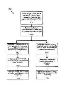
[00100] Attention is now directed to Fig. 17 which depicts a flowchart
of a
method 1700 for detecting and classifying a particle (or particles), according
to non-
limiting implementations. The particle or particles can comprise one or more
of a solid
particle, a solid spherical particle, a liquid particle and a liquid spherical
particle. In order
to assist in the explanation of method 1700, it will be assumed that method
1700 is
performed using system 3000. Furthermore, the following discussion of method
1700 will
lead to a further understanding of system 3000 and its various components.
However, it is
to be understood that system 3000 and/or method 1700 can be varied, and need
not work
exactly as discussed herein in conjunction with each other, and that such
variations are
within the scope of present implementations.
[00101] It is appreciated that, in some implementations, method 1700 is
implemented in system 3000 by processing unit 3012 of computing device 3011.
Indeed,
method 1700 is one way in which computing device 3011 can be configured. It is
to be
emphasized, however, that method 1700 need not be performed in the exact
sequence as
shown, unless otherwise indicated; and likewise various blocks may be
performed in
parallel rather than in sequence; hence the elements of method 1700 are
referred to herein
27
CA 02809596 2013-03-15
Agent Docket: P3683PC00
as "blocks" rather than "steps". It is also to be understood, however, that
method 1700
can be implemented on variations of system 3000 as well. For example, method
1700
could employ one, two, three or more ultrasound transducers.
1001021 At block 1710, light source 3001 and ultiasound transducers
3003-3005
are controlled to irradiate particle 3002 with light and an ultrasound pulse.
In some
implementations, only light source 3001 is employed to irradiate particle
3002. hi some
implementations, only one or more of ultrasound transducers 3003-3005 is
employed to
irradiate particle 3002. In some implementations, both light source 3001 and
at least one
of ultrasound transducers 3003-3005 are employed to irradiate particle 3002.
In some
implementations, light source 3001 comprises a laser, which, in a non-limiting
example,
could comprise a pulsed laser. It is understood that the term "irradiate"
comprises any
suitable mode of exposing particle 3002 to radiation, including by, but not
limited to,
electromagnetic radiation such as light, microwaves, radio waves, heat and
ultrasound.
[00103] In some implementations, the ultrasound pulse is in a range of
about 100
MHz to about 1000 MHz. In some implementations, the ultrasound pulse is in a
range up
to about 1000 MHz, including, but not limited to a range of about 100 MHz to
about 1000
MHz. For example, the ultrasound pulse could be in a range of about 10 MHz up
to about
1000 MHz.
[00104] In some implementations, controlling one or more of light
source 3001
and ultrasound transducers 3003-3005 to irradiate particle 3002 comprises
alternately
irradiating the particle with one of the light and the ultrasound pulse and
then the other of
the light and the ultrasound pulse.
[001051 At block 1720, a feature associated with particle 3002 is
determined by
processing ultrasound data resulting from particle 3002 being irradiated. In
some
implementations, one or more of ultrasound transducers 3003-3005 is configured
to
measure one or more of a photoacoustic wave and a pressure wave resulting from
the
irradiation of particle 3002 by the light and the ultrasound pulse. In some
implementations, the ultrasound data comprises data received from one or more
of
ultrasound transducers 3003-3005 when the one or more of ultrasound
transducers 3003-
3005 is measuring the photoacoustic wave and/or pressure wave. In some
implementations, the feature comprises one or more of an amplitude and an
intensity of a
28
CA 02809596 2013-03-15
Agent Docket: P3683PC00
pressure wave received by one or more of ultrasound transducers 3003-3005. In
some
implementation, the feature comprises a power spectrum of the particle. In
some
implementations, the ultrasound data comprises data resulting from detecting
one or more
of a photoacoustic pulse and an ultrasound pulse.
[00106] For example, at block 1730, a power spectrum of particle 3002 is
determined by processing ultrasound data resulting from particle 3002 being
irratliated. In
some implementations, ultrasound data comprises any information suitable for
system
3000 or similar system to determine a power spectrum of particle 3002 after or
while
particle 3002 is being irradiated. In some implementations, the ultrasound
data is
indicative of one or more of an ultrasound wave and a scattered ultrasound
wave
produced when the particle is irradiated. In some implementations, ultrasound
data
comprises at least one signal, such as photoacoustic signal and/or ultrasonic
signal.
[00107] In some implementations, the ultrasound data is received from
at least one
ultrasound transducer, which in turn measures a received ultrasound pulse from
particle
3002 and converts the received ultrasound pulse into the ultrasound data. In
related
implementations, the at least one of ultrasound transducer comprises one or
more of
ultrasound transducers 3003-3005 and a further ultrasound transducer.
[00108] As a non-limiting example, when block 1710 is performed using a
single
light source and a single ultrasound transducer, according to some
implementations, the
ultrasound transducer could be employed to irradiate particle 3002 and a
further
ultrasound ultrasound transducer could be employed to receive the ultrasound
pulse and
convert the received ultrasound pulse to the ultrasound data. Likewise, in
some
implementations, when block 1710 is performed using one or more light sources
and
more than one ultrasound transducer, at least a further ultrasound transducer
could be
employed to receive the ultrasound pulse and convert the received ultrasound
pulse to the
ultrasound data. In some implementations, at least one of the ultrasound
transducers
employed to irradiate particle 3002 is also employed to receive the ultrasound
pulse and
convert the received ultrasound pulse to the ultrasound data. Other
combinations of
ultrasound transducers employed to irradiate particle 3002 and/or receive the
ultrasound
pulse and convert the received ultrasound pulse to the ultrasound data will
occur to
persons skilled in the art and are within the scope of present
implementations.
29
CA 02809596 2013-03-15
Agent Docket: P3683PC00
1001091 In some implementations, the power spectrum is determined by
applying
a Fast Fourier Transform (FFT) to the ultrasound data. It is understood that
any suitable
technique for determining the power spectrum of particle 3002 while or after
particle
3002 has been irradiated are within the scope of present implementations.
[001101 At block 1740, the determined power spectrum of particle 3002 is
compared to a reference power spectrum. In some implementations, the reference
power
spectrum comprises one or more of a control power spectrum and a theoretical
model
power spectrum. In some implementation, the control power spectrum comprises a
power
spectrum derived from a sample particle having known properties. In some
implementation, the theoretical model power spectrum can be based upon one or
more of
an ultrasound scattering model, photoacoustic generation model or a finite
element model
(FEM). As a non-limiting example, the theoretical model power spectrum can
comprise
one or more of a Diebold model. Anderson model and/or a Faxan model. It is
understood
that the reference power spectrum comprises any known or theoretical model
power
spectrum suitable for comparison with the power spectrum of particle 3002
after particle
has been irradiated.
[001111 At block 1750, at least one property of particle 3002 is
determined based
on the comparison between the determined power spectrum and the reference
power
spectrum. As a non-limiting example, the determined property or properties
could
comprise one or more of the size, orientation, morphology, composition, sound
speed,
density and elasticity of particle 3002,
[001121 As another example, at block 1760 one or more of an amplitude
and an
intensity of a pressure wave received by one or more of ultrasound transducers
3003-
3005 is determined based on the processing of ultrasound data at block 1720.
In some
implementations, ultrasound data comprises any information suitable for system
3000 or
similar system to determine an amplitude and an intensity of a pressure wave
received by
one or more of ultrasound transducers 3003-3005, and emitted by particle 3002,
after or
while particle 3002 is being irradiated. In some implementations, light-based
analysis
techniques to assist in determining the feature of particle 3002. For example,
one or more
of photoacoustics, fluorescence, light scattering, light transmission and
absorbance can be
used to assist in determining the feature of the particle 3002.
CA 02809596 2013-03-15
Agent Docket: P3683PC00
1001131 At block 1770, the determined amplitude and/or intensity is
compared to a
reference, such as a reference data set or a control sample.
[00114] At block 1780, at least one property of particle 3002 is
determined based
on the comparison performed at block 1770. For example, one or more of a type
(e.g.
such as a cell type), a count and a state (e.g. such as whether particle 3002
is a live cell or
a dead cell) of particle 3002.
1001151 In some implementations, the ultrasound data is processed to
determine
features or characteristics in a range of about 100 MHz to about 1000 MHz of
the power
spectrum. In some implementations, the ultrasound data is processed to
determine
features or characteristics in a range up to about 1000 MHz, including, but
not limited to
a range of about 100 MHz to about 1000 MHz. For example, the ultrasound data
could be
processed to determine features or characteristics in a range of about 10 MHz
up to about
1000 MHz.
[00116] Those skilled in the art will appreciate that in some
implementations, the
functionality of system 3000 and/or computing device 3011 can be implemented
using
pre-programmed hardware or firmware elements (e.g., application specific
integrated
circuits (AS1Cs), electrically erasable programmable read-only memories
(EEPROMs),
etc.), or other related components. In other implementations, the
functionality of system
3000 and/or computing device 3011 can be achieved using a computing apparatus
that
has access to a code memory (not shown) which stores computer-readable program
code
for operation of the computing apparatus. The computer-readable program code
could be
stored on a computer readable storage medium which is fixed, tangible and
readable
directly by these components, (e.g., removable diskette, CD-ROM, ROM, fixed
disk,
USB drive). Furthermore, it is appreciated that the computer-readable program
can be
stored as a computer program product comprising a computer usable medium.
Further, a
persistent storage device can comprise the computer readable program code. It
is yet
further appreciated that the computer-readable program code and/or computer
usable
medium can comprise a non-transitory computer-readable program code and/or non-
transitory computer usable medium. Alternatively, the computer-readable
program code
could be stored remotely but transmittable to these components via a modem or
other
interface device connected to a network (including, without limitation, the
Internet) over
31
CA 02809596 2013-03-15
Agent Docket: P3683PC00
a transmission medium. The transmission medium can be either a non-mobile
medium
(e.g., optical and/or digital and/or analog communications lines) or a mobile
medium
(e.g., microwave, infrared, free-space optical or other transmission schemes)
or a
combination thereof.
[00117] Persons
skilled in the art will appreciate that there are yet more alternative
implementations and modifications possible, and that the above examples are
only
illustrations of one or more implementations. The scope, therefore, is only to
be limited
by the claims appended hereto.
32