Note: Descriptions are shown in the official language in which they were submitted.
CA 02809859 2013-03-19
MICROWAVE-SHIELDED TISSUE SENSOR PROBE
BACKGROUND
1. Technical Field
[0001] The present disclosure relates to systems and methods for providing
energy to
biological tissue and, more particularly, to systems and methods for precisely
sensing thermal
parameters of tissue during a microwave ablation procedure.
2. Background of Related Art
[0002] Energy-based tissue treatment is well known in the art. Various
types of energy (e.g.,
electrical, ultrasonic, microwave, cryogenic, thermal, laser, etc.) are
applied to tissue to achieve a
desired result. Electrosurgery involves application of high radio frequency
electrical current to a
surgical site to cut, ablate, coagulate or seal tissue. In monopolar
electrosurgery, a source or
active electrode delivers radio frequency energy from the electrosurgical
generator to the tissue
and a return electrode carries the current back to the generator. In monopolar
electrosurgery, the
source electrode is typically part of the surgical instrument held by the
surgeon and applied to the
tissue to be treated. A return electrode is placed remotely from the active
electrode to carry the
current back to the generator. In tissue ablation electrosurgery, the radio
frequency energy may
be delivered to targeted tissue by an antenna or probe.
[0003] There are several types of microwave antenna assemblies in use,
e.g., monopole,
dipole and helical, which may be used in tissue ablation applications. In
monopole and dipole
antenna assemblies, microwave energy generally radiates perpendicularly away
from the axis of
the conductor. Monopole antenna assemblies typically include a single,
elongated conductor. A
typical dipole antenna assembly includes two elongated conductors, which are
linearly aligned
1
CA 02809859 2013-03-19
and positioned end-to-end relative to one another with an electrical insulator
placed between
them. Helical antenna assemblies include a helically-shaped conductor
connected to a ground
plane. Helical antenna assemblies can operate in a number of modes including
normal mode
(broadside mode), in which the field radiated by the helix is maximum in a
perpendicular plane
to the helix axis, and axial mode (end fire mode), in which maximum radiation
is along the helix
axis. The tuning of a helical antenna assembly may be determined, at least in
part, by the
physical characteristics of the helical antenna element, e.g., the helix
diameter, the pitch or
distance between coils of the helix, and the position of the helix in relation
to the probe assembly
to which the helix is mounted.
[0004] The typical microwave antenna has a long, thin inner conductor that
extends along the
longitudinal axis of the probe and is surrounded by a dielectric material. An
outer conductor
surrounds the dielectric material and extends along the axis of the probe. In
another variation of
the probe that provides effective outward radiation of energy or heating, a
portion or portions of
the outer conductor are selectively removed. This type of construction is
typically referred to as
a "leaky waveguide" or "leaky coaxial" antenna. Another variation on the
microwave antenna
probe involves having the tip formed in a uniform spiral pattern, such as a
helix, to provide the
necessary configuration for effective radiation. This variation can be used to
direct energy in a
particular direction, e.g., perpendicular to the axis, in a forward direction
(i.e., towards the distal
end of the antenna), or any combination of these directions.
[0005] Invasive procedures and devices have been developed in which a
microwave antenna
probe is either inserted directly into a point of treatment via a normal body
orifice or
percutaneously inserted. Such invasive procedures and devices potentially
provide better
temperature control of the tissue being treated. Because of the small
difference between the
2
CA 02809859 2013-03-19
temperature required for denaturing malignant cells and the temperature
injurious to healthy
cells, a known heating pattern and predictable temperature control is
important so that heating is
confined to the tissue to be treated. For instance, hyperthermia treatment at
the threshold
temperature of about 41.5 C generally has little effect on most malignant
growth of cells.
However, at slightly elevated temperatures above the approximate range of 43 C
to 45 C or with
rapid bursts of elevated temperatures, thermal damage to most types of normal
cells is routinely
observed. Accordingly, great care must be taken not to exceed these
temperatures along the
length of the ablation probe when it is placed adjacent to healthy tissue.
[0006]
In the case of tissue ablation, a high radio frequency electrical current in
the range of
about 500 MHz to about 10 GHz is applied to a targeted tissue site to create
an ablation volume,
which may have a particular size and shape. The ablation volume is correlated
to antenna
design, antenna tuning, antenna impedance and tissue impedance. Tissue
impedance may
change during an ablation procedure due to a number of factors, e.g., tissue
denaturization or
desiccation occurring from the absorption of microwave energy by tissue.
Changes in tissue
impedance may cause an impedance mismatch between the probe and tissue, which
may affect
delivery of microwave ablation energy to targeted tissue. The temperature
and/or impedance of
targeted tissue, and of non-targeted tissue and adjacent anatomical
structures, may change at
varying rates, which may be greater, or less than, expected rates. A surgeon
may need to
perform an ablation procedure in an incremental fashion in order to avoid
exposing targeted
tissue and/or adjacent tissue to excessive temperatures and/or denaturation.
In certain
circumstances, a surgeon may need to rely on experience and/or published
ablation probe
parameters to determine an appropriate ablation protocol (e.g., ablation time,
ablation power
level, and the like) for a particular patient.
3
CA 02809859 2013-03-19
[0007] One way to monitor and control the temperature of tissue during a
tissue ablation
procedure is to provide feedback from a tissue sensor probe. If, however, the
tissue sensor probe
is positioned near a microwave ablation probe, the electromagnetic fields
produced by the
microwave ablation probe may reduce the accuracy and precision of the
measurements of the
tissue sensor probe either through direct thermal agitation of the thermal
physics of the tissue
sensor probe or through induced electrical current in the tissue sensor probe.
One solution to this
problem is to maintain an appropriate distance between the tissue sensor probe
and the
electromagnetic fields produced by the microwave field during a surgical
procedure. It is
difficult, however, to predict the location and boundary of this
electromagnetic field. In addition,
electromagnetic energy from other sources may interfere with the operation of
the tissue sensor
probe.
SUMMARY
[0008] The disclosed tissue sensor probe and corresponding surgical
ablation system
increases the speed and accuracy of temperature measurements taken near an
ablation probe
during a microwave ablation procedure. This is achieved by shielding a
temperature sensor
within the probe from electromagnetic radiation produced by the microwave
ablation probe and
maximizing the response time of thermal energy transfer to the temperature
sensor.
[0009] In one aspect, the present disclosure is directed to an
electromagnetic surgical
ablation system that includes a tissue sensor probe that is configured to
accurately sense tissue
temperature at or near an ablation surgical site. The electromagnetic surgical
ablation system
also includes a generator that selectively provides surgical ablation energy
to an ablation probe.
The surgical ablation system further includes an ablation probe operatively
coupled to the
generator and configured to receive ablation energy from the generator and to
deliver the
4
CA 02809859 2013-03-19
ablation energy to tissue. The surgical ablation system further includes a
controller operatively
coupled to the generator. The controller includes a processor and a memory
operatively coupled
to the processor.
[0010] The tissue sensor probe is operatively coupled to the controller.
The tissue sensor
probe includes an electrically-conductive enclosure. At least a portion of the
electrically-
conductive enclosure is formed of a thermally-conductive material. The
electrically-conductive
enclosure may include an elongated cylindrical shaft having a distal end and a
proximal end, a
thermally-conductive tip electrically coupled to the distal end of the
elongated cylindrical shaft,
and a cap electrically coupled to the proximal end of the elongated
cylindrical shaft. At least one
of the electrically-conductive elongated cylindrical shaft, the electrically-
conductive tip, and the
electrically-conductive cap may include at least one of stainless steel,
copper, and aluminum.
[0011] The tissue sensor probe also includes a material of high thermal
conductivity disposed
in the electrically-conductive enclosure and in thermal association with the
thermally-conductive
material. At least a portion of the material of high thennal conductivity may
be disposed within
the electrically-conductive tip. The material of high thermal conductivity may
have low
electrical conductivity. The material of high thermal conductivity may include
at least one of
silver, gold, carbon nanotube, diamond, copper, aluminum, a thermally-
conductive gel, and a
thermally-conductive polymer.
[0012] The tissue sensor probe further includes a temperature sensor
disposed in the material
of high thettnal conductivity and electrically isolated from the electrically-
conductive enclosure.
The temperature sensor may include a plurality of temperature sensors disposed
along the length
of the electrically-conductive elongated cylindrical shaft.
CA 02809859 2013-03-19
[0013] The temperature sensor may be configured to provide a temperature
sensor signal
corresponding to a temperature of body tissue. The temperature sensor may
include at least one
of a fluoroptic temperature sensor, a thermocouple, a thermistor, a resistance
temperature
detector, or an infrared thermometer.
[0014] The surgical ablation system may further include an actuator
operatively coupled to
the controller and configured to selectively activate the generator. The
actuator may be selected
from a group consisting of a handswitch, a footswitch, and an orally-activated
switch. The tissue
sensor probe may also include a handle disposed at a proximal end of the
tissue sensor probe.
The handle may include a grip-enhancing feature.
[0015] In another aspect, the present disclosure is directed to a tissue
sensor probe. The
tissue sensor probe includes an electrically-conductive elongated cylindrical
shaft having a distal
end and a proximal end. The tissue sensor probe also includes an electrically-
conductive tip
coupled to the distal end of the electrically-conductive cylindrical shaft.
The electrically-
conductive tip closes the distal end of the electrically-conductive
cylindrical shaft and has a high
thermal conductivity. The temperature sensor further includes an electrically-
conductive cap
coupled to the proximal end of the elongated cylindrical shaft to close the
proximal end of the
electrically-conductive cylindrical shaft. At least one of the electrically-
conductive elongated
cylindrical shaft, the electrically-conductive tip, and the electrically-
conductive cap may be
composed of at least one of stainless steel, copper, and aluminum.
[0016] The tissue sensor probe also includes a material of high thermal
conductivity disposed
in the distal end of the elongated cylindrical shaft. The material of high
thermal conductivity is
disposed in the distal end of the elongated cylindrical shaft so that it is in
thermal association
with the electrically-conductive tip.
6
CA 02809859 2013-03-19
[0017] The tissue sensor probe further includes a temperature sensor
disposed in the material
of high thermal conductivity and electrically isolated from the electrically-
conductive cylindrical
shaft and the electrically-conductive tip. In some embodiments, the
temperature sensor may be
configured to provide a temperature sensor signal corresponding to a
temperature of body tissue.
The temperature sensor may include at least one of a fluoroptic temperature
sensor, a
thermocouple, a thermistor, a resistive temperature detector, and an infrared
thermometer. In
some embodiments, the temperature sensor may include a plurality of
temperature sensors
disposed along the length of the electrically-conductive elongated cylindrical
shaft configured to
measure a temperature profile or gradient.
[0018] The tissue sensor probe may include a handle disposed at a proximal
end of the
elongated cylindrical shaft. In some embodiments, the material of high thermal
conductivity
may have low electrical conductivity. The material of high thermal
conductivity may include at
least one of silver, gold, carbon nanotube, diamond, copper, and aluminum. At
least a portion of
the material of high thermal conductivity may be disposed within the
electrically-conductive tip.
BRIEF DESCRIPTION OF THE DRAWINGS
[0019] The above and other aspects, features, and advantages of the present
disclosure will
become more apparent in light of the following detailed description when taken
in conjunction
with the accompanying drawings in which:
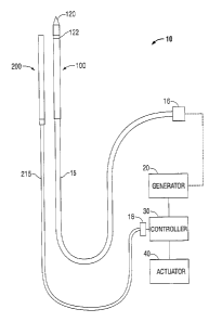
[0020] Fig. 1 is a diagram of a microwave ablation system having an
electromagnetic
surgical ablation probe and a tissue sensor probe in accordance with
embodiments of the present
disclosure;
7
CA 02809859 2013-03-19
[0021] Fig. 2 is a block diagram of a microwave ablation system having an
electromagnetic
surgical ablation probe and a tissue sensor probe in accordance with
embodiments of the present
disclosure;
[0022] Fig. 3 is a side, cross-sectional view of a tissue sensor probe in
accordance with
embodiments of the present disclosure;
[0023] Fig. 4 is a side, cross-sectional view of a tissue sensor probe in
accordance with other
embodiments of the present disclosure; and
[0024] Fig. 5 is a flowchart showing a method of operation of a microwave
ablation system
having a tissue sensing probe in accordance with embodiments of the present
disclosure.
DETAILED DESCRIPTION
[0025] Particular embodiments of the present disclosure will be described
hereinbelow with
reference to the accompanying drawings; however, it is to be understood that
the disclosed
embodiments are merely exemplary of the disclosure, which may be embodied in
various forms.
Well-known functions or constructions are not described in detail to avoid
obscuring the present
disclosure in unnecessary detail. Therefore, specific structural and
functional details disclosed
herein are not to be interpreted as limiting, but merely as a basis for the
claims and as a
representative basis for teaching one skilled in the art to variously employ
the present disclosure
in virtually any appropriately detailed structure.
[0026] In the drawings and in the descriptions that follow, the term
"proximal," as is
traditional, shall refer to the end of the instrument that is closer to the
user, while the term
"distal" shall refer to the end that is farther from the user.
8
CA 02809859 2013-03-19
,
[0027] Fig. 1 shows an embodiment of a microwave ablation system 10 in
accordance with
the present disclosure. The microwave ablation system 10 includes an
electromagnetic surgical
ablation probe 100 having a tapered distal tip 120 and a feed point 122. The
ablation probe 100
is operatively connected by a cable 15 to connector 16, which may further
operatively connect
ablation probe 100 to a generator assembly 20. Generator assembly 20 may be a
source of
ablation energy, e.g., microwave or RF energy in the range of about 915 MHz to
about 2.45
GHz. The disclosed system 10 includes a tissue sensor probe 200 that is
configured to sense at
least one operative parameter, e.g., a tissue temperature. In other
embodiments, the tissue sensor
probe 200 may be configured to sense a tissue dielectric parameter, e.g., a
relative permittivity, a
dielectric constant, a dielectric loss factor and/or conductivity. The tissue
sensor probe 200 is
operatively connected by a cable 215 to connector 18, which may further
operatively connect
tissue sensor probe 200 to a controller assembly 30. An actuator 40 is
operatively coupled to the
controller to enable a user, e.g., a surgeon, to selectively activate and de-
activate the delivery of
ablation energy to patient tissue. Controller 30 is operatively coupled to
generator 20 to enable
communication therebetween, such as without limitation, a control signal
and/or a status signal.
[0028] Fig. 2 illustrates a functional block diagram of an ablation system
10 in accordance
with the present disclosure. The system 10 includes a controller 30 that
includes one or more
processors 31 operatively coupled to memory 32, a database 33, and a
temperature sensor circuit
35. In other embodiments, the controller 30 may include one or more processors
31 operatively
coupled to other sensor circuits (not explicitly shown), e.g., a dielectric
sensor circuit, that are
coupled to corresponding sensors in the tissue sensor probe 200. Processor(s)
31 may be
configured to execute a set of programmed instructions for performing a method
of microwave
ablation as disclosed herein.
9
CA 02809859 2013-03-19
[0029] Controller 30 includes actuator interface 36 that is adapted to
facilitate operative
coupling with actuator 40 and/or a generator interface 37 that is adapted to
facilitate operative
coupling with generator 20. Actuator 40 may be any suitable actuator, such as
without
limitation, a footswitch, a handswitch (which may be mounted on an ablation
probe 100 and/or a
tissue sensor probe 200), an orally-activated switch (e.g., a bite-activated
switch and/or a breath-
actuated switch), and the like.
[0030] The processor(s) 31, memory 32, database 33, temperature sensor
circuit 35, actuator
interface 36 and/or generator interface 37 may be separate components or may
be integrated,
such as in one or more integrated circuits. The various components in the
controller 30 may be
coupled by one or more communication buses or signal lines 38. Memory 30
and/or database 33
may include a set of executable instructions for perfoiming a method of
microwave ablation as
described herein. One or more elements of ablation system 10 may be coupled
using hard-wired
connections and/or a wireless link. During use, tissue sensor probe 200 may be
positioned in
tissue T in proximity to ablation probe 100 to obtain at least one tissue
parameter.
[0031] Embodiments of tissue sensor probe 200 in accordance with the
present disclosure are
now described with reference to Figs. 3 and 4. The tissue sensor probe 200 may
be a needle-like
device having a small gauge size (e.g., 17 gauge or smaller). The tissue
sensor probe 200
includes a temperature sensor 231 and interfaces with a microwave generator
and controller to
enable feedback and control using tissue temperature measurements from the
tissue sensor probe
200. For example, the user may use the controller to set limits on the
temperature of target tissue
and apply high frequency energy to the target tissue based on temperature
limits and the
measured tissue temperature.
CA 02809859 2013-03-19
[0032] Referring to Fig. 3, the tissue sensor probe 200 is configured to
form a "Faraday
cage" or a "Faraday shield." A Faraday cage is an enclosure or a container
made of electrically-
conductive material that can shield its interior from external electromagnetic
radiation if the
electrically-conductive material has an appropriate thickness. In operation,
an external electrical
field causes the electrical charges within the Faraday cage's electrically-
conductive material to
redistribute so as to cancel the external electrical field's effects in the
Faraday cage's interior.
[0033] The tissue sensor probe 200 includes an electrically-conductive
enclosure configured
as a Faraday cage, which contains a temperature sensor 231. In this way, the
temperature sensor
231 is isolated from any electromagnetic radiation, including microwave
radiation, originating
external to the probe 200. The probe 200 includes an elongated, generally-
cylindrical shaft 223
having a distal end 213 and a proximal end 216. In some embodiments, the
elongated shaft 223
is a cylindrical hypo-tube made of stainless steel or other electrically-
conductive metal such as
aluminum or copper.
[0034] The distal end 213 of the shaft 223 includes an electrically-
conductive tip 221, such
as a metal trocar tip. The tip 221 is made of an electrically-conductive
material that also has
high thermal conductivity, such as copper, silver, gold, or any alloys of
these materials. In some
embodiments, the electrically-conductive material may also have physical
properties that allow
the tip 221 to keep its sharpness for long periods.
[0035] An electrical connection is made completely around the junction
between the tip 221
and the elongated shaft 223, either by close physical contact or by soldering
or laser welding, to
form a tight, electrically-conductive enclosure that can block electromagnetic
radiation, such as
microwave radiation. A thermally-conductive material 233 is disposed inside
the elongated shaft
223 at its distal end 216 directly behind and in contact with the tip 221. In
some embodiments,
11
CA 02809859 2013-03-19
the tip 221 may include a hollow center in which thermally-conductive material
233d is
disposed. For example, the thermally-conductive material 233d may be a high
thermally-
conductive gel.
[0036] The thermally-conductive material 233 may have high thermal
conductivity, but low
electrical conductivity. The thermally-conductive material 233 may include
gold, silver, copper,
a thermally-conductive gel or polymer, or any combination of these materials.
A temperature
sensor 231, such as a thermistor or thermocouple, is disposed inside the
thermally-conductive
material 233 in such a way as to be electrically isolated from the
electrically-conductive tip 221
and elongated shaft 223. The temperature sensor 231 may be affixed within the
tissue sensor
probe 200 using a thermally-conductive adhesive or epoxy. The thermally-
conductive material
233 rapidly transfers heat from the body tissue to the temperature sensor 231
to provide accurate
and precise temperature measurements of the body tissue.
[0037] The temperature sensor 231 is connected to electrically-insulated
leads 235, such as
wires coated with plastic insulation or a vapor-deposited insulator. The wires
235 emerge from
the theimally-conductive material and are coupled to a shielded cable 215 at
the proximal end
213 of the tissue sensor probe 200. The wires 235 may twist about each other
to form a twisted
pair. The far proximal end of the elongated shaft 223 is capped or closed with
an electrically-
conductive metallic plate 237. A bulkhead-like port 225, which maintains
electrical isolation
from the external environment, passes through the plate 237 and enables a
transition to the
shielded cable 215. The twisted pair of tissue sensor probe wires 235 passes
through the port
225 into the shielded cable 215. The shield of the cable 215 may be a wire
braid or solid
cylindrical conductor. An ergonomic handle 227, such as molded plastic that
provides an
12
CA 02809859 2013-03-19
attractive device appearance, is positioned at the proximal end 213 of the
tissue sensor probe
200.
[0038] Thus, the cylindrical elongated shaft 223, the tip 221, and the cap
237 form an
electrically-conductive enclosure that isolates the temperature sensor 231
from any
electromagnetic interference external to the tissue sensor probe 220. At the
same time, the
thermally-conductive tip 221 and the thermally-conductive material 233 can
quickly transmit
thermal energy at or near the exterior of the tissue sensor probe 200 to the
temperature sensor
231 to ensure a fast thermal response by the temperature sensor 231.
10039] The type of electrically-conductive material used to form the
cylindrical elongated
shaft 223, the tip 221, and the cap 237, and other parameters of the
electrically-conductive
material (e.g., thickness) may be selected based on parameters of
electromagnetic radiation
emitted by the microwave probe (e.g., frequency, phase, or polarization). The
parameters for the
electrically-conductive material may also be selected based on parameters of
the electromagnetic
radiation from another source.
[0040] The surgical ablation system may include multiple tissue sensor
probes 200. The
tissue sensor probes 200 may be placed at the margin of a target tissue
structure to make sure that
critical tissue structures are not harmed by elevated temperatures created by
a microwave
ablation probe (e.g., ablation probe 200).
[0041] Fig. 4 illustrates a side, cross-sectional view of a tissue sensor
probe 200 in
accordance with another embodiment of the present disclosure. The thermally-
conductive
material 233d may extend within a hollow portion of the tip 221. Thermally-
conductive material
233b, 233c may also be disposed within the elongated shaft 223 at different
positions along the
length of the elongated shaft 223. Temperature sensors 231b, 231c are disposed
within the
13
CA 02809859 2013-03-19
thermally-conductive material 233b, 233c and connect to the leads or wires
235. In this
configuration, the temperature of tissue near each temperature sensor 233a-
233c may be
measured to obtain a temperature gradient or profile of the tissue along the
length of the
elongated shaft 223. For example, the tissue sensor probe 200 may be oriented
parallel to an
ablation probe to measure the temperature radiating up the ablation probe. To
ensure accurate
temperature measurements, the elongated shaft 223 or those portions of the
elongated shaft 223
that make contact with the thermally-conductive material 233b, 233c may be
made of a
thermally-conductive material.
[0042] The probe 200 may include one or more absorbent sleeves 214 disposed
on the
elongated shaft 223 and adapted to attract and absorb moisture, e.g., steam
and/or condensed
water vapor, which may be released as a byproduct of an ablation procedure and
collect on the
elongated shaft 223. In some embodiments, absorbent sleeve(s) 214 is slidably
disposed on the
elongated shaft 223 to enable the selective absorption of moisture, and/or to
enable a surgeon to
position sleeve 214 according to surgical requirements. Sleeve(s) 214 may be
formed from any
suitable biocompatible absorbent material, including without limitation paper-
based material
composed of virgin wood pulp that is obtained from certified forests.
[0043] Probe 200 includes a handle 227 positioned at a proximal end 213 of
the probe 200.
Handle 227 may include grip-enhancing features such as, without limitation,
knurling, ridges,
coatings (e.g., silicone-based or rubberized coating) disposed on at least a
part of an outer surface
of the handle 227. Probe 200 includes a cable 215 extending from the probe 200
that is adapted
to operatively couple the temperature sensor 231 with temperature sensor
circuit 35. The
elongated shaft 223 of the probe 200 may have any suitable length and/or
diameter suitable for
14
CA 02809859 2013-03-19
use in an ablation procedure. In some embodiments, the elongated shaft 223 has
a length of
about 10 cm to about 30 cm.
[0044] Tissue sensor probe 200 includes a temperature sensor 231 disposed
at a distal end
211 of the probe shaft 223. Temperature sensor 231 may include any suitable
temperature-
sensing transducer, including without limitation, a fluoroptic (e.g., fiber
optic) sensor, a
thermocouple, a thennistor, an infrared thermometer (e.g., emissive
measurement), a resistance
thermometer (also referred to as a resistive temperature detector or resistive
thermal device
(RTD)), or other temperature sensor now or in the future known. Temperature
sensor 231 is
operatively coupled to temperature sensor circuit 35 via connection element
218, which may
include electrical and/or fiber optic conductors. Temperature sensor circuit
35 is adapted to
receive a temperature measurement signal from temperature sensor 231 to
determine a tissue
temperature, which, in turn, may be utilized by controller 30 and/or generator
20.
[0045] Fig. 5 illustrates a method 300 for operating a microwave ablation
system 10, which
includes the generator 20, the controller 30, the ablation probe 100, and the
tissue sensor probe
200. The method 300 begins in step 301 wherein one or more initializations may
be performed,
e.g., power-on self test (POST), memory allocation, input/output (I/O)
initialization, and the like.
In step 305, it is determined whether an activation signal is present, e.g.,
whether actuator 40 has
been engaged by a user to cause delivery of ablation energy to the tissue. If
no activation signal
is present, the process iterates until an activation signal is detected (e.g.,
the system enters a
"wait state").
[0046] Upon receipt of an activation signal, an ablation generator, e.g.,
generator 20, is
activated in step 315 to deliver ablation energy to tissue via the ablation
probe 100. In step 315,
an ablation control signal may be generated by controller 30 and conveyed to
ablation generator
CA 02809859 2013-03-19
20 via generator interface 37. In step 320, a tissue temperature measurement T
is obtained.
Tissue temperature measurement T is obtained from temperature sensor 231 of
tissue sensor
probe 200.
[0047] In step 345, a tissue temperature measurement is used to determine a
tissue status S.
Tissue status S may be indicative of whether the sensed tissue has received
insufficient,
sufficient, and/or excessive ablation energy, e.g., whether the tissue is
underablated
("undercooked"), ablated ("cooked"), or hyper-ablated ("charred"). Other
tissue statuses are
envisioned, for example, a near-ablated ("pre-cooked") status. A lookup table
(not explicitly
shown) may be utilized to ascertain tissue status based upon temperature. In
other embodiments,
the lookup table may be used to ascertain tissue status based upon
temperature, permittivity, and
loss factor. For example, the lookup table may have a three-dimensional
organization whereby a
first table dimension is representative of tissue temperature, a second table
dimension is
representative of tissue permittivity, and a third table dimension is
representative of tissue loss
factor. In this manner, each of temperature, permittivity, and loss factor are
thus used as indices
into the three dimensional table to identify a particular tissue status
corresponding thereto. In
some embodiments, the lookup table may be included within database 33.
[0048] In step 350, a tissue status signal may be reported to a user via
any suitable manner of
communication, such as without limitation, an audible signal, a visual signal,
a haptic (tactile)
signal, and/or a combination of these signals. By way of example only, an
underablated status
may be excluded from reporting, since this is routinely observed and expected
during an initial
phase of an ablation procedure. As tissue approaches ablated status, e.g.,
attains near-ablated
status, a first tissue status signal, e.g., a short audible signal, may be
issued to apprise a user
accordingly. When tissue reaches ablated status, a second tissue status signal
may be issued,
16
CA 02809859 2013-03-19
e.g., a more urgent audible signal, alone or in combination with a visual
signal. In some
embodiments, multiple tissue statuses may be utilized to convey a continuous
indication of
ablation progress. In this manner, a user may be assisted in the accurate and
timely assessment
of ablation progress, in real-time, during an ablation procedure.
[0049] A tissue status may be associated with a terminal condition whereby
attainment of a
terminal status indicates that the ablation procedure is complete and/or that
delivery of ablation is
to be terminated. In step 355, a determination is made whether the currently-
identified tissue
status is a terminal status. If the current tissue status is not a terminal
status, the process iterates
step 305 whereupon presence of an activation signal is confirmed, and the
ablation procedure
continues as just described. Conversely, if a terminal status is reached, the
ablation process
concludes at step 360.
[0050] It is to be understood that the steps of the method provided herein
may be performed
in combination and/or in a different order than presented herein without
departing from the scope
and spirit of the present disclosure.
[0051] During use, tissue sensor probe 200 is positioned at a boundary of
the operative site
that corresponds to an outer limit of the desired ablation region. As ablation
energy is applied to
targeted tissue (e.g., generator 20 is activated and ablation probe 100 is
applied to the operative
site), tissue sensor probe 200 provides tissue parameters (e.g., temperature
and dielectric
properties) to controller 30. As the ablated region expands, controller 30
continues to monitor
tissue status at the probed location. When a tissue terminal status is
detected (e.g., tissue is
"cooked"), controller 30 causes generator 20 to be deactivated, thus enabling
a surgeon to
perform a precisely-formed ablation, which may lead to improved operative
outcomes, reduced
operative and/or recovery times, and enhanced patient satisfaction. A distal
end 213 of tissue
17
CA 02809859 2013-03-19
sensor probe 200 may be generally positioned coincident with a plane radially
extending
transversely from feed point 122 of the ablation probe 100.
[0052] Tissue sensor probe 200 may be positioned between an ablation region
and an
adjacent anatomical structure, which may be a critical structure to be
protected from receipt of
excessive ablation energy, increased temperature, and/or undesired
denaturization which may
occur as a side effect of an ablation procedure. In this instance, a tissue
terminal status may
reflect a threshold at which an ablation procedure is suspended in order to
protect a critical
structure from damage.
[0053] The described embodiments of the present disclosure are intended to
be illustrative
rather than restrictive, and are not intended to represent every embodiment of
the present
disclosure. Further variations of the above-disclosed embodiments and other
features and
functions, or alternatives thereof, may be made or desirably combined into
many other different
systems or applications without departing from the spirit or scope of the
disclosure as set forth in
the following claims both literally and in equivalents recognized in law. The
claims can
encompass embodiments in hardware, software, or a combination thereof
18