Note: Descriptions are shown in the official language in which they were submitted.
WO 2012/030996 CA 02810008 2013-02-28 PCT/US2011/050019
PROSTHETIC TISSUE VALVE
Cross-Reference to Related Applications
[0001] This application claims the benefit of the filing date of U.S. Patent
Application
Serial No. 12/875,727, filed September 3, 2010, which is a continuation-in-
part of U.S. Patent
Application Serial No. 11/958,405, filed December 18, 2007, now abandoned, and
U.S.
Patent Application Serial No. 11/958,407, filed December 18, 2007, now
abandoned, all of
which are hereby incorporated by reference in their entireties. This
application also claims
the benefit of the filing date of U.S. Provisional Patent Application Serial
No. 61/295,503,
filed January 15, 2010, which is hereby incorporated by reference in its
entirety.
Field of the Invention
[0002] The invention generally relates to a prosthetic tissue valve for
replacing
defective aortic, pulmonary, mitral or tricuspid valves. More specifically,
the invention
relates to a prosthetic tissue valve that is substantially planar prior to
implantation in an
annulus and substantially non-planar following implantation in an annulus.
Background of the Invention
[0003] In general, two types of artificial heart valves are used to replace
defective
heart valves: mechanical valves and tissue valves. Although implantation of
artificial heart
valves has traditionally occurred through open heart surgery, research and
experimentation
are being done to develop valves that can be placed in a patient
percutaneously, thereby
avoiding open heart surgery.
[0004] Implantation of mechanical valves, which are durable, requires open
heart
surgery, risks peri-valvular leakage on the outside of the valve between the
valve and the
attachment wall, and requires a lifetime of administration of anti-coagulants,
which requires
close (usually bi-weekly) monitoring in order to avoid either bleeding or
thrombotic/embolic
stroke. Mechanical valves also risk development of stenosis at the valve
replacement site,
and incur chronic hemolysis (damage to red blood cells by the mechanical
action of the
valve).
[0005] Tissue valves typically last from 10 to 15 years in less active and
elderly adults
and are of porcine or human origin. They fail because the tissue of the valve
begins to wear,
at least in part because the valves are retrieved after already having
undergone partial
lifetimes of use. Tissue valves in younger people wear out more quickly due to
the more
1
WO 2012/030996 CA 02810008 2013-02-28PCT/US2011/050019
active blood flow in younger people, which causes rapid calcification and
places great
mechanical demands on the valves. The risk of death or serious complications
from surgical
valve replacement is typically from 1% to 5% depending on the health of the
patient and the
skill of the surgeon. Therefore, it is preferred that a valve only be replaced
one time.
[0006] Mechanical valves last longer in younger patients because the patients
are still
growing. However, pediatric valve replacements are particularly challenging
because the
patients frequently outgrow the implanted mechanical valve and require
surgical intervention
to replace the pediatric valve with a larger valve.
[0007] Progressive deterioration of a tissue valve can lead to stenosis,
which manifests
itself as an obstruction of forward flow through the valve when the valve is
in its open
position. More commonly, deterioration of a valve produces tears in the valve
leaflets that
cause regurgitation, which manifests itself as a leakage in the valve when the
valve is in its
closed position.
[0008] Known synthetic valves, although configured to mimic native valves,
never
assimilate fully into the surrounding tissue following implantation. In
addition, attachment
of known synthetic valves is accomplished using a ring that remains in a
single plane
following implantation, thereby risking perivalvular leakage in the same
manner as the
attachments of mechanical valves.
[0009] The tricuspid valve separates the right atrium from the right
ventricle, and the
mitral valve separates the left atrium from the left ventricle. The annuluses
in which these
valves are mounted typically comprise dense fibrous rings that are attached
either directly or
indirectly to the atrial and ventricular muscle fibers. In a valve replacement
operation, the
damaged leaflets are excised and the annulus is sculpted to receive a
replacement valve.
Ideally, the annulus presents relatively healthy tissue which can be formed by
a surgeon into
a substantially uniform ledge that projects into the opening created after a
native valve is
removed. The time and spatial constraints imposed by surgery, however, often
dictate that
the shape of the resulting annulus is less than perfect for attachment of a
sewing ring.
Moreover, the leaflets of the valve and the annulus may be calcified, and
complete annular
debridement, or removal of the hardened tissue, can result in a larger opening
and a more
gradually sloped annulus ledge for attachment of the sewing ring. In short,
the contours of
the resulting annulus vary widely after the natural valve has been excised.
2
WO 2012/030996 CA 02810008 2013-02-28PCT/US2011/050019
[0010] Conventional placement of a valve is intra-annular, with a valve body
deep
within the narrowest portion of the annulus to enhance any seal effected by
the sewing
ring/suture combination and reduce the chance of perivalvular leakage.
Surgeons report
using at least 30 simple sutures or 20 mattress-type sutures to prevent
leakage.
[0011] The implantation of a prosthetic heart valve, including mechanical
valves and
bioprosthetic valves (i.e., "tissue" valve), requires a great deal of skill
and concentration
given the delicate nature of the native heart tissue, the spatial constraints
of the surgical field
and the criticality of achieving a secure and reliable implantation. It is of
equal importance
that the valve have characteristics that promote a long valve life and have
minimal impact on
the physiological makeup of the heart environment.
[0012] Given the uneven nature of the annuluses, the design of the sewing
ring and the
method by which the sewing ring is fixed into place are perhaps the most
crucial aspects of
prosthetic heart valve implantation. Due to the inability of conventional
sewing rings to
easily stretch, if the selected size of the sewing ring is even slightly too
small, attachment can
only be achieved by placing undue tension on the tissue and sutures. As a
result, a great deal
of care and accuracy by the surgeon is needed in the selection of a valve size
that precisely
matches the valve annulus of the patient. Unfortunately, standard sizing tools
are provided in
increments based on an overall opening size, and may not be able to accurately
measure a
less than optimally formed annulus. The surgeon thus must select an
approximate valve size.
[0013] Accordingly, there is a need in the art of valve replacement
procedures for a
valve having the benefits of a tissue valve and the longevity of a mechanical
valve, without
the side effects or disadvantages of either. Surgical outcomes would also
benefit greatly by
an improved sewing ring, permitting improved tissue attachment in all valve
replacements.
SUMMARY OF THE INVENTION
[0014] In one aspect, a valve disclosed herein is designed to replace a
native valve
such as the aortic, pulmonary, mitral, or tricuspid valves in the heart of a
subject. In one
aspect, the valve can have a plurality of leaflets that extend generally
inwardly relative to a
valve circumference toward a radial center point of the valve such that at
least a portion of
each leaflet contacts its adjacent leaflets. When placed on a flat surface in
an unstressed
position before attachment of the valve in the subject, the valve is
substantially flat or planar
and can therefore, in a further aspect, be formed from a substantially planer
material. In one
aspect, the valve can have a sewing ring to which the leaflets are attached
and the sewing ring
3
WO 2012/030996 CA 02810008 2013-02-28PCT/US2011/050019
can be attached to the valvular annulus at the site of valve replacement. In
various aspects, it
is contemplated that the sewing ring can be less than about 5 mm wide, and
more preferably
less than about 1 mm wide, thereby maximizing the portion of the luminal space
that is
available for blood flow.
[0015] In another aspect, the sewing ring and the leaflets of the valve can
be made of a
biointegrating material such that, over time in the body, the leaflets develop
material
properties substantially similar to or identical to the material properties of
native tissue found
in the body of the subject. In one aspect, the biointegrating material used to
make the sewing
ring and the valve can be an extracellular matrix material.
[0016] Although theoretically any extracellular matrix material can be used
for this
purpose, preferred extracellular matrix materials are exogenous mammalian
extracellular
matrices, such as those derived from porcine or bovine sources. In one aspect,
the
extracellular matrices can be derived from such tissues as small intestine
submucosa
(SIS), stomach submucosa (SS), liver basement membrane (LBM), urinary bladder
submucosa (UBS), and in general any other sources of extracellular matrix
material that
are retrievable from a mammal. The advantage of using the extracellular matrix
materials
from mammalian sources is that these materials are known to regenerate tissue
at the site
where they are placed in a human or other mammal. In use, the extracellular
matrix material
of the sewing ring and the valve can be in communication with the circulation
of a subject
and can develop into human tissue after about 3 to 6 months in the subject's
body. Thus, the
regenerated tissue will be like new tissue with the coordinate lifespan of new
tissue, and will
not need to be replaced. In addition, with pediatric patients, the leaflet
tissue can grow with
the patient and expand as the patient's heart tissue grows to adult
proportions, thus eliminating
the risk of needing a second or subsequent surgery to replace the valve or the
sewing ring.
[0017] In one aspect, the circumference of the valve can be defined by the
sewing
ring. In this aspect, the circumference of an outer portion of the sewing ring
is formed to be
larger than the circumference of the annulus of the valve lumen where the
replacement is to
occur. In one aspect, the circumference of the valve can range from about 60
mm to about
220 mm. The ratio of the operative valve circumference to the annular
circumference can
range from about 1.01:1 to about 3.00:1. Similarly, the operative valve
diameter can be
configured to be larger than the diameter of the annulus, and the valve
diameter can range
from about 20 mm to about 70 mm. Optionally, the ratio of the operative valve
diameter to
the diameter of the annulus of the valve lumen can range from about 1.01:1 to
about 3.00:1.
4
WO 2012/030996 CA 02810008 2013-02-28PCT/US2011/050019
[0018] In another aspect, although the claimed valve and sewing ring are
generally
planar in an unstressed position outside the body, upon attachment of the
valve to the annulus
in a biased position, they become substantially non-planar. In this aspect,
when the valve is
attached to the annulus in the biased position, the valve is configured to
function much like a
native valve and work to control blood flow like a native valve does. Thus,
using either
intermittent or continuous attachment points (such as suture), the edge of the
valve is
attached to the interior wall of the annulus in a sinusoidal or wave-like
pattern so that
each leaflet has substantially consistent high and low attachment points that
vary from the
plane of the annulus. This attachment means forms leaflets that are configured
to form a
valve in the annulus that will approximate or mimic the characteristics of a
native tissue
valve having native tissue leaflets with a rise and fall of leaflet tissue
providing for a
substantially unidirectional flow of blood into a right ventricle, pulmonary
artery, left
ventricle, and aorta.
[0019] Preferred attachment means include using multiple sutures along the
sewing
ring, forming attachment of the sewing ring in an up and down configuration
along the
annular region to generally position the sewing ring at the location of the
annulus of the
defective valve, and directing three-dimensional structural formation of the
leaflets, which
structure directs the leaflets to function similarly to the function of native
leaflets in healthy
native valves.
[0020] In operation, an edge portion of the valve can be wrapped around or
otherwise
attached to the sewing ring, if a sewing ring is used. In one aspect, where
the sewing ring is
constructed of extracellular matrix material, the extracellular matrix
material can be rolled to
form several layers in a tubular configuration forming the sewing ring by
attachment of the
two ends of the rolled material. Alternatively, additional ring-like pieces
can be formed from
extracellular matrix material and can be laminated or otherwise coupled to the
edge portion of
the valve to form the sewing ring. As a still further alternative, a circular
or linear strip of
material having a width can be sewn, glued, or otherwise attached to itself,
thereby forming a
tear drop-like tube that extends for a length and can either be attached at
the two ends of the
extracellular matrix material or extend for a circular distance in a ring
formation.
5
WO 2012/030996 CA 02810008 2013-02-28 PCT/US2011/050019
BRIEF DESCRIPTION OF THE DRAWINGS
[0021] These and other features of the preferred embodiments of the invention
will
become more apparent in the detailed description in which reference is made to
the appended
drawings wherein:
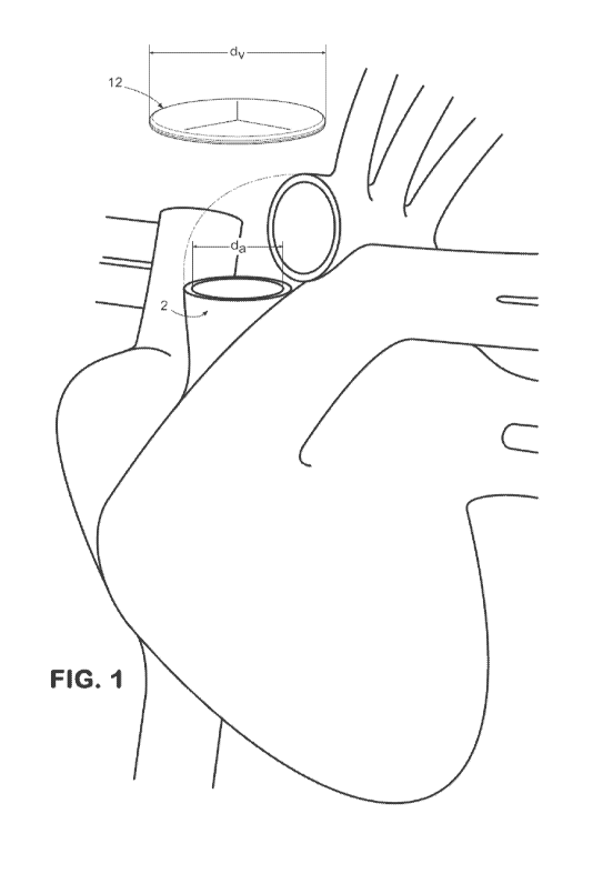
[0022] Figure 1 depicts a perspective view of a valve as described herein
positioned
relative to an annulus of the heart.
[0023] Figure 2A depicts a top view of an exemplary planar valve with
substantially
triangular leaflets in an unstressed position before implantation in an
annulus in a non-planar
configuration, as described herein. Figure 2B depicts a top view of an
exemplary planar
valve with substantially triangular leaflets and a sewing ring in an
unstressed position before
implantation in an annulus in a non-planar configuration, as described herein.
[0024] Figure 3A depicts a perspective view of the valve of Figure 2B
positioned
relative to an annulus of the heart. Figure 3B depicts a perspective view of
the valve of
Figure 2B in a biased, non-planar position following implantation in the
annulus of the heart,
as described herein. Figure 3C depicts a top view of the valve of Figure 3B.
[0025] Figure 4A depicts a top view of an exemplary planar valve with
substantially
rounded leaflets in an unstressed position before implantation in an annulus
in a non-planar
configuration, as described herein. Figure 4B depicts a top view of an
exemplary planar
valve with substantially rounded leaflets and a sewing ring in an unstressed
position before
implantation in an annulus in a non-planar configuration, as described herein.
[0026] Figure 5A depicts a perspective view of the valve of Figure 4B
positioned
relative to an annulus of the heart. Figure 5B depicts a perspective view of
the valve of
Figure 4B in a biased, non-planar position following implantation in the
annulus of the heart,
as described herein. Figure 5C depicts a top view of the valve of Figure 5B.
[0027] Figure 6 depicts a top view of an exemplary planar valve with
substantially
triangular leaflets prior to folding of the outer edge portion of the valve,
as described herein.
[0028] Figure 7 depicts a top view of an exemplary planar valve with
substantially
rounded leaflets prior to folding of the outer edge portion of the valve, as
described herein.
[0029] Figure 8A depicts a cross-sectional view of an exemplary sewing ring
rolled
from a piece of extracellular material, as described herein. Figure 8B depicts
a cross-
sectional view of an exemplary sewing ring formed into a tear drop shape, as
described
6
WO 2012/030996 CA 02810008 2013-02-28 PCT/US2011/050019
herein. Figure 8C depicts a cross-sectional view of an exemplary sewing ring
having a
plurality of laminated sheets of extracellular matrix material.
DETAILED DESCRIPTION OF THE INVENTION
[0030] The present invention may be understood more readily by reference to
the
following detailed description, examples, drawings, and claims, and their
previous and
following description. However, before the present devices, systems, and/or
methods are
disclosed and described, it is to be understood that this invention is not
limited to the specific
devices, systems, and/or methods disclosed unless otherwise specified, as such
can, of course,
vary. It is also to be understood that the terminology used herein is for the
purpose of
describing particular aspects only and is not intended to be limiting.
[0031] As used in the specification and the appended claims, the singular
forms "a,"
"an" and "the" include plural referents unless the context clearly dictates
otherwise. Thus,
for example, reference to a "leaflet" can include two or more such leaflets
unless the context
indicates otherwise.
[0032] Ranges may be expressed herein as from "about" one particular value,
and/or
to "about" another particular value. When such a range is expressed, another
aspect includes
from the one particular value and/or to the other particular value. Similarly,
when values are
expressed as approximations, by use of the antecedent "about," it will be
understood that the
particular value forms another aspect. It will be further understood that the
endpoints of each
of the ranges are significant both in relation to the other endpoint, and
independently of the
other endpoint.
[0033] As used herein, the terms "optional" or "optionally" mean that the
subsequently described event or circumstance may or may not occur, and that
the description
includes instances where said event or circumstance occurs and instances where
it does not.
[0034] Without the use of such exclusive terminology, the term "comprising"
in the
claims shall allow for the inclusion of any additional element--irrespective
of whether a given
number of elements are enumerated in the claim, or the addition of a feature
could be
regarded as transforming the nature of an element set forth in the claims.
Except as
specifically defined herein, all technical and scientific terms used herein
are to be given as
broad a commonly understood meaning as possible while maintaining claim
validity.
[0035] Described herein are valves and replacement leaflets for controlling
fluid flow
in a lumen having an annulus. In one aspect, the valve is suitable for
replacing an aortic,
7
WO 2012/030996 CA 02810008 2013-02-28 PCT/US2011/050019
pulmonary, mitral, or tri-cuspid valve in the heart of a subject. In another
aspect, the valve
can comprise at least one leaflet configured to selectively prevent undesired
regurgitation of
blood flow therethrough the valve. For example, the valve can comprise a
single leaflet that
is sized to prevent blood flow therethrough the valve when the leaflet is
selectively positioned
in a blocking position. Alternatively, the valve can comprise a plurality of
leaflets.
Optionally, the at least one leaflet can be attached to a sewing ring. In a
further aspect, a
single leaflet as described herein can be used as a replacement leaflet for
controlling fluid
flow through an annulus. In a further aspect, the valve can have a
circumference and a
diameter that are larger than the circumference and diameter of the annulus.
[0036] In one aspect, as shown in Figures 6 and 7, it is contemplated that
the leaflets
of the valve 12 can be created from a substantially planar piece of material,
such as, for
example and without limitation, a substantially planar piece of extracellular
matrix material
as defined herein. In this aspect, the leaflets can be defined by cutting or
stamping out
selected portions of the planar piece of material using conventional
techniques. For example,
as depicted in Figures 6, the leaflets of the valve 12 can be cut from the
substantially planar
piece of material in substantially triangular shapes. Alternatively, as
depicted in Figures 7,
the leaflets of the valve 12 can have substantially rounded shapes.
[0037] In another aspect, and with reference to Figures 6 and 7, prior to
preparation of
the valve 12 for implantation within the annulus 2, a circumference and, thus,
an outer edge
portion 15 of the valve can be defined. In this aspect, the outer edge portion
15 of the valve
12 can have a width E that ranges from about 3 mm to about 6 mm, and more
preferably is
about 5 mm. It is contemplated that the outer edge portion 15 of the valve can
be rolled to
create an attachment surface. In one aspect, the attachment surface can be
configured for
direct attachment thereto the annulus 2. Alternatively, the attachment surface
can be
configured for attachment thereto a sewing ring.
[0038] Optionally, in one exemplary aspect, as depicted in Figures 6 and 7,
during the
process of defining the leaflets and outer edge portion, an uncut portion 17
along the
operative circumference of the valve 12 can also be defined. In this aspect,
the uncut portion
17 can have a substantially consistent width U along the operative
circumference of the valve
12. Where an uncut portion 17 is defined in the valve 12, it is contemplated
that the width U
of the uncut portion can range from about 1 mm to about 6 mm, and more
preferably from
about 4 mm to about 5 mm.
8
WO 2012/030996 CA 02810008 2013-02-28PCT/US2011/050019
[0039] Figures 2A and 4A each depict an exemplary valve 12 as it appears
after it has
been prepared for implantation (after the outer edge of the valve has been
rolled up) but
before attachment to the annulus 2. More particularly, Figure 2A depicts an
exemplary valve
12 having substantially triangular leaflets, while Figure 4A depicts an
exemplary valve
having substantially rounded leaflets. It is contemplated that the
circumference of the valve
12 following the rolling of the outer edge portion 15 of the valve can
correspond to an
operative circumference of the valve. Similarly, the diameter of the valve 12
following the
rolling of the outer edge portion 15 of the valve can correspond to an
operative diameter (dv)
of the valve. As used herein, the operative diameter (dv) of the valve 12
corresponds to the
portion of the valve that is configured to span across the annulus 2 after
attachment of the
valve thereto the annulus. Thus, as used herein, the operative diameter (dv)
does not factor in
outer edge portion 15, which is rolled up prior to attachment of the valve 12
thereto the
annulus 2.
[0040] In another aspect, the valve 12 can comprise at least one leaflet. In
this aspect,
the at least one leaflet can comprise a plurality of leaflets. In an
additional aspect, leaflets 28,
30, and 32 can have distal end portions that extend inwardly relative to the
circumference of
the valve generally toward a radial center 20 of the valve.
[0041] Optionally, the valve 12 can comprise a sewing ring 40. In one aspect,
the
sewing ring 40 can be attached to the rolled up outer edge portion 15 of the
valve 12. In
another aspect, before attachment to the annulus, the sewing ring 40 can be
substantially
semi-lunar or circular with an inner portion and an outer portion. In this
aspect, the inner
portion of the sewing ring can be attached to the valve, while the outer
portion of the sewing
ring 40 can define an operative circumference of the sewing ring and, thus,
the operative
circumference of the valve 12. Similarly, the outer diameter of the sewing
ring 40 can define
the operative diameter of the sewing ring and, thus, the operative diameter
(dv) of the valve
12.
[0042] Figures 2A and 4A depict valve 12 and sewing ring 40 as they are
before
attachment to the annulus. More particularly, Figure 2A depicts an exemplary
valve 12
having substantially triangular leaflets and sewing ring 40, while Figure 4A
depicts an
exemplary valve having substantially rounded leaflets and sewing ring 40. In
one aspect, the
valve 12 can comprise at least one leaflet. In this aspect, the at least one
leaflet can comprise
a plurality of leaflets. In one aspect, leaflets 28, 30, and 32 can have
distal end portions that
9
WO 2012/030996 CA 02810008 2013-02-28PCT/US2011/050019
extend inwardly relative to the inner portion of the sewing ring 40 generally
toward a radial
center 20 of the valve.
[0043] In one aspect, the operative circumference of the valve 12 can be
larger than
the circumference of the annulus. In this aspect, when the annulus is located
in a heart valve,
including, for example and without limitation, an aortic valve, a pulmonary
valve, a tricuspid,
or a bicuspid (mitral) valve, the ratio of the operative circumference of the
valve to the
circumference of the annulus can range from about 1.01:1 to about 3.00:1, more
preferably
from about 1.40:1 to about 2.40:1, and most preferably from about 1.70:1 to
about 2.10:1. In
addition to the ratios serving as the endpoints of the ranges set forth above,
the disclosed
ranges also include all ratios falling between the endpoint ratios. It is
contemplated that,
because the operative circumference of the valve 12 is greater than the
circumference of the
annulus 2, the valve can form a substantially sinusoidal or wave-like pattern
upon attachment
to the annulus in the biased position. In another aspect, the operative
circumference of the
valve can range from about 60 mm to about 220 mm, more preferably from about
80 mm to
about 190 mm, and most preferably from about 100 mm to about 140 mm.
Optionally, it is
contemplated that the valves and sewing rings described herein can be provided
in a series
of different circumferences, thereby permitting a surgeon to select an
appropriately sized
valve or sewing ring depending on the dimensions of the annulus, which can be
determined
during a surgical procedure.
[0044] Similarly, in another aspect, and as shown in Figures 3A and 5A, the
operative
diameter (dv) of the valve 12 can be greater than the diameter (da) of the
annulus 2. In this
aspect, when the annulus is located in a heart valve, including, for example
and without
limitation, an aortic valve, a pulmonary valve, a tricuspid, or a bicuspid
(mitral) valve, the
ratio of the operative diameter (dv) of the valve to the diameter (da) of the
annulus 2 can range
from about 1.01:1 to about 3.00:1, more preferably from about 1.40:1 to about
2.40:1, and
most preferably from about 1.70:1 to about 2.10:1. In addition to the ratios
serving as the
endpoints of the ranges set forth above, the disclosed ranges also include all
ratios falling
between the endpoint ratios. In another aspect, the operative diameter (dv) of
the valve can
range from about 20 mm to about 70 mm, more preferably from about 25 mm to
about 60
mm, and most preferably from about 35 mm to about 45 mm. Optionally, it is
contemplated
that the valves and sewing rings described herein can be provided in a series
of different
diameters, thereby permitting a surgeon to select an appropriately sized valve
or sewing ring
10
WO 2012/030996 CA 02810008 2013-02-28PCT/US2011/050019
depending on the dimensions of the annulus, which can be determined during a
surgical
procedure.
[0045] As shown in Figures 6-7, in one aspect, each leaflet can have an edge
length L
corresponding to the total length of an inner edge of each leaflet that
extends inwardly
relative to the operative circumference of the valve 12 generally toward a
radial center 20 of
the valve. In an additional aspect, when the leaflets are substantially
triangular as shown in
Figure 6, the edge length L of each leaflet can range from about 10 mm to
about 70 mm,
more preferably from about 15 mm to about 60 mm, and most preferably from
about 25 mm
to about 45 mm. In this aspect, it is contemplated that the ratio between the
edge length L of
each leaflet to the diameter (da) of the annulus 2 can range from about 0.5:1
to about 3:1, and
more preferably from about 1:1 to about 2:1. In addition to the ratios serving
as the
endpoints of the ranges set forth above, the disclosed ranges also include all
ratios falling
between the endpoint ratios. In another aspect, when the leaflets are
substantially rounded as
shown in Figure 7, the edge length L of each leaflet can range from about 15
mm to about 60
mm, more preferably from about 20 mm to about 50 mm, and most preferably from
about 25
mm to about 35 mm. In this aspect, it is contemplated that the ratio between
the edge length
L of each leaflet to the diameter (da) of the annulus 2 can range from about
1:1 to about 2:1,
and more preferably from about 1.20:1 to about 1.40:1. In addition to the
ratios serving as
the endpoints of the ranges set forth above, the disclosed ranges also include
all ratios falling
between the endpoint ratios.
[0046] In an additional aspect, and as shown in Figures 6-7, each leaflet can
have a
height H. In this aspect, it is contemplated that each leaflet can have an
apex corresponding
to the point along edge length L of each leaflet that is farthest from the
operative
circumference of the valve 12, and the height H of each leaflet can correspond
to the distance
between the apex of each leaflet and the operative circumference of the valve.
In one aspect,
when the leaflets are substantially triangular as shown in Figure 6, the
height H of each
leaflet can range from about 10 mm to about 35 mm, more preferably from about
12 mm to
about 30 mm, and most preferably from about 17 mm to about 23 mm. In this
aspect, it is
contemplated that the ratio between the height H of each leaflet to the
diameter (da) of the
annulus 2 can range from about 0.3:1 to about 2:1, more preferably from about
0.5:1 to about
1.5:1, and most preferably from about 0.7:1 to about 1.1:1. In addition to the
ratios serving as
the endpoints of the ranges set forth above, the disclosed ranges also include
all ratios falling
between the endpoint ratios. Optionally, in this aspect, it is contemplated
that the ratio
11
WO 2012/030996 CA 02810008 2013-02-28PCT/US2011/050019
between the height H of each leaflet and the width U of the uncut portion 17
can range from
about 2:1 to about 7:1, and more preferably from about 4:1 to about 5:1. In
addition to the
ratios serving as the endpoints of the ranges set forth above, the disclosed
ranges also include
all ratios falling between the endpoint ratios. In another aspect, when the
leaflets are
substantially rounded as shown in Figure 7, the height H of each leaflet 28,
30, 32 can range
from about 5 mm to about 30 mm, more preferably from about 10 mm to about 25
mm, and
most preferably from about 12 mm to about 18 mm. In this aspect, it is
contemplated that the
ratio between the height H of each leaflet to the diameter (da) of the annulus
2 can range from
about 0.3:1 to about 1:1, more preferably from about 0.4:1 to about 0.9:1, and
most
preferably from about 0.5:1 to about 0.8:1. In addition to the ratios serving
as the endpoints
of the ranges set forth above, the disclosed ranges also include all ratios
falling between the
endpoint ratios. Optionally, in this aspect, it is contemplated that the ratio
between the height
H of each leaflet and the width U of the uncut portion 17 can range from about
1:1 to about
5:1, and more preferably from about 3:1 to about 4:1. In addition to the
ratios serving as the
endpoints of the ranges set forth above, the disclosed ranges also include all
ratios falling
between the endpoint ratios.
[0047] In a further aspect, attachment of the valve 12 can occur at a
plurality of
attachment points on the operative circumference of the valve, such as points
22, 24, and 26,
as depicted in Figures 2A, 2B, and 3C for a valve having substantially
triangular leaflets as
described herein, and in Figures 4A, 4B, and 5C for a valve having
substantially curved
leaflets as described herein. Points 22, 24, and 26 can be radially aligned
with points where
adjacent leaflets 28, 30, and 32 contacted one another prior to attachment of
the valve 12
thereto the interior surface of the annulus 2. In this aspect, for a valve 12
having a sewing
ring 40, attachment of the valve can occur at a plurality of attachment points
on the outer
portion of the sewing ring. As depicted in Figures 3B and 5B, the outer edge
portion of the
valve 12 can be attached to the interior wall of the valve annulus 2 in a
substantially
sinusoidal or wave-like pattern. It is contemplated that the substantially
sinusoidal pattern
formed by the valve 12 can promote substantially unidirectional blood flow
therethrough the
valve. It is further contemplated that blood flow can occur through the
annulus 2 in an axial
direction from points 14, 16, and 18 to points 22, 24, and 26.
[0048] In one aspect, it is contemplated that the plurality of attachment
points can be
substantially equally spaced along the circumference of the valve. In this
aspect, for a valve
12 having a sewing ring 40, the plurality of attachment points can be
substantially equally
12
WO 2012/030996 CA 02810008 2013-02-28PCT/US2011/050019
spaced along the outer portion of the sewing ring. In another aspect, the
plurality of
attachment points can comprise at least three attachment points. In a further
aspect, the
plurality of attachment points can comprise six attachment points
corresponding to points 22,
24, 26 and also points 14, 16 and 18. It is contemplated that more points in
between these
equally spaced points can also be used for attachment consistent with the wave-
like pattern
formed by the sewing ring when the valve is attached in the biased position.
In another
aspect, the spacing between the attachment points of the plurality of
attachment points can be
minimized such that the attachment points are placed substantially
contiguously along the
outer portion of the sewing ring. Attachment can be, without limitation, by
suture using
absorbable or permanent sutures. The exact knot tying technique can be
selected at the
preference of the operating physician.
[0049] As shown in Figures 3B and 5B, it is contemplated in one aspect, that
the valve
12, in the biased position, will be attached to the interior surface of the
annulus such that the
first portions 60 of the outer edge portion of the valve that are adjacent to
the base juncture of
the respective adjoining leaflets of the valve are positioned substantially co-
planar relative to
each other or are generally the most upstream portion of the outer edge
portion of the valve.
In this aspect, the medial portions 62 of the outer edge portion of the valve
12 (medial
between the respective adjoining first portions) would extend downward and be
coupled to
the interior surface of the annulus 2 at a position downstream of the first
portions of the outer
edge portion of the valve. In one aspect, the medial portions of the outer
edge portion of the
valve can be substantially co-planar to each other downstream of the first
portions of the
outer edge portion of the valve.
[0050] In a further aspect, and with reference to Figures 3B-3C and 5B-5C,
upon
attachment of the valve thereto the annulus in the biased position, at least a
portion of leaflets
28, 30, and 32 can be superposed relative to at least a portion of adjacent
leaflets. In this
aspect, it is contemplated that, in the biased position, at least a portion of
leaflets 28, 30, and
32 can be superposed relative to at least a portion of the other leaflets of
the at least one
leaflet, including non-adjacent leaflets. It is further contemplated that, in
the biased position,
at least a portion of leaflets 28, 30, and 32 can underlie at least a portion
of the adjacent
leaflets of the at least one leaflet. It is still further contemplated that,
in the biased position, at
least a portion of leaflets 28, 30, and 32 can overlie at least a portion of
the adjacent leaflets
of the at least one leaflet. In another aspect, it is contemplated that the
leaflets are configured
such that, upon attachment of the valve thereto the annulus in the biased
position, the leaflets
13
WO 2012/030996 CA 02810008 2013-02-28PCT/US2011/050019
can selectively move to an overlapping or otherwise blocking position that is
sufficient to
selectively prevent undesired regurgitation blood flow therethough the valve.
In a further
aspect, it is contemplated that the sinusoidal method of attaching the valve
in the
biased position can produce a tight and conforming fit between the valve and
the
annulus such that the likelihood of perivalvular leakage is reduced.
[0051] With reference to Figures 2A-3A and 4A-5A, it is contemplated that, in
one
aspect, the valve 12 can be substantially planar in an unstressed or pre-
insertion position
before attachment to an interior surface of an annulus 2 of a valvular lumen.
As illustrated in
Figures 3B-3C and 5B-5C, it is contemplated that the valve can be
substantially non-planar
upon attachment in a biased position at the annulus. In another aspect, the
distal end portions
of the respective leaflets can be configured to ensure adequate operational
overlay with the
other leaflets to prevent undesired directional passage of blood therethough
the valve when
the valve is attached to the annulus in the biased position. It is also
contemplated that
portions of the distal edges of the respective leaflets can partially overlap
other respective
leaflets or can otherwise be in contact with each other to effect the desired
directional passage
of blood therethough the valve. Although not specifically indicated in Figures
3C and 5C, it
is contemplated that the portion of each leaflet that overlies or underlies
adjacent leaflets can
be curved in a manner consistent with the curvature of the remainder of the
leaflet.
[0052] In one aspect, the valve 12, including the sewing ring 40 and the
leaflets 28, 30,
and 32, can comprise a biointegrating material. In another aspect, the
biointegrating material
can comprise an extracellular matrix material. In a further aspect, the
extracellular matrix
material can comprise mammalian extracellular matrix material that is obtained
from
mammalian tissue sources. In one exemplary embodiment, the sewing ring and the
leaflets comprise mammalian extracellular matrix material.
[0053] Mammalian tissue sources are in general any tissue having an
extracellular
matrix that can be isolated from the mammal and decellularized. Thus, for
example,
mammalian organs are tissue sources. For example and without limitation, the
tissue sources
can be any mammalian tissue, for example and without limitation, the small
intestine, large
intestine, stomach, lung, liver, kidney, pancreas, placenta, heart, bladder,
prostate, tissue
surrounding growing enamel, tissue surrounding growing bone, fetal tissue from
any
mammalian organs, and the like.
14
WO 2012/030996 CA 02810008 2013-02-28PCT/US2011/050019
[0054] The forms of the extracellular matrices that make up the extracellular
matrix
material are, without limitation, generally particulates, liquids, gels,
pastes, emulsions, or
suspensions. Liquid extracellular matrices are generally thin emulsions or
suspensions that
are injectable and fluid. Suspension, emulsion or gel extracellular matrices
can be
substantially thicker and have more body and substance than liquids, but
suspensions,
emulsions or gels can also be injected if they are not too thick.
Extracellular matrices in the
form of pastes or near-solid gels or plugs are more concentrated than liquids
or injectable
emulsions. Particulate extracellular matrices are powders that are formed from
a lyophilized
sheet of extracellular matrix material that is broken up into fine powder or
particulate.
Particulates can be used dry as a powder. Particulate extracellular matrices
can also be
reconstituted in a suitable buffer such as saline to transition into a liquid
or semi-solid form.
[0055] Extracellular matrix material can be obtained from the tissues of
mammals by
processes such as described in U.S. Patent No. 5,554,389, U.S. Patent No.
4,902,508, and
U.S. Patent No. 5,281,422, which are specifically incorporated by reference in
their entirety.
Enamel matrices are described in U.S. Patent No. 7,033,611 and U.S. Patent
Publication No.
2005/0043216, which are specifically incorporated by reference in their
entirety. For
example, the urinary bladder submucosa (UBS) is an extracellular matrix that
has the tunica
mucosa (which includes the transitional epithelial layer and the tunica
propria), a submucosal
layer, three layers of muscularis, and the adventitia (a loose connective
tissue layer). This
general configuration is true also for small intestine submucosa (SIS) and
stomach
submucosa (SS). However, it is contemplated that any configuration of
extracellular matrix
tissue layers, including, for example and without limitation, epithelial
basement membrane,
tunica propria, stratum compactum, lamina muscularis mucosa, tunica submucosa,
tunica
muscularis, and tunica serosa, can be used to produce the extracellular matrix
material.
[0056] Other sources of extracellular matrix material include tissues such as
the liver
and pancreas, which have an additional tissue layer called a basement
membrane. For
example, the extracellular matrix material can comprise the liver basement
membrane (LBM)
of mammals prepared by the process described in U.S. Patent No. 6,379,710,
which is
specifically incorporated by reference in its entirety. Basement membranes
generally do not
demonstrate the kind of tensile strength found in submucosa. However, other
useful
properties may be opportunistically employed from the extracellular matrices
of such tissues
as the liver, pancreas, placenta and lung tissues, all of which have either
basement membrane
for extracellular matrix or interstitial membrane (as with the lung). For
example, pancreatic
15
WO 2012/030996 CA 02810008 2013-02-28PCT/US2011/050019
extracellular membranes support beta islet cells which are critical to
pancreatic function.
Also, for example, the liver is one tissue known to be able to regenerate
itself and, therefore,
special qualities may be present in the LBM that help facilitate that process.
The
extracellular matrices surrounding developing tooth enamel and developing bone
also have
particular advantages over other matrices in that they support the growth and
differentiation
of the hard tissues of bone and enamel.
[0057] In some aspects, the extracellular matrix material can be from dermis.
For
example, AlloDerm0, produced by LifeCell Corporation, is an acellular tissue
matrix which
is produced from normal human skin using processing techniques established to
remove the
epidermis and cells within the dermis without significantly altering the
normal biochemistry
and molecular architecture of the connective tissue matrix. The resulting
product is in a
freeze-dried form allowing extended shelf life and ease of shipping without
degradation or
loss of the normal tissue matrix components. AlloDerm0 can retain decorin,
hyaluronic acid,
chondroitin sulfates, nidogen, growth factors and other biochemical proteins
present in
normal soft tissues. Additionally, AlloDerm0 can contain the basement
membranes of
vascular channels and the orientation of elastin and collagen fibers of the
starting dermal
tissue.
[0058] In some aspects, the extracellular matrix material can be obtained
from fascia.
In some aspects, the extracellular matrix material can be from parenchymal
tissue. In other
aspects, the extracellular matrix material can be from pericardium. In still
other aspects, the
extracellular matrix material can be myocardial extracellular matrix. In
additional aspects,
the extracellular matrix material can be from decellularized heart tissue,
produced, for
example, by coronary artery perfusion with detergents (Ott, HC, et al. Nat
Med. 2008
Feb;14(2):213-21).
[0059] In some aspects, the extracellular matrix material can comprise a
collagen
scaffold derived from a mammalian tissue or organ source. The collagen
scaffold can in some
aspects comprise the basement membrane of the mammalian tissue source.
[0060] In some aspects, the extracellular matrix material can be produced in
vitro. For
example, the extracellular matrix material can be produced from a culture of
mammalian
cells. The extracellular matrix material can be produced from proteins
extracted from
mammalian tissue/organs. For example, in some aspects, the extracellular
matrix material
comprises an artificial collagen scaffold synthesized from collagen extracted
from a
16
WO 2012/030996 CA 02810008 2013-02-28PCT/US2011/050019
mammalian tissue or organ source. Collagen from mammalian sources can be
retrieved from
matrix-containing tissues and used to form a matrix composition. Extracellular
matrices can
be synthesized from cell cultures as in the product manufactured by
MatrigelTM. In addition,
dermal extracellular matrix material, subcutaneous extracellular matrix
material, large
intestine extracellular matrix material, placental extracellular matrix
material, omentum
extracellular matrix material, heart extracellular matrix material, and lung
extracellular matrix
material, can be used, derived and preserved similarly as described herein for
the SIS, SS,
LBM, and UBS materials. Other organ tissue sources of basement membrane for
use in
producing the extracellular matrix material include the spleen, lymph nodes,
salivary glands,
prostate, pancreas and other secreting glands. In general, any tissue of a
mammal that has an
extracellular matrix can be used for developing the extracellular matrix
material.
[0061] Collagenous matrix can be selected from a variety of commercially
available
collagen matrices or can be prepared from a wide variety of natural sources of
collagen.
Collagenous matrix for use in accordance with the disclosed compositions and
methods can
comprise highly conserved collagens, glycoproteins, proteoglycans, and
glycosaminoglycans
in their natural configuration and natural concentration. Collagens can be
from animal
sources, from plant sources, or from synthetic sources, all of which are
available and standard
in the art.
[0062] The extracellular matrix material can be made from a plurality of
mammalian
tissue sources. Specifically, the extracellular matrix material can be made
from two
mammalian tissue sources, three mammalian tissue sources, four mammalian
tissue sources,
five mammalian tissue sources, six mammalian tissue sources, and conceivably
up to ten or
more tissue sources. These tissue sources can be from the same mammal (for
example the
same cow, the same pig, the same rodent, the same human, etc.), different
mammalian
animals of the same species, (e.g. cow 1 and cow 2, pig 1 and pig 2, rodent 1
and rodent 2,
human 1 and human 2, etc.), or different species of mammals (for example LBM
from a pig,
SIS from a cow, and UBS from a dog), all mixed together to form the
extracellular matrix
material).
[0063] The extracellular matrix material can also be a gel matrix combined
with a
particulate matrix, where the gel is applied to a space or cavity and dusted
with powder-like
particulates to increase the concentration of matrix at the surface of the
cavity. The
extracellular matrix material can be two or more liquid matrices (from
different tissue
sources) combined together. The extracellular matrix material can be two or
more suspension
17
WO 2012/030996 CA 02810008 2013-02-28PCT/US2011/050019
matrices (from different tissue sources) combined together. The extracellular
matrix material
can be two or more particulate matrices (from different tissue sources)
combined together.
The particulate matrices combined together can be applied to an annulus as a
particulate or as
a rehydrated suspension, where saline or other suitable buffer is applied to
the particulate
mixture and that hydrated composition is applied to the annulus in the
individual being
treated. The particulate can also be dusted onto a sheet of matrix before or
after placement at
the annulus. The extracellular matrix material can be a liquid mixture of two
or more
extracellular matrices. With this dusting embodiment, the liquid, gel,
suspension or emulsion
can be from a single mammalian tissue source, and dusted with a particulate
extracellular
matrix from either the same or a different mammalian tissue source.
Accordingly, the
suspension, emulsion, gel or liquid can be SIS, and the particulate can be
SIS, or the
suspension, emulsion, gel or liquid can be SIS and the particulate can be SS,
or LBM, or
UBS. The suspension, emulsion, gel or liquid can be a mixture of SIS and LBM
and the
particulate for dusting can be from SS. These examples are not meant to be
exhaustive of the
possible combinations of elements in the extracellular matrix material.
[0064] The extracellular matrix material can further include one or more
additional
components to aid in some aspect of the tissue regenerative process or the
generation of new
tissue, however the biological activity is characterized. The additional
component can be any
component that somehow serves the extracellular matrix material and its
purpose in the
mammalian body. Thus, the additional component can help to regenerate tissue,
heal a
wound, better recruit stem cells, manipulate the immune environment in a
beneficial way,
therapeutically treat the local environment, or otherwise contribute to some
aspect of the
process for which the extracellular matrix material is being used.
[0065] In one aspect, the additional component can be one or more cells. In
some
aspects, the additional component can be non-native cells, i.e., cells that
are heterologous to
the mammalian ECM. In some aspects, the additional component can be stem
cells. A non-
exhaustive list of stem cells include a human embryonic stem cell, a fetal
cardiomyocyte, a
myofibroblast, a mesenchymal stem cell, an autotransplanted expanded
cardiomyocyte, an
adipocyte, a totipotent cell, a pluripotent cell, a blood stem cell, a
myoblast, an adult stem
cell, a bone marrow cell, a mesenchymal cell, an embryonic stem cell, a
parenchymal cell, an
epithelial cell, an endothelial cell, a mesothelial cell, a fibroblast, an
osteoblast, a
chondrocyte, an exogenous cell, an endogenous cell, a stem cell, a
hematopoietic stem cell, a
pluripotent stem cell, a bone marrow-derived progenitor cell, a progenitor
cell, a myocardial
18
WO 2012/030996 CA 02810008 2013-02-28PCT/US2011/050019
cell, a skeletal cell, a fetal cell, an embryonic cell, an undifferentiated
cell, a multi-potent
progenitor cell, a unipotent progenitor cell, a monocyte, a cardiomyocyte, a
cardiac myoblast,
a skeletal myoblast, a macrophage, a capillary endothelial cell, a xenogenic
cell, an allogenic
cell, an adult stem cell, and a post-natal stem cell. In some aspects, the
stem cells have the
potential to differentiate into cardiac tissue cells. Thus, in some aspects,
the stem cells can be
pluripotent. In other aspects, the stem cells can be angioblasts or
hemangioblasts. In
additional aspects, the stem cells can be myoblasts. The stem cells described
herein can be
derived and maintained using standard methods for stem cell culture.
[0066] In another aspect, the additional component can be a drug, including
any
known or newly discovered substance that can be administered to the heart of a
subject. For
example, the additional component can be an antithrombotic agent, including,
for example,
and without limitation, antiplatelet drugs, anticoagulants, and thrombolytic
drugs. Exemplary
antiplatelet drugs include, for example and without limitation, Aspirin,
Clopidogrel,
Prasugrel, Ticlopidine, Cilostazol, Abciximab, Eptifibatide, Tirofiban, and
Dipyridamole.
Exemplary anticoagulants include, for example and without limitation,
Coumadins,
Acenocoumarol, Phenprocoumon, Phenindione, Heparin, Low Molecular Weight
Heparin,
Fondaparinux, Idraparinux, Agratroban, Lepirudin, Bivalirudin, and Dabigatran.
Exemplary
thrombolytic drugs include, for example and without limitation, Alteplase,
Reteplase,
Tenecteplase, Anistreplase, Streptokinase, and Urokinase.
[0067] In a further aspect, the additional component can be a protein. In
this aspect,
the additional component can be an exogenous protein, such as those normally
found in
mammalian ECM. Thus, it is contemplated that the additional component can be,
for
example and without limitation, a collagen, a proteoglycan, a
glycosaminoglycan (GAG)
chain, a glycoprotein, a growth factor, a cytokine, a cell-surface associated
protein, a cell
adhesion molecule (CAM), an angiogenic growth factor, an endothelial ligand, a
matrikine, a
matrix metalloprotease, a cadherin, an immunoglobulin, a fibril collagen, a
non-fibrillar
collagen, a basement membrane collagen, a multiplexin, a small-leucine rich
proteoglycan,
decorin, biglycan, a fibromodulin, keratocan, lumican, epiphycan, a heparan
sulfate
proteoglycan, perlecan, agrin, testican, syndecan, glypican, serglycin,
selectin, a lectican,
aggrecan, versican, nuerocan, brevican, cytoplasmic domain-44 (CD-44),
macrophage
stimulating factor, amyloid precursor protein, heparin, chondroitin sulfate B
(dermatan
sulfate), chondroitin sulfate A, heparan sulfate, hyaluronic acid, fibronectin
(Fn), tenascin,
elastin, fibrillin, laminin, nidogen/entactin, fibulin I, fibulin II,
integrin, a transmembrane
19
WO 2012/030996 CA 02810008 2013-02-28PCT/US2011/050019
molecule, platelet derived growth factor (PDGF), epidermal growth factor
(EGF),
transforming growth factor alpha (TGF-alpha), transforming growth factor beta
(TGF-I3),
fibroblast growth factor-2 (FGF-2) (also called basic fibroblast growth factor
(bFGF)),
thrombospondin, osteopontin, angiotensin converting enzyme (ACE), or a
vascular epithelial
growth factor (VEGF). Thus, in addition to one or more extracellular matrix
tissues, the
disclosed extracellular matrix material can comprise collagen I and III,
elastin, laminin,
CD44, hyaluronan, syndecan, bFGF, HGF, PDGF, VEGF, Fn, tenascin, heparin,
heparan
sulfate, chondroitin sulfate B, integrins, decorin, TGF-I3, or a combination
thereof
[0068] It is contemplated that once the extracellular matrix material is in
the body of
the subject, at least a portion of the extracellular matrix material can
integrate into the host
tissue and develop substantially the same properties as proximate native
material.
Specifically, the extracellular matrix material can be in cellular
communication with the
blood supply of a subject. It is contemplated that at least 70% of the
extracellular matrix
material can fully integrate into the host tissue. More preferably, it is
contemplated that at
least 80% of the extracellular matrix material can fully integrate into the
host tissue. Most
preferably, it is contemplated that at least 90% of the extracellular matrix
material can fully
integrate into the host tissue.
[0069] It is contemplated that extracellular matrix material can be harvested
and
processed as described in U.S. Patent No. 5,554,389 (UBS), U.S. Patent No.
6,099,567 (SS),
and U.S. Patent No. 6,379,710 (LBM), as well as U.S. Patent No. 4,902,508,
U.S. Patent
No. 4,956,178, U.S. Patent No. 5,275,826, U.S. Patent No. 5,516,533, U.S.
Patent No.
5,573,784, U.S. Patent No. 5,711,969, U.S. Patent No. 5,755,791, U.S. Patent
No.
5,955,110, U.S. Patent No. 5,968,096, U.S. Patent No. 5,997,575, and U.S.
Patent No. 6,653,291
(SIS), which are specifically incorporated by reference in their entirety. In
one aspect, it is
contemplated that the valve 12 and sewing ring 40 described herein can be
stamped out of a
sheet of extracellular matrix material. For example, and without limitation,
it is contemplated
that the valve 12 as depicted in Figures lA and 1B, and the sewing ring as
depicted in Figure
3C, can be stamped out of a substantially planar sheet of extracellular matrix
material. In an
additional aspect, the valve 12 and the sewing ring 40 can be continuous and
can be formed
or stamped out of a plane of laminate sheets of matrix material. In another
aspect, the
extracellular matrix material can be single sheets, multi-laminate sheets, or
some other
configuration of extracellular matrix that lends itself to the formation of
sheet-like leaflets.
For example and without limitation, the valve 12 and the sewing ring 40 can be
stamped out
20
WO 2012/030996 CA 02810008 2013-02-28PCT/US2011/050019
of a larger laminate sheet of 2 ply, 3 ply, 4, ply, 5 ply, 6 ply, 7 ply, 8
ply, 9 ply, and 10 ply
extracellular matrix. It is further contemplated that the extracellular matrix
material can be
selectively formed at an appropriate width for the valve being replaced.
[0070] In a further aspect, the extracellular matrix material of the valve 12
and sewing
ring 40 can have a desired elastic modulus. For example, and without
limitation, the desired
elastic modulus of the extracellular matrix material can range from about 5 to
about 15, more
preferably from about 7 to about 13, and most preferably from about 8 to about
12. It is
contemplated that the desired elastic modulus can be selected to substantially
correspond to
the elastic modulus of native tissue surrounding the site of implantation of
the valve, thereby
improving integration of the valve into the host tissue. It is contemplated
that the source of
the extracellular matrix material, including, for example and without
limitation, urinary
bladder submucosa, small intestine submucosa, stomach submucosa, and liver
basement
membrane, can be selected depending on the desired elastic modulus.
[0071] In one aspect, Figure 8A depicts a sewing ring 40 constructed from a
rolled
piece of extracellular matrix material as described herein. In this aspect,
the rolled piece of
extracellular matrix material can define a cross-sectional core 42. The sewing
ring 40 can
have a point of attachment 44 where two ends of the sewing ring are attached
to one another.
In another aspect, and referring to Figure 8B, the sewing ring 40 can be
defined by a tightly
configured roll of extracellular matrix material. In this aspect, the
extracellular matrix
material can be folded to itself and attached at point 46 with suture or glue
or other
attachment means. The sewing ring 40 can have a cross-sectional core 48 that
illustrates the
resulting tear-drop configuration of the sewing ring when it is attached to
itself In another
aspect, as shown in Figure 8C, the sewing ring 40 can be formed from a
plurality of
laminated sheets 52 of extracellular matrix material. It is contemplated that
the plurality of
sheets 52 of extracellular matrix material can comprise multiple types of
extracellular matrix
material, as described herein. It is further contemplated that the sheets 52
of extracellular
material can be laminated together using any conventional biocompatible means
for
lamination of two structures. For example, it is contemplated that the sheets
52 of
extracellular material can be laminated together using a biodegradable
material.
[0072] In exemplary aspects, it is contemplated that the sewing ring 40 can
be attached
to the attachment surface defined by the outer edge portion 15 of the valve 12
as described
herein. It is further contemplated that the outer edge portion 15 of the valve
12 can be
formed in the same manner as the sewing ring 40, as described herein, to
thereby define the
21
WO 2012/030996 CA 02810008 2013-02-28PCT/US2011/050019
attachment surface, which can be configured for attachment to a sewing ring or
for direct
attachment to the inner surface of the annulus 2.
[0073] In an additional aspect, it is contemplated that the sewing ring can
have a
minimal width compared to the area defined by the annulus. In this aspect, the
width of the
sewing ring can be less than about 5 mm, and more preferably less than about 1
mm. It is still
further contemplated that the tight fit between the sewing ring and the
annulus, coupled with
the minimal width of the sewing ring, can maximize the portion of the lumen
available for
accommodating blood flow following attachment of the valve in the biased
position.
[0074] It is contemplated that the extracellular matrix material of the
sewing ring can
be used with the leaflets of a trileaflet valve or with other valves such as
pulmonary, aortic,
mitral or tricuspid valves. The sewing ring can be used with mechanical or
tissue valves.
[0075] In addition to comprising extracellular matrix material, the sewing
ring 40 can
further comprise metal, or a mixture of conventional metals or alloys. In one
aspect, the
sewing ring can also comprise a shape memory activated (SMA) material such as,
for example
and without limitation, Nitinol or other conventional SMA materials. It is
contemplated that
the sewing ring can be a synthetic or polymeric material, such as, for example
and without
limitation, silicone, rubber, plastic, or the like. In one aspect, the sewing
ring can be
constructed like catheter tubing, with a woven support of metal wire embedded
within the
reinforced plastic of the tubing. In another exemplary aspect, the sewing ring
can comprise an
extracellular matrix material and a conventional polymeric material. In this
aspect, it is
contemplated that the extracellular matrix material can be subjected to a
conventional
electrospinning process and then applied to the polymeric material to produce
the sewing ring.
[0076] In one aspect, it is contemplated that the sewing ring can comprise a
biodegradable material. In this aspect, it is contemplated that the
biodegradable material can be
configured to degrade following significant integration of the extracellular
matrix material into
the host tissue of the subject. More generally, it is contemplated that the
sewing ring can be
made of any material suitable for the purpose identified in the definition of
a sewing ring. It is
further contemplated that the functionality of the sewing ring can be
maintained by ensuring
that the sewing ring possesses sufficient flexibility to permit the larger
circumference of the
sewing ring to be placed into the smaller circumference of the annulus in a
non-planar
attachment configuration.
22
WO 2012/030996 CA 02810008 2013-02-28PCT/US2011/050019
[0077] It is further contemplated that each configuration of the sewing ring
imparts
different advantages, and it is contemplated that different valves will be
more or less
appropriately suited for the different variations of sewing ring. For example,
the sewing ring
40 of rolled extracellular matrix has a point where the ring is attached to
itself. It is
contemplated that this point of attachment would be considered a weak point in
the sewing
ring, and the ring needs to be attached to itself and the annulus with
particular care and
reinforcement so that the ring does not yield or break free at the point of
attachment.
Accordingly, it is contemplated that because sewing ring 40, while unitary, is
non-tubular,
attachment of the ring to the annulus will require attendant care to that
aspect of its
configuration. In one aspect, it is specifically contemplated that running
sutures that
surround the ring 40 will securely attach the ring to the annulus. Optionally,
suturing through
the ring itself can be used. This securing methodology may be difficult due to
the dense and
strong nature of the extracellular matrix material. However, it is
contemplated that sutures
can be accomplished with conventional stitches or mattress stitches depending
on the
surgeon's assessment of the situation.
[0078] It is still further contemplated that attachment of the valve can be
accomplished
percutaneously without open heart surgery. In use, the valve can be guided to
the site of
replacement after the defective valve has been removed, and the sewing ring
can be
systematically sutured or otherwise attached to the annular region in a biased
position as
described herein, using a visualization technique enabling manipulations in
the body within
the view of a camera that shows the manipulations to the surgeon.
[0079] In further aspects, methods are provided for using the valves and
replacement
leaflets as described herein to control fluid flow in a lumen having an
annulus. In one aspect,
the methods comprise providing at least one replacement leaflet having the
characteristics of
the leaflets described herein. In another aspect, the methods comprise
securely attaching the
at least one replacement leaflet to the annulus in a desired position. It is
contemplated that
the at least one leaflet can comprise a single leaflet that is used to replace
a single defective
leaflet located therein an annulus in a heart of a subject with a blood
supply. It is further
contemplated that the at least one replacement leaflet can promote vascular
development
within the subject by permitting cellular communication between the at least
one leaflet and
the blood supply of the subject. Thus, it is further contemplated that the at
least one
replacement leaflet can effectively behave as a native leaflet after
attachment in the desired
position within the subject's heart.
23
WO 2012/030996 CA 02810008 2013-02-28PCT/US2011/050019
[0080] In another aspect, the method can comprise providing a valve as
described
herein. In this aspect, the method can comprise securely attaching the outer
portion of the
valve to the annulus in a biased position as described herein. Optionally, in
an additional
aspect, the valve can comprise a sewing ring as described herein. In this
aspect, the method
can comprise securely attaching the outer portion of the sewing ring to the
annulus such that
the valve is in a biased position as described herein.
[0081] In a further aspect, a kit having a valve as described herein can be
assembled.
Optionally, the kit can comprise a sewing ring as described herein.
Additionally, it is
contemplated that the sewing rings as described herein can be provided
separately for
attaching any number of valves.
Experimental Data in Support of Concept
[0082] In one long-term animal study, four clinically normal swine were used
to study
the effectiveness of porcine small intestine submucosa as cardiac pulmonary
valve leaflets.
Matheny, et al., Porcine Small Intestine Submucosa as a Pulmonary Valve
Leaflet Substitute,
The Journal of Heart Valve Disease 2000; 9:769-775. In this study, each swine
had one
pulmonary valve leaflet excised and replaced with a leaflet produced from a
layer of porcine
small intestine submucosa. The leaflets were secured within the annulus using
a suture line.
The swine were individually sacrificed at 56, 63, 88, and 111 days following
implantation of
the leaflets.
[0083] The leaflet removed 63 days after implantation was securely attached
to the
annulus along the entire suture line. Although one fenestration was present,
complete
organization of the leaflet was observed. The apical portion of the leaflet
consisted of mature
and moderately dense fibrous connective tissue, while the basal portion of the
leaflet had less
dense and mucinous tissue. Complete endothelialization of the leaflet was
observed.
[0084] The leaflet removed 88 days after implantation was also securely
attached to
the annulus along the entire suture line. No fenestrations were present, and
the basal portion
of the leaflet was cellular and mature connective tissue. The apical portion
of the leaflet was
notably larger in comparison to the leaflet removed 63 days after
implantation. The apical
portion formed a largely acellular nodule composed of serum, cellular debris,
and leukocytes
in a dense network of fibrin (an organized thrombus). A layer of residual and
acellular
matrix was observed at the center of the thrombus. Endothelial cell coverage
of the leaflet
was continuous.
24
WO 2012/030996 CA 02810008 2013-02-28PCT/US2011/050019
[0085] The leaflet removed 111 days after implantation was securely and
continuously
attached to the annulus along the entire suture line without evidence of
thrombus. The leaflet
was observed to possess gross characteristics similar to those of normal
leaflet tissue.
Specifically, the leaflet was observed to have histologically identifiable
features and was
composed of collagenous tissue containing indistinct layers of viable cells.
The histological
organization of the leaflet was comparable to the organization observed in the
adjacent native
leaflets. The surfaces of the leaflet were completely lined with endothelial
cells.
[0086] Although several embodiments of the invention have been disclosed in
the
foregoing specification, it is understood by those skilled in the art that
many modifications and
other embodiments of the invention will come to mind to which the invention
pertains, having
the benefit of the teaching presented in the foregoing description and
associated drawings.
It is therefore understood that the invention is not limited to the specific
embodiments disclosed
herein, and that many modifications and other embodiments of the invention are
intended
to be included within the scope of the invention. Moreover, although specific
terms are
employed herein, they are used only in a generic and descriptive sense, and
not for the
purposes of limiting the described invention.
[0087] Various publications are referenced in this document. These
publications in their
entireties are hereby incorporated by reference into this application in order
to more fully
describe the state of the art to which the disclosed system and method
pertains. The references
disclosed are also individually and specifically incorporated by reference
herein for the
material contained in them that is discussed in the sentence in which the
reference is relied
upon.
25