Note: Descriptions are shown in the official language in which they were submitted.
WO 2012/027804 CA 02810043 2013-03-01 PCT/BE2011/000055
APPARATUS FOR AUTOMATICALLY DIAGNOSING EMPHYSEMA
Background and Summary
BACKGROUND OF THE INVENTION
A. Field of the Invention
The present invention relates generally to diagnosing chronic obstructive
pulmonary disease
(COPD) and estimating of the severity thereof, in particularly the occurrence
of emphysema.
More particularly the present invention relates to a system and method for
computerized
quantification of airway collapse during forced expiration or total
exhalation. Such airway
collapse is correlated with the presence of emphysema that has been verified
on computer
tomography (CT) scan and that is due to the loss of alveolar attachments in
emphysema.
Moreover the present invention provides a computerized apparatus for detection
of emphysema,
a computerized system for detection of emphysema or a method of computer-aided
detection of
emphysema. This can also be used for quantifying the severity of emphysema in
patients with
COPD by an automated analysis of the amount (volume) and/or speed (flow) of a
total or forced
exhalation or the processing of the graphics of a pneumotachograph of a total
or forced
exhalation. Such measurements are obtainable by a spirometer.
The apparatus, system or method of present invention correlates airway
collapse during forced
expiration in COPD with presence and severity of emphysema. It was
demonstrated that airway
collapse during forced expiration in COPD correlates with presence and
severity of emphysema.
Several documents are cited throughout the text of this specification. Each of
the documents
herein (including any manufacturer's specifications, instructions etc.) are
herby incorporated by
reference; however, there is no admission that any document cited is indeed
prior art of the
present invention.
B. Description of the Related Art
Worldwide, COPD ranked as the sixth leading cause of death in 1990. It is
projected to be the
fourth leading cause of death worldwide by 2030 due to an increase in smoking
rates and
demographic changes in many countries. COPD is the 4th leading cause of death
in the U.S., and
1
CA 02810043 2013-03-01
WO 2012/027804 PCT/BE2011/000055
the economic burden of COPD in the U.S. in 2007 was $42.6 billion in health
care costs and lost
productivity. The diagnosis of COPD requires lung function tests such as
respirometry.
Emphysema is a long-term, progressive disease of the lung. It is included in a
group of diseases
called COPD. Emphysema is called an obstructive lung disease because the
destruction of lung
tissue around smaller airways, called alveoli, makes these airways unable to
hold their functional
shape upon exhalation.
Forced expiratory volumes and flows or the expiratory volumes and flows of
total exhalation are
used to diagnose COPD. However the current methods and systems in the art do
not accurately
distinguish airway obstruction from emphysema and are thus not suitable for
accurate
distinguishing emphysema from COPD and for estimating the severity of
emphysema.
Thus, there is a need in the art for more accurate diagnosis systems and
methods to detect
emphysema and assess the severity of emphysema in a group of diseases called
COPD.
Present invention provides such by computerized quantification of airway
collapse during forced
expiration and demonstrates correlations with the presence of emphysema on CT
scan.
[5
SUMMARY OF THE INVENTION
The present invention solves the problems of the related art of inaccurate
diagnosing of
emphysema by providing an apparatus for diagnosing emphysema , the apparatus
comprising a)
a device for receiving and sensing forced or total expiration of the
respiratory system of a patient
!O and further comprising b) a calculator or signal processor calculating the
flow volume curve or
relationship from said forced or total airway expiration, characterized in
that the calculator or
signal processor comprises 1) a first calculating means for calculating the
flow-volume curve or
the flow-volume relationship and 2) a second calculating means for
automatically calculating the
angle in the expiratory flow-volume curve between the two best fitting linear
regression curves
on flow-volume curve.
In accordance with the purpose of the invention, as embodied and broadly
described herein, the
invention is broadly drawn to an apparatus for diagnosing emphysema whereby
the apparatus
having a) a signal input to receive electrical signals of an electrical
signals produced by volume
sensor and flow sensors of a device for receiving and sensing forced or total
expiration of the
2
WO 2012/027804 CA 02810043 2013-03-01 PCT/BE2011/000055
respiratory system of a patient and b) a calculator or signal processor
calculating the flow
volume curve or relationship from said forced or total airway expiration,
characterized in that
the calculator or signal processor comprises 1) a first calculating means for
calculating the flow-
volume curve or the flow-volume relationship and 2) a second calculating means
for
automatically calculating the angle in the expiratory flow-volume curve
between the two best
fitting linear regression curves on the flow-volume curve corresponding to
entire exhalation
flow rate and exhalation volume of a patients respiratory system.
This apparatus of present invention or described above can further comprise
calculating means
for calculating the peak-to-surface area. Moreover in the apparatus of present
invention or as
0 described above the signal processor or calculator further can comprise a
mathematical model to
compare said angle with that of a plurality of chronic obstructive pulmonary
disease (COPD)
patients with no emphysema. Moreover the signal processor or calculator
further comprising a
mathematical model to compare said angle with that of a plurality of COPD
patients affected
with emphysema. In yet another embodiment the signal processor or calculator
further comprises
5 a mathematical model to compare said angle with that of a plurality of
COPD patients affected
with a defined seriousness or with defined progress of emphysema. Furthermore
the signal
processor or calculator in the apparatus here above described can further
comprise a
mathematical model to compare said angle with that of a control or the signal
processor
comprises a mathematical model that is described to automatically calculate
expiratory airway
!O collapse due to loss of alveolar attachments in emphysema.
The device for receiving and sensing forced or total expiration of the
respiratory system of a
patient can be for present invention a spirometer.
Furthermore the present invention concerns the use of the apparatus of any one
of the
embodiments described here above to define emphysema or for diagnosing
emphysema; to
define the seriousness or progress of emphysema or to define a respiratory
therapy medication.
This is a first time accurate diagnosing of emphysema without radiological
imaging such as CT
scanning.
Another embodiment of present invention is a system for diagnosing an
emphysema disorder in a
chronic obstructive pulmonary disease (COPD) patient, the system comprising:
1) a sampling
30 device to obtain total expiration or exhalation of the respiratory system
of the subject; 2) a
detection device generating flow and volume data of said total expiration or
exhalation; and 3) a
computer loaded model to calculate the flow-volume loop; the two best fitting
linear regression
3
CA 02810043 2013-03-01
WO 2012/027804 PCT/BE2011/000055
curves on flow-volume loop from peak flow to end of expiration and the angle
between both
regression lines and further loaded with a flow-volume loop reference profile
for a emphysema
disorder; wherein the computer receives and compares subject's flow-volume
loop profile with
the reference flow-volume loop profile.
Yet another embodiment of present invention is a system for diagnosing an
emphysema disorder
in a chronic obstructive pulmonary disease (COPD) patient, the system
comprising: a sampling
device to obtain total expiration or exhalation of the respiratory system of
the subject; a detection
device generating flow and volume data of said total expiration or exhalation;
and a computer
loaded model to calculate the flow-volume loop; the two best fitting linear
regression curves on
[0 flow-volume loop from peak flow to end of expiration and the angle between
both regression
lines and further loaded with a flow-volume loop reference profile for an
emphysema disorder;
wherein the computer receives and compares subject's flow-volume loop profile
with the
reference flow-volume loop profile.
Another aspect of the invention is a method of diagnosing an emphysema
disorder in a subject
[5 with COPD, the method comprising: generating flow and volume data of total
expiration or
forced exhalation of a subject, calculating a profile of the two best fitting
linear regression
curves on flow-volume loop from peak flow and the angle between both
regression lines, thereby
providing; obtaining a reference profile for emphysema or for no emphysema;
and comparing the
subject profile with the reference profile, wherein a match of the subject
profile to the reference
!O profile indicates that the subject has emphysema or has no emphysema.
Further scope of applicability of the present invention will become apparent
from the detailed
description given hereinafter. However, it should be understood that the
detailed description and
specific examples, while indicating preferred embodiments of the invention,
are given by way of
illustration only, since various changes and modifications within the spirit
and scope of the
l5 invention will become apparent to those skilled in the art from this
detailed description. It is to
be understood that both the foregoing general description and the following
detailed description
are exemplary and explanatory only and are not restrictive of the invention,
as claimed.
The present invention relates to an apparatus for diagnosing emphysema , the
apparatus
comprising a) a device for receiving and sensing forced or total expiration of
the respiratory
30 system of a patient and further comprising b) a calculator or signal
processor calculating the
flow volume curve or relationship from said forced or total airway expiration,
characterized in
that the calculator or signal processor comprises 1) a first calculating means
for calculating the
4
CA 02810043 2013-03-01
WO 2012/027804 PCT/BE2011/000055
flow-volume curve or the flow-volume relationship and 2) a second calculating
means for
automatically calculating the angle in the expiratory flow-volume curve
between the two best
fitting linear regression curves on flow-volume curve. This apparatus can
further comprise a
calculating means for calculating the peak-to-surface area. The object of the
present invention is
also a an apparatus for diagnosing emphysema whereby the apparatus having a) a
signal input to
receive electrical signals of an electrical signals produced by volume sensor
and flow sensors of
a device for receiving and sensing forced or total expiration of the
respiratory system of a patient
and b) a calculator or signal processor calculating the flow volume curve or
relationship from
said forced or total airway expiration, characterized in that the calculator
or signal processor
[0 comprises 1) a first calculating means for calculating the flow-volume
curve or the flow-volume
relationship and 2) a second calculating means for automatically calculating
the angle in the
expiratory flow-volume curve between the two best fitting linear regression
curves on the flow-
volume curve corresponding to entire exhalation flow rate and exhalation
volume of a patients
respiratory system. This apparatus can further comprise a calculating means
for calculating the
5 peak-to-surface area.
With respect to the signal processor or calculator, it is noted that it is
advantageous if this signal
processor or calculator further comprising a mathematical model to compare
said angle with that
of plurality of chronic obstructive pulmonary disease (COPD) patients with no
emphysema. A
further disadvantageous aspect is also, that the signal processor or
calculator further comprising
!O a mathematical model to compare said angle with that of plurality of COPD
patients affected
with emphysema or that the signal processor or calculator further comprising a
mathematical
model to compare said angle with that of plurality of COPD patients affected
with a defined
seriousness or with defined progress of emphysema. In a particular embodiment
the signal
processor or calculator in these apparatus of present invention further
comprises a mathematical
model to compare said angle with that of a control. In yet another embodiments
these
apparatuses are characterized in that the apparatus comprises a signal
processor comprising a
mathematical model that is described to automatically calculate expiratory
airway collapse due
to loss of alveolar attachments in emphysema.
In an advantageous embodiment, the apparatuses according to the present
invention further
10 comprises a device for receiving and sensing forced or total expiration of
the respiratory system
of a patient is a spirometer.
5
CA 02810043 2013-03-01
WO 2012/027804 PCT/BE2011/000055
The object of the present invention is also to provide a use of the
apparatuses according to the
present invention to define emphysema or for diagnosing emphysema.
The use of the apparatuses according to the present invention van be for any
of the following: to
define the seriousness or progress of emphysema; to define a respiratory
therapy medication; to
define emphysema or for diagnosing emphysema without radiological imaging such
as CT
scanning; to define emphysema or for diagnosing emphysema whereby an angle
smaller than
140 is indicative for emphysema; to define emphysema or for diagnosing
emphysema whereby
an angle smaller than 1350 is indicative for emphysema; to define emphysema or
for diagnosing
emphysema whereby an angle smaller than 1300 is indicative for emphysema; to
define
[0 emphysema or for diagnosing emphysema whereby an angle smaller than 125
is indicative for
emphysema or to define emphysema or for diagnosing emphysema whereby an angle
smaller
than 120 is indicative for emphysema.
Another object of the present invention is to provide a system for diagnosing
a emphysema
disorder in a chronic obstructive pulmonary disease (COPD) patient, the system
can comprise: 1)
[5 a sampling device to obtain total expiration or exhalation of the
respiratory system of the
subject; 2) a detection device generating flow and volume data of said total
expiration or
exhalation; and 3) a computer loaded a model a model to calculate the flow-
volume loop; the
two best fitting linear regression curves on flow-volume loop from peak flow
to end of
expiration and the angle between both regression lines and further loaded with
a flow-volume
?.0 loop reference profile for a emphysema disorder; wherein the computer
receives and compares
subject's flow-volume loop profile with the reference flow-volume loop profile
or the system can
comprise a sampling device to obtain total expiration or exhalation of the
respiratory system of
the subject; a detection device generating flow and volume data of said total
expiration or
exhalation; and a computer loaded model to calculate the flow-volume loop; the
two best fitting
)..5 linear regression curves on flow-volume loop from peak flow to end of
expiration and the angle
between both regression lines and further loaded with a flow-volume loop
reference profile for
an emphysema disorder; wherein the computer receives and compares subject's
flow-volume
loop profile with the reference flow-volume loop profile.
Yet another object of the present invention is to provide to provide method of
diagnosing an
30 emphysema disorder in a subject with COPD, the method comprising:
generating flow and
volume data of total expiration or forced exhalation of a subject, calculating
a profile of the two
best fitting linear regression curves on flow-volume loop from peak flow and
the angle between
both regression lines, thereby providing; obtaining a reference profile for
emphysema or for no
6
CA 02810043 2013-03-01
WO 2012/027804 PCT/BE2011/000055
emphysema; and comparing the subject profile with the reference profile,
wherein a match of the
subject profile to the reference profile indicates that the subject has
emphysema or has no
emphysema.
Some embodiments of the invention are set forth in claim format directly
below:
Detailed Description
DETAILED DESCRIPTION OF EMBODIMENTS OF THE INVENTION
The following detailed description of the invention refers to the accompanying
drawings. The
same reference numbers in different drawings identify the same or similar
elements. Also, the
following detailed description does not limit the invention. Instead, the
scope of the invention is
[0 defined by the appended claims and equivalents thereof.
Several documents are cited throughout the text of this specification. Each of
the documents
herein (including any manufacturer's specifications, instructions etc.) are
hereby incorporated by
reference; however, there is no admission that any document cited is indeed
prior art of the
present invention.
5 The present invention will be described with respect to particular
embodiments and with
reference to certain drawings but the invention is not limited thereto but
only by the claims. The
drawings described are only schematic and are non-limiting. In the drawings,
the size of some of
the elements may be exaggerated and not drawn to scale for illustrative
purposes. The
dimensions and the relative dimensions do not correspond to actual reductions
to practice of the
:0 invention.
Furthermore, the terms first, second, third and the like in the description
and in the claims, are
used for distinguishing between similar elements and not necessarily for
describing a sequential
or chronological order. It is to be understood that the terms so used are
interchangeable under
appropriate circumstances and that the embodiments of the invention described
herein are
:5 capable of operation in other sequences than described or illustrated
herein.
Moreover, the terms top, bottom, over, under and the like in the description
and the claims are
= used for descriptive purposes and not necessarily for describing relative
positions. It is to be
understood that the terms so used are interchangeable under appropriate
circumstances and that
7
CA 02810043 2013-03-01
WO 2012/027804 PCT/BE2011/000055
the embodiments of the invention described herein are capable of operation in
other orientations
than described or illustrated herein.
It is to be noticed that the term "comprising", used in the claims, should not
be interpreted as
being restricted to the means listed thereafter; it doe not exclude other
elements or steps. It is
thus to be interpreted as specifying the presence of the stated features,
integers, steps or
components as referred to, but doe not preclude the presence or addition of
one or more other
features, integers, steps or components, or groups thereof. Thus, the scope of
the expression "a
device comprising means A and B" should not be limited to the devices
consisting only of
components A and B. It means that with respect to the present invention, the
only relevant
[ 0 components of the device are A and B.
Reference throughout this specification to "one embodiment" or "an embodiment"
means that a
particular feature, structure or characteristic described in connection with
the embodiment is
included in at least one embodiment of the present invention. Thus,
appearances of the phrases
"in one embodiment" or "in an embodiment" in various places throughout this
specification are
5 not necessarily all referring to the same embodiment, but may. Furthermore,
the particular
features, structures or characteristics may be combined in any suitable
manner, as would be
apparent to one of ordinary skill in the art from this disclosure, in one or
more embodiments.
Similarly it should be appreciated that in the description of exemplary
embodiments of the
invention, various features of the invention are sometimes grouped together in
a single
0 embodiment, figure, or description thereof for the purpose of streamlining
the disclosure and
aiding the understanding of one or more of the various inventive aspects. This
method of
disclosure, however, is not to be interpreted as reflecting an intention that
the claimed invention
requires more features than are expressly recited in each claim. Rather, as
the following claims
reflect, inventive aspects lie in less than all features of a single foregoing
disclosed embodiment.
5 Thus, the claims following the detailed description are hereby expressly
incorporated into this
detailed description, with each claim standing on its own as a separate
embodiment of this
invention.
Furthermore, while some embodiments described herein include some but not
other features
included in other embodiments, combinations of features of different
embodiments are meant to
) be within the scope of the invention, and form different embodiments, as
would be understood
by those in the art. For example, in the following claims, any of the claimed
embodiments can
be used in any combination.
8
WO 2012/027804 CA 02810043 2013-03-01PCT/BE2011/000055
In the description provided herein, numerous specific details are set forth.
However, it is
understood that embodiments of the invention may be practiced without these
specific details. In
other instances, well-known methods, structures and techniques have not been
shown in detail in
order not to obscure an understanding of this description.
Forced expiratory volumes and flows are used to diagnose COPD and estimate the
severity
thereof They do not accurately distinguish airway obstruction from emphysema.
We
investigated whether a computerized detection and quantification of airway
collapse during
forced expiration and measurement of the amount (volume) and/or speed (flow)
of a total or
forced exhalation correlated with the presence of emphysema on CT scan. The
amount
0 (volume) and/or speed (flow) of a total or forced exhalation can be
assessed by a spirometer. A
spirometer is an apparatus for measuring the volume of air inspired and
expired by the lungs. It
is a precision differential pressure transducer for the measurements of
respiration flow rates. The
spirometer records the amount of air and the rate of air that is breathed in
and out over a
specified period of time. An incentive spirometer is used to help patients
improve the
5 functioning of their lungs. Tank-type spirometer works as the same
principle as the gasometer. A
canister of soda is usually attached to absorb carbon dioxide and a kymograph
trace is produced
to record changes in total volume gas. From this, vital capacity, tidal
volume, breathing rate and
ventilation rate (=tidal volume x breathing rate) can be calculated. From the
overall decline on
the graph, the oxygen uptake can also be measured
!O EXAMPLES
Example 1: 513 patients with >15 pack-years and > 50 years were enrolled.
Electronic data of
the best spirometry (ATS/ERS criteria) were used to calculate the two best
fitting linear
regression curves on flow-volume loops from peak flow to end of expiration.
The angle between
both regression lines (AC) was used as surrogate marker of airway collapse. AC
was related to
l5 diffusing capacity over alveolar ventilation (KCO), another functional
variable known to be
associated with emphysema but NOT accessible with spirometry in a general
practise. AC was
also related to semi-quantitative visual scores of emphysema on CT.
In 93% of patients (n=477) the computer model resulted in a correct
quantification of mean AC,
156 7 in healthy subjects (n=138) and 135 10 in COPD patients (n=339). In
subjects with
30 FEV1/FVC ratio > 0.7, ACs were not different between emphysema and non-
emphysema
subgroups. In COPD patients however, the mean AC in the emphysema subgroup
(n=238) was
significantly lower as compared to the non-emphysema group (n=101) (130 vs.
145 ), even
9
CA 02810043 2013-03-01
WO 2012/027804 PCT/BE2011/000055
when stratifying for GOLD stage (p<0.0001). Multivariate analysis retained KCO
as the best
indicator of emphysema on CT scan. When considering only spirometry, AC was
clearly the best
predictor of visually scored emphysema (R2=0.43, p< 0.0001), whereas other
variables such as
FEV1, FEV1/FVC ratio, time of expiration, peak flow to surface ratio and SVC-
VC difference
did not further contribute to the regression model. Finally, receiver
operating curves defined
130 as best cut-off for emphysema with a specificity of 95% and sensitivity
of 52%
(AUC=0.82, p < 0.0001).
Airway collapse on expiratory flow reflected by AC, correlates with severity
of emphysema. A
0 cut-off of 130 may identify emphysema in general practice with high
certainty. Other
embodiments of the invention will be apparent to those skilled in the art from
consideration of
the specification and practice of the invention disclosed herein. It is
intended that the
specification and examples be considered as exemplary only. Each and every
claim is
incorporated into the specification as an embodiment of the present invention.
Thus, the claims
5 are part of the description and are a further description and are in
addition to the preferred
embodiments of the present invention. Each of the claims set out a particular
embodiment of the
invention. The following terms are provided solely to aid in the understanding
of the invention.
Example 2: a calculation process.
As first COPD calculator the peak-to-surface value of flow-volume curve or the
flow-volume
;0 relationship. The peak-to-surface value is the peak value divided by the
surface under the
measured curve. As second COPD indicator the angle between two lines is
calculated, which
lines describe the point cloud between the peak and the maximal volume in the
meaning that the
mean square error between the data and fitted curve are minimal. Since it is
initially not known
which angle points to select or which portion of the data to fit with curve
one and which of the
;5 remaining portion to fit with curve 2, the automated program makes
different runs. For each run
the automated program selects a new angle point with a 10 points interval
starting at peakflow.
For instance 1) for the first run (run 1) we select the angle point lx10 (= 10
samples) after the
peak and a fit run is carried out 2) for the second run (run 2) an angle point
is selected at 2x10 (=
20 samples) of the peak and a fit is carried out and 3) such operations with
the same or similar
,0 sample gaps are repeated. Such examples are demonstrated in the figures 1 ¨
3. Figure 1
demonstrates the best fitting linear regression curves at run 2 (the angle
point at 2x10 (= 20
samples) of the peak. The fit is acceptable but not yet optimal. Figure 2
demonstrates the fit for
the repetitive runs at run 6 (the angle point being selected at 6 x 10 (= 60
samples) of the peak.
10
CA 02810043 2013-03-01
WO 2012/027804
PCT/BE2011/000055
This already demonstrates a best fit. Figure 3 demonstrates a fit for run 50;
i.e. if we select an
angle point at 50x10 (= 500 samples) of the peak. Here the fit is suboptimal.
Figure 4
demonstrates the goodness of fit of all runs and shows the mean square error
for al the runs.
Hereby is clearly demonstrated that the best total fit occurred at run 6
(angle point selected at
6x10 (= 60 samples) of the peak. An overview of all runs is provided in figure
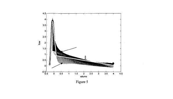
5. Figure 5
demonstrates the fits for all runs (run 1 to run 5000) which is designate by
the single line arrow
).The fit of the best run is designated by double line arrow >). this concerns
run six
with angular point at 6*10 = 60 sample points of the peak. The MSE (mean
square error) was
hereby minimal. This figure 5 demonstrates the calculated two best fitting
linear regression
[0 curves on flow-volume loops from peak flow to end of expiration and the
angle between both
regression lines (AC).
Particular and preferred aspects of the invention are set out in the
accompanying independent
and dependent claims. Features from the dependent claims may be combined with
features of the
[5 independent claims and with features of other dependent claims as
appropriate and not merely as
explicitly set out in the claims. Thus, the claims following the detailed
description are hereby
expressly incorporated into this detailed description, with each claim
standing on its own as a
separate embodiment of this invention.
!,0
11
WO 2012/027804 CA 02810043 2013-03-01
PCT/BE2011/000055
Drawing Description
BRIEF DESCRIPTION OF THE DRAWINGS
The present invention will become more fully understood from the detailed
description given
herein below and the accompanying drawings which are given by way of
illustration only, and
thus are not limitative of the present invention, and wherein:
Figure 1: concerns a COPD analysis by the measurement of the amount (volume)
and/or speed
(flow) of air of a forced exhalation and displays the fit through two linear
regression line
(defining a relationship between the flow and the volume) on 20 samples points
of the peak of
the flow volume curve from said forced airway expiration.
[0 Figure 2: concerns a COPD analysis by the measurement of the amount
(volume) and/or speed
(flow) of air of a forced exhalation and displays the fit through two linear
regression line
(defining a relationship between the flow and the volume) on 60 samples points
of the peak of
the flow volume curve from said forced airway expiration.
Figure 3: concerns a COPD analysis by the measurement of the amount (volume)
and/or speed
[5 (flow) of air of a forced exhalation and displays the fit through two
linear regression line
(defining a relationship between the flow and the volume) on 500 samples
points of the peak of
the flow volume curve from said forced airway expiration.
Figure 4: displays the MSE (mean square error) of different runs. A minimums
is reached at run
6, e.g. if we select an angular point on 6*10 = 60 sampling points from the
peak. Bij dit
!O minimum hebben we m.a.w. de beste globale fit.
Figure 5: demonstrates the fits for all runs (designate by the single line
arrow The fit of the
best run is designated by double line arrow > ). this concerns run six with
angular point at
6*10 = 60 sample points of the peak. The MSE (mean square error) was hereby
minimal. This
figure demonstrates the calculated two best fitting linear regression curves
on flow-volume loops
from peak flow to end of expiration and the angle between both regression
lines (AC)
Figures 6 to 15 concerns measurement of the amount (volume) and/or speed
(flow) (designate by
the single line arrow ). of air of a forced exhalation and displays the
fit on 10 different
patients. These figures demonstrate the calculated two best fitting linear
regression curves
12
CA 02810043 2013-03-01
WO 2012/027804
PCT/BE2011/000055
(designated by double line arrow > )on flow-volume loops from peak
flow to end of
expiration and the angle between both regression lines (AC)
= Figure 6: Peak to surface = 1.3893 and Angle = 109.4244
= Figure 7: Peak_to_surface = 0.7073 and Angle = 154.0281
= Figure 8: Peak_to_surface: 0.5547 and Angle: 154.5251
= Figure 9: Peak_to_surface: 1.3299 and Angle:132.5744
= Figure 10: Peak_to_surface: 0.9722 and Angle: 149.8245
= Figure 11: Peak_to_surface: 1.5545 and Angle: 140.2217
= Figure 12: Peak_to_surface: 0.5386 and Angle: 159.2410
= Figure 13 : Peak_to_surface: 0.9283 and Angle: 158.3582
= Figure 14: Peak_to_surface: 2.1694 and Angle: 117.7351
= Figure 15: Peak_to_surface: 0.4691 and Angle: 166.5198
Figure 16 a ¨ c provides an overview of COPD analysis on the patient as
described in example 1
. The receiver operating characteristic demonstrated that 130 provides the
best cut off value to
diagnose emphysema in the flow-volume curve that represents the measurement of
the amount
(volume) and/or speed (flow) of air of a forced exhalation of a COPD patient
(figure 16a). The
specificity was 95%. The sensitivity was 52% (area under the curve AUC= 0.82,
p< 0.0001)
There are none or very few patients with an angle smaller than 130 who do not
have the
emphysema disorder as could be confirmed via a visual score system on CT
¨images. Figure 16b
demonstrates that lower FEV1% relates to more patients with emphysema but is
also common in
COPD patients without emphysema and can therefore not be used for
discrimination. Figure 16c
demonstrates that diffusing capacity corrected for alveolar volume, a
previously accepted non
spirometry-derived predictor for emphysema, is almost equally efficient as the
angle of collaps, a
variable which can be obtained from every spirometry.
Accordingly, the present invention provides a system for diagnosing a
emphysema disorder in a
chronic obstructive pulmonary disease (COPD) patient, the system comprising:
1) a sampling
device to obtain total expiration or exhalation of the respiratory system of
the subject; 2) a
detection device generating flow and volume data of said total expiration or
exhalation; and 3) a
computer loaded a model a model to calculate the flow-volume loop; the two
best fitting linear
regression curves on flow-volume loop from peak flow to end of expiration and
the angle
between both regression lines and further loaded with a flow-volume loop
reference profile for
13
CA 02810043 2013-03-01
WO 2012/027804 PCT/BE2011/000055
a emphysema disorder; wherein the computer receives and compares subject's
flow-volume
loop profile with the reference flow-volume loop profile. The present
invention also provides a
system for diagnosing a emphysema disorder in a chronic obstructive pulmonary
disease
(COPD) patient, the system comprising: a sampling device to obtain total
expiration or
exhalation of the respiratory system of the subject; a detection device
generating flow and
volume data of said total expiration or exhalation; and a computer loaded
model to calculate the
flow-volume loop; the two best fitting linear regression curves on flow-volume
loop from peak
flow to end of expiration and the angle between both regression lines and
further loaded with a
flow-volume loop reference profile for an emphysema disorder; wherein the
computer receives
and compares subject's flow-volume loop profile with the reference flow-volume
loop profile.
In one embodiment of the invention, a method of diagnosing an emphysema
disorder in a
subject with COPD is provided whereby the method comprising: generating flow
and volume
data of total expiration or forced exhalation of a subject, calculating a
profile of the two best
fitting linear regression curves on flow-volume loop from peak flow and the
angle between both
regression lines, thereby providing; obtaining a reference profile for
emphysema or for no
emphysema; and comparing the subject profile with the reference profile,
wherein a match of the
subject profile to the reference profile indicates that the subject has
emphysema or has no
emphysema.
This invention accordingly provides the advantage that emphysema disorder can
be distinguished
in a in a chronic obstructive pulmonary disease (COPD) patient group.
In an advantageous embodiment, the method or system according to the present
invention and
described here above comprise a calculating means for calculating the peak-to-
surface area. In
any of the different embodiments it comprises a mathematical model that
compares said angle
with that of plurality of chronic obstructive pulmonary disease (COPD)
patients with no
emphysema. Hereby the mathematical model can compare the angle with that of
plurality of
COPD patients affected with emphysema; the mathematical model can compare said
angle with
that of plurality of COPD patients affected with a defined seriousness or with
defined progress of
emphysema; the mathematical model compares said angle with that of a control
and/or the
mathematical model compares automatically calculates expiratory airway
collapse due to loss of
alveolar attachments in emphysema.
The present invention also provides uses of the method or system according to
the present
invention for any of the following: to define emphysema or for diagnosing
emphysema; to define
14
CA 02810043 2013-03-01
WO 2012/027804 PCT/BE2011/000055
the seriousness or progress of emphysema; to define a respiratory therapy
medication; to define
emphysema or for diagnosing emphysema without radiological imaging such as CT
scanning; to
define emphysema or for diagnosing emphysema whereby an angle smaller than
1400 is
indicative for emphysema; to define emphysema or for diagnosing emphysema
whereby an angle
smaller than 135 is indicative for emphysema; to define emphysema or for
diagnosing
emphysema whereby an angle smaller than 130 is indicative for emphysema; to
define
emphysema or for diagnosing emphysema whereby an angle smaller than 125 is
indicative for
emphysema and to define emphysema or for diagnosing emphysema whereby an angle
smaller
than 120 is indicative for emphysema.
The present invention also provides an apparatus for diagnosing emphysema ,
the apparatus
comprising a) a device for receiving and sensing forced or total expiration of
the respiratory
system of a patient and further comprising b) a calculator or signal processor
calculating the
flow volume curve or relationship from said forced or total airway expiration,
characterized in
that the calculator or signal processor comprises 1) a first calculating means
for calculating the
[5 flow-volume curve or the flow-volume relationship and 2) a second
calculating means for
automatically calculating the angle in the expiratory flow-volume curve
between the two best
fitting linear regression curves on flow-volume curveõ whereby the signal
processor or
calculator further comprising a mathematical model to compare said angle with
that of a COPD
disorder patient subgroups or with a reference angle of one or more such COPD
disorder
!O subgroups.
The present invention further provides an apparatus for diagnosing emphysema
whereby the
apparatus having a) a signal input to receive electrical signals of an
electrical signals produced
by volume sensor and flow sensors of a device for receiving and sensing forced
or total
expiration of the respiratory system of a patient = and b) a calculator or
signal processor
calculating the flow volume curve or relationship from said forced or total
airway expiration,
characterized in that the calculator or signal processor comprises 1) a first
calculating means for
calculating the flow-volume curve or the flow-volume relationship and 2) a
second calculating
means for automatically calculating the angle in the expiratory flow-volume
curve between the
two best fitting linear regression curves on the flow-volume curve
corresponding to entire
;0 exhalation flow rate and exhalation volume of a patients respiratory
system, whereby the signal
processor or calculator further comprising a mathematical model to compare
said angle with that
of a COPD disorder patient subgroups or with a reference angle of one or more
such COPD
disorder subgroups. These apparatuses can comprise a signal processor or
calculator comprising
15
CA 02810043 2013-03-01
WO 2012/027804 PCT/BE2011/000055
a with mathematical model to compare said angle with that of plurality of
chronic obstructive
pulmonary disease (COPD) patients with no emphysema or with a reference angle
of COPD but
no emphysema.
In an embodiment of the apparatus the signal processor or calculator comprises
a mathematical
model to compare said angle with that of plurality of COPD patients affected
with emphysema or
with a reference angle for COPD and emphysema
In another embodiment of the apparatus the signal processor or calculator
comprises a
mathematical model to compare said angle with that of plurality of COPD
patients affected with
a defined seriousness or with defined progress of emphysema.
In another embodiment of the apparatus the signal processor or calculator
comprises a
mathematical model to compare said angle with that of a control.
Furthermore the apparatuses according to the present invention can be
characterized in that the
apparatus comprises a signal processor comprising a mathematical model that is
described to
automatically calculate expiratory airway collapse due to loss of alveolar
attachments in
emphysema.
It is an object of the present invention to provide such apparatus with a
calculating means that is
adapted for calculating the peak-to-surface area. This provides the surprising
advantage that
emphysema disorder can be distinguished in a in a chronic obstructive
pulmonary disease
(COPD) patient group by a device for receiving and sensing forced or total
expiration of the
respiratory system of a patient for instance by a spirometer. A simple, fast
and accurate
technology to discover emphysema disorder and advocate a proper corresponding
treatment.
The present invention also provides uses of the an apparatuses according to
present invention as
described in this application here above to define emphysema or for diagnosing
emphysema
without radiological imaging such as CT scanning; to define emphysema or for
diagnosing
emphysema without radiological imaging such as CT scanning; to define the
seriousness or
progress of emphysema; to define a respiratory therapy medication; to define
emphysema or for
diagnosing emphysema whereby an angle smaller than 140 is indicative for
emphysema; to
define emphysema or for diagnosing emphysema whereby an angle smaller than 135
is
indicative for emphysema; to any one of the claims 1 to 8, to define emphysema
or for
diagnosing emphysema whereby an angle smaller than 130 is indicative for
emphysema; to
define emphysema or for diagnosing emphysema whereby an angle smaller than 125
is
16
WO 2012/027804 CA 02810043 2013-03-01 PCT/BE2011/000055
indicative for emphysema and/or to define emphysema or for diagnosing
emphysema whereby
an angle smaller than 1200 is indicative for emphysema.
=
17