Note: Descriptions are shown in the official language in which they were submitted.
CA 02810679 2014-07-21
WO 2012/034056 PCT/US2011/051062
INDWELLING LLTMINAL DEVICES
FIELD OF INVENTION
[0002] The invention relates generally to medical devices for the treatment of
various medical conditions and specifically to indwelling catheters used to
treat a
patient's bloodstream.
BACKGROUND OF THE INVENTION
[0003] Implanted medical devices such as venous and arterial catheters,
neurological prostheses, wound drains, urinary catheters, central venous
catheters, peritoneal catheters, shunts, and other luminal indwelling devices,
are
useful for treating various medical conditions. However, a drawback of
implanted
medical devices is the risk of infection while the medical device is inserted
in the
body, and thereafter. Such risk exists even though the medical devices are
sterilized and carefully packaged to guard against introduction of microbes or
pathogens during implantation or insertion of the medical device. For example,
there is a risk of serious nosocomial infections when using catheters for
hemodialysis procedures. In fact, central venous catheters account for most
nosocomial catheter-related bloodstream infections.
[0004] When catheters and other indwelling luminal devices are inserted into
body cavities such as the urinary tract, venous or arterial vessels, bacteria
or other
microbes can be picked up from the skin and carried into the insertion site
where
bacterial or microbial colonization may ensue. Infections may derive from an
interaction of the microbes and the catheter micro-surface. Once infected, the
microorganisms adhere to the catheter micro-surface and rapidly become encased
in a polysaccharide matrix or biofilm, which protects the microorganisms from
a
host's defenses.
1
CA 02810679 2013-03-06
WO 2012/034056 PCT/US2011/051062
[0005] In the case of urinary and venous catheters, there is a significant
threat
of microbial growth along the exterior surface or outer wall of the catheter
and,
especially for catheters used long-term, there is a significant threat of
microbial
growth along the interior surface or inner wall. This can lead to chronic
urinary
tract infections (CUTI), or septicemia in the case of venous and arterial
catheters,
thrombolytic emboli, stenosis, and thrombosis resulting from infections, and
other
life threatening complications, especially among the elderly and immuno-
compromised patients. Thus, there is a need for the development of better
methods of preventing and treating infections caused by the insertion of
catheters
into a patient's body.
[0006] In addition to antimicrobials, other therapeutic agents may help reduce
complications associated with chronically implanted indwelling medical devices
in
the body of a patient. Such medications include anti-inflammatories, anti-
proliferatives and anti-coagulating agents or a combination thereof. However,
to
be effective the therapeutic agent should be delivered to a substantial
portion of
the surface of the indwelling medical device. Without such therapeutic agents,
there is a risk that portions of the medical device will become compromised
and
cause an inflammatory response and/or allow tissue in-growth over surfaces of
the
indwelling portion of the medical device.
[0007] Other drawbacks of conventional indwelling catheters include a
significant crossing profile, lack of convenience, and tissue damage to the
areas to
which the catheters are deployed. For example, indwelling catheters are
typically
used only periodically. As a result, inconvenient characteristics of
catheters, such
as being difficult to thread or insert catheter bodies, add to the treatment
time and
potential discomfort of therapy provided by the catheter. Also, as discussed
above, indwelling catheters, such as central venous catheters, may cause
damage
to a patient's vasculature.
[0008] Accordingly, there is a need for a medical device that can effectively
deliver a therapeutic agent to a substantial portion of its surface, e.g. a
substantial
length of the outer surface of an indwelling catheter. In addition, improved
devices
are needed which feature a lower profile, more convenient method of use, and
reduce tissue damage caused to a patient's anatomy.
2
CA 02810679 2013-03-06
WO 2012/034056 PCT/US2011/051062
SUMMARY OF THE INVENTION
[0009] One embodiment of the invention comprises an indwelling catheter
having a central tube with at least one lumen and an outer jacket surrounding
said
tube. In various embodiments, the indwelling catheter is a central venous
catheter.
In various embodiments, the catheter further comprises a plurality of grooves
in the
outer surface of the central tube, wherein the grooves and jacket form a
plurality of
channels extending along at least a portion of the longitudinal axis of the
catheter.
[0010] In one embodiment, the central tube is internally segmented into a
plurality of lumens. In another embodiment, the central tube comprises two
lumens. In another embodiment, the outer surface of the central tube is
generally
circular. In another embodiment, the outer surface of the central tube is
generally
an oval. In another embodiment, the cross-section of the lumen is d-shaped. In
another embodiment, the cross-section of the lumen is circular.
[0011] In various embodiments, said lumens are each in fluid communication
with at least one extension tube. In another embodiment, the extension tubes
each comprise a connector hub. In another embodiment, the channels are in
fluid
communication with an extension tube. In another embodiment, at least two
extension tubes are in fluid communication with different channels. In another
embodiment, the channels are in deflectable legs which can be positioned
against
the walls.
[0012] In various embodiments, the jacket is permeable. In various
embodiments, the jacket is porous. In another embodiment, the jacket comprises
ePTFE.
[0013] In various embodiments, the catheter further comprises a secondary
tube. In various embodiments, the secondary tube concentrically surrounds the
central tube and jacket. In other embodiments, the secondary tube spirals
around
the surface of the central tube and jacket. The secondary tube may also
comprise
a sleeve inserted into the patient's vasculature.
DESCRIPTION OF THE DRAWINGS
[0014] Figure 1 illustrates an exemplary medical device which comprises an
indwelling catheter and extension tubes.
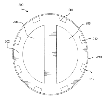
[0015] Figures 2A and 2B illustrate cross sections of two exemplary indwelling
catheters at "A-A" in Figure 1.
3
CA 02810679 2014-07-21
WO 2012/034056 PCT/US2011/051062
[0016] Figures 3A and 36 illustrate the distal end of the indwelling catheters
depicted in Figures 2A and 2B.
[0017] Figures 4A and 4B illustrate, respectively, a perspective view and a
cross section of an exemplary medical device comprising a "Chinese lantern"
type
anchoring device.
[0018] Figure 5 illustrates a cross section of an exemplary medical device.
[0019] Figures 6A and 6B illustrate side views of an exemplary medical device.
[0020] Figure 7 illustrates a side view of another exemplary medical device,
comprising a "pigtail" type anchoring device.
[0021] Figure 8 illustrates a side view of an exemplary medical device
comprising a secondary tube.
[0022] Figure 9 illustrates a side view of another exemplary medical device
comprising a secondary tube.
[0023] Figures 10A and 106 illustrate a side view of an exemplary medical
device comprising a secondary tube.
[0024] Figures 11A and 11B illustrate an exemplary medical device comprising
a port.
[0025] Figures 12A and 12B illustrate an exemplary medical device comprising
a support wire path and support wire.
[0026] Figure 13 illustrates an exemplary medical device comprising a docking
station.
DETAILED DESCRIPTION OF THE INVENTION
[0027] While the present invention will hereinafter be described in connection
with the preferred embodiments, it will be understood that it is not intended
to limit
the invention to these embodiments. Instead, it
is intended to cover all
alternatives, modifications, and equivalents as may be included within the
scope of the invention as described herein.
[0028] For the purposes of the following description and the claims appended
hereto, the term "distal" refers to those portions of a medical device, such
an
indwelling catheter, and those portions of components of the medical device
which
are nearest the insertion tip, that is, the end of the medical device that is
inserted
into an area of a patient's body, such as a blood vessel. Conversely, the
relative
4
CA 02810679 2013-03-06
WO 2012/034056 PCT/US2011/051062
term "proximal" refers to those portions of a medical device and those
portions of
components which are farthest from the insertion tip of the catheter.
[0029] Various exemplary medical devices in accordance with the disclosure
comprise a central tube with at least one lumen, a second lumen, and an outer
jacket concentrically surrounding the central tube. In various exemplary
embodiments, the second lumen is comprised within the central tube and the two
lumens are of equal cross-sectional surface area. In other exemplary
embodiments, the second lumen is configured annularly between the outer
surface
of the central tube and the inner surface of the outer jacket. In yet other
exemplary
embodiments, the medical device comprises a secondary tube which comprises
the second lumen.
[0030] In another embodiment of the invention, the medical device comprises
an indwelling catheter. Said indwelling catheter can comprise a portion that
is
accessible from outside the body once said indwelling portion is inserted into
the
body. Any catheter used for medical treatment can generally be used for the
present invention. Suitable catheters include, but are not limited to, venous,
arterial, urinary catheters, wound drains, central venous catheters,
peritoneal
catheter, percutaneous catheters, sheaths and trocars, drainage catheters,
endoscopes and endoscopic catheters, and gastrointestinal catheters. In
addition
to catheters, other medical devices that are insertable into the body of a
patient,
and accessible through the skin or other method once implanted can be used in
the present invention. For example, the following other indwelling medical
devices
may be used: cannulas, cardiac pacing leads or lead tips, cardiac
defibrillator
leads or lead tips, implantable vascular access ports, blood tubing, vascular
or
other grafts, intra-aortic balloon pumps, heart valves, cardiovascular
sutures, total
artificial hearts and ventricular assist pumps.
[0031] Referring to the drawings, like reference numbers represent like or
corresponding elements in the drawings. The drawings illustrate one embodiment
of the instant invention. Other medical devices are also contemplated as part
of
the instant invention.
[0032] With reference to FIG. 1, exemplary medical device 100 is illustrated.
Medical device 100 comprises an indwelling catheter 104. Medical device 100
further comprises extension tubes 106, 108 and 110, which are in fluid
communication with lumens and channels (or annual spaces) within indwelling
CA 02810679 2013-03-06
WO 2012/034056 PCT/US2011/051062
catheter 104. Medical device 100 further comprises connector hubs 112, 114 and
116, which are attached to the proximal ends of extension tubes 106, 108 and
110,
respectively.
[0033] The proximal portion of medical device 100 comprises a central tube 102
which houses a plurality of single lumen proximal extension tubes, 106, 108
and
110. Proximal extension tubes 106, 108, and 110 each have a distal end and a
proximal end. The distal end of each proximal extension tube is connected to
the
proximal end of central tube 102 such that the single lumen of each proximal
extension tube is in fluid communication with one of the plurality of lumens
of
central tube 102. In addition, at least one single lumen extension tube is
attached
to a plurality of channels (not shown). In another embodiment, several
extension
tubes can be attached to the plurality of channels. In another embodiment,
extension tubes 106, 108 and 110 may be removable.
[0034] In one embodiment, medical device 100 comprises a polymer, such as
polyethylene, polyurethane, polycarbonates, ethyl vinyl acetate, polyamides
(such
as PEBAX 0, a registered trademark of Arkema), polyimides, or similar
material.
[0035] FIG. 2A illustrates a cross-sectional view of an exemplary medical
device 200 at "A-A" in FIG. 1. Central tube 202 of medical device 200 is
generally
circular. Central tube 202 comprises a biocompatible polymeric material. A
biocompatible material is hereby defined as a material being suited for and
meeting the purpose and requirements of a medical device, used for either long
or
short term implants or for non-implantable applications. Long term implants
are
defined as items implanted for more than 30 days. Exemplary polymeric
materials
include without limitation polyurethane and its copolymers, silicone and its
copolymers, ethylene vinyl-acetate, polyethylene terephtalate, thermoplastic
elastomers, polyvinyl chloride, polyolefins, cellulosics, polyam ides,
polyether-
amides, polyesters, polyimides, polysulfones, polytetrafluorethylenes,
polycarbonates, acrylonitrile butadiene styrene copolymers, acrylics,
polylactic
acid, polyglycolic acid, polycaprolactone, polylactic acid-polyethylene oxide
copolymers, cellulose, collagens, and chitins, and other various copolymers.
[0036] In an aspect of exemplary medical device 200, central tube 202 is
internally segmented into lumens 206 and 208. Lumens 206 and 208 are parallel
and have substantially the same "d-shaped" cross-sectional surface area.
6
CA 02810679 2013-03-06
WO 2012/034056 PCT/US2011/051062
[0037] In various exemplary embodiments, central tube 202 and jacket 210
comprise a highly flexible material, such as ePTFE. In this configuration,
central
tube 202 and jacket 210 are sufficiently flexible such that they are buoyant
in the
blood flow within the vessel. The flexible material also provides adequate
support
to allow medical device 200 to retain its overall shape while in operation.
For
example, central tube 202 has sufficient column strength to prevent the tube
from
collapsing when vacuum or suction is applied to it during a medical procedure.
This flexibility and buoyancy helps to reduce the force and/or impact at which
that
medical device 200 makes contact with the walls of the treatment vessel,
thereby
minimizing tissue damage and reducing the likelihood of occurrences of
vascular
stenosis.
[0038] In various exemplary embodiments, central tube 202 further comprises
heparin. Heparin may be attached to any surface of the central tube 202. A
coating of heparin can help prevent and/or reduce thrombosis formation on or
in
central tube 202. In one exemplary embodiment, the surfaces of lumens 206 and
208 comprise a heparin coating. In another embodiment, the outer surface of
central tube 202 comprises a heparin coating. The heparin coating and method
of
attaching a heparin coating is taught in U.S. Patent 6,559,132, which is
incorporated by reference herein in its entirety for all purposes.
[0039] One embodiment of the invention comprises an indwelling medical
device that can effectively deliver a therapeutic agent evenly along the
length, of
said medical device. For the purposes of this invention "length" comprises at
least
a portion of the length of a medical device and its surface (i.e. outer wall),
unless
otherwise stated.
[0040] In this regard, in exemplary embodiments, central tube 202 further
comprises a plurality of grooves 204 extending along its longitudinal axis.
Grooves
204 may be positioned in the outer surface of central tube 202. In this
configuration, the combination of grooves 204 and outer jacket 210 forms a
plurality of channels 212 extending along the longitudinal axis of said
central tube
and jacket. In one aspect of the exemplary embodiment, grooves 204 are formed
during extrusion of central tube 202. In another aspect, grooves 204 are
formed
by cutting the grooves into the outer wall of central tube 202, such as, for
example,
with a laser. Further, grooves 204 may be formed by reflowing the outer
surface of
central tube 202 around longitudinal features.
7
CA 02810679 2013-03-06
WO 2012/034056 PCT/US2011/051062
[0041] One of the advantages of having channels 212 along the longitudinal
axis of the medical device is that when a fluid is infused into the channels,
it evenly
distributes the fluid along its length which can then diffuse to the outer
wall of the
medical device and/or to the surrounding environment in which the medical
device
is dwelling.
[0042] Grooves 204 may be configured in any shape, e.g. square, round or
combinations thereof. Grooves 204 may continue down the entire length of
central
tube 202 or a portion thereof. In various exemplary embodiments, channels 212,
which are formed by grooves 204 interfacing with jacket 210, carry liquids
and/or
gases from the proximal end to the distal end of medical device 202.
[0043] Jacket 210 of medical device 200 may comprise a permeable and/or
porous material. Examples of such porous materials include, but are not
limited to,
porous fluoropolymers such as expanded polytetrafluoroethylene (ePTFE),
expanded high density polyethylene (HDPE). Other non-porous polymers such as
polyesters, polyurethanes, polyethylenes, polyimides, etc. can also be of
utility
provided they are processed to have pores. Examples of such processes include
laser perforation and pin perforations. Jacket 210 may also comprise semi-
permeable films, such as polyurethanes, silicones, and polyether-amides. As
used
herein, the term "porous" describes a material that contains small or
microscopic
openings, or pores. Without limitation, "porous" is inclusive of materials
that
possess pores that are observable under microscopic examination. The term
"porous" describes a material through which fluids (liquid and/or gas) can
penetrate through bulk flow. A permeable material prevents bulk flow while
allowing selective molecules to pass, while a porous material can allow bulk
flow
while restricting flow of certain size particles.
[0044] Selecting porosity and/or permeability of the jacket material can
generate a back pressure within channels 212 which aids in an even
distribution of
fluid down the length of the device, as opposed to a more porous or permeable
structures that provide for flow to only a section of channels 212 with the
least
resistance. For example, if medical device 200 is located against the wall of
a
vessel, flow may be restricted and therapeutic agents will only be delivered
to the
sections that are not in contact with the vessel. If there was a jacket 210
that
comprises a material with sufficient back pressure, the effect of wall contact
on
distribution will be minimized. Such a material can be designed, inter alia,
by
8
CA 02810679 2013-03-06
WO 2012/034056 PCT/US2011/051062
adjusting the porosity and/or permeability of the jacket material. Furthermore
said
jacket material may be adjusted by taking into account (or adjusting) the
physical
properties of the fluid (including any active agents and/or excipients in the
fluid) by
methods known in the art.
[0045] A therapeutic agent is a drug or agent that can elicit a bioactive
response. Examples of the therapeutic agents or drugs useful in this invention
include prochlorperazine edisylate, ferrous sulfate, aminocaproic acid,
mecaxylamine hydrochloride, procainamide hydrochloride, amphetamine sulfate,
methamphetamine hydrochloride, benzphetamine hydrochloride, isoproteronol
sulfate, phenmetrazine hydrochloride, bethanechol chloride, methacholine
chloride, pilocarpine hydrochloride, atropine sulfate, scopolamine bromide,
isopropamide iodide, tridihexethyl chloride, phenformin hydrochloride,
methylphenidate hydrochloride, theophylline cholinate, cephalexin
hydrochloride,
diphenidol, meclizine hydrochloride, prochlorperazine maleate,
phenoxybenzamine, thiethylperazine maleate, anisindione, diphenadione,
erythrityl
tetranitrate, digoxin, isoflurophate, acetazolamide, methazolamide,
bend roflumethiazide, chlorpropamide, tolazamide, chlormadinone acetate,
phenaglycodol, allopurinol, aluminum aspirin, methotrexate, acetyl
sulfisoxazole,
hydrocortisone, hydrocorticosterone acetate, cortisone acetate, dexamethasone
and its derivatives such as betamethasone, triamcinolone, methyltestosterone,
17-
13-estradiol, ethinyl estradiol, ethinyl estradiol 3-methyl ether,
prednisolone, 17- p -
hydroxyprogesterone acetate, 19-nor-progesterone, norgestrel, norethindrone,
norethisterone, norethiederone, progesterone, norgesterone, norethynodrel,
indomethacin, naproxen, fenoprofen, sulindac, indoprofen, nitroglycerin,
isosorbide
dinitrate, propranolol, timolol, atenolol, alprenolol, cimetidine, clonidine,
imipramine, levodopa, chlorpromazine, methyldopa, dihydroxyphenylalanine,
theophylline, calcium gluconate, ketoprofen, ibuprofen, atorvastatin,
simvastatin,
pravastatin, fluvastatin, lovastatin, cephalexin, erythromycin, haloperidol,
zomepirac, ferrous lactate, vincamine, phenoxybenzamine, diltiazem, milrinone,
captropril, mandol, quanbenz, hydrochlorothiazide, ranitidine, flurbiprofen,
fenbufen, fluprofen, tolmetin, alclofenac, mefenamic, flufenamic, difuninal,
nimodipine, nitrendipine, nisoldipine, nicardipine, felodipine, lidoflazine,
tiapamil,
gallopamil, amlodipine, mioflazine, lisinopril, enalapril, captopril,
ramipril,
enalaprilat, famotidine, nizatidine, sucralfate, etintidine, tetratolol,
minoxidil,
9
CA 02810679 2013-03-06
WO 2012/034056 PCT/US2011/051062
chlordiazepoxide, diazepam, amitriptylin, and imipramine. Further examples are
proteins and peptides which include, but are not limited to, insulin,
colchicine,
glucagon, thyroid stimulating hormone, parathyroid and pituitary hormones,
calcitonin, renin, prolactin, corticotrophin, thyrotropic hormone, follicle
stimulating
hormone, chorionic gonadotropin, gonadotropin releasing hormone, bovine
somatotropin, porcine somatropin, oxytocin, vasopressin, prolactin,
somatostatin,
lypressin, pancreozymin, luteinizing hormone, LHRH, interferons, interleukins,
growth hormones such as human growth hormone, bovine growth hormone and
porcine growth hormone, fertility inhibitors such as the prostaglandins,
fertility
promoters, growth factors, and human pancreas hormone releasing factor. In an
exemplary embodiment, the therapeutic agent is a steroid, such as
dexamethasone. Additional exemplary embodiments comprise therapeutic agents
consisting of mixtures of anti-microbials, antivirals, antibiotics,
antibacterial agents,
anti-inflammatory agents, anti-proliferative agents, anti-coagulating agents,
hemostatic agents, decongestants, hemorroidal treatments, and/or analgesics.
[0046] The fluid interaction with jacket 210 is important for even
distribution. If
the therapeutic agent to be delivered to the vasculature is of high viscosity
or has a
surface energy that restricts and/or prevents passage through jacket 210, the
pore
size, structure and/or surface energy of jacket 210 can be tailored to obtain
the
optimized fluid mechanics for a desired dosing regime. In an exemplary
embodiment, a therapeutic agent is substantially evenly distributed along the
length of the medical device. In an aspect of these embodiments, jacket 210 is
a
highly porous material, which allows a substantial amount of therapeutic agent
to
evenly perfuse out along the length of medical device 200. In other aspects of
these embodiments, jacket 210 has a low degree of porosity, and therefore the
therapeutic agent may dwell within the channel 212 and wick out slowly over a
period of time.
[0047] For example, if wicking of the therapeutic agent is used to either
provide
for a slow delivery (spanning the course of multiple hours, days and/or
treatment
cycles) or a more even delivery of a therapeutic agent, then the
microstructure and
surface energy of jacket 210 must be tailored to allow for wicking of the
therapeutic
agent. Wicking may be used to provide a therapeutic agent to the areas of
outer
jacket 210 that are over the non-grooved portions of the surface of central
tube
202.
CA 02810679 2013-03-06
WO 2012/034056 PCT/US2011/051062
[0048] In addition, channels 212 may be designed to function as a reservoir to
allow for the storage of an appropriate amount of a therapeutic agent. For
instance, if the device is intended to supply a therapeutic agent to the
surface of
jacket 210 over the course of multiple days, and the rate of delivery is
known, the
required volume for channels 212 to function as reservoirs can be calculated.
If
additional volume is required, an additional reservoir can be located external
to the
patient or in an additional volume internal to the central tube 202.
[0049] In various exemplary embodiments, jacket 210 further comprises a
heparin coating. In various embodiments, both jacket 210 and the outer surface
of
central tube 202 comprise a heparin coating.
[0050] In various exemplary embodiments, jacket 210 may further comprise a
coating of therapeutic agents. In some exemplary embodiments, the therapeutic
agent would be bound to the outer surface of jacket 210. In other embodiments,
the therapeutic agent would pass through jacket 210 into the vasculature via
pores
or permeations in jacket 210.
[0051] In other exemplary embodiments, jacket 210 comprises a hydrogel
coating. In these configurations, therapeutic agents may be absorbed by the
hydrogel as they exit the pores and/or permeations in jacket 210. The
therapeutic
agents may also be formed as a hydrogel and applied to the surface of or
otherwise dissociating from jacket 210. The therapeutic agent would be
released
by the hydrogel into the blood stream at a significantly slower rate,
preventing the
drug from sloughing off of jacket 210. In yet other exemplary embodiments, a
beneficial gas, such as nitrous oxide, may be passed through channels 212 and
out of jacket 210 into the patient's blood stream.
[0052] With reference to FIG. 2B an exemplary medical device 300 is
illustrated. Medical device 300 comprises a central tube 302, grooves 304, and
an
outer jacket 310. In this exemplary embodiment, central tube 302 is generally
oval
in shape, and is segmented into lumens 306 and 308. Lumens 306 and 308 are
substantially parallel and have the same general circular shape. In an aspect
of
the exemplary embodiment, lumens 306 and 308 may have substantially the same
cross-sectional surface area.
[0053] FIGs. 3A and B illustrate the distal end of the exemplary medical
devices
depicted in FIGs. 2A and 2B. Specifically, FIG. 3A illustrates the distal end
of
medical device 200. FIG. 3B illustrates the distal end of medical device 300.
As
11
CA 02810679 2013-03-06
WO 2012/034056 PCT/US2011/051062
illustrated in FIGs. 3A and 3B, the lumens 206, 208, 306 and 308 extend from
the
proximal to the distal end of the medical device. However, in a preferred
embodiment, the channels will not be open at the distal end so that when fluid
is
infused into the channel, the fluid will not flow out of the distal end of the
channel.
[0054] With reference to FIGs. 4A and 4B, an exemplary medical device 400 is
illustrated. Medical device 400 further comprises an anchor segment 420 of
outer
tube 404. Anchor segment 420 may be located at the distal end of outer tube
404.
In an aspect of the exemplary embodiment, anchor segment 420 may comprise a
series of "legs" which are in contact with the vessel walls, and act to center
and
stabilize medical device 400 within the vessel. Such a configuration may help
to
minimize the risk of venous stenosis by preventing medical device 400 from
damaging and/or abrading adjacent tissues.
[0055] In an aspect of the exemplary embodiment, the anchor segment 420
may be configured as a "Chinese lantern" shape. In this configuration, the
legs of
anchor segment 420 may be deployed from a relaxed configuration, in which they
are substantially parallel to central tube 402, to an expanded configuration,
in
which they contact the vessel walls in a "Chinese lantern" shape. In a
preferred
embodiment, central tube 402 is fixedly attached to the distal end of
anchoring
segment 420. Central tube 402 is partially withdrawn axially through outer
tube
404, which causes the legs of anchoring segment 420 to expand and contact the
walls of the treatment vessel.
[0056] Anchor segment 420 may further comprise at least one individual lumen
within each "leg" with communication to the outside surface via holes or
ports. In
this configuration, therapeutic agent may be delivered through the individual
lumens directly to the point where the legs of anchor segment 420 contact the
vessel wall. In another aspect of the exemplary embodiment, the legs of anchor
segment 420 are configured as a tubular construct having a plurality of
grooves on
their outer periphery. The grooves may deliver therapeutic agents directly to
the
vessel walls. Outer tube 404 may comprise a material of sufficient porosity
and/or
permeability to deliver therapeutic agents at the points which outer tube 404
contacts the vessel wall, thereby efficiently delivering smaller doses of
therapeutic
agents directly to targeted tissue and preventing drugs from washing
downstream
into the circulatory system.
12
CA 02810679 2013-03-06
WO 2012/034056 PCT/US2011/051062
[0057] With initial reference to FIG. 5, in various exemplary embodiments,
medical device 500 comprises a central tube 502, an outer tube 504 and an
outer
jacket 510. Outer tube 504 concentrically surrounds central tube 502, creating
an
annular lumen 508. Outer jacket 510 concentrically surrounds the outer surface
of
outer tube 504. Central tube further comprises a central lumen 506.
[0058] In various exemplary embodiments, outer tube 504 further comprises a
plurality of grooves 512 extending along its longitudinal axis. Grooves 512
may be
positioned in the outer surface of outer tube 504. In this configuration, a
plurality of
channels extend along the longitudinal axis of outer tube 504 and outer jacket
510.
As discussed earlier in relation to exemplary medical device 200, grooves 512
may
be formed, for example, by cutting grooves into the outer wall of outer tube
504,
reflowing the outer surface of outer tube 504, or created during extrusion of
outer
tube 504.
[0059] In various exemplary embodiments, these channels may be in fluid
communication with a supply of beneficial drugs or agents. As previously
discussed in relation to exemplary medical device 200, the microstructure of
jacket
510 and position and configuration of the channels may control the flow rate
of a
beneficial drug or agent into the treatment vessel.
[0060] With reference to FIGs. 6A and 6B, in various exemplary embodiments,
medical device 600 comprises a central tube 602, an outer tube 604, and a
jacket
610. Outer tube 604 concentrically surrounds central tube 602, creating an
annular lumen 608. Jacket 610 concentrically surrounds outer tube 604. In
accordance with an aspect of the exemplary embodiment, central tube 602 may be
longer than outer tube 604 and jacket 610, therefore protruding from outer
tube
604 and jacket 610 at their distal ends.
[0061] In various exemplary embodiments, outer tube 604 further comprises a
plurality of grooves extending along its longitudinal axis. These grooves may
be
positioned in the outer surface of outer tube 604. In this configuration, a
plurality of
channels extending along the longitudinal axis of outer tube 604 and outer
jacket
610. These channels may be in fluid communication with a supply of beneficial
drugs or agents. As previously discussed in relation to exemplary medical
device
200, the microstructure of jacket 610 and position and configuration of
channels
may control the flow rate of a beneficial drug or agent into the treatment
vessel.
13
CA 02810679 2013-03-06
WO 2012/034056 PCT/US2011/051062
[0062] In various exemplary embodiments, medical device 600 may further
comprise a distal tip 616 attached to the distal end of central tube 602.
Distal tip
616 may comprise opening 606 and terminate in a tip. In various exemplary
embodiments, distal tip 616 is wedge, "duck-bill," or flapper-shaped. However,
any
shape of distal tip 616 which allows for treated blood to exit through opening
606 is
within the scope of the present disclosure.
[0063] In this configuration, the distal tip 616 protrudes from the outer tube
604
when medical device 600 is in operation, as illustrated in FIG. 6B. During
operation, blood to be treated flows in to medical device 600 through annular
lumen 608. The blood is treated outside of the body, and the treated blood
flows
back in to the vessel through opening 606 and as the blood exits distal tip
616.
When medical device 600 is not in operation, as illustrated in FIG. 6A, distal
tip
616 may be retracted such that it seats inside the distal end of outer tube
604,
sealing the end of annular lumen 608. Any shape and configuration of distal
tip
616 which provides a return path for treated blood and is capable of sealing
lumen
608 when the medical device is not in operation is within the scope of the
present
disclosure. In addition, any shape of lumen, including annular or one or more
channels, which provides an outflow path for blood from the vessel is within
the
scope of the present invention.
[0064] In various exemplary embodiments, distal tip 616 comprises a relatively
soft biocompatible material, such a silicone. In such embodiments, central
tube
602 may comprise a more rigid biocompatible material, such as polyurethane. A
more rigid material than is used in other exemplary embodiments is permissible
in
this configuration because central tube 602 is only exposed when medical
device
600 is in operation, and the end of the tube features a relatively soft distal
tip 616.
Therefore, central tube 602 is unlikely to make inadvertent contact with the
vessel
walls and cause unintended tissue damage. However, central tube 602 may
comprise any material which is biocompatible and provides sufficient
structure,
including materials discussed in regards to other exemplary embodiments.
[0065] With reference to FIG. 7, medical device 700 may comprise a central
tube 702 which contains a single lumen 706 and extends beyond the distal end
of
an outer tube 710. In various exemplary embodiments, the portion of central
tube
702 which extends beyond the distal end of outer tube 710 may change shape and
configuration. For example, the exposed portion of central tube 702 may be
14
CA 02810679 2013-03-06
WO 2012/034056 PCT/US2011/051062
configured in a "pigtail" configuration. In this embodiment, the "pigtail" is
made by
forming the distal end of central tube 702 into a spiral at the distal region
large
enough in diameter to contact adjacent tissues. In this configuration, the
"pigtail"
shape of the exposed portion of central tube 702 positions and centers medical
device 700 in the treatment vessel by contacting the side walls of the vessel.
This
positioning helps to reduce inadvertent contact between medical device 700 and
the walls of the treatment vessel, minimizing damage to the tissue and
reducing
the risk of vascular stenosis.
[0066] In various exemplary embodiments, medical devices in accordance with
the present disclosure may further comprise a secondary tube. As illustrated
in
FIG. 8, secondary tube 803 may be configured to spiral along the outside of
central
tube 802. In this configuration, secondary tube 803 houses second lumen 808.
[0067] Secondary tube 803 may comprise, for example, a biocompatible
material. Such materials may include olefin polymers, polyethylene,
polypropylene, polyvinyl chloride, polytetrafluoroethylene which is not
expanded,
fluorinated ethylene propylene copolymer, polyvinyl acetate, polystyrene,
poly(ethylene terephthalate), naphthalene dicarboxylate derivatives, such as
polyethylene naphthalate, polybutylene naphthalate, polytrimethylene
naphthalate
and trimethylenediol naphthalate, polyurethane, polyurea, silicone rubbers,
polyamides, polycarbonates, polyaldehydes, natural rubbers, polyester
copolymers, styrene-butadiene copolymers, polyethers, such as fully or
partially
halogenated polyethers, copolymers, and combinations thereof. Also,
polyesters,
including polyethylene terephthalate (PET) polyesters, polypropylenes,
polyethylenes, polyurethanes, polyolefins, polyvinyls, polymethylacetates,
polyamides, naphthalane dicarboxylene derivatives, and natural silk may be
used.
[0068] In an aspect of the exemplary embodiments, secondary tube 803 may
provide structural support to medical device 800. Central tube 802 and
secondary
tube 803 may comprise a flexible material, such as ePTFE, which allows central
tube 802 and secondary tube 803 to be in a collapsed configuration when
medical
device 800 is not in use. When medical device 800 is in operation, blood
returning
to the vessel from treatment inflates secondary tube 803, providing structural
support to central tube 802. When medical device 800 is not in operation,
central
tube 802 will float in the flow of blood within the treatment vessel. This
configuration reduces the force and/or impact with which medical device 800
CA 02810679 2014-07-21
WO 2012/034056 PCT/US2011/051062
contacts the walls of the treatment vessel, decreasing tissue damage and
reducing
the risk of vascular stenosis.
[0069] With reference to FIG. 9, an exemplary medical device 900 comprises a
secondary tube 903 which surrounds central tube 902. In various exemplary
embodiments, central tube 902 and secondary tube 903 may comprise multiple
configurations. For example, central tube 902 and secondary tube 903 may
comprise an expanded configuration when medical device 900 is in operation and
a collapsed configuration when medical device 900 is not in operation. As used
herein, "expanded" means being swelled, unfurled or otherwise having an
increased diameter and/or an increased volume. "Collapsed" means being
compressed, closed, furled or otherwise having a decreased diameter and/or a
decreased volume.
[0070] For example, secondary tube 903 may comprise an inflatable sleeve
which extends from the distal end of medical device 900 to the proximal end of
central tube 902. When medical device 900 is in operation, secondary tube 903
inflates to an expanded configuration and provides structural support to
central
tube 902. In an aspect of these exemplary embodiments, secondary tube 903
inflates to a diameter that makes contact with the inner vessel walls. When
medical device 900 is not in operation, secondary tube 903 deflates to a
collapsed
configuration in which the tube does not contact the inner vessel walls. The
expansion and collapse of secondary tube 903 may reduce the formation of
biofilm
and/or biofouling on the outside surface of the tube.
[0071] In various exemplary embodiments, secondary tube 903 may comprise a
perforated material, such as ePTFE. In this configuration, treated blood is
returned
to the treatment vessel through the walls of secondary tube 903. In an aspect
of
various exemplary embodiments, therapeutic agent may also pass through the
secondary tube 903 and into the treatment vessel. Secondary tube 903 may also
be rendered elastomeric by the incorporation of an elastomeric compound such
as
is taught in U.S. Patent Application Publication 2004/0024448 to Chang et al.
[0072] With reference to FIGs. 10A and 10B, an exemplary medical device
1000 comprises a central tube 1002 and a secondary tube 1003. In various
exemplary embodiments, secondary tube 1003 is a sleeve which may be
implanted in a patient's vasculature. As illustrated in FIG. 10A, central tube
1002
16
CA 02810679 2013-05-29
may be inserted into secondary tube 1003 to provide treatment to a patient's
vasculature, and removed once treatment has been completed.
[0073] In various exemplary embodiments, secondary tube 1003 may comprise
an expanded configuration when medical device 1000 is in operation and a
collapsed configuration when medical device 1000 is not in operation. For
example, secondary tube 1003 may comprise a collapsible sleeve which extends
from the distal end of medical device 1000 to the proximal end of central tube
1002. When medical device 1000 is in operation, central tube 1002 is inserted
into
secondary tube 1003, opening secondary tube 1003 to an expanded configuration.
When medical device 1000 is not in operation, central tube 1002 is removed
from
secondary tube 1003, allowing secondary tube 1003 to collapse.
[0074] Secondary tube 1003 may comprise, for example, a highly flexible
biocompatible polymeric material such as ePTFE. As illustrated in FIG. 10B,
secondary tube 1003 is flexible enough that, in the absence of central tube
1002, it
collapses within the treatment vessel and seals itself. This prevents blood
from
flowing back into secondary tube 1003 when treatment is not being provided to
the
patient.
[0075] In other exemplary embodiments, secondary tube 1003 may further
comprise an anchoring segment. In an aspect of these exemplary embodiments,
the anchor segment may be configured as a "Chinese lantern" shape. In this
configuration, the legs of the anchor segment may be deployed from a collapsed
configuration, in which they are substantially parallel to central tube 1002,
to an
expanded configuration, in which they contact the vessel walls in a "Chinese
lantern" shape. The legs of the anchor segment may contact the walls of the
treatment vessel, stabilizing and centering medical device 1000 while
treatment is
delivered to the patient. Any shape or configuration of the anchor segment
which
centers and stabilizes medical device 1000 within the treatment vessel is
within the
scope of the invention.
[0076] With reference to FIG. 11A, an exemplary medical device 1100 comprises
a catheter body 1101, which houses a central tube 1102. Medical device 1100
further comprises a port 1105 with a port opening 1107 and a secondary tube
1103. Port 1105 is installed in the patient's skin, and secondary tube 1103 is
installed between the subdural portion of port 1105 and a treatment vessel
1111.
17
CA 02810679 2013-05-29
In this configuration, port opening 1105 is in fluid communication with
treatment
vessel 1111 through secondary tube 1103.
[0077] Catheter body 1101 may be attached to port 1105 and port opening
1107. Central tube 1102 may then be inserted through port opening 1107 into
secondary tube 1103 as shown in FIG. 11B. Central tube 1102 is advanced
through secondary tube
1103 into treatment vessel 1111. Once central tube 1102 is in position within
treatment vessel 1111, treatment may begin. When treatment has completed,
central tube 1102 may be removed from treatment vessel 1111 and secondary
tube 1103 for cleaning or disposal. Port opening 1107 may then be sealed to
prevent fluid leakage from the patient's vasculature.
[0078] With reference to FIG. 12, an exemplary medical device 1200 comprises
a central tube 1202, jacket 1210, support wire path 1213, and a support wire
1214.
In this configuration, support wire path 1213 is integral to jacket 1210, and
may
comprise a spiral-shaped groove in jacket 1210. As illustrated in FIG. 12A,
support wire 1214 may be inserted into support wire path 1213, providing shape
and structural support to central tube 1202. Support wire 1214 may comprise a
metallic material, such as a stainless steel or nitinol stylet. Any material
which
provides sufficient support such that support wire 1214 has adequate strength
to
maintain the desired shape of central tube 1202 and jacket 1210 is within the
scope of the present disclosure.
[0079] Once support wire 1214 is inserted into support wire path 1213,
treatment may be provided to the patient's vasculature. When treatment has
concluded, support wire 1214 may be removed from support wire path 1213,
allowing central tube 1202 to adopt a relaxed configuration.
[0080] Central tube 1202 may comprise a relatively flexible material, such as
ePTFE. The relatively flexible material allows central tube 1202 to float in
the flow
of blood within the treatment vessel, minimizing inadvertent contact with the
vessel
walls and decreasing potential tissue damage. Central tube 1202 may comprise
any biocompatible material which allows central tube 1202 to assume a relaxed
configuration after support wire 1214 is removed.
[0081] With reference to FIG. 13, an exemplary medical device 1300 comprises
a catheter tube 1302 and docking station 1315. In this configuration, catheter
tube
1302 is positioned inside of docking station 1315. Treated blood is pumped in
to
docking station 1315. The pressure of the returning blood flow causes catheter
18
CA 02810679 2013-03-06
WO 2012/034056 PCT/US2011/051062
tube 1302 to telescope out of docking station 1315, through a port, and into
the
treatment vessel for the duration of treatment. Once treatment has completed,
catheter tube 1302 may be retracted into docking station 1315. In another
embodiment, central tube 1303 may be passed through docking station 1315 and
inserted into catheter tube 1302 to unfurl, expand, extend and/or provide
support
for catheter tube 1302 during a treatment. In another embodiment, after
treatment,
central tube 1303 is removed from catheter tube 1302 allowing retraction
and/or
retracting catheter tube 1302 into docking station 1315. Between treatments, a
substantial portion and/or distal end of catheter tube 1302 may remain within
docking station 1315.
[0082] In various exemplary embodiments, such as those illustrated in FIG. 1,
the proximal portion of central tube 102 may comprise a plurality of single-
lumen
extension tubes 106, 108 and 110. In addition, each extension tube can
comprise
a connector hub 112, 114 and 116. Connector hubs 112, 114 and 116 may be
configured for selective sealable attachment between the proximal end of the
proximal extension tubes and legs of a fluid exchange device, or other device,
such as a syringe. In one embodiment, connector hubs 112, 114 and 116 are
connectable with mating compression fittings. In another embodiment, connector
hubs 112, 114 and 116 comprise luer-type fittings. Any attachment means which
permits connector hubs 112, 114 and 116 to maintain proper fluid communication
with a fluid exchange device is within the scope of the present disclosure.
[0083] In various exemplary embodiments, such as those illustrated in the
various figures, the central tube, jacket, and various lumens may be
disposable.
Such disposable embodiments may be discarded after a single treatment is
completed. In other embodiments, various components of the medical devices
may be sterilized for re-use. For example, in various exemplary embodiments,
medical devices may be exposed to ultrasonic energy as a component of the
sterilization cycle. However, any technique which sufficiently sterilizes and
prepares exemplary medical devices for re-use is within the scope of the
present
disclosure.
[0084] In various exemplary embodiments, medical devices of the present
disclosure may be cleaned and/or sterilized while they are positioned in a
patient's
vasculature. For example, ultrasonic energy may be applied to an exemplary
medical device which is positioned in a patient. The resultant vibration may
reduce
19
CA 02810679 2014-07-21
WO 2012/034056 PCT/US2011/051062
or remove biofouling, such as biofilm, that has accumulated on the surface of
the
portion of medical device in the patient's body.
[0085] Numerous characteristics and advantages of the present invention have
been set forth in the preceding description, including preferred and alternate
embodiments together with details of the structure and function of the
invention.
The disclosure is intended as illustrative only and as such is not intended to
be
exhaustive. It will be evident to those skilled in the art that various
modifications
may be made, especially in matters of structure, materials, elements,
components,
shape, size and arrangement of parts within the principals of the invention,
to the
full extent indicated by the broad, general meaning of the terms in which the
appended claims are expressed. To the extent that these various modifications
do
not depart from the scope of the invention as described herein, they are
intended
to be encompassed therein. The scope of the claims should not be limited by
the
embodiments set forth in the description, but should be given the broadest
interpretation consistent with the description as a whole.