Note: Descriptions are shown in the official language in which they were submitted.
CA 02811675 2016-08-05
ESTIMATION OF DISTANCES AND SIZE OF LESIONS IN THE COLON
WITH AN IMAGING CAPSULE
TECHNICAL FIELD
The present disclosure relates generally to imaging the insides of a patient's
colon using an intra-lumen imaging capsule and more specifically to estimating
the
distance from the capsule to the internal walls of the colon and estimating
the size of
lesions thereof.
BACKGROUND
One method of examining the gastrointestinal tract for the existence of polyps
and other clinically relevant features that may provide an indication
regarding the
potential of cancer is performed by swallowing an imaging capsule that will
travel
through the entire gastrointestinal (GI) tract and view the patient's
situation from the
inside. In a typical case the trip can take between 24-48 hours, after which
the
imaging capsule exits in the patient's feces. Typically the patient swallows a
contrast
agent to enhance the imaging ability of the imaging capsule. Then the patient
swallows the imaging capsule to examine the gastrointestinal tract while
flowing
through the contrast agent. The imaging capsule typically includes a radiation
source,
for example including a radioisotope that emits X-rays or Gamma rays. The
radiation
is typically collimated to allow it to be controllably directed in a specific
direction
during the imaging process. In an exemplary case the imaging capsule is
designed to
. measure Comptoi back-scattering and transmits the measurements (e.g.
count rate) to
an external analysis device, for example a computer or other dedicated
instruments.
In a typical implementation a radio-opaque contrast agent is used so that a
position with a polyp will have less contrast agent and will measure a large
back-
scattering count to enhance accuracy of the measurements. Alternatively, other
methods may be used to image the
gastrointestinal tract.
-1-
CA 02811675 2016-08-05
US Patent No. 7,787,926 to Kimchy describes details related to the
manufacture and use of such an imaging capsule.
One challenge in estimating the distance from the imaging capsule to the inner
. walls of the colOn is that the measurements are affected by the
radiation blocking
ability of the contents surrounding the imaging capsule: generally the
contrast agent.
The blocking ability of the contrast agent is dependent on the concentration
of the
contrast agent. Generally the patient can swallow a contrast agent of a
specific
concentration, however while advancing through the GI tract the water
contained in
the colon contents is absorbed by the colon leaving a less diluted solution
having a
higher concentration of contrast agent surrounding the imaging capsule.
Additionally
= in some cases the patient is required to drink more contrast agent at
specific times to
assure proper functionality of the imaging capsule. Therefore at any specific
position
the concentration is not known. As a result the distance measurements may not
be
accurate as desired.
There is thus a need for improved methods of measuring the distance from the
imaging capsule to the walls of the colon.
27-
=
CA 02811675 2013-03-19
WO 2012/038960 PCT/1L2011/000749
SUMMARY
An aspect of an embodiment of the disclosure relates to a system and method
for
measuring distances inside a patient's colon and optionally using the
measurements to
construct an image of the inside of the colon. The patient swallows a radio
opaque
contrast agent and then swallows an imaging capsule. The imaging capsule emits
radiation at its current location in the colon and then detects photons that
are returned
from interactions of the radiation with an inner wall of the colon and the
contents of the
colon, for example the contrast agent.
Two types of interactions with the radiation produce most of the returned
photons:
1. X-ray florescence;
2. Compton back-scattering.
The photons of each type of interaction have specific ranges of energy and can
be
identified by the energy level of the detected photons. The system counts the
photons for
each energy level and then summates the photons with energy levels
corresponding to X-
ray florescence interactions to form a first count and the photons with energy
levels
corresponding to Compton back-scattering to form a second count. The first
count and
second count are then used to determine the distance from the imaging capsule
to the
inner wall of the colon and to determine the concentration of the contrast
agent at the
location of the imaging capsule.
In an exemplary embodiment of the disclosure, the emitting and detecting are
performed on the entire circumference of the inner wall of the colon at the
location of
the imaging capsule. Optionally, the emitting and detecting are performed
repeatedly
along the length of the colon as the imaging capsule progresses.
In an exemplary embodiment of the disclosure, the information from the
detecting
is transmitted wirelessly to an external processing device (e.g. a computer)
having a
program that handles the information. Optionally, the external computer counts
the
photons according to their energy level and summates them according to the
type of
interaction that they initiated from. Alternatively, the imaging capsule may
summate the
photons according to the type of interaction and transmit the results to the
computer.
In an exemplary embodiment of the disclosure, the determined distances are
used
to determine the size and location of polyps inside the colon and to construct
images of
the inside of the colon.
-3-
CA 02811675 2013-03-19
WO 2012/038960 PCT/1L2011/000749
BRIEF DESCRIPTION OF THE DRAWINGS
The present disclosure will be understood and better appreciated from the
following detailed description taken in conjunction with the drawings.
Identical
structures, elements or parts, which appear in more than one figure, are
generally labeled
with the same or similar number in all the figures in which they appear,
wherein:
Fig. 1 A is a schematic cross sectional side view of an imaging capsule
deployed in a patient's colon, according to an exemplary embodiment of the
disclosure;
Fig. 1B is a schematic cross sectional view of an imaging capsule deployed
in a patient's colon, according to an exemplary embodiment of the disclosure;
Fig. 2 is a schematic illustration of a graph of a count of detected photons,
according to an exemplary embodiment of the disclosure;
Fig. 3 is a schematic illustration of images of the inside of a colon,
according
to an exemplary embodiment of the disclosure;
Fig. 4 is a schematic illustration of an experiment demonstrating the
calculation of distances in the colon, according to an exemplary embodiment of
the
disclosure;
Fig. 5 is a schematic illustration of a graph depicting the experimental
results
showing the relationship of the photon count, distance from the radiation
source and
concentration of the contrast agent, according to an exemplary embodiment of
the
disclosure;
Fig. 6A is a schematic illustration of a graph depicting a surface
representing
the distance as a function of the count and contrast agent concentration for X-
Ray
florescence, according to an exemplary embodiment of the disclosure;
Fig. 6B is a schematic illustration of a graph depicting a surface
representing
the distance as a function of the count and contrast agent concentration for
Compton
back-scattering, according to an exemplary embodiment of the disclosure;
Fig. 7 is a schematic illustration of a graph depicting an estimation of
distance and concentration for a specific photon count, according to an
exemplary
embodiment of the disclosure; and
Figures 8A, 8B and 8C are schematic graphs that demonstrate the
relationship between an estimated distance and a real distance as a function
of
concentration, according to an exemplary embodiment of the disclosure.
-4-
CA 02811675 2013-03-19
WO 2012/038960 PCT/1L2011/000749
DETAILED DESCRIPTION
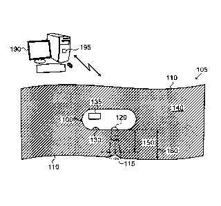
Fig. 1 A is a schematic cross sectional side view of an imaging capsule 100
deployed in a patient's colon 105, and Fig. 1B is a schematic cross sectional
view of an
imaging capsule 100 deployed in a patient's colon 105, according to an
exemplary
embodiment of the disclosure. In an exemplary embodiment of the disclosure,
the patient
first drinks a contrast agent 140 that mixes with the colon contents. The
contrast agent
140 assists in enabling the imaging capsule 100 to perform measurements and
form a 3-
dimensional image of colon 105 from the inside. Optionally, the contrast agent
140
includes water mixed with a radio opaque material with a relatively high
atomic number
such as, for example, Barium (atomic number 56) or Iodine (atomic number 53).
After
drinking the contrast agent 140 the patient swallows imaging capsule 100.
Imaging
capsule 100 travels through the patient's GI tract and through the colon until
it exits in
the patient's feces.
In an exemplary embodiment of the disclosure, imaging capsule 100 includes
a radiation emitter 120 and a radiation detector 130. In some aspects, the
radiation
emitter 120 provides a collimated radiation beam that emits radiation while
rotating 360
degrees inside imaging capsule 100 to scan the entire inner circumference of
the colon
walls 110 as the imaging capsule progresses through the colon. In an exemplary
embodiment of the disclosure, radiation detector 130 rotates with radiation
emitter 120
to detect the photons that are returned from interactions with the emitted
radiation. In
some aspects, radiation detector 130 may include detectors surrounding the
outer
circumference of imaging capsule 100 to detect radiation from all sides of
imaging
capsule 100. In some aspects, radiation detector 130 may be a solid state
detector, for
example a Cadmium Telluride (CdT1) compound serving as a detector. In an
exemplary
embodiment of the disclosure, imaging capsule 100 emits X-ray radiation and
measures
photons returned by two physical phenomenon causing interactions with the
radiation. In
an exemplary embodiment of the disclosure, the two physical phenomenons are
Compton back-scattering and X-ray fluorescence. The measured photons related
to these
phenomenon are used to determine the distance 160 from imaging capsule 100 to
the
surrounding walls 110 of the colon or the distance 150 to polyps 115 extending
from the
inner walls 110 of the colon 105.
In an exemplary embodiment of the disclosure, imaging capsule 100 includes
a transmitter 135 (e.g. an RF transmitter) to transmit the measurements to an
external
-5-
CA 02811675 2013-03-19
WO 2012/038960 PCT/1L2011/000749
processing device 190 for processing. In an exemplary embodiment of the
disclosure,
processing device 190 is a general purpose computer with an executable program
195
that accepts the measurements from the imaging capsule 100. Optionally,
program 195
determines the distances (e.g. 150 and 160) inside colon 105 and constructs a
3
dimensional image of the colon for a medical practitioner to view. Optionally,
the
processing device 190 also determines the width 170 and height (160-150) of
polyps
extending from the colon walls 110. In an exemplary embodiment of the
disclosure,
imaging capsule 100 travels in the longitudinal direction through the colon.
The imaging
capsule 100 may be off center sometimes during the journey. In an exemplary
embodiment of the disclosure, program 195 compensates for deviations from the
center
by using the measurements that are performed on the entire circumference
inside the
colon and adjusting the results if necessary.
In some embodiments of the disclosure, imaging capsule 100 may include an
internal processing device and transmit 3-dimensional images directly to an
external
viewing device for the medical practitioner to view.
In an exemplary embodiment of the disclosure, the radiation emitter emits X-
ray radiation, for example between 10 to 100 KeV (e.g. 59.4 KeV). Optionally,
the X-
ray photons interact with the contrast agent, the contents of the colon and
the tissue of
the colon walls 110. The interactions cause the return of photons to detector
130 based
on two physical phenomenons:
1. Compton back-scattering (CMT) ¨ The X-ray photons emitted from
imaging capsule 100 collide with the electrons of the colon content and the
tissue of the
colon walls 110 and provide back-scattered photons of specific energies, which
are
detected by detector 130. Additionally, the backscattered photons are
attenuated by the
distance traveled. The larger the distance that the back-scattered photons
travel through
the contrast agent 140 the less the number of back-scattered photons that will
be detected
since the contrast agent enhances absorption of the photons. When a polyp 115
exists on
the colon wall 110 the distance is shorter, less contrast agent absorbs the
photons and
more will be detected by detector 130.
2. X-ray Florescence (XRF) ¨ The X-ray photons emitted from the imaging
capsule interact with the atoms of the contrast agent and the rest of the
contents of the
colon 105. The interactions cause ionization, which yields a florescent photon
flux with
specific energy levels from the heavy atoms in the contrast agent such as
Iodine or
-6-
CA 02811675 2013-03-19
WO 2012/038960 PCT/1L2011/000749
Barium. Additionally, the larger the distance from imaging capsule 100 the
more X-ray
florescence will be detected and the shorter the distance the less X-ray
florescence will
be detected.
The photon energy (KeV) for the photons released by each of the two
physical phenomenons is different so the results from each phenomenon can be
analyzed
independently. Fig. 2 is a schematic illustration of a graph 200 of a count of
detected
photons, according to an exemplary embodiment of the disclosure. In a typical
case the
X-ray florescence forms the two highest peaks on the right side of the graph
(lower
energies) and the Compton back-scattering forms the highest peak on the left
side of the
graph (higher energies). The energies of the peaks are generally known since
they
depend mainly on the energy of the emitted radiation, the compounds in the
contrast
agent and the geometry between the radiation emitted and the detector's
position relative
to the emitter.
Fig. 3 is a schematic illustration of images 300 of a colon, according to an
exemplary embodiment of the disclosure. Image 310 shows a computer
reconstructed
cross sectional perspective view of the inside of colon 105 with a polyp 115
on the
bottom surface. Image 310 is reconstructed based on the measurements of
imaging
capsule 100. Image 320 shows a longitudinal side view of the inside of the
colon 105
with polyp 115 and image 330 shows a cross sectional view of the colon at the
position
of the polyp 115.
Following are details of an experiment 400 conducted to demonstrate the
connection between the distances (150, 160 and 170) and the results measured
from
Compton back-scattering and X-ray florescence as described above. Fig. 4 is a
schematic
illustration of the setup of experiment 400 to demonstrate the calculation of
distances in
the colon 105, according to an exemplary embodiment of the disclosure. In an
exemplary
embodiment of the disclosure, a tank 410 of water mixed with a contrast agent
430 is
used to demonstrate colon 105. A slab 420 of plastic with the same density as
water is
used to demonstrate the colon tissue and the tissues beyond. A collimated
radiation
source 440 emitting X-ray radiation at 59.4 keV (e.g. using an Am241 radiation
source)
is used to provide X-ray radiation. A solid state (CdT1) radiation detector
450 counts
photons that are released responsive to the X-ray radiation. The measurements
are
provided to a transmitter 460 that transmits the measurements wirelessly to
processing
device 190, such as, for example, a computer that executes program 195.
-7-
CA 02811675 2013-03-19
WO 2012/038960 PCT/1L2011/000749
In an exemplary embodiment of the disclosure, slab 420 was positioned at
various distances (e.g. 0-30mm) relative to the radiation source 440 to see
the effect on
the measurements. Additionally, the measurements were repeated for various
concentrations of contrast agent 430, for example 1% - 8%. The graph in Fig. 2
shows a
typical spectrum with two areas:
1. Area 210 representing the results from X-ray florescence with 2 peaks, for
example one large and one smaller between 30 KeV and 35 KeV, and
2. Area 220 representing the results from Compton back-scattering with a
peak, for example between 40-45 KeV.
The results of area 210 and area 220 for various distances and contrast agent
concentrations were integrated and provided in graphical form. Fig. 5 is a
schematic
illustration of a graph 500 depicting the experimental results showing the
relationship of
the photon count, distance from the radiation source and concentration of the
contrast
agent, according to an exemplary embodiment of the disclosure. The lower lines
correspond to X-ray florescence and the upper lines correspond to Compton back-
scattering. Each line represents a different concentration percentage for
various
distances. As shown in graph 500 the more concentrated the contrast agent the
greater
the count for X-ray florescence and the lower the count for Compton back-
scattering.
Likewise the greater the distance from the radiation source the greater the
count for X-
ray florescence and the lower the count for Compton back-scattering.
In an exemplary embodiment of the disclosure, program 195 is required to
determine the distance L as a function of the counts (I) of the X-ray
florescence and
Compton back-scattering (i.e. L=L(Iafr IxRF)).
Fig. 6A is a schematic illustration of a graph 600 depicting a surface
representing the distance (L) as a function of the count (I) and contrast
agent
concentration (Ro) for X-Ray florescence, and Fig. 6B is a schematic
illustration of a
graph 650 depicting a surface representing the distance (L) as a function of
the count (I)
and contrast agent concentration (Ro) for Compton back-scattering, according
to an
exemplary embodiment of the disclosure.
In an exemplary embodiment of the disclosure, for specific count values
(IcNIT , IxRF) at a specific moment (when the imaging capsule is at a specific
position) a
set of 2 functions can be obtained from the surfaces in graphs 600 and 650
providing an
-8-
CA 02811675 2013-03-19
WO 2012/038960 PCT/1L2011/000749
estimated distance (LEST) as a function of the concentration 430 (a line on
the surface
representing a specific concentration):
LEST= LCmT(RO, IcmT=constant); and
LEST= LxRF (Ro, IxRF=constant).
Optionally, program 195 finds the intersection point of the 2 curves yielding
the estimated distance LEST and the concentration (Ro). Fig. 7 is a schematic
illustration
of a graph 700 depicting an estimation of the distance LEST and concentration
(Ro) for a
specific photon count, according to an exemplary embodiment of the disclosure.
In an exemplary embodiment of the disclosure, during live application of
imaging capsule 100 through a patient's colon 105, various disturbances may
hinder the
calculations described above and disturb the smoothness of the results, for
example the
concentration of the contrast agent varies throughout the colon 105.
Additionally, the
concentration is lower at the beginning and increases toward the exit from the
colon due
to absorption of water from the colon leaving the molecules of the contrast
agent at a
higher concentration. In order to overcome disturbances the following method
and
assumptions are used:
1. The contrast agent concentration (Ro) is assumed to change gently along
the colon tract.
2. The results of the concentration will be calculated based on the estimation
calculations used above.
3. The concentration for a sequence of positions will be filtered by a
regression to provide a smooth function.
4. The smoothed concentration function will be used to estimate the distance
160 either using the Compton back-scattering curve or the X-ray florescence
curve (as
shown in Fig. 7):
LEsT= LcmT(ROsmooth, IcmT=constant) or LEsT=LxRF (Rosmooth, IxRF=constant).
In an exemplary embodiment of the disclosure, the performance of the
estimation calculation is evaluated by comparing the estimated distance (LEST)
to the real
(LREAL) distance in the experiment described above. Figures 8A, 8B and 8C are
schematic graphs that demonstrate the relationship between the estimated
distance and
the real distance as a function of the concentration (Ro). The figures show
two dotted
outer lines showing the boundaries of the results based on the measurements
and two
inner lines one showing the standard deviation of the measured results and one
showing
-9-
CA 02811675 2013-03-19
WO 2012/038960 PCT/1L2011/000749
the mean of the measured results. Fig. 8A shows the relationship for Ro=8%,
Fig. 8B
shows the relationship for Ro=6% and Fig. 8C shows the relationship for Ro=4%.
The
results of the graph show that good results can be obtained for distances up
to 20mm
with a concentration of 8% and larger distances for lower concentration.
Typically
imaging capsule 100 will travel along the longitudinal direction, which has a
typical
diameter of 30-40 mm and a maximum of up to about 50mm. However it should be
noted that during movement, the colon typically contracts to less than 50% of
its normal
diameter leaving a short distance between the colon wall 110 and imaging
capsule 100 in
the order of 5 ¨ 15 mm at the most.
In an exemplary embodiment of the disclosure, after calculating the distance
from imaging capsule 100 to the colon walls 110 other measurements may be
calculated
based on the results. In an exemplary embodiment of the disclosure, the width
(D) 170
(Fig. 1B) of a polyp 115 can be estimated by calculating an angle (A) 180
enclosing the
polyp 115, for example the angle between two scanning positions during
rotation of the
radiation source where the length is larger than the length over width D
because of the
polyp 115 or that the length is substantially the same as the rest of the
circumference
except over width D. Additionally, geometric calculations can be used to
determine the
width of polyp 115, for example by calculating D=2*L*Tan(Al2).
It should be appreciated that the above described methods and apparatus may
be varied in many ways, including omitting or adding steps, changing the order
of steps
and the type of devices used. It should be appreciated that different features
may be
combined in different ways. In particular, not all the features shown above in
a particular
embodiment are necessary in every embodiment of the disclosure. Further
combinations
of the above features are also considered to be within the scope of some
embodiments of
the disclosure.
It will be appreciated by persons skilled in the art that the present
disclosure
is not limited to what has been particularly shown and described hereinabove.
-10-