Note: Descriptions are shown in the official language in which they were submitted.
CA 02811888 2013-03-20
WO 2011/034620 PCT/US2010/002568
1
INTEGRATED CARTRIDGE
RELEVANT APPLICATIONS
This application claims the priority of U.S. Provisional Application No.
61/272,397, filed on September 21, 2009, which is incorporated herein by
reference in its
entirety.
TECHNICAL FIELD
The technical field is biotechnology and, more specifically, methods and
apparatus
for analysis of biomolecules.
BACKGROUND
It is desirable to have an analytical instrument that possesses both sample
preparation and sample analysis functions. It is also desirable to have an
analytical
instrument that is light and small and can be produced at a low cost.
However,
microfluidic challenges have impeded the development of such analytical
devices. These
challenges are due, in part, to the fluid dynamics at small scales. For
example, as the
diameter of a microfluidic channel decreases, the pressure drop across the
channel
increases by the 4th power, according to the Hagen¨Poiseuille equation. When
employing
complex microfluidic geometries, these large pressure drops can result in flow
patterns
that are very difficult to predict, particularly with air bubbles in the
system. The thermal
expansion of air is more than five times greater than liquid, causing
additional challenges.
SUMMARY
An integrated cartridge for sample processing and analysis is disclosed. The
integrated cartridge contains a sample preparation chamber having a sample
inlet and a
sample outlet, and a sample purification chamber adapted to receive a
replaceable sample
purification unit containing a housing and an extraction filter inside the
housing. The
extraction filter specifically binds to a molecule of interest. The sample
purification
chamber has a sample inlet that is in fluid communication with the sample
outlet of the
sample preparation chamber.
Also disclosed is a microarray-based sample analysis (MBSA) system. The
MBSA system includes a cartridge holder adapted to receive a detachable
cartridge that is
configured to receive a detachable, replaceable sample analysis unit having a
reaction
CA 02811888 2013-03-20
WO 2011/034620 PCT/US2010/002568
2
chamber and a microarray, a fluid control subsystem that controls fluid flow,
and an
optical subsystem configured to capture an image of the microarray.
BRIEF DESCRIPTION OF DRAWINGS
The detailed description will refer to the following drawings in which:
Figures lA and 1B are schematic drawings showing embodiments of the integrated
cartridge.
Figure 2 is a schematic drawing showing an integrated cartridge with cell
lysis
means.
Figure 3A-3C are schematics showing different embodiments of the protein
purification components of a dual-function integrated cartridge.
Figure 4 is a block diagram of a microarray-based sample analysis (MBSA)
system.
Figure 5 is a schematic of the fluidic subsystem showing pumps and selection
valves (SV) connected to the integrated cartridge.
Figure 6A is a schematic showing an integrated cartridge residing in a fluidic
manifold without the thermocycler bladders.
Figure 6B is a schematic showing an integrated cartridge residing in a fluidic
manifold with the thermocycler bladders.
Figure 7 is a schematic showing the location of the on-board microfluidic
cartridge
valves in an integrated cartridge.
Figure 8 is a schematic showing an embodiment of an on-board microfluidic
cartridge valve.
Figure 9 is a schematic showing an embodiment of the chamber arrangement
within a flow cell.
Figure 10 is a diagram showing PCR/APEX allele signal ratio results obtained
for
an eye color SNP at position RS1800407. PCR was performed in a Akonni flow
cell
positioned in a bladder thermocycler, in 0.2 ml PCR tube positioned in a MJ
thermal
cycler, and in Cepheid tube positioned in the bladder thermocycler. APEX was
performed
offline for one hour. The results indicate comparable APEX signals for all
three PCR
approaches.
Figure 11A is a schematic of an embodiment of the optical subsystem with a
laser
light source.
CA 02811888 2013-03-20
WO 2011/034620 PCT/US2010/002568
3
Figure 11B shows a typical image of a Cy3 array with a pitch of 300 gm taken
with the optical subsystem described in Figure 1 1A with the co-axial
illumination.
Figure 12A schematic of another embodiment of the optical subsystem with a
laser
light source.
Figure 12B shows a typical image of am 11x18 Cy3 array taken with the optical
subsystem of Figure 12A.
Figure 13A is a schematic showing an embodiment of the optical subsystem with
a
high-brightness LED.
Figures 13B is a schematic showing an optical train for fluorescence and FII
imaging.
Figures 13C is a schematic showing another optical train for fluorescence and
FII
imaging.
Figures 13D is a schematic showing a collimated source used in Figures 13B and
13C.
Figure 14A is an FII image of a gel array.
Figure 14B is a diagram showing the normalized pixel intensity profile of an
array
element in Figure 14A.
Figure 15 is a composite of images showing a gel array image processed by
different methods available in the ImageJ software. Panel A, original image;
Panel B,
ImageJ processing: Process => Find Edges; Panel C, ImageJ processing: Process
=>
Filters => Median, R=5; Panel D, ImageJ processing: Process => Filters =>
Mean, R=5;
Panel E, ImageJ processing: Image => Adjust Threshold; Panel F, ImageJ
processing:
Process => Filters => Maximum.... R = 2; Panel G, ImageJ processing: Process
=> Binary
=> Find Maxima; Panel H, Superposition of the images showing respectively spot
centers
found and spot boundaries detected.
Figure 16 is a composite showing evaluation of spot morphology. Panel A: image
obtained in the fluorescence imaging mode; Panel B: FII image of the same
array obtained
using oblique illumination with collimated beam; Panel C: Image processing in
ImageJ
using Process => Find Edges; Panel D: Image processing in ImageJ using Process
=>
Filters => Median, R=2; Image => Adjust threshold.
Figures 17A and 17B are graphs showing evaluation of morphology for the gel
spots in panel A of Figure 15
CA 02811888 2013-03-20
WO 2011/034620 PCT/US2010/002568
4
Figure 18A is picture showing detection of S. pyogenes with a prototype MBSA
system and an Aurora imager.
Figure 18B is picture showing detection of S. pyogenes with a prototype MBSA
system and an Akonni imager.
Figure 19A is a picture showing detection of B. anthracis with a prototype
MBSA
system and the imaging approach shown in Figure 11A.
Figure 19B is an array map of the microarray used in Figure 19A.
Figure 20 is diagram showing PCR results of S. pyogenes DNA extracted using an
integrated cartridge with a bead mixer. Mixer 1 and mixer 2: S. pyogenes DNA
extracted
in two separated experiment using an integrated cartridge with a bead mixer.
Vortex: S.
pyogenes DNA extracted using standard vortexing method. NTC: negative control.
Figure 21A shows the microarray results for 6-plex PCR products. The upper
left
panel is a microarray image with arrows pointing to the six PCR product
generated in the
multiplex PCR. The upper right panel is the microarray map. The lower panel
shows
probe number/primer identifier/fluorescent signal intensities for PCR product
generated
under three different annealing temperatures.
Figure 21B is a diagram showing real-time PCR analysis of cartridge-purified
blood DNA.
Figure 22 shows thermocycling profile with PID pump and heater control. Line A
shows the temperature of the hot zone, line B shows the cold zone, line C
shows the
temperature of a thermocouple sandwiched between the bladders, and line D
shows the
temperature of the working fluid just prior to entering the bladder.
Figure 23 is a drawing showing an embodiment of a dual-function integrated
cartridge.
Figure 24 is a schematic showing an embodiment of a complete MBSA system.
DETAILED DESCRIPTION
This description is intended to be read in connection with the accompanying
drawings, which are to be considered part of the entire written description of
this invention.
The drawing figures are not necessarily to scale and certain features of the
invention may
be shown exaggerated in scale or in somewhat schematic form in the interest of
clarity and
conciseness. In the description, relative terms such as "front," "back," "up,"
"down," "top"
and "bottom," as well as derivatives thereof, should be construed to refer to
the orientation
as then described or as shown in the drawing figure under discussion. These
relative terms
CA 02811888 2013-03-20
WO 2011/034620 PCT/US2010/002568
are for convenience of description and normally are not intended to require a
particular
orientation. Terms concerning attachments, coupling and the like, such as
"connected" and
"attached," refer to a relationship wherein structures are secured or attached
to one another
either directly or indirectly through intervening structures, as well as both
movable or rigid
5 attachments or relationships, unless expressly described otherwise.
In describing preferred embodiments of the present invention, specific
terminology
is employed for the sake of clarity. However, the invention is not intended to
be limited to
the specific terminology so selected. It is to be understood that each
specific element
includes all technical equivalents which operate in a similar manner to
accomplish a
similar purpose.
An integrated cartridge for sample processing and analysis is disclosed. The
integrated cartridge includes a sample preparation chamber, a sample
purification chamber
and a detachable sample analysis unit. The sample preparation chamber has a
sample inlet
and sample outlet and is in fluid communication with the sample purification
chamber.
The sample purification chamber contains an extraction filter that
specifically binds to a
molecule of interest. The detachable sample analysis unit includes at least
one sample
analysis chamber that contains a microarray.
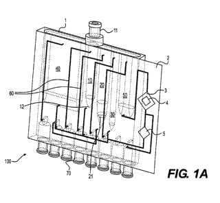
Figure 1A shows an embodiment of an integrated cartridge. The integrated
cartridge allows for the extraction of biomolecules, such as polypeptides and
polynucleotides, and subsequent analysis of the biomolecules within the same
cartridge.
In this embodiment, the integrated cartridge 100 contains a cartridge body 1
and a sample
analysis unit 2. The cartridge body 1 contains a sample preparation chamber
10, a sample
purification chamber 20 in fluid communication with the sample preparation
chamber 10,
an sample elution chamber 30 in fluid communication with the sample
purification
chamber 20, a waste chamber 40 in fluid communication with the sample
purification
chamber 20, a product waste chamber 50 in fluid communication with the sample
analysis
unit 2 when it is attached to the cartridge body 1, fluidic channels 60 that
connect the
chambers to each other, and a fluidic interface 70 that allows the integrated
cartridge 100
to be connected to a cartridge base (not shown). In one embodiment, the sample
analysis
unit 2 is an integrated part of the cartridge body 1. In another embodiment,
the sample
analysis unit 2 is detachable from the cartridge body 1. Figure 1B is a
schematic drawing
CA 02811888 2013-03-20
WO 2011/034620 PCT/US2010/002568
6
showing the same cartridge 100 with on-board valves 80 that control fluid flow
in each
chamber.
The sample preparation chamber 10 has a sample inlet 11 at an upper portion of
the
chamber and a sample outlet 12 at a lower portion of the chamber. In the
embodiments
shown in Figures 1A and 1B, the sample inlet 11 is located at the top of the
sample
preparation chamber 10 and the sample outlet 12 is located at the bottom of
the sample
preparation chamber 10. In one embodiment, the sample inlet 11 is a dome
valve. When
connected to a pump, the sample outlet 12 may also serve as an air inlet.
Sample mixing
inside the sample preparation chamber may be achieved by pumping a gas, such
as air or
an inert gas (e.g., nitrogen), into the sample preparation chamber 10 from the
sample
outlet 12. The gas forms bubbles that migrate from the bottom to the top of
the chamber
10 and mix the sample during the process.
In another embodiment, the gas is heated to provide a means of uniformly
heating
the sample/reagent mixture in the sample preparation chamber 10.
The sample purification chamber 20 contains an extraction filter 21 that binds
specifically to an analyte. Examples of analytes include, but are not limited
to,
polynucleotides, polypeptides, lipids, and polysaccharides. In one embodiment,
the
extraction filter is a silica extraction filter that specifically binds to
DNA. In another
embodiment the extraction filter is a porous material that specifically binds
to a protein.
In one embodiment, the extraction filter 21 is located in a removable sample
purification
unit 22 that can be easily detached from the cartridge body 1 and replaced
with a new
sample purification unit 22. The sample purification unit 22 comprises a
housing with an
inlet and an outlet, and an extraction filter 21 that specifically binds to an
analyte. In one
embodiment, the sample purification unit 22 is in the form of a pipet tip. In
a preferred
embodiment, the pipet tip is sized to fit within the confines of the sample
purification
chamber 20. In another preferred embodiment, the extraction filter 21 is the
glass frit
described in US Patent Application Serial Nos. 11/933,113 and 12/213,942, both
of which
are herein incorporated by reference in their entirety.
The elution chamber 30 is connected to both the sample purification chamber 20
and sample analysis unit 2 when it is attached to the cartridge body 1. In
certain
embodiments, the elution chamber 30 is eliminated from the integrated
cartridge and the
sample purification chamber 20 is connected directly to the sample analysis
unit 2.
CA 02811888 2013-03-20
WO 2011/034620 PCT/US2010/002568
7
The integrated cartridge 100 is designed to operate in the upright position as
shown
in Figures IA and 1B. The sample preparation chamber and sample purification
chamber
are are designed to have a diameter that is sufficient to allow bubbles to
rise to the top.
The sample analysis unit 2 may contain one or more chambers for sample
analysis.
In one embodiment, the sample analysis unit 2 contains a microarray chamber 3
that
contains a microarray 4 for the analysis of the analyte. The microarray 4 can
be a
microarray of any type. In one embodiment, the microarray 4 is a DNA array. In
another
embodiment, the microarray 4 is a protein or peptide array. In another
embodiment, the
microarray is a gel element array described in e.g., US Patent Nos. 5,741,700,
5,770,721,
5,981,734, and 6,656,725, and US Patent Application Nos. 10/068,474,
11/425,667 and
60/793,176, which are hereby incorporated by reference in their entirety. In
yet another
embodiment, the microarray 4 is an antibody array.
In one embodiment, the microarray chamber 3 also serves as a reaction chamber
for an amplification reaction, such as polymerase chain reaction (PCR) or
arrayed-primer
extension (APEX).
In another embodiment, the sample analysis unit 2 further contains an
amplification chamber 5. In one embodiment, the amplification chamber 5 is a
PCR
chamber that can be heated and cooled repetitively to amplify a DNA target
inside the
amplification chamber 5.
The sample analysis unit 2 and/or the cartridge body 1, are made from
materials
capable of withstanding thermocycling, and immune to solvents such as ethanol.
The
cartridge body can be machined or injection molded.
In one embodiment, the microarray chamber 3 and/or the amplification chamber 5
have a hydrophilic interior surface. In a preferred embodiment, the microarray
chamber 3
and/or the amplification chamber 5 are hydrophilic flow cells described in US
Patent
Application Serial No. 12/149,865, which is incorporated herein by reference
in its
entirety.
During operation, the cartridge 100 is inserted into a fluidic docking
station. In
one embodiment, fluidic docking station engages with the cartridge 100 through
luer
tapered bosses that activate luer-activated valves on the cartridge. A sample,
such as a cell
suspension, is loaded into the sample preparation chamber 10 of the integrated
cartridge
100 through the sample inlet 11. A cell lysis buffer is added to the sample
preparation
CA 02811888 2013-03-20
WO 2011/034620 PCT/US2010/002568
8
chamber 10, and mixed with the sample by air bubbles that migrate from the
bottom to the
top of the sample preparation chamber 10. The air bubbles are controlled by
regulated air
flow from an air pump. This air may be heated for protocols that require
incubation at an
elevated temperature. The mixed sample is then introduced into the sample
purification
chamber 20, which contains the extraction filter 21 that specifically binds to
the analyte of
interest. The sample passes through the extraction filter 21 to allow the
analyte of interest
to bind to the extraction filter 21. In certain embodiments, the sample is
cycled back and
forth across the extraction filter 21 a number of times to improve efficiency
of analyte
binding. The unbound sample is then directed to the waste chamber 40 through
on-board
cartridge pin valves 80 and fluidic channels 60. Leaving the waste in the
cartridge
prevents contamination with subsequent samples.
The extraction filter 21 is washed with a wash buffer one or more times. Used
wash buffer is directed to the waste chamber 40 through on-board cartridge
valves 80 and
fluidic channels 60. In one embodiment, the wash buffer is an ethanol-based
wash buffer
and is cycled 5 times through the extraction filter 21. An elution buffer is
then introduced
into the sample purification chamber 20 through the extraction filter 21. The
eluted
analyte is directed into the elution chamber 30, which removes the bubbles in
the eluant.
The eluted analyte is then used for the subsequent sample analysis in the
sample analysis
unit. Aliquots of the eluted analyte may also be removed for other types of
analysis. In
one embodiment, an eluted DNA sample is directed into the amplification
chamber 5 for
PCR amplification. Temperature change in the amplification chamber 5 may be
achieved
by oscillating two temperature fluids through flexible bladders that makes
intimate contact
with the amplification chamber 5, as described, for example, in described in
US Patent
Application Serial Nos 11/843,843 and 12/232,669, which are incorporated
herein by
reference in their entirety. The on-board cartridge valves 80 switch between
thermally-
controlled reservoirs to provide a rapid change in temperature. The
amplification product
is directed into the microarray chamber for hybridization to the microarray.
Integrated cartridge with cell lysis beads
In certain embodiments, the sample preparation chamber 10 further includes
sample lysis means for lysing cell samples. In an embodiment shown in Figure
2, the
sample preparation chamber 10 contains a plurality of cell lysis beads 13 and
a magnetic
stirring element 14 with a magnet 15. A suspension of intact cells is added to
the sample
CA 02811888 2013-03-20
WO 2011/034620 PCT/US2010/002568
9
preparation chamber 10 and a rotating magnetic field is applied to the sample
preparation
chamber 10 to rotate the magnetic stirring element 14 at a rotation speed
sufficient to lyse
the cells.
As used herein, the term "cell" refers to eukaryotic cells, prokaryotic cells,
and
components or fragments thereof. The term "cell" includes parasites, bacteria,
bacteria
spores, fungi, virus particles, as well as an aggregation of cells such as
multi-cell
organisms, tissues and fragments thereof. The term "cell suspension" refers to
a mixture
of cells and a liquid medium, wherein the cells are suspended in the liquid
medium.
Preferably, the cells are suspended at a concentration that is not too thick
or viscous to
interfere with the movement of the magnetic stir element.
In a certain embodiment, eukaryotic or prokaryotic cells are suspended in the
concentration range of 1x10² to 1x10⁵ cells/ml. In another embodiment,
eukaryotic or
prokaryotic cells are suspended in the concentration range of 1x10³ to 1x10⁴
cells/ml. In
other embodiment, virus particles are suspended in the concentration range of
1x10⁷ to
1x10'3 particles/ml. In yet other embodiment, virus particles are suspended in
the
concentration range of 1x10⁹ to 1x10" particles/ml.
The liquid medium can be isotonic, hypotonic, or hypertonic. In some
embodiments, the liquid medium is aqueous. In certain embodiments, the liquid
medium
contains a buffer and/or at least one salt or a combination of salts. In some
embodiments,
the pH of the liquid medium ranges from about 5 to about 8, from about 6 to 8,
or from
about 6.5 to about 8.5. A variety of pH buffers may be used to achieve the
desired pH.
Suitable buffers include, but are not limited to, Tris, MES, Bis-Tris, ADA,
ACES, PIPES,
MOPSO, Bis-Tris propane, BES, MOPS, TES, HEPES, DIPSO, MOBS, TAPSO,
HEPPSO, POPSO, TEA, HEPPS, Tricine, Gly-Gly, Bicine, and a phosphate buffer
(e.g.,
sodium phosphate or sodium-potassium phosphate, among others). The liquid
medium
may comprise from about 10 mM to about 100 mM buffer, about 25 mM to about 75
mM
buffer, or from about 40 mM to about 60 mM buffer, among others. The type and
amount
of the buffer used in the liquid medium can vary from application to
application. In some
embodiments, the liquid medium has a pH of about 7.4, which can be achieved
using
about 50 mM Tris buffer. In some embodiments the liquid medium is water.
The cell lysis beads can be any particle-like or bead-like material that has a
hardness greater than the hardness of the cells to be lysed. The cell lysis
beads may be
CA 02811888 2013-03-20
WO 2011/034620 PCT/US2010/002568
made of plastic, glass, ceramics, or any other non-magnetic materials, such as
non-
magnetic metal beads. In one embodiment, the cell lysis beads are rotationally
symmetric
to one axis (e.g., spherical, rounded, oval, elliptic, egg-shaped, and droplet-
shaped
particles). In other embodiments, the cell lysis beads have polyhedron shapes.
In other
5
embodiments, the cell lysis beads are irregular shaped particles. In yet other
embodiments,
the cell lysis beads are particles with protrusions.
In one embodiment, the cell lysis beads have diameters in the range of 10-
1,000
gm. In other embodiments, the cell lysis beads have diameters in the range of
20-400 p.m.
In yet other embodiments, the cell lysis beads have diameters in the range of
50-200 pm.
10 The
magnetic stir element can be of any shape and has dimensions that match the
container. In other words, the magnetic stir element should be small enough to
be placed
\
into the container and to spin or stir within the container. It is within the
knowledge of a
person of ordinary skill in the art to choose a magnetic stir element of
appropriate sizes for
a given container. The magnetic stir element can be a bar-shaped, cylinder-
shaped, rod-
shaped, cross-shaped, V-shaped, triangular, rectangular or disc-shaped stir
element. In
one embodiment, the magnetic stirring element has a rectangular shape. In
another
embodiment, the magnetic stirrer has a two-pronged tuning fork shape. In yet
another
embodiment, the magnetic stirrer has a V-like shape. In certain embodiments,
the
magnetic stir element is coated with a chemically inert material, such as
plastics, glass or
porcelain.
In one embodiment, the cell lysis beads 13 and/or the magnetic stirring
element 14
are pre-packed into the sample preparation chamber 10. In another embodiment,
the cell
lysis beads 13 and/or the magnetic stirring element 14 are pre-packed into a
removable
sample lysis unit that can be easily placed into the sample preparation
chamber 10 and
discarded after cell lysis. In yet another embodiment, the cell lysis beads 13
and/or the
magnetic stirring element 14 are added to the sample preparation chamber 10
with the cell
suspension. The cell lysis beads 13, the magnetic stirring element 14 and the
cell
suspension may be placed into the sample preparation chamber 10 in any order.
In one
embodiment, the cell suspension is loaded into the sample preparation chamber
10 first
and followed with either the cell lysis beads or the magnetic stirring
element. In another
embodiment, the hard beads and/or the magnetic stirring element are placed
into the
sample preparation chamber 10 first, followed with the cell suspension.
CA 02811888 2013-03-20
WO 2011/034620 PCT/US2010/002568
11
The cartridge is then placed in close proximity to a magnetic stirrer. The
cell
suspension is stirred with the magnetic stirring element at a rotation speed
sufficient to
lyse the cells inside the container. The optimal stirring speed and duration
can be
empirically determined based on the cells, viruses or tissues to be lysed. The
appropriate
rotation speed is application dependent and can be determined by a person of
ordinary
skill in the art. Generally speaking, the rotational speed sufficient to lyse
the cells is
determined by factors such as the type of cells, the concentration of cell
suspension, the
size and shape of the magnetic stirring element, the amount, size, shape and
hardness of
the hard beads, and the size, shape and interior surface roughness of the
container. In
certain embodiments, the container in the shape of a test tube or Eppendorf
tube is placed
in a rack on a standard laboratory magnetic stirrer plate and is stirred at
the highest speed
setting.
In certain embodiments, lysing of particular cell types can be facilitated by
adding
additives to the cell suspension prior to the stirring step. Examples of
additives include
enzymes, detergents, surfactants and other chemicals such as bases and acids.
It has been
found that alkaline conditions (e.g. 10 mM NaOH) may enhance the lysis
efficiency for
certain types of cells. It should be noted, however, that the additive should
not interfere
with the downstream reactions in the sample analysis process. The cell
suspension may
also be heated during stirring to enhance the lysis efficiency.
Other embodiments of the bead/magnetic stirrer lysis methods and systems are
described in U.S. Provisional Application No. 61/272,396, which is
incorporated herein by
reference in its entirety.
Besides the bead/magnetic stirrer method, cells in the sample preparation
chamber
10 may be lysed with other methods such as bead beating, vortexing, sonication
and
chemical lysis.
To avoid problems commonly associated with microfluidic devices, the
integrated
cartridge may contain one or more of the following features (1) The
microfluidic channels
in the integrated cartridge are sufficiently large (0.2-1 mm, preferably 0.5
mm in diameter)
to reduce backpressure and enable reproducible injection molded features,
which can be
highly variable when creating microfluidic geometries below 0.1 mm. (2) The
interior
surface of the microfluidic channels are fully covered, or partially covered
or coated with a
hydrophilic film to reduce bubble trapping inside the microfluidic channels.
(3) The
CA 02811888 2013-03-20
WO 2011/034620 PCT/US2010/002568
12
reaction chambers (i.e., the sample preparation chamber, the sample
purification chamber
and the sample elution chamber) in the integrated cartridge are designed in a
vertically-
oriented tower shape to ensure that bubbles rise to the top of the chamber due
to the
density difference of air compared with water. The tower design also allows
for sample
mixing in the tower by flowing air into the sample from the bottom of the
tower. The
liquid is vigorously mixed by the air bubbles rising from the bottom to the
top. (5) The
extraction filter in the integrated cartridge has a large porosity to minimize
back pressure.
(6) The on-board cartridge valves in the integrated cartridge shut off fluidic
pathways and
prevent liquids from flowing in unwanted paths. (7) The cartridge is designed
so that the
liquid is controlled by precision pumps and selection valves off the
disposable cartridge.
The liquid metering occurs on volumes greater than 10 L, to prevent small air
bubbles
from causing large variations in reagent proportions.
Examples of the hydrophilic material that can be used for the hydrophilic
cover or
coating include, but are not limited to, hydrophilic polymers such as poly(N-
vinyl lactams),
poly(vinylpyrrolidone), poly(ethylene oxide), poly(propylene oxide),
polyacrylamides,
cellulosics, methyl cellulose, polyanhydrides, polyacrylic acids, polyvinyl
alcohols,
polyvinyl ethers, allcylphenol ethoxylates, complex polyol mono-esters,
polyoxyethylene
esters of oleic acid, polyoxyethylene sorbitan esters of oleic acid, and
sorbitan esters of
fatty acids; inorganic hydrophilic materials such as inorganic oxide, gold,
zeolite, and
diamond-like carbon; and surfactants such as Triton X-100, Tween, Sodium
dodecyl
sulfate (SDS), ammonium lauryl sulfate, alkyl sulfate salts, sodium lauryl
ether sulfate
(SLES), alkyl benzene sulfonate, soaps, fatty acid salts, cetyl
trimethylammonium
bromide (CTAB) a.k.a. hexadecyl trimethyl ammonium bromide,
allcyltrimethylammonium salts, cetylpyridinium chloride (CPC), polyethoxylated
tallow
amine (POEA), benzalkonium chloride (BAC), benzethonium chloride (BZT),
dodecyl
betaine, dodecyl dimethylamine oxide, cocamidopropyl betaine, coco ampho
glycinate
alkyl poly(ethylene oxide), copolymers of poly(ethylene oxide) and
poly(propylene oxide)
(commercially called Poloxamers or Poloxamines), alkyl polyglucosides, fatty
alcohols,
cocamide MEA, cocamide DEA, cocamide TEA. Surfactants can be mixed with
reaction
polymers such as polyurethanes and epoxies to serve as a hydrophilic coating.
Simplified integrated cartridge
CA 02811888 2013-03-20
WO 2011/034620 PCT/US2010/002568
13
Also disclosed is a simplified cartridge for sample processing. The cartridge
includes a sample preparation chamber having an inlet and an outlet and a
sample
purification chamber having an inlet and an outlet. The sample preparation
chamber
contains a magnetic stir element and a plurality of cell lysis beads disposed
therein. The
sample purification chamber contains an extraction filter and is in fluidic
communication
with the sample preparation chamber.
In one embodiment, the magnetic stir element and the plurality of cell lysis
beads
are pre-packed in a container that can be easily inserted or removed from the
sample
preparation chamber and the extraction filter is pre-packed in a container
that can be easily
inserted or removed from the sample purification chamber.
Dual-function integrated cartridge
Also disclosed is a dual-function cartridge that contains both protein and
nucleic
acid purification capabilities. Briefly, the dual-function cartridge contains
a nucleic acid
purification module and a protein purification module. Each module contains a
sample
purification chamber and a sample elution chamber. In one embodiment, the
modules
share a single sample preparation chamber and/or a single waste chamber. In
another
embodiment, the modules share a single sample preparation chamber, but each
module
contains its own waste chamber. In yet another embodiment, each module has its
own
sample preparation chamber and waste chamber. Appropriate cartridge base setup
can be
constructed to allow simultaneous purification of nucleic acid and protein or
serial
purification of nucleic acid and protein.
In one embodiment, a sample is processed in a serial fashion. As shown in
Figure
3A, the sample is processed for nucleic acid purification first. The nucleic
acid-deprived
sample in the nucleic waste chamber is aspirated into a second purification
chamber (e.g.,
a TruTip) for protein purification and is then dispensed into a second waste
chamber. The
reagents are driven by a bi-directional pump such as the milliGAT pump.
Protein washing
buffer may be added directly to the second purification chamber from the
buffer reservoir
(not shown).
In another embodiment, the integrated cartridge is modified so that the
cartridge
valve on the second purification chamber may serve as a syringe-type pump. As
shown in
Figure 3B, only a single waste chamber is needed. Specifically, the cartridge
is modified
to have a reservoir pocket connected to the cartridge valve on the second
purification
CA 02811888 2013-03-20
WO 2011/034620 PCT/US2010/002568
14
chamber (shown as the "syringe pump" in Figure 3B), so that liquid in the
nucleic acid
waste chamber may fill the reservoir pocket (similar to filling the barrel of
a syringe)
during the "withdraw" state of the "syringe pump", and flow into the second
purification
chamber during the "dispense" state of the "syringe pump." The size of the
reservoir
pocket may vary depending on the size and function of the integrated
cartridge. In certain
embodiments, the reservoir has a volume of 0.01-10 ml, 0.2-5 ml, 0.5-3 ml, or
1-5 ml.
The shape and exact location of the reservoir may be adjusted based on the
overall design
of the integrated cartridge. A check valve is installed between the nucleic
acid waste
chamber and the second purification chamber to ensure unidirectional flow of
the liquid.
The washing step in the second purification chamber can be accomplished by
closing the
cartridge valve, open vent (not shown) in the nucleic acid waste chamber and
continuously
flow the protein washing buffer from the buffer reservoir into the second
purification
chamber through the "syringe pump."
In yet another embodiment, the second purification chamber is attached to a
gear
pump that continuously recirculates solution through the frit in the second
purification
chamber (Figure 3C). The gear pump can operate bi-directionally and no check
valve is
needed between the nucleic acid waste chamber and the second purification
chamber. The
protein washing step may be accomplished by closing the cartridge valve,
opening the
vent in the nucleic acid waste chamber and continuously flowing the protein
wash buffer
from the buffer reservoir into the second purification chamber through the
gear pump.
The microarray-based sample analysis (MBSA) system
Also disclosed is a microarray-based sample analysis (MBSA) system. As shown
in Figure 4, an embodiment of the MBSA system 200 includes a cartridge holder
210, a
fluid control subsystem 220, an optical subsystem 230, control software 240
and,
optionally, a thermocycler 250.
The cartridge holder 210 is configured to be connected to an integrated
cartridge
100 through a fluidic manifold 212. The cartridge holder 210 also holds the
integrated
cartridge 100 in a position to facilitate interaction between various
components of the
integrated cartridge and the subsystems of the MBSA system. For example, a
properly
attached cartridge would position the PCR chamber 5 of the cartridge between
the heating
elements of the thermocycler 250 and allow optical interrogation of the
microarray 4 by
the optical subsystem.
CA 02811888 2013-03-20
WO 2011/034620 PCT/US2010/002568
The fluid control subsystem 220 controls the fluid movement within the MBSA
system 200. The subsystem includes multiple fluid containers, pumps, valves
and tubings.
The fluid control subsystem 220 is directly connected to the integrated
cartridge 100
through the fluidic manifold 212
5 The
optical subsystem 230 is designed to capture images of the microarray 4 of the
sample analysis unit 2 after hybridization to a sample. In certain
embodiments, the optical
subsystem is specifically designed for low-level fluorescence detection on
microarrays. In
one embodiment, the optical subsystem uses confocal or quasi-confocal laser
scanners that
acquire the microarray image pixel by pixel in the process of interrogating
the object plane
10
with a tightly focused laser beam. The laser scanners offer the advantages of
spatially
uniform sensitivity, wide dynamic range, and efficient rejection of the out-of-
focus stray
light. The laser scanners, however, are very delicate and expensive devices.
In another embodiment, the optical subsystem uses imaging devices with flood
illumination, in which all the microarray elements (features) are illuminated
15
simultaneously, and a multi-element light detector, such as a CCD camera,
acquires the
image of microarray either all at once or in a sequence of a few partial
frames that are
subsequently stitched together. Compared to laser scanners, CCD-based imaging
devices
have simpler designs and lower cost. CCD-based imaging systems are an
attractive option
for both stand-alone and built-in readers in cost-sensitive applications
relying on
microarrays of moderate complexity (i.e., having a few hundred or fewer array
elements).
Commercial instruments typically use cooled CCD cameras and employ expensive
custom-designed objective lenses with an enhanced light-collection capability
that helps to
balance, to some extent, the low efficiency of the excitation scheme.
In another embodiment, the optical subsystem contains an imaging device that
uses
a non-cooled CCD camera. Although non-cooled cameras typically have a
noticeably
higher dark current as compared to the cooled models, the optical subsystem
could provide
the required sensitivity without using exposures in excess of a few seconds by
(1)
increasing the excitation intensity, or (2) employing an objective lens with
high light
collection efficiency; or (3) using the above two approaches in combination.
The light
source can be a conventional light source, such as a metal halide or mercury
bulb, a laser-
based system, or a high-intensity LED.
CA 02811888 2013-03-20
WO 2011/034620 PCT/US2010/002568
16
In another embodiment, the optical subsystem has a fluorescence-independent
imaging (FII) mode as a supplementary imaging mode of microarray reader
operation.
The FII mode allows imaging the array elements regardless of their
fluorescence level.
The practical implementation of FII is technically challenging in both
microarray
scanners and imagers using flood illumination. The problem is especially
difficult when
the microarrays to be imaged are the mainstream planar arrays, because the
layer of
biomolecular probes immobilized on the microarray substrate is too thin to
produce a
noticeable change in the intensity of light used for probing the slide
surface.
In one embodiment, the present invention uses dark field illumination in
reflected
light for imaging gel arrays printed on opaque (black) plastic substrates. In
another
embodiment, the present invention uses oblique illumination in transmitted
light for
imaging gel arrays printed on transparent (glass) slides. In both cases, the
light source used
for FII could be any light source emitting within the transmission band of the
reader's
emission filter.
The control software 240 is designed to control all the components of the MBSA
system, including the fluid control subsystem 220, the optical subsystem 230,
the
thermocycler 250 and any additional subsystems. The control software 240 may
be
installed on any computer that is connected to the MBSA system and provides a
user
interface for the MBSA system. In one embodiment, the MBSA system includes a
controller that contains a processor, memory, and a user interface
The thermocycler 250 is configured to provide heating and cooling to the PCR
chamber 5 of the integrated cartridge 100. In one embodiment, the thermocycler
250 is a
bladder thermocycler described in US Patent Application Serial Nos. 11/843,843
and
12/232,669, both of which are herein incorporated by reference in their
entirety.
In another embodiment, the MBSA system further includes an isothermal heating
system for the microarray chamber 3. In one embodiment, the isothermal heating
system
contains an air heating unit and a blower that blows heated air through a
nozzle to the back
side of the microarray chamber. Thermal control is achieved with a
thermocouple at the
nozzle or near the microarray chamber.
In another embodiment, the microarray chamber is heated by a single sided
bladder
so that the microarray chamber can be imaged with the optical system from the
other side.
CA 02811888 2013-03-20
WO 2011/034620 PCT/US2010/002568
17
This embodiment would allow real-time PCR monitoring of the gel-elements in
the array
as described elsewhere (Khodakov et al., BioTechniques, (2008) 44:241-248).
In another embodiment, the MBSA system further includes a cell lysis
subsystem.
In one embodiment, the cell lysis subsystem includes a magnetic stirrer that
produces a
rotating magnetic field. In another embodiment, the cell lysis subsystem
produces
sonication or vibration to the integrated cartridge to facilitate cell lysis.
EXAMPLES
Example 1: Components of the microarray based sample analysis (MBSA) system
A. The fluidic subsystem
The fluidic subsystem consists of three types of functional components:
bidirectional microfluidic pumps, selection valves, and cartridge "pin"
valves. Figure 5 is
a schematic of the fluidic subsystem showing pumps and selection valves
connected to the
integrated cartridge. The fluidic layout uses a combination of bidirectional
pumps and
selection valves, available from Global FIA. As shown in Figure 5, the fluidic
subsystem
is connected to the integrated cartridge through a fluidic manifold 90 and
keeps the
various reagents in storage containers outside the integrated cartridge 100.
Figure 6A shows an embodiment of a fluidic manifold 90. In this embodiment,
the manifold 90 allows two fluidic fittings 91 to be connected for each
cartridge port 72.
The manifold has eight bosses 94 with luer tapers. These bosses have a fluidic
channel
that provides a flow path from the fluidic fittings to the cartridge port. In
this embodiment,
the fluidic manifold 90 contains a holder 96 that is configured to hold a pair
of
thermocycler bladders, such as those described in US Patent Application Serial
Nos.
11/843,843 and 12/232,669, in such a manner so that the amplification chamber
5 of the
sample analysis unit 2 of the integrated cartridge 100 is positioned between
the two
thermocycler bladders 92 when the integrated cartridge 100 resides on the
fluidic manifold
90. Figure 6B shows a fluidic manifold 90 with the integrated cartridge 100
and the
thermocycler bladders 92.
Liquid flow within the integrated cartridge 100 is also controlled by on-board
cartridge valves 80. Figure 7 shows the location of the on-board microfluidic
cartridge
valves 80 in an embodiment of integrated cartridge 100. Figure 8 shows an
embodiment
of a valve 80. In this embodiment, the valve 80 is made of a machined plastic
body 82, a
spring 84, an inner o-ring 86 and an outer o-ring 88, and are screwed in
place. The
CA 02811888 2013-03-20
WO 2011/034620 PCT/US2010/002568
18
machined parts can also be injection molded to reduce cost. In another
embodiment, the
valves further contain a valve alignment fixture (not shown) for rapid and
consistent
insertion on the cartridge. The valves can be secured on the cartridge by
laser welding,
heat staking, ultrasonic welding or snap fitting in lieu of being screwed in
place.
The on-board valves 80 may be opened and closed by manually turning screws to
push the valve shaft in and out. Alternatively, an automated linear actuator
panel with
supporting electronics and software may be implemented to control the on-board
cartridge
valves electronically based on the required protocol.
In one embodiment, the fluidic subsystem includes two bidirectional pumps: one
for sample preparation and one for polymerase chain reaction (PCR) and/or
APEX. These
pumps connect to a carrier solution and the center of a selection valve. The
pumps have
two types of holding coils associated with them: a circular coil to provide
mixing by the
"racetrack" effect and a switchback coil that alternates between streamlines
that undergo
the racetrack effect.
In this embodiment, the fluidic subsystem contains three 10-port selection
valves
to provide flexibility and to isolate sample preparation from the PCR/APEX
fluidics. One
of the selection valves serves to aliquot sample preparation reagents to the
cartridge with
an embedded Akonni TruTip, another serves to aliquot PCR and APEX pellet
rehydration
buffers to the cartridge, and the third serves as a means of venting or
applying positive
pressure to reservoirs on the cartridge. Increasing the number of ports per
selection valve
is one way to increase the number of cartridges per instrument without adding
hardware
complexity.
The cartridge pin valves serve two purposes: (1) they allow a single reagent
supply
line to serve multiple reservoirs on the cartridge and (2) they control liquid
movement that
would otherwise be directed in unwanted pathways due to the compliance of air.
Miniature linear actuators open and close the cartridge pin valves with forces
less than
35N.
Small holders were designed and implemented to rigidly position the actuators
concentric with the shaft of the pin valves. The holder design allows the
actuator to be
rotated along the axis of the pin valve shaft to tightly pack multiple
actuators into place.
This design is necessary because the body of the actuator protrudes from the
axis, giving it
a quasi-elliptical profile (i.e., the actuators are not axisymmetric). The
actuators are
CA 02811888 2013-03-20
WO 2011/034620 PCT/US2010/002568
19
secured to an I-Beam to rigidly secure them to the instrument. The arm of the
actuators
penetrate through holes on a second I-Beam, which provides support for the
cartridge.
This second I-Beam has a linear pattern of threaded holes that allow standoffs
to be
fastened to it. The standoff also has a threaded hole that accepts a bolt,
which penetrates
through the cartridge. The bolts keep the cartridge in a fixed position with
respect to x, y
and z dimensions. In one embodiment, the pin valves are secured by screws. In
another
embodiment, multiple pin valves are being actuated by a single actuator.
B. The bladder thermocycler subsystem
The bladder thermocycler subsystem is described in U.S. Patent Application
Serial
Nos. 11/843,843 and 12/232,669, both of which are incorporated herein by
reference in
their entirety.
In one embodiment, the bladder thermocycler subsystem consists of 2 pumps, 3
three-way valves, three heaters (two for the denaturing flow loop and one for
the
annealing/extension flow loop), 2 reservoirs that serve as bubble traps and
refilling access,
a radiator, and a bladder assembly having two flexible bladders facing each
other. In this
embodiment, the pump is a diaphragm pump with a flow rate range of 0.15-2.00
liters per
min, and operates up to 82 C. To protect the temperature of the pump from the
heaters, a
PVC fitting was used to insulate the heater from the pump.
The ramp times with this system are approximately 10 C/sec for both heating
and
cooling. For preliminary evaluation of performance for this iteration of the
bladder
thermocycler, a PCR reaction was performed in a Cepheid Smart Cycler tube
(flat reaction
tube) inserted between the bladders in the bladder assembly of the bladder
thermocycler
and in a standard 0.2 ml PCR tube inserted in an MJ Research thermocycler.
Both
thermocyclers generated equivalent levels of product. Figure 9 shows an
embodiment of a
detachable sample analysis unit 2. In this embodiment, the detachable sample
analysis
unit 2 is a flow cell having an array chamber 22 with a microarray 23, an
inlet port 24 and
an outlet port 25, and a PCR chamber 26 with an inlet port 27 and an outlet
port 28. As
shown in Figure 6B, the flow cell is attached to one side of the integrated
cartridge so that
the PCR chamber is extended from the side of the integrated cartridge into the
bladder
heating units of the thermocycler which is attached to the fluidic manifold.
Figure 10
demonstrates PCR in a flow cell and subsequent APEX using the bladder
thermocycler,
CA 02811888 2013-03-20
WO 2011/034620 PCT/US2010/002568
showing equivalent discrimination in both a tube-based format and a Cepheid
Smart
Cycler tube on the bladder thermocycler.
In one embodiment, the MSTA system is designed for room temperature
hybridizations. In another embodiment, the MSTA system comprises an isothermal
5 heating system for heating the array chamber. The isothermal control can
be achieved by
blowing heated air at the back side of the array chamber with a thermocouple
at the nozzle
of the air flow tube. In certain embodiments, the isothermal heating system is
capable of
maintaining the temperature of the array chamber in the ranges of 20 C-65 C,
20 C-75 C,
20 C-85 C or 20 C-95 C.
10 C. The optical subsystem
Figure 11A is a schematic of an embodiment of an optical subsystem. The low-
cost optical subsystem is developed for reading arrays of a relatively low
complexity
(number of array elements < 50). The CCD camera used in this subsystem of the
reader is
a miniature non-cooled camera of 1/3" optical format with resolution 659 x 493
pxl and a
15 pixel size of 7.4x7.4 gm (e.g. a Prosilica model EC650).
Both the objective and camera lenses are identical F/1.5 single-element
aspheric
lenses such as Edmund Optics part # NT47-732. The lenses have an effective
focal length
of 37.5 mm. Since the magnification of the imaging system is equal to 1, the
pixel size in
the object plane is the same as the pixel size of the CCD sensor (7.4 gm).
20 The useful field of view (FOV) is about 2 mm in diameter, which is
enough for
imaging arrays comprising up to 50 spots under assumption that the array pitch
is 300 gm.
The FOV-limiting factor is the field curvature, which predictably is
relatively high for this
simplistic optical setup.
Another distinctive feature of the optical design (in addition to the
objective lens
with high light-collection efficiency ensured by the low f-number of 1.5) is
the co-axial
illumination scheme that employs a 15 mW green (532 nm) DPSS laser and a
multimode
optical fiber that is used not only for beam delivery, but also for beam
shaping and speckle
suppression.
Because of the multimode nature of the fiber, the Gaussian intensity
distribution in
the laser beam at the fiber input face is transformed into a top-hat-like
distribution at the
fiber output. The collimating and beam-focusing lenses work together to
project the
image of the fiber end face onto the object plane of the objective lens. As a
result, the
CA 02811888 2013-03-20
WO 2011/034620 PCT/US2010/002568
21
intensity distribution in the object plane is similar to that at the fiber
output, whereas the
size of the illuminated spot is determined by the fiber core diameter and the
magnification
factor of the two-lens projection system.
The speckle suppression is achieved with the help of a miniature vibrating
motor
attached to the fiber (see Figure 11A). Vibrating the fiber results in rapid
modulation of
the phase shifts between different fiber modes, which translates into high-
frequency
intensity oscillations in the speckle pattern. The latter are effectively
smoothed out in the
process of taking the image even for exposures as short as 100 ms.
In one embodiment, a low-cost optical subsystem scans a microarray and
stitches
the images together to generate a complete picture of the microarray. In
another
embodiment, arrays on multiple cartridges are consecutively scanned using a
linear motion
system.
Figure 11B shows a typical image of a Cy3 array with a pitch of 300 gm taken
with the optical subsystem described in Figure 11A with the co-axial
illumination.
Figure 12A is a diagram of another optical subsystem. This version of optical
subsystem was developed for imaging arrays with the number of features up to
200, which
is sufficient for many diagnostic assays. Expanding the field of view calls
for the
objective lens with a high degree of aberration correction. It also makes it
necessary to
use a lens with a higher f-number. An example of an objective that satisfies
the above
requirements is a Leica's infinity-corrected Planapo 2x lens with a working
distance of 39
mm and a numerical aperture of 0.234. It should be noted that the objective
and camera
lenses in the imaging optical path of the optical subsystem are shown as
single-element
lenses for the sake of simplicity only. In fact, both these lenses are highly
corrected multi-
element optics that work at infinity conjugates and have the focal length
equal to each
other. So the magnification factor of the entire lens system is 1.
In another embodiment, the focal length of the beam-focusing lens was
increased
to expand the illuminated spot in the object plane to about 8 mm. It was found
experimentally that with the spot diameter that large, the non-uniformity of
illumination
caused by the oblique angle of beam incidence did not exceed 5%. Figure 12B
represents
a typical image of am 1 1x18 Cy3 array taken with the optical subsystem
employing the
oblique illumination scheme (Figure 12A). As described above, the array pitch
is 300 gm,
and a characteristic diameter of the array spots is about 120 gm.
CA 02811888 2013-03-20
WO 2011/034620 PCT/US2010/002568
22
Figure 13A shows another embodiment of the optical subsystem. In this
embodiment, the laser in the oblique illumination design is replaced with a
high-brightness
light-emitting diode (LED). The LED illuminator shown implements a Kohler
illumination scheme (A.V.Arecchi, T. Messadi, and R.J. Koshel, Field Guide to
Illumination, SPIE Press Book, Vol. FG11, August 31, 2007, ISBN:
9780819467683,
which is incorporated herein by reference) for projection systems and includes
a clean-up
filter placed between the collector and condenser lenses and a beam-steering
mirror
directing the excitation light at the object plane at oblique angle. The
filter is intended for
rejecting the spectral components of LED light overlapping with the emission
spectrum of
the dye used for DNA labeling (e.g. Cy3). Although the embodiment shown in
Figure
13A is a preferred one, it will be apparent to those skilled in the art that
the beam delivery
system with a mirror may be modified without departing from the scope and
spirit of the
invention. In particular, a liquid light guide or fiber optic bundle could be
introduced in the
optical train to facilitate beam delivery from a remote light source.
In other embodiments, the optical subsystem comprises a miniature low-power
LED for FM Figure 13B shows an embodiment of an optical train capable of both
the
fluorescence imaging and FII using oblique illumination in transmitted light.
Figure 13C
shows an embodiment of an optical train capable of both the fluorescence
imaging and FIT
using darkfield illumination in reflected light. The optical train in Figure
13C requires
less space behind the slide holder (i.e., the object plane). Figure 13D shows
an exemplary
technical implementation of a collimated source. The light emitter used is a
low-intensity
yellow LED with a peak emission wavelength of 591 nm.
Figure 14A shows an exemplary image of Akonni MRSA assay slide recorded
using oblique illumination with a collimated beam of LED light source. Figure
14B shows
the normalized pixel intensity profile of an array element in Figure 14A.
Although
images with the highest contrast were typically obtained using oblique
illumination with a
collimated light source, other optical arrangements for oblique illumination
are also
possible. The general requirement for such schemes is asymmetrical
distribution of
illumination intensity between different azimuths. The FII array image can be
processed
for the purposes of gridding and spot morphology analysis using relatively
simple image
processing tools, such as the image processing tools in the ImageJ software
(http://rsbweb.nih.gov/ij/). Figure 15 is a composite of images showing a gel
array image
CA 02811888 2013-03-20
WO 2011/034620 PCT/US2010/002568
23
processed by different methods available in the ImageJ software. Figure 16 is
a
composite showing evaluation of spot morphology using the ImageJ software.
Figures 17A and 17B are graphs showing evaluation of morphology for the gel
spots in panel A of Figure 15. In this study, FII imaging using oblique
illumination with a
collimated beam allowed both detection of array spot centers and
identification of
damaged spots with considerable morphology abnormalities. The data suggest
that, for
the best sensitivity of morphology analysis, the spots need to be
characterized by a
combination of parameters. For example, by spot area and parameters of best
fitting ellipse;
It should be noted, however, that array image QC cannot rely solely on the FII
imaging mode: some of the image features that may seem innocuous in an FII
image can,
in fact, be a source of considerable interference for spot fluorescence
intensity
measurements (e.g., the particle marked by the arrow in panels A and D of
Figure. 16).
Example 2: Detection of bacteria in test samples with integrated cartridge and
the
microarray based sample analysis system.
The sequence of events for the MBSA system are as follows. The user introduces
a sample into the integrated cartridge. The system then performs the following
automated
steps: prepare the samples, perform PCR using a Bladder ThermocyclerTm, mix
the PCR
product with a hybridization and/or APEX reaction mixture, transfer the
mixture to the
microarray chamber, perform hybridization to the microarray, and detect the
microarray
image.
In one embodiment, the sequence of steps for preparing samples on the
cartridge
are: introduce the sample into the sample tower, introduce the bind buffer
into the sample
tower, mix with air, transport the sample mix to the TruTip tower, toggle
between the
towers, dispense to waste, introduce the wash buffer to the TruTip tower,
toggle between
the towers, dispense to waste, introduce the elution buffer, and dispense to
the elution
tower.
The sequence of steps for PCR are: add PCR mix to the elution tower or
reconstitute a lyophilized reagent pellet with elution buffer following sample
processing,
dispense to PCR chamber on flow cell, close all cartridge pin valves, and
start
thermocycling.
CA 02811888 2013-03-20
WO 2011/034620 PCT/US2010/002568
24
The sequence of steps for APEX are: flush PCR chamber with APEX buffer and
add to APEX reservoir, thoroughly mix, add APEX reagent to array chamber,
incubate at
65 C, wash, and image.
In one experiment, 500 1.11, of 105cfu/mL of Streptococcus pyogenes, which had
been lysed offline by vortexing for 2 minutes, was introduced into the sample
chamber of
an integrated cartridge. The cartridge was connected to a prototype MBSA
system
comprising a cartridge adaptor, a fluidic control subsystem for sample
preparation and
processing, a thermal control subsystem for PCR amplification, an optical
subsystem and a
data acquisition board and PC for image acquisition.
The following steps are controlled by the analytical system. Bind buffer was
added to the sample mixing chamber, and mixed with the sample by air bubbles
that
migrate from the bottom of the chamber to the top of the chamber. The air
bubbles were
controlled by regulated air flow from the instrument pump. The mixed sample
was then
introduced into the sample purification chamber, which contains the silica
extraction filter
that binds specifically to DNA, and cycled 5 times back and forth across the
matrix to
improve binding efficiency. The unbound material was then directed to the
waste
chamber in the cartridge by means of on-board cartridge valves. Leaving the
waste in the
cartridge prevents contamination with subsequent samples.
A wash buffer was
subsequently toggled 5 times between the TruTip tower and the sample
preparation tower
and then directed to the waste chamber. One hundred and twenty microliters of
elution
volume was then introduced from the analytical system onto the cartridge
through the
TruTip tower and then directed into the elution chamber, which removes the
bubbles.
A 10 ul aliquot of the eluted DNA was removed and processed on a Roche 480
real-time PCR Light Cycler. A. second aliquot of the eluted DNA was also
removed from
the elution chamber and amplified "off-line" in a representative flow cell. A
PCR master
mix is then introduced into the elution chamber. The eluted sample was mixed
with the
master mix by bubbling air from the bottom of the elution chamber. The mixed
sample
was then directed, via on-board valves into the PCR chamber of a flow cell
attached to the
integrated cartridge, which was positioned between two compliant bladders of
the
thermocycler when the cartridge is inserted in the fluidic docking station.
A
manufacturing benefit to the use of compliant bladders is that because they
expand and
make intimate contact with the cartridge flow cell, they can accommodate a
large range of
CA 02811888 2013-03-20
WO 2011/034620 PCT/US2010/002568
flow cell thicknesses and positional variations without changing the design.
Forty cycles
of two-temperature PCR was performed by oscillating two temperature fluids
through the
bladder using the Bladder ThermocyclerTm method. This method is based on
flowing
heated liquids into a flexible membrane ("bladder") that makes intimate
contact with the
5 PCR
chamber. Valves switch between thermally-controlled reservoirs to provide a
rapid
change in temperature. This thermocycling approach results in PCR protocols
that are
approximately 4X faster than conventional slide thermocyclers, which rely on
Peltiers for
heating and cooling.
Following amplification, the PCR product was automatically transferred to the
10
microarray chamber of the flow cell through the on-board cartridge valves and
hybridized
to the microarray in the microarray chamber for one hour at room temperature.
The
microarray was then read with an Aurora PortArray 5000 imager. Figure 18A
shows an
image of the hybridized microarray taken from the Aurora Port Array 5000. The
positive
signals are indicated by the boxes.
15 The
second aliquot of the eluted DNA was amplified "off-line" in a flow cell
identical to the flow cell attached to the integrated cartridge. Briefly, the
DNA sample
was manually loaded into the PCR chamber of the flow cell and amplified as
described
above. The amplification product was then manually loaded into the microarray
chamber
and hybridized to the microarray in the microarray chamber for one hour at
room
20
temperature. The microarray was then read with an Akonni imager specifically
designed
for gel element arrays and flow cell assemblies. Figure 18B shows an image of
the
hybridized microarray taken from the Akonni imager. The positive signals are
indicated
by the boxes.
In another experiment, 500 ill Bacillus anthracis sample (104 cfu /ml in
water), was
25
processed and analyzed using the procedure described above. Figure 19A shows
an
image of the microarray that indicates positive detection of Bacillus
anthracis in the test
sample. Figure 19B is the array map for the microarray used in Figure 19A.
In another experiment, 500 I Streptococcus pyogenes sample (104 cfu/ml in
water)
was processed using an integrated cartridge with a magnetic bead mixer in the
sample
preparation chamber. Glass beads were added to the chamber and the bead mixer
was
agitated to lyse the organism in the chamber. Figure 20 shows PCR results of
DNA
extracted with the mixer. The vortexing method was accomplished by mixing
beads to
CA 02811888 2013-03-20
WO 2011/034620 PCT/US2010/002568
26
sample volume at a ratio of 50/50 and vortexed for 2 minutes in a 1.5mL micro
centrifuge
tube.
Example 3: Genotyping of single-nucleotide polymorphism (SNP typing) with
integrated cartridge and the microarray based sample analysis system
This example demonstrates the feasibility of a microfluidic-controlled system
for
SNP-typing of physical appearance markers that could be utilized for forensic
applications.
The experimental setting combines the components for sample preparation, PCR
amplification and Allele-specific Arrayed Primer Extension (AS-APEX) to form
an
integrated system to detect markers for eye color. The system contains a
liquid handling
sub-system (e.g., pumps), thermal cycling sub-system (e.g., Akonni Bladder
Thermocycler), optical instrument (e.g., Akonni Reader), a disposable,
integrated cartridge
(i.e., Akonni TruTip, Akonni PCR/TruArray flow cell, microfluidic circuits,
and
microfluidic valves) and a cartridge docking station.
The six SNPs listed in Table 1 are used as test models for the experiment.
These
SNPs have been reported in the literature as major indicators of eye color.
Table 1. Six SNPs Identified as Major Determinants for Eye Color
SNP-ID Chr Position Gene Common Minor
Notes
Allele/ Allele/
Eye color Eye color
rs12913832 15 26039213 HERC2 G / Blue A / Brown
T/ associated with
rs1800407 15 25903913 OCA2 C/ Brown T/ Blue
Green/Hazel
rs12896399 14 91843416 SLC24A4 T/ Blue G/ Brown
C/ associated with
rs16891982 5 33987450 SLC45A2 G/ Blue C/ Brown
Black hair
rs1393350 11 88650694 TYR G/ Brown A/ Blue
rs12203592 6 341321 IRF4 C/ Brown T/ Blue
Sequence information for the SNPs was compiled and forward and reverse PCR
primers were synthesized. Single-plex PCR reactions for each amplicon were
tested and
optimized. Systematic pooling of the primers in a multiplex format to produce
a standard
6-plex PCR reaction for the eye color amplicons was achieved. This standard 6-
plex PCR
reaction was converted to a low temperature (LowTemp) 6-plex PCR reaction.
LowTemp
CA 02811888 2013-03-20
WO 2011/034620 PCT/US2010/002568
27
PCR involves using a chemical to supplement temperature annealing stringency.
By
adding formamide (a common PCR additive) to the master mix, the denaturing
temperature was reduced to 85 C instead of the normal 95 C. This strategy
allows more
choices of plastics and adhesives for our integrated cartridge; thereby
lowering
manufacturing costs. In addition, the lower temperature allows more material
choices for
the bladder thermocycler components and plumbing. In one embodiment, a single
enzyme
is used for both PCR and APEX.
Figure 21A displays a microarray image and fluorescent intensities of gel spot
signals for each amplicon generated in the LowTemp 6-plex PCR. All six
amplicons were
detected, with amplicon signal intensities exceeding the acceptable threshold
for a positive
call on the array. While relative signal intensities vary amongst the
products, the signal
intensities can be fine tuned by adjusting primer concentrations in the
multiplex PCR
and/or length of primers used for APEX.
In the APEX approach, primers are designed so that a single primer is
immobilized
within each gel element on the array such that the ending 3' base is at the
SNP site. A
separate primer is designed for each SNP to be detected. For instance, if
there is a
possibility for an A or a C at a certain SNP site, then a separate primer is
designed for
each, one ending in a 3' A and one in a 3' C. Extension by polymerase is
inhibited if the 3'
nucleotide of the primer is mismatched to the target. In the presence of the
correct target
and matched 3' base, polymerase incorporates fluorescently labeled nucleotides
to produce
the final signal.
In one experiment, genomic DNA was purified from 50 ul blood sample using the
integrated cartridge. In this process, all sample and sample waste along with
all
reagents/buffers that came in contact with the sample were maintained on the
cartridge.
Used reagents/buffers were moved and stored in the cartridge waste chamber.
The entire
process was performed under computer control. Real-time PCR results on
extracted,
purified DNA sample that was manually removed from the cartridge displayed a
strong
positive PCR signal (Figure 21B). More importantly, this purified DNA was
successfully
used for manual PCR and APEX to call the correct genotype (Table 2).
Briefly,
automated, integrated cartridge-purified DNA was used for (manual PCR (20 ng
DNA)/APEX typing. The fluorescent intensities of each primer on the array were
imaged,
CA 02811888 2013-03-20
WO 2011/034620 PCT/US2010/002568
28
and raw signal intensity data was used to generate primer (allele) signal
ratio values for
each SNP. The called genotype matched the genomic DNA sequencing results.
Table 2: Genotyping of blood samples using primer (allele) signal ratio values
Blood DNA Ratios Results
Sample ID Description Average
Average Grand Average
50 Rs1393350: G/A 0.75 0.77 0.76
A/G
53 Rs1393350: A/G 1.33 1.30, 1.32
23 Rs16891982: C/G 5.33 5.48 5.40
24 Rs16891982: G/C 0.19 0.18: 0.19
77 Rs1800407: G/A 5.11 5.13 5.12
78 Rs1800407: A/G 0.20 0.20 0.20
33 Rs12913832: A/G 3.62 3.64" 3.63
A
34 Rs12913832: G/A 0.28 0.27= 0.28
66 Rs12896399: GfT 0.34 0.62 0.48
69 Rs12896399: T/G 2.91 1.62- 2.26
71 Rs12203592: C/T 31.55 10.83 21.19
73 Rs12203592:T/C 0.03 0.09 0.06
Example 4: Software Development: DX3000 Automated Task Execution Program
A software program, designated DX3000 Automated Task Execution Program was
created to control the integrated assay system. Code was written in Labview on
a National
Instruments industrial computer (NI 3110) to control the three major sub-
systems (Fluidic
Handling Sub-System, Thermocycler Sub-System, and Optical Sub-System). The NI
3110
has a dual-core processor where one core executes tasks for Windows and the
other core
executes tasks for a Real-Time operating system (OS). This architecture allows
for the
execution of high level Windows tasks such as managing the user-interface,
serial
communication and image processing, as well as low-level deterministic tasks
such as
control of the heaters, linear actuators, thermocycler pumps, and precisely
timed events.
The Real-Time OS communicates with the Ethercat I/0 module, which scans analog
input,
analog output, and digital I/0 modules.
Each of the three major sub-systems utilize resources from both the Windows
and
Real-Time OS and require communication between the two operating systems.
Within the
Windows environment a sequence of tasks, created by the user, is managed and
communicated to the appropriate process on either the Real-Time OS or the
Windows OS
via shared variables. Tasks associated with the Fluidic Handling Sub-System
include:
CA 02811888 2013-03-20
WO 2011/034620 PCT/US2010/002568
29
change position of the selection valve(s), close/open cartridge valve(s), and
dispense/aspirate with microfluidic pump. Tasks associated with the
Thermocycler Sub-
System include: warm up the thermocycler and initiate thermocycling. And tasks
associated with the Optical Sub-System include: initiate APEX heating and
acquire an
image. Sequences can be saved and imported into the DX3000 Automated Task
Execution Program.
The components, classified as being controlled by the Fluidic Handling Sub-
System, include: 2 bi-directional pumps, 3 selection valves, and 8 linear
actuators, which
open and close the cartridge valves. The bi-directional pumps and the
selection valves are
controlled by a USB serial port. Serial commands are communicated to them via
Labview
drivers, developed by Global FIA. The control commands for the pump are
direction,
flow rate, volume and address, and the control commands for the selection
valve include
selection valve port number and address. Whereas these commands are executed
entirely
within Windows, the linear actuators are controlled primarily from the Real-
Time
Operating System. Linear actuators, available from Firgelli Technologies Inc.
(Victoria,
Canada) are controlled by an H bridge of 4 MOSFETs for each valve. Two digital
I/0
lines per actuator trigger the actuator to move forward, move backward, or
remain at rest.
When the actuator is at rest, no power is required. The actuators include an
internal
potentiometer to provide feedback of the position of the actuator arm. A
calibration
routine is used to determine the extents to which the actuator arm reaches,
which
correspond to a closed or an open position for the cartridge valves. A
comparison
algorithm is used to compare the desired with the actual location of the arm.
The
appropriate movements are executed to reach the desired location within a pre-
determined
tolerance window. Following the calibration routine, each, all, or some
combination of the
cartridge valves can be closed or opened as a task in the sequencer.
The components, classified as being controlled by the Thermocycler Sub-System,
include: 2 diaphragm pumps, 3 three-way valves, 3 coiled heaters (two for the
hot zone
and one for the cold zone), and a fan to provide cooling through a miniature
radiator.
Darlington BJT transistors source current to the diaphragm pumps in proportion
to an
analog output signal that is controlled by the DX3000 Automated Task Execution
Program.
Linearly proportional relays control the amplitude of an AC signal that powers
the heaters
and an AC fan for the radiator. An analog voltage output signal controls the
output of
CA 02811888 2013-03-20
WO 2011/034620 PCT/US2010/002568
these relays. Three in-line thermistors, exposed to the recirculating flow,
are located after
the hot zone and cold zone heaters and prior to the bladder.
Two separate virtual
processes control the recirculating temperature of the hot zone fluidic loop
and the cold
zone fluidic loop. These virtual processes include an algorithm for PID
control of the
5 temperature in the loop. The heater PID is used for a wide range of
temperature control,
but is slow responding. The pump flow rate PID has a narrow range of
temperature
control, but is fast responding. So, both PID loops are used to achieve rapid
switching
between the hot and cold zones with stable plateaus, as shown in Figure 22.
The radiator
fan speed is typically maintained at 40% power.
10 The
components, classified as being controlled by the Optical Sub-System, include:
an LED, a camera, a heater, and an air pump. The LED, camera and air pump are
turned
on and off by solid state relays. A pulse width modulation algorithm controls
the duty
cycle of AC power to the heater using a solid state relay. The air pump flow
rate is
maintained at a constant rate. A thermocouple, internal to the heater,
provides feedback to
15 a PID virtual interface.
Example 5: Protein and nucleic acid purification on the same cartridge.
The integrated cartridge can be modified to contain both protein and nucleic
acid
purification capabilities. Figure 23 shows an embodiment of a dual-function
integrated
cartridge 300. Briefly, the cartridge 300 contains a protein purification
module and a
20 nucleic acid purification module. The protein purification module
includes a protein waste
tower T1, a protein purification tower T2, a protein elution tower T3,
cartridge pin valves
CV1, CV2, CV3 and CV4 and Luer-activated valves LV1, LV2, LV3 and LV4. The
nucleic acid purification module includes a nucleic acid waste tower T4, a
sample
preparation tower T5, a nucleic acid purification tower T6, a nucleic acid
elution tower T7,
25 cartridge pin valves CV5, CV6, CV7, CV8 and CV9, and Luer-activated
valves LV5, LV6,
LV7, LV8 and LV9.
In an embodiment, nucleic acid is purified via the following procedure:
1) A. Close all CVs
B. Add 5004 sample to T5 through sample inlet
30 C. Open CV6 and CV7
D. Add 500 ilL cell lysis solution to T5 through LV5 (with LV7 venting to
atmosphere)
CA 02811888 2013-03-20
WO 2011/034620 PCT/US2010/002568
31
2) Add air for mixing to T5 through LV5 (with LV7 venting to
atmosphere)
3) Incubate for 5 minutes at room temperature
4) A. Close CV6, Open CV8 (CV7 remains open, CV8 will stay open
until the
start of Protein Purification protocol)
B. Flow air through LV7 (with LV8 venting to atmosphere)
C. Flow air through LV8 (with LV7 venting to atmosphere)
Repeat Steps B & C five times. Mixture will flow back and forth from T5
to T6, ending in T6
5) A. Close CV7, Open CV2
B. Flow air through LV8 (with LV2 venting to atmosphere) to push 1 mL
of mixture into T1
6) A. Open CV6, Close CV2
B. Add wash to T6 through LV5 (with LV8 venting to atmosphere)
C. Close CV6, Open CV7
D. Flow air through LV8 (with LV7 venting to atmosphere)
E. Flow air through LV7 (with LV8 venting to atmosphere)
Repeat Steps D & E five times. Wash will flow back and forth from T5 to
T6, ending in T6
E. Close CV7, Open CV5
F. Flow air through LV8 (with LV6 venting to atmosphere) to push wash
into T4
7) A. Open CV6, Close CV5
B. Add air to T6 through LV5 (with LV8 venting to atmosphere)
8) A. Add elution to T6 through LV5 (with LV8 venting to
atmosphere)
B. Close CV6, Open CV9
C. Flow air through LV8 (with LV9 venting to atmosphere)
D. Flow air through LV9 (with LV8 venting to atmosphere)
Repeat Steps C & D five times. Elution will flow back and forth from T6 to
T7, ending in T7
The proteins in the 1 ml sample mixture stored in T1 (see step 5B) is purified
via
the following procedure:
9) A. Open CV1 and CV2, close all other CVs
CA 02811888 2013-03-20
WO 2011/034620 PCT/US2010/002568
32
,
B. Add protein binding solution to T1 through LV1 (with LV2 venting to
atmosphere)
10) Add air for mixing to T1 through LV1 (with LV2 venting to atmosphere)
11) Incubate for 2 minutes at room temperature
12) A. Open CV3, Close CV1 (CV2 remains open, CV3 will stay open until the
end of the protocol)
B. Pull pump arm backwards to pull 250 lit of solution from T1 through
T2 via the connecting channel (with LV2 & LV3 not venting)
C. Push pump arm forward to push 250 I., of solution from T1 through T2
via the connecting channel (with LV2 & LV3 not venting)
Repeat Steps B & C ten to fifteen times. Solution will flow in a single
direction loop due to one-way check valve on cartridge, which is located
directly above CV2 and directly below T1.
D. Open CV5, Close CV2
E. Flow air through LV3 to push solution into T4 (with LV6 venting to
atmosphere)
Note: Volume of Protein Waste Chamber (T1) is now full, so Nucleic Acid
Waste Chamber is being used (T4)
F. Open CV1, Close CV5
13) A. Add wash to T2 through LV1 (with LV3 venting to atmosphere)
B. Open CV5, Close CV1
C. Flow air through LV3 to push wash into T4 (with LV6 venting to
atmosphere)
D. Open CV1, Close CV5
Repeat Steps A - D five times. Wash will be added to T2 and then flow into
T4
14) Add air to T2 through LV1 (with LV3 venting to atmosphere)
15) Increase flow rate of air to blow out residual liquid
16) A. Add elution to T2 through LV1 (with LV3 venting to atmosphere)
B. Close CV1, Open CV4
C. Flow air through LV3 (with LV4 venting to atmosphere)
D. Flow air through LV4 (with LV3 venting to atmosphere)
CA 02811888 2013-03-20
WO 2011/034620 PCT/US2010/002568
33
Repeat Steps C & D five times. Elution will flow back and forth from T2 to
T3, ending in T3
In one embodiment, the sample lysis solution is 6M guanidine isothiocyanate.
In
another embodiment, the protein binding solution is 0.1% trifluoroacetic acid.
Figure 24 shows a prototype MBSA system 500 that include an integrated
cartridge 510 with a sample analysis unit 2 (i.e., a flow cell with an array
chamber and a
PCR chamber), a fluid control subsystem 520, an optical subsystem 530, a
bladder
thermocycler subsystem 550, and a fluidic manifold 560. In this embodiment,
the fluid
control subsystem 520 contains a setup capable of parallel processing of
protein and
nucleic acid in a dual-function integrated cartridge. The setup includes two
pumps 521
and 523, and three selection valves 525, 527 and 529. One pump and one
selection valve
would be for nucleic acid extraction and the other pump and another selection
valve for
protein. The third selection valve would be used to open and close the vents.
Such a
setting would allow parallel processing of protein and nucleic acid
simultaneously. The
optical subsystem 530 includes a CCD camera 531, lens assembly 533 and
illumination
assembly 535. The bladder thermocycler subsystem 550 includes of two pumps 551
and
552, three heaters 553, 554 and 555 (two for the denaturing flow loop and one
for the
annealing/extension flow loop), two reservoirs 556 and 557 that serve as
bubble traps and
refilling access, and a bladder assembly 558 having two flexible bladders
facing each
other.
The present invention has been described in terms of specific embodiments
incorporating details to facilitate the understanding of the principles of
construction and
operation of the invention. Such references herein to specific embodiments and
details
thereof are not intended to limit the scope of the claims appended hereto. It
will be
apparent to those skilled in the art that modifications may be made in the
embodiment
chosen for illustration without departing from the spirit and scope of the
invention. All the
references cited in the specification are hereby incorporated by reference in
their entirety.