Note: Descriptions are shown in the official language in which they were submitted.
CA 02812208 2013-03-21
WO 2012/037667
PCT/CA2011/001080
SYNTHESIS OF FLUORESCENT NOBLE METAL NANOPARTICLES
Field:
A process for the synthesis of fluorescent noble metal, in particular gold
nanoparticles over the range of 4 ¨ 300 nm is described. These particles have
advantageous properties, including the ability to produce a wide range of
particle sizes,
highly stable particles both in vitro and in vivo, and the ability to use a
variety of
organic dyes (for example, any Alexa Fluor dye). The fluorescent nanoparticles
may be
used as a reagent in many different applications, including in vitro cell
labelling, in vitro
diagnostic assay labelling, and in vivo tracking or tumor targeting.
Background:
Nanomaterials containing noble metals are commonly used in biological and
medical applications. For example, nanomaterials containing gold or silver are
commonly injected into animals as delivery vehicles for drug targeting to
diseased
tissues. While these nanomaterials have favourable properties for biomedical
applications such as the production of heat, the scattering of light, and high
surface
area ¨to-volume ratio, they are difficult to measure and detect in a
biological system.
Typically, destructive techniques such as inductively-couple plasma atomic
emission
spectroscopy is used for analyzing nanomaterials containing noble metals.
Therefore,
a primary drawback of using noble metal containing nanomaterials at a size of
5 nm
and above in biology and medicine is the lack of fluorescence emission.
Furthermore,
the design of noble-metal containing nanomaterials with fluorescence is also
limited
because of the quenching effects of noble metals on the fluorophores. For
example,
the adsorption of an organic dye molecule (alexa-fluoro 750) will be quenched
when
they are in contact with the surface of the noble metal.
Summary:
This description provides a process for the production of fluorescent- noble
metal containing nanoparticles which comprises:
(1) Providing a platform of nanoparticles;
(2) Covering the surfaces of the nanoparticles to saturation with thiol-
terminated polymers by one of the following methods:
1. mixing the nanoparticles with methoxy-(polyethylene glycol)-thiol and
biotin-(polyethylene glycol)- thiol;
2. coordinating thiol and biotin thiol to the surfaces of the nanoparticles by
a non-covalent bond; and
1
CA 02812208 2013-03-21
WO 2012/037667
PCT/CA2011/001080
3. directly conjugating methoxy-thiol and biotin-thiol to the surfaces of the
nanoparticles,
so that the polymers bind to the surfaces of the particles as a brush layer
via the thiol
ends so that the biotin or methoxy ends are free; and
(3) Homogeneously mixing the resulting biotin nanoparticles with
fluorescent avidin or a derivative thereof in proportions such that the final
concentration
is 1 biotin molecule for every 10 to 1000 avidin molecules in the fluorescent
multi-
coloured nanoparticle-avidin complexes, each being capable of having a
different
targeting molecule, and which may be mixed with biotin related targets, and
the
fluorescent labeled avidin or a derivative thereof being spaced away from the
particle
surface, thus reducing or removing the potential quenching of the dye.
Nanoparticles (with a diameter of between 1-100 nm) provide a platform, to
which many different fluorophores and other detection molecules can be added.
A
method has been optimized to construct a fluorescent nanoparticle that allows
many
fluorophores to bind to a single particle. Using a highly specific combination
of
reagents, the process also allows targeting molecules e.g., antibodies,
peptides, or
aptamers to be included, allowing the fluorescent nanoparticles to bind
specifically to
the desired target.
This approach has several advantages:
1.) Many fluorophores can be bound to a single target, increasing the
sensitivity of detection.
2.) The present approach attaches multiple targeting molecules (i.e.
antibodies) to a single particle. This improves the overall ability of that
fluorescent nanoparticle to specifically bind and hold its target.
3.) Using the same method, it is possible to prepare fluorescent nanoparticles
with many different fluorophores (many different fluorescence colours), and
as many different targeting molecules (different antibodies for different
targets). This allows for true multiplex capability with a single protocol.
4.) Attachment of fluorescent dyes to a metallic nanoparticle
surface, if
designed properly, causes further enhancement of the fluorescent signal
(increased brightness).
The present synthesis protocol produces enhancement, which further improves
sensitivity of detection.
2
CA 02812208 2013-03-21
WO 2012/037667
PCT/CA2011/001080
Detailed Description:
Nanoparticle Platform
To exemplify the present process, gold nanoparticles were used as a platform.
These were synthesized using a technique whereby a gold chloride solution was
mixed
with a specific number of gold nanoparticle "seeds" (12 nm diameter particles
synthesized by the classic sodium citrate reduction method), and then
hydroquinone
was added as a specific reducing agent to cause reduced gold in solution to
deposit on
the nanoparticle seeds, thus growing them to the desired size.
Alternative metallic nanoparticle materials can be used, including but not
limited
to silver, platinum, palladium, copper, and rhodium. The primary role of the
nanoparticle is to provide a surface on which other materials can be added.
Such
alternative nanoparticles are commercially available from numerous vendors.
"Nanoparticles" useful in the practice of the invention include all those
consisting of noble metals such as gold, silver, platinum, palladium, copper,
and
rhodium, as well as silica and polymer. The size of the nanoparticles is
preferably from
about 1 nm to about 400 nm (mean diameter), more preferably from about 5 to
about
50 nm, most preferably from about 10 to about 30 nm. The nanoparticles may
also be
non-spherical such as rods. Other nanoparticles useful in the invention
include silica
and polymer (e.g. latex or polystyrene) nanoparticles.
Methods of making metal nanoparticles are well-known in the art. See, e.g.,
Schmid, G. (ed.) Clusters and Colloids (VCH, Weinheim, 1994); Hayat, M. A.
(ed.)
Colloidal Gold: Principles, Methods, and Applications (Academic Press, San
Diego,
1991); Massart, R., IEEE Transactions On Magnetics, 17, 1247 (1981); Ahmadi,
T. S.
et al., Science, 272, 1924 (1996); Henglein, A. et al., J. Phys. Chem., 99,
14129
(1995); Curtis, A. C., et al., Angew. Chem. Int. Ed. Engl., 27, 1530 (1988),
Gentry S.T.
et al, Langmuir, 25, 2613 (2009), Cason J.P. et at, Journal of Physical
Chemistry B,
104, 1217 (2000). However, these methods yield somewhat heterogeneous
nanoparticles and each method may have limited nanoparticle size ranges.
The term "antibody or antibodies" refers to an immunoglobulin which
.. specifically binds to and is thereby defined as complementary with a
particular spatial
and polar organization of another molecule. The antibody can be monoclonal or
polyclonal and can be prepared by techniques that are well known in the art
such as
immunization of a host and collection of sera (polyclonal) or by preparing
continuous
hybrid cell lines and collecting the secreted protein (monoclonal), or by
cloning and
.. expressing nucleotide sequences or mutagenized versions thereof coding at
least for
3
CA 02812208 2013-03-21
WO 2012/037667
PCT/CA2011/001080
the amino acid sequences required for specific binding of natural antibodies.
Antibodies may include a complete immunoglobulin or fragment thereof, which
immunoglobulins include the various classes and isotypes, such as IgA, IgD,
IgE,
IgG1 , IgG2a, IgG2b and IgG3, IgM, etc. Fragments thereof may include Fab, Fv
and
F(ab')2, Fab', and the like. In addition, aggregates, polymers, and conjugates
of
immunoglobulins or their fragments can be used where appropriate so long as
binding
affinity for a particular molecule is maintained.
Surface Conjugation
The nanoparticle surface may be covered to saturation with any substance
.. capable of stabilizing the nanoparticle surface, forming a "brush" type
layer on the
outer surface, and containing biotin on the peripheral termini. This could be
a polymer,
for example poly-(ethylene glycol) (PEG), a nucleic acid, or a peptide or
protein.
Additionally, these substances could be mixed in different ratios on the
particle
surface, or a single substance without biotin could be mixed with the same
substance
with biotin. For example, two derivatives of PEG could be used,
methoxypoly(ethylene
glycol)-thiol (mPEG-SH) (molecular weight about 10,000 amu) and
biotinpoly(ethylene
glycol)-thiol (molecular weight about 10,000 amu). The two polymers may be
mixed
with the gold nanoparticles, after which the polymers bind to the particle
surface as a
brush layer via thiol-gold co-ordination. This means the thiol end of the
polymer is
attached to the nanoparticle surface, while the methoxy or biotin ends are
presented
outwardly to the solution. This allows the biotin to bind with avidin,
streptavidin or
neutravidin (or any other derivative or modification of avidin) molecules that
are added
into the solution. In the present description, the particle platform was
optimized for use
with fluorescently labelled streptavidin.
The goal of the first approach is that fluorescent gold nanoparticles are
created
by the direct adsorption of fluorescently-labeled PEG onto the surface of the
nanoparticle. The surface can also contain a population of fluorescently-
labeled PEG
and/or biotin or oligonucleotide-labeled PEG. The purpose of the fluorescence
is for
detection and the purpose of the biotin or oligonucleotide is for tagging to
other
biological molecules. These molecules can be bound to the surface of the gold
nanoparticles through thiol chemistry.
The goal of this approach is to provide a) a biomolecule on the particle
surface
that can be bound by a recognition molecule (biotin and streptavidin), b) to
provide a
very high degree of stability to the particle surface, and c) to space the
fluorescently
.. labelled streptavidin away from the particle surface, thus reducing or
removing
4
CA 02812208 2013-03-21
WO 2012/037667
PCT/CA2011/001080
potential quenching of the dye. Alternatively, the PEG polymer could be
replaced by
any other polymer that performs the same tasks. These polymers could be either
co-
ordinated to the particle surface via a non covalent bond such as the thiol-
gold
interaction, or via a direct conjugation for polymer-based particles.
The percentage of the particle surface covered by methoxy versus biotin-PEG-
SH depends on the size of the particle and the density of fluorescently
labelled
streptavidin after binding. The number of streptavidins coated onto the
nanoparticle
surface was optimized to ensure the surface is not saturated (ie density of
protein is
too high) with strepatavidin as this would cause self-quenching and would
prevent the
use of this technology in fluorescence applications.
The macromolecule streptavidin is actually a homotetramer, with each tetramer
having the capacity to bind 4 biotin molecules. Addition of streptavidin into
a solution
of nanoparticles covered with biotin may therefore have the following
outcomes:
1.) A single streptavidin molecule binds 4 biotins on a single nanoparticle,
and will be unable to bind any further biotin molecules; or
2.) A single streptavidin molecule binds biotin molecules on 2-4
nanoparticles, cross-linking those particles and being unable to bind any
further biotin molecules; or
3.) A single streptavidin molecule binds 1-3 biotin molecules on a single
nanoparticle, but does not become saturated, because the number of
streptavidins in solution far outnumbers the available biotins. The
streptavidin molecules are therefore still able to bind further biotin
molecules.
Assembly
As described above, there are a number of different possible outcomes from
mixing fluorescently labelled streptavidin with biotin-coated nanoparticles.
The present
process was optimized to produce outcome (3), as this outcome allows
additional
biotinylated molecules (i.e. antibodies) to be added into the solution to bind
to the gold
nanoparticle-streptavidin construct. Optimization of the assembly process
appears to
be highly specific, and will not function correctly otherwise. The details of
the
synthesis protocol and assembly are as follows.
1. Density of biotin-PEG-SH on the nanoparticle surface.
The density of biotin-PEG-SH on the particle determines i) how many
streptavidins can be bound, ii) whether dye self-quenching occurs due to high
density,
5
CA 02812208 2013-03-21
WO 2012/037667
PCT/CA2011/001080
and iii) what manner of biotin-streptavidin binding occurs, due to the spatial
organization of available biotins.
2. Homogeneous mixing of the biotin-GNP and fluorescent streptavidin
Fluorescently labeled streptavidin must be added to the biotin-GNP mix in a
.. manner that favors outcome 3) described above. The relative concentrations
of the
two reagents, the volumes of mixing, and the method of mixing were optimized.
3. Homogeneous mixing of the GNP-streptavidin constructs and
biotinylated antibodies.
Similar to the above, once GNP-streptavidin complexes have been formed,
biotin-labeled targeting molecules (i.e. antibodies) can be added, which will
complex
with the GNP-streptavidin. The proportions of molecules needed to favour no
cross-
linking of particles were optimized by targeting molecules with multiple
biotins. This
included optimizing the volume and concentration of the biotin-targeting
molecule
added, and the method of mixing.
Examples:
The following examples are included to illustrate, but not to limit the
invention.
Overview
The experiments performed to optimize the steps above evolved through two
different analytical phases. In (1) and (2) above, changes in the fluorescent
signal on
particles, and also for shifts in their electrophoretic mobility were
primarily studied. Gel
electrophoresis combined with fluorescent and while light imaging were
conducted to
quantify the success of these optimization steps. In (3), the primary concern
was
whether successful binding biotin-labeled biomolecules to the particle surface
had
been obtained. To visualize this, gel electrophoresis was used to first look
for a shift in
electrophoretic mobility, and then to test for specific binding of the
constructs to targets
and non-targets by adsorbing target and non-target molecules onto the surface
of
ELISA plates (as per normal protocols), incubated the particles with the
adsorbed
molecules, washed off excess and unbound constructs, and imaged for
specificity and
signal intensity. For protein targets, the enzyme-linked immunosorbent assay
(ELISA)
.. is the standard detection technique. The ELISA is an extremely general
technique
which relies on target-specific antibody labelling and calorimetric readout
based either
on fluorophores or chromophores.
Brief Description of the Drawings:
The present invention is further described by the accompanying figures
wherein:
6
CA 02812208 2013-03-21
WO 2012/037667
PCT/CA2011/001080
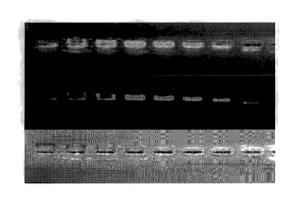
Figure 1 illustrates an electrophoretic gel that shows electrophoretic
mobility
(EB) and fluorescence signal (Fl) in a biotin-density dependent manner, which
includes
a top panel that is an overlay of fluorescence and white light, a middle panel
that is
fluorescence only, and the bottom panel that is white light showing absorbance
and
migration of the gold nanoparticles;
Figure 2 illustrates an electrophoretic gel conducted to test gel shift by
migration. From left to right are the fluorescence and white light overlay,
then the
white light only showing migration of the particles, and finally the
fluorescence only
channel; and
Figure 3 shows the fluorescence signal arising from NP coated particles with a
PEG spacer of 5000 kDa, equivalent to 5 nm with the brightness of the signal
dependent upon the number of fluorophores coated onto the surface.
Optimizing the available biotin binding sites on the nanoparticles
The number of binding sites on the surfaces of the particles determines not
only how many streptavidins can be bound, but also the manner of streptavidin
binding
(see above). The range of biotin concentrations on the particle surface, and
the ability
of streptavidin binding were examined. The PEG's were mixed with the particles
at
different ratios, excess was washed off, and then electrophoretic mobility
(EM) and
fluorescence signal (Fl) in a biotin-density dependent manner were examined.
Referring to Figure 1, the top panel is an overlay of fluorescence and white
light, the
middle is fluorescence only, and the bottom panel is white light showing
absorbance
and migration of the gold nanoparticles. Left to right, is high to low density
of biotin-
PEG on the particles surface (from 5x biotin-PEGs added per nm2 to 0.04 per
nm2).
This shows how the migration of the particles changes depending on the number
of
streptavidin bound, due to the change in size and charge. It also shows that
the signal
increases to a maximum at approximately 0.6x biotin-PEG. As biotin density
increases
beyond this ratio, the density of streptavidin increases on the nanoparticle
surface to
the point that self-quenching from the dye occurs.
In the above example and with other similar experiments conducted, the
relationship between the density of biotin-PEG on the particle surface and the
brightness of the resulting particles were examined. These studies were,
however,
conducted without first investigating the effect of the homogeneous mixing of
streptavidin-A750 and particles or whether the streptavidin that bound to the
particle
could bind additional biotin-labelled biomolecules.
7
CA 02812208 2013-03-21
WO 2012/037667
PCT/CA2011/001080
After characterizing the above, the amount of biotin-PEG on the particle
surface
that would allow streptavidin binding in a manner that didn't cause cross-
linking of the
particles by streptavidin binding, and that would allow biotin-labelled
biomolecules to
bind the streptavidin was assessed. We determined by this characterizing the
effect of
different biotin densities binding to streptavidin, washing unbound
streptavidin, binding
to biotinylated biological molecules, and finally analyzing using gel
electrophoresis
(where we are looking for shifts in the gel to indicate the differential
binding between
different binding densities and the streptavidin).
Particles having 0.1x biotin-PEG molecules per nm2 were first synthesized.
Streptavidin-A750 was mixed with the biotin-GNP by adding it in a small volume
scintillation vial, 150 pl total volume in phosphate buffered saline (PBS).
The
streptavidin-A750 in 150 pl total PBS was then added while rapidly stirring
the gold
nanoparticles (GNP) solution. After incubating for 5 minutes, the product was
centrifuged, and excess streptavidin was removed by washing. Biotin-
transferrin was
added to the streptavidin-coated nanoparticles. The product was then added to
an
agarose gel to test gel-shift by migration. Referring now to Figure 2, from
left to right is
the fluorescence and white light overlay, then the white light only showing
migration of
the particles, and finally the fluorescence only channel. The left hand
treatment is the
particles prior to spinning and washing the excess streptavidin away. The
middle
column is the washed biotin-GNP, bound with streptavidin, then biotin-
transferrin was
added. The final column is the washed biotin-GNP with streptavidin added, but
with no
biotin-transferrin added. The two outer treatments shows that the streptavidin
bound
the biotin-GNP without causing cross-linking of the particles (no fluorescence
is visible
in the well of the left treatment, demonstrating that no cross-linking
occurred, which
would "aggregate" the particles and prevent migration). The right treatment
shows that
the washing procedure caused a small amount of particle aggregation, visible
by the
fluorescent particles stuck in the well. The middle column shows a greater
degree of
particle cross-linking by addition of the biotin-transferrin. It also shows a
gel-shift due
to addition of the biotin-transferrin to the particles. Overall, our results
demonstrate
that the streptavidin was successfully added to the GNP in a manner that did
not cause
cross linking and allowed binding of biotin-labelled biomolecules. The gel-
shift proves
this. A small amount of cross-linking may occur with the particles, which
indicates that
the number of biotin-labelled biomolecules added to the solution, the
proportions of
which, and also the method of adding and mixing can be optimized further to
reduce
cross linking and aggregation.
8
CA 02812208 2013-03-21
WO 2012/037667
PCT/CA2011/001080
In final experiments, the addition of biotin-transferrin to the constructs of
GNP-
streptavidin was optimized. The volumes used for mixing, and the ratio of
biotin-
transferrin to GNP-streptavidin were examined. So were total signal, migration
and
cross-linking examined using gel electrophoresis. Once optimized, the ability
of the
constructs to bind their targets in an ELISA format was examined. Successful
ELISA
results indicated the labelling of gold nanoparticles with the protein
transferrin by
comparison the ELISA experiment of transferrin-coated gold nanoparticles with
albumin-coated gold nanoparticles. The constructs were incubated in the wells,
excess
was washed off, and fluorescence resulting from specific binding was measured.
Final Product
Following the optimized protocol, biotin-GNP was synthesized and complexes
of 3 fluorescently coloured nanoparticles were constructed, each having a
different
targeting molecule. Each different nanoparticle was able to specifically bind
their
respective targets, and did not bind non-specifically to other targets. The
number of
different fluorescent probes that can be constructed with this approach was
limited by
a) the number of commercially available fluorescently labelled streptavidins,
or b) the
number of possible fluorescently labeled streptavidin molecules, synthesized
by
covalent attachment of fluorophores to the streptavidin, and c) the number of
biotin-
targeting molecules available or d) the number of biotin-targeting molecules
synthesized by conjugating biotin to a targeting molecule. For example,
lnvitrogen
currently sells 40-50 variations of streptavidin labelled with fluorophores,
and a number
of quantum dot-conjugates of streptavidin that could be used. Because new
fluorophores are constantly being developed, there is really no upper limit to
the
number of potential conjugates that could be used. Similarly, because biotin
is readily
conjugated to any targeting molecule of interest, there is an almost limitless
variety of
combinations of fluorophores and targeting molecules that can be combined on
the
present platform.
Assay Kit
This description also encompasses an assay kit of fluorescent, functionalized
nanoparticles, with various components included in the kit, as well as a
specific
protocol for the end user to assemble the components. In broad terms there is
provided an assay kit of fluorescent, functionalized noble metal nanoparticles
comprising the following separate components:
- Solution A comprising an aqueous solution of nanoparticles;
- Solution B comprising a "conjugation buffer";
9
CA 02812208 2013-03-21
WO 2012/037667
PCT/CA2011/001080
- Solution C comprising streptavidin-fluorophore conjugate stock solutions
in
a conjugation buffer, each with a different fluorophore;
and instructions for assembling the components in accordance with the process
as
defined above for the production of fluorescent nanoparticles.
More specifically the kit may comprise the following components:
- Solution A comprising a stock vial of concentrated nanoparticles in for
example, aqueous solution;
- Solution B comprising a "conjugation buffer" or more specifically
a
phosphate buffered saline plus surfactant such as 0.1% Tween 20TM
(weight-to-volume);
- Solution C comprising a number, for example 2 to 20, of streptavidin-
fluorophore conjugate stock solutions in conjugation buffer, each with a
different fluorophore;
- A wash buffer, for example a phosphate buffered saline plus 0.5% Tween
20TM (W/V) ;
- A binding buffer or phosphate buffered saline plus a surfactant such as
0.05% Tween 2OTM (w/v); and
- A number of scintillation vials and stir bars to allow assembly.
The specific kit also contains a protocol for preparing the reagent. For
example, the kit contains specific instructions for the end user such as:
1. Add a small volume (10 pL) of Solution A to 500 pl of Solution B in a 1.5
mL
Eppendorf tube. Pipette this solution into an empty scintillation vial and
stir rapidly
on a magnetic stir plate.
2. Add a small volume (10 pL) of Solution C to 500 pL of Solution C in a 1.5
mL
Eppendorf tube. Pipette this solution into the scintillation vial while
stirring rapidly.
Incubate 15 minutes at room temperature, while stirring.
3. Centrifuge the reaction for 3 minutes at 2000 x g. Remove 900 pL of
supernatant
without disturbing the pellet. Add 900 pL of Wash Buffer, vortex to mix, and
repeat
centrifuge and wash a total of 3 times. Resuspend the pellet with 900 pi_ of
Solution B.
4. Prepare a solution of the biotinylated targeting molecule (user provided)
in Solution
B, to a final concentration of 1 mg/mL.
5. Add 10 pL of the biotinylated targeting molecule to the nanoparticle
solution and
then incubate 30 minutes.
CA 02812208 2016-10-18
6. Repeat the wash step described in step 3, a total of 3 times. After the
last wash,
remove as much supernatant liquid as possible without disturbing the pellet.
Resuspend the nanoparticles in 1 mL of Solution D.
7. The fluorescent, functionalized nanoparticles are now ready for use.
Optimize the
volume of nanoparticle reagent needed for your specific assay.
=
Most particularly there is provided particles synthesized on the nanometer
scale that have many potential applications, for instance as biomedical
devices
(diagnostic and therapeutic agents). The present process optimizes the
synthesis of
fluorescent gold nanoparticles in particular, generally over the range of 10 -
100 nm.
These particles have advantageous properties, including the ability to produce
a wide
range of particle sizes, highly stable particles both in vitro and in vivo,
and the ability
to use a variety of organic dyes (for example, any Alexa Fluor dye). Because
of the
flexibility to apply any fluorescent dye, the nanoparticle can have a wide
range of
fluorescent properties. The particles consist of a gold cluster core, a poly-
(ethylene
glycol) (PEG) outer brush layer, and a biotin linker presented on the outside
of a
proportion of the PEG layer that provides a rapid, easy means of attaching a
fluorescence dye by biotin-avidin (or derivative of avidin) linkage. The
materials used
in synthesizing these fluorescent particles have previously been approved for
internal
use by the U.S. FDA. These particles are comparable in many ways to
fluorescent
nanocrystals, quantum dots. Fluorescent gold nanoparticles are considered to
be
applicable as a reagent including but not limited to in vitro cell labelling,
in vitro
diagnostic assay labelling, and in vivo tracking or tumor targeting.
The scope of the claims should not be limited by the preferred embodiments
set forth in the examples, but should be given the broadest interpretation
consistent
.. with the description as a whole.
11