Note: Descriptions are shown in the official language in which they were submitted.
CA 02812785 2013-03-21
WO 2012/054565
PCT/US2011/056826
METHODS AND COMPOSITIONS FOR MODULATING THE WNT PATHWAY
CROSS REFERENCE TO RELATED APPLICATION
This application claims the benefit of U.S. Provisional Application No.
61/394,840, filed
October 20, 2010, the disclosure of which is incorporated herein by reference
in its entirety.
TECHNICAL FIELD
The present invention relates generally to the field of Wnt pathway
regulation. More
specifically, the invention concerns modulators of the Wnt signaling pathway,
and uses of said
modulators.
BACKGROUND
The Wnt/13-catenin signaling pathway is essential from embryonic development
to adult
organism homeostasis, and if deregulated, can induce diseases ranging from
osteoporosis to
cancer (1-4). The first Wnt gene, originally named int-1 (5), was discovered
in 1982 and later
reclassified as the founding member of the Wnt gene family upon discovery of
its homolog Wg in
Drosophila (6, 7). Within the last three decades, proteins constituting the
core of the Wnt/[3-
catenin signaling have been identified which define off and on states of this
pathway. In the
absence of Wnt ligand, intracellular 13-catenin is part of a complex formed by
Axin, APC, GSK3
and CK1 which phosphorylates and target 13-catenin for degradation by the
proteasome upon
ubiquitination by 13-Trcp (2). Wnt/13-catenin signaling is initiated by
binding of the secreted Wnt
to its co-receptors Frizzled (Fz) (8) and low density lipoprotein receptor-
related protein 5 or 6 (9,
10). Wnt mediated binding of Fz to LRP induces the formation of a ternary
complex at the cell
surface (10, 11) which results in association of the protein Dishevelled (Dvl)
with the intracellular
domain of Fz and the phosphorylation of the LRP6 C-terminal PPPSPxS motif by
the protein
kinases GSK3 and CK1, two events necessary for the recruitment of Axin to the
plasma
membrane (12-15). Wnt mediated displacement of Axin induces the stabilization
of the 13-catenin
cytoplasmic pool, and allows its translocation to the nucleus, where it acts
as a co-transcriptional
factor in complex with TCF/LEF to activate expression of the Wnt target genes
(2).
The Wnt/13-catenin pathway has been linked to metabolic disorders (16),
neurodegeneration (17, 18), and numerous types of cancers (1, 2, 4). A more
established link
exists between mutations of the APC protein, which prevent full 13-catenin
regulation, and
colorectal cancers (4, 19, 20). Of particular note is the genetic relationship
between LRP5 and
bone homeostasis. Loss of function mutations in LRP5 cause the autosomal
recessive disorder
osteoporosis-pseudoglioma syndrome (OPPG), characterized by low bone mass,
ocular defects
CA 02812785 2013-03-21
WO 2012/054565
PCT/US2011/056826
and a predisposition to fractures (21). Conversely, additional genetic
characterization of LRP5
revealed mutations translating in a high bone-mass density phenotype (22-24).
At the cell surface, Wnt/13-catenin signaling is regulated by two groups of
secreted
proteins with distinct modes of action. First, the soluble Frizzled-related
protein, or sFRPs (25),
have a similar fold to the cysteine-rich domain (CRD) of the Frizzled receptor
(26) and inhibit the
Wnt/13-catenin pathway by directly binding to the Wnt protein. A second type
of Wnt-binding
inhibitors, the Wnt inhibitory factor (WIF) is composed instead of a WIF
domain and five EGF
domains (27), which indicates that the Wnt proteins can interact with
structurally different
inhibitors. The second class of Wnt inhibitors is composed of the Dickkopf
(Dkk) (28, 29) and
WISE/Sclerostin (30-32) families of proteins. These proteins inhibit the
Wnt/13-catenin signaling
pathway by directly competing with Wnt for binding to its co-receptors LRP5
and LRP6 (29, 33).
Both Dkkl and Sclerostin (SOST) have been shown to be directly involved in
bone growth
regulation by LRP5. In particular, Sclerostin loss of function is responsible
for sclerosteosis and
Van Buchem diseases (34, 35); the unusually dense and strong bone observed in
these conditions
is similar to the hBMD phenotype caused by to LRP5 gain-of-function mutations.
Dkkl
mutations causing comparable effects have not been found, even though the
function of Dkkl in
murine bone development is comparable to that of Sclerostin (36).
At present, parathyroid hormone (PTH) represents the only FDA-approved bone-
forming
product available on the market, but PTH has been associated with safety
issues such as
hypercalcemia and osteosarcoma (37). Other treatments, such as biphosphonate
and antibodies
targeting the receptor activator of nuclear factor-KB (RANKL), target the
osteoclast cell subtype
which has the effect of decreasing bone resorption (38). Alternatively, the
Wnt/13-catenin
signaling pathway stimulates osteoblastogenesis (39) and, therefore,
stimulation of Wnt signaling
can induce bone formation (40). With an aging population pre-disposed to
fractures, osteoporosis
and rheumatoid arthritis, there is a need for safe and therapeutically
effective bone anabolic
agents.
SUMMARY
The invention provides compounds that modulate the Wnt pathway and methods
of using the same. One aspect of the invention provides for a compound that
inhibits the
binding of Dkkl and/or SOST to LRP6 and/or LRP5. In one embodiment, the
compound does
not inhibit the binding of a Wnt to LRP6 and/or LRP6. In one embodiment, the
compound does
not inhibit binding of Wnt9B to LRP6 and/or LRP5.
One aspect of the invention provides for an isolated peptide comprising the
amino acid
sequence XoX1X2X3 where X0 is N; X1 is A, S, F, T, Y, L, or K, or R; X2 is I
or V; and X3 is K, R,
2
CA 02812785 2013-03-21
WO 2012/054565
PCT/US2011/056826
or H. In one embodiment, the peptide comprises the amino acid sequence
X1XoX1X2X3X4, where
X_1 is P, S, C, or G; X0 is N; X1 is A, S, F, T, Y, L, or K, or R; X2 is I or
V; X3 is K, R, or H; and
X4 is F, T, Y, L, or V. In one embodiment, the peptide comprises an amino acid
sequence
selected from the group consisting of N XIIK, N XIVK, N X1 IR, N X1 VR, N X1
IH, and N
XIVH, where X1 is A, S, F, T, Y. In one embodiment, the peptide is selected
from among the
peptides of Family 1 (Figure 1 ). In one embodiment, at least one amino acid
of the peptide is
substituted with an amino acid analog. In one embodiment, the peptide
comprises an amino acid
analog. In one embodiment, the peptide inhibits the binding of Dkklto LRP6 and
does not inhibit
the binding of Wnt9B to LRP6. In one embodiment, the peptide binds to the El
13-prope11er of
LRP6. In one embodiment, the peptide interacts with at at least one, at least
two, at least three, at
least four, at least five, at least six, at least seven, at least eight, at
least nine, at least ten, at least
eleven, or all of the amino acid residues R28, E51, D52, V70, S71, E73, L95,
S96, D98, E115,
R141, and N185 of the El 13-prope11er of LRP6.
One aspect of the invention provides for an isolated cyclic peptide comprising
the amino
acid sequence: X0X1X2X3, where X0 is N; X1 is F, Y, L, A, R, or S; X2 is I or
V; and X3 is K, R, or
H. In one embodiment, the cyclic peptide comprises the amino acid sequence
X_IX0X1X2X3X4,
where X_1 is P, S, C, or G; X0 is N; X1 is F, Y, L, A, R, or S; X2 is I or V;
X3 is K, R, or H; and X4
is F, T, Y, L, or V. In one embodiment, the cyclic peptide comprises an amino
acid sequence
from the group consisting of N XIIK, N XIVK, N X1 IR, N X1 VR, N X1 IH, and N
XIVH, where
X1 is F, Y, L, A, R, or S. In one embodiment, the cyclic peptide is selected
from among the
peptides of Family 2 (Figure 2). In one embodiment, at least one amino acid of
the cyclic
peptide is substituted with an amino acid analog. In one embodiment, the
cyclic peptide
comprises an amino acid analog. In one embodiment, the cyclic peptide inhibits
the binding of
Dkklto LRP6 amd does not inhibit the binding of Wnt9B to LRP6. In one
embodiment, the
cyclic peptide binds to the El 13-prope11er of LRP6. In one embodiment, the
cyclic peptide
interacts with at at least one, at least two, at least three, at least four,
at least five, at least six, at
least seven, at least eight, at least nine, at least ten, at least eleven, or
all of the amino acid
residues R28, E51, D52, V70, S71, E73, L95, S96, D98, E115, R141, and N185 of
the El 13-
propeller of LRP6.
One aspect of the invention provides for an isolated peptide comprising the
amino acid
sequence: X_IX0X1X2, where X_1 is W, L, Y, F, or I; X0 is D or E; X1 is F, W,
I, S, or Y; and X2 is
M. In one embodiment, the peptide comprises the amino acid sequence:
X_2X_IX0X1X2X3, where
X_2 is V, I, L, or F ;X_1 is W, L, Y, F, or I; X0 is D or E; X1 is F, W, I, S,
or Y; X2 is M; and X3 is
W, M, A, or G. In one embodiment, the peptide is selected from among the
peptides of Family 3
(Figure 3)..
3
CA 02812785 2013-03-21
WO 2012/054565
PCT/US2011/056826
One aspect of the invention provides for an isolated peptide selected from
among the
peptides of Family 4 (Figure 4).
One aspect of the invention provides for a method for screening for a compound
that
inhibits the interaction of Dkkl and LRP6 comprising contacting a test
compound with LRP6, or
functional equivalent thereof, and determining the level of binding of the
test compound to the
LRP6, or functional equivalent thereof, in the presence and the absence of a
peptide ligand that
inhibits the interaction of Dkkl with LRP6 wherein a change in level of
binding in the presence
or absence of the peptide ligand indicates that the test compound inhibits the
interaction of Dkkl
with LRP6 and wherein the peptide ligand comprises an amino acid sequence
selected from the
group consisting of the amino acid sequences of a) Family 1 (Figure 1); b)
Family 2 (Figure
2); c) Family 3 (Figure 3); and d) Family 4 (Figure 4). In one embodiment, the
peptide ligand
is labeled with a detectable label.
One aspect of the invention provides for a method for screening for a compound
that
inhibits the interaction of Dkkl and LRP5 comprising contacting a test
compound with LRP5, or
functional equivalent thereof, and determining the level of binding of the
test compound to the
LRP5, or functional equivalent thereof, in the presence and the absence of a
peptide ligand that
inhibits the interaction of Dkkl with LRP5 wherein a change in level of
binding in the presence
or absence of the peptide ligand indicates that the test compound inhibits the
interaction of Dkkl
with LRP5 and wherein the peptide ligand comprises an amino acid sequence
selected from the
group consisting of the amino acid sequences of a) Family 1 (Figure 1); b)
Family 2 (Figure
2); c) Family 3 (Figure 3); and d) Family 4 (Figure 4). In one embodiment, the
peptide ligand
is labeled with a detectable label.
BRIEF DESCRIPTION OF THE DRAWINGS
Figure 1. Exemplary peptides of Family 1.
Figure 2. Exemplary peptides of Family 2.
Figure 3. Exemplary peptides of Family 3.
Figure 4A-C. Exemplary peptides of Family 4.
Figure 5. Detailed view of the CDR H3 interaction with residues of the LRP6
groove
showing the network of interactions made by the NAVK sequence.
Figure 6. Detail of the interactions made by antibody CDRs other than H3.
Figure 7. (A) Alignment of primary sequences from Dkk 1, Dkk2, Dkk4,
Sclerostin, and
Wise. (B) Examples of peptides based on proteins with "NXI" motif
4
CA 02812785 2013-03-21
WO 2012/054565
PCT/US2011/056826
Figure 8. Competition binding between Dkkl and other Wnt pathway inhibitors.
The
indicated LRP6 construct was preloaded onto biosensor tips. Dkkl (100 nM) (or
buffer control) and
the test ligand (100 nM) were loaded sequentially onto the LRP6 tips. (A) Dkk2
competition with
Dkkl. (B) Sclerostin competition with Dkkl. Percent binding in the presence of
Dkkl is shown
relative to buffer control.
Figure 9. Binding determinants in the Wnt inhibitors Dkkl and sclerostin (A)
The conserved
Asn and Ile residues of the "NXI" motif are important for Dkkl and sclerostin
binding to LRP6
El E2. (B) Dkkl has two independent binding regions, one that recognizes LRP6
El E2 and one that
recognizes LRP6 E3E4. Substitutions in the "NXI" motif (N40A, 142E) affect
binding to LRP6 El E2
io but not to E3E4, whereas substitutions in the C-terminal cysteine-rich
domain (H204E, K211E)
affect binding to LRP6 E3E4 but not to El E2. In each case, mutant proteins
retain binding to LRP6
E1E4.
Figure 10. Cartoon depicting the different Dkkl-LRP6 El E4 complexes studied
by SEC-
MALS and possible models for the interaction. Predicted molecular weights for
each individual
molecule or complex are indicated, with experimentally observed weights shown
below. The
observed molecular weights are consistent with 1:1 complex formation between
LRP6 El E4 and
each of the Dkkl variants. The data are not consistent with model 3 (showing a
2:1 stoichiometry).
The data are instead consistent with either model 4, in which one Dkkl
molecule can bridge two
LRP6 binding sites, or model 5/6, in which only one or the other site is
accessible to bound Dkkl.
Figure 11. Wnt binding to LRP6 El E4 in the presence or absence of Dkkl or
sclerostin.
Dkkl (125 nM) inhibits binding of both Wnt3A and Wnt9B (125 nM each), while
sclerostin (125
nM) only inhibits binding of Wnt9B.
Figure 12. Induction of a Wnt/13-catenin reporter in the presence or absence
of wild-type and
mutant inhibitors. Cells were transfected by Wntl (binds to LRP6 El E2). Dkkl
and sclerostin
variants, or the control inhibitor Fz8 CRD, were used at the indicated doses.
Figure 13. Introduction of LRP5 BMD substitutions into LRP6 El E2 lowers
affinity for
Wnt inhibitors. The five substitutions characterized are indicated on the y-
axis. Steady-state affinity
measurements were made for Wnt9b, Dkkl, and sclerostin binding to each of the
LRP6 variants.
Differences in binding to Wnt9b were minor (< 5-fold change compared to wild
type), while binding
to Dkkl and sclerostin was more significantly impacted (10-250-fold losses in
affinity compared to
wild type).
Figure 14. Conserved motifs present in phage clones selected from linear and
cyclic peptide
libraries against LRP6 El E2 (A) Linear peptides of Exemplary Family 1. (B)
Cyclic peptides of
Exemplary Family 2.
5
CA 02812785 2013-03-21
WO 2012/054565
PCT/US2011/056826
Figure 15. Conserved motifs present in phage clones selected from linear and
cyclic peptide
libraries against LRP5 El (A) Linear peptides of Exemplary Family 3. (B)
Cyclic peptides of
Exemplary Family 4.
Figure 16. Co-crystal structures of LRP6 El and peptides discovered from phage-
display
libraries. (A) Peptide Ac-SNSIKFYA-am from Exemplary Family 1. (B) Peptide Ac-
GSLCSNRIKPDTHCSS-am (disulfide), a CX9C class member of Exemplary Family 2.
(C) Peptide
Ac-CNSIKLC-am (disulfide), a CX5C class member of Exemplary Family 2. (D)
Peptide Ac-
CNSIKCL-am (disulfide), a CX4C class member of Exemplary Family 2.
Figure 17. Structure-activity study of the Dkkl 7-mer peptide. The indicated
peptides were
synthesized by standard Fmoc procedures, and IC50 values were determined as
described in Example
1. (A) C-terminal and N-terminal truncations. (B) Substitutions at position
"X" of the "NXI" motif
Figure 18A and B. Structure-activity study of the Dkkl 7-mer peptide showing
effects of
substitution of the N, S, I, and K residues. The indicated peptides were
synthesized by standard
Fmoc procedures, and IC50 values were determined as described in Example 1.
Figure 19. Structure-activity study of a linear peptide from Exemplary Family
1.
Substitutions were made in the Ile position of the "NXI" motif The indicated
peptides were
synthesized by standard Fmoc procedures, and IC50 values were determined as
described in Example
1.
Figure 20. Transfer of the "NXI" epitope to a structured peptide scaffold. (A)
Design of the
structured mimetic. The residues N100¨V100b from the antibody complex
structure were overlaid
on a representative structure of a Bowmain-Birk inhibitory (BBI) loop peptide
(PDB code 1GM2)
(42). Apart from an amide bond rotation preceding the branched hydrophobic
residue, the
conformations of the peptides are similar. The positions of side chain I3-
carbons for the three-residue
motif coincide. Sequences of the BBI loop template and the "NXI"-containing
BBI mimetic are
shown. (B) The BBI mimetic binds to LRP6 El, while a control peptide lacking
the conserved Asn
does not.
Figure 21. Design of a amide-cyclized variant of the Dkkl 7-mer peptide. (A)
Structure of
the Dkkl peptide taken from the complex with LRP6 El is shown at top. The side
chain of Ser2
points toward the side chain of Asn7 with a short gap between. Below is a
model in which Ser2 is
substituted by Lys, and Asn7 by Asp. The side chains are joined by an amide
bond between the Lys
c-amine and the Asp carboxylate. (B) Competition binding data indicate that
the cyclized peptide
binds to LRP6 El.
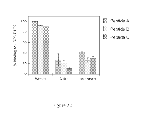
Figure 22. LRP6 El-binding peptides inhibit binding of Wnt inhibitors, but not
of Wnt9B,
to LRP6 El E2. Binding was assessed by biolayer interferometry, as described
in Example 1.
6
CA 02812785 2013-03-21
WO 2012/054565
PCT/US2011/056826
Immobilized LRP6 El E2 was exposed to protein ligand (Wnt 9b, Dkkl, or
sclerostin) present in
solution at a concentration three-fold higher than the measured dissociation
constant for El E2.
Competing peptides were added at a saturating level (20-fold higher than the
measured IC50 value).
Peptide A: Ac-NSNAIKN-am; Peptide B: Ac-CNSIKFCG-am (disulfide); Peptide C: Ac-
GSLCSNRIKPDTHCSS-am (disulfide)
DISCLOSURE OF THE INVENTION
General Techniques
The practice of the present invention will employ, unless otherwise indicated,
conventional
techniques of molecular biology (including recombinant techniques),
microbiology, cell biology,
biochemistry, and immunology, which are within the skill of the art. Such
techniques are explained
fully in the literature, such as, "Molecular Cloning: A Laboratory Manual",
second edition (Sambrook
et al., 1989); "Oligonucleotide Synthesis" (M. J. Gait, ed., 1984); "Animal
Cell Culture" (R. I.
Freshney, ed., 1987); "Methods in Enzymology" (Academic Press, Inc.); "Current
Protocols in
Molecular Biology" (F. M. Ausubel et al., eds., 1987, and periodic updates);
"PCR: The Polymerase
Chain Reaction", (Mullis et al., ed., 1994); "A Practical Guide to Molecular
Cloning" (Perbal Bernard
V., 1988).
Definitions
The term "amino acid" within the scope of the present invention is used in its
broadest
sense and is meant to include the naturally- occurring L -amino acids or
residues. The commonly
used one- and three-letter abbreviations for naturally-occurring amino acids
are used herein
(Lehninger, Biochemistry, 2d ed., pp. 71-92, (Worth Publishers: New York,
1975). The term
includes D-amino acids as well as chemically-modified amino acids such as
amino acid analogs,
naturally- occurring amino acids that are not usually incorporated into
proteins such as
norleucine, and chemically-synthesized compounds having properties known in
the art to be
characteristic of an amino acid. For example, analogs or mimetics of
phenylalanine or proline,
which allow the same conformational restriction of the peptide compounds as
natural Phe or Pro,
are included within the definition of amino acid. Such analogs and mimetics
are referred to
herein as "functional equivalents" of an amino acid. Other examples of amino
acids are listed by
Roberts and Vellaccio, The Peptides: Analysis, Synthesis, Biology, Eds. Gross
and Meiehofer,
Vol. 5, p. 341 (Academic Press, Inc.: N.Y. 1983).
In certain embodiments, variants of compounds, such as peptide variants having
one or more
amino acid substitutions, are provided. Conservative substitutions are shown
in Table 1 under the
heading of "conservative substitutions." More substantial changes are provided
in Table 1 under the
7
CA 02812785 2013-03-21
WO 2012/054565
PCT/US2011/056826
heading of "exemplary substitutions," and as further described below in
reference to amino acid side
chain classes.
TABLE 1
Original Exemplary
Conservative
Residue Substitutions
Substitutions
Ala (A) Val; Leu; Ile Val
Arg (R) Lys; Gln; Asn Lys
Asn (N) Gln; His; Asp, Lys; Arg Gln
Asp (D) Glu; Asn Glu
Cys (C) Ser; Ala Ser
Gln (Q) Asn; Glu Asn
Glu (E) Asp; Gln Asp
Gly (G) Ala Ala
His (H) Asn; Gln; Lys; Arg Arg
Ile (I) Leu; Val; Met; Ala; Phe; Norleucine Leu
Leu (L) Norleucine; Ile; Val; Met; Ala; Phe Ile
Lys (K) Arg; Gln; Asn Arg
Met (M) Leu; Phe; Ile Leu
Phe (F) Trp; Leu; Val; Ile; Ala; Tyr Tyr
Pro (P) Ala Ala
Ser (S) Thr Thr
Thr (T) Val; Ser Ser
Trp (W) Tyr; Phe Tyr
Tyr (Y) Trp; Phe; Thr; Ser Phe
Val (V) Ile; Leu; Met; Phe; Ala; Norleucine Leu
Amino acids may be grouped according to common side-chain properties:
(1) hydrophobic: Norleucine, Met, Ala, Val, Leu, Ile;
(2) neutral hydrophilic: Cys, Ser, Thr, Asn, Gln;
(3) acidic: Asp, Glu;
(4) basic: His, Lys, Arg;
(5) residues that influence chain orientation: Gly, Pro;
8
CA 02812785 2013-03-21
WO 2012/054565
PCT/US2011/056826
(6) aromatic: Trp, Tyr, Phe.
Non-conservative substitutions will entail exchanging a member of one of these
classes for
another class.
Synthetic peptides, synthesized for example by standard solid-phase synthesis
techniques,
are not limited to amino acids encoded by genes and therefore allow a wider
variety of
substitutions for a given amino acid. Amino acids that are not encoded by the
genetic code are
referred to herein as "amino acid analogs" and include, for example, those
described in WO
90/01940 and in the table below (Table 2), as well as, for example, 2-amino
adipic acid (Aad) for
Glu and Asp; 2-aminopimelic acid (Apm) for Glu and Asp; 2-aminobutyric (Abu)
acid for Met,
Leu, and other aliphatic amino acids; 2-aminoheptanoic acid (Ahe) for Met,
Leu, and other
aliphatic amino acids; 2-aminoisobutyric acid (Aib) for Gly; cyclohexylalanine
(Cha) for Val,
Leu and Ile; homoarginine (Har) for Arg and Lys; 2,3-diaminopropionic acid
(Dap) for Lys, Arg,
and His; N-ethylglycine (EtGly) for Gly, Pro, and Ala; N-ethylglycine (EtGly)
for Gly, Pro, and
Ala; N-ethylasparagine (EtAsn) for Asn, and Gln; hydroxylysine (Hyl) for Lys;
allohydroxylysine
(AHyl) for Lys; 3-(and 4-)hydroxyproline (3Hyp, 4Hyp) for Pro, Ser, and Thr;
allo-isoleucine
(A11e) for Ile, Leu, and Val; 4-amidinophenylalanine for Arg; N-methylglycine
(MeGly,
sarcosine) for Gly, Pro, and Ala; N-methylisoleucine (Mae) for Ile; norvaline
(Nva) for Met and
other aliphatic amino acids; norleucine (Nle) for Met and other aliphatic
amino acids; ornithine
(Orn) for Lys, Arg and His; citrulline (Cit) and methionine sulfoxide (MSO)
for Thr, Asn, and
Gln; and N-methylphenylalanine (MePhe), trimethylphenylalanine, halo-(F-, Cl-,
Br-, or I-
)phenylalanine, or trifluorylphenylalanine for Phe.
Table 2
Examples of hydrophobic amino acid analogs that may be incorporated into the
peptides of the
inventionl
Name Common abbreviation
Cyclohexylglycine Chg
Cyclopentylglycine Cpg
Cyclobutylalanine
Cyclopropylalanine
tert-Leucine Tle
Norleucine Nle
Norvaline Nva
2-Aminobutyric acid Abu
9
CA 02812785 2013-03-21
WO 2012/054565
PCT/US2011/056826
1Non-genetically encoded amino acids corresponding to those used in Example
13. This list is not
meant to be exhaustive and other substitutions may be contemplated.
"Percent (%) amino acid sequence identity" with respect to a peptide or
polypeptide
sequence is defined as the percentage of amino acid residues in a candidate
sequence that are
identical with the amino acid residues in the specific peptide or polypeptide
sequence, after
aligning the sequences and introducing gaps, if necessary, to achieve the
maximum percent
sequence identity, and not considering any conservative substitutions as part
of the sequence
identity. Alignment for purposes of determining percent amino acid sequence
identity can be
achieved in various ways that are within the skill in the art, for instance,
using publicly available
computer software such as BLAST, BLAST-2, ALIGN or Megalign (DNASTAR)
software.
Those skilled in the art can determine appropriate parameters for measuring
alignment, including
any algorithms needed to achieve maximal alignment over the full length of the
sequences being
compared. For purposes herein, however, % amino acid sequence identity values
are generated
using the sequence comparison computer program ALIGN-2. The ALIGN-2 sequence
comparison computer program was authored by Genentech, Inc. and the source
code has been
filed with user documentation in the U.S. Copyright Office, Washington D.C.,
20559, where it is
registered under U.S. Copyright Registration No. TXU510087. The ALIGN-2
program is
publicly available through Genentech, Inc., South San Francisco, California.
In situations where ALIGN-2 is employed for amino acid sequence comparisons,
the %
amino acid sequence identity of a given amino acid sequence A to, with, or
against a given amino
acid sequence B (which can alternatively be phrased as a given amino acid
sequence A that has or
comprises a certain % amino acid sequence identity to, with, or against a
given amino acid
sequence B) is calculated as follows:
100 times the fraction X/Y
where X is the number of amino acid residues scored as identical matches by
the sequence
alignment program ALIGN-2 in that program's alignment of A and B, and where Y
is the total
number of amino acid residues in B. It will be appreciated that where the
length of amino acid
sequence A is not equal to the length of amino acid sequence B, the % amino
acid sequence
identity of A to B will not equal the % amino acid sequence identity of B to
A.
Unless specifically stated otherwise, all % amino acid sequence identity
values used
herein are obtained as described in the immediately preceding paragraph using
the ALIGN-2
computer program.
CA 02812785 2013-03-21
WO 2012/054565
PCT/US2011/056826
An "isolated" compound is one which has been separated from a component of its
natural
environment. In some embodiments, a compound, such as a peptide, is purified
to greater than
95% or 99% purity as determined by, for example, electrophoretic (e.g., SDS-
PAGE, isoelectric
focusing (IEF), capillary electrophoresis) or chromatographic (e.g., ion
exchange or reverse phase
HPLC). For review of methods for assessment of purity, see, e.g., Flatman et
al., J. Chromatogr.
B 848:79-87 (2007).
An "isolated" nucleic acid refers to a nucleic acid molecule that has been
separated from a
component of its natural environment. An isolated nucleic acid includes a
nucleic acid molecule
contained in cells that ordinarily contain the nucleic acid molecule, but the
nucleic acid molecule
is present extrachromosomally or at a chromosomal location that is different
from its natural
chromosomal location.
The term "vector," as used herein, is intended to refer to a nucleic acid
molecule capable of
transporting another nucleic acid to which it has been linked. One type of
vector is a "plasmid", which
refers to a circular double stranded DNA loop into which additional DNA
segments may be ligated.
Another type of vector is a phage vector. Another type of vector is a viral
vector, wherein additional
DNA segments may be ligated into the viral genome. Certain vectors are capable
of autonomous
replication in a host cell into which they are introduced (e.g., bacterial
vectors having a bacterial origin
of replication and episomal mammalian vectors). Other vectors (e.g., non-
episomal mammalian
vectors) can be integrated into the genome of a host cell upon introduction
into the host cell, and
thereby are replicated along with the host genome. Moreover, certain vectors
are capable of directing
the expression of genes to which they are operatively linked. Such vectors are
referred to herein as
"recombinant expression vectors" (or simply, "recombinant vectors"). In
general, expression vectors
of utility in recombinant DNA techniques are often in the form of plasmids. In
the present
specification, "plasmid" and "vector" may be used interchangeably as the
plasmid is the most
commonly used form of vector.
"Polynucleotide," or "nucleic acid," as used interchangeably herein, refer to
polymers of
nucleotides of any length, and include DNA and RNA. The nucleotides can be
deoxyribonucleotides,
ribonucleotides, modified nucleotides or bases, and/or their analogs, or any
substrate that can be
incorporated into a polymer by DNA or RNA polymerase, or by a synthetic
reaction. A polynucleotide
may comprise modified nucleotides, such as methylated nucleotides and their
analogs. If present,
modification to the nucleotide structure may be imparted before or after
assembly of the polymer. The
sequence of nucleotides may be interrupted by non-nucleotide components. A
polynucleotide may be
further modified after synthesis, such as by conjugation with a label. Other
types of modifications
include, for example, "caps", substitution of one or more of the naturally
occurring nucleotides with an
analog, internucleotide modifications such as, for example, those with
uncharged linkages (e.g., methyl
phosphonates, phosphotriesters, phosphoamidates, carbamates, etc.) and with
charged linkages (e.g.,
11
CA 02812785 2013-03-21
WO 2012/054565
PCT/US2011/056826
phosphorothioates, phosphorodithioates, etc.), those containing pendant
moieties, such as, for example,
proteins (e.g., nucleases, toxins, antibodies, signal peptides, poly-L-lysine,
etc.), those with
intercalators (e.g., acridine, psoralen, etc.), those containing chelators
(e.g., metals, radioactive metals,
boron, oxidative metals, etc.), those containing alkylators, those with
modified linkages (e.g., alpha
anomeric nucleic acids, etc.), as well as unmodified forms of the
polynucleotide(s). Further, any of the
hydroxyl groups ordinarily present in the sugars may be replaced, for example,
by phosphonate groups,
phosphate groups, protected by standard protecting groups, or activated to
prepare additional linkages
to additional nucleotides, or may be conjugated to solid or semi-solid
supports. The 5' and 3' terminal
OH can be phosphorylated or substituted with amines or organic capping group
moieties of from 1 to
20 carbon atoms. Other hydroxyls may also be derivatized to standard
protecting groups.
Polynucleotides can also contain analogous forms of ribose or deoxyribose
sugars that are generally
known in the art, including, for example, 2'-0-methyl-, 2'-0-allyl, 2'-fluoro-
or 2'-azido-ribose,
carbocyclic sugar analogs, alpha-anomeric sugars, epimeric sugars such as
arabinose, xyloses or
lyxoses, pyranose sugars, furanose sugars, sedoheptuloses, acyclic analogs and
abasic nucleoside
analogs such as methyl riboside. One or more phosphodiester linkages may be
replaced by alternative
linking groups. These alternative linking groups include, but are not limited
to, embodiments wherein
phosphate is replaced by P(0)S("thioate"), P(S)S ("dithioate"), "(0)NR2
("amidate"), P(0)R, P(0)OR',
CO or CH2 ("formacetal"), in which each R or R' is independently H or
substituted or unsubstituted
alkyl (1-20 C) optionally containing an ether (-0-) linkage, aryl, alkenyl,
cycloalkyl, cycloalkenyl or
araldyl. Not all linkages in a polynucleotide need be identical. The preceding
description applies to all
polynucleotides referred to herein, including RNA and DNA.
"Oligonucleotide," as used herein, generally refers to short, generally single
stranded,
generally synthetic polynucleotides that are generally, but not necessarily,
less than about 200
nucleotides in length. The terms "oligonucleotide" and "polynucleotide" are
not mutually exclusive.
The description above for polynucleotides is equally and fully applicable to
oligonucleotides.
The term "LRP6", as used herein, refers to any native LRP6 from any vertebrate
source,
including mammals such as primates (e.g. humans) and rodents (e.g., mice and
rats), unless
otherwise indicated. The term encompasses "full-length," unprocessed LRP6 as
well as any form
of LRP6 that results from processing in the cell. The term also encompasses
naturally occurring
variants of LRP6, e.g., splice variants or allelic variants. The amino acid
sequence of an
exemplary human LRP6 is provided in NCBI accession number AAI43726,
Strausberg, R. L., et
al., Proc. Natl. Acad. Sci. U.S.A. 99 : 16899-16903 (2002) (He, X, et al.,
Development,
131:1663-1677 (2004); Chen, M., et al., J. Biol. Chem., 284:35040-35048
(2009).
The term "LRP5", as used herein, refers to any native LRP5 from any vertebrate
source,
including mammals such as primates (e.g. humans) and rodents (e.g., mice and
rats), unless
otherwise indicated. The term encompasses "full-length," unprocessed LRP5 as
well as any form
12
CA 02812785 2013-03-21
WO 2012/054565
PCT/US2011/056826
of LRP5 that results from processing in the cell. The term also encompasses
naturally occurring
variants of LRP5, e.g., splice variants or allelic variants. The amino acid
sequence of an
exemplary human LRP5 is provided in NCBI accession number 075197, Hey, P.J.,
et al, Gene
216 (1), 103-111 (1998).
As used herein, "treatment" (and grammatical variations thereof such as
"treat" or
"treating") refers to clinical intervention in an attempt to alter the natural
course of the individual
being treated, and can be performed either for prophylaxis or during the
course of clinical
pathology. Desirable effects of treatment include, but are not limited to,
preventing occurrence or
recurrence of disease, alleviation of symptoms, diminishment of any direct or
indirect
pathological consequences of the disease, preventing metastasis, decreasing
the rate of disease
progression, amelioration or palliation of the disease state, and remission or
improved prognosis.
In some embodiments, compounds of the invention are used to delay development
of a disease or
to slow the progression of a disease.
The terms "antibody" and "immunoglobulin" are used interchangeably in the
broadest
sense and include monoclonal antibodies (for e.g., full length or intact
monoclonal antibodies),
polyclonal antibodies, multivalent antibodies, multispecific antibodies (e.g.,
bispecific antibodies
so long as they exhibit the desired biological activity) and may also include
certain antibody
fragments (as described in greater detail herein). An antibody can be human,
humanized and/or
affinity matured.
"Antibody fragments" comprise only a portion of an intact antibody, wherein
the portion
preferably retains at least one, preferably most or all, of the functions
normally associated with
that portion when present in an intact antibody. In one embodiment, an
antibody fragment
comprises an antigen binding site of the intact antibody and thus retains the
ability to bind
antigen. In another embodiment, an antibody fragment, for example one that
comprises the Fc
region, retains at least one of the biological functions normally associated
with the Fc region
when present in an intact antibody, such as FcRn binding, antibody half life
modulation, ADCC
function and complement binding. In one embodiment, an antibody fragment is a
monovalent
antibody that has an in vivo half life substantially similar to an intact
antibody. For example
such an antibody fragment may comprise on antigen binding arm linked to an Fc
sequence
capable of conferring in vivo stability to the fragment.
The term "monoclonal antibody" as used herein refers to an antibody obtained
from a
population of substantially homogeneous antibodies, i.e., the individual
antibodies comprising the
population are identical except for possible naturally occurring mutations
that may be present in
minor amounts. Monoclonal antibodies are highly specific, being directed
against a single
antigen. Furthermore, in contrast to polyclonal antibody preparations that
typically include
13
CA 02812785 2013-03-21
WO 2012/054565
PCT/US2011/056826
different antibodies directed against different determinants (epitopes), each
monoclonal antibody
is directed against a single determinant on the antigen.
The monoclonal antibodies herein specifically include "chimeric" antibodies in
which a
portion of the heavy and/or light chain is identical with or homologous to
corresponding
sequences in antibodies derived from a particular species or belonging to a
particular antibody
class or subclass, while the remainder of the chain(s) is identical with or
homologous to
corresponding sequences in antibodies derived from another species or
belonging to another
antibody class or subclass, as well as fragments of such antibodies, so long
as they exhibit the
desired biological activity (U.S. Patent No. 4,816,567; and Morrison et al.,
Proc. Natl. Acad. Sci.
USA 81:6851-6855 (1984)).
"Humanized" forms of non-human (e.g., murine) antibodies are chimeric
antibodies that
contain minimal sequence derived from non-human immunoglobulin. For the most
part,
humanized antibodies are human immunoglobulins (recipient antibody) in which
residues from a
hypervariable region of the recipient are replaced by residues from a
hypervariable region of a
non-human species (donor antibody) such as mouse, rat, rabbit or nonhuman
primate having the
desired specificity, affinity, and capacity. In some instances, framework
region (FR) residues of
the human immunoglobulin are replaced by corresponding non-human residues.
Furthermore,
humanized antibodies may comprise residues that are not found in the recipient
antibody or in the
donor antibody. These modifications are made to further refine antibody
performance. In
general, the humanized antibody will comprise substantially all of at least
one, and typically two,
variable domains, in which all or substantially all of the hypervariable loops
correspond to those
of a non-human immunoglobulin and all or substantially all of the FRs are
those of a human
immunoglobulin sequence. The humanized antibody optionally will also comprise
at least a
portion of an immunoglobulin constant region (Fc), typically that of a human
immunoglobulin.
For further details, see Jones et al., Nature 321:522-525 (1986); Riechmann et
al., Nature
332:323-329 (1988); and Presta, Curr. Op. Struct. Biol. 2:593-596 (1992). See
also the following
review articles and references cited therein: Vaswani and Hamilton, Ann.
Allergy, Asthma &
Immunol. 1:105-115 (1998); Harris, Biochem. Soc. Transactions 23:1035-1038
(1995); Hurle and
Gross, Curr. Op. Biotech. 5:428-433 (1994).
A "human antibody" is one which possesses an amino acid sequence which
corresponds
to that of an antibody produced by a human and/or has been made using any of
the techniques for
making human antibodies as disclosed herein. This definition of a human
antibody specifically
excludes a humanized antibody comprising non-human antigen-binding residues.
An "affinity matured" antibody is one with one or more alterations in one or
more CDRs
thereof which result in an improvement in the affinity of the antibody for
antigen, compared to a
14
CA 02812785 2013-03-21
WO 2012/054565
PCT/US2011/056826
parent antibody which does not possess those alteration(s). Preferred affinity
matured antibodies
will have nanomolar or even picomolar affinities for the target antigen.
Affinity matured
antibodies are produced by procedures known in the art. Marks et al.
Bio/Technology 10:779-
783 (1992) describes affinity maturation by VH and VL domain shuffling. Random
mutagenesis
of CDR and/or framework residues is described by: Barbas et al. Proc Nat.
Acad. Sci, USA
91:3809-3813 (1994); Schier et al. Gene 169:147-155 (1995); Ye1ton et al. J.
Immunol.
155:1994-2004 (1995); Jackson et al., J. Immunol. 154(7):3310-9 (1995); and
Hawkins et al, J.
Mot. Biol. 226:889-896 (1992).
A "disorder" is any condition that would benefit from treatment with a
io substance/molecule or method of the invention. This includes chronic and
acute disorders or
diseases including those pathological conditions which predispose the mammal
to the disorder in
question. Non-limiting examples of disorders to be treated herein include
disorders of processes
that are activated or inhibited by Wnt signaling. Such processes include, for
example, cell
proliferation, cell fate specification, and stem cell self-renewal in
different cancer types, and
developmental processes. The compounds of the invention are useful, for
example, in the
treatment of Wnt mediated disorders of the bones or skeletal system. Examples
of skeletal or
bone disorders that can be treated using the compounds of the invention
include osteoporosis,
osteoarthritis, bone fractures, and bone lesions and various forms of bone
degeneration.
The terms "cell proliferative disorder" and "proliferative disorder" refer to
disorders that
are associated with some degree of abnormal cell proliferation. In one
embodiment, the cell
proliferative disorder is cancer.
"Tumor", as used herein, refers to all neoplastic cell growth and
proliferation, whether
malignant or benign, and all pre-cancerous and cancerous cells and tissues.
The terms "cancer",
"cancerous", "cell proliferative disorder", "proliferative disorder" and
"tumor" are not mutually
exclusive as referred to herein.
The terms "cancer" and "cancerous" refer to or describe the physiological
condition in
mammals that is typically characterized by unregulated cell
growth/proliferation. Examples of
cancer include but are not limited to, carcinoma, lymphoma, blastoma, sarcoma,
and leukemia.
More particular examples of such cancers include squamous cell cancer, small-
cell lung cancer,
non-small cell lung cancer, adenocarcinoma of the lung, squamous carcinoma of
the lung, cancer
of the peritoneum, hepatocellular cancer, gastrointestinal cancer, pancreatic
cancer, glioblastoma,
cervical cancer, ovarian cancer, liver cancer, bladder cancer, hepatoma,
breast cancer, colon
cancer, colorectal cancer, endometrial or uterine carcinoma, salivary gland
carcinoma, kidney
cancer, liver cancer, prostate cancer, vulval cancer, thyroid cancer, hepatic
carcinoma and various
types of head and neck cancer.
CA 02812785 2013-03-21
WO 2012/054565
PCT/US2011/056826
An "effective amount" refers to an amount effective, at dosages and for
periods of time
necessary, to achieve the desired therapeutic or prophylactic result.
A "therapeutically effective amount" of a substance/molecule of the invention,
agonist or
antagonist may vary according to factors such as the disease state, age, sex,
and weight of the
individual, and the ability of the substance/molecule, agonist or antagonist
to elicit a desired response
in the individual. A therapeutically effective amount is also one in which any
toxic or detrimental
effects of the substance/molecule, agonist or antagonist are outweighed by the
therapeutically
beneficial effects. A "prophylactically effective amount" refers to an amount
effective, at dosages and
for periods of time necessary, to achieve the desired prophylactic result.
Typically but not necessarily,
since a prophylactic dose is used in subjects prior to or at an earlier stage
of disease, the
prophylactically effective amount will be less than the therapeutically
effective amount.
The term "cytotoxic agent" as used herein refers to a substance that inhibits
or prevents
the function of cells and/or causes destruction of cells. The term is intended
to include
radioactive isotopes (e.g., At211, 1131, 1125, y90, Re186, Re188, sm153,
Bi212, -.32
f and radioactive
isotopes of Lu), chemotherapeutic agents e.g. methotrexate, adriamicin, vinca
alkaloids
(vincristine, vinblastine, etoposide), doxorubicin, melphalan, mitomycin C,
chlorambucil,
daunorubicin or other intercalating agents, enzymes and fragments thereof such
as nucleolytic
enzymes, antibiotics, and toxins such as small molecule toxins or
enzymatically active toxins of
bacterial, fungal, plant or animal origin, including fragments and/or variants
thereof, and the
various antitumor or anticancer agents disclosed below. Other cytotoxic agents
are described
below. A tumoricidal agent causes destruction of tumor cells.
Compounds and Methods
The Dickkopf (Dkk) and WISE/Sclerostin (SOST) family of proteins inhibit the
Wnt/13-
catenin signaling pathway by directly competing with Wnt for binding to its
LRP5 and LRP6 co-
receptors. Provided herein are compounds that modulate the interaction of DKK1
with LRP5
and/or LRP6 and compounds that modulate the interaction of SOST with LRP5
and/or LRP6. In
some embodiments, a compound modulates the interactions of both Dkkl and SOST
with LRP5/
and or LRP6.
In one embodiment, the compound inhibits the interaction of Dkkl with LRP5
and/or
LRP6. In one embodiment, the compound inhibits the interaction of SOST with
LRP5 and/or
LRP6. In one embodiment, the compound inhibits the interactions of both Dkkl
and SOST with
LRP5 and/or LRP6.
In one embodiment, the compound competes for binding to LRP6 with Dkkl. In one
embodiment, the compound competes for binding to LRP6 with SOST. In one
embodiment, the
compound competes for binding to LRP5 with Dkkl. In one embodiment, the
compound
16
CA 02812785 2013-03-21
WO 2012/054565
PCT/US2011/056826
competes for binding to LRP5 with SOST. In one embodiment, the compound binds
to a Dkkl
binding site on LRP6. In one embodiment, the compound binds to a SOST binding
site on LRP6.
In one embodiment, the compound binds to a Dkkl binding site on LRP5. In one
embodiment,
the compound binds to a SOST binding site on LRP5. In one embodiment, the
compound binds
to the El 13-prope11er of LRP6. In one embodiment, the compound binds to the
El 13-prope11er of
LRP5. In one embodiment, the compound interacts with at least one, at least
two, at least three, at
least four, at least five, at least six, at least seven, at least eight, at
least nine, at least ten, at least
eleven, or all of the amino acid residues R28, E51, D52, V70, S71, E73, L95,
S96, D98, E115,
R141, and N185 of the El 13-prope11er of LRP6. In one embodiment, the compound
interacts with
at least one, at least two, at least three, at least four, at least five, at
least six, at least seven, at least
eight, at least nine, at least ten, at least eleven, or all of the amino acid
residues R28, E63, D64,
V82, S83, E85, V108, S109, D111, E128, R154, and N198 of the El 13-prope11er
of LRP5. By
directly binding to the Dkkl or SOST binding site, the compound provides a
targeted approach to
modulating the Wnt pathway signaling associated with binding of Dkkl and SOST.
In one
embodiment, the compound modulates Wnt pathway signaling associated with
binding of Dkkl
to LRP5 or LRP6. In one embodiment, the compound modulates Wnt pathway
signaling
associated with binding of SOST to LRP5 or LRP6. In one embodiment, the
compound
modulates the Wnt pathway signaling associated with binding of Dkkl and/or
SOST to LRP5 or
LRP6 without modulating the serotonin pathway.
In some embodiments, the compound inhibits the interaction of Dkkl with LRP5
and/or
LRP6 and does not inhibit the interaction of a Wnt with LRP5 or LRP6. In some
embodiments,
the compound inhibits the interaction of SOST with LRP5 and/or LRP6 and does
not inhibit the
interaction of a Wnt with LRP5 or LRP6. In one embodiment, the Wnt is Wnt3a.
In one
embodiment, the Wnt is Wnt9b. This selective inhibition serves to prevent
inhibition of the Wnt
signaling pathway by the inhibitors Dkkl or SOST while allowing for the
stimulation of the
pathway by Wnt molecules. As a result, the compounds serve to promote bone
growth and repair
associated with the Wnt pathway.
In some embodiments, the compounds find use in the treatment of various
skeletal
disorders that can benefit from the promotion of bone growth such as, for
example, osteoporosis,
rheumatoid arthritis, bone degradation or degeneration which can occur due to
a number of
conditions including, for example, cancers such as multiple myeloma, and in
the treatment of
bone fractures or other bone deficiencies associated with low bone density or
low bone strength.
In one aspect of the invention, the compound is a peptide. In one embodiment,
the
compound is a linear peptide. In embodiment, the linear peptide is from 3 to
100, 3 to 50, 3 to 30,
3 to 20, 3 to 10, 3 to 9, 3 to 8, 3 to 7, 3 to 6, 3 to 5, or 3 to 4 amino
acids in length. In one
embodiment, the linear peptide is from 4 to 10, 5 to 8, 6 to 7 amino acids in
length. In one
17
CA 02812785 2013-03-21
WO 2012/054565
PCT/US2011/056826
embodiment, the linear peptide is 3, 4, 5, 6, 7, 8, 9, or 10 amino acids in
length. In another
embodiment, the compound is a cyclic peptide. In embodiment, the cyclic
peptide is from 5 to
100, 5 to 50, 5 to 30, 5 to 20, 5 to 10, 7 to 20. 7 to 17, 7 to 16, 7 to 17, 7
to 18, 7 to 19, or 7 to 20
amino acids in length. In one embodiment, the cyclic peptide is 5, 6, 7, 8, 9,
10, 11, 12, 13, 14,
15, 16, 17, 18, 19, or 20 amino acids in length.
In a further embodiment, the peptide is a structured peptide or a peptide that
adopts a
well-defined conformation in the absence of binding to the target (adoptive
peptide). This
conformation adopted by the peptide is similar to the conformation of the
bound-state structure of
the peptide. In some embodiments, the structured peptide or adoptive peptide
has enhanced
therapeutic efficacy as compared to an unstructured peptide. In one
embodiment, the structured
peptide or adoptive peptide has one or more of the characteristics of enhanced
target binding,
enhanced stability, and enhanced bioavailability as compared to an
unstructured peptide.
In one aspect, the invention provides a linear peptide of Family 1 comprising
the amino
acid sequence: X0X1X2X3 where X0 is an asparagine (N) residue. The peptides of
Family 1 bind
to the El 13-prope11er of LRP6. In some embodiments, peptides of Family 1 also
bind to LRP5.
In one embodiment, X0 is N; Xi is A, S, F, T, Y, L, K or R; X2 1S I or V; and
X3 is K, R , or H. In
one embodiment, X0 is N; X1 is A, S, F, T, Y, L, K, or R; X2 is I; and X3 is
K, R, or H. In one
embodiment, X0 is N; X1 is A, S, F, T, Y, L, K, or R; X2 is I or V; and X3 is
K. In one
embodiment, X0 is N; X1 is A, S, F, T, Y, L, K, or R; X2 is V; and X3 is K, R,
or H. In one
embodiment, X0 is N; X1 is A, S, F, T, Y, L, K, or R; X2 is I; and X3 is K. In
one embodiment, X0
is N; X1 is A, S, F, T, Y, L, K, or R; X2 is I; and X3 is R. In one
embodiment, X0 is N; X1 is A, S,
F, T, Y, L, K, or R; X2 is V; and X3 is K. In one embodiment, X0 is N; X1 is
A, S, F, T, Y, L, K,
or R; X2 is V; and X3 is R, or H.
In other embodiments, the linear peptide of Family 1 further comprises
additional amino
acid residues on either side of X0X1X2X3. In one embodiment, the invention
provides for a
peptide of Family 1 comprising the amino acid sequence: X_IX0X1X2X3X4, where
x0 is N. In one
embodiment, X1 is P, S, C, or G; X0 is N; X1 is A, S, F, T, Y, L, K, or R; X2
is I or V; X3 is K, R,
or H;and -4 X is F, T, Y, L, or V. In one embodiment, X1 is P, S, C, or G; X0
is N; X1 is A, S, F, T,
Y, L, K, or R; X2 is I; X3 is K, R, or H; and X4 is F, T, Y, L, or V. In one
embodiment, X1 is P,
S, C, or G; X0 is N; X1 is A, S, F, T, Y, L, K, or R; X2 is I or V; X3 is K;
and X4 is F, T, Y, L, or
V. In one embodiment, the invention provides for a peptide of Family 1
comprising the amino
acid sequence: X_IX0X1X2X3X4X5, where x0 is N. In one embodiment, X1 is P, S,
C, or G; X0 is
N; X1 is A, S, F, T, Y, L, K, or R; X2 is I or V; X3 is K, R, or H; X4 is F,
T, Y, L, or V; and X5 is
F, T, Y, L, or V. In one embodiment, X1 is P, S, C, or G; X0 is N; X1 is A, S,
F, T, Y, L, K, or R;
X2 1S I; X3 is K, R , or H; X4 is F, T, Y, L, or V; and X5 is F, T, Y, L, or V
. In one embodiment,
18
CA 02812785 2013-03-21
WO 2012/054565
PCT/US2011/056826
X_1 is P, S, C, or G; X0 is N; X1 is A, S, F, T, Y, L, K, or R; X2 is I or V;
X3 is K; X4 is F, T, Y, L,
or V; and X5 is F, T, Y, L, or V.
In one embodiment, the peptide of Family 1 comprises a peptide selected from
the group
consisting of N XIIK , N XIVK , N X1 IR, N X1 VR, N X1 IH, and N XIVH , where
X1 is A, S, F,
T, Y, R, or K. Exemplary peptides of Family 1 are shown in Figure 1.
In another aspect, the invention provides a cyclic peptide of Family 2
comprising the
amino acid sequence: X0X1X2X3, where X0 is N. The peptides of Family 2 bind to
the El 13-
propeller of LRP6. In some embodiments, peptides of Family 2 also bind to
LRP5. In one
embodiment, X0 is N; Xi is F, Y, L, A, R, or S; X2 1S I or V; and X3 is K, R ,
or H. In one
embodiment, X0 is N; X1 is F, Y, L, A, R, or S; X2 is I; and X3 is K, R, or H.
In one embodiment,
X0 is N; Xi is F, Y, L, A, R, or S; X2 1S I or V; and X3 is K; X4 is F, T, Y,
L, or V. In one
embodiment, X0 is N; X1 is F, Y, L, A, R, or S; X2 is I; and X3 is K. In one
embodiment, X0 is N;
Xi is F, Y, L, A, R, or S; X2 is I; and X3 is R. In one embodiment, X0 is N;
X1 is F, Y, L, A, R, or
S; X2 is V; and X3 is K. In one embodiment, X0 is N; Xi is F, Y, L, A, R, or
S; X2 is V; and X3 is
R.
In other embodiments, the cyclic peptide of Family 2 further comprises
additional amino
acid residues on either side of X0X1X2X3 In one embodiment, the invention
provides a cyclic
peptide of Family 2 comprising the amino acid sequence: X_IX0X1X2X3X4, where
X0 is N. In one
embodiment, X_1 is P, S, C, or G; X0 is N; X1 is F, Y, L, A, R, or S; X2 is I
or V; X3 is K, R, or H;
and X4 is F, T, Y, L, or V. In one embodiment, X_1 is P, S, C, or G; X0 is N;
X1 is F, Y, L, A, R,
or S; X2 is I; X3 is K, R, or H; and X4 is F, T, Y, L, or V. In one
embodiment, X_1 is P, S, C, or G;
X0 is N; Xi is F, Y, L, A, R, or S; X2 1S I or V; X3 is K; and X4 is F, T, Y,
L, or V. In another
embodiment, the invention provides for a peptide of Family 1 comprising the
amino acid
sequence: X1XoX1X2X3X4X5, where X0 is N. In one embodiment, X_1 is P, S, C, or
G; X0 is N;
X1 is F, Y, L, A, R, or S; X2 is I or V; X3 is K, R, or H; X4 is F, T, Y, L,
or V; and X5 is F, T, Y,
L, or V. In one embodiment, X_1 is P, S, C, or G; X0 is N; X1 is F, Y, L, A,
R, or S; X2 is I; X3 is
K, R, or H; X4 is F, T, Y, L, or V; and X5 is F, T, Y, L, or V. In one
embodiment, X_1 is P, S, C,
or G; X0 is N; X1 is F, Y, L, A, R, or S; X2 is I or V; X3 is K; X4 is F, T,
Y, L, or V; and X5 is F, T,
Y, L, or V.
In one embodiment, the peptide of Family 2 comprises a peptide selected from
the group
consisting of N XIIK , N XIVK, N X1 IR, N X1 VR, N X1 IH, and N XIVH , where
X1 is F, Y, L,
A, R, or S.
Exemplary peptides of Family 2 are shown in Figure 2.
In another aspect, the invention provides a linear peptide of Family 3
comprising the
amino acid sequence: X_IX0X1X2, where X0 is D or E and X2 1S M. The peptides
of Family 3 bind
to the El 13-prope11er of LRP5. In some embodiments, X_1 is W, L, Y, F, or I;
X0 is D or E; X1 is
19
CA 02812785 2013-03-21
WO 2012/054565
PCT/US2011/056826
F, W, I, S, or Y; and X2 is M. In one embodiment, X_1 is W, L, Y, F, or I; X0
is D; X1 is F, W, I,
S, or Y; and X2 is M. In one embodiment, X_1 is W, L, Y, F, or I; X0 is E; X1
is F, W, I, S, or Y;
X2 is M; and X3 is W, M, A, or G. In one embodiment, X_1 is F; X0 is E; X1 is
I; X2 is M; and X3
is W.
In other embodiments, the linear peptide of Family 3 further comprises
additional amino
acid residues on either side of X_IX0X1X2. In one embodiment, the linear
peptide of Family 3
comprises the amino acid sequence: X_2X_IX0X1X2X3, where X0 is D or E and x2
is M. In one
embodiment, X2 is V, I, L, or F ;X_1 is W, L, Y, F, or I; X0 is D or E; X1 is
F, W, I, S, or Y; X2 is
M; and X3 is W, M, A, or G. In one embodiment, X2 is V, I, L, or F ;X_1 is W,
L, Y, F, or I; X0 is
D; X1 is F, W, I, S, or Y; X2 is M; and X3 is W, M, A, or G. In one
embodiment, X2 is V, I, L, or
F ; X_1 is W, L, Y, F, or I; X0 is E; X1 is F, W, I, S, or Y; X2 is M; and X3
is W, M, A, or G. In one
embodiment, X2 is V; X1 is F; X0 is E; X1 is I; X2 is M; and X3 is W. In
another embodiment,
the invention provides a linear peptide of Family 3 comprising the amino acid
sequence: X_3X_2X_
1X0X1X2X3, where X0 is D or E and x2 is M. In one embodiment, x3 is H, F, N,
or Q; X2 is V, I,
L, or F ;X_1 is W, L, Y, F, or I; X0 is D or E; X1 is F, W, I, S, or Y; X2 is
M; and X3 is W, M, A, or
G. In one embodiment, x3 is H, F, N, or Q; X2 is V, I, L, or F ;X_1 is W, L,
Y, F, or I; X0 is D;
X1 is F, W, I, S, or Y; X2 is M; and X3 is W, M, A, or G. In one embodiment,
x3 is H, F, N, or Q;
X2 is V, I, L, or F ;X_1 is W, L, Y, F, or I; X0 is E; X1 is F, W, I, S, or Y;
X2 is M; and X3 is W, M,
A, or G. In one embodiment, x3 is H; X2 is V; X1 is F; X0 is E; X1 is I; X2 is
M; and X3 is W.
Exemplary peptides of Family 3 are shown in Figure 3.
In another aspect, the invention provides a cyclic peptide of Family 4. The
peptides of
Family 4 bind to the El 13-prope11er of LRP5. In some embodiments, the
invention provides a
peptide of Family 4 as shown in Figure 4.
In some embodiments, the peptides of the invention bind their target with a Kd
of less
than 100 uM, less than 50 uM, less than 20 uM, less than 10 uM, less than 5
uM, less than 1 uM,
less than 0.5 uM, less than 0.1 uM, or less than 0.01 uM. In some embodiments,
the peptides of
the invention bind their target with a IC50 of less than 100 uM, less than 50
uM, less than 20 uM,
less than 10 uM, less than 5 uM, less than 1 uM, less than 0.5 uM, less than
0.1 uM, or less than
0.01 uM.
In some embodiments, the peptides of the invention comprise amino acid
analogs. In
some embodiments, the peptides of the invention comprise the peptides of
Family 1, Family 2,
Family 3, and/or Family 4 where at least one amino acid of the peptide is
substituted with an
amino acid analog. Specific examples of amino acid analog substitutions
include, but are not
limited to, 2-amino adipic acid (Aad) for Glu and Asp; 2-aminopimelic acid
(Apm) for Glu and
Asp; 2-aminobutyric (Abu) acid for Met, Leu, and other aliphatic amino acids;
2-aminoheptanoic
acid (Ahe) for Met, Leu, and other aliphatic amino acids; 2-aminoisobutyric
acid (Aib) for Gly;
cyclohexylalanine (Cha) for Val, Leu and Ile; homoarginine (Har) for Arg and
Lys; 2,3-
CA 02812785 2013-03-21
WO 2012/054565
PCT/US2011/056826
diaminopropionic acid (Dap) for Lys, Arg, and His; N-ethylglycine (EtGly) for
Gly, Pro, and Ala;
N-ethylglycine (EtGly) for Gly, Pro, and Ala; N-ethylasparagine (EtAsn) for
Asn, and Gln;
hydroxylysine (Hyl) for Lys; allohydroxylysine (AHyl) for Lys; 3-(and 4-
)hydroxyproline (3Hyp,
4Hyp) for Pro, Ser, and Thr; allo-isoleucine (AIle) for Ile, Leu, and Val; 4-
amidinophenylalanine
for Arg; N-methylglycine (MeGly, sarcosine) for Gly, Pro, and Ala; N-
methylisoleucine (MeIle)
for Ile; norvaline (Nva) for Met and other aliphatic amino acids; norleucine
(Nle) for Met and
other aliphatic amino acids; ornithine (Orn) for Lys, Arg and His; citrulline
(Cit) and methionine
sulfoxide (MSO) for Thr, Asn, and Gln; and N-methylphenylalanine (MePhe),
trimethylphenylalanine, halo-(F-, Cl-, Br-, or I-)phenylalanine, or
trifluorylphenylalanine for Phe.
More specific examples of compounds of in the invention include an
oligonucleotide
(which may be an aptamer), antibodies including, without limitation, poly- and
monoclonal
antibodies and antibody fragments, single-chain antibodies, anti-idiotypic
antibodies, and
chimeric or humanized versions of such antibodies or fragments, as well as
human antibodies and
antibody fragments. Alternatively, the compound may be a closely related
protein, for example, a
mutated form of Dkkl or SOST that recognizes LRP5 or LRP6 but imparts no
additional effect,
thereby competitively inhibiting the action of wild type Dkkl or SOST. As
noted above, the
compound, in some embodiments, inhibits the action of Dkkl or SOST but does
not inhibit
interactions of Wnt molecules with LRP5 or LPR6.
Additional compounds of the invention include small molecules that interfere
with the
interaction of Dkkl with LRP5 and/or LRP6 or the interaction of SOST with LRP5
and/or LRP6.
Examples of small molecules include, but are not limited to, peptide-like
molecules and synthetic
non-peptidyl organic or inorganic compounds.
These small molecules can be identified by any one or more of the screening
assays
discussed herein and/or by any other screening techniques well known for those
skilled in the art.
As described herein, a compound of the invention can be a peptide. Methods of
obtaining
such peptides are well known in the art, and include screening peptide
libraries for binders to a
suitable target antigen. In one embodiment, suitable target antigens would
comprise LRP5 or
LRP6 (or portion thereof that comprises binding site for Dkkl or SOST), which
is described in
detail herein. For e.g., a suitable target antigen is the El 13-p ropeller of
LRP6 or LRP5. Libraries
of peptides are well known in the art, and can also be prepared according to
art methods. See, for
e.g., Clark et al., U.S. Pat. No. 6,121,416. Libraries of peptides fused to a
heterologous protein
component, such as a phage coat protein, are well known in the art, for e.g.,
as described in Clark
et al., supra. Variants of a first peptide binder can be generated by
screening mutants of the
peptide to obtain the characteristics of interest (e.g., enhancing target
binding affinity, enhanced
pharmacokinetics, reduced toxicity, improved therapeutic index, etc.).
Mutagenesis techniques
21
CA 02812785 2013-03-21
WO 2012/054565
PCT/US2011/056826
are well known in the art. Furthermore, scanning mutagenesis techniques (such
as those based on
alanine scanning) can be especially helpful to assess structural and/or
functional importance of
individual amino acid residues within a peptide.
Vector Construction
Polynucleotide sequences encoding the peptides described herein can also be
obtained
using standard recombinant techniques. Desired polynucleotide sequences may be
isolated and
sequenced from appropriate source cells. Source cells for antibodies would
include antibody
producing cells such as hybridoma cells. Alternatively, polynucleotides can be
synthesized using
nucleotide synthesizer or PCR techniques. Once obtained, sequences encoding
the
immunoglobulins are inserted into a recombinant vector capable of replicating
and expressing
heterologous polynucleotides in a host cell. Many vectors that are available
and known in the art
can be used for the purpose of the present invention. Selection of an
appropriate vector will
depend mainly on the size of the nucleic acids to be inserted into the vector
and the particular host
cell to be transformed with the vector. Each vector contains various
components, depending on
its function (amplification or expression of heterologous polynucleotide, or
both) and its
compatibility with the particular host cell in which it resides. The vector
components generally
include, but are not limited to: an origin of replication (in particular when
the vector is inserted
into a prokaryotic cell), a selection marker gene, a promoter, a ribosome
binding site (RBS), a
signal sequence, the heterologous nucleic acid insert and a transcription
termination sequence.
In general, plasmid vectors containing replicon and control sequences which
are derived
from a species compatible with the host cell are used in connection with these
hosts. The vector
ordinarily carries a replication site, as well as marking sequences which are
capable of providing
phenotypic selection in transformed cells. For example, E. coli is typically
transformed using
pBR322, a plasmid derived from an E. coli species. pBR322 contains genes
encoding ampicillin
(Amp) and tetracycline (Tet) resistance and thus provides easy means for
identifying transformed
cells. pBR322, its derivatives, or other microbial plasmids or bacteriophage
may also contain, or
be modified to contain, promoters which can be used by the microbial organism
for expression of
endogenous proteins.
In addition, phage vectors containing replicon and control sequences that are
compatible
with the host microorganism can be used as transforming vectors in connection
with these hosts.
For example, bacteriophage such as WEM.TM.-11 may be utilized in making a
recombinant
vector which can be used to transform susceptible host cells such as E. coli
LE392.
Either constitutive or inducible promoters can be used in the present
invention, in
accordance with the needs of a particular situation, which can be ascertained
by one skilled in the
art. A large number of promoters recognized by a variety of potential host
cells are well known.
22
CA 02812785 2013-03-21
WO 2012/054565
PCT/US2011/056826
The selected promoter can be operably linked to cistron DNA encoding a
polypeptide described
herein by removing the promoter from the source DNA via restriction enzyme
digestion and
inserting the isolated promoter sequence into the vector of choice. Both the
native promoter
sequence and many heterologous promoters may be used to direct amplification
and/or expression
of the target genes. However, heterologous promoters are preferred, as they
generally permit
greater transcription and higher yields of expressed target gene as compared
to the native target
polypeptide promoter.
Promoters suitable for use with prokaryotic hosts include the PhoA promoter,
the 13-
galactamase and lactose promoter systems, a tryptophan (trp) promoter system
and hybrid
io promoters such as the tac or the trc promoter. However, other promoters
that are functional in
bacteria (such as other known bacterial or phage promoters) are suitable as
well. Their nucleotide
sequences have been published, thereby enabling a skilled worker operably to
ligate them to
cistrons encoding the target light and heavy chains (Siebenlist et al. (1980)
Cell 20: 269) using
linkers or adaptors to supply any required restriction sites.
In some embodiments, each cistron within a recombinant vector comprises a
secretion
signal sequence component that directs translocation of the expressed
polypeptides across a
membrane. In general, the signal sequence may be a component of the vector, or
it may be a part
of the target polypeptide DNA that is inserted into the vector. The signal
sequence selected for the
purpose of this invention should be one that is recognized and processed (i.e.
cleaved by a signal
peptidase) by the host cell. For prokaryotic host cells that do not recognize
and process the signal
sequences native to the heterologous polypeptides, the signal sequence is
substituted by a
prokaryotic signal sequence selected, for example, from the group consisting
of the alkaline
phosphatase, penicillinase, Ipp, or heat-stable enterotoxin II (STII) leaders,
LamB, PhoE, PelB,
OmpA and MBP.
Prokaryotic host cells suitable for expressing polypeptides include
Archaebacteria and
Eubacteria, such as Gram-negative or Gram-positive organisms. Examples of
useful bacteria
include Escherichia (e.g., E. coli), Bacilli (e.g., B. subtilis),
Enterobacteria, Pseudomonas species
(e.g., P. aeruginosa), Salmonella typhimurium, Serratia marcescans,
Klebsiella, Proteus, Shigella,
Rhizobia, Vitreoscilla, or Paracoccus. Preferably, gram-negative cells are
used. Preferably the
host cell should secrete minimal amounts of proteolytic enzymes, and
additional protease
inhibitors may desirably be incorporated in the cell culture.
Polypeptide Production
Host cells are transformed or transfected with the above-described expression
vectors and
cultured in conventional nutrient media modified as appropriate for inducing
promoters, selecting
transformants, or amplifying the genes encoding the desired sequences.
23
CA 02812785 2013-03-21
WO 2012/054565
PCT/US2011/056826
Transfection refers to the taking up of an expression vector by a host cell
whether or not
any coding sequences are in fact expressed. Numerous methods of transfection
are known to the
ordinarily skilled artisan, for example, CaPO4 precipitation and
electroporation. Successful
transfection is generally recognized when any indication of the operation of
this vector occurs
within the host cell.
Transformation means introducing DNA into the prokaryotic host so that the DNA
is
replicable, either as an extrachromosomal element or by chromosomal integrant.
Depending on
the host cell used, transformation is done using standard techniques
appropriate to such cells. The
calcium treatment employing calcium chloride is generally used for bacterial
cells that contain
substantial cell-wall barriers. Another method for transformation employs
polyethylene
glycol/DMSO. Yet another technique used is electroporation.
Prokaryotic cells used to produce the polypeptides of the invention are grown
in media
known in the art and suitable for culture of the selected host cells. Examples
of suitable media
include Luria broth (LB) plus necessary nutrient supplements. In preferred
embodiments, the
media also contains a selection agent, chosen based on the construction of the
expression vector,
to selectively permit growth of prokaryotic cells containing the expression
vector. For example,
ampicillin is added to media for growth of cells expressing ampicillin
resistant gene.
Any necessary supplements besides carbon, nitrogen, and inorganic phosphate
sources
may also be included at appropriate concentrations introduced alone or as a
mixture with another
supplement or medium such as a complex nitrogen source. Optionally the culture
medium may
contain one or more reducing agents selected from the group consisting of
glutathione, cysteine,
cystamine, thioglycollate, dithioerythritol and dithiothreitol.
The prokaryotic host cells are cultured at suitable temperatures. For E. coli
growth, for
example, the preferred temperature ranges from about 20 C to about 39 C, more
preferably from
about 25 C to about 37 C, even more preferably at about 30 C. The pH of the
medium may be
any pH ranging from about 5 to about 9, depending mainly on the host organism.
For E. coli, the
pH is preferably from about 6.8 to about 7.4, and more preferably about 7Ø
If an inducible promoter is used in the expression vector, protein expression
is induced
under conditions suitable for the activation of the promoter. For example, if
a PhoA promoter is
used for controlling transcription, the transformed host cells may be cultured
in a phosphate-
limiting medium for induction. A variety of other inducers may be used,
according to the vector
construct employed, as is known in the art.
Polypeptides described herein expressed in a microorganism may be secreted
into and
recovered from the periplasm of the host cells. Protein recovery typically
involves disrupting the
microorganism, generally by such means as osmotic shock, sonication or lysis.
Once cells are
24
CA 02812785 2013-03-21
WO 2012/054565
PCT/US2011/056826
disrupted, cell debris or whole cells may be removed by centrifugation or
filtration. The proteins
may be further purified, for example, by affinity resin chromatography.
Alternatively, proteins
can be transported into the culture media and isolated therefrom. Cells may be
removed from the
culture and the culture supernatant being filtered and concentrated for
further purification of the
proteins produced. The expressed polypeptides can be further isolated and
identified using
commonly known methods such as fractionation on immunoaffinity or ion-exchange
columns;
ethanol precipitation; reverse phase HPLC; chromatography on silica or on a
cation exchange
resin such as DEAE; chromatofocusing; SDS-PAGE; ammonium sulfate
precipitation; gel
filtration using, for example, Sephadex G-75; hydrophobic affinity resins,
ligand affinity using a
113 suitable antigen immobilized on a matrix and Western blot assay.
Besides prokaryotic host cells, eukaryotic host cell systems are also well
established in
the art. Suitable hosts include mammalian cell lines such as CHO, and insect
cells such as those
described below.
Polypeptide Purification
Polypeptides that are produced may be purified to obtain preparations that are
substantially homogeneous for further assays and uses. Standard protein
purification methods
known in the art can be employed. The following procedures are exemplary of
suitable
purification procedures: fractionation on immunoaffinity or ion-exchange
columns, ethanol
precipitation, reverse phase HPLC, chromatography on silica or on a cation-
exchange resin such
as DEAE, chromatofocusing, SDS-PAGE, ammonium sulfate precipitation, and gel
filtration
using, for example, Sephadex G-75.
Determination of the ability of a candidate substance/molecule compound of the
invention to inhibit binding of Dkkl with LRP5 and/or LRP6 and SOST with LRP5
and/or LRP6,
can be performed by testing the modulatory capability of the compound in in
vitro or in vivo
assays, which are described in the Examples section.
Pharmaceutical Compositions and Modes of Administration
Various compounds (including peptides, etc.) may be employed as therapeutic
agents.
One embodiment provides pharmaceutical compositions or medicaments containing
the
compounds of the invention and a therapeutically inert carrier, diluent or
excipient, as well as
methods of using the compounds of the invention to prepare such compositions
and medicaments.
In one example, compounds may be formulated by mixing at ambient temperature
at the
appropriate pH, and at the desired degree of purity, with physiologically
acceptable carriers, i.e.,
carriers that are non-toxic to recipients at the dosages and concentrations
employed into a
galenical administration form. The pH of the formulation depends mainly on the
particular use
and the concentration of compound, but preferably ranges anywhere from about 3
to about 8. In
CA 02812785 2013-03-21
WO 2012/054565
PCT/US2011/056826
one example, a compound is formulated in an acetate buffer, at pH 5. In
another embodiment, the
compounds are sterile. The compound may be stored, for example, as a solid or
amorphous
composition, as a lyophilized formulation or as an aqueous solution.
Compositions are formulated, dosed, and administered in a fashion consistent
with good
medical practice. Factors for consideration in this context include the
particular disorder being
treated, the particular patient being treated, the clinical condition of the
individual patient, the
cause of the disorder, the site of delivery of the agent, the method of
administration, the
scheduling of administration, and other factors known to medical
practitioners.
The pharmaceutical composition (or formulation) for application may be
packaged in a
variety of ways depending upon the method used for administering the drug.
Generally, an article
for distribution includes a container having deposited therein the
pharmaceutical formulation in
an appropriate form. Suitable containers are well-known to those skilled in
the art and include
materials such as bottles (plastic and glass), sachets, ampoules, plastic
bags, metal cylinders, and
the like. The container may also include a tamper-proof assemblage to prevent
indiscreet access
to the contents of the package. In addition, the container has deposited
thereon a label that
describes the contents of the container. The label may also include
appropriate warnings.
Sustained-release preparations may be prepared. Suitable examples of sustained-
release
preparations include semipermeable matrices of solid hydrophobic polymers
containing a
compound, which matrices are in the form of shaped articles, e.g. films, or
microcapsules.
Examples of sustained-release matrices include polyesters, hydrogels (for
example, poly(2-
hydroxyethyl-methacrylate), or poly(vinylalcohol)), polylactides, copolymers
of L-glutamic acid
and gamma-ethyl-L-glutamate, non-degradable ethylene-vinyl acetate, degradable
lactic acid-
glycolic acid copolymers such as the LUPRON DEPOTTm (injectable microspheres
composed of
lactic acid-glycolic acid copolymer and leuprolide acetate), and poly-D-(+3-
hydroxybutyric acid.
In one example, the pharmaceutically effective amount of the compound of the
invention
administered parenterally per dose will be in the range of about 0.01-100
mg/kg, alternatively
about 0.1 to 20 mg/kg of patient body weight per day, with the typical initial
range of compound
used being 0.3 to 15 mg/kg/day. In another embodiment, oral unit dosage forms,
such as tablets
and capsules, preferably contain from about 5-100 mg of the compound of the
invention.
The compounds of the invention may be administered by any suitable means,
including
oral, topical (including buccal and sublingual), rectal, vaginal, transdermal,
parenteral,
subcutaneous, intraperitoneal, intrapulmonary, intradermal, intrathecal and
epidural and
intranasal, and, if desired for local treatment, intralesional administration.
Parenteral infusions
include intramuscular, intravenous, intraarterial, intraperitoneal, or
subcutaneous administration.
26
CA 02812785 2013-03-21
WO 2012/054565
PCT/US2011/056826
The compounds of the present invention may be administered in any convenient
administrative form, e.g., tablets, powders, capsules, solutions, dispersions,
suspensions, syrups,
sprays, suppositories, gels, emulsions, patches, etc. Such compositions may
contain components
conventional in pharmaceutical preparations, e.g., diluents, carriers, pH
modifiers, sweeteners,
bulking agents, and further active agents.
A typical formulation is prepared by mixing a compound of the present
invention and a
carrier or excipient. Suitable carriers and excipients are well known to those
skilled in the art and
are described in detail in, e.g., Ansel, Howard C., et al., Ansel's
Pharmaceutical Dosage Forms
and Drug Delivery Systems. Philadelphia: Lippincott, Williams & Wilkins, 2004;
Gennaro,
Alfonso R., et al. Remington: The Science and Practice of Pharmacy.
Philadelphia: Lippincott,
Williams & Wilkins, 2000; and Rowe, Raymond C. Handbook of Pharmaceutical
Excipients.
Chicago, Pharmaceutical Press, 2005. The formulations may also include one or
more buffers,
stabilizing agents, surfactants, wetting agents, lubricating agents,
emulsifiers, suspending agents,
preservatives, antioxidants, opaquing agents, glidants, processing aids,
colorants, sweeteners,
perfuming agents, flavoring agents, diluents and other known additives to
provide an elegant
presentation of the drug (i.e., a compound of the present invention or
pharmaceutical composition
thereof) or aid in the manufacturing of the pharmaceutical product (i.e.,
medicament).
An example of a suitable oral dosage form is a tablet containing about 25 mg,
50 mg, 100
mg, 250 mg or 500 mg of the compound of the invention compounded with about 90-
30 mg
anhydrous lactose, about 5-40 mg sodium croscarmellose, about 5-30 mg
polyvinylpyrrolidone
(PVP) K30, and about 1-10 mg magnesium stearate. The powdered ingredients are
first mixed
together and then mixed with a solution of the PVP. The resulting composition
can be dried,
granulated, mixed with the magnesium stearate and compressed to tablet form
using conventional
equipment. An example of an aerosol formulation can be prepared by dissolving
the compound,
for example 5-400 mg, of the invention in a suitable buffer solution, e.g. a
phosphate buffer,
adding a tonicifier, e.g. a salt such sodium chloride, if desired. The
solution may be filtered, e.g.,
using a 0.2 micron filter, to remove impurities and contaminants.
An embodiment, therefore, includes a pharmaceutical composition comprising a
compound, or a stereoisomer or pharmaceutically acceptable salt thereof In a
further
embodiment includes a pharmaceutical composition comprising a compound, or a
stereoisomer or
pharmaceutically acceptable salt thereof, together with a pharmaceutically
acceptable carrier or
excipient.
The formulation herein may also contain more than one active compound as
necessary for
the particular indication being treated, preferably those with complementary
activities that do not
adversely affect each other. Alternatively, or in addition, the composition
may comprise an agent
27
CA 02812785 2013-03-21
WO 2012/054565
PCT/US2011/056826
that enhances its function, such as, for example, a cytotoxic agent, cytokine,
chemotherapeutic
agent, or growth-inhibitory agent, or growh-enhancing agent. Such molecules
are suitably
present in combination in amounts that are effective for the purpose intended.
Screening Methods
In another aspect, the invention provides a method of screening for a compound
that
inhibits Dkkl and/or SOST interactions with LRP5 and/or LRP6. The method
comprises
screening for a compound that binds (preferably, but not necessarily,
specifically) to LRP5 and/or
LRP6 and inhibits the specific binding of Dkkl and/or SOST to these receptors.
This invention encompasses methods of screening candidate or test compounds to
io identify those that inhibit the interactions of Dkkl with LRP5 and/or
LRP6 and compounds that
inhibit the interaction of sclerostin with LRP5 and/or LRP6. In one
embodiment, the compounds
do not inhibit Wnt signaling, Screening assays are designed to identify
compounds that bind or
complex with LRP5 and/or LRP6, or otherwise interfere with the interaction of
LRP5 and/or
LRP6 with Dkkl and/or SOST. Such screening assays will include assays amenable
to high-
throughput screening of chemical libraries, making them particularly suitable
for identifying
small molecule drug candidates.
The assays can be performed in a variety of formats, including protein-protein
binding
assays, biochemical screening assays, immunoassays, and cell-based assays,
which are well
characterized in the art.
In one embodiment, the assay calls for contacting the candidate compound with
a LRP5
or LRP6 (or equivalent thereof) under conditions and for a time sufficient to
allow these two
components to interact. In one embodiment, the candidate compound is contacted
with the 13-
propeller domain of El of LRP6. In one embodiment, the candidate compound is
contacted with
the 13-prope11er domain of El of LRP5. In binding assays, the interaction is
binding and the
complex formed can be isolated or detected in the reaction mixture. In a
particular embodiment, a
candidate compound is immobilized on a solid phase, e.g., on a microtiter
plate, by covalent or
non-covalent attachments. Non-covalent attachment generally is accomplished by
coating the
solid surface with a solution of the substance/molecule and drying.
Alternatively, an immobilized
affinity molecule, such as an antibody, e.g., a monoclonal antibody, specific
for the
substance/molecule to be immobilized can be used to anchor it to a solid
surface. The assay is
performed by adding the non-immobilized component, which may be labeled by a
detectable
label, to the immobilized component, e.g., the coated surface containing the
anchored component.
When the reaction is complete, the non-reacted components are removed, e.g.,
by washing, and
complexes anchored on the solid surface are detected. When the originally non-
immobilized
component carries a detectable label, the detection of label immobilized on
the surface indicates
28
CA 02812785 2013-03-21
WO 2012/054565
PCT/US2011/056826
that complexing occurred. Where the originally non-immobilized component does
not carry a
label, complexing can be detected, for example, by using a labeled antibody
specifically binding
the immobilized complex.
In other embodiments, interactions between a candidate compound and LRP5 or
LRP6,
or functionally equivalent portions thereof such as the 13-prope11er domain of
El of LRP6 or 13-
propeller domain of El of LRP5, can be assayed by methods well known for
detecting protein-
protein interactions. Such assays include traditional approaches, such as,
e.g., cross-linking, co-
immunoprecipitation, and co-purification through gradients or chromatographic
columns. In
addition, protein-protein interactions can be monitored by using a yeast-based
genetic system
described by Fields and co-workers (Fields and Song, Nature (London), 340:245-
246 (1989);
Chien et al., Proc. Natl. Acad. Sci. USA, 88:9578-9582 (1991)) as disclosed by
Chevray and
Nathans, Proc. Natl. Acad. Sci. USA, 89: 5789-5793 (1991). Many
transcriptional activators,
such as yeast GAL4, consist of two physically discrete modular domains, one
acting as the DNA-
binding domain, the other one functioning as the transcription-activation
domain. The yeast
expression system described in the foregoing publications (generally referred
to as the "two-
hybrid system") takes advantage of this property, and employs two hybrid
proteins, one in which
the target protein is fused to the DNA-binding domain of GAL4, and another, in
which candidate
activating proteins are fused to the activation domain. The expression of a
GAL 1-lacZ reporter
gene under control of a GAL4-activated promoter depends on reconstitution of
GAL4 activity via
protein-protein interaction. Colonies containing interacting polypeptides are
detected with a
chromogenic substrate for 13-ga1actosidase. A complete kit (MATCHMAKER) for
identifying
protein-protein interactions between two specific proteins using the two-
hybrid technique is
commercially available from Clontech. This system can also be extended to map
protein domains
involved in specific protein interactions as well as to pinpoint amino acid
residues that are crucial
for these interactions.
Another aspect of the invention provides for an assay that involves using the
peptides
described herein to screen test compounds for their ability to inhibit
interaction of Dkkl or SOST
to an LRP5 or LRP6 target molecule. LRP5 or LRP6 target molecules include the
full length
LRP5 or LRP6 molecules as well as functionally equivalent portions thereof
such as the 13-
propeller domain of El of LRP6 or 13-prope11er domain of El of LRP5. In one
embodiment, the
assay comprises contacting a test compound with LRP5 or LRP6 target molecule
in the presence
or absence of a peptide selected from among the peptides of invention, for
example a peptide
from Family 1, Family 2, Family 3, or Family 4. This peptide is referred to as
the peptide ligand
in the context of an assay. If the test compound competes for binding with or
displaces the
peptide ligand from the LRP5 or LPR6 target molecule, then the test compound
is selected as a
compound that inhibits the interaction of Dkkl or SOST with the target
molecule. The selected
29
CA 02812785 2013-03-21
WO 2012/054565
PCT/US2011/056826
test compound can further be evaluated for specific desirable characteristics,
such as the ability to
promote bone growth, using assays well-known in the art or those described
herein, as well as for
its effect on the binding of Wnt ligands to the LRP5 or LPR6 target molecule.
The ability of a test compound to inhibit the binding of a peptide ligand to
the LPR5 or
LRP6 target molecule may be assessed by techniques well known in the art.
Either the target
molecule, peptide ligand, or test compound can be labeled with a detectable
label to facilitate
monitoring of assay interactions. Such labels include radioactive isotope,
fluorescent labels,
chemiluminescent labels, phosphorescent labels, magnetic particles, dyes,
metal particles,
enzymes, etc. Examples of such labels include, but are not limited to biotin,
fluorescein, Texas
red, Lucifer yellow, and rhodamine. Other labeling methods include enzymatic
tracers, such as
alkaline phosphatase, horseradish peroxidase, and glucose oxidase.
Such screening assays will include assays amenable to high-throughput
screening of
chemical libraries, making them particularly suitable for identifying small
molecule drug
candidates. Small molecules contemplated include synthetic organic or
inorganic compounds. The
assays can be performed in a variety of formats, including protein-protein
binding assays,
biochemical screening assays, immunoassays and cell based assays, which are
well characterized
in the art.
A test compound can be any type of molecule, including, for example, a
peptide, a
peptidomimetic, a peptoid such as vinylogous peptoid, a polynucleotide, or a
small organic
molecule.
The following are examples of the methods and compositions of the invention.
It is
understood that various other embodiments may be practiced, given the general
description provided
above.
All references cited herein, including patent applications and publications,
are incorporated
by reference in their entirety.
EXAMPLES
Example 1
MATERIALS & METHODS
Materials. Highly pure Wnt3a, Wnt9b, Dkk2, Dkk3 and Dkk4 were obtained as
carrier-
free proteins from R&D Systems (Minneapolis, MN) for use in the binding and
cell assays.
Genens encoding Dkkl, Sclerostin and LRP6 proteins were cloned into a modified
pAcGP67
baculovirus DNA transfer vector (BD Pharmingen) for baculovirus generation and
extracellular
CA 02812785 2013-03-21
WO 2012/054565
PCT/US2011/056826
expression in Tni insect cells (Expression Systems, LLC, Woodland CA.) as
previously described
(11).
Protein expression and purification. All LRP6, Dkkl and sclerostin proteins
used for
this study were expressed and purified according to previously described
protocols (11). Pure
proteins can be then concentrated to 10 [LM stocks and stored at -80 C. The
anti-LRP6 El
YW210.09 Fab was expressed by growing transformed E. co/i 34B8 (Stratagene) in
low-
phosphate AP5 medium at 30 C for 24 h (43) and was purified over a protein G
affinity column
(GE Healthcare) (44). Fab-containing fractions were further purified by
passage over a SP-
Sepharose column (GE Healthcare). Protein concentration was determined by
absorbance at 280
nm.
Protein complex crystallization. Purified LRP6 E1E2 was incubated with
YW210.09
Fab overnight to form a stable complex, followed by purification of the
complex over a Superdex
S200 gel-filtration column (GE Healthcare). Fractions containing the complex
were pooled and
concentrated to 8 mg/mL, then dialyzed into a buffer containing 10 mM Tris pH
8, 300 mM NaC1
and 2.5% glycerol. Crystals were obtained from a solution of 0.2M ammonium
formate and 20%
PEG 3350 (w/v). Because only LRP6E1 was visible in the solved structure, the
crystal was
analyzed by mass spectrometry, revealing degradation of LRP6 E2 beyond Arg
335. The complex
of purified LRP6 El and Dkkl peptide was crystallized from 0.1 M potassium
thiocyanate and 30
% (w/v) PEG MME 2000 or from 0.2 M NaC1, 0.1 M Tris pH 8, 25% (w/v) PEG 3,350.
Crystallization of additional peptides in complex with LRP6 El was achieved by
micro-seeding
the original co-crystals (containing Dkkl peptide) in the presence of an
excess of the peptide of
interest (1 to 2 mM final concentration) and LRP6 El. Seeded crystals grew in
2 or 3 days from
one (or both) of the two original Dkkl peptide crystallization conditions and
were found to
contain the peptide of interest.
Data collection and structure determination. The diffraction data were
collected using
a monochromatic X-ray beam (12398.1eV) at the Advanced Light Source (ALS) beam
line 5Ø2.
The X-ray detection device was an ADSC quantum-210 CCD detector placed 350 mm
away from
the crystal. Alternatively, data were collected at Stanford Synchrotron
Radiation Laboratory
(SSRL71), Advanced Photon Souce (APS211DF), or in-house using a Rigaku X-ray
generator
model 007HF coupled to a Rigaku CCD camera (007HF/Saturn 944+);. Prior to data
collection,
crystals were transferred into cryo-protective solutions containing 25%
glycerol, followed by
flash freezing in liquid nitrogen. Rotation method was applied to a single
crystal for collection of
the complete data set, with 1 oscillation per frame and total wedge size of
180 . The data were
then indexed, integrated, and scaled using program HKL2000 (45). The LRP6
El/Fab structure
was phased by the molecular replacement (MR) method using program Phaser
(CCP4, Daresbury,
England). Matthews' coefficient calculation results indicated that each
asymmetric unit was
31
CA 02812785 2013-03-21
WO 2012/054565
PCT/US2011/056826
composed of one Fab/E1 complex and 54% solvent. Therefore the MR calculation
was directed to
search for one set of three subunits including the N-terminal domains of the
Fab, the C-terminal
domain of the Fab, and the 13-prope11er domain of El. The N- and C-terminal
domains were
searched separately, considering the Fab elbow angle as a variable. The search
models of Fab
subunits were derived from the crystal structure of an HGFA/Fab complex (46).
The search
model of the 13-prope11er domain was a homology model generated through the
ESyPred3D web
server (47).; the structure of the extracellular domain of LDL receptor (48)
was used as the
homology modeling template. The difference electron-density map calculated
using the MR
solution revealed the EGF domain structure. LRP6-peptide complex structures
were determined
by molecular replacement, using the LRP6E1 domain from the Fab complex as the
search model.
Peptides were built manually into the electron density. Manual rebuilding was
done with the
program COOT (49). Structure refinement was carried out with programs REFMAC5
(50) and
PHENIX (51) using the maximum likelihood target functions, anisotropic
individual B-factor
refinement (peptide complexes only.
Binding Assays. Binding kinetics were measured by biolayer interferometry
using an
Octet Red instrument (ForteBio) as previously described (11). Streptavidin
(SA) biosensors were
loaded with biotinylated hLRP6 in 50 mMTris, pH 8, 300 mM NaC1, 5% (v/v)
glycerol, and
0.05% (w/v) Triton X-100. The loaded biosensors were washed in the same buffer
before carrying
out association and dissociation measurements for the indicated times. The Kd
of each interaction
was determined using steady state-analysis through the Octet Red software
v6.3. Each reported
value represents an average of three or more experiments at different
concentrations, with a fitted
experimental curve for which the square of the correlation coefficient (R2) is
above 0.96.
Alternatively, affinities were determined by fluorescence polarization (FP). A
fluorescein-modified peptide probe (30 nM) was mixed with LRP5 or LRP6 El
domain at a
concentration suitable for the affinity of the particular target-probe
combination (approximately
Kd). Competing test agent (protein or peptide) was then added and FP monitored
as a function of
concentration of the test agent. Inhibition constants were obtained by fitting
the resulting curves
to standard equations using the program KaleidaGraph (Synergy Software).
Peptide affinities were also determined by competition with phage displaying a
binding
peptide (competition phage ELISA). Serial dilutions of test peptide were mixed
with an
appropriate (non-saturating) concentration of phage before exposing the
mixture to target and
allowing it to reach equilibrium. After washing to remove unbound material,
bound phage were
detected by incubation with anti-M13 antibody-horseradish peroxidase (HRP)
conjugate and
exposure to a suitable colorometric HRP substrate.
Finally, peptide affinities were also determined by competition ELISA. Maxi-
Sorb
32
CA 02812785 2013-03-21
WO 2012/054565
PCT/US2011/056826
plates (Nunc) were coated with streptavidin or NeutrAvidin (5 iLtg/mL in
phosphate-
buffered saline (PBS); overnight, 4 C) then blocked with 0.2% bovine serum
albumin
(BSA) in PBS (1 h, room temperature). A solution (500 nM in PBS) of
biotinylated El-
binding peptide Ac- GSLCSNRIKPDTHCSSK(biotin)-am (disulfide) was added to each
well for 30 min, and the wells were then washed 3 x with PBS containing 0.05%
Tween-
20 to remove excess peptide. His-tagged El domain or FLAG-tagged E1E2 protein
(5-10
nM final concentration) was preincubated for 15 minutes with serially diluted
test peptide
before addition of the mixture to wells of the assay plate for 30 min. Wells
were washed
and then probed for bound LRP6 by addition of Qiagen penta His-HRP conjugate
or
Sigma anti-FLAG M2 HRP conjugate (1:2000 dilution in PBS, 0.2% BSA, 0.05%
Tween-20) for 30 min. After washing, TMB substrate was added (Kirkegaard and
Perry
Laboratories). Wells were quenched with 1 M H3PO4 and plates read at 450 nm.
Inhibition constants were obtained by fitting the resulting curves to standard
four-
parameter equations using the program KaleidaGraph (Synergy Software).
Light-scattering experiments. Aliquots of 110 1 of protein, or protein
complexes
equilibrated overnight, were analyzed by SEC-MALS (Dawn Helios 2 with QELS
HPLC coupled
to Optilab Rex, Wyatt Technologies) as previously described (11).
Phage display. Phage-displayed peptide libraries (approximately 2 x 10¹⁰
unique
members) were constructed as described (41) and cycled through four rounds of
solution binding
selection against LRP6 E1E2, El or E3E4, or against LRP5 El. Individual phage
clones that
bound to LRP6 in a phage ELISA were subjected to DNA sequence analysis.
Cellular fi-catenin Assay. Wnt signaling was assessed either in mouse
fibroblast L-cells
or in HEK293s cells. The luciferase reporter assay in 293 cells was performed
as described
(52). The mouse fibroblast L-cell imaging assay was conducted essentially as
described(53). Cells
were treated with Wnt3a, Fz8 CRD ((US Patent Publication 20080299136; (54),
LRP6, or Dkkl,
or combinations of these proteins, as indicated and processed after an
additional 6 h at 37 C / 5%
CO2.
Calvariae bone models. Calvariae are harvested and cultured as previously
described
(52, 55). Calvariae are cultured in tissue culture plates in BGJb medium
supplemented with 0.1%
bovine serum albumin and 100 U/ml each of penicillin and streptomycin for 1
day before treating
with appropriate concentrations of peptide or protein for 7 days. The bones
are cultured in a
humidified atmosphere of 5% CO2 at 37 C. Mouse calvariae are imaged with a
[tCT 40
(SCANCO Medical, Basserdorf, Switzerland) x-ray micro-CT system. Micro-CT
scans are
analyzed with Analyze (AnalyzeDirect Inc., Lenexa, KS, USA). Alternatively,
calvariae are
33
CA 02812785 2013-03-21
WO 2012/054565
PCT/US2011/056826
stained histologically to view areas of calcification. All experiments using
mice are performed in
accordance with Genentech Institutional Animal Care and Use Committee
guidelines.
Example 2
Structure of the LRP6 E1-YW210.09 Fab complex.
The crystal structure of the first 13-prope11er and EGF domain of LRP6 (El
domain) in
complex with a Fab from the anti-LRP6 antibody YW210.09 (W02011119661) was
determined
by molecular replacement and refined to 1.9 A resolution with an R and Rfree
of 0.175 and 0.220
respectively. The crystallographic asymmetric unit is composed of one LRP6 El
domain and one
YW210.09 Fab. Interpretable electron density allowed tracing of the residues
A1a20 to Lys324 for
the El domain. With the exception of Fab heavy chain residues Ser127 to
Thr131, residues Aspl
to G1u213 and Glul to Lys214 could be traced for the Fab light chain and heavy
chain,
respectively. (Kabat numbering is used throughout (56)).
The LRP6 El domain is assembled in a modular architecture that comprises a 13-
prope11er
module and an epidermal growth factor (EGF) like module. The 13-prope11er
consists of six blades
formed by a four-stranded anti-parallel 13-sheet arranged radially, with the N-
terminal edge facing
the center channel and the YWTD motifs located in the second strand of each
blade. The LRP6
El 13-prope11er structure closely resembles that of LDLr (57) with an rmsd of
0.83 A when
superimposed over 245 Ca atoms, despite a sequence identity of only 36%. Most
of the conserved
residues are concentrated around the YWTD core motifs forming the 13-sheets,
essential to the 13-
propeller structural integrity. In contrast, the surface residues are highly
diverse, as might be
expected from the functional diversity of these receptors. LRP6 uses its EGF-
like domain to lock
down the first and sixth blades of the propeller (to maintain its mechanical
strength). The EGF-
like module extends out C-terminally from the 13-prope11er via a ten-residue
linker and then folds
back on to the bottom side of 13-prope11er, docking to a surface between the
third and fourth
blades. The interaction between EGF-like domain and 13-propeller is extensive,
as indicated by the
large total buried surface area of 1226 A2 and a shape complimentarity score
of 0.74 (58). Three
residues in the first 13-strand of the EGF module, Leu296, Leu298 and Met299,
constitute a
hydrophobic core that packs into a complementary cavity of the 13-prope11er;
the hydrophobic core
is surrounded by a number of direct or water-mediated polar interactions.
These features are also
observed in LDLr structures (48, 57).
YW210.09 Fab recognizes a region at the top center of the 13-prope11er, an
area that is
frequently found to be involved in protein-protein interactions (59). The
paratope is composed of
residues from five of the CDRs, including three heavy chain CDRs (H1, H2, H3)
and two light
chain CDRs (L1 and L3). Antibody binding to the 13-prope11er buries a total
area of 1691 A2, with
34
CA 02812785 2013-03-21
WO 2012/054565
PCT/US2011/056826
a shape complementarity score of 0.76. An acidic patch occupies roughly a
third of the total
surface area on this side of the 13-propeller but barely overlaps with the
YW210 epitope. Antibody
heavy chain and light chain recognize discrete areas. Direct contacts formed
by the heavy chain
CDRs represent 80% of the buried surface area, with CDR H3 alone accounting
for over 50%.
This segment is composed of 17 residues, among which residues His98 to Lys100c
form direct
contacts with the 13-propeller. Importantly, Asnl 00 of the antibody makes a
pair of hydrogen
bonds with Asnl 85 of LRP6, forming a "hand shake" interaction (Figure 5). In
addition, the
unusual main chain conformation through Vall 00b and Lys100c positions a
carbonyl group that
interacts with Arg28 of LRP6 in the "back", and two NH groups which interact
with the acidic
patch through two water molecules (Watl and Wat2) in the "front" (Figure 5).
The Lys 100c side
chain also neutralizes in part the acidic patch by hydrogen bonding with Va170
and Ser96 main
chain carbonyls of LRP6. Arg141 of LRP6 is anchored in the middle and
interacts with the
bridging water Wat2, Asn185 of LRP6, and Ala100a of YW210.09. Arg141 appears
to integrate
the two hydrogen-bond networks. Additionally, the Vail 00b side chain docks
into a hydrophobic
cavity in the center channel of the 13-propeller. Therefore, the short,
contiguous YW210.09 H3
sequence NAVK exhibits an unusually significant degree of interaction with the
13-propeller El of
LRP6. The other CDRs interact with residues along the perimeter of the top of
the 13-propeller.
H1 and H2 contact the fifth and sixth blades, while Ll and L3 contact the
sixth, the first, and the
second blades (Figure 6). Crystal packing interactions are not directly
involved in the areas
where the YW210.09 contacts the LRP6 epitope, indicating that the crystal
structure should
reflect how the two molecules interact in solution.
Example 3
YW210.09 H3 loop sequence presents an "NXI" motif conserved among Dkks,
sclerostin and
wise.
The interaction between the distinct CDR H3 NAVKN motif and LRP6 El 13-
propeller is
highly similar to the interaction reported between laminin and nidogen (60).
In both cases,
significant contacts are made through the Asn handshake described above and a
branched
hydrophobic residue entering a hydrophobic cavity formed by the top of 13-
propeller center
channel. In contrast to LDLr, the center of the nidogen and LRP6 El channels
is closed off from
solvent by a tryptophan residue held in place by a nearby phenylalanine side
chain, or "Phe
shutter" (60). This feature has been proposed to be predictive of YWTD
propeller domains that
can bind to low molecular-weight ligands (60). A short sequence of human Dkkl
(NAIKN; amino
acids 40 to 44) is nearly identical to the motif found in the CDR H3 loop of
YW210.09 (Figure
7). This motif is strictly conserved among multiple Dkk family members from
different species,
CA 02812785 2013-03-21
WO 2012/054565
PCT/US2011/056826
with the exception of Dkk3. Strong conservation suggests that this segment of
Dkks 1, 2, and 4
has an important function. The conserved motif is found near the N-terminus of
Dkkl, a region
which is predicted to be disordered and which has not been identified
previously as functionally
important (61). Additionally, this motif appears in two other proteins
regulating Wnt signaling via
interaction with LRP5/6, namely sclerostin (32) and wise (30) (Figure 7A).
Sclerostin and wise
belong to the super-family of cystine-knot proteins (62) and display the motif
in the extended
loop 2, also called the "heel" of this well-defined fold (63, 64). In the case
of sclerostin, the"heel"
has been mapped as the binding epitope for a neutralizing antibody (63),
suggesting that the
region may be functionally important. No details of the sclerostin or wise
interaction with LRP5
or LRP6 have been reported.
Example 4
Peptides from Dkkl and sclerostin bind to the top of the LRP6 J3-propeller.
Seven-residue peptides from Dkkl and Sost were synthesized by standard Fmoc
procedures. Figure 7B. These peptides include the "NXI" motif described above.
Affinities of the
peptides for LRP6 E1E2 were determined by competition phage ELISA (41). The
Dkkl peptide
binds with relatively high affinity, while the sclerostin peptide binds about
10-fold more weakly
(IC5os 4 [LM and 45 [tM, respectively) These values are comparable to the
affinity of a laminin
peptide for the nidogen 0-propeller (65). To understand the detailed
interactions of the peptides
with LRP6, we determined high-resolution co-crystal structures of the peptides
bound to the
LRP6 El 13-propeller. Structures were determined by molecular replacement and
refined to 1.9
and 1.5 A resolution for Dkkl and Sost peptides, respectively (Figure 8).
Remarkably, the
peptides show very similar bound-state conformations compared to the antibody
loop, in each
case placing the key asparagine side chain in position for the "handshake"
interaction described
above. Peptide isoleucine residues occupy the hydrophobic pocket where the
valine side chain of
the antibody loop interacts. The overall comparison of the antibody loop and
the Dkkl peptide is
especially striking; alignment of antibody residues Va199 to Lys100c
(backbones Ca-to-Ca,
including also the Lys 0-carbon and the entire side chains of Asn100, Ala100a,
and Va1100b)
with the equivalent atoms of the Dkkl peptide shows that the conformations are
essentially
identical (RMSD of 0.14 A over 26 atoms). In addition to the core "NXI" motif,
basic side chains
in each peptide interact with the acidic patch on LRP6, despite their
different relative positions in
the sequence. For the Dkkl peptide, lysine immediately follows the isoleucine
residue; the c-
amino group of lysine occupies a small acidic cleft in a manner very similar
to the interaction of
the analogous lysine from the antibody loop. In the case of the Sost peptide,
the isoleucine is
followed by an intervening glycine before the basic arginine residue. This
reorients the peptide
36
CA 02812785 2013-03-21
WO 2012/054565
PCT/US2011/056826
backbone and places the arginine side chain in a more peripheral location on
the acidic patch of
LRP6. These peptide structures demonstrate that binding to LRP6 El is driven
by both an
extremely well-defined core motif (interactions of the Asn and Ile side
chains) and by interactions
with a surrounding surface capable of a range of supporting contacts. This
latter group of
interactions is likely responsible not only for additional affinity but also
for specificity. For
example, a related peptide from laminin requires Asn and Val residues for high-
affinity binding to
nidogen (65), and these form very similar contacts to those seen for the "NXI"
motif in the LRP6
complex structures (60). However, high-affinity interaction with nidogen
requires an additional
contact from an Asp that occurs two residues before the Asn of the core motif
(60, 65). This Asp
forms a salt bridge with a surface Arg that is present in nidogen but not in
LRP5 or LRP6.
Overall, the binding properties of the Dkkl and sclerostin peptides are
consistent with the idea
that the "NXI" motif observed in multiple Wnt pathway inhibitors (Figure 7) is
important for the
binding of these proteins to LRP5 and LRP6 and, therefore, for their
inhibitory activity.
Example 5
Mapping of interactions between Wnt pathway inhibitors and the individual 13-
prope11ers of
LRP6.
The interaction of the different Dkks and sclerostin with various domains of
LRP6 was
measured using a biolayer interferometry assay (11). Purified receptors
contained individual 13-
propeller-EGF-like units (El, E2, or E4), two 13-prope11ers (E1E2 or E3E4), or
four 13-prope11ers
(El E4) ) ¨ as follows:
Human LRP6: construct El E4 - amino acids A20 - Q1253 of LPR6; construct El E2
-
amino acids A20 - E631 of LPR6; construct E3E4 amino acids E631 ¨ Q1253 of
LPR6;
construct El - amino acids A20 ¨ D325 of LPR6; construct E2 - amino acids D235
¨ E631 of
LPR6; construct E4 - T933 ¨ Q1253. . The individual LRP6 13-propeller E3 could
not be
expressed.
Human LRP5 : construct El - amino acids P33-R348 of LRP5
Dkkl can bind to both the El E2 and the E3E4 regions of LRP6 (11). This study
extends
that finding by showing that both Dkkl and Dkk2 bound to LRP6 El E2 with high
affinity (22 and
53 nM, respectively). Furthermore, Dkkl and Dkk2 also bound to E3E4 (51 and 38
nM,
respectively). In contrast, high-affinity interactions were not observed for
Dkk3 and Dkk4. Dkk3
failed to bind to any LRP6 construct tested, in agreement with a recent report
(66). Dkk4 showed
some evidence of very weak binding to LRP6 E1E4 and E3E4 but, interestingly,
did not bind to
E1E2. Further analysis of Dkkl and Dkk2 binding to the individual 13-
prope11ers indicates that
37
CA 02812785 2013-03-21
WO 2012/054565
PCT/US2011/056826
they each bind with high affinity only to El. Binding to E4 was undetectable,
indicating that the
observed interaction with E3E4 is likely driven by a high-affinity interaction
with E3. Partial
binding to E2 was detectable only at very high Dkk concentrations, consistent
with either very
weak binding or with a non-specific effect. Binding of sclerostin was mapped
in a similar manner.
Sclerostin binds only to E 1 E4, E 1 E2, and El, with only very weak or non-
specific binding to E2.
To assess whether Dkkl, Dkk2, and sclerostin might bind to the same site on
LRP6 El,
Dkk2 or sclerostin binding was measured in the presence of preloaded Dkkl (100
nM). Dkk2
binding was inhibited only slightly (Figure 8A), suggesting that the binding
sites for Dkkl and
Dkk2 do not significantly overlap. In contrast, sclerostin binding is very
strongly inhibited in the
presence of Dkkl (Figure 8B), suggesting overlapping binding sites for these
two inhibitors. This
conclusion is consistent with the peptide interaction studies above showing
that the "NXI" motifs
of Dkkl and sclerostin interact with LRP6 El in a similar manner.
Example 6
The "NXI" motif is important for binding of Dkkl and sclerostin to LRP6 El.
Based on the combined results of the peptide and domain mapping studies, it
was
hypothesized that binding of Dkkl and sclerostin to LRP6 El is mediated
primarily by the "NXI"
motif present in each protein. To test this idea, the key contact residues in
the motif were
substituted with amino acids predicted to disrupt the interaction (Asn-to-Ala;
or Ile-to-Glu).
Notably, substitutions of the analogous residues in laminin dramatically
impact binding to
nidogen, with a losses in affinity of 3000- to 50,000-fold (67). The Asn40Ala
substitution in
Dkkl resulted in a 75-fold loss in affinity for LRP6 El E2, while the Ile42Glu
substitution largely
abolished binding (>364-fold effect) (Figure 9A). The impact of the
substitutions on sclerostin
binding is clearly evident but not as strong, with 14- and 19-fold losses in
affinity for the
Asnl 1 7Ala and Ilell9Glu substitutions, respectively (Figure 9A). These data
are consistent with
an important role for the "NXI" motif, especially for Dkkl.
Example 7
Amino acid substitutions in CRD2 of Dkkl disrupt binding to LRP6 E3E4.
An important role for the C-terminal region of Dkk proteins has been proposed;
for
example, it has been shown that mRNAs encoding human Dkkl or Dkk2 lacking the
first
cysteine-rich domain (CRD1) can inhibit xWnt8 signaling when injected into
Xenopus embryos
(61). To date, however, there is no complete structure available of any Dkk
family member, nor
of any complex with an interaction partner. An experimental structure of CRD2
from mouse
Dkk2 has been computationally docked onto a homology model of LRP5 E3 (68).
Based on this
38
CA 02812785 2013-03-21
WO 2012/054565
PCT/US2011/056826
model of the complex, substitution of mouse Dkkl residues His210, Lys217 or
Arg242
(corresponding to human Dkkl residues 204, 211, and 236, respectively) with
Glu was predicted
to interfere with binding (68) and, in each case, was found to disrupt both
binding to cells
transfected with LRP6 and the ability of Dkkl to inhibit Wnt3a signaling (69).
As described in
Example 5, Dkkl can bind to both the El and, presumably, the E3 domains of
LRP6. We
therefore suspected that the affinity for LRP6 E3 might be much lower for
human Dkkl
incorporating the reported amino acid substitutions in CRD2 and that binding
to El would be
unaffected. This hypothesis was tested with Dkkl mutants H204E and K211E, with
the results
shown in Figure 9B. Indeed, these mutations interfered with binding to LRP6
E3E4 but not to
E1E2. In agreement with results described above, the converse was true for
"NXI" motif
substitutions. Interestingly, neither CRD2 nor "NXI" motif substitutions had
more than a slight
effect on Dkkl binding to E1E4. Taken together, this suggests that that Dkkl
binds
independently to two different sites on E1E4 (2:1 complex; cartoon 3 in Figure
10), or
alternatively, that Dkkl can bind to two sites that are mutually exclusive
(i.e., alternative 1:1
complexes; cartoons 5 and 6 in Figure 10). A third possibility is that a
single Dkkl moelcule
binds to sites on El and E3 simultaneously (cartoon 4 in Figure 10); however
the lack of any
substantial "avidity effect" for wild-type Dkkl compared to the two classes of
mutants would
appear to make this less likely. In addition, no formation of a ternary
complex of E1E2, Dkkl,
and E3E4 was observed (11). To distinguish 2:1 and 1:1 binding models,
complexes of Dkkl
variants with E1E4 were analyzed by size-exclusion chromatography coupled with
light-
scattering detection. These data show that all of the Dkkl E1E4 complexes with
LRP6 E1E4
exhibit 1:1 stoichiometry (Figure 10). Overall, the affinity measurements and
light-scattering data
suggest the existence of two independent, but mutually exclusive, binding
modes between Dkkl
and LRP6.
Example 8
Sclerostin regulates only a subset of Wnts whereas Dkkl act as a broad
inhibitor of the pathway.
As described above (Example 5), sclerostin binds to LRP6 El and does not
interact with
the E3E4 region of LRP6. Wnt9b also binds to the E1E2 region but not to the
E3E4 region (11).
Accordingly, sclerostin inhibits Wnt9b binding to LRP6 El E4 (Figure 11). In
contrast, sclerostin
is unable to inhibit the binding of Wnt3a to LRP6 E1E4 (Figure 11), in
agreement with previous
observations that Wnt3a does not bind to E1E2 but instead binds to the E3E4
region of LRP6
(11). The situation with Dkkl is more complex, as Dkkl can bind with high
affinity to both El E2
and E3E4 fragments of LRP6 (11), apparently through two distinct modes of
interaction
39
CA 02812785 2013-03-21
WO 2012/054565
PCT/US2011/056826
(Examples 5-7). Accordingly, Dkkl inhibits the binding of both Wnt3a and Wnt9b
to LRP6
E1E4 (Figure 11).
To test whether the observed effects on binding between purified proteins were
relevant
to cellular signaling, Dkkl and sclerostin activities were tested further in a
Wnt-dependent
TOPbrite luciferase reporter assay (52). Cells stably transfected with
reporter were transfected
transiently with Wntl. The Wnt-transfected cells were treated with purified
Dkkl or sclerostin
variants, and the effect on reporter induction was measured (Figure 12). For
wild-type Dkkl and
sclerostin, strong inhibition is observed of Wntl-dependent signaling. This is
consistent with
earlier observations that Wntl belongs to a class of Wnts signaling through
the E1E2 portion of
LRP5/6 (11, 52). The Dkkl and sclerostin mutants show activities consistent
with their binding to
LRP6 (Figure 12). Sclerostin Ilel 19Glu ("NXI" motif) is impaired relative to
wild-type in its
ability to inhibit Wntl-driven signaling, as is Dkkl Ile42G1u. In contrast,
Dkkl Lys211Glu
(CRD2) efficiently inhibits Wntl signaling, consistent with the retained
ability of this mutant to
bind to El E2 (Figure 9B).
Taken together, the binding data and the effects on Wnt signaling in the
cellular assay
confirm that the conserved "NXI" motif is functionally relevant for Dkkl and
sclerostin inhibition
of those Wnts signaling through binding to El E2, and that the Dkkl CRD2
interaction with E3E4
is important only for inhibition of a different subset of Wnt ligands. In
addition, the data show
that Dkkl inhibits Wnt signaling broadly (through two distinct binding modes),
while sclerostin is
more selective.
Example 9
Human bone mineral density (BMD) mutations disrupt Dkkl and sclerostin binding
to LRP6
El E2 without affecting Wnt9b binding.
Understanding the Dkkl and sclerostin interaction with the first 13-prope11er
of LRP6
sheds light on the mechanism of LRP5 gain-of-function mutations. These single
amino-acid
substitutions in LRP5 El lead to significant increases in bone strength and
thickness in affected
individuals (22-24). Over the last eight years, a total of nine LRP5 gain-of-
function mutations (at
seven positions) have been described (23). Each of these seven amino acids is
strictly conserved
between LRP5 and LRP6. Overall, the El 13-prope11ers of LRP5 and LRP6 are
highly conserved
(68% identical), and, significantly, their top interacting surfaces are almost
entirely identical.
From a structural point of view, the most striking of the BMD mutations is the
substitution of
Asnl 98 with Ser (24); Asnl 98 corresponds to LRP6 Asnl 85 that is engaged in
the "handshake"
interaction with the "NXI" motif found in Dkkl and sclerostin (Examples 2 and
4). Mapping the
sites of BMD mutations on the surface of the LRP6 El/Dkkl peptide complex
revealed that in
CA 02812785 2013-03-21
WO 2012/054565
PCT/US2011/056826
addition to Asn185, LRP6 residues Asp98, Arg141, and A1a201, either make
direct contacts with
the peptide or are immediately adjacent to the binding pocket. Accordingly,
these mutations can
be predicted to disrupt Dkkl and sclerostin binding to LRP6 El.
The other three sites of mutation are more distant from the bound peptide, but
the
substitutions might be expected to have indirect effects on the integrity of
the peptide binding
pocket. LRP6 residue G1y158 is present on a surface loop and might be expected
to influence the
conformation of Trp157. The indole ring of Trp157 sits next to the BMD
mutation site Arg141,
where it may screen the hydrogen bond between the Arg side chain and the
carbonyl group of the
peptide Asn from solvent. The indole of Trp157 also makes up one wall of the
pocket
surrounding the Asn-Asn "handshake". Adjacent to the Glyl 58 loop is a second
indole side chain,
that of Trp183; this indole forms a second wall of the Asn-Asn pocket. A1a201,
another BMD
mutation site, is on a surface loop on the other side of Tip183 from G1y158.
Incorrect positioning
of the side chains of Tip157 or Trp183 would be expected to disrupt binding of
the "NXI"
peptide. BMD sites Thr240 and A1a229 are positioned away from the surface of
the protein near
the ends of adjacent I3-strands. Thr240 occurs in one of the characteristic
"YWTD" repeats
present in this class of propeller proteins. The Thr240 hydroxyl group
hydrogen bonds to the
backbone amide of A1a229; substitution at either residue might be expected to
cause
destabilization of the protein. In addition, A1a229 lies immediately under the
"Phe shutter" (see
Example 3) thought to be important for closing off the bottom of the ligand
binding site from
solvent (60).
Cells transfected with LRP5 variants carrying BMD mutations show reduced
binding to
sclerostin and are less sensitive to sclerostin inhibition of Wntl Ob or Wnt6
signaling (70). To
further test the effects of the BMD substitutions, we introduced several of
them into LRP6 El E2,
with the results shown in Figure 13. LRP6 E1E2 Gly158Val could not be
expressed in insect
cells. This observation is in line with the extremely low levels of expression
observed in
mammalian cells for the corresponding LRP5 mutant (70), suggesting that
substitution of this
residue is structurally destabilizing. All of the other LRP6 mutations we
tested disrupt the binding
of both Dkkl and sclerostin to LRP6 E1E2. Mutation of Asn185 to Ser
significantly disrupts
binding with a losses in affinity of 183- and 59-fold for Dkkl and sclerostin,
respectively.
Similarly, Arg141Met induces losses in affinity of 29- and 31-fold for Dkkl
and sclerostin,
respectively. Importantly, there is little to no effect on the binding of
Wnt9b binding to the LRP6
variants . These results support the idea that the gain of function resulting
from BMD mutations
does not result from a gain in affinity for Wnt ligands, but instead from a
selective loss in affinity
for Wnt inhibitors. Importantly, not only is the binding to sclerostin
affected (70), but the binding
of Dkkl to its El interaction site is also impaired.
41
CA 02812785 2013-03-21
WO 2012/054565
PCT/US2011/056826
Example 10
LRP5 and LRP6 El 13-propellers are highly specific peptide recognition
modules.
The LPR6 (3-propellers were probed for peptide binding specificity using phage
display.
Naive libraries of linear or cyclic peptides were used for solution binding
experiments against
LRP6 E1E2 or E3E4, or LRP5 El (41). Each target was used to perform four
rounds of binding
selection. A dramatic enrichment was observed for binding to specific target
over binding to
BSA, with 1000- and 6000-fold enrichment for LRP6 E1E2 and E3E4, respectively.
Similar
strong enrichment was observed for selection against LRP5 El domain.
Individual phage clones
were screened for binding to the target of interest and also for binding to
other LRP6 13-propeller
io constructs. Phage selected against LRP6 El E2 or E3E4 constructs were
remarkably specific: all
isolated clones bound only to the original target with no cross-binding to
other LRP6 constructs.
In addition, phage specific for El E2 bound only to the El domain, while phage
selected against
E3E4 appear to be specific for E3. Sequences of peptides were obtained from
sequencing phage
clones of interest. Particularly promising clones were used to design
secondary libraries for
affinity maturation; these libraries were subjected to additional rounds of
selection and screening.
LRP6 El peptide sequence motifs (Figure 14) are remarkably consistent with the
"NXI"
motif found in Dkkl, sclerostin and wise. For both linear and cyclic peptides
libraries, a strictly
conserved Asn is present (position 0). At position + 2, there is invariably a
branched hydrophobic
residue, with Ile being present in the overwhelming majority of cases. The
strong selection for
these residues in specifically binding phage confirms the importance of these
two residues in the
"NXI" motif For peptides derived from linear libraries, a residue that could
render a turn, such as
Pro, Ser, Cys or Gly, is preferred at the -1 position. Ser is the most
preferred for the +1 position,
followed by hydrophobic residues such as Phe, Trp, Tyr and Leu. As observed in
the Dkkl
sequence, Lys is the most preferred residue at position +3, with Arg and His
as the second and
third most common residues. Finally, hydrophobic residues are preferred at
position +4 and +5.
Cyclic libraries that included a wide range of loop lengths between the two
cysteines yielded,
after binding selection, cyclic peptides of only four types. These differ both
in loop length and in
the position of the "NXI" motif relative to the Cys residues. In addition,
residue preferences at
positions flanking the conserved Asn and Ile residues are different for
different cycle types. For
example, the preference for Lys at +3 is considerably relaxed for cycles of
the type "CNXIXC".
In other cases, for example cycles of the type "CXNXIKX4C", the underlined Lys
is nearly
invariant. These results are consistent not only with a strong specificity for
the "NXI" motif, but
also with distinct conformational preferences (and potentially binding
contacts) for the different
types of cyclic peptides.
42
CA 02812785 2013-03-21
WO 2012/054565
PCT/US2011/056826
Peptides binding to LRP5 El were obtained in the same manner as described
above for
LRP6 E1E2. Like LRP6, LRP5 yielded distinct linear and cyclic peptide motifs
(Figure 15).
However, these motifs were rather different from those binding to LRP6 El. In
particular, these
peptides do not contain the "NXI" motif The linear peptides instead show a
conserved acidic
position (position 0), with hydrophobic amino acids at positions +2, +3 and -
1, (Met, Trp, and
Phe, respectively). Matured clones show a very strong preference for His at -3
and Arg at -5. Two
of the three cyclic peptide families also have a conserved acidic residue, but
their sequence
patterns are otherwise distinct from that of the linear family.
Example 11
Synthesis of peptides identified from phage library selections confirms that
they bind to
LRP6 El.
Several peptides from Exemplary Families 1 and 2 were chemically synthesized
to assess
whether they bound to target (LRP6 El) outside of the context of display on
phage particles. In
general, these synthetic peptides were capable of binding to target.
Affinities for the linear
peptides of Exemplary Family 1 were in the same range as the Dkkl 7-mer
peptide (low
micromolar), while cyclic peptides from Exemplary Family 2 had affinities of
low micromolar to
mid-nanomolar. To understand how these phage-derived peptides recognized LRP6
El, several
co-crystal structures were determined (Figure 16). The four peptides in the
structures shown all
contain "NXI" motifs; accordingly, all four peptides bind to the same site as
Dkkl and sclerostin
peptides and the Asn and Ile residues of each peptide occupy the same sites
described above for
other structures. In addition, the peptide structures show some unique
features. The "CX9C"
cyclic peptide shown in part B places an N-terminal acetyl group into a third
shallow pocket on
the surface of LRP6 (top center). This pocket is not occupied by the Dkkl
peptide. Interestingly,
several residues of this peptide (those after the Lys shown toward botton
left) are not visible in
the electron density, suggesting that they are dynamic in the bound state. The
peptides in the
structures shown in parts C and D are closely related in sequence, differing
only in reversal of the
last two residues. This residue reversal has the additional effect of
contracting the cycle size from
"CX5C" to "CX4C". It can be seen that the two peptides make slightly different
contacts with the
protein; in particular, the Lys side chain interaction is different, and,
accordingly, peptide affinity
is affected.
43
CA 02812785 2013-03-21
WO 2012/054565
PCT/US2011/056826
Example 12
Determination of the minimal binding sequence of the Dkkl peptide and
substitution of
individual residues in a minimized analogue.
To determine whether all seven residues of the Dkkl peptide were necessary for
binding
to LRP6 El, several shorter peptides were synthesized. These peptides lacked
one or more
residues taken from the N-terminus or from the C-terminus. Removal of three
residues from
either end completely abolished binding. These deletions were sufficient to
remove either the
conserved Asn or the conserved Ile of the "NXI" motif This confirms the
importance of both of
these residues for binding to the LRP6 site. Lesser deletions generally
preserved binding, with
effects on affinity of no more than 3-fold. Figure 17 (A and B).
Results from a substitution study are shown in Figure 18. Substitutions of the
Asn residue
in the peptide Ac-NSIKGY-am confirmed the importance of this residue in
binding to LRP6 El
domain. In particular, the normally conservative substitution Gln resulted in
complete loss of
detectable binding. Substitutions of the S, I, and K residues were generally
more tolerated.
Replacement of Ser with Ala or with basic residues Lys, Arg, His, or c,c-
dimethyl Lys slightly
improved affinity, although basic residues with shorter side chains, such as
Orn, Dab, and Dap
showed lower affinity as the length of the side chain decreased. Many
hydrophobic substitutions
for Ile were tolerated, although amino acids with larger side chains, such as
Phe, were not.
Relatively long side chains such as those of Leu or Met caused significant
loss of affinity. The 13-
methyl group of Ile appears to have minimal importance, as Nva bound with
affinity similar to the
Ile-containing parent, and a similar pattern was also observed for Val and Abu
analogues.
However, substitution of the Ile residue with a charged residue (Glu)
abolished binding. Finally,
the Lys residue could be replaced by a variety of basic amino acids. Of these,
Orn and
Arg peptides retained affinity close to that of the Lys parent, while amino
acids with
shorter side chains (Dap and Dab) reduced peptide affinity. Substitution of Na-
methyl
amino acids at any position tested (S, I, or K) caused substantial loss of
affinity (binding
not detected).
Example 13
Further exploration of the hydrophobic pocket.
The preference for side chains at the Ile position of the "NXI" motif was
explored in the
context of a peptide from Exemplary Family 1 (the parent peptide is the same
as that shown in
Figure 16 A). Ten additional peptides were synthesized, each with a different
hydrophobic amino
acid in place of the Ile residue (Table 2; Figure 19). With the exception of
the cyclohexylglycine
44
CA 02812785 2013-03-21
WO 2012/054565
PCT/US2011/056826
(Chg) substitution, each of the peptides bound to LRP6. The peptide
incorporating the non-
genetically encoded amino acid norvaline (Nva) was equipotent to the parent
Ile peptide. In
addition, the Tle peptide bearing three 13-methyl groups was equipotent with
the Val peptide
(bearing two such methyl groups). From both of these comparisons, it can be
inferred that LRP6
El can accommodate one extra 13-methyl group on the peptide side chain (or a
loss of such a
methyl group) with no deleterious effect on affinity.
Example 14
Transfer of the "NXI" motif to a structured peptide scaffold.
A peptide having both the side chains necessary to make specific contacts and
a well-
defined (and appropriate) conformation in solution might be expected to
exhibit higher affinity
for a target protein. With this idea in mind, the strucure of ligands bound to
LRP6 was compared
to published peptide structures. The unusual backbone conformation around the
Asn of the "NXI"
motif appeared matched to a class of plant protease inhibitors (Bowman-Birk
inhibitors; BBI).
Short, disulfide-bonded inhibitory peptides can be taken from the larger,
natural inhibitors, and
structures of some of these peptides have been determined (42). Comparing over
several residues,
the backbone conformation of the "NXI" motif overlaid closely with the BBI
peptide structure
(Figure 20A). The only significant sequence difference was a BBI loop Lys (the
P1 determinant
for trypsin inhibition) compared to the Asn required for binding to LRP6.
Synthesis of a BBI-
related peptide with this single amino acid change yielded a peptide with
affinity for LRP6 (22
[tIVI; Figure 20B). Notably, this BBI mimetic has no equivalent to the Lys
residue in Dkkl that
interacts with the acidic patch of LRP6. This shows that the "NXI" motif is
sufficient to bind to
LRP6 El.
Example 15
Peptide cyclization strategies other than disulfide bonds.
As described in the preceding Example, cyclization of peptides can improve
affinity for a
target protein. In addition, cyclization may, in some cases, enhance stability
in biological settings
or otherwise improve the properties of a peptide for use in modulating a
biological effect. It is
therefore of interest to define a variety of cyclization methods for a given
peptide. The structure
of the Dkkl peptide bound to LRP6 suggested such a strategy (Figure 21A). The
bound peptide is
bent in a way that places sidechains of the second (Ser) and seventh (Asn)
residues pointing
toward one another. The distance is such that it might be joined by amide bond
formation
between a Lys side chain (in place of the Ser) and an Asp side chain (in place
of the Asn). The
CA 02812785 2013-03-21
WO 2012/054565
PCT/US2011/056826
target cyclic peptide was synthesized and found to bind to LRP6 with affinity
equivalent to the
parent Dkkl peptide (Figure 21B).
Example 16
"NXI" motif peptides inhibit binding of Wnt inhibitors to LRP6 but do not
inhibit Wnt
binding.
A therapeutic strategy directed at stimulation or restoration of Wnt-
stimulated bone
growth might be most effective if the action of inhibitors can be eliminated
without interference
with positive signaling by Wnt ligands. The possibility that inhibitor binding
and Wnt binding
might be separable (because of distinct epitopes on LRP5/6) was suggested by
experiments with
BMD mutant analogues of LRP6 (Example 9). To support the conclusions from
protein
mutagenesis, peptides were assayed for inhibitory activity toward binding of
various ligands to
LRP6. Three different peptides inhibited binding of the inhibitors Dkkl and
sclerostin to LRP6
El E2 without affecting the binding of Wnt9B (Figure 22). This shows that low
molecular-weight
ligands can recapitulate the effect of BMD mutations.
Example 17
Compounds can be tested in an ex vivo bone growth assay.
To assess whether a peptide or other agent might have useful effects on bone
growth, an
ex vivo bone growth assay is used. This assay follows the development of the
skulls (calvaria) of
mouse embryos in culture. The developing bone produces a number of relevant
cell types, for
example osteoblasts, and the dissected calvaria are sufficiently complex to
respond to treatments
in a manner indicative of potential in vivo responses. In addition, the
calvaria assay is more
convenient than treatment of an animal. In general, calvaria are harvested and
split into halves for
assay as previously described (see Example 1) (52, 55). Peptides are dissolved
in water at 50
times the target assay concentration then diluted fresh daily into assay
medium. The medium is
changed daily for 7 days. At the end of this growth period, samples are
processed for analysis as
described (52, 55). Histological staining (alizarin red/alcian blue) reveals
areas of calcification in
red.
References
1. Clevers H. 2006. Cell 127: 469-80
2. MacDonald BT, Tamai K, He X. 2009. Dev Cell 17: 9-26
3. Nusse R. 2008. Cell Res 18: 523-7
4. Polakis P. 2007. Curr Opin Genet Dev 17: 45-51
46
CA 02812785 2013-03-21
WO 2012/054565
PCT/US2011/056826
5. Nusse R, Varmus HE. 1982. Cell 31: 99-109
6. Nusse R, Brown A, PapkoffJ, Scambler P, Shackleford G, et al. 1991. Cell
64: 231
7. Rijsewijk F, Schuermann M, Wagenaar E, Parren P, Weigel D, Nusse R.
1987. Cell 50:
649-57
8. Bhanot P, Brink M, Samos CH, Hsieh JC, Wang Y, et al. 1996. Nature 382:
225-30
9. Pinson KI, Brennan J, Monkley S, Avery BJ, Skarnes WC. 2000. Nature 407:
535-8
10. Tamai K, Semenov M, Kato Y, Spokony R, Liu C, et al. 2000. Nature 407:
530-5
11. Bourhis E, Tam C, Franke Y, Bazan JF, Ernst J, et al. 2010. J Biol Chem
285: 9172-9
12. Noordermeer J, Klingensmith J, Perrimon N, Nusse R. 1994. Nature 367:
80-3
13. Tamai K, Zeng X, Liu C, Zhang X, Harada Y, et al. 2004. Mol Cell 13:
149-56
14. Zeng X, Huang H, Tamai K, Zhang X, Harada Y, et al. 2008. Development
135: 367-75
15. Zeng X, Tamai K, Doble B, Li S, Huang H, et al. 2005. Nature 438: 873-7
16. Mani A, Radhakrishnan J, Wang H, Mani MA, Nelson-Williams C, et al.
2007. Science
315: 1278-82
17. Caricasole A, Copani A, Caraci F, Aronica E, Rozemuller AJ, et al.
2004. J Neurosci 24:
6021-7
18. De Ferrari GV, Papassotiropoulos A, Biechele T, Wavrant De-Vrieze F,
Avila ME, et al.
2007. Proc Nall Acad Sci USA 104: 9434-9
19. Kinzler KW, Nilbert MC, Vogelstein B, Bryan TM, Levy DB, et al. 1991.
Science 251:
1366-70
20. Nishisho I, Nakamura Y, Miyoshi Y, Miki Y, Ando H, et al. 1991. Science
253: 665-9
21. Gong Y, Slee RB, Fukai N, Rawadi G, Roman-Roman S, et al. 2001. Cell
107: 513-23
22. Boyden LM, Mao J, Belsky J, Mitzner L, Farhi A, et al. 2002. N Engl J
Med 346: 1513-
21
23. Rickels MR, Zhang X, Mumm S, Whyte MP. 2005. J Bone Miner Res 20: 878-
85
24. Whyte MP, Reinus WH, Mumm S. 2004. N Engl J Med 350: 2096-9; author
reply -9
25. Hoang B, Moos M, Jr., Vukicevic S, Luyten FP. 1996. J Biol Chem 271:
26131-7
26. Dann CE, Hsieh JC, Rattner A, Sharma D, Nathans J, Leahy DJ. 2001.
Nature 412: 86-90
27. Hsieh JC, Kodjabachian L, Rebbert ML, Rattner A, Smallwood PM, et al.
1999. Nature
398: 431-6
28. Glinka A, Wu W, Delius H, Monaghan AP, Blumenstock C, Niehrs C. 1998.
Nature 391:
357-62
29. Semenov MV, Tamai K, Brott BK, Kuhl M, Sokol S, He X. 2001. Curr Biol
11: 951-61
30. Itasaki N, Jones CM, Mercurio S, Rowe A, Domingos PM, et al. 2003.
Development 130:
4295-305
31. Li X, Zhang Y, Kang H, Liu W, Liu P, et al. 2005. J Biol Chem 280:
19883-7
32. Semenov M, Tamai K, He X. 2005. J Biol Chem 280: 26770-5
33. Bafico A, Liu G, Yaniv A, Gazit A, Aaronson SA. 2001. Nat Cell Biol 3:
683-6
34. Balemans W, Ebeling M, Patel N, Van Hul E, Olson P, et al. 2001. Hum
Mol Genet 10:
537-43
35. Balemans W, Patel N, Ebeling M, Van Hul E, Wuyts W, et al. 2002. J Med
Genet 39: 91-
7
36. Li X, Liu P, Liu W, Maye P, Zhang J, et al. 2005. Nat Genet 37: 945-52
37. Rawadi G. 2008. Curr Drug Targets 9: 581-90
38. Roux S. 2010. Joint Bone Spine
39. Bennett CN, Longo KA, Wright WS, Suva LJ, Lane TF, et al. 2005. Proc
Natl Acad Sci
USA 102: 3324-9
40. Walsh NC, Gravallese EM. 2010. Immunol Rev 233: 301-12
41. Tonikian R, Zhang Y, Boone C, Sidhu SS. 2007. Nat Protoc 2: 1368-86
42. Brauer AB, Kelly G, McBride JD, Cooke RM, Matthews SJ, Leatherbarrow
RJ. 2001. J
Mol Biol 306: 799-807
43. Lee CV, Liang WC, Dennis MS, Eigenbrot C, Sidhu SS, Fuh G. 2004. J Mol
Biol 340:
1073-93
44. Fellouse FA, Esaki K, Birtalan S, Raptis D, Cancasci VJ, et al. 2007. J
Mol Biol 373:
924-40
47
CA 02812785 2013-03-21
WO 2012/054565
PCT/US2011/056826
45. Otwinowski ZaM, W. 1997. Methods in Enzymology 276: 307-26
46. Wu Y, Eigenbrot C, Liang WC, Stawicki S, Shia S, et al. 2007. Proc Natl
Acad Sci USA
104: 19784-9
47. Lambert C, Leonard N, De Bolle X, Depiereux E. 2002. Bioinformatics 18:
1250-6
48. Rudenko G, Henry L, Henderson K, Ichtchenko K, Brown MS, et al. 2002.
Science 298:
2353-8
49. Emsley P, Cowtan K. 2004. Acta Crystallogr D Biol Crystallogr 60: 2126-
32
50. Murshudov GN, Vagin AA, Dodson EJ. 1997. Acta Cryst D53: 240-55
51. Adams PD, Afonine PV, Bunkoczi G, Chen VB, Davis IW, et al. 2010. Acta
Crystallogr
D Biol Crystallogr 66: 213-21
52. Gong Y, Bourhis E, Chiu C, Stawicki S, DeAlmeida VI, et al. 2010. PLoS
ONE 5:
el2682
53. Hannoush RN. 2008. PLoS One 3: e3498
54. DeAlmeida VI, Miao L, Ernst JA, Koeppen H, Polakis P, Rubinfeld B.
2007. Cancer Res
67: 5371-9
55. Mohammad KS, Chirgwin JM, Guise TA. 2008. Methods Mol Biol 455: 37-50
56. Wu TT, Kabat EA. 1970. J Exp Med 132: 211-50
57. Jeon H, Meng W, Takagi J, Eck MJ, Springer TA, Blacklow SC. 2001. Nat
Struct Biol 8:
499-504
58. Lawrence MC, Colman PM. 1993. J Mol Biol 234: 946-50
59. Springer TA. 1998. J Mol Biol 283: 837-62
60. Takagi J, Yang Y, Liu JH, Wang JH, Springer TA. 2003. Nature 424: 969-
74
61. Brott BK, Sokol SY. 2002. Mol Cell Biol 22: 6100-10
62. McDonald NQ, Hendrickson WA. 1993. Cell 73: 421-4
63. Lintern KB, Guidato S, Rowe A, Saldanha JW, Itasaki N. 2009. J Biol
Chem 284: 23159-
68
64. Veverka V, Henry AJ, Slocombe PM, Ventom A, Mulloy B, et al. 2009. J
Biol Chem
284: 10890-900
65. Poschl E, Fox JW, Block D, Mayer U, Timpl R. 1994. EMBO J13: 3741-7
66. Nakamura RE, Hackam AS. Growth Factors
67. Poschl E, Mayer U, Stetefeld J, Baumgartner R, Holak TA, et al. 1996.
EMBO J15:
5154-9
68. Chen L, Wang K, Shao Y, Huang J, Li X, et al. 2008. J Biol Chem 283:
23364-70
69. Wang K, Zhang Y, Li X, Chen L, Wang H, et al. 2008. J Biol Chem 283:
23371-5
70. Semenov MV, He X. 2006. J Biol Chem 281: 38276-84
71. Bourhis, E. et al., 2011. Structure, 19: 1433-1442
48