Note: Descriptions are shown in the official language in which they were submitted.
CA 02813353 2013-04-02
WO 2012/057916
PCT/US2011/049831
1
CATHETER WITH CORONARY SINUS OSTIUM ANCHOR
FIELD OF THE INVENTION
The present invention relates to methods and systems for cardiac treatment
and, more particularly, to systems and methods of use thereof facilitating the
positioning of minimally-invasive devices in confined, tortuous physiological
areas.
BACKGROUND OE TI IL INVENTION
During a number of interventional procedures related to cardiac treatment, a
physician manipulates catheters and/or leads inside the heart chambers and
associated
vasculature. Two such cardiac procedures include the treatment of an
arrhythmia
(such as atrial fibrillation) and cardiac pacing. Atrial fibrillation, which
refers to an
arrhythmia in which the atria (upper chambers of the heart) stop contracting
as they
fibrillate, is the most commonly experienced heart rhythm abnormality. Both
treatment procedures involve directing a catheter or other intravascular
device to
designated areas in or about the chambers of the heart. For example, an
arrhythmia
treatment procedure may include directing an ablation device through a blood
vessel
leading to the heart for subsequent treatment of portions of an atrial wall
where the
problematic tissue is located. Biventricular pacing also involves the routing
and
positioning of one or more intravascular devices in proximity to specific
locations of
the heart, such as the coronary ostium.
A common technique facilitating or easing the directing and placement of the
selected medical devices within the heart involves fluoroscopic imaging.
However,
numerous factors render current approaches using current fluoroscopically
guided
techniques somewhat cumbersome and lengthy for arrhythmia treatment, such as
inadequate three-dimensional reconstruction of the left atrium using some
currently
available technologies, the inability of the physician to visualize particular
tissue sites
(such as the pulmonary vein ostia), the varying size of the pulmonary veins
and thus
the pulmonary vein ostia, the difficulty in keeping the selected medical
devices stable
at the pulmonary vein ostia and other important sites in the left atrium due
to the
complex geometry of these areas.
Similar difficulties are present for placing cardiac leads or other diagnostic
and therapeutic devices within the coronary sinus. The anatomical structures
in and
around the coronary sinus itself are not depicted very well by typical
fluoroscopic
CA 02813353 2013-04-02
WO 2012/057916
PCT/US24111/049831
2
systems since they do not present sufficient contrast to the surrounding
anatomical
structures. Moreover, cannulating the coronary sinus may be challenging as a
result of
an enlarged right atrium, rotation of the heart, or presence of a Thebesian
valve (a
valve close to the opening of the coronary sinus), and coronary sinus stenosis
(occlusion) has also been reported in patients with prior coronary artery
bypass
surgery, further complicating the intended treatment procedure.
The difficulties described above may result in extended times to complete a
designated procedure and potentially expose patients to undesired risks
associated
with such prolonged procedures. In view of such limitations and difficulties,
there
remains a need in the art for improved methods and apparatus for expeditiously
directing and placing medical devices in and around the heart for subsequent
diagnosis or treatment.
SUMMARY OF THE INVENTION
The present invention advantageously provides methods and apparatus for
directing and placing medical devices in and around the heart for subsequent
treatment. In particular, a medical device is provided, including an elongate
body
having a proximal portion and a distal portion; an electrode on the distal
portion; a
chamber positioned on the elongate body between approximately lcm to 10cm
proximally of the electrode; and a cryogenic coolant source in fluid
communication
with the chamber. The chamber may be defined by a balloon, and the device may
also
include a temperature sensor in thermal communication with the chamber; an
expandable chamber disposed on the elongate body between the first chamber and
the
electrode; at least one of an electrical current generator, a voltage sensing
apparatus,
and an electrical property assessment device in electrical communication with
the
electrode, such as a cardiac pacing/stimulation and/or radiofrequency signal
source, a
voltage measuring apparatus, an impedance measurement device, or the like.
A method of operating a medical device in a patient is provided, including
positioning a first chamber of a medical device adjacent a first tissue
region; directing
a cryogenic coolant into the first chamber; anchoring the first chamber to the
first
tissue region through cryoadhesion; positioning at least one of a diagnostic
element
and a therapeutic element of the medical device adjacent a second tissue
region: and
operating the at least one of a diagnostic element and therapeutic element
proximate
CA 02813353 2013-04-02
WO 2012/057916
PCT/US2011/049831
3
to the second tissue region. The diagnostic element or therapeutic element may
include a cardiac lead or a thermal ablation element, and operating the
ablation
element may include ablating at least a portion of the second tissue region.
Positioning the diagnostic element or therapeutic element may include moving
the
diagnostic element or therapeutic element with respect to the first chamber.
The first
tissue region may include cardiac tissue; the second tissue region may include
a
portion of the coronary sinus; and the method may also include anchoring a
second
chamber of the medical device to a portion of the coronary sinus; measuring a
temperature of the first chamber; obtaining positional information of the at
least one
of a diagnostic element and a therapeutic element; and/or storing the
positional
information. Obtaining positional information may include at least one of
conducting
a current through the diagnostic element or therapeutic element and/or
measuring a
voltage with the at least one of a diagnostic element and a therapeutic
element. The
method may also include determining an impedance between the diagnostic or
therapeutic element and a reference electrode located on or in the patient.
The method
may also include obtaining positional information of the first chamber and
storing the
positional information, where obtaining positional information includes at
least one of
conducting a current through at least a portion of the first chamber and/or
measuring a
voltage with at least a portion of the first chamber. The method may also
include
registering the obtained positional information to a historical positional
information
set and/or determining an impedance between the portion of the first chamber
and a
reference electrode located on or in the patient.
A method of treating a patient is also provided, including performing a first
procedure, the first procedure including: positioning at least one of a
diagnostic
element and a therapeutic element of a first medical device adjacent a first
tissue
region; operating the at least one of a diagnostic element and therapeutic
element
proximate to the first tissue region; obtaining positional information of the
at least one
of a diagnostic element and a therapeutic element; generating a first map
based on the
positional information; and performing a second procedure, the second
procedure
including: positioning at least one of a diagnostic element and a therapeutic
element
of a second medical device adjacent a second tissue region; operating the at
least one
of a diagnostic element and therapeutic element proximate a tissue region;
obtaining
CA 02813353 2013-04-02
WO 2012/057916
PCT/US2011/049831
4
positional information of the at least one of a diagnostic element and a
therapeutic
element; generating a second map based on the positional information;
registering the
second map to the first map. The first procedure may include positioning a
first
chamber of the first medical device adjacent a third tissue region different
from the
first tissue region; and anchoring the first chamber to the third tissue
region. The
second procedure may include positioning a first chamber of the second medical
device adjacent a fourth tissue region different from the second tissue
region; and
anchoring the first chamber to the fourth tissue region. Also, the first
tissue region and
the second tissue region may be substantially co-located or otherwise include
substantially the same tissue region within the patient.
A method of treating cardiac tissue is also provided, including directing a
distal portion of a medical device into a coronary sinus; positioning a first
chamber of
a medical device adjacent an atrial wall; directing a cryogenic coolant into
the first
chamber; anchoring the first chamber to the atrial wall through cryoadhesion;
and
positioning a cardiac lead through at least a portion of the coronary sinus
with the
distal portion. The method may include measuring a temperature of the first
chamber;
evacuating coolant from the first chamber; and/or removing the first chamber
from the
atrial wall once a predetermined threshold temperature of the first chamber is
reached.
The method may also include anchoring a second chamber of the medical device
to a
portion of the coronary sinus, where anchoring the second chamber includes
introducing a cryogenic fluid into the second chamber to cryoadhere the second
chamber to the coronary sinus; and/or perfusing blood flow through at least a
portion
of the second chamber.
BRIEF DESCRIPTION OF THE DRAWINGS
A more complete understanding of the present invention, and the attendant
advantages and features thereof, will be more readily understood by reference
to the
following detailed description when considered in conjunction with the
accompanying
drawings wherein:
FIG. 1 is an illustration of an embodiment of a medical system constructed in
accordance with the principles of the present invention;
FIG. la is an illustration of an embodiment of an anchoring element for a
medical device constructed in accordance with the principles of the present
invention;
CA 02813353 2013-04-02
WO 2012/057916
PCT/US2011/049831
FIG. lb is another illustration of an embodiment of an anchoring element for a
medical device constructed in accordance with the principles of the present
invention;
FIG. lc is yet another illustration of an embodiment of an anchoring element
for a medical device constructed in accordance with the principles of the
present
5 invention;
FIG. 2 is an illustration of an embodiment of a medical device constructed in
accordance with the principles of the present invention;
FIG. 3 is an exemplary use of an embodiment of a medical system in
accordance with the principles of the present invention;
FIG. 4 is another exemplary use of an embodiment of a medical system in
accordance with the principles of the present invention; and
FIG. 5 is an illustration of an embodiment of a position sensing system
constructed in accordance with the principles of the present invention.
DETAILED DESCRIPTION OF THE INVENTION
The present invention advantageously provides methods and apparatus for
expeditiously directing and placing medical devices in and around the heart
for
subsequent diagnosis or treatment. Referring now to the drawing figures in
which like
reference designations refer to like elements, an embodiment of a medical
system
constructed in accordance with principles of the present invention is shown in
FIG. 1
and generally designated as "10." Of note, the device components have been
represented where appropriate by conventional symbols in the drawings, showing
only those specific details that are pertinent to understanding the
embodiments of the
present invention so as not to obscure the disclosure with details that will
be readily
apparent to those of ordinary skill in the art having the benefit of the
description
herein. Moreover, while certain embodiments or figures described herein may
illustrate features not expressly indicated on other figures or embodiments,
it is
understood that the features and components of the system and devices
disclosed
herein are not necessarily exclusive of each other and may be included in a
variety of
different combinations or configurations without departing from the scope and
spirit
of the invention.
Referring now to FIGS. 1, the system 10 generally includes a medical device
12 that may be coupled to a control unit or operating console 14. The medical
device
CA 02813353 2013-04-02
WO 2012/057916
PCT/US2011/049831
6
12 may include an elongate body 16 passable through a patient's vasculature
and/or
proximate to a tissue region for diagnosis or treatment, such as a catheter,
sheath, or
intravascular introducer. The elongate body 16 may define a proximal portion
18 and
a distal portion 20, and may further include one or more lumens disposed
within the
elongate body 16 thereby providing mechanical, electrical, and/or fluid
communication between the proximal portion 18 of the elongate body 16 and the
distal portion 20 of the elongate body 16, as discussed in more detail below.
The medical device 12 may further include a diagnostic or treatment
element(s) 22 on the distal portion 20 of the elongate body 16 for assessing
or
measuring a property or characteristic of a tissue site (e.g., cardiac signal
mapping,
tissue composition assessments, tissue contact assessment, or the like) and/or
for
delivering or otherwise transmitting a therapeutic or diagnostic signal or
energy to a
tissue site (e.g., electrical energy delivery, tissue ablation, cardiac
pacing, or the like).
The treatment element(s) 22 may deliver, for example, radiofrequency enemy,
cryogenic therapy, or the like to a tissue area in proximity to the distal
portion 20 of
the medical device 12. For example, the diagnostic or treatment element 22 may
include one or more electrodes or electrically conductive portions of
electrodes. The
electrode(s) may include variations in their number, arrangement,
configuration, or
shape and may be constructed from conductive materials such as silver,
platinum or
gold, for example. The electrode(s) may constitute an electrically conductive
portion
operable as a temporarily positionable or implantable cardiac lead for cardiac
pacing
and/or other electrophysiological functions or treatments. The electrode(s)
may be
coupled to or otherwise be in electrical communication with a power delivery
and/or
measurement source 24 in the control unit 14 operable to deliver or measure a
characteristic of a particular energy (such as a radiofrequency ablation
signal, a
cardiac pacing signal, or other therapeutic or diagnostic signal, and/or
properties
thereof, for example) to the medical device 12 during a designated medical
procedure.
The medical device 12 may include one or more anchoring elements 26a to
facilitate the secure placement or coupling of a portion of the medical device
12 to a
designated or selected tissue site, such as within one or more chambers of a
heart or
its associated vascular pathways. The anchoring element 26a may be located
along a
length of the elongate body 16 with sufficient distance from the distal
portion 20
CA 02813353 2013-04-02
WO 2012/057916
PCT/US2011/049831
7
and/or the diagnostic/treatment element 22 to allow the secure placement of
the
anchoring element 26a while permitting the distal portion 20 and/or the
diagnostic/treatment element 22 to be manipulated or otherwise positioned and
re-
positioned around a particular physiological region or structure (and vice
versa). In
particular, the anchoring element 26a may be located between approximately lcm
and
10cm proximally of the treatment element 22. The spacing between the anchoring
element 26a and the treatment/diagnostic element 22 may allow the anchoring
element 26a to engage a portion of the right atrial wall, while allowing the
treatment
element 22 to be maneuvered and directed into and around portions of the
coronary
sinus, for example.
The anchoring element 26a may include a thermally conductive segment
defining an interior chamber or fluid circulation passage allowing the
introduction of
a fluid, such as a cryogenic coolant therein. The anchoring element 26a may
thus
have its temperature significantly reduced to enable cryoadhesive engagement
between the anchoring element 26a and an adjacent or nearby tissue structure
or
region. As used herein, cryoadhesion is referred to as the contact freezing
and
resulting bond (which is facilitated by the moisture on the tissue) formed
between a
cryogenically cooled structure and the adhered tissue segment. The anchoring
element
26a may include an expandable element, such as a balloon, that can be inflated
or
otherwise expanded to contact and engage tissue in its proximity. The
expandable
element may have any of a myriad of shapes, and may further include one or
more
material layers providing for puncture resistance, radiopacity, or the like.
In addition,
the anchoring element 26a and/or a portion thereof may be electrically
conductive and
coupled to an electrical signal generator and/or electrical measurement and/or
sensing
apparatus, as described in more detail below. The expandable element may
circumscribe a portion of the elongate body 16 in a substantially coaxial
configuration, or may alternatively traverse only a portion of the
circumference of the
elongate body 16 or have an otherwise eccentric configuration. The elongate
body 16
may include an injection lumen 28 and an exhaust lumen 30 defining a fluid
flow path
therethrough in fluid communication with an interior chamber defined by the
expandable element. In addition, the fluid injection and/or exhaust lumens may
be
slidably positionable and movable within the expandable element to direct
coolant or
CA 02813353 2013-04-02
WO 2012/057916
PCT/US2011/049831
8
fluid dispersion towards a desired portion of the expandable element, such as
distal or
proximal rx)rtion 18.
The anchoring element 26a may include a variety of configurations in addition
or alternatively to the expandable element described above. For example, as
shown in
FIG. la, the anchoring element 26a may include a substantially linear,
elongated
thermal segment disposed on or embedded in a portion of the elongate body. As
shown in FIG. lb, the anchoring element may include a malleable or non-
compliant
disc. FIG. lc illustrates the anchoring element defining one or more
extendable
protrusions or fingers for engaging a targeted tissue region. Each of these
variations
may include an internal chamber or passage for the introduction of a fluid or
coolant
to facilitate cryoadhesion between the anchoring element 26a and a tissue
structure.
In addition, the elongate body 16 may include a guide wire or stylet lumen 32
movably disposed within and/or extending along at least a portion of the
length of the
elongate body 16 for over-the-wire applications and/or for use with a stylet.
The
guide wire lumen 32 may define a proximal end and a distal end, and the guide
wire
lumen 32 may be movably disposed within the elongate body 16 to also
facilitate or
allow manipulation of the anchoring element 26a. For example, the anchoring
element
26a may include a proximal end coupled to a first portion 34 of the elongate
body 16
while the distal portion 20 of the anchoring element may be coupled to a
second
portion 36 of the elongate body 16 that is movable in conjunction with the
movement
of the guide wire lumen 32. That is, the first portion 34 of the elongate body
16 may
be movable with respect to the second portion 36 of the elongate body 16,
where the
movement is actuated by or otherwise coupled to movement of the guide wire
lumen
32. As such, due to the movable nature of the guide wire lumen 32 and/or the
second
portion of the elongate body 16 with respect to the first portion of the
elongate body
16, any axial and/or longitudinal movement of the guide wire lumen 32 may act
to
extend or tension (and oppositely, retract or loosen) the anchor element 26a.
As such,
where the anchoring element 26a includes an expandable element, the guide wire
lumen 32 and/or the second portion 36 of the elongate body 16 may be used to
controllably extend or retract the expandable element from a lengthened state
to a
shortened state for ease of insertion, positioning, and/or removal of the
medical device
12. Positioning and manipulation of the portions of the medical device at the
distal
CA 02813353 2013-04-02
WO 2012/057916
PCT/US2011/049831
9
portion 20 may also be performed by direct action onto the proximal end of the
medical device and/or elongate body 16, through direct use of a handle for
example.
The first and second portions of the elongate body 16, the anchoring element
26a, and/or the treatment/diagnostic element(s) 22 may also be controllably
moved
with respect to each other through other linked couplings, or may be
completely
separate and independent of one another. For example, the second portion 36 of
the
elongate body 16 may be at least partially slidably disposed within the first
portion 34
of the elongate body 16 in a telescoping arrangement to allow the length of
second
portion (and thus the distal portion 20 and diagnostic/treatment element of
the medical
device 12) to be controllably selected and deployed to reach targeted tissue
sties or
structures once the anchoring element 26a is engaged. Further, the distal
portion and
the diagnostic/treatment element 22 may be separate from the elongate body 16.
The
diagnostic/treatment element 22 may be mounted or otherwise located on a
secondary
device or elongate body that is passable through the elongate body 16,
allowing
increased independent and separate operation between the anchoring element 26a
and
the distal treatment/diagnostic portion or element of the system 10. The
controlled
deployment and/or retraction of the second portion may be achieved through the
use
of one or more steering wires or other actuation mechanisms as known in the
art.
Now referring to FIG. 2, the medical device 12 may include a second
anchoring element 26b, such as an expandable element or other
deployable/retractable
structure, disposed on the elongate body 16 between the anchoring element 26a
and
the treatment/diagnostic element 22 at the distal portion 20. The second
anchoring
26b element may be operated independently of the anchoring element 26a. To
that
end, the second anchoring element 26b may be in fluid communication with an
independent arrangement of inflation lumen(s), exhaust lumen(s), and/or fluid
sources
(not shown), as well as independently operated steering or deflection
mechanisms (not
shown). The second anchoring element 26b may provide for additional engagement
of
a portion of the medical device 12 to a desired tissue region or anatomical
structure
while still allowing the treatment/diagnostic element 22 to be maneuvered for
use.
The second anchoring element 26b, or portions of the medical device 12 in
proximity
to the second anchoring element, may define one or more perfusion passages 38
providing for the flow of blood or other fluids through and/or around the
medical
CA 02813353 2013-04-02
WO 2012/057916
PCT/US2011/049831
device 12 when in use in constricted areas, such as an ostium or other portion
of the
coronary sinus.
Of note, while an example of a suitable anchoring element may include an
expandable or inflatable member or balloon, other controllably deployable
and/or
5 retractable structures and mechanisms may be employed to facilitate
engagement of a
portion of the medical device 12 with a desired tissue region or structure.
For
example, one or more arms (not shown) may be extendable from a portion of the
elongate body 16, or the elongate body 16 may define one or more deformable
regions (not shown) that can be bowed outward to contact and engage adjacent
tissue
10 structures.
Referring again to I:1G. 1, the medical device 12 may include a handle 40
coupled to the proximal portion 18 of the elongate body 16, where the handle
40 may
include an element such as a lever or knob 42 for manipulating portions of the
elongate body 16 and/or additional components of the medical device 12 14. The
handle 40 can further include circuitry for identification and/or use in
controlling of
the medical device 12 or another component of the system 10. Additionally, the
handle 40 may be provided with a fitting 44 for receiving a guide wire that
may be
passed into the guide wire lumen 32. The handle 40 may also include connectors
46
that are matable directly to a fluid supply/exhaust and/or control unit 14 or
indirectly
by way of one or more umbilicals.
Continuing to refer to FIG. 1, the medical device 12 may include an actuator
element 48 that is movably coupled to the proximal portion 18 of the elongate
body
16 and/or the handle 40. The actuator element 48 may be further coupled to the
guide
wire lumen 32 and/or second portion of the elongate body 16 such that
manipulating
the actuator element in a longitudinal direction causes the guide wire lumen
32 and/or
the second portion of the elongate body 16 to slide towards either of the
proximal or
distal portions of the medical device 12. As a portion of either and/or both
the
anchoring elements 26a, 26b may be coupled to the guide wire lumen 32 and/or
the
second portion of the elongate body 16, manipulation of the actuator element
48 may
further provide for the controllable deployment and retraction of the
anchoring
element(s). The actuator element 48 may include a thumb-slide, a push-button,
a
rotating lever, or other mechanical structure for providing a movable coupling
to the
CA 02813353 2013-04-02
WO 2012/057916
PCT/US2011/049831
11
elongate body 16 (or portion thereof), the handle 40, the guide wire lumen 32,
and/or
the anchoring element(s) 26a, 26b. Moreover, the actuator element 48 may be
movably coupled to the handle 40 such that the actuator element is movable
into
individual, distinct positions, and is able to be releasably secured in any
one of the
distinct positions.
The system 10 may further include one or more sensors to monitor the
operating parameters throughout the system 10, including for example,
pressure,
temperature, flow rates, volume, or the like in the control unit 14 and/or the
medical
device 12, in addition to monitoring, recording or otherwise conveying
measurements
or conditions within the medical device 12 or the ambient environment at the
distal
portion 20 or anchoring elements of the medical device 12. For example, a
temperature sensor 50 may be disposed in thermal communication with a portion
of
the anchoring element. The sensor(s) may be in communication with the control
unit
14 for initiating or triggering one or more alerts or therapeutic delivery
modifications
during operation of the medical device 12. One or more valves, controllers, or
the like
may be in communication with the sensor(s) to provide for the controlled
dispersion
or circulation of fluid through the injection lumens/fluid paths of the
medical device
12. Such valves, controllers, or the like may be located in a portion of the
medical
device 12 and/or in the control unit 14.
In an exemplary system, a fluid supply including a coolant, cryogenic
refrigerant, or the like, an exhaust or scavenging system (not shown) for
recovering or
venting expended fluid for re-use or disposal, as well as various control
mechanisms
for the medical device 12 and the anchoring element(s) may be housed in the
control
unit 14. In addition to providing an exhaust function for the fluid supply,
the control
unit 14 may also include pumps, valves, controllers or the like to recover
and/or re-
circulate fluid delivered to the handle 40, the elongate body 16, anchoring
element(s)
and/or treatment/diagnostic element(s) 22 of the medical device 12. The
control unit
14 may also include the power source 24 in electrical communication with the
electrode(s). The control unit 14 may include one or more controllers,
processors,
and/or software modules containing instructions or algorithms to provide for
the
automated operation and performance of the features, sequences, or procedures
described herein.
CA 02813353 2013-04-02
WO 2012/057916
PCT/US2011/049831
12
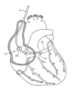
Now referring to FIGS. 3-4, exemplary methods of use of the medical system
are illustrated. The medical device may be positioned near a region of tissue
targeted for a therapeutic or diagnostic procedure, and the anchoring
element(s) 26a,
26b may be cryoadhered to a portion of the tissue before, during, or after
placement or
5 positioning of the treatment/diagnostic element 22. For example, as shown
in FIG. 3,
a portion of the elongate body 16 may be routed through the vasculature of a
patient
and into a chamber of the heart, such as the right atrium 52. The
introduction, routing,
and/or positioning of the medical device 12 may be facilitated with one or
more
sheaths, introducer devices, and/or other secondary devices. The medical
device 12
10 may be positioned such that the anchoring element is in proximity to a
region of the
atrial wall near the coronary sinus 54, while the distal portion 20 of the
medical
device 12 including the treatment/diagnostic element 22 is directed into the
coronary
sinus 54. The handle 40 and/or proximal end 18 of the medical device 12 may be
used
to exert a pressure or torque on the anchoring element 26a and/or the
treatment/diagnostic element 22 to obtain a desired location or position of
the device
12 with respect to the surrounding anatomy. Once in a generally desired
position, the
anchoring element 26a may be actuated or otherwise operated to engage the
atrial
wall or surrounding tissue. For example, where the anchoring element includes
the
expandable element, a cryogenic cooling fluid may be introduced into the
expandable
element to significantly reduce the temperature of the anchoring element. The
reduced
temperature promotes or otherwise results in cryoadhesion between the cooled
anchoring element and the tissue. Of note, the reduced temperatures obtained
in the
anchoring element 26 may be sufficiently cold to promote adhesion between the
tissue
and the anchoring element 26, while minimizing the likelihood of creating a
permanent tissue lesion or otherwise adversely affecting cardiac conduction or
function through the adhered tissue segment. Such temperatures may include,
for
example, those temperatures typically used for cardiac mapping using a
cryogenic
device, e.g., approximately -25 C or higher.
Now referring to FIG. 4, where multiple anchoring elements are included, an
exemplary method of use may include positioning the anchoring elements into a
generally desired position. For example, the anchoring clement 26a may be
positioned adjacent the atrial wall, while the second anchoring element 26b
may be
CA 02813353 2013-04-02
WO 2012/057916
PCT/US2011/049831
13
routed into the ostium of the coronary sinus, with the distal portion 20 of
the elongate
body 16 extending even further into the coronary sinus. The anchors may then
be
deployed, which may include the introduction of a coolant into the individual
anchors
to effectuate a cryoadhesive engagement between the individual anchoring
elements
and their surrounding tissue. To reduce any undesired effects from the
obstruction of
blood flow through the coronary ostium, external fluid flow may be perfused
through
the perfusion passages 38 of the second anchoring element 26b and/or portion
of the
elongate body 16.
Engaging the tissue with the anchoring element(s) provides a substantially
fixed position to aid in either securing the previously-attained position of
the distal
portion 20 of the medical device 12 or providing a substantially secure
location from
which the distal portion 20 of the elongate body 16 and thus the
diagnostic/treatment
element can be moved around to more effectively reach a targeted tissue
location,
such as a branch of the coronary sinus and/or blood vessels connected or
otherwise in
proximity thereto. Alternatively, the anchoring element(s) can be used to
secure the
device after a desirable position of the proximal and/or distal portion has
been
attained. Moreover, while a particular example of a targeted tissue location
and/or
structure can include cardiac tissue and the coronary sinus, the anchoring and
positioning of the medical device may be implemented in other areas, including
a
trans-septal crossing site (such as an intraventricular septum), an
intravascular
insertion point, and an epicardial-to-endocardial site (and vice versa), for
example.
Once the medical device 12 has been suitably anchored and the distal portion
20 directed to the targeted site, the treatment/diagnostic element 22 may then
be used
in accordance with the aim of the particular procedure. Such procedures may
include,
for example, electrically stimulating or pacing cardiac tissue, electrically
mapping
aberrant electrical activity, ablating a problematic tissue segment, measuring
an
electrical characteristic of the tissue, or the like, using the components of
the medical
system 10 as described herein.
Upon completing the selected treatment or diagnostic regimen, the anchoring
elements may be retracted or otherwise released from their engagement with the
tissue. Where cryoadhesion is the engagement mechanism, removal of the
anchoring
element(s) 26a, 26b may be prolonged until a temperature of the anchoring
element(s)
CA 02813353 2013-04-02
WO 2012/057916
PCT/US2011/049831
14
or tissue has reached a predetermined, thawed temperature to reduce the
likelihood of
any unwanted injury to the tissue that could otherwise result. Retraction
and/or
removal of the anchoring elements, the treatment/diagnostic element 22, and/or
portions of the elongate body 16 may be facilitated, at least in part, through
the
manipulation of the handle, actuator element 48, the relative movement between
portions of the elongate body 16, and/or other steering modalities as
described herein.
Of note, maneuvering and directing the medical device 12 to the desired
locations may be aided in part by fluoroscopy or other imaging means to the
extent
portions of the device 12 and/or surrounding tissue structures can be
visualized. For
example, the formation of ice crystals between the medical device 12 and the
adhered
tissue can be imaged or otherwise located using imaging methodologies able to
distinguish the properties of frozen ice or tissue segments as compared to non-
frozen
tissue regions, such as ultrasound.
In addition, the anchoring element(s) 26 and/or the treatment/diagnostic
element 22 may provide reference points for locating and/or mapping a position
of the
medical device 12. For example, the medical device may be used in conjunction
with
a position sensing module or system 55 that is operable to map and illustrate
mapped
and saved points. The position sensing system can be used to determine the
location
or position of the anchoring element(s) 26 and/or the treatment/diagnostic
element 22
by generating a voltage in a patient and calculating an impedance at the
anchoring
element(s) 26 and/or the treatment/diagnostic element 22. The calculated
impedance
may then be used to determine the position of the electrode as in a patient or
other
appropriate conducting medium.
In particular, as shown in FIG. 5, a portion of the anatomy of a patient 56
can
be mapped by identifying a plurality of points to generate a map within the
patient 56
by determining a relative location of the medical device 12. The plurality of
points
can be illustrated individually, or sequentially, or a surface can be
illustrated with or
without the plurality of points to illustrate or identify a portion of the
anatomy of the
patient 56. The map data can be generated or acquired with the position
sensing
system 55 that can acquire multiple points of or within the patient 56. The
position
sensing system 55 can obtain or otherwise measure voltage, impedance, acoustic
properties (e.g., sound and ultrasound), time-of-travel information, magnetic
field
CA 02813353 2013-04-02
WO 2012/057916
PCT/US2011/049831
strengths, for example. The position sensing system 55 can include an
impedance or
electrical potential (EP) system, an electromagnetic (EM), and/or optical
tracking
system that can be integrated with or operate independently and separate from
the
control unit 14.
5 The position sensing system 55 may include a processor, controller or
driving
unit that includes one or more input or output connectors to interconnect with
a
plurality of current conducting or drive patches or electrodes connected
directly with
the patient 56, and may also be coupled to the anchoring element(s) 26 and/or
the
treatment/diagnostic element 22 to obtain, sense, or otherwise record an
electrical
10 measurement or property thereof during a procedure. The current
patches/reference
electrodes can include electrically conductive segments placed on or in the
patient to
create three substantially orthogonal voltage or current axes within the
patient 56. For
example, a first y-axis patch 58a and a second y-axis patch 58b can be
positioned on
an exterior of the patient 56 to form a y-axis (such as an axis that is
generally
15 superior-inferior of a patient as illustrated in FIG. 5) with a
conductive path such that
the conducted current establishes a voltage potential gradient substantially
along this
axis and between the patches 58a and 58b. A related y-axis current flows from
the
first y-axis patch 58a to the second y-axis patch 58b substantially along the
y-axis.
Likewise, a first x-axis patch 60a and a second x-axis patch 60b can be
connected
with the patient 56 to create a x-axis (such as an axis that is generally
medial-lateral of
a patient) with a voltage gradient substantially along the x-axis between the
patches
60a and 60b and a corresponding x-axis current flowing between patches 60a and
60b.
Finally, a first z-axis patch 62a and a second z-axis patch 62b can be
connected with a
patient 56 to create a z-axis (such as an axis that is generally anterior-
posterior of a
patient) with a voltage potential gradient substantially along the z-axis
between the
patches 62a and 62b with a corresponding z-axis current flowing between the
patches
62a and 62b. The three axes are generally formed to have an organ or area of
interest
that the common intersection or origin of each of the axes x, y, z.
Accordingly, the
patches can be positioned on or in the patient 56 to achieve the selected
placement of
the axes x, y, z relative to the patient 56. Alternatively to surface-
positioned reference
electrodes or patches, an electrically conductive segment or electrode may be
placed
CA 02813353 2013-04-02
WO 2012/057916
PCT/US2011/049831
16
inside the patient in the form of a secondary catheter or medical device, a
permanently
implanted electrically conductive segment or lead, or the like.
The current applied between the related patches generates a small or micro-
current, which can be about 1 microampere (muA) to about 100 milliamperes
(mA),
in the patient along the axis between the respective patch pairs. The induced
current
can be of a different frequency for each of the related patch pairs to allow
for
distinguishing which axis is being measured or can be of a single frequency
and time
multiplexed. The current induced in the patient 56 will generate a voltage
gradient
across different portions, such as the heart, that can be measured with an
electrically
conductive portion of the anchoring element(s) 26 and/or the
treatment/diagnostic
element 22. The sensed voltage can be used to identify a position of the
anchoring
element(s) 26 and/or the treatment/diagnostic element 22 along an axis
(whereby each
axis can be identified by the particular frequency of the current being
measured) to
generally determine a position along each of the three axes. Although a
voltage can be
sensed, an impedance can also be calculated or measured to determine a
location in a
similar manner. The position of the anchoring element(s) 26 and/or the
treatment/diagnostic element 22 with respect to each of the three axes can be
used as
map data for the surrounding physiological area. The anchoring element(s) 26
and/or
the treatment/diagnostic element 22 can be moved through various portions in
the
patient 56 while obtaining, sensing or otherwise recording the voltages,
substantially
continuously or as selected intervals, among the three axes to determine
multiple three
dimensional positions of the anchoring element(s) 26 and/or the
treatment/diagnostic
element 22.
The saved points may be used to create a map generated with the anchoring
element(s) 26a, 26b and/or the treatment/diagnostic element 22 that can be
used to
determine a location of a later positioned anchoring element(s) 26 and/or the
treatment/diagnostic element 22 (such as in a subsequent follow-up procedure).
The
discussion herein may refer to map data or map data points and will be
understood to
include individual acquired data points, illustrated individual or managed
points an
algorithm process applied to acquired data points to improve visual display by
eliminating regions of especially high density and useful in modulating
characteristics
of rendered surfaces, a rendered surface, or any appropriate manner of
illustrating the
CA 02813353 2013-04-02
WO 2012/957916
PCT/US2011/949831
17
acquired map data. Once the map has been created of the patient 56 or a
portion of the
patient 56, either with or without a surface rendered relative to the
individual points, a
procedure can be guided or navigated using the map data.
The map data can be used to identify various anatomical features. In addition,
instruments, including the medical device 10 and/or other instruments separate
or
complementary to the medical device 12 in a common procedure or subsequent
procedures, can be navigated relative to the patient 56 using the map data.
For
example, a sterilized or "new" medical device 12 may be used in a later
procedure to
create a second map having one or more points using the methodology described
above. The second map may be compared and registered to the first map to match
the
location and reference points from the first procedure to the locations and
reference
points in the second procedure. Significant physiological changes are unlikely
to
occur to anatomical structures or features (such as the superior vena cava,
inferior
vena cava, the coronary sinus, and/or portions thereof) between procedures,
which
allows such anatomical structures to be matched or synchronized between the
two
maps. The second procedure can thus proceed with knowledge of the previous
locations and reference points where therapy and/or diagnostic measurements
were
conducted in the earlier procedure with respect to the medical device as
positioned in
the second procedure. In a particular example, obtaining a second map,
registering it
or matching it the first map, and proceeding with the secondary procedure can
allow a
physician or user to "touch up" or otherwise create additional ablative
segments in the
treatment of an arrhythmia to lesions or specific treatment areas or location
initially
created during a first, previous procedure.
Identification of implants, ablation or cannulation procedures, or other
procedures can be performed to guide initial procedures as well as providing a
guiding, navigational history for later treatments or procedures. Accordingly,
a
procedure can be navigated and performed precisely and repeatably with the
generated map data in an initial treatment as well as subsequent procedures. A
display
device (either separate or integral with the control unit 14, for example) can
be used to
display the map data and/or illustrate icons representing various portions or
reference
points relative to the patient 56. The registering or correlation between the
two maps
can be performed by one or more processors in the system 55, and presented to
an
CA 02813353 2014-11-25
18
end-user in a visual format. Moreover, the system 55 may include one or more
storage
components to store map data recorded or otherwise obtained during one or more
procedures for later use in subsequent procedures. In addition, the map data
can be
generated in a substantially three dimensional or even four dimensional
manner.
Accordingly, the display can include a three dimensional viewing, simulated
three
dimensional viewing, or even four dimensional viewing, such as to illustrate a
change
in the patient 56 over time.
The map generated with the position sensing system can be used to guide or
navigate the medical device 12 to a selected location without the use of other
prior or
concurrent imaging devices, such as an external fluoroscope, magnetic
resonance
imaging, ultrasound, as described in more detail in Application Ser. No.
12/424,013,
filed April 15, 2009, entitled -LOCATING A MEMBER IN A STRUCTURE-.
In addition, unless mention was made above to the contrary, it should be noted
that all of the accompanying drawings are not to scale. A variety of
modifications and
variations are possible in light of the above teachings.