Note: Descriptions are shown in the official language in which they were submitted.
CA 02814117 2013-04-26
METHODS OF USING LIGHT TO REPAIR HERNIA DEFECTS
BACKGROUND
Technical Field
[0002] The present disclosure relates to hernia repair methods. More
particularly, the
present disclosure relates to methods for positioning a surgical patch to a
tissue site of a hernia
using light.
Description of Related Art
[00031 A hernia is a protrusion of a tissue, structure, or part of an
organ through injured
muscle tissue or an injured membrane by which the tissue, structure, or organ
is normally
contained. Some examples of hernias include: abdominal hernias, diaphragmatic
hernias and
hiatal hernias (for example, para-esophageal hernia of the stomach), pelvic
hernias, for example,
obturator hernia, anal hernias, hernias of the nucleus pulposus of the
intervertebral discs,
intracranial hernias, and Spigelian hernias.
[00041 Hernias may be surgically repaired, and are principally repaired by
pushing back,
or "reducing", the herniated tissue, and then reinforcing the defect in
injured muscle tissue (an
operation called herniorrhaphy). Modern muscle reinforcement techniques
involve placement of
a surgical patch, such as a surgical mesh, near the injured tissue or defect
to support the defect.
1
CA 02814117 2013-04-26
The surgical patch is either placed over the defect (anterior repair) or under
the defect (posterior
repair).
[0005] A variety of different fixation devices are used to anchor the
surgical patch to the
tissue. For example, a needled suture may be passed through or around the
tissue near the defect
to hold the surgical patch in a position which spans the injured tissue, In
other examples, staples,
tacks, clips and pins are also known to be passed through or around the tissue
near the defect to
anchor the surgical patch in a position which spans the injured tissue.
[00061 When applying a surgical patch during minimally invasive surgery,
it is
imperative that the surgeon know the precise location, size and shape of the
hernia defect in
order to properly place the surgical patch. However, since the bounds of the
hernia defect are
generally internal, visibility is often limited and placement of the surgical
patch can be
cumbersome. Thus, a continuing need still exits to provide a means for
facilitating the
effectiveness of the placement of surgical patches used to surgically repair
hernias.
SUMMARY
[00071 Accordingly, a hernia repair method includes the step of
identifying a hernia
defect in a patient, the hernia defect having a size, a location, and a shape.
The method involves
positioning a dispensing instrument laparoscopically into the patient adjacent
the hernia defect.
10008] According to one step, the method includes dispensing one or more
light pipes
from the dispensing instrument at predetermined locations. The light pipes may
be fiber optic.
The method may include the step of positioning the dispensing instrument
adjacent a corner or
an extreme of the hernia defect prior to dispensing the one or more light
pipes.
2
CA 02814117 2013-04-26
[0009] The method also involves advancing the one or more light pipes
through the
patient's skin. The method may further include piercing the patient's skin
with that one or more
light pipes, The method may further comprise the step of advancing the one or
more light pipes
to a position immediately adjacent the hernia defect after advancing the one
or more light pipes
through the patient's skin.
[0010] The method may include the step of bundling a plurality of light
pipes. Another
step includes coupling a light source to the one or more light pipes. The
method also involves
generating a pattern of light that indicates one or more of the size, the
location, and the shape of
the hernia defect. The method further includes the step of forming an outline
of the hernia
defect, in vivo, with the pattern of light being formed from the positioning
of the plurality of
light pipes at the predetermined locations.
[0011] One step includes positioning a surgical patch adjacent the hernia
defect in
accordance with the pattern of light. According to one step, the method
involves generating a
pattern of light that can be visualized, ex vivo, through the surgical patch
when the surgical patch
is positioned over the hernia defect in vivo. According to one step, the
method involves
removing the one or more light pipes from the patient after positioning the
surgical patch
adjacent the hernia defect.
BRIEF DESCRIPTION OF THE DRAWINGS
[0012] The above and other aspects, features, and advantages of the
present disclosure
will become more apparent in light of the following detailed description when
taken in
conjunction with the accompanying drawings in which:
3
CA 02814117 2013-04-26
[0013] FIG. 1 is a cross-sectional view illustrating a tear in an
abdominal wall;
[0014] FIG. 2 is a cross-sectional view illustrating a ventral hernia;
[0015] FIG. 3 is a perspective view of a hernia repair system in
accordance with the
present disclosure;
[0016] FIGS. 4-8 are progressive views illustrating a deployment of a
light pipe of the
hernia repair system of FIG, 3 into tissue;
[0017] FIG. 9 is perspective view of a plurality of light pipes disposed
in tissue after
being deployed from the hernia repair system of FIG. 3;
[0018] FIG. 10 is a perspective view of another embodiment of a hernia
repair system
deploying a plurality of light pipes in tissue in accordance with the present
disclosure; and
[0019] FIGS. 11-13 are progressive views illustrating a surgical patch
being positioned
adjacent a hernia defect with the aid of a plurality of light pipes in
accordance with the present
disclosure.
DETAILED DESCRIPTION
[0020] The present disclosure relates to methods for surgeries such as
transluminal
and/or endoluminal placement of a surgical patch at a surgical site. As used
herein the term
"surgical patch" is used to refer to any type of patch for use in surgical
procedures, such as, for
example, meshes that can be attached to the abdominal wall. Although described
herein with
reference to a hernia surgical patch, the methods of the disclosure may be
used in any surgical
repair.
4
CA 02814117 2013-04-26
[00211 In the drawings and in the description that follows, the term
"proximal," as is
traditional, will refer to an end of a device that is closer to the user,
while the term "distal" will
refer to the end of the device that is farther from the user.
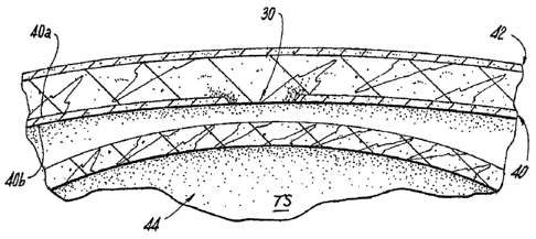
[0022] Referring now in specific detail to the drawings, in which like
numbers identify
similar or identical elements, FIG. 1 illustrates a hernia that may involve a
defect 30 such as a
tear in the abdominal wall 40. The abdominal wall 40 is defined by an external
side 40a and an
internal side 40b. A surface tissue 42, which covers the external side 40a of
abdominal wall 40,
may or may not be immediately affected by this defect 30. An internal organ 44
located below
the internal side 40b of the abdominal wall 40 may not protrude until some
form of exertion or
use of the muscle located at the abdominal wall 40 forces the internal
orgari44 into the defect 30.
Depending on the size and location of the defect 30, exertion may not be
needed to cause the
organ to protrude. As shown in FIG. 2, a hernia occurs when an internal organ
44 protrudes into
the defect 30 of abdominal wall 40. Oftentimes the protrusion creates a bulge
46 in the surface
tissue 42.
[0023] As depicted in FIG. 3, a hernia repair system 100 includes an
access port 110, a
dispensing instrument 120, and one or more light pipes 130.
[0024] The access port 110 includes a seal assembly 112 at a proximal end
and cannula
114 at a distal end. The seal assembly 112 accommodates the dispensing
instrument 120 in a
substantially sealed relationship. The seal assembly 112 includes an
insufflation valve 116 to
selectively permit the passage of insufflation fluids therethrough to create a
working space in an
underlying tissue site.
[0025] The dispensing instrument 120 includes an actuation assembly 122 at
a proximal
end and a shaft 124 at a distal end. The shaft 124 extends from the actuation
assembly 122. The
CA 02814117 2013-04-26
shaft 124 defines a lumen 126 therethrough to accommodate the one or more
light pipes 130.
The shaft 124 is movable via the actuation assembly 122 to dispense the one or
more light pipes
130 at predetermined locations within a patient. In particular, as illustrated
in FIG. 3, the distal
end of the shaft 124 may be rotatable, pivotable, and/or articulable to orient
the distal end of the
shaft 124 in a particular orientation relative to the hernia defect 30.
[0026] Each light pipe 130 includes a distal tip 132, which may be
sharpened to pierce
tissue, and an elongated member 134. The elongated member 134 may define a
lumen 136
therethrough to permit the passage of light therethrough when coupled to a
light source 150 (see
FIG. 11). Alternatively, the elongated member 134 may include any suitable
electrical and/or
mechanical and/or chemical components configured to emit light from the distal
end of the
elongated member 134 (e.g., like a flashlight). The elongated member 134 may
be rigid or
flexible. The light pipes 130 may be fiber optic,
[0027] In use, a hernia defect 30 is identified in a patient. As can be
appreciated, each
hernia defect 30 has a particular size, location, and shape and therefore
proper placement of a
surgical patch 160 during minimally invasive surgery is facilitated when a
practitioner can
ascertain the size, location, and shape from an ex vivo location. Thus, in
order to be able to
perceive the hernia defect 30 from an ex vivo location, the practitioner
inserts the access port
110, namely the cannula 114 into tissue adjacent the hernia defect 30 (see
FIG. 4). With
continued reference to FIGS. 4-5, the underlying tissue site "TS" may be
insufflated when the
insuffiation valve 116 is coupled to an insufflation source 118 to create a
working space. In this
respect, the practitioner may then laparoscopically advance the dispensing
instrument 120 into
the patient adjacent the hernia defect 30 to facilitate placement of one or
more light pipes 30 in
position about the hernia defect 30, which is best depicted in FIG. 5. As
depicted in FIG. 10, any
6
CA 02814117 2013-04-26
number of access ports 110 and/or dispensing instruments 120 may be used to
position the one or
more light pipes 130 in tissue. In some modes of operation, the practitioner
may even directly
laparoscopically advance the one or more light pipes 30 through the access
port 110 or directly
laparoscopically advance the one or more light pipes 30 through an incision
(without the access
port 110) by virtue of the sharpened distal tip 132, where appropriate.
[0028] Turning now to FIGS. 6-9, one or more light pipes 130 may then be
dispensed
from the dispensing instrument 120 at predetermined locations about the hernia
defect 30 to
create a pattern about the defect 30 that is commensurate with the size,
location, orientation
and/or shape of the defect 30. For example, the dispensing instrument 120 may
be positioned
adjacent one or more corners and/or extremes of the hernia defect 30 prior to
dispensing the one
or more light pipes 130 to generate the pattern. The one or more light pipes
130 can then be
dispensed with sufficient force to pierce and advance through the patient's
skin, e.g. surface
tissue 42. The one or more light pipes 130 may include sharpened tips 132 to
further facilitate
the penetration of the patient's skin. After the ends, e.g., the sharpened
tips 132 are positioned
so that they extend externally, the one or more light pipes 130 may be
advanced to a position
immediately adjacent the hernia defect 30 or the abdominal wall 40. In this
respect, the one or
more light pipes 130 may be pulled proximally through the pierced skin until
they are positioned
snug against the hernia defect 30 or the abdominal wall 40, depending upon the
desired position.
[0029] Once in the snug position, the one or more light pipes 130 are most
suitably
configured in the pattern. The pattern may extend along the defect 30 and/or
along an area
immediately adjacent the defect 30. The pattern may have any suitable
geometry, size, etc. for
facilitating the placement of a surgical patch 160 adjacent the defect 30.
7
CA 02814117 2013-04-26
[0030] As illustrated in FIGS. 11-13, when the one or more light pipes
130 are coupled to
a light source 150 or are otherwise adapted to emit light (e.gõ selectively
via a switch coupled to
the one or more light pipes or autonomously by fluorescent chemicals or the
like), one or more
points of light are formed about the defect 30 corresponding to the pattern to
indicate the size,
location, orientation and/or shape of the defect 30.
[0031] With reference to FIG. 11, when there is a plurality of light
pipes 130, the
plurality may be bundled together via a bundling member 140. The light source
150 may then be
coupled to the one or more light pipes 130, either individually, collectively,
or by groups of light
pipes 130. The bundling member 140 may include a light source 150. As depicted
in FIG, 12,
the light source 150 generates the pattern of light "P" via points of light
Pl, P2, P3, P4, P5, P6,
etc. that indicate the size, the location, the orientation and/or the shape of
the hernia defect 30.
In this regard, the light emitted from the light source 150 will form an
outline of the hernia defect
30, in vivo, so as to be viewable ex vivo so that the surgical patch 160 may
be positioned
adjacent the hernia defect 30 with any suitable instrument 200 (e.g., a
grasper) in accordance
with the outline/pattern of light. As can be appreciated, when the surgical
patch 160 is positioned
in vivo over the hernia defect 30, the generated pattern of light can be
visualized, ex vivo,
through the surgical patch 110 and the tissue. After the surgical patch 160 is
placed in a desired
position adjacent the hernia defect 30, the surgical patch 160 may be secured
to the defect 30 by
any suitable means (e.g., glue, tack, staple, suture, etc.) and the one or
more light pipes 130 may
then be removed from the patient either individually, collectively, or by
groups of light pipes 130
(see FIG. 13).
[0032] The presently disclosed surgical patch may be any type of patch
for use in
surgical repair and suitable for use in situ. The surgical patch may be any
suitable shape (i.e.,
8
CA 02814117 2013-04-26
circular, noncircular, etc.) and may include one or more layers. The surgical
patch may be made
of multiple fibers, or may be made of a single fiber, The fibers may be a
monofilament or multi-
filament.
[0033] The fibers forming the presently disclosed patch may be made from
a natural
material or a synthetic material. The fibers may be biodegradable or non-
biodegradable. Any
combination of natural, synthetic, bioadegradable and non-biodegradable
materials may be used
to form the fibers, The term "biodegradable" as used herein is defined to
include both
bioabsorbable and bioresorbable materials. By biodegradable, it is meant that
the materials
decompose, or lose structural integrity under body conditions (e.g. enzymatic
degradation or
hydrolysis) or are broken down (physically or chemically) under physiologic
conditions in the -
body such that the degradation products are excretable or absorbable by the
body.
[0034] The surgical patch of the present disclosure may be formed using
any method
suitable to forming patch structures, including but not limited to knitting,
weaving, non-woven
techniques, and the like. Suitable techniques for making the surgical patch
are within the
purview of those skilled in the art.
[00351 The surgical patch may be any shape or size suitable for covering
the herniated
area and securing the patch to surrounding tissue. The surgical patch may be
preformed to a
certain size, such as, for example, a 9 cm diameter round patch or 50 cm x 50
cm square patch.
In embodiments, the surgical patch may be cut to a particular size and shape
as needed.
[0036] In addition, the surgical patch of the present disclosure may be
rolled, folded, or
otherwise oriented so that the surgical patch forms a shape more suitable for
placement adjacent
a hernia defect.
9
CA 02814117 2013-04-26
[00371
While several embodiments of the disclosure have been shown in the drawings,
it
is not intended that the disclosure be limited thereto, as it is intended that
the disclosure be as
broad in scope as the art will allow and that the specification be read
likewise, Therefore, the
above description should not be construed as limiting, but merely as
exemplifications of
particular embodiments. Those skilled in the art will envision other
modifications within the
scope and spirit of the claims appended hereto.